Review

A Representative Clinical Course of Progression, with Molecular Insights, of Hormone Receptor-Positive, HER2-Negative Bone Metastatic Breast Cancer

by
Elizabeth Magno
and
Karen M. Bussard
*
Department of Pharmacology, Physiology and Cancer Biology, Thomas Jefferson University, Philadelphia, PA 19107, USA
*
Author to whom correspondence should be addressed.
Int. J. Mol. Sci. 2024, 25(6), 3407; https://doi.org/10.3390/ijms25063407
Submission received: 13 February 2024 / Revised: 11 March 2024 / Accepted: 12 March 2024 / Published: 17 March 2024
(This article belongs to the Special Issue Hormone Receptor in Breast Cancer)

Abstract

:
Despite treatment advances, breast cancer remains a leading cause of death of women in the United States, mostly due to metastatic disease. Bone is a preferential site for breast cancer metastasis, and most metastatic breast cancer patients experience bone involvement at the time of death. The majority of patients with bone metastatic breast cancer are first diagnosed with and treated for early-stage disease, and from development of early-stage breast cancer to the recurrence of cancer in the bones, up to 30 years may elapse. Throughout this timeframe, a typical patient undergoes many treatments that have effects on the bone microenvironment. Therefore, this review explores the clinical course of a representative patient with hormone receptor-positive bone metastatic breast cancer, examining key treatment options at each stage and their effects on preventing and treating bone metastases.

1. Introduction

Breast cancer is the most frequently diagnosed malignancy among women in the United States, excluding skin cancers [1]. Approximately 1 out of every 8 (13%) women will be diagnosed with breast cancer during their lifetime [1]. Breast cancer is the second leading cause of death among women in the United States, and although the disease predominately affects women, 1% of breast cancer cases and deaths occur in men [1]. In 2024 alone, 42,780 patients are expected to die from the disease [2]. The majority of breast cancer-related deaths occur from metastatic disease, characterized by the spread of cancer cells from the breast to other tissues, leading to their proliferation at secondary sites [3]. Up to 30% of patients diagnosed with breast cancer will ultimately develop metastases [4]. Common metastatic sites include the bones, lungs, liver, and brain, with bone being the most prevalent location [5]. Among all patients who die from breast cancer, approximately 73% exhibit bone involvement at the time of death, representing the highest percentage of bone involvement among all cancers [6]. While each patient’s journey is unique, this review illustrates a representative case of bone metastatic breast cancer, featuring a patient with the most statistically common clinical features: Hormone Receptor (HR) positive, Human Epidermal growth factor Receptor 2 (HER2)-negative breast cancer without germline mutation. From development of early-stage breast cancer to the recurrence of cancer in the bones, up to 30 years may elapse [7]. A common clinical course of progression is presented, highlighting key events, including presentation to the clinic, primary and adjuvant treatment involving locoregional and/or systemic therapy, progression to metastasis, and treatment of bone metastatic breast cancer. At each juncture, therapeutic options and their effects on the bone microenvironment are discussed.

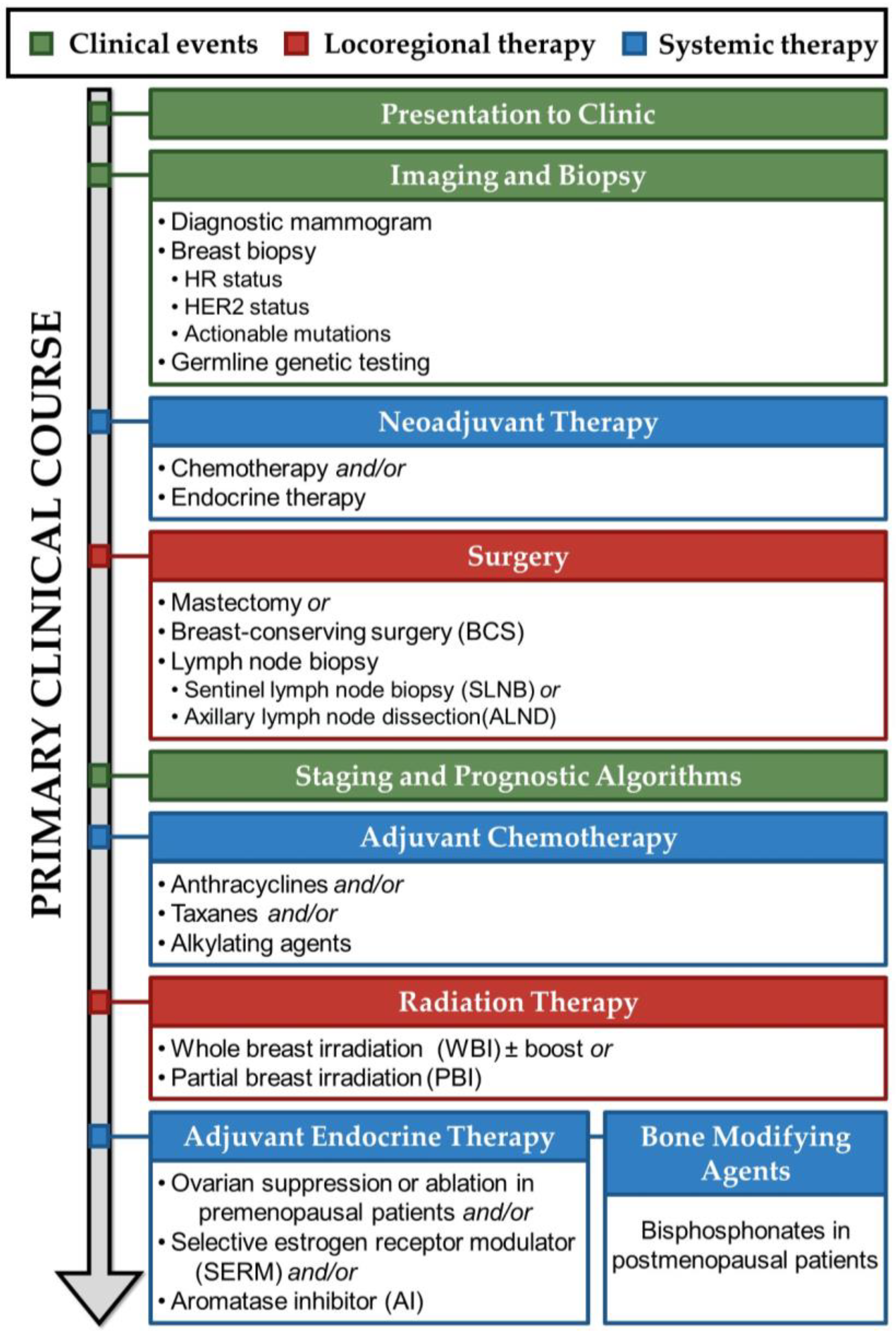
2. Presentation to Clinic, Imaging, Biopsy, and Staging

While a minority of patients initially present with widespread metastatic disease, 92% of breast cancers in the United States are diagnosed in local or regional stages [1]. Importantly, among patients who ultimately develop metastatic breast cancer, 74% are initially diagnosed with early-stage breast cancer [8]. Patients with early-stage, primary breast cancer often present to the clinic after discovering a palpable mass within the breast, either through self-examination, clinician detection, or a suspicious finding on screening mammography. Diagnostic mammography is then used to identify the location and relative size of the primary tumor. In individuals with dense breasts, breast ultrasound and Magnetic Resonance Imaging (MRI) may also be used to better visualize the suspected tumor [9]. A sample of the primary tumor is then obtained via core biopsy, fine needle aspiration, or incisional biopsy and profiled in order to assess tumor hormone-receptor status, molecular markers and mutational status, and histology [9]. These characteristics from biopsy tissue are synthesized to inform treatment decisions and determine a patient’s eligibility for certain therapies.
Tumor expression of hormone receptors is critical in determining both prognostic and therapy predictive information [10]. Cancers with as few as 1% of cells positive for Estrogen Receptor (ER) or Progesterone Receptor (PR) expression via ImmunoHistoChemistry (IHC) staining are considered ER or PR-positive, respectively [11]. Cancers that are either ER-positive, PR-positive, or both are considered Hormone Receptor (HR)-positive. ER itself is a DeoxyriboNucleic Acid (DNA)-binding transcription factor that is responsive to its ligands, including estrogen, the main hormone regulating mammary gland development. Estrogens are believed to stimulate breast cancer cell growth by associating with regulatory elements in the genome, enhancing the transcription of genes such as MYeloCytomatosis (MYC) and CyCliN D1 (CCND1) and promoting tumor cell growth and proliferation [12]. Because ER-positive breast cancers are responsive to circulating estrogens which stimulate breast cancer cell growth, suppression of endogenous estrogen and consequent abrogation of ER-signaling is a critical part of ER-positive breast cancer management. Meanwhile, PR is dually expressed in more than 50% of ER-positive cancers [13]. PR is an ER-induced gene, and PR positivity is thought to reflect a cancer that is driven by an active ERα complex and therefore likely to respond to endocrine therapies [14]. Patients with ER-negative, PR-positive cancers may therefore be considered for endocrine therapies, but data on this group are limited [9].
Along with HR status, HER2 expression is among the most important clinical and prognostic factors to determine for breast cancer patients. HER2 is a membrane tyrosine kinase and oncogene that is overexpressed and gene amplified in about 20% of breast cancers [15]. Transcription factors activated by the HER2 pathway regulate genes involved in cell proliferation, survival, differentiation, angiogenesis, and invasion and metastasis [15]. HER2 amplification and resultant overexpression confer clinical aggressiveness, predicting poorer overall survival and shorter time to relapse [16]. However, patients with tumors overexpressing HER2 protein are candidates for HER2-targeted therapies, including adjuvant trastuzumab, which significantly improves disease-free survival [9]. Therefore, determining HER2 expression via IHC is a critical component of molecular tumor diagnostics. HR-positive/HER2-negative breast cancers are by far the most common subtype in the United States, comprising 68% of all diagnosed invasive female breast cancers, followed by HR-positive/HER2-positive and HR-negative/HER2-negative (triple negative) breast cancer, each at ten percent overall [1]. HR-positive/HER2-negative cancers are significantly associated with bone relapse compared with HER2-positive or HR-negative subtypes [17]. In addressing bone metastatic breast cancer, HR-positive/HER2-negative patients are therefore among the most vulnerable populations to focus on.
The genomic landscape of breast cancers is heterogeneous but follows a general pattern, with tumors broadly classified into four intrinsic subtypes: luminal A, luminal B, HER2-enriched, and basal-like (also termed triple-negative) [18]. Each subtype bears specific natural gene expression patterns by gene expression array, and subtypes differ in natural histories, response to therapy, and epidemiologic frequencies [19].
Furthermore, some breast cancer subtypes are associated with particular somatic mutations; somatic mutations in only three genes (Tumor Protein P53 (TP53), Phosphatidylinositol-4,5-Bisphosphonate 3-Kinase Catalytic Subunit Alpha (PIK3CA), and GATA Binding Protein 3 (GATA3)) are represented at >10% incidence across all subtypes [20]. Luminal breast cancers have the most heterogeneous mutations, with Luminal A cancers demonstrating PIK3CA mutations most frequently, followed by Mitogen-Activated Protein Kinase Kinase Kinase 1 (MAP3K1), GATA3, TP53, Cadherin 1 (CDH1), and Mitogen-Activated Protein Kinase Kinase 4 (MAP2K4) alterations, while luminal B cancers have frequent TP53 and PIK3CA mutations (29% each). In contrast, basal-like cancers have few shared mutations with the luminal subtypes, and instead are characterized by a very high frequency of TP53 mutations (84%) and loss of function [20].
Microarray analysis to determine the intrinsic subtype of a given tumor is costly, difficult to standardize, and not commercially available in the United States, so intrinsic subtype classification is limited to research settings [21]. Clinically, expression of ER, PR, and HER2 by IHC staining is often used as a surrogate for intrinsic subtype classification, with luminal subtypes defined by presence of ER staining (highest in A and lower in B), HER2-enriched by HER2 positivity, and basal-like by absence of ER, PR, and HER2. However, these clinical markers are a poor heuristic for intrinsic subtype determination. For example, when analyzed by microarray, 5% of clinical Luminal A samples were actually the basal-like subtype, and 30% of HER2-negative tumors were the HER2-enriched subtype [22]. Therefore, clinicians use factors other than intrinsic subtype to stratify risk, including multigene assays such as OncoType Dx, MammaPrint, Prosigna, EndoPredict, and Breast Cancer Index (BCI) to assess genomic level information that predicts recurrence risk and/or potential therapy benefit for certain populations [23].
Beyond tumor-level genomic analysis, germline genetic testing may also be performed in a newly diagnosed patient to evaluate hereditary breast cancer genes [9]. In contrast to somatic mutations, which develop in specific cells over time, germline mutations are inherited and are present in every cell throughout the body. Often, these germline mutations disrupt the normal functioning of processes like DNA repair or regulation of cell proliferation, thereby predisposing patients to cancer development [24]. Most significantly, inherited mutations in the DNA repair genes BRCA1/2 DNA Repair Associated (BRCA1/2) incur a lifetime risk of breast cancer of approximately 50% [24]. Pathogenic germline variants in Partner And Localizer of BRCA2 (PALB2), ATM Serine/Threonine Kinase (ATM), BRCA1 Associated RING Domain 1 (BARD1), and Checkpoint Kinase 2 (CHEK2) individually increase breast cancer risk, while TP53, Phosphatase And Tensin Homolog (PTEN), Serine/Threonine Kinase 11 (STK11), Neurofibromin 1 (NF1), and CDH1 mutations increase breast cancer risk in association with genetic syndromes [24,25]. Some germline mutations make patients eligible for targeted therapies, such as poly adenosine diphosphate (ADP)-ribose polymerase (PARP) inhibitors in patients with BRCA1/2 mutations. Although germline mutations are more common within certain ethnic groups, they have low prevalence among the general population (<7% for BRCA1 DNA Repair Associated (BRCA1), the most common germline mutation) [26].
At diagnosis, in addition to surveying the molecular characteristics of the primary tumor, a patient with breast cancer is also assessed for cancer spread. Axillary lymph nodes (ALNs), which drain lymph fluid from the breast, are biopsied in order to determine if cancer cells have spread from the primary tumor into secondary locations via the lymphatic system. If signs or symptoms of metastatic disease are present, systemic imaging is used to evaluate the extent of metastasis. The patient’s cancer is then staged using the traditional anatomic Tumor (T), Lymph Node (N), Metastases (M) (TNM) system, based on the size of the primary tumor, the number of regional lymph nodes with cancer cells present, and the number of distant metastases. Tumors are also graded based on histological stage, which is determined based on tissue architectural distortion in biopsy samples. The TNM staging, histological staging, molecular markers, and HR status are synthesized to generate both clinical and pathological prognostic stages.

3. Treatment of Primary Hormone Receptor-Positive Breast Cancer

The therapeutic management of primary breast cancer is complex, varied, and multimodal. Treatment may begin with neoadjuvant systemic therapy but almost always includes local therapy of the affected breast, such as surgery in order to remove the primary tumor, and subsequent radiation therapy (RT) to eradicate any remaining cancer cells in the breast. Whole-body systemic therapy, such as chemotherapy, endocrine therapy, targeted therapy, or immunotherapy, is then often used to kill any remaining cancer cells and reduce the risk of later recurrence. For such cases, primary cancer treatment involves a sequential approach: neoadjuvant hormone therapy to shrink a tumor, then surgery, followed by adjuvant systemic therapy and radiation, then several years of adjuvant endocrine therapy (Figure 1) [9].

3.1. Neoadjuvant Hormone Therapy for Primary Hormone Receptor-Positive Breast Cancer

Prior to surgery, neoadjuvant therapy may be used for patients with hormone receptor-positive breast cancer, in order to reduce the size of a patient’s primary tumor for future surgical removal [27]. Both chemotherapy and endocrine therapy may be employed in the neoadjuvant setting. In particular, endocrine therapy reduces the amount of circulating estrogen and/or progesterone, and can only be used for patients with hormone receptor-positive cancers. This is because HR-positive cancers are responsive to estrogen and progesterone [28]. Most often, neoadjuvant endocrine therapy is used in post-menopausal women, and it is especially advantageous for patients with larger tumors [29]. By reducing the size of the tumor, surgeries that spare more breast tissue, such as a lumpectomy, may be used instead of mastectomy [27,30]. Furthermore, neoadjuvant endocrine therapy may also provide prognostic information based on a patient’s response, which can better inform adjuvant treatment regimens [23,31].
Common neoadjuvant endocrine therapies include either tamoxifen or aromatase inhibitors, such as letrozole, anastrozole, or exemestane. Importantly, comprehensive studies have been carried out to examine the efficacy of aromatase inhibitors versus tamoxifen as neoadjuvant treatments. In both the Immediate Preoperative Anastrozole, tamoxifen, or Combined with Tamoxifen (IMPACT) and PRe-Operative “Arimidex” Compared to Tamoxifen (PROACT) trial, the aromatase inhibitor anastrozole was as effective, and in some cases more effective than tamoxifen for reducing tumor volume [29,32]. Furthermore, letrozole has been shown to be more effective than tamoxifen as a neoadjuvant treatment in post-menopausal women with primary hormone receptor-positive breast cancers [33]. Thus, neoadjuvant endocrine therapy that uses an aromatase inhibitor may offer more benefit for patients than tamoxifen.
In some patients with hormone receptor-positive, HER2-negative breast cancer, chemotherapy may also be used as a neoadjuvant treatment [34]. There is one main consideration when deciding if chemotherapy should be used as a neoadjuvant treatment (as opposed to post-surgical treatment): is it necessary to obtain pathological or genomic information from a patient’s tumor to help select an appropriate chemotherapeutic agent? If no pathological or genomic information is needed, neoadjuvant chemotherapy is appropriate to use [34]. If genomic and/or pathological information about the tumor is needed, however, then chemotherapy should be used after surgery [34]. Interestingly, a landmark clinical trial showed no difference between overall survival for the use of certain neoadjuvant or adjuvant chemotherapy regimens [35].

3.2. Locoregional Therapy for Primary Hormone Receptor-Positive Breast Cancer

Patients and clinicians have a number of surgical options for removal of breast tumors. Breast Conserving Surgery (BCS), commonly referred to as lumpectomy, entails the removal of the visible tumor and some surrounding normal-looking tissue, while preserving the remaining breast tissue. After tumor removal, surgical margins are microscopically evaluated, and if cancer cells are detected, a second surgery removes additional breast tissue around the tumor site. BCS is followed by radiation therapy of the affected breast, with or without regional lymph nodes, to eradicate any remaining malignant cells. The alternative to BCS is mastectomy, which removes all of the breast. Total mastectomy, also termed simple mastectomy, removes the entire breast, while radical mastectomy removes all breast tissue, axillary lymph nodes along the breast, and, when necessary, underlying chest wall muscle. Mastectomy is indicated in cases of widespread tumors, diffusely positive margins, cancer spreading to chest muscles, and during pregnancy [9]. Selecting the appropriate surgical intervention entails shared decision-making between patients and clinicians, considering factors like long-term survival, the risk of local recurrence, and the impact on cosmetic outcomes and overall quality of life. Overall, BCS with radiation and mastectomy show similar overall survival outcomes for patients [36], though some studies suggest improved survival [37,38] and fewer post-surgical complications [39] with BCS.
In addition to removing the primary tumor from the breast, nearby axillary lymph nodes are assessed to determine the extent of breast cancer cell spread. Axillary Lymph Node Dissection (ALND) is a traditional approach that removes and assesses at least 10 lymph nodes for the presence of breast cancer cells. However, ALND is associated with high complication rates, including lymphedema and sensory loss, and has largely been replaced by Sentinel Lymph Node Biopsy (SLNB) [40]. A Sentinel Lymph Node (SLN) is any lymph node that receives direct lymphatic drainage from a primary tumor site [40]. In SLNB, a radiopharmaceutical tracer and/or visible dye are injected peritumorially and monitored for spread to identify draining sentinel lymph nodes, which take up the tracer and dye. The SLNs are then excised and microscopically evaluated by Hematoxylin and Eosin (H&E) staining [41]. If the SLNs do not contain malignant cells, it is presumed that the cancer is contained within the breast. If cancer cells are identified, further ALND is indicated. Importantly, axillary lymph node status, not tumor size, predicts locoregional recurrence and overall survival [42]. Involvement of axillary lymph nodes at diagnosis is also the single greatest clinical risk factor for later development of bone metastasis [43]. Patients with axillary lymph nodes positive for cancer cells are therefore considered for systemic adjuvant treatment, Radiation Therapy (RT), and bisphosphonate therapy [9].

3.3. Systemic Therapy for Primary Hormone Receptor-Positive Breast Cancer

In contrast to locoregional treatments, which remove cancer within the breast and surrounding structures, systemic therapy treats the entire body to reduce the risk of cancer recurrence. The decision to administer adjuvant systemic therapy after primary treatments such as surgery is guided by the patient’s individual risk of relapse and the anticipated effectiveness of targeted treatments. Clinicians use algorithms incorporating prognostic factors like age, comorbidity, tumor size, grade, and the number of involved lymph nodes to estimate recurrence risk [31]. Additionally, multigene assays like Oncotype DX assess somatic mutations present within tumor tissue to determine the potential benefits of adding adjuvant chemotherapy to endocrine therapy for specific patient populations [23]. The potential survival benefits of adjuvant therapy are carefully weighed against considerations of toxicity and the patient’s existing comorbidities, in order to assess whether chemotherapy should be administered [9].

3.4. Adjuvant Chemotherapy for Primary Hormone Receptor-Positive Breast Cancer

Numerous cytotoxic chemotherapy regimens for breast cancer have undergone extensive evaluation in large-scale clinical trials to assess both efficacy and toxicity. Consequently, several preferred regimens have emerged for the treatment of early breast cancer [9]. While individual drugs within these classes may be interchangeable, three primary drug classes are commonly employed: anthracyclines, taxanes, and alkylating agents.
Anthracyclines, exemplified by doxorubicin and its derivative epirubicin, integrate into the DNA of cancer cells, disrupting topoisomerase-II-mediated DNA repair. Additionally, they generate free radicals that inflict damage on cellular membranes, DNA, and proteins [44]. In preclinical mouse models, doxorubicin treatment causes bone loss through an interplay between oxidative stress and induction of Tumor Growth Factor-Beta (TGFβ) signaling [45]. TGF-β has both tumor-suppressive and tumor-promoting functions, but in the context of the cancer-bone microenvironment, it stimulates breast cancer cells to secrete ParaThyroid Hormone-Related Protein (PTHrP), which begins a “vicious cycle” that results in bone-resorbing osteoclast formation and activation [46]. This osteoclast-mediated bone destruction liberates growth factors stored in the bone matrix, fueling the proliferation of cancer cells [47]. Furthermore, patients treated with cyclophosphamide and doxorubicin undergo a reduction in Bone-Mineral Density (BMD) [48] with increasing bone resorption markers, which are predictors of bone metastasis [49]. It has also been demonstrated that primary breast cancer patients with low BMD have greater numbers of disseminated tumor cells in the bone than patients with normal bone density [50]. Therefore, the anthracycline-induced reduction in BMD that occurs during treatment of the primary breast tumor may contribute to later bone metastases by increasing bone turnover and allowing disseminated tumor cells to seed the bone.
Taxanes, such as docetaxel and paclitaxel, impede movement and function of microtubules, thereby altering cell migration and cell cycle progression [51]. The specific role of taxanes in bone homeostasis is not well studied [52].
Alkylating agents, including cyclophosphamide and platinum-based chemotherapies like carboplatin, create permanent cross-linkages within and between adjacent DNA strands, inducing apoptosis in cancer cells [53]. An adverse side effect of alkylating agents is inadvertent induction of menopause, which is defined as the permanent cessation of menses [54]. Premenopausal patients treated with modern regimens of alkylating agents are at intermediate risk of experiencing permanent chemotherapy-induced amenorrhea [55]. Cyclophosphamide, in particular, is highly gonadotoxic, causing a significant loss of ovarian primordial follicle reserve [56]. Anthracylines, too, pose an intermediate to high risk of permanent amenorrhea, with doxorubicin inducing DNA double-stranded breaks that cause primordial follicle apoptosis [55,57]. However, for both agents, patient age at the time of treatment, baseline ovarian reserve, and specific treatment regimen determine individual risk of ovarian toxicity [55].
Menopause, both natural and induced, leads to a precipitous drop in hormones levels as ovaries cease to synthesize estrogen and progesterone [58]. Estrogen is one of the main hormonal regulators of bone and causes pleotropic effects. Long-term estrogen deficiency in post-menopausal women leads to a slight increase in bone formation and a large increase in bone resorption, causing an overall net loss of bone mass [59]. Consequently, patients who experience chemotherapy-induced ovarian failure develop rapid and highly significant decreases in BMD that are detectable within 6 months of starting chemotherapy [58]. Decreasing BMD following the loss of estrogen is associated with increased expression of bone turnover markers [60], which are predictors for metastatic recurrence [49].
Preferred treatment regimens of cytotoxic chemotherapies in early breast cancer include (1) doxorubicin plus cyclophosphamide followed by paclitaxel (termed “AC-T”, after doxorubicin’s brand name, Adriamycin) and (2) docetaxel plus cyclophosphamide (termed “TC”, after one of docetaxel’s brand names, Taxotere). Trials examining outcomes of the two regimens have found them equivocal. However, no trials have examined the influence of each regimen on bone health and subsequent development of bone metastases [61].

3.5. Radiation Therapy for Primary Hormone Receptor-Positive Breast Cancer

After adjuvant chemotherapy, radiation may be employed. Whole Breast Irradiation (WBI) has been shown to reduce the risk of local recurrence and risk of death from breast cancer [62]. For patients with high-risk characteristics (such as age less than 50 years, high-grade disease, or focally positive margins), radiation to the tumor bed has been shown to reduce local relapse [63]. Radiation can also be applied to the chest wall and regional lymph node basins when indicated. In contrast to WBI, Partial Breast Irradiation (PBI), a targeted radiation therapy administered exclusively to the breast tissue surrounding the site of tumor removal, may be considered for certain low-risk patients [64]. Whether utilizing WBI or PBI, radiotherapy is highly localized to the area at risk of disease recurrence, and concerted effort is made to spare surrounding tissue and structures [9]. As such, nearby skeletal structures are rarely affected, though rib fracture can occur as a very rare, late complication [65].

3.6. Endocrine Therapy for Primary Hormone Receptor-Positive Breast Cancer

Following chemotherapy, endocrine therapy modulates the production or activity of a patient’s hormones to prevent HR-positive cancers from responding to them. Because HR-positive cancers are responsive to estrogen or progesterone, endocrine therapy seeks to reduce the amount of, mainly, circulating estrogen (as PR activity is driven partly, but not exclusively, by ER-mediated transcription, and the role of progesterone alone in breast carcinogenesis is not well defined) [28].
Tamoxifen is the most established adjuvant endocrine therapy. For those diagnosed with ER-positive breast cancer, the use of adjuvant tamoxifen leads to a 39% reduction in the annual likelihood of recurrence and a 31% decrease in the annual likelihood of mortality [66]. Importantly, these benefits remain consistent, regardless of factors such as chemotherapy utilization, patient age, menopausal status, or ALN status [66]. Tamoxifen belongs to a class of drugs called Selective-Estrogen Receptor Modulators (SERMs) that have mixed estrogen agonist or antagonist effects based on target tissue. Tamoxifen and raloxifene, another SERM member, both have estrogen antagonist effects in the breast and agonist effects in the bone, making them useful for both breast cancer and fracture prevention [67].
In patients who are premenopausal at diagnosis, endocrine therapies are often combined with ovarian suppression. The ovaries are premenopausal patients’ main source of estradiol, so abrogating ovarian function decreases estrogen production [68]. Therapeutic options include ovarian suppression or ovarian ablation [12]. Ovarian suppression utilizes pharmacotherapy to temporarily halt ovarian estrogen production. The most common agents, Luteinizing Hormone-Releasing Hormone (LHRH) agonists, suppress hormonal release from the pituitary gland and suppress estrogen production throughout the duration of drug administration [69]. After discontinuation, estrogen production and menstruation resume. Ovarian suppression is generally the preferred modality among premenopausal patients [69], but permanent ovarian ablation through either surgical oophorectomy or ovarian irradiation remains an important therapeutic option. Ovarian ablation may be employed to manage suboptimal ovarian suppression or instances of “breakthrough” ovarian function associated with pharmacologic agents. Additionally, it may be considered for patients with an inherited cancer syndrome elevating the risk of ovarian cancer or for those who do not wish to pursue pregnancy [70]. Importantly, among pre-menopausal women under the age of 45, both ovarian ablation and suppression significantly decrease the risk of recurrence and mortality from breast cancer, with no demonstrated distinction between the two methods [71].
Following therapy-induced ovarian suppression or after natural menopause, patients exclusively generate estrogen in extragonadal sites such as adipose tissue, kidneys, skin, and the brain [72]. While extragonadal estrogen generally functions locally as a paracrine factor, it can still contribute to the growth of ER-positive breast cancers [72]. Estrogen is a steroid hormone derived from androgen precursors, and the enzyme aromatase converts androstenedione and testosterone into estrone and estradiol [68]. Therefore, Aromatase Inhibitors (AIs) are used to block aromatase activity, impeding the production of estrogens and the growth of ER-positive breast cancers. AIs include anastrozole, letrozole, and exemestane.
For HR-positive cancers, endocrine therapy is indicated in the majority of cases, regardless of menopausal status, age, or HER2 status of the tumor [12]. However, the specifics of endocrine treatment depend on these individual factors. Overall recommendations for adjuvant endocrine therapy are complicated, but generally consist of SERMs and/or AIs for up to 10 years [9].

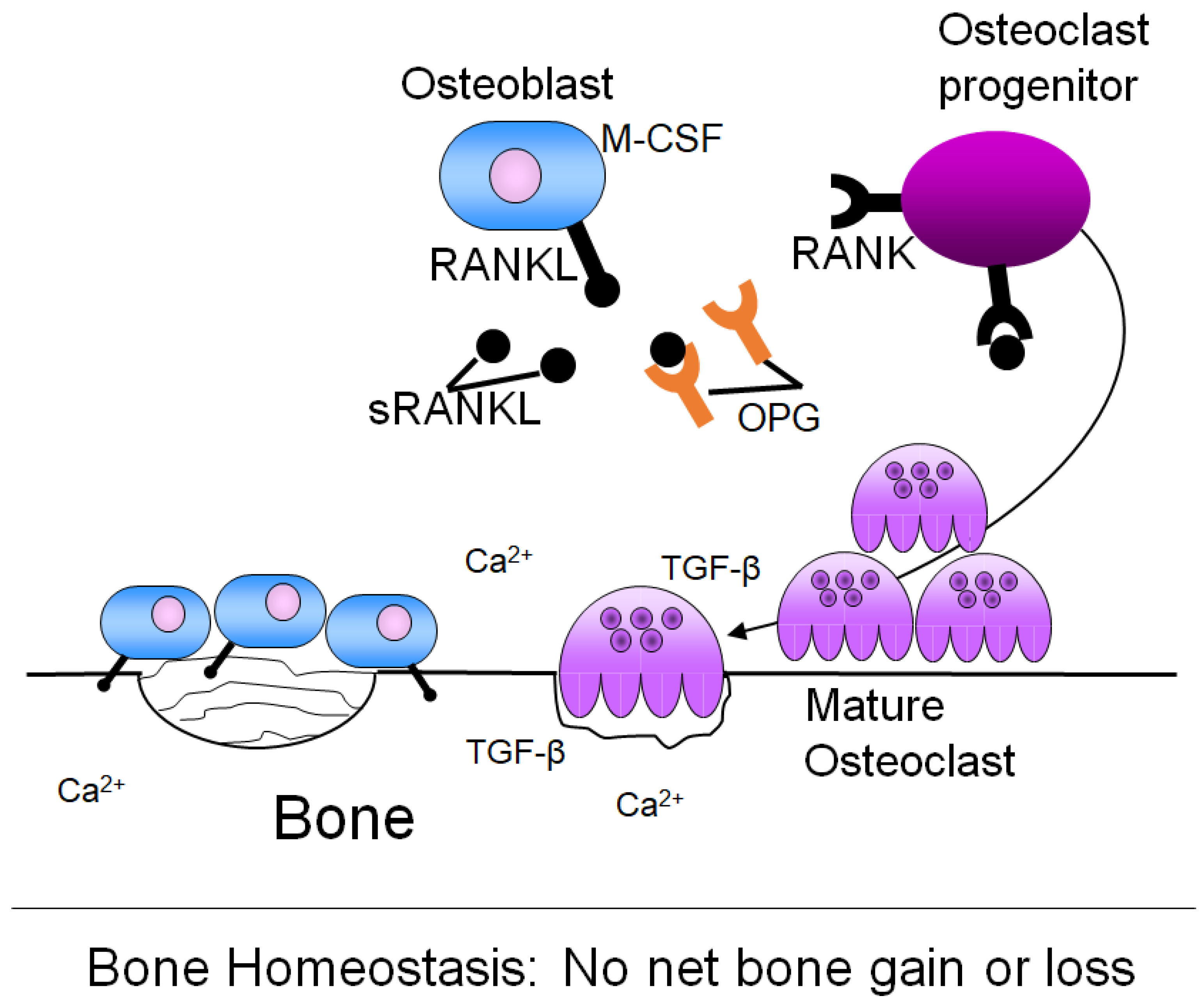
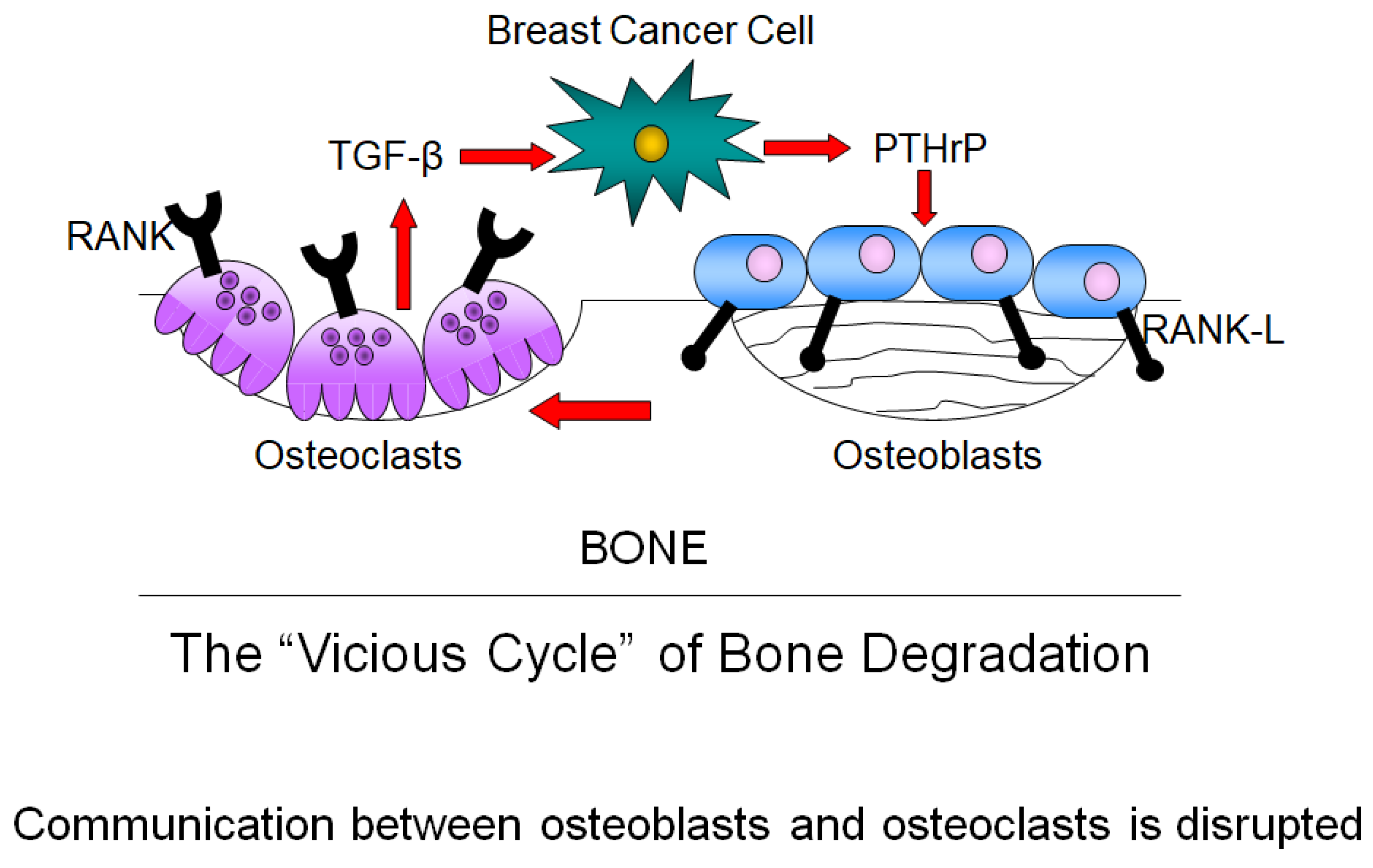
4. Bone Remodeling and Its Dysregulation in Cancer

Bone is constantly being built and degraded in response to the body’s needs, including in response to mechanical stress and to maintain mineral homeostasis [73]. Bone remodeling is a tightly regulated process, mainly involving three different cell types: osteoblasts, which deposit new bone matrix; osteocytes, which are mature bone cells; and osteoclasts, which resorb bone [74].
Osteoblasts are the body’s bone-building cells and are derived from mesenchymal stromal cells located in the bone marrow [74]. Osteoblasts undergo a distinct differentiation process characterized by three main stages of growth: proliferation, extracellular matrix maturation, and extracellular matrix mineralization [75]. Each stage is marked by specific factor expression [75]. Once osteoblasts are fully differentiated, they are capable of laying down bone matrix, which is composed of hydroxyapatite, type I collagen, non-collagenous proteins, and water [74,76,77,78]. After synthesizing new bone matrix, an osteoblast either undergoes apoptosis or becomes embedded in the bone as an osteocyte [79]. Osteocytes are the mechanosensors of bone and communicate between osteoblasts and osteoclasts to initiate bone remodeling [80].
Osteoclasts are the bone cells responsible for matrix resorption. Osteoclasts are derived from the monocytes that are located in the bone marrow stroma [81]. Bone marrow monocytes are activated to form osteoclasts through ligands expressed on osteoblasts. Osteoblasts express the Receptor-Activator for NFκB Ligand (RANKL) on their membrane surface, as well as secrete soluble RANKL (sRANKL) [82]. Both sRANKL and RANKL can bind to the receptor RANK found on the surface of bone marrow monocytes. Osteoblasts also produce OsteoProteGerin (OPG), which is a decoy receptor for sRANKL and RANKL. Osteoclast formation is regulated by the ratio of OPG:RANKL [83]. Osteoblasts additionally produce Macrophage Colony Stimulating Factor (M-CSF), which, in the presence of RANKL, promotes cellular fusion of several monocytes to form one large, multinucleated osteoclast [81]. Activated osteoclasts then bind to the bone matrix through αvβ3, αvβ5, α2β1 integrins located on the membrane surface and secrete acid and lysosomal enzymes to degrade bone [79,81].
The relative activities of osteoblasts and osteoclasts are normally tightly coupled and regulated in order to maintain a balance between bone formation and degradation (Figure 2). Cells in the bone marrow, especially stromal and immune cells, produce cytokines and growth factors that influence the activities of osteoblasts and osteoclasts [84,85]. However, this balance between bone synthesis and resorption is upset in several pathological conditions, including bone metastatic breast cancer, resulting in osteoclast activity in excess of bone deposition by osteoblasts and subsequently net bone loss [85].
Breast cancer metastasis to bone disrupts the tightly regulated balance between osteoblasts and osteoclasts. In the original model proposed by Guise, breast cancer cells overproduce PTHrP [86,87]. This then activates osteoblasts to produce RANKL, which binds to the RANK receptor on osteoclast precursors, inducing osteoclast formation and bone matrix destruction. TGF-β is released from the destroyed bone matrix and stimulates cancer cells to produce more PTHrP [82]. This feed-forward loop establishes a “vicious cycle”, resulting in constitutive osteoclast activation, an inability of osteoblasts to lay down bone matrix, and sustained bone degradation (Figure 3) [88,89]. Ultimately, osteolytic lesions form at sites of metastases [90,91], which lead to intractable bone pain and pathologic fractures [92]. Breast cancer can rarely also cause osteoblastic or mixed osteoblastic-osteolytic disease, but the great majority are osteolytic—about 85 percent of all breast cancer bone lesions [92].

Adjuvant Bone-Modifying Agents for Early Hormone Receptor-Positive Breast Cancer

Bone-modifying agents are used to ameliorate the therapy-induced loss of BMD seen in patients treated with cytotoxic chemotherapy and endocrine therapy. In addition, in the adjuvant setting, bone-modifying agents administered at the time of primary treatment play a role as preventive measures against later bone recurrence and fractures [9]. The first class of antiresorptive drugs are bisphosphonates such as alendronate, risedronate, ibandronate, and zoledronic acid. Bisphosphonates are highly effective antiresorptive agents composed of synthetic pyrophosphate analogs that bind strongly to the bone mineral and incorporate into the bone matrix, where they can remain pharmacologically effective for many years [93]. When osteoclasts attempt to resorb bone mineral, they also endocytose the bisphosphonates, which contain a molecular bond that the osteoclast is unable to hydrolyze. Ultimately, this perturbs mature osteoclast intracellular metabolism and induces osteoclast apoptosis, preventing osteoclast-mediated bone resorption [93]. Incorporating bisphosphonate therapy during primary cancer treatment can help prevent bone loss and mitigate the increased bone remodeling associated with cytotoxic chemotherapy and endocrine therapy [94].
For breast cancer patients who are postmenopausal (natural or induced), treatment with adjuvant bisphosphonates reduces overall cancer recurrence, distant recurrence, bone recurrence, and, importantly, increases disease-free and overall survival by around 25–30% [95]. However, the same anti-cancer benefits are not observed in premenopausal patients who retain ovarian function and concomitant estrogen production [66]. The reason for this disparity remains unclear, but preclinical evidence in mouse models suggests that the presence of reproductive hormones can prevent bisphosphonate-mediated cancer inhibition. In mice with disseminated breast cancer cells, administration of zoledronic acid only reduced tumor growth in ovariectomized mice, which represent surgical menopause, and not in mice with intact ovaries [96]. This supports the evidence seen in large-scale human clinical trials, which demonstrated that treating premenopausal patients with bisphosphonates had no effect on overall cancer recurrence, distant recurrence, bone recurrence, disease-free survival, or overall survival. As such, adjuvant bisphosphates are only considered for patients who are postmenopausal at the time of treatment [9].
The other antiresorptive agent used in clinical practice is denosumab, a human monoclonal antibody that binds the cytokine RANKL, which is normally produced by osteoblasts and binds to osteoclasts to induce osteoclast maturation and proliferation. By binding to RANKL, denosumab prevents its interaction with osteoclast precursors, halting osteoclast maturation and proliferation. RANKL inhibition therefore reduces osteoclast-mediated bone resorption [97]. Despite early evidence that postmenopausal early breast cancer patients treated with AIs and adjuvant denosumab show a reduction in clinical fractures [98] and disease-free survival [99], a large randomized trial showed that denosumab did not improve disease-related outcomes above the known beneficial effects of denosumab on skeletal health [100]. Therefore, denosumab is not currently recommended in the adjuvant setting [9].

5. Progression to Metastasis

5.1. The Metastatic Cascade

Cancer cell dissemination to secondary organs occurs through a series of coordinated steps. First, the growing primary tumor undergoes diversification through phenotypic instability to generate variant cell(s) with metastatic properties [101,102]. Proliferation of the primary tumor may be supported by growth factors produced by the primary niche or produced by the tumor. Next, the formation of new blood vessels occurs, which provide an endless supply of nutrients to the tumor. Subsequently, local invasion occurs, whereby the extracellular matrix is destroyed via matrix metalloproteinases produced by the tumor cells [101]. Tumor cells additionally experience an increase in motility and reduction in adherence-dependence, which facilitates their invasion. It is postulated that one way this conversion may occur is through epithelial-to-mesenchymal transition [103]. Next, the invasive cancer cells gain access to the circulation, either by direct entry into a blood vessel or via indirect access through the lymphatic system [101]. The process by which cancer cells gain access to the circulation is called intravasation. Intravasation can occur as a result of chemotaxis in response to a soluble chemotactic factor gradient or by physical pressure [103].
Chemokines and cytokines have been heavily implicated in cancer cell chemotaxis to bone as a secondary site [104,105,106,107]. Elevated levels of IL-8 production by human breast cancer cells have been correlated with increased bone metastasis in vivo [105,107]. Furthermore, IL-8 has been implicated with enhanced cell motility, invasion, and metastatic potential [108,109,110]. As another example, increased Monocyte Chemoattractant Protein-1 (MCP-1) expression has been associated with increased cancer cell proliferation and invasion [111,112,113]. MCP-1 has also been implicated as a chemoattractant for metastatic cancer cells [114]. As a final example, InterLeukin-6 (IL-6) has been shown to increase the migration of breast cancer cells, and it also enhances the survival of invasive tumor cells by acting as an anti-apoptotic factor [115,116].
After circulating through the vasculature, and/or responding to a chemoattractant gradient, disseminated tumor cells adhere to the vascular endothelium in their secondary site. Extravasation into the secondary niche then occurs, facilitated by additional cancer cell production of matrix metalloproteinases that destroy the surrounding tissue [101]. For bone in particular, disseminated tumor cells enter the bone in blood vessels called vascular sinusoids. Blood flow within the sinusoids is sluggish, which allows for the normal movements of lymphoid and hematopoietic cells [46,117]. However, this sluggish blood flow also enables disseminated tumor cells to easily enter the bone niche [46]. Breast cancer cells frequently metastasize to long bones, including the femur, whereby vascular sinusoids are located near the bone ends, regions of high metabolic bone turnover [82,118]. Growth factors, cytokines, and chemokines, including TGF-beta, Insulin Growth Factor (IGF), Bone Morphogenetic Proteins (BMPs), IL-6, and InterLeukin-8 (IL-8), are found in abundance as part of normal bone remodeling, making this region of the bone an especially attractive site for metastatic cell survival [118]. Once in the secondary site, disseminated tumor cells must have the ability to respond to local growth factors in order to colonize, grow, and survive [46]. Responding to factors present in the secondary microenvironment is a major rate limiting step in the metastatic cascade and distinguishes a metastatic cancer cell from a cell solely capable of dissemination without colonization [46,119].

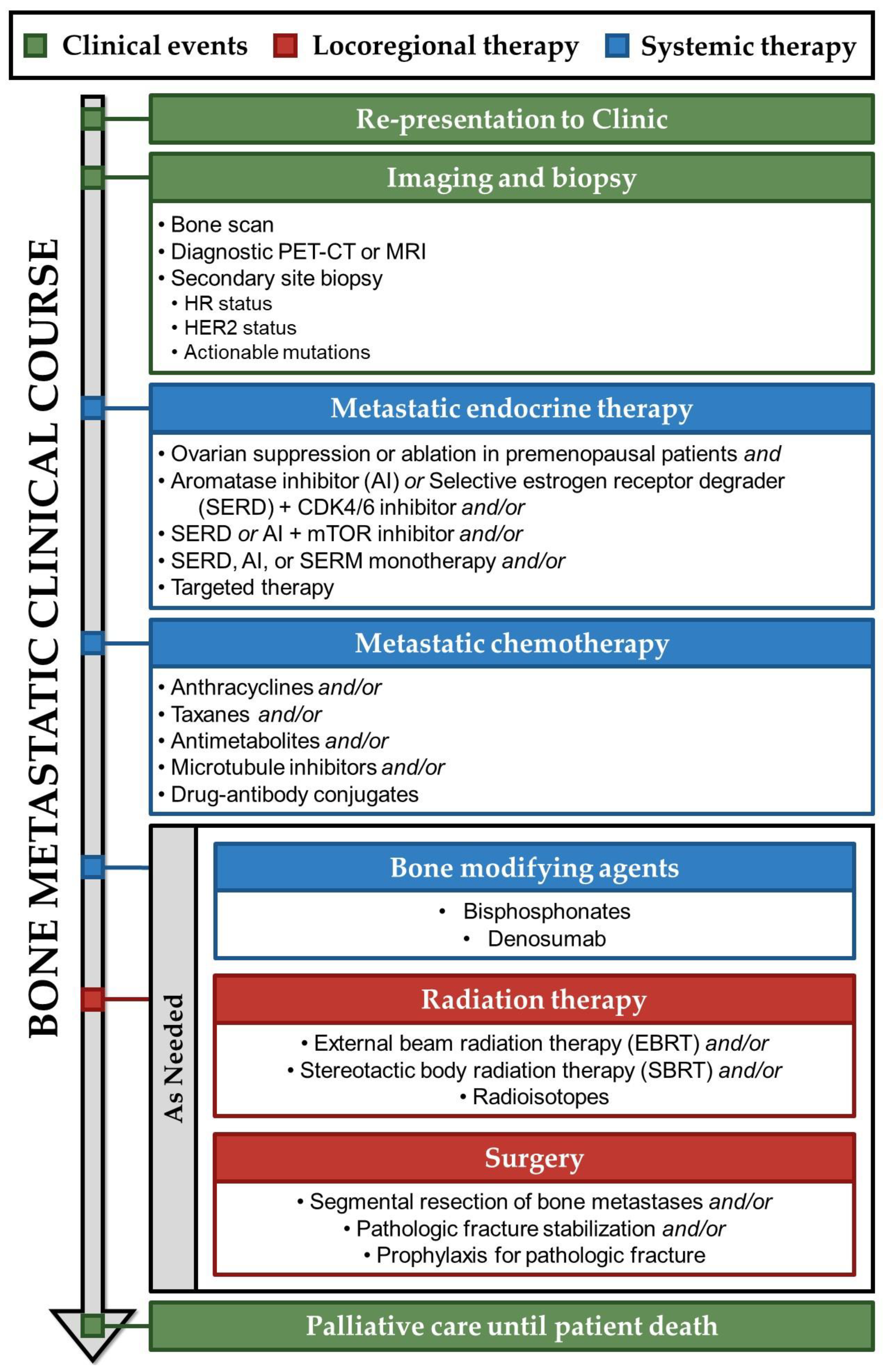
5.2. Clinical Metastatic Recurrence

Despite best therapeutic efforts, some patients with primary breast cancer will progress to metastatic disease. Unlike other subtypes of breast cancers in which risk of recurrence decreases 5 years after diagnosis, the recurrence risk for HR-positive breast cancer remains steady from 5 years to at least 20 years, with some patients experiencing recurrence three decades after initial diagnosis [7]. Therefore, long-term monitoring is a critical part of breast cancer clinical care. Regular history and physical examinations are recommended every 4 to 6 months for the first 5 years after primary therapy and annually thereafter, in conjunction with annual mammography [9]. However, without any clinical evidence of metastatic disease, evaluation of serum tumor markers, routine bone scans, Computed Tomography (CT) scans, Magnetic Resonance Imaging (MRI) scans, Positron Emisson Tomography (PET) scans, or ultrasound examinations in the asymptomatic patient provide no survival advantage and are, therefore, not recommended [9,120]. Instead, patients should be instructed to monitor for signs of recurrence [9].
Among patients who develop metastatic breast cancer, 45% experience their initial metastasis in the bone, a considerably higher occurrence than in the lungs (19%), liver (5%), or brain (2%) [121]. Patients with bone metastatic breast cancer may present with bone pain, pathologic fractures, symptoms of spinal cord compression including incontinence and muscle weakness, or hypercalcemia, which can cause nausea and altered mental status [122]. Each of these signs in a patient with a history of breast cancer warrants further follow-up and begins the clinical course outlined in Figure 4.
Upon a patient presenting to the clinic, a history, physical exam, and blood tests should be obtained, including liver function tests and alkaline phosphatase levels. Alkaline phosphatase is a serum marker of both hepatobiliary pathology and bone turnover and mineralization [123]. Bone-specific alkaline phosphatase is expressed on the cell surface of osteoblasts, and serum levels correlate with increased osteoblastic activity, which occurs in both osteoblastic and osteolytic lesions [86,123]. In osteoblastic lesions, this elevation correlates with local stimulation of osteoblasts, while in osteolytic lesions, it is secondary to local bone destruction and subsequent compensatory bone formation [86]. Patients with active bone metastases typically have elevated levels of alkaline phosphatase and should be followed with skeletal scintigraphy (bone scan), radiographs of any long bones that are painful or appear abnormal on bone scan, and diagnostic PET-CT or MRI [9].
Biopsy tissue should be acquired, if possible, from the site of recurrence and tested for molecular characteristics, HR status, and staging, as in primary disease [9]. Importantly, metastatic tumors may differ from the primary lesion, especially in HR and HER2 expression. In a large-scale study, 16% of patients who previously had HR-positive, HER2-negative primary tumors had changes in HR/HER2 expression in metastatic sites [124]. Of this 16%, 67% lost HR receptors, 25% gained HER2 receptors, and 8% both lost HR and gained HER2 receptors [124]. HR discordance with loss of HR status was significantly associated with a worse overall survival and has implications for therapeutic options [124]. Therapy choices can also be guided by tumor biomarkers. Unlike in primary disease, where tumor genomics are indirectly assessed by multigene assay, metastatic breast cancer is tested directly for actionable somatic mutations, in order to determine a patient’s eligibility for targeted therapy. For example, demonstrable mutation in PIK3CA using Next-Generation Sequencing (NGS) of tumor tissue or Circulating Tumor Deoxyribonucleic Acid (ctDNA) in blood renders a patient eligible for second-line Phosphatidylinositol-4,5-Bisphosphate 3-Kinase (PIK3) inhibitors, including alpelisib [125]. PIK3CA or AKT Serine/Threonine Kinase 1 (AKT1) activating mutations or PTEN alterations detected by NGS determine eligibility for the AKT inhibitor capivasertib, and acquired Estrogen Receptor 1 (ESR1) mutations at recurrence or progression on endocrine therapy make a patient eligible for the estrogen receptor antagonist elacestrant [25]. Clinicians may also test for Neurotrophic Receptor Tyrosine Kinase (NTRK) fusions, microsatellite instability-high/deficient mismatch repair biomarkers, tumor mutational burden, and Ret Proto-Oncogene (RET)-fusion in certain circumstances [125].

6. Treatment of Bone Metastatic Breast Cancer

Metastatic breast cancer treatment shares many features with primary treatment. However, treatment plans are more highly individualized, focusing on a patient’s specific site of recurrence for symptom relief and slowing of disease progression. It is estimated that about half of patients with bone relapse present with bone-only metastases and half present with other organ involvement [17]. Compared to patients with visceral involvement, patients with bone-only metastases have markedly longer overall survival, and a minority never develop extra-osseous metastases along their disease course [126]. For some patients with oligometastatic, bone-only disease, aggressive locoregional therapy may be pursued with curative intent [127]. However, most patients with bone metastases are treated extensively with endocrine therapy, chemotherapy, bone-modifying agents, and locoregional therapy to reduce or prevent tumor growth in both skeletal and extra-skeletal sites [126,128].
In the widely metastatic stage, breast cancer is treatable, but is not generally considered curable [9]. Therefore, treatment objectives shift to optimizing patient quality of life and, if possible, prolonging time to progression of disease and death [129]. Treatments with minimal toxicity are consequently preferred to maintain patient quality of life, and, except in cases of immediately life-threatening organ dysfunction, a trial of endocrine therapy should be the initial treatment for patients with bone-metastatic HR-positive disease [130].

6.1. Endocrine Therapy for Bone Metastatic Breast Cancer

Metastatic endocrine therapy employs many of the same principles and therapies that are used in the adjuvant setting, with important additions. The objective of endocrine therapy remains to decrease circulating estrogen or ER activity and prevent estrogen-related tumor growth in hormone-responsive cancers [131]. Notably, treatment guidelines pertain to both postmenopausal patients and premenopausal patients, who require ovarian suppression or ablation. Premenopausal patients are frequently underrepresented in clinical trials for metastatic breast cancer, so treatment strategies for premenopausal patients are typically extrapolated from postmenopausal patient data, with the addition of ovarian suppression or ablation [132]. As previously discussed, both natural and induced menopause profoundly decrease BMD through rapid decline in estrogen levels [59].
HR-positive cancers commonly lose endocrine sensitivity over time [131]. As a result, the usual treatment approach involves a sequential strategy, where one endocrine therapy is pursued until intolerable toxicity or progression occurs, followed by a switch to another endocrine therapy [133]. The optimal sequence for endocrine treatment remains unclear and is influenced by factors such as previous treatments, tolerance, and patient preference [9]. First-line metastatic endocrine therapy commonly includes AIs, as in primary disease. AIs are typically administered with a Cyclin-Dependent Kinase (CDK) 4/6 inhibitor, such as palbociclib, ribociclib, or abemaciclib. Cyclin-dependent kinases 4 and 6 are enzymes that mediate transition through the cell cycle and guard genome integrity [134]. ER-positive breast cancer cells are reliant on CDK4/6 for cell cycle progression [135]. Inhibition of CDK4/6 in combination with AIs in metastatic HR-positive, HER2-negative breast cancer has been shown to improve progression-free survival and objective response rate compared to AI monotherapy [136,137,138]. However, recently reported trials suggest that overall survival benefits are not consistent among the three approved CDK4/6 inhibitors when combined with AIs [135]. Only the addition of ribociclib to AIs has yet been shown to increase overall survival over AIs alone [139]. Though no trials have yet reported the effects of CDK4/6 inhibitor on bone outcomes, preclinical data interestingly report that only ribociclib has no toxic effects on osteoblasts in-vitro. The preserved viability of osteoblasts in the bone–tumor microenvironment could play a role in the overall survival benefit associated with ribociclib plus AI [134].
As an alternative to AIs, CDK4/6 inhibitors may instead be combined with fulvestrant, a selective estrogen receptor degrader/downregulator (SERD) and ER antagonist [140]. This is particularly beneficial for patients who experience progressive disease during AI treatment or develop a recurrence within one year of adjuvant endocrine therapy [141]. Fulvestrant plus ribociclib [142] or abemaciclib [143] has been shown to increase overall survival over fulvestrant monotherapy. Fulvestrant may also be combined with an AI, which has been shown to improve overall survival over AI alone [144,145]. SERDs like fulvestrant induce accelerated degradation of ER, resulting in a pure antiestrogenic effect on HR-positive breast cancer cells [140]. While fulvestrant is a pure anti-estrogen, it does not appear to decrease bone mass, in contrast to other estrogen modulators such as SERMs and AIs. Though clinical data are surprisingly limited, fulvestrant administration does not seem to increase markers of bone turnover [146,147].
For patients with metastatic breast cancer, next-line endocrine therapy options are pursued when disease progression occurs on first-line therapeutic regimens. Often, this is due to acquired endocrine resistance, which is associated with aberrant activation of the Mammalian Target Of Rapamycin (mTOR) signal transduction pathway [148]. Co-targeting the mTOR pathway and ER can restore sensitivity to patients with endocrine therapy-resistant advanced breast cancer [149]. Everolimus is an oral inhibitor of mTOR that has been shown to increase progression-free survival when added to AI [148] or fulvestrant [150] and overall survival when added to tamoxifen [151]. It should be noted, however, that there have been some reports of therapeutic resistance to everolimus in patients with AI resistance and estrogen-depleted-resistant breast cancer [152]. Regardless, everolimus has been reported to have bone-protective effects in metastatic disease. In a large clinical trial, adding everolimus to the AI exemestane reduced bone turnover markers when compared to exemestane alone [153]. It also decreased progression of pre-existing bone lesions and development of new bone lesions [153]. This finding was supported by preclinical work that showed that everolimus reduced osteoclast progenitor maturation in-vitro and osteoclast quantity and concomitant osteoclast-mediated bone resorption in an ovariectomized mouse model of metastatic breast cancer [154]. Other subsequent-line endocrine therapies may include SERD, SERM, or AI monotherapy, but these remain less well defined [9]. Providers and patients should engage in shared decision-making when considering subsequent lines of therapy and evaluate potential clinical risks, benefits, and patient goals and preferences.

6.2. Chemotherapy for Bone Metastatic Breast Cancer

When resistance to endocrine therapies becomes evident, the use of chemotherapies and targeted treatments is appropriate [141]. Chemotherapy is also indicated for patients who have symptomatic or rapidly progressive visceral metastasis [9]. Various chemotherapy protocols are considered suitable, including sequential monotherapy or select combinations in certain patients. Although combination chemotherapy may provide a longer time to progression, it is also associated with greater toxicity and generally provides only marginal survival benefit [155]. Therefore, sequential monotherapy is typically preferred, except in patients with rapid, symptomatic disease progression [9]. Agents are continued until progression or unacceptable toxicity occurs, decided in concert with the patient.
Many of the same chemotherapeutic agents are used in both metastatic and primary breast cancer treatment. These include the anthracycline doxorubicin and the taxane paclitaxel. Other treatments are used primarily in the metastatic setting, including anti-metabolites, microtubule inhibitors, and antibody-drug conjugates.
Anti-metabolites, which are incorporated into cancer cell nucleic acids and interfere with their synthesis, include capecitabine and gemcitabine [51]. Capecitabine is an oral, well-tolerated agent that increases objective response rate compared to traditional regimens [156] and improves quality of life in patients with heavily pretreated metastatic breast cancer [157]. An anti-metabolite alternative is gemcitabine, which is also efficacious in pretreated metastatic breast cancer patients [158]. A conjugate drug of gemcitabine and the bisphosphonate ibandronate was shown to localize to the bone microenvironment in a mouse model of osteosarcoma, a primary bone cancer [159]. When administered with docetaxel, the combination reduced tumor burden and improved mouse survival, while preserving bone architecture as measured by microCT. This innovative approach allows for targeted drug delivery to the bone microenvironment, allowing lower and less toxic doses of systemic drugs to be administered.
Microtubule inhibitors, which disrupt cell division in cancer cells, include eribulin and vinorelbine [51]. Eribulin is indicated for patients who have previously received an anthracycline, a taxane, and at least two chemotherapeutic regimens for the treatment of metastatic disease [9]. Importantly, single-agent eribulin has been demonstrated to increase overall survival in patients with heavily pretreated metastatic breast cancer over physician’s choice of chemotherapy [160]. However, in a recent preclinical study, mice treated with eribulin experienced a decrease in BMD as a result of increased osteoclast numbers and activity. The mechanism for this is thought to be indirect, with eribulin treatment causing a decrease in the RANKL decoy receptor OPG. With diminished OPG, the relative availability of RANKL increases, allowing the RANK/RANKL system to promote increased osteoclastogenesis [161]. Unfortunately, no clinical data have yet been reported regarding eribulin effects on bone in humans, but this preclinical study suggested that eribulin may cause bone loss in patients with metastatic breast cancer. An additional microtubule inhibitor option is vinorelbine, which has also demonstrated survival benefits in previously treated metastatic breast cancer, but little data on its effects on bone have been reported [162].
Finally, recent evidence has supported the use of drug–antibody conjugates in metastatic breast cancer. Sacituzumab Povitecan (SG) delivers SN-38, a topoisomerase I inhibitor, linked to an antibody targeting Trophoblast cell-surface antigen 2 (Trop-2), an epithelial antigen highly expressed in breast cancer [163]. SG is newly indicated for patients with metastatic unresectable breast cancer who are refractory to endocrine therapy and have received at least two prior lines of chemotherapy for metastatic disease [164]. In this patient population, SG has shown improvement in progression-free survival over physician’s choice of chemotherapy, but overall survival results have not yet been determined [163]. Given its recent introduction into the clinic, no information has yet been reported regarding SG effects on bone.

6.3. Bone-Modifying Agents for Bone Metastatic Breast Cancer

Endocrine and chemotherapeutic agents have profound effects on the bone microenvironment, and most therapies in the metastatic setting ultimately reduce BMD. As in adjuvant treatment of primary breast cancer, bone-modifying agents can help ameliorate therapy-induced loss of BMD, and their administration is standard of care in treating patients with bone metastatic breast cancer with life expectancy beyond 3 months [9]. Bisphosphonate usage has been rigorously demonstrated to reduce the risk of Skeletal Related Events (SREs) and bone pain and increase the time to SRE development [165]. However, unlike in the adjuvant setting where bisphosphonate administration has been shown to increase overall survival, bisphosphonates in metastatic breast cancer are a palliative measure only: no survival benefits have been demonstrated [165]. Compared to bisphosphonates, denosumab further reduces the risk of SRE by 22%, but demonstrates no additional benefits for time to SRE, bone pain, or overall survival [165].

6.4. Radiation Therapy for Bone Metastatic Breast Cancer

Radiation therapy has historically been used in bone metastatic breast cancer for analgesic purposes. The exact mechanism of RT-induced pain relief remains unclear, but it is thought that decreased tumor burden alleviates the mass effect on surrounding nerve structures [166] and decreased osteoclast-mediated bone destruction relieves inflammatory pain [167,168]. Evidence suggests that, beyond pain palliation, RT may have a role in SRE prevention. In a recent study, patients with asymptomatic high-risk bone metastases of either lung, breast, prostate, or other cancers were treated with either prophylactic RT or standard of care without RT. Patients who received prophylactic RT had significantly fewer SREs and improved overall survival [169]. Though a larger clinical trial is warranted, these results may suggest a wider role for RT in bone metastatic breast cancer beyond palliation.
Patients with bone metastatic breast cancer receive RT in a number of ways, including External Beam Radiation Therapy (EBRT), Stereotactic Body Radiation Therapy (SBRT), and through radioisotopes. EBRT is the standard of care for patients with painful bone metastases [169]. Compared to EBRT, SBRT administers a higher dose fraction of radiation but is more highly localized to its target, sparing nearby structures. SBRT is a reasonable option for patients with limited metastatic disease, with the intention of eradicating all known active disease and preventing disease progression [170]. Additionally, SBRT is used in the treatment of spinal metastases, where precise localization is critical to avoid excess irradiation of the spinal cord [170]. Increasingly, SBRT is being adopted for treatment of non-spinal bone metastases, but data in this setting are limited and outside the scope of professional society guidelines [171,172]. Studies comparing EBRT and SBRT for control of bone pain have produced mixed results, with some finding SBRT superior and others showing no appreciable difference between the two methods [173].
For patients with diffuse bone pain who are not eligible for palliative local RT with EBRT or SBRT, radioisotopes may be indicated [174]. Radioisotopes are given systemically and are localized to sites of active bone turnover, emitting high-energy β particles to provide cytotoxic irradiation of nearby cancer cells [175]. In the United States, strontium-89 is FDA-approved for treatment of painful bone metastases and samarium-153 is approved exclusively for osteoblastic bone lesions, which are present in some metastatic breast cancer cases [175]. Radium-233 is approved for pain relief in castration-resistant prostate cancer, where it also has been demonstrated to increase overall survival [176]. However, it remains poorly evaluated in metastatic breast cancer. Isolated case reports suggest that it may provide effective and tolerable pain relief, though with no reported survival outcomes [176].

6.5. Surgery for Bone Metastatic Breast Cancer

Beyond RT, surgery can be effective in ameliorating pain and responding to SREs, though with considerably more morbidity. Because the bone microenvironment surrounding skeletal metastases undergoes aberrant remodeling, pathological fracture healing is greatly impaired compared to normal bone [177]. As a result, most pathological fractures require surgical fixation for stabilization [178]. Surgical intervention to address impending fractures provides improved outcomes [178], but providers should strongly consider the life expectancy of the patient before undertaking preventative surgery requiring extensive recovery [178].

7. Palliative Care until Patient Death

Together, a patient and provider decide whether disease is being controlled and the toxicity of treatment is acceptable [179]. Should the burden of anticancer therapy outweigh its benefits, goals of care, prognosis, and end-of-life care should be discussed. Ideally, a multidisciplinary team consults to address physical, psychosocial, and practical interventions. The goal for each patient is to provide a death free from avoidable distress and suffering for patients, families, and caregivers; in general accord with the patient’s and family’s wishes; and consistent with clinical, cultural, and ethical standards [179]. The 5-year survival rate for patients with metastatic breast cancer is 29% [1].

8. Concluding Remarks

Patients diagnosed with HR-positive early breast cancer undergo aggressive treatment in hopes of eradicating local disease and preventing future recurrence. While treatment for early breast cancer is generally very effective, most of the therapeutic agents have deleterious effects on skeletal health that lead to bone remodeling and lowering of BMD. These skeletal changes wrought by chemotherapy and endocrine therapy may predispose patients to future bone metastases. In the adjuvant setting, antiresorptive bisphosphonates have been demonstrated to reduce this risk in postmenopausal women.
Despite advances in early breast cancer treatment, some patients progress to metastatic disease, and the most common site for dissemination is the bone. HR-positive/HER2-negative tumors are particularly associated with bone relapse. Patients with HR-positive bone metastatic breast cancer encounter distinct challenges to the integrity of their skeletal system. Initially, the widespread presence of cancer in the bone triggers a destructive local cycle. Subsequently, patients undergo endocrine therapy that requires elimination of estrogen signaling, abolishing estrogen’s protective effects on bone. Lastly, chemotherapy contributes to a further reduction in BMD, collectively rendering patients susceptible to SREs including pathologic fractures, intractable pain, and spinal cord compression.
The treatment approach for patients with bone metastatic breast cancer requires a highly individualized approach. Patients may be eligible for multiple lines of therapy, and clinicians therefore have discretion in choosing an appropriate treatment strategy. Therapy choice is dependent on a given patient’s previous treatments and response. For example, one patient may be successful with an AI and CDK4/6 inhibitor in the first-line metastatic setting, while another patient who previously had disease progression on adjuvant AI may be instead offered fulvestrant with a CDK4/6 inhibitor as a first-line metastatic treatment. Practitioners also must consider if an individual patient is eligible for targeted therapies based on tumor-specific somatic mutations. For example, PIK3 inhibitors like alpelisib or the novel AKT inhibitor capivasertib have been demonstrated to increase survival only in patients with PIK3CA, AKT1, or PTEN mutations. Identifying which patients may benefit from which therapies is a critical component of metastatic breast cancer care. Additionally, bone metastatic breast cancer can cause a variety of clinical sequelae, which may present differently in each patient. For instance, while some patients may experience spinal instability, others may develop hypercalcemia, and care must address each patient’s individual needs in symptom management and palliation. Finally, each patient has unique goals, values, and preferences for metastatic breast cancer treatment. Some patients may wish to continue anticancer treatment for as long as possible, and others elect to focus care on maintaining quality of life. Practitioners and patients must engage in shared decision-making to collectively decide on the best course of individual treatment.
Clinicians have a diverse armamentarium of therapies at their disposal to combat metastatic breast cancer and palliate symptoms, including endocrine therapy, chemotherapy, bone-modifying agents, and radiation therapy. Despite this, bone metastatic breast cancer remains an incurable disease, though several effective treatment strategies have emerged that extend progression-free survival and overall survival. CDK4/6 inhibitors have been heralded as a breakthrough since their introduction into the clinic and have provided the single greatest improvement in overall survival of any therapy for advanced HR-positive, HER2-negative breast cancer to date. The addition of CDK4/6 inhibitor ribociclib to AI therapy was demonstrated to improve overall survival for patients with metastatic breast cancer by more than 12 months over AI alone [139]. CDK4/6 inhibitors have proven so effective in metastatic settings that abemaciclib has been newly approved for HR-positive/HER2-negative early-breast cancer patients at high-risk of recurrence [180,181]. In this way, CDK4/6 inhibitors represent an ongoing strategy in combatting metastatic breast cancer; optimizing primary breast cancer treatment in order to prevent metastasis development.
Despite their efficacy, most patients eventually develop resistance to CDK4/6 inhibitors. To address this, a current major endeavor in metastatic breast cancer research focuses on overcoming or preventing therapy resistance. A novel small molecule PI3K inhibitor, inavolisib, has more than doubled progression-free survival when combined with CDK4/6 inhibitor palbociclib plus fulvestrant in patients with recurrent PIK3CA-mutated, HR–positive, HER2-negative advanced breast cancer [182]. Though no results are yet available regarding overall survival outcomes, this combinatorial strategy incorporating targeted therapy for eligible patients is among the most promising treatment options. Likewise, targeted therapy such as AKT inhibitor capivasertib in patients with PIK3CA, AKT1, or PTEN mutations, or estrogen receptor antagonist elacestrant in patients with acquired ESR1 mutations have improved progression-free survival in those patient populations [183,184]. Precision therapy based on individual tumor characteristics is a growing part of next-generation metastatic cancer care.
Unlike targeted therapy, immunotherapy has had little success in treatment of HR-positive, HER2-negative breast cancer, despite revolutionizing the treatment of other cancer types. Immune checkpoint inhibitors such as pembrolizumab, which have been successful in other cancers, are currently only indicated for metastatic breast cancer patients with MicroSatellite Instability-High (MSI-H) or MisMatch Repair deficient (dMMR) tumors, having shown little efficacy in other HR-positive populations [9]. However, another type of immunotherapy, antibody-drug conjugates, have shown promise using antibodies to specifically target breast cancer cells and deliver chemotherapy. Sacituzumab govitecan is the first of its class in HER2-negative breast cancer and represents a next-generation approach to cancer care.
As advances in the treatment of metastatic breast cancer lead to extended patient survival, care and consideration need to be given to preserving skeletal health and enhancing the overall quality of life for patients as they navigate an extended lifespan.

Author Contributions

Conceptualization, E.M. and K.M.B.; writing—original draft preparation, E.M.; writing—review and editing, K.M.B.; visualization, E.M. and K.M.B.; supervision, K.M.B. All authors have read and agreed to the published version of the manuscript.

Funding

This research was funded by The Department of Defense, grant number W81XWH-21-1-0001. The APC was funded by the Department of Defense, grant number W81XWH-21-1-0001.

Institutional Review Board Statement

Not applicable.

Informed Consent Statement

Not applicable.

Data Availability Statement

Not applicable.

Conflicts of Interest

The authors declare no conflicts of interest.

References

  1. Giaquinto, A.N.; Sung, H.; Miller, K.D.; Kramer, J.L.; Newman, L.A.; Minihan, A.; Jemal, A.; Siegel, R.L. Breast Cancer Statistics, 2022. CA Cancer J. Clin. 2022, 72, 524–541. [Google Scholar] [CrossRef]
  2. Siegel, R.L.; Giaquinto, A.N.; Jemal, A. Cancer statistics, 2024. CA Cancer J. Clin. 2024, 74, 12–49. [Google Scholar] [CrossRef]
  3. Gaorav, P.G.; Joan, M. Cancer Metastasis: Building a Framework. Cell 2006, 127, 679–695. [Google Scholar] [CrossRef]
  4. Sanmartin, M.C.; Borzone, F.R.; Giorello, M.B.; Pacienza, N.; Yannarelli, G.; Chasseing, N.A. Bone marrow/bone pre-metastatic niche for breast cancer cells colonization: The role of mesenchymal stromal cells. Crit. Rev. Oncol. Hematol. 2021, 164, 103416. [Google Scholar] [CrossRef] [PubMed]
  5. Wu, Q.; Li, J.; Zhu, S.; Wu, J.; Chen, C.; Liu, Q.; Wei, W.; Zhang, Y.; Sun, S. Breast cancer subtypes predict the preferential site of distant metastases: A SEER based study. Oncotarget 2017, 8, 27990–27996. [Google Scholar] [CrossRef] [PubMed]
  6. Suva, L.J.; Washam, C.; Nicholas, R.W.; Griffin, R.J. Bone metastasis: Mechanisms and therapeutic opportunities. Nat. Rev. Endocrinol. 2011, 7, 208–218. [Google Scholar] [CrossRef] [PubMed]
  7. Pan, H.; Gray, R.; Braybrooke, J.; Davies, C.; Taylor, C.; McGale, P.; Peto, R.; Pritchard, K.I.; Bergh, J.; Dowsett, M.; et al. 20-Year Risks of Breast-Cancer Recurrence after Stopping Endocrine Therapy at 5 Years. N. Engl. J. Med. 2017, 377, 1836–1846. [Google Scholar] [CrossRef] [PubMed]
  8. Mariotto, A.B.; Etzioni, R.; Hurlbert, M.; Penberthy, L.; Mayer, M. Estimation of the Number of Women Living with Metastatic Breast Cancer in the United States. Cancer Epidemiol. Biomark. Prev. 2017, 26, 809–815. [Google Scholar] [CrossRef] [PubMed]
  9. National Comprehensive Cancer Network. NCCN Clinical Practice Guidelines in Oncology (NCCN Guidelines®) for Breast Cancer [v.1.2024]. 2024. Available online: https://www.nccn.org/professionals/physician_gls/pdf/breast.pdf (accessed on 29 January 2024).
  10. Nicolini, A.; Ferrari, P.; Duffy, M.J. Prognostic and predictive biomarkers in breast cancer: Past, present and future. Semin. Cancer Biol. 2018, 52, 56–73. [Google Scholar] [CrossRef] [PubMed]
  11. Allison, K.H.; Hammond, M.E.H.; Dowsett, M.; McKernin, S.E.; Carey, L.A.; Fitzgibbons, P.L.; Hayes, D.F.; Lakhani, S.R.; Chavez-Macgregor, M.; Perlmutter, J.; et al. Estrogen and Progesterone Receptor Testing in Breast Cancer: American Society of Clinical Oncology/College of American Pathologists Guideline Update. Arch. Pathol. Lab. Med. 2020, 144, 545–563. [Google Scholar] [CrossRef]
  12. Carroll, J.S. Mechanisms of oestrogen receptor (ER) gene regulation in breast cancer. Eur. J. Endocrinol. 2016, 175, R41–R49. [Google Scholar] [CrossRef]
  13. Orrantia-Borunda, E.; Anchondo-Nuñez, P.; Acuña-Aguilar, L.E.; Gómez-Valles, F.O.; Ramírez-Valdespino, C.A. Subtypes of Breast Cancer; Exon Publications: Brisbane, QLD, Australia, 2022; pp. 31–42. [Google Scholar] [CrossRef]
  14. Lee, Y.; Gorski, J. Estrogen-induced transcription of the progesterone receptor gene does not parallel estrogen receptor occupancy. Proc. Natl. Acad. Sci. USA 1996, 93, 15180–15184. [Google Scholar] [CrossRef]
  15. Gutierrez, C.; Schiff, R. HER2: Biology, Detection, and Clinical Implications. Arch. Pathol. Lab. Med. 2011, 135, 55–62. [Google Scholar] [CrossRef]
  16. Slamon, D.J.; Clark, G.M.; Wong, S.G.; Levin, W.J.; Ullrich, A.; McGuire, W.L. Human Breast Cancer: Correlation of Relapse and Survival with Amplification of the HER-2/neu Oncogene. Science 1987, 235, 177–182. [Google Scholar] [CrossRef] [PubMed]
  17. Soni, A.; Ren, Z.; Hameed, O.; Chanda, D.; Morgan, C.J.; Siegal, G.P.; Wei, S. Breast Cancer Subtypes Predispose the Site of Distant Metastases. Am. J. Clin. Pathol. 2015, 143, 471–478. [Google Scholar] [CrossRef] [PubMed]
  18. Parker, J.S.; Mullins, M.; Cheang, M.C.; Leung, S.; Voduc, D.; Vickery, T.; Davies, S.; Fauron, C.; He, X.; Hu, Z.; et al. Supervised risk predictor of breast cancer based on intrinsic subtypes. J. Clin. Oncol. 2009, 27, 1160–1167. [Google Scholar] [CrossRef] [PubMed]
  19. Goldhirsch, A.; Wood, W.C.; Coates, A.S.; Gelber, R.D.; Thürlimann, B.; Senn, H.J. Strategies for subtypes—Dealing with the diversity of breast cancer: Highlights of the St Gallen International Expert Consensus on the Primary Therapy of Early Breast Cancer 2011. Ann. Oncol. 2011, 22, 1736–1747. [Google Scholar] [CrossRef] [PubMed]
  20. Koboldt, D.C.; Fulton, R.S.; McLellan, M.D.; Schmidt, H.; Kalicki-Veizer, J.; McMichael, J.F.; Fulton, L.L.; Dooling, D.J.; Ding, L.; Mardis, E.R.; et al. Comprehensive molecular portraits of human breast tumours. Nature 2012, 490, 61–70. [Google Scholar] [CrossRef]
  21. Schettini, F.; Brasó-Maristany, F.; Kuderer, N.M.; Prat, A. A perspective on the development and lack of interchangeability of the breast cancer intrinsic subtypes. NPJ Breast Cancer 2022, 8, 85. [Google Scholar] [CrossRef] [PubMed]
  22. Soldato, D.; Miller, K.D. Flashback Foreword: Supervised Risk Predictor of Breast Cancer. J. Clin. Oncol. 2023, 41, 4189–4190. [Google Scholar] [CrossRef]
  23. Andre, F.; Ismaila, N.; Allison, K.H.; Barlow, W.E.; Collyar, D.E.; Damodaran, S.; Henry, N.L.; Jhaveri, K.; Kalinsky, K.; Kuderer, N.M.; et al. Biomarkers for Adjuvant Endocrine and Chemotherapy in Early-Stage Breast Cancer: ASCO Guideline Update. J. Clin. Oncol. 2022, 40, 1816–1837. [Google Scholar] [CrossRef] [PubMed]
  24. Hu, C.; Hart, S.N.; Gnanaolivu, R.; Huang, H.; Lee, K.Y.; Na, J.; Gao, C.; Lilyquist, J.; Yadav, S.; Boddicker, N.J.; et al. A Population-Based Study of Genes Previously Implicated in Breast Cancer. N. Engl. J. Med. 2021, 384, 440–451. [Google Scholar] [CrossRef] [PubMed]
  25. National Comprehensive Cancer Network. NCCN Clinical Practice Guidelines in Oncology (NCCN Guidelines®) for Genetic/Familial High-Risk Assessment: Breast, Ovarian, and Pancreatic [V3.2024]. 2024. Available online: https://www.nccn.org/professionals/physician_gls/pdf/genetics_bop.pdf (accessed on 12 February 2024).
  26. Balmaña, J.; Díez, O.; Rubio, I.T.; Cardoso, F. BRCA in breast cancer: ESMO Clinical Practice Guidelines. Ann. Oncol. 2011, 22, vi31–vi34. [Google Scholar] [CrossRef] [PubMed]
  27. Spring, L.M.; Gupta, A.; Reynolds, K.L.; Gadd, M.A.; Ellisen, L.W.; Isakoff, S.J.; Moy, B.; Bardia, A. Neoadjuvant Endocrine Therapy for Estrogen Receptor-Positive Breast Cancer: A Systematic Review and Meta-analysis. JAMA Oncol. 2016, 2, 1477–1486. [Google Scholar] [CrossRef] [PubMed]
  28. Trabert, B.; Sherman, M.E.; Kannan, N.; Stanczyk, F.Z. Progesterone and Breast Cancer. Endocr. Rev. 2020, 41, 320–344. [Google Scholar] [CrossRef] [PubMed]
  29. Smith, I.E.; Dowsett, M.; Ebbs, S.R.; Dixon, J.M.; Skene, A.; Blohmer, J.U.; Ashley, S.E.; Francis, S.; Boeddinghaus, I.; Walsh, G. Neoadjuvant treatment of postmenopausal breast cancer with anastrozole, tamoxifen, or both in combination: The Immediate Preoperative Anastrozole, Tamoxifen, or Combined with Tamoxifen (IMPACT) multicenter double-blind randomized trial. J. Clin. Oncol. 2005, 23, 5108–5116. [Google Scholar] [CrossRef] [PubMed]
  30. Van de Loo, M.E.; Andour, L.; van Heesewijk, A.E.; Oosterkamp, H.M.; Liefers, G.J.; Straver, M.E. Neoadjuvant endocrine treatment in hormone receptor-positive breast cancer: Does it result in more breast-conserving surgery? Breast Cancer Res. Treat. 2024, 1–12. [Google Scholar] [CrossRef]
  31. Olivotto, I.A.; Bajdik, C.D.; Ravdin, P.M.; Speers, C.H.; Coldman, A.J.; Norris, B.D.; Davis, G.J.; Chia, S.K.; Gelmon, K.A. Population-Based Validation of the Prognostic Model ADJUVANT! for Early Breast Cancer. J. Clin. Oncol. 2005, 23, 2716–2725. [Google Scholar] [CrossRef]
  32. Cataliotti, L.; Buzdar, A.U.; Noguchi, S.; Bines, J.; Takatsuka, Y.; Petrakova, K.; Dube, P.; Tosello de Oliveira, C. Comparison of anastrozole versus tamoxifen as preoperative therapy in postmenopausal women with hormone receptor-positive breast cancer. Cancer 2006, 106, 2095–2103. [Google Scholar] [CrossRef]
  33. Eiermann, W.; Paepke, S.; Appfelstaedt, J.; Llombart-Cussac, A.; Eremin, J.; Vinholes, J.; Mauriac, L.; Ellis, M.; Lassus, M.; Chaudri-Ross, H.A.; et al. Preoperative treatment of postmenopausal breast cancer patients with letrozole: A randomized double-blind multicenter study. Ann. Oncol. 2001, 12, 1527–1532. [Google Scholar] [CrossRef]
  34. Korde, L.A.; Somerfield, M.R.; Carey, L.A.; Crews, J.R.; Denduluri, N.; Hwang, E.S.; Khan, S.A.; Loibl, S.; Morris, E.A.; Perez, A.; et al. Neoadjuvant Chemotherapy, Endocrine Therapy, and Targeted Therapy for Breast Cancer: ASCO Guideline. J. Clin. Oncol. 2021, 39, 1485–1505. [Google Scholar] [CrossRef] [PubMed]
  35. Fisher, B.; Brown, A.; Mamounas, E.; Wieand, S.; Robidoux, A.; Margolese, R.G.; Cruz, A.B., Jr.; Fisher, E.R.; Wickerham, D.L.; Wolmark, N.; et al. Effect of preoperative chemotherapy on local-regional disease in women with operable breast cancer: Findings from National Surgical Adjuvant Breast and Bowel Project B-18. J. Clin. Oncol. 1997, 15, 2483–2493. [Google Scholar] [CrossRef]
  36. Fisher, B.; Anderson, S.; Bryant, J.; Margolese, R.G.; Deutsch, M.; Fisher, E.R.; Jeong, J.-H.; Wolmark, N. Twenty-Year Follow-up of a Randomized Trial Comparing Total Mastectomy, Lumpectomy, and Lumpectomy plus Irradiation for the Treatment of Invasive Breast Cancer. N. Engl. J. Med. 2002, 347, 1233–1241. [Google Scholar] [CrossRef]
  37. Agarwal, S.; Pappas, L.; Neumayer, L.; Kokeny, K.; Agarwal, J. Effect of Breast Conservation Therapy vs Mastectomy on Disease-Specific Survival for Early-Stage Breast Cancer. JAMA Surg. 2014, 149, 267. [Google Scholar] [CrossRef] [PubMed]
  38. Hwang, E.S.; Lichtensztajn, D.Y.; Gomez, S.L.; Fowble, B.; Clarke, C.A. Survival after lumpectomy and mastectomy for early stage invasive breast cancer. Cancer 2013, 119, 1402–1411. [Google Scholar] [CrossRef]
  39. Chatterjee, A.; Pyfer, B.; Czerniecki, B.; Rosenkranz, K.; Tchou, J.; Fisher, C. Early postoperative outcomes in lumpectomy versus simple mastectomy. J. Surg. Res. 2015, 198, 143–148. [Google Scholar] [CrossRef] [PubMed]
  40. Mansel, R.E.; Fallowfield, L.; Kissin, M.; Goyal, A.; Newcombe, R.G.; Dixon, J.M.; Yiangou, C.; Horgan, K.; Bundred, N.; Monypenny, I.; et al. Randomized Multicenter Trial of Sentinel Node Biopsy Versus Standard Axillary Treatment in Operable Breast Cancer: The ALMANAC Trial. JNCI J. Natl. Cancer Inst. 2006, 98, 599–609. [Google Scholar] [CrossRef] [PubMed]
  41. Giuliano, A.E.; Hawes, D.; Ballman, K.V.; Whitworth, P.W.; Blumencranz, P.W.; Reintgen, D.S.; Morrow, M.; Leitch, A.M.; Hunt, K.K.; McCall, L.M.; et al. Association of Occult Metastases in Sentinel Lymph Nodes and Bone Marrow With Survival Among Women With Early-Stage Invasive Breast Cancer. JAMA 2011, 306, 385. [Google Scholar] [CrossRef]
  42. Beenken, S.W.; Urist, M.M.; Zhang, Y.; Desmond, R.; Krontiras, H.; Medina, H.; Bland, K.I. Axillary Lymph Node Status, But Not Tumor Size, Predicts Locoregional Recurrence and Overall Survival After Mastectomy for Breast Cancer. Ann. Surg. 2003, 237, 732–739. [Google Scholar] [CrossRef]
  43. Colleoni, M.; O’Neill, A.; Goldhirsch, A.; Gelber, R.D.; Bonetti, M.; Thürlimann, B.; Price, K.N.; Castiglione-Gertsch, M.; Coates, A.S.; Lindtner, J.; et al. Identifying Breast Cancer Patients at High Risk for Bone Metastases. J. Clin. Oncol. 2000, 18, 3925–3935. [Google Scholar] [CrossRef]
  44. Thorn, C.F.; Oshiro, C.; Marsh, S.; Hernandez-Boussard, T.; McLeod, H.; Klein, T.E.; Altman, R.B. Doxorubicin pathways. Pharmacogenetics Genom. 2011, 21, 440–446. [Google Scholar] [CrossRef]
  45. Rana, T.; Chakrabarti, A.; Freeman, M.; Biswas, S. Doxorubicin-Mediated Bone Loss in Breast Cancer Bone Metastases Is Driven by an Interplay between Oxidative Stress and Induction of TGFβ. PLoS ONE 2013, 8, e78043. [Google Scholar] [CrossRef]
  46. Bussard, K.M.; Gay, C.V.; Mastro, A.M. The bone microenvironment in metastasis; what is special about bone? Cancer Metastasis Rev. 2008, 27, 41–55. [Google Scholar] [CrossRef] [PubMed]
  47. Seoane, J.; Gomis, R.R. TGF-β Family Signaling in Tumor Suppression and Cancer Progression. Cold Spring Harb. Perspect. Biol. 2017, 9, a022277. [Google Scholar] [CrossRef] [PubMed]
  48. Peyman, H.; May, Z.; Carolin, M.; Ute, A.; Matthias, K. The influence of chemotherapy on bone mineral density, quantitative ultrasonometry and bone turnover in pre-menopausal women with breast cancer. Eur. J. Cancer 2009, 45, 3205–3212. [Google Scholar] [CrossRef]
  49. Lipton, A.; Chapman, J.-A.W.; Demers, L.; Shepherd, L.E.; Han, L.; Wilson, C.F.; Pritchard, K.I.; Leitzel, K.E.; Ali, S.M.; Pollak, M. Elevated Bone Turnover Predicts for Bone Metastasis in Postmenopausal Breast Cancer: Results of NCIC CTG MA.14. J. Clin. Oncol. 2011, 29, 3605–3610. [Google Scholar] [CrossRef] [PubMed]
  50. Kraemer, B.; Rothmund, R.; Banys, M.; Krawczyk, N.; Solomayer, E.-F.; Mack, C.; Wallwiener, D.; Fehm, T. Impaired Bone Microenvironment: Correlation between Bone Density and Presence of Disseminated Tumor Cells. Anticancer. Res. 2011, 31, 4423–4428. [Google Scholar] [PubMed]
  51. Tilsed, C.M.; Fisher, S.A.; Nowak, A.K.; Lake, R.A.; Lesterhuis, W.J. Cancer chemotherapy: Insights into cellular and tumor microenvironmental mechanisms of action. Front. Oncol. 2022, 12, 960317. [Google Scholar] [CrossRef] [PubMed]
  52. Bjarnason, N.H.; Hitz, M.; Jorgensen, N.R.; Vestergaard, P. Adverse bone effects during pharmacological breast cancer therapy. Acta Oncol. 2008, 47, 747–754. [Google Scholar] [CrossRef]
  53. Colvin, O.M. An overview of cyclophosphamide development and clinical applications. Curr. Pharm. Des. 1999, 5, 555–560. [Google Scholar] [CrossRef]
  54. Lower, E.E.; Blau, R.; Gazder, P.; Tummala, R. The Risk of Premature Menopause Induced by Chemotherapy for Early Breast Cancer. J. Women’s Health Gend. Based Med. 1999, 8, 949–954. [Google Scholar] [CrossRef]
  55. Reynolds, A.C.; McKenzie, L.J. Cancer Treatment-Related Ovarian Dysfunction in Women of Childbearing Potential: Management and Fertility Preservation Options. J. Clin. Oncol. 2023, 41, 2281–2292. [Google Scholar] [CrossRef]
  56. Oktem, O.; Oktay, K. Quantitative assessment of the impact of chemotherapy on ovarian follicle reserve and stromal function. Cancer 2007, 110, 2222–2229. [Google Scholar] [CrossRef] [PubMed]
  57. Soleimani, R.; Heytens, E.; Darzynkiewicz, Z.; Oktay, K. Mechanisms of chemotherapy-induced human ovarian aging: Double strand DNA breaks and microvascular compromise. Aging 2011, 3, 782–793. [Google Scholar] [CrossRef] [PubMed]
  58. Shapiro, C.L.; Manola, J.; Leboff, M. Ovarian Failure after Adjuvant Chemotherapy Is Associated with Rapid Bone Loss in Women with Early-Stage Breast Cancer. J. Clin. Oncol. 2001, 19, 3306–3311. [Google Scholar] [CrossRef] [PubMed]
  59. Khosla, S.; Oursler, M.J.; Monroe, D.G. Estrogen and the skeleton. Trends Endocrinol. Metab. 2012, 23, 576–581. [Google Scholar] [CrossRef] [PubMed]
  60. Garnero, P.; Sornay-Rendu, E.; Chapuy, M.C.; Delmas, P.D. Increased bone turnover in late postmenopausal women is a major determinant of osteoporosis. J. Bone Miner. Res. 1996, 11, 337–349. [Google Scholar] [CrossRef] [PubMed]
  61. Nisha, Y.; Dubashi, B.; Bobby, Z.; Sahoo, J.P.; Kayal, S. Effect of cytotoxic chemotherapy on bone health among breast cancer patients. Does it require intervention? Support. Care Cancer 2021, 29, 6957–6972. [Google Scholar] [CrossRef] [PubMed]
  62. Clarke, M.; Collins, R.; Darby, S. Effects of radiotherapy and of differences in the extent of surgery for early breast cancer on local recurrence and 15-year survival: An overview of the randomised trials. Lancet 2005, 366, 2087–2106. [Google Scholar] [CrossRef] [PubMed]
  63. Jones, H.; Antonini, N.; Hart, A. Impact of pathological characteristics on local relapse after breast-conserving therapy: A subgroup analysis of the EORTC boost versus no boost trial. J. Clin. Oncol. 2009, 27, 4939–4947. [Google Scholar] [CrossRef]
  64. Shaitelman, S.F.; Anderson, B.M.; Arthur, D.W.; Bazan, J.G.; Bellon, J.R.; Bradfield, L.; Coles, C.E.; Gerber, N.K.; Kathpal, M.; Kim, L.; et al. Partial Breast Irradiation for Patients with Early-Stage Invasive Breast Cancer or Ductal Carcinoma In Situ: An ASTRO Clinical Practice Guideline. Pract. Radiat. Oncol. 2023, 14, 112–132. [Google Scholar] [CrossRef]
  65. Nicholls, L.; Gorayski, P.; Harvey, J. Osteoradionecrosis of the Ribs following Breast Radiotherapy. Case Rep. Oncol. 2015, 8, 332–338. [Google Scholar] [CrossRef]
  66. Early Breast Cancer Trialists’ Collaborative Group (EBCTCG). Effects of chemotherapy and hormonal therapy for early breast cancer on recurrence and 15-year survival: An overview of the randomised trials. Lancet 2005, 365, 1687–1717. [Google Scholar] [CrossRef]
  67. Dutertre, M.; Smith, C.L. Molecular Mechanisms of Selective Estrogen Receptor Modulator (SERM) Action. J. Pharmacol. Exp. Ther. 2000, 295, 431–437. [Google Scholar] [PubMed]
  68. Caciolla, J.; Bisi, A.; Belluti, F.; Rampa, A.; Gobbi, S. Reconsidering Aromatase for Breast Cancer Treatment: New Roles for an Old Target. Molecules 2020, 25, 5351. [Google Scholar] [CrossRef] [PubMed]
  69. Fleming, G.F.; Rajagopal, P. Point: LHRH Agonists vs Ovarian Ablation for Suppression of Ovarian Function in Premenopausal Breast Cancer Patients. Starting with Reversible OFS Using LHRH Agonists Is Preferred for Most Patients. Oncology 2018, 32, 168–170. [Google Scholar]
  70. Poorvu, P.D.; Partridge, A.H. Counterpoint: LHRH Agonists vs Ovarian Ablation for Suppression of Ovarian Function in Premenopausal Breast Cancer Patients. Ovarian Ablation Is a Viable Option for Select Patients. Oncology 2018, 32, 168, 171, 179. [Google Scholar] [PubMed]
  71. Gray, R.G.; Bradley, R.; Braybrooke, J.; Clarke, M.; Hills, R.K.; Peto, R.; Bergh, J.C.S.; Swain, S.M.; Davidson, N.E.; Francis, P.A.; et al. Effects of ovarian ablation or suppression on breast cancer recurrence and survival: Patient-level meta-analysis of 14,993 pre-menopausal women in 25 randomized trials. J. Clin. Oncol. 2023, 41, 503. [Google Scholar] [CrossRef]
  72. Cui, J.; Shen, Y.; Li, R. Estrogen synthesis and signaling pathways during aging: From periphery to brain. Trends Mol. Med. 2013, 19, 197–209. [Google Scholar] [CrossRef]
  73. Tahara, R.K.; Brewer, T.M.; Theriault, R.L.; Ueno, N.T. Bone Metastasis of Breast Cancer; Springer International Publishing: New York, NY, USA, 2019; pp. 105–129. [Google Scholar] [CrossRef]
  74. Marks, S.C., Jr.; Odgren, P.R. Structure and development of the skeleton. In Principles of Bone Biology; Bilezikian, J.P., Raisz, L.G., Rodan, G.A., Eds.; Academic Press: San Diego, CA, USA, 2002; Volume 1, pp. 3–16. [Google Scholar]
  75. Lian, J.B.; Stein, G.S. Concepts of osteoblast growth and differentiation: Basis for modulation of bone cell development and tissue formation. Crit. Rev. Oral Biol. Med. 1992, 3, 269–305. [Google Scholar] [CrossRef]
  76. Alberts, B.; Johnson, A.; Lewis, J.; Raff, M.; Roberts, K.; Walter, P. Molecular Biology of the Cell, 4th ed.; Garland Science: New York, NY, USA, 2002; p. 1308. [Google Scholar]
  77. Hancox, N.M. Biology of Bone; University Press: Cambridge, UK, 1972. [Google Scholar]
  78. Baron, R. General principles of bone biology. In Primer on the Metabolic Bone Diseases and Disorders of Mineral Metabolism; Favus, M.J., Ed.; American Society for Bone and Mineral Research: Washington, DC, USA, 2003; pp. 1–8. [Google Scholar]
  79. Kanis, J.A.; McCloskey, E.V. Bone turnover and biochemical markers in malignancy. Cancer 1997, 80, 1538–1545. [Google Scholar] [CrossRef]
  80. Bonewald, L.F. The amazing osteocyte. J. Bone Miner. Res. 2011, 26, 229–238. [Google Scholar] [CrossRef]
  81. Takahashi, N.; Udagawa, N.; Takami, M.; Suda, T. Cells of bone: Osteoclast generation. In Principles of Bone Biology; Bilezikian, J.P., Raisz, L.G., Rodan, G.A., Eds.; Academic Press: San Diego, CA, USA, 2002; Volume 1, pp. 109–126. [Google Scholar]
  82. Mastro, A.M.; Gay, C.V.; Welch, D.R.; Donahue, H.J.; Jewell, J.; Mercer, R.; DiGirolamo, D.; Chislock, E.M.; Guttridge, K. Breast cancer cells induce osteoblast apoptosis: A possible contributor to bone degradation. J. Cell Biochem. 2004, 91, 265–276. [Google Scholar] [CrossRef]
  83. Renema, N.; Navet, B.; Heymann, M.F.; Lezot, F.; Heymann, D. Rank-rankl signalling in cancer. Biosci. Rep. 2016, 36, e00366. [Google Scholar] [CrossRef]
  84. Roodman, G.D. Biology of osteoclast activation in cancer. J. Clin. Oncol. 2001, 19, 3562–3571. [Google Scholar] [CrossRef] [PubMed]
  85. Yoneda, T. Mechanisms of preferential metastasis of breast cancer to bone. Int. J. Oncol. 1996, 9, 103–109. [Google Scholar] [CrossRef] [PubMed]
  86. Mundy, G.R. Metastasis to bone: Causes, consequences and therapeutic opportunities. Nat. Rev. Cancer 2002, 2, 584–593. [Google Scholar] [CrossRef] [PubMed]
  87. Guise, T.A.; Yin, J.J.; Taylor, S.D.; Kumagai, Y.; Dallas, M.; Boyce, B.F.; Yoneda, T.; Mundy, G.R. Evidence for a causal role of parathyroid hormone-related protein in the pathogenesis of human breast-cancer-mediated osteolysis. J. Clin. Investig. 1996, 98, 1544–1549. [Google Scholar] [CrossRef] [PubMed]
  88. Stewart, A.F.; Vignery, A.; Silverglate, A.; Ravin, N.D.; Livolsi, V.; Broadus, A.E.; Baron, R. Quantitative bone histomorphology in humoral hypercalcemia of malignancy: Uncoupling of bone cell activity. J. Clin. Endocrinol. Metab. 1982, 55, 219–227. [Google Scholar] [CrossRef] [PubMed]
  89. Mercer, R.; Miyasaka, C.; Mastro, A.M. Metastatic breast cancer cells suppress osteoblast adhesion and differentiation. Clin. Exp. Metas. 2004, 21, 427–435. [Google Scholar] [CrossRef] [PubMed]
  90. Martin, T.J.; Moseley, J.M. Mechanisms in the skeletal complications of breast cancer. Endocr. Rel. Cancer 2000, 7, 271–284. [Google Scholar] [CrossRef]
  91. Guise, T.A.; Mundy, G.R. Cancer and bone. Endocr. Rev. 1998, 19, 18–54. [Google Scholar]
  92. Kozlow, W.; Guise, T.A. Breast Cancer Metastasis to Bone: Mechanisms of Osteolysis and Implications for Therapy. J. Mammary Gland. Biol. Neoplasia 2005, 10, 169–180. [Google Scholar] [CrossRef]
  93. Rogers, M.J.; Gordon, S.; Benford, H.L.; Coxon, F.P.; Luckman, S.P.; Monkkonen, J.; Frith, J.C. Cellular and molecular mechanisms of action of bisphosphonates. Cancer 2000, 88, 2961–2978. [Google Scholar] [CrossRef] [PubMed]
  94. Khan, M.N.; Khan, A.A. Cancer Treatment–Related Bone Loss: A Review and Synthesis of the Literature. Curr. Oncol. 2008, 15, 30–40. [Google Scholar] [CrossRef]
  95. Gnant, M.; Mlineritsch, B.; Stoeger, H.; Luschin-Ebengreuth, G.; Heck, D.; Menzel, C.; Jakesz, R.; Seifert, M.; Hubalek, M.; Pristauz, G.; et al. Adjuvant endocrine therapy plus zoledronic acid in premenopausal women with early-stage breast cancer: 62-month follow-up from the ABCSG-12 randomised trial. Lancet Oncol. 2011, 12, 631–641. [Google Scholar] [CrossRef] [PubMed]
  96. Ottewell, P.D.; Wang, N.; Brown, H.K.; Reeves, K.J.; Fowles, C.A.; Croucher, P.I.; Eaton, C.L.; Holen, I. Zoledronic Acid Has Differential Antitumor Activity in the Pre- and Postmenopausal Bone Microenvironment In Vivo. Clin. Cancer Res. 2014, 20, 2922–2932. [Google Scholar] [CrossRef] [PubMed]
  97. Hanley, D.A.; Adachi, J.D.; Bell, A.; Brown, V. Denosumab: Mechanism of action and clinical outcomes. Int. J. Clin. Pract. 2012, 66, 1139–1146. [Google Scholar] [CrossRef] [PubMed]
  98. Gnant, M.; Pfeiler, G.; Dubsky, P.C.; Hubalek, M.; Greil, R.; Jakesz, R.; Wette, V.; Balic, M.; Haslbauer, F.; Melbinger, E.; et al. Adjuvant denosumab in breast cancer (ABCSG-18): A multicentre, randomised, double-blind, placebo-controlled trial. Lancet 2015, 386, 433–443. [Google Scholar] [CrossRef] [PubMed]
  99. Gnant, M.; Pfeiler, G.; Steger, G.G.; Egle, D.; Greil, R.; Fitzal, F.; Wette, V.; Balic, M.; Haslbauer, F.; Melbinger-Zeinitzer, E.; et al. Adjuvant denosumab in postmenopausal patients with hormone receptor-positive breast cancer (ABCSG-18): Disease-free survival results from a randomised, double-blind, placebo-controlled, phase 3 trial. Lancet Oncol. 2019, 20, 339–351. [Google Scholar] [CrossRef]
  100. Robert, C.; Dianne, M.F.; Carlos, B.; Miguel, M.; Hiroji, I.; Roberto, H.; John, G.; Alvaro Montaño, P.; Katia, T.; Ines, D.; et al. Adjuvant denosumab in early breast cancer (D-CARE): An international, multicentre, randomised, controlled, phase 3 trial. Lancet Oncol. 2020, 21, 60–72. [Google Scholar] [CrossRef]
  101. Chambers, A.F.; Groom, A.C.; MacDonald, I.C. Dissemination and growth of cancer cells in metastatic sites. Nat. Rev. Cancer 2002, 2, 563–572. [Google Scholar] [CrossRef] [PubMed]
  102. Hanahan, D.; Weinberg, R.A. Hallmarks of cancer: The next generation. Cell 2011, 144, 646–674. [Google Scholar] [CrossRef]
  103. Welch, D.R.; Hurst, D.R. Defining the Hallmarks of Metastasis. Cancer Res. 2019, 79, 3011–3027. [Google Scholar] [CrossRef] [PubMed]
  104. Guise, T.A. Molecular mechanisms of osteolytic bone metastases. Cancer 2000, 88, 2892–2898. [Google Scholar] [CrossRef]
  105. Bendre, M.; Montague, D.C.; Peery, T.; Akel, N.S.; Gaddy, D.; Suva, L.J. Interleukin-8 stimulation of osteoclastogenesis and bone resorption is a mechanism for the increased osteolysis of metastatic bone disease. Bone 2003, 33, 28–37. [Google Scholar] [CrossRef] [PubMed]
  106. Bendre, M.; Gaddy, D.; Nicholas, R.W.; Suva, L.J. Breast cancer metastasis to bone. Clin. Ortho. Rel. Res. 2003, 415S, S39–S45. [Google Scholar] [CrossRef]
  107. Bendre, M.; Gaddy-Kurten, D.; Foote-Mon, T.; Akel, N.S.; Skinner, R.A.; Nicholas, R.W.; Suva, L.J. Expression of interleukin 8 and not parathyroid hormone-related protein by human breast cancer cells correlates with bone metastasis in vivo. Cancer Res. 2002, 62, 5571–5579. [Google Scholar]
  108. Arenberg, D.; Kunkel, S.; Polverini, P.; Glass, M.; Burdick, M.D.; Strieter, R.M. Inhibition of interleukin-8 reduces tumorigenesis of human non-small cell lung cancer in SCID mice. J. Clin. Investig. 1996, 97, 2792–2802. [Google Scholar] [CrossRef]
  109. Kim, S.; Uehara, H.; Karashima, T.; Mccarty, M.; Shih, N.; Fidler, I.J. Expression of interleukin 8 correlates with angiogenesis, tumorigenicity, and metastasis of human prostate cancer cells implanted orthotopically in nude mice. Neoplasia 2001, 3, 33–42. [Google Scholar] [CrossRef]
  110. Singh, R.; Varney, M. IL-8 expression in malignant melanoma: Implications in growth and metastases. Histol. Histopathol. 2000, 15, 843–849. [Google Scholar] [PubMed]
  111. Lu, Y.; Cai, Z.; Galson, D.L.; Xiao, G.; Liu, Y.; George, D.E.; Melhem, M.F.; Yao, Z.; Zhang, J. Monocyte chemotactic protein-1 (MCP-1) acts as a paracrine and autocrine factor for prostate cancer growth and invasion. Prostate 2006, 66, 1311–1318. [Google Scholar] [CrossRef] [PubMed]
  112. Mestdagt, M.; Polette, M.; Buttice, G.; Noel, A.; Ueda, A.; Foidart, J.-M.; Gilles, C. Transactivation of MCP-1/CCL2 by β-catenin/TCF-4 in human breast cancer cells. Int. J. Cancer 2006, 118, 35–42. [Google Scholar] [CrossRef] [PubMed]
  113. Koide, N.; Nishio, A.; Sato, T.; Sugiyama, A.; Miyagawa, S. Significance of macrophage chemoattractant protein-1 expression and macrophage infiltration in squamous cell carcinoma of the esophagus. Am. J. Gastroenterol. 2004, 99, 1667–1674. [Google Scholar] [CrossRef] [PubMed]
  114. Johrer, K.; Janke, K.; Krugmann, J.; Fiegl, M.; Greil, R. Transendothelial migration of myeloma cells is increased by tumor necrosis factor (TNF)-alpha via TNF receptor 2 and autocrine upregulation of MCP-1. Clin. Cancer Res. 2004, 10, 1901–1910. [Google Scholar] [CrossRef] [PubMed]
  115. Badache, A.; Hynes, N.E. Interleukin 6 inhibits proliferation and, in cooperation with epidermal growth factor receptor autocrine loop, increases migration of T47D breast cancer cells. Cancer Res. 2001, 61, 383–391. [Google Scholar]
  116. Kishimoto, T. Interleukin-6: From basic science to medicine—40 years in immunology. Annu. Rev. Immunol 2005, 23, 1–21. [Google Scholar] [CrossRef]
  117. Mazo, I.B.; von Andrian, U.H. Adhesion and homing of blood-borne cells in bone marrow microvessels. J. Leukoc. Biol. 1999, 66, 25–32. [Google Scholar] [CrossRef]
  118. Shupp, A.B.; Kolb, A.D.; Mukhopadhyay, D.; Bussard, K.M. Cancer Metastases to Bone: Concepts, Mechanisms, and Interactions with Bone Osteoblasts. Cancers 2018, 10, 182. [Google Scholar] [CrossRef]
  119. Bissell, M.J.; Hines, W.C. Why don’t we get more cancer? A proposed role of the microenvironment in restraining cancer progression. Nat. Med. 2011, 17, 320–329. [Google Scholar] [CrossRef]
  120. Runowicz, C.D.; Leach, C.R.; Henry, N.L.; Henry, K.S.; Mackey, H.T.; Cowens-Alvarado, R.L.; Cannady, R.S.; Pratt-Chapman, M.L.; Edge, S.B.; Jacobs, L.A.; et al. American Cancer Society/American Society of Clinical Oncology Breast Cancer Survivorship Care Guideline. J. Clin. Oncol. 2016, 34, 611–635. [Google Scholar] [CrossRef]
  121. Coleman, R.E.; Rubens, R.D. The clinical course of bone metastases from breast cancer. Br. J. Cancer 1987, 55, 61–66. [Google Scholar] [CrossRef]
  122. Mayo Clinic. Bone Metastasis. Available online: https://www.mayoclinic.org/diseases-conditions/bone-metastasis/symptoms-causes/syc-20370191 (accessed on 23 January 2024).
  123. Karhade, A.V.; Thio, Q.C.B.S.; Kuverji, M.; Ogink, P.T.; Ferrone, M.L.; Schwab, J.H. Prognostic value of serum alkaline phosphatase in spinal metastatic disease. Br. J. Cancer 2019, 120, 640–646. [Google Scholar] [CrossRef]
  124. Grinda, T.; Joyon, N.; Lusque, A.; Lefèvre, S.; Arnould, L.; Penault-Llorca, F.; Macgrogan, G.; Treilleux, I.; Vincent-Salomon, A.; Haudebourg, J.; et al. Phenotypic discordance between primary and metastatic breast cancer in the large-scale real-life multicenter French ESME cohort. NPJ Breast Cancer 2021, 7, 41. [Google Scholar] [CrossRef] [PubMed]
  125. Henry, N.L.; Somerfield, M.R.; Dayao, Z.; Elias, A.; Kalinsky, K.; McShane, L.M.; Moy, B.; Park, B.H.; Shanahan, K.M.; Sharma, P.; et al. Biomarkers for Systemic Therapy in Metastatic Breast Cancer: ASCO Guideline Update. J. Clin. Oncol. 2022, 40, 3205–3221. [Google Scholar] [CrossRef] [PubMed]
  126. Bertho, M.; Fraisse, J.; Patsouris, A.; Cottu, P.; Arnedos, M.; Pérol, D.; Jaffré, A.; Goncalves, A.; Lebitasy, M.-P.; D’Hondt, V.; et al. Real-life prognosis of 5041 bone-only metastatic breast cancer patients in the multicenter national observational ESME program. Ther. Adv. Med. Oncol. 2021, 13, 175883592098765. [Google Scholar] [CrossRef] [PubMed]
  127. Gion, M.; Saavedra, C.; Perez-Garcia, J.; Cortes, J. Oligometastatic Disease: When Stage IV Breast Cancer Could Be “Cured”. Cancers 2022, 14, 5229. [Google Scholar] [CrossRef] [PubMed]
  128. D’Oronzo, S.; Coleman, R.; Brown, J.; Silvestris, F. Metastatic bone disease: Pathogenesis and therapeutic options: Up-date on bone metastasis management. J. Bone Oncol. 2019, 15, 100205. [Google Scholar] [CrossRef] [PubMed]
  129. Higgins, M.J.; Wolff, A.C. Therapeutic options in the management of metastatic breast cancer. Oncology 2008, 22, 614–623, discussion 623, 627–619. [Google Scholar] [PubMed]
  130. Rugo, H.S.; Rumble, R.B.; Macrae, E.; Barton, D.L.; Connolly, H.K.; Dickler, M.N.; Fallowfield, L.; Fowble, B.; Ingle, J.N.; Jahanzeb, M.; et al. Endocrine Therapy for Hormone Receptor–Positive Metastatic Breast Cancer: American Society of Clinical Oncology Guideline. J. Clin. Oncol. 2016, 34, 3069–3103. [Google Scholar] [CrossRef] [PubMed]
  131. Hanker, A.B.; Sudhan, D.R.; Arteaga, C.L. Overcoming Endocrine Resistance in Breast Cancer. Cancer Cell 2020, 37, 496–513. [Google Scholar] [CrossRef]
  132. Bardia, A.; Hurvitz, S. Targeted Therapy for Premenopausal Women with HR+, HER2 Advanced Breast Cancer: Focus on Special Considerations and Latest Advances. Clin. Cancer Res. 2018, 24, 5206–5218. [Google Scholar] [CrossRef]
  133. Burstein, H.J.; Somerfield, M.R.; Barton, D.L.; Dorris, A.; Fallowfield, L.J.; Jain, D.; Johnston, S.R.D.; Korde, L.A.; Litton, J.K.; Macrae, E.R.; et al. Endocrine Treatment and Targeted Therapy for Hormone Receptor–Positive, Human Epidermal Growth Factor Receptor 2–Negative Metastatic Breast Cancer: ASCO Guideline Update. J. Clin. Oncol. 2021, 39, 3959–3977. [Google Scholar] [CrossRef]
  134. Iuliani, M.; Simonetti, S.; Ribelli, G.; Napolitano, A.; Longo, U.; Vincenzi, B.; Orsaria, P.; Denaro, V.; Tonini, G.; Santini, D.; et al. Biological Effects of Cyclin-Dependent Kinase Inhibitors Ribociclib, Palbociclib and Abemaciclib on Breast Cancer Bone Microenvironment. Int. J. Mol. Sci. 2022, 23, 2477. [Google Scholar] [CrossRef]
  135. Morrison, L.; Loibl, S.; Turner, N.C. The CDK4/6 inhibitor revolution—A game-changing era for breast cancer treatment. Nat. Rev. Clin. Oncol. 2023, 21, 89–105. [Google Scholar] [CrossRef] [PubMed]
  136. Finn, R.S.; Martin, M.; Rugo, H.S.; Jones, S.; Im, S.A.; Gelmon, K.; Harbeck, N.; Lipatov, O.N.; Walshe, J.M.; Moulder, S.; et al. Palbociclib and Letrozole in Advanced Breast Cancer. N. Engl. J. Med. 2016, 375, 1925–1936. [Google Scholar] [CrossRef] [PubMed]
  137. Goetz, M.P.; Toi, M.; Campone, M.; Sohn, J.; Paluch-Shimon, S.; Huober, J.; Park, I.H.; Trédan, O.; Chen, S.C.; Manso, L.; et al. MONARCH 3: Abemaciclib As Initial Therapy for Advanced Breast Cancer. J. Clin. Oncol. 2017, 35, 3638–3646. [Google Scholar] [CrossRef] [PubMed]
  138. Hortobagyi, G.N.; Stemmer, S.M.; Burris, H.A.; Yap, Y.S.; Sonke, G.S.; Paluch-Shimon, S.; Campone, M.; Petrakova, K.; Blackwell, K.L.; Winer, E.P.; et al. Updated results from MONALEESA-2, a phase III trial of first-line ribociclib plus letrozole versus placebo plus letrozole in hormone receptor-positive, HER2-negative advanced breast cancer. Ann. Oncol. 2018, 29, 1541–1547. [Google Scholar] [CrossRef] [PubMed]
  139. Hortobagyi, G.N.; Stemmer, S.M.; Burris, H.A.; Yap, Y.-S.; Sonke, G.S.; Hart, L.; Campone, M.; Petrakova, K.; Winer, E.P.; Janni, W.; et al. Overall Survival with Ribociclib plus Letrozole in Advanced Breast Cancer. N. Engl. J. Med. 2022, 386, 942–950. [Google Scholar] [CrossRef] [PubMed]
  140. Carlson, R.W. The history and mechanism of action of fulvestrant. Clin. Breast Cancer 2005, 6 (Suppl. S1), S5–S8. [Google Scholar] [CrossRef] [PubMed]
  141. Sukhun, S.A.; Temin, S.; Barrios, C.H.; Antone, N.Z.; Guerra, Y.C.; Chavez-MacGregor, M.; Chopra, R.; Danso, M.A.; Gomez, H.L.; Homian, N.D.M.; et al. Systemic Treatment of Patients With Metastatic Breast Cancer: ASCO Resource–Stratified Guideline. JCO Glob. Oncol. 2024, 10, e2300285. [Google Scholar] [CrossRef] [PubMed]
  142. Slamon, D.J.; Neven, P.; Chia, S.; Jerusalem, G.; De Laurentiis, M.; Im, S.; Petrakova, K.; Valeria Bianchi, G.; Martín, M.; Nusch, A.; et al. Ribociclib plus fulvestrant for postmenopausal women with hormone receptor-positive, human epidermal growth factor receptor 2-negative advanced breast cancer in the phase III randomized MONALEESA-3 trial: Updated overall survival. Ann. Oncol. 2021, 32, 1015–1024. [Google Scholar] [CrossRef]
  143. Llombart-Cussac, A.; Sledge, G.; Toi, M.; Neven, P.; Sohn, J.H.; Inoue, K.; Pivot, X.; Okera, M.; Masuda, N.; Kaufman, P.A.; et al. Abstract PD13-11: PD13-11 Final Overall Survival Analysis of Monarch 2: A Phase 3 trial of Abemaciclib Plus Fulvestrant in Patients with Hormone Receptor-Positive, HER2-Negative Advanced Breast Cancer. Cancer Res. 2023, 83, PD13-11. [Google Scholar] [CrossRef]
  144. Mehta, R.S.; Barlow, W.E.; Albain, K.S.; Vandenberg, T.A.; Dakhil, S.R.; Tirumali, N.R.; Lew, D.L.; Hayes, D.F.; Gralow, J.R.; Linden, H.M.; et al. Overall Survival with Fulvestrant plus Anastrozole in Metastatic Breast Cancer. N. Engl. J. Med. 2019, 380, 1226–1234. [Google Scholar] [CrossRef] [PubMed]
  145. Ellis, M.J.; Llombart-Cussac, A.; Feltl, D.; Dewar, J.A.; Jasiówka, M.; Hewson, N.; Rukazenkov, Y.; Robertson, J.F. Fulvestrant 500 mg Versus Anastrozole 1 mg for the First-Line Treatment of Advanced Breast Cancer: Overall Survival Analysis From the Phase II FIRST Study. J. Clin. Oncol. 2015, 33, 3781–3787. [Google Scholar] [CrossRef] [PubMed]
  146. Kuter, I.; Gee, J.M.W.; Hegg, R.; Singer, C.F.; Badwe, R.A.; Lowe, E.S.; Emeribe, U.A.; Anderson, E.; Sapunar, F.; Finlay, P.; et al. Dose-dependent change in biomarkers during neoadjuvant endocrine therapy with fulvestrant: Results from NEWEST, a randomized Phase II study. Breast Cancer Res. Treat. 2012, 133, 237–246. [Google Scholar] [CrossRef] [PubMed]
  147. Agrawal, A.; Hannon, R.A.; Cheung, K.L.; Eastell, R.; Robertson, J.F.R. Bone turnover markers in postmenopausal breast cancer treated with fulvestrant—A pilot study. Breast 2009, 18, 204–207. [Google Scholar] [CrossRef]
  148. Baselga, J.; Campone, M.; Piccart, M.; Burris, H.A.; Rugo, H.S.; Sahmoud, T.; Noguchi, S.; Gnant, M.; Pritchard, K.I.; Lebrun, F.; et al. Everolimus in Postmenopausal Hormone-Receptor–Positive Advanced Breast Cancer. N. Engl. J. Med. 2012, 366, 520–529. [Google Scholar] [CrossRef]
  149. Royce, M.; Bachelot, T.; Villanueva, C.; Özgüroğlu, M.; Azevedo, S.J.; Cruz, F.M.; Debled, M.; Hegg, R.; Toyama, T.; Falkson, C.; et al. Everolimus Plus Endocrine Therapy for Postmenopausal Women With Estrogen Receptor–Positive, Human Epidermal Growth Factor Receptor 2–Negative Advanced Breast Cancer: A Clinical Trial. JAMA Oncol. 2018, 4, 977–984. [Google Scholar] [CrossRef]
  150. Kornblum, N.; Zhao, F.; Manola, J.; Klein, P.; Ramaswamy, B.; Brufsky, A.; Stella, P.J.; Burnette, B.; Telli, M.; Makower, D.F.; et al. Randomized Phase II Trial of Fulvestrant Plus Everolimus or Placebo in Postmenopausal Women with Hormone Receptor–Positive, Human Epidermal Growth Factor Receptor 2–Negative Metastatic Breast Cancer Resistant to Aromatase Inhibitor Therapy: Results of PrE. J. Clin. Oncol. 2018, 36, 1556–1563. [Google Scholar] [CrossRef]
  151. Bachelot, T.; Bourgier, C.; Cropet, C.; Ray-Coquard, I.; Ferrero, J.-M.; Freyer, G.; Abadie-Lacourtoisie, S.; Eymard, J.-C.; Debled, M.; Spaëth, D.; et al. Randomized Phase II Trial of Everolimus in Combination with Tamoxifen in Patients with Hormone Receptor–Positive, Human Epidermal Growth Factor Receptor 2–Negative Metastatic Breast Cancer with Prior Exposure to Aromatase Inhibitors: A GINECO Study. J. Clin. Oncol. 2012, 30, 2718–2724. [Google Scholar] [CrossRef]
  152. Kimura, M.; Hanamura, T.; Tsuboi, K.; Kaneko, Y.; Yamaguchi, Y.; Niwa, T.; Narui, K.; Endo, I.; Hayashi, S.I. Acquired resistance to everolimus in aromatase inhibitor-resistant breast cancer. Oncotarget 2018, 9, 21468–21477. [Google Scholar] [CrossRef] [PubMed]
  153. Gnant, M.; Baselga, J.; Rugo, H.S.; Noguchi, S.; Burris, H.A.; Piccart, M.; Hortobagyi, G.N.; Eakle, J.; Mukai, H.; Iwata, H.; et al. Effect of Everolimus on Bone Marker Levels and Progressive Disease in Bone in BOLERO-2. JNCI J. Natl. Cancer Inst. 2013, 105, 654–663. [Google Scholar] [CrossRef] [PubMed]
  154. Browne, A.J.; Kubasch, M.L.; Göbel, A.; Hadji, P.; Chen, D.; Rauner, M.; Stölzel, F.; Hofbauer, L.C.; Rachner, T.D. Concurrent antitumor and bone-protective effects of everolimus in osteotropic breast cancer. Breast Cancer Res. 2017, 19, 92. [Google Scholar] [CrossRef] [PubMed]
  155. Dear, R.F.; McGeechan, K.; Jenkins, M.C.; Barratt, A.; Tattersall, M.H.; Wilcken, N. Combination versus sequential single agent chemotherapy for metastatic breast cancer. Cochrane Database Syst. Rev. 2013, 2013, Cd008792. [Google Scholar] [CrossRef] [PubMed]
  156. Oshaughnessy, J.A.; Blum, J.; Moiseyenko, V.; Jones, S.E.; Miles, D.; Bell, D.; Rosso, R.; Mauriac, L.; Osterwalder, B.; Burger, H.U.; et al. Randomized, open-label, phase II trial of oral capecitabine (Xeloda) vs. a reference arm of intravenous CMF (cyclophosphamide, methotrexate and 5-fluorouracil) as first-line therapy for advanced/metastatic breast cancer. Ann. Oncol. 2001, 12, 1247–1254. [Google Scholar] [CrossRef] [PubMed]
  157. Fumoleau, P.; Largillier, R.; Clippe, C.; Dièras, V.; Orfeuvre, H.; Lesimple, T.; Culine, S.; Audhuy, B.; Serin, D.; Curé, H.; et al. Multicentre, phase II study evaluating capecitabine monotherapy in patients with anthracycline- and taxane-pretreated metastatic breast cancer. Eur. J. Cancer 2004, 40, 536–542. [Google Scholar] [CrossRef] [PubMed]
  158. Vernieri, C.; Prisciandaro, M.; Milano, M.; Cona, M.S.; Maggi, C.; Brambilla, M.; Mennitto, A.; Fabbroni, C.; Farè, E.; Cresta, S.; et al. Single-Agent Gemcitabine vs. Carboplatin-Gemcitabine in Advanced Breast Cancer: A Retrospective Comparison of Efficacy and Safety Profiles. Clin. Breast Cancer 2019, 19, e306–e318. [Google Scholar] [CrossRef] [PubMed]
  159. Farrell, K.B.; Zinnen, S.; Thamm, D.H.; Karpeisky, A. Gemcitabine–Ibandronate Conjugate Enables the Bone-Targeted Combination Therapy in Bone Cancer: Synthesis and Efficacy in Combination with Docetaxel. Bioconjugate Chem. 2021, 32, 2530–2539. [Google Scholar] [CrossRef]
  160. Cortes, J.; O’Shaughnessy, J.; Loesch, D.; Blum, J.L.; Vahdat, L.T.; Petrakova, K.; Chollet, P.; Manikas, A.; Diéras, V.; Delozier, T.; et al. Eribulin monotherapy versus treatment of physician’s choice in patients with metastatic breast cancer (EMBRACE): A phase 3 open-label randomised study. Lancet 2011, 377, 914–923. [Google Scholar] [CrossRef]
  161. Ishizaka, T.; Horiuchi, K.; Kondo, S.; Isaji, M.; Nakagawa, T.; Inoue, M.; Rikitake, H.; Taguchi, E.; Susa, M.; Yoda, M.; et al. Eribulin mesylate induces bone mass loss by promoting osteoclastic bone resorption in mice. Bone Rep. 2023, 18, 101693. [Google Scholar] [CrossRef] [PubMed]
  162. Jones, S.; Winer, E.; Vogel, C.; Laufman, L.; Hutchins, L.; O’Rourke, M.; Lembersky, B.; Budman, D.; Bigley, J.; Hohneker, J. Randomized comparison of vinorelbine and melphalan in anthracycline-refractory advanced breast cancer. J. Clin. Oncol. 1995, 13, 2567–2574. [Google Scholar] [CrossRef] [PubMed]
  163. Rugo, H.S.; Bardia, A.; Marmé, F.; Cortes, J.; Schmid, P.; Loirat, D.; Trédan, O.; Ciruelos, E.; Dalenc, F.; Pardo, P.G.; et al. Sacituzumab Govitecan in Hormone Receptor–Positive/Human Epidermal Growth Factor Receptor 2–Negative Metastatic Breast Cancer. J. Clin. Oncol. 2022, 40, 3365–3376. [Google Scholar] [CrossRef] [PubMed]
  164. Moy, B.; Rumble, R.B.; Carey, L.A. Chemotherapy and Targeted Therapy for Endocrine-Pretreated or Hormone Receptor–Negative Metastatic Breast Cancer: ASCO Guideline Rapid Recommendation Update. J. Clin. Oncol. 2023, 41, 1318–1320. [Google Scholar] [CrossRef]
  165. O’Carrigan, B.; Wong, M.H.; Willson, M.L.; Stockler, M.R.; Pavlakis, N.; Goodwin, A. Bisphosphonates and other bone agents for breast cancer. Cochrane Database Systematic Reviews 2017, 10, 1–213. [Google Scholar] [CrossRef]
  166. Goblirsch, M.; Mathews, W.; Lynch, C.; Alaei, P.; Gerbi, B.J.; Mantyh, P.W.; Clohisy, D.R. Radiation Treatment Decreases Bone Cancer Pain, Osteolysis and Tumor Size. Radiat. Res. 2004, 161, 228–234. [Google Scholar] [CrossRef]
  167. Hoskin, P.J.; Stratford, M.R.; Folkes, L.K.; Regan, J.; Yarnold, J.R. Effect of local radiotherapy for bone pain on urinary markers of osteoclast activity. Lancet 2000, 355, 1428–1429. [Google Scholar] [CrossRef]
  168. Zhen, G.; Fu, Y.; Zhang, C.; Ford, N.C.; Wu, X.; Wu, Q.; Yan, D.; Chen, X.; Cao, X.; Guan, Y. Mechanisms of bone pain: Progress in research from bench to bedside. Bone Res. 2022, 10, 44. [Google Scholar] [CrossRef]
  169. Gillespie, E.F.; Yang, J.C.; Mathis, N.J.; Marine, C.B.; White, C.; Zhang, Z.; Barker, C.A.; Kotecha, R.; McIntosh, A.; Vaynrub, M.; et al. Prophylactic Radiation Therapy Versus Standard of Care for Patients With High-Risk Asymptomatic Bone Metastases: A Multicenter, Randomized Phase II Clinical Trial. J. Clin. Oncol. 2024, 42, 38–46. [Google Scholar] [CrossRef] [PubMed]
  170. American Society for Radiation Oncology. Stereotactic Body Radiation Therapy (SBRT) Model Coverage Policy. Available online: https://www.astro.org/ASTRO/media/ASTRO/Daily%20Practice/PDFs/ASTROSBRTModelPolicy.pdf (accessed on 12 February 2024).
  171. Nguyen, T.K.; Sahgal, A.; Dagan, R.; Eppinga, W.; Guckenberger, M.; Kim, J.H.; Lo, S.S.; Redmond, K.J.; Siva, S.; Stish, B.J.; et al. Stereotactic Body Radiation Therapy for Nonspine Bone Metastases: International Practice Patterns to Guide Treatment Planning. Pract. Radiat. Oncol. 2020, 10, e452–e460. [Google Scholar] [CrossRef] [PubMed]
  172. Lutz, S.; Balboni, T.; Jones, J.; Lo, S.; Petit, J.; Rich, S.E.; Wong, R.; Hahn, C. Palliative radiation therapy for bone metastases: Update of an ASTRO Evidence-Based Guideline. Pract. Radiat. Oncol. 2017, 7, 4–12. [Google Scholar] [CrossRef] [PubMed]
  173. Wong, H.C.Y.; Chan, A.W.; Johnstone, P.; Simone, C.B., II; Navarro-Domenech, I.; Hoskin, P.; Johnstone, C.; Recht, A.; Menten, J.; van der Linden, Y.M.; et al. A critical appraisal of the four systematic reviews and meta-analysis on stereotactic body radiation therapy versus external beam radiotherapy for painful bone metastases and where we go from here. Ann. Palliat. Med. 2023, 12, 1318–1330. [Google Scholar] [CrossRef] [PubMed]
  174. Felice, F.D.; Piccioli, A.; Musio, D.; Tombolini, V. The role of radiation therapy in bone metastases management. Oncotarget 2017, 8, 25691–25699. [Google Scholar] [CrossRef] [PubMed]
  175. Finlay, I.G.; Mason, M.D.; Shelley, M. Radioisotopes for the palliation of metastatic bone cancer: A systematic review. Lancet Oncol. 2005, 6, 392–400. [Google Scholar] [CrossRef] [PubMed]
  176. Kluetz, P.G.; Pierce, W.; Maher, V.E.; Zhang, H.; Tang, S.; Song, P.; Liu, Q.; Haber, M.T.; Leutzinger, E.E.; Al-Hakim, A.; et al. Radium Ra 223 dichloride injection: U.S. Food and Drug Administration drug approval summary. Clin. Cancer Res. 2014, 20, 9–14. [Google Scholar] [CrossRef] [PubMed]
  177. Gainor, B.J.; Buchert, P. Fracture healing in metastatic bone disease. Clin. Orthop. Relat. Res. 1983, 178, 297–302. [Google Scholar] [CrossRef]
  178. Soeharno, H.; Povegliano, L.; Choong, P.F. Multimodal Treatment of Bone Metastasis—A Surgical Perspective. Front. Endocrinol. 2018, 9, 518. [Google Scholar] [CrossRef]
  179. National Comprehensive Cancer Network. NCCN Clinical Practice Guidelines in Oncology (NCCN Guidelines®) for Palliative Care [v.2.2023]. 2023. Available online: https://www.nccn.org/professionals/physician_gls/pdf/palliative.pdf (accessed on 29 January 2024).
  180. Rastogi, P.; O’Shaughnessy, J.; Martin, M.; Boyle, F.; Cortes, J.; Rugo, H.S.; Goetz, M.P.; Hamilton, E.P.; Huang, C.-S.; Senkus, E.; et al. Adjuvant Abemaciclib Plus Endocrine Therapy for Hormone Receptor–Positive, Human Epidermal Growth Factor Receptor 2–Negative, High-Risk Early Breast Cancer: Results From a Preplanned monarchE Overall Survival Interim Analysis, Including 5-Year Efficacy Outcomes. J. Clin. Oncol. 2024, 42, 987–993. [Google Scholar] [CrossRef]
  181. US FDA Broadens Indication for Verzenio (abemaciclib) in HR+, HER2-, Node-Positive, High Risk Early Breast Cancer. News Release. Eli Lilly and Company. 3 March 2023. Available online: https://investor.lilly.com/news-releases/news-release-details/us-fda-broadens-indication-verzenior-abemaciclib-hr-her2-node (accessed on 29 January 2024).
  182. Jhaveri, K.; Im, S.-A.; Saura, C.; Juric, D.; Loibl, S.; Kalinsky, K.; Schmid, P.; Loi, S.; Thanopoulou, E.; Shankar, N.; et al. Inavolisib or placebo in combination with palbociclib and fulvestrant in patients with PIK3CA-mutated, hormone receptor-positive, HER2-negative locally advanced or metastatic breast cancer: Phase III, INAVO120 primary analysis. In Proceedings of the 2023 San Antonio Breast Cancer Symposium, San Antonio, TX, USA, 5–9 December 2023. [Google Scholar]
  183. Turner, N.C.; Oliveira, M.; Howell, S.J.; Dalenc, F.; Cortes, J.; Gomez Moreno, H.L.; Hu, X.; Jhaveri, K.; Krivorotko, P.; Loibl, S.; et al. Capivasertib in Hormone Receptor–Positive Advanced Breast Cancer. N. Engl. J. Med. 2023, 388, 2058–2070. [Google Scholar] [CrossRef]
  184. Bidard, F.-C.; Kaklamani, V.G.; Neven, P.; Streich, G.; Montero, A.J.; Forget, F.; Mouret-Reynier, M.-A.; Sohn, J.H.; Taylor, D.; Harnden, K.K.; et al. Elacestrant (oral selective estrogen receptor degrader) Versus Standard Endocrine Therapy for Estrogen Receptor–Positive, Human Epidermal Growth Factor Receptor 2–Negative Advanced Breast Cancer: Results from the Randomized Phase III EMERALD Trial. J. Clin. Oncol. 2022, 40, 3246–3256. [Google Scholar] [CrossRef]
Figure 1. Representative timeline of primary breast cancer clinical course for a hormone receptor-positive, HER2-negative patient. Following presentation to the clinic, a patient undergoes diagnostic imaging and breast biopsy, on which molecular testing is performed. Neoadjuvant therapy may be employed to reduce tumor burden. Next, surgery removes the primary tumor, along with all or part of the breast, and lymph nodes are surveyed for spread of cancer cells outside of the breast. Information garnered from the breast and lymph node biopsies are synthesized using staging and prognostic algorithms, which help health care providers decide which therapeutics to administer. Adjuvant chemotherapy may be administered to reduce the risk of later cancer recurrence, followed by radiation therapy of the local site. Finally, adjuvant endocrine therapy is administered concurrently with bone modifying agents to reduce cancer recurrence risk and deleterious effects on skeletal health.
Ijms 25 03407 g001
Figure 2. Bone homeostasis. In the healthy adult, there is a tightly regulated balance between osteoblast bone deposition and osteoclast bone resorption, such that there is no net bone gain or loss. To initiate bone resorption, osteoblasts express RANKL on their cell surface. Alternatively, osteoblasts can also express sRANKL. RANKL then binds the RANK receptor on osteoclast precursors, leading to formation of mature osteoclasts and bone resorption. M-CSF is another molecule produced by osteoblasts that can initiate osteoclast formation. OPG, also produced by osteoblasts, is a decoy receptor for RANKL that regulates osteoclast formation. Once bone resorption occurs, growth factors and minerals stored in the bone, including TGF-beta and/or calcium (Ca2+) are released. A signal is then sent to osteoblasts to migrate to the site of resorbed bone and synthesize bone matrix to fill in the hole. Thus, homeostasis is maintained, with no net bone gain or loss.
Ijms 25 03407 g002
Figure 3. The “vicious cycle” of bone degradation. When breast cancer cells enter the bone microenvironment, they produce PTHrP that induces osteoblasts to increase their expression of RANKL. RANKL then binds the RANK receptor on osteoclast precursors, leading to formation of mature osteoclasts and bone resorption. Growth factors stored in the bone, including TGF-beta are released, which feed back onto breast cancer cells to continue their release of PTHrP. This feed-forward loop causes an overactivation of osteoclast formation and sustained bone resorption. Osteolytic lesions occur due to an inability of the osteoblasts to deposit bone matrix and fill the lesions.
Ijms 25 03407 g003
Figure 4. Representative timeline of bone metastatic breast cancer clinical course. A patient presents to the clinic with metastatic disease following signs or symptoms of recurrence or findings on history and physical examination. Diagnostic imaging and biopsy, preferably of the secondary site, are performed to discern molecular characteristics of the metastasis, which may differ from the original primary cancer. A trial of endocrine therapy is then performed in concert with administration of bone modifying agents. If progression occurs on endocrine therapy, chemotherapy is administered. Palliative radiation therapy or surgery are used to control pain or pathologic fracture within the metastatic site and may be administered at any time along the patient’s clinical course. Finally, when disease progresses or the burdens of treatment outweigh the benefits, palliative care is provided to the patient until death.
Ijms 25 03407 g004
Disclaimer/Publisher’s Note: The statements, opinions and data contained in all publications are solely those of the individual author(s) and contributor(s) and not of MDPI and/or the editor(s). MDPI and/or the editor(s) disclaim responsibility for any injury to people or property resulting from any ideas, methods, instructions or products referred to in the content.

