Communication

The Molecular Epidemiology of Pneumococcal Strains Isolated from the Nasopharynx of Preschool Children 3 Years after the Introduction of the PCV Vaccination Program in Poland

by
Karolina Kielbik
1,*,
Ewelina Grywalska
2,
Andrzej Glowniak
3,
Grażyna Mielnik-Niedzielska
4 and
Izabela Korona-Glowniak
1,*
1
Department of Pharmaceutical Microbiology, Faculty of Pharmacy, Medical University of Lublin, 20-093 Lublin, Poland
2
Department of Clinical Immunology, Faculty of Medicine, Medical University of Lublin, 20-093 Lublin, Poland
3
Department of Cardiology, Medical University of Lublin, 20-093 Lublin, Poland
4
Department of Pediatric Otolaryngology, Phoniatrics and Audiology, Medical University of Lublin, 20-093 Lublin, Poland
*
Authors to whom correspondence should be addressed.
Int. J. Mol. Sci. 2023, 24(9), 7883; https://doi.org/10.3390/ijms24097883
Submission received: 21 March 2023 / Revised: 19 April 2023 / Accepted: 25 April 2023 / Published: 26 April 2023
(This article belongs to the Special Issue Antibiotic Resistance: Appearance, Evolution, and Spread 2.0)

Abstract

:
The genetic mechanisms of resistance, clonal composition, and the occurrence of pili were analyzed in 39 pneumococcal strains isolated from healthy children in the southeastern region of Poland. Strains with resistance to combinations of erythromycin, clindamycin, and tetracycline were found in clonal groups (CGs) related to Tennessee 23F-4 and Taiwan 19F-14 clones. Capsular switching possibly occurred in the Spain 9V-3 clone and its variants to serotypes 35B and 6A, as well as DLVs of Tennessee 23F-4 to serotype 23A. The double-locus variants of Colombia 23F-26 presented serotype 23B. The major transposons carrying the erythromycin and tetracycline resistance genes were Tn6002 (66.6%), followed by Tn916 (22.2%) and Tn2009 (11.1%). The macrolide efflux genetic assembly (MEGA) element was found in 41.7% of all erythromycin-resistant isolates. The majority of the isolates carrying the PI-1 gene belonged to the CGs related to the Spain 9V-3 clone expressing serotypes 35B and 6A, and the presence of both PI-1 and PI-2 was identified in CG4 consisting of the isolates related to the Taiwan 19F-14 clone expressing serotypes 19F and 19A. Importantly, in the nearest future, the piliated strains of serogroups 23B, 23A, and 35B may be of concern, being a possible origin of the emerging clones of piliated non-vaccine pneumococcal serotypes in Poland. This study reveals that nasopharyngeal carriage in children is an important reservoir for the selection and spreading of new drug-resistant pneumococcal clones in the community after the elimination of vaccine serotypes.

1. Introduction

Streptococcus pneumoniae is one of the major bacterial pathogens colonizing the nasopharynx of young children, mainly asymptomatically. Colonized children have been found to be the main reservoir of pneumococci, playing a key role in the dissemination and selection of multidrug-resistant strains [1]. Moreover, pneumococci are essential etiological agents of pneumonia, bacteremia, meningitis, and acute otitis media in both children and adults [2]. The treatment of pneumococcal infections has become challenging due to the spreading of penicillin-resistant strains and the rapid development of resistance to other antibiotics, including macrolides.
S. pneumoniae is armed with several virulence factors, including capsular polysaccharide, the main virulence factor, and many proteins involved in the development of pneumococcal disease, namely pneumolysin (Ply), the major autolysin (LytA), two neuraminidases (NanA and NanB), choline-binding protein A (CbpA), hyaluronate lyase (Hyl), and the pili, i.e., multimetric filaments [3,4]. Pneumococcal pathogenesis is based on these proteins and enzymes, which have distinct roles in different host niches and stages of infection [3,4]. Pneumococci may possess two different pilus islets (PIs) that encode the structural genes necessary for the biosynthesis of two antigenically different types of pili, PI-1 and PI-2. These surface-exposed hair-like structures favor nasopharyngeal colonization and invasion [5].
Poland is one of the last countries in Europe that introduced mandatory anti-pneumococcal vaccinations at no charge. In 2017, the 10-valent pneumococcal conjugate vaccine (PCV-10) was added to the childhood vaccination schedule for all children born that year without follow-up. In the preceding 10-year period, PCVs were recommended to all children for a fee and free of charge to high-risk groups. The 13-valent pneumococcal conjugate vaccine (PCV-13) was then available for personal requirements and was often used [6].
In our previous studies carried out in the prevaccination period in Poland in 2002–2003, the rate of S. pneumoniae carriage ranged from 33% to 44% in healthy preschool children [7,8] and in the 2011–2012 period, in children with recurrent upper respiratory tract infections, the observed rate of carriage ranged from 64.1% to 70.2% [9,10]. In a recent study completed in 2020 (three years after the introduction of mandatory PCV), the colonization of S. pneumoniae in healthy children aged 1–6 years was halved to 23.3%. However, due to the replacement with non-vaccine serotypes, there was no difference in carriage prevalence in vaccinated and unvaccinated children [11]. The introduction of PCVs is expected to cause serotype replacement and alters the genetic composition of pneumococci in response to vaccine-induced selective pressure [12,13]. Considering the high recombination rates in pneumococci [14], the determination of their genetic diversity and serotype expression, especially antibiotic-resistant strains, can be useful to monitor the evolution of this bacterium under antibiotic and vaccine pressure. The distribution of pneumococcal serotypes and genotypes varies among countries [15].
Multilocus sequence typing (MLST) is considered the standard method for the investigation of genetic diversity among isolates and the epidemiological surveillance of S. pneumoniae from different continents [16].
Multidrug resistance in S. pneumoniae strains, mainly resistance to tetracycline encoded by tet(M), macrolides encoded by erm(B) and/or mef(A/E), and chloramphenicol encoded by cat, is generally associated with possessing mobile genetic elements, including Tn916 and Tn5252 families, as well as their unique ability to transformation and recombination resulting from genetic plasticity [17]. Pneumococcal resistance to erythromycin and tetracycline is related to the insertion of the resistance gene erm(B) or mef(A/E) (or both) into the transposons of the Tn916 family forming Tn6002, Tn1545 (also carrying the kanamycin resistance gene aph3-III), and Tn3872 (carrying erm(B) within Tn917 and transposase genes tnpA and tnpR) [17]. Moreover, two composite elements of the Tn916 family, containing tet(M) plus MEGA (macrolide efflux genetic assembly) carrying the mef(E) gene (Tn2009) and tet(M), erm(B), and MEGA (Tn2010) have been described [18,19].
The aim of this study was to characterize the S. pneumoniae strains detected in preschool children 3 years after the introduction of the vaccination program in Poland. Serotypes; antimicrobial susceptibility patterns; genotypic characteristics, including the transposons with resistance genes; and pilus prevalence were analyzed, followed by the clonality analysis of the isolates performed using the MLST method. Moreover, we investigated the prevalence of pilus in pneumococcal isolates and their association with serotypes and antibiotic resistance.

2. Results

The 39 pneumococcal strains isolated from 176 healthy children aged 1–6 years (prevalence—22.2%) were characterized for genotypic properties. They were identified with the PCR detection of lytA and ply genes, and all of them were identified as capsular with the detection of the cpsA gene. However, only 28.2% of the isolates (n = 11) amplified the targets for PI-1 and 28.2% (n = 11) for PI-2. Six strains expressed both pilus islets (15.4%). Serotype determination and antibiotic resistance patterns of these strains were presented previously [20]. They were from 10 different serotypes. Notably, 17 out of the 39 pneumococcal isolates (43.6%) were susceptible to all the tested antimicrobial agents. They belonged to serotype 23A (five isolates), serotype 23B (four isolates), 35F/47F (three isolates), 10A (two isolates), and 6A, 15B, and 35B (one isolate per each serotype).

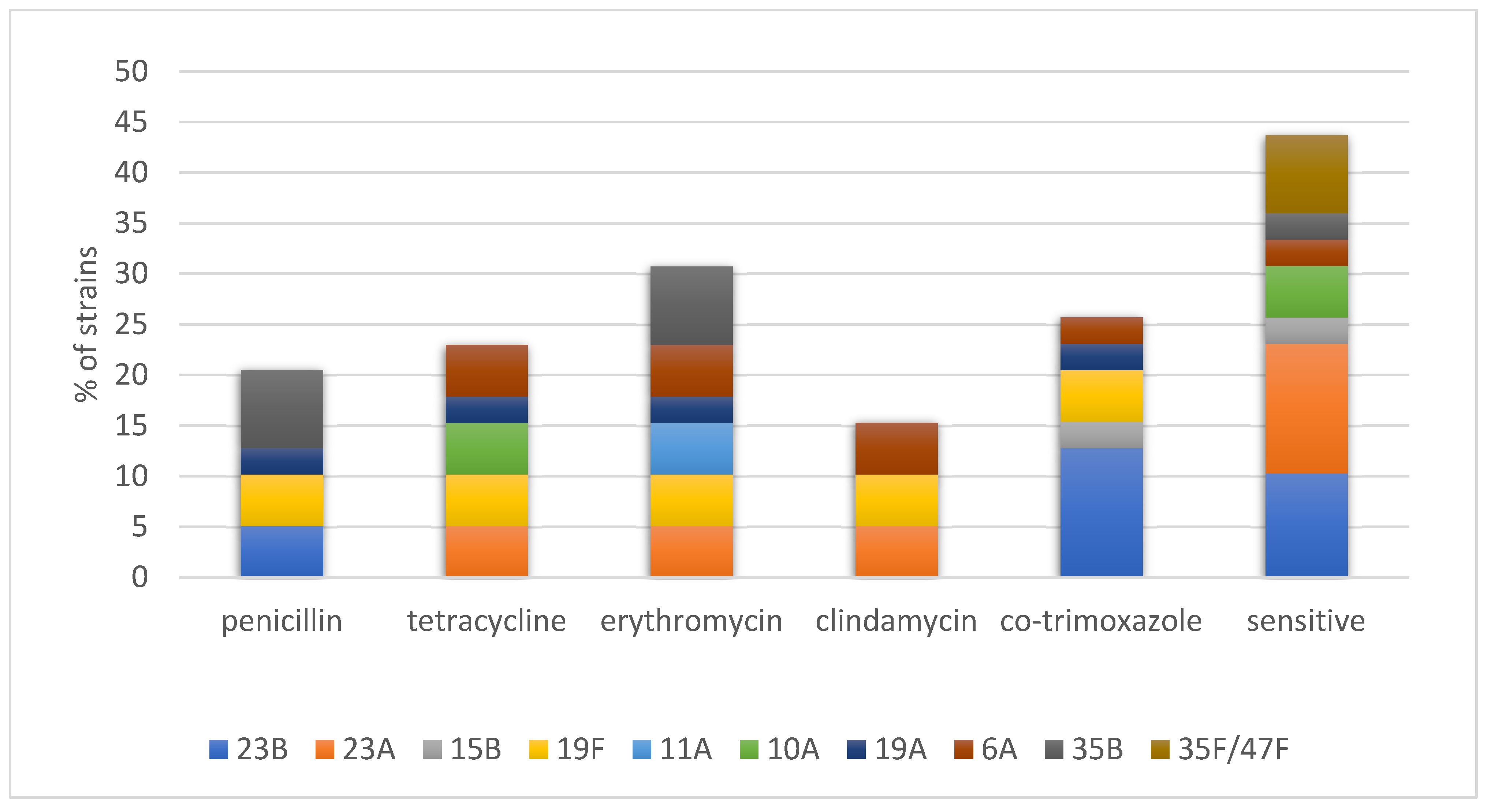
2.1. Resistance Genes and Presumptive Transposons

Of the 39 isolates, 8 (20.5%) were not susceptible to penicillin (Figure 1). Resistance to erythromycin, clindamycin, tetracycline, and cotrimoxazole was 30.7% (12 isolates), 15.4% (6 isolates), 23.1% (9 isolates), and 25.6% (10 isolates), respectively. Stratifying the tested population by vaccination status, 7 out of 28 (25%) isolates were collected from vaccinated children, and 5 of the 11 (45%) isolates collected from unvaccinated children were resistant to erythromycin (p = 0.26). There were no significant differences in resistance to penicillin and co-trimoxazole between these groups (21% vs. 18%, p = 1.0; and 25% vs. 27%, p = 1.0, respectively). Interestingly, resistance to tetracycline was significantly higher among the isolates obtained from unvaccinated children than the isolates from the vaccinated group (55% vs. 11%, RR 5.1, 95%CI 1.5–16.9, p = 0.0078).
All tetracycline-resistant isolates were tet(M)-gene-positive. The analyses of erythromycin-resistant strains revealed that 50% of the isolates were resistant to macrolide lincosamide–streptogramin B (MLSB) with a constitutive phenotype. These isolates harbored the erm(B) gene only. The macrolide (M) resistance phenotype was observed in 50% of the isolates and harbored the mef(E) gene (Table 1). None of the isolates possessed both macrolide resistance genes.
The most frequent transposon carrying the erythromycin and tetracycline resistance genes was Tn6002 (66.6%), followed by Tn916 (22.2%) and Tn2009 (11.1%). The MEGA element was found in 41.7% of all erythromycin-resistant isolates (Table 1). Stratifying our population by vaccination status, in 50% of the erythromycin-resistant isolates collected from unvaccinated children, 66.7% and 33.3% had erm(B) and mef(E) genes, respectively. Amongst the erythromycin-resistant isolates collected from vaccinated children, the percentage was the opposite (33.3% and 66.7% isolates with erm(B) and mef(E), respectively). The same prevalence was observed with the mobile elements containing these genes, namely Tn916 family transposons with erm(B) and tet(M) genes and the MEGA element not integrated into transposon from the Tn916 group. Out of the eight pneumococcal strains resistant to erythromycin and/or tetracycline isolated from unvaccinated children, 75% transposons were detected from the Tn916 family (Tn916—25% and Tn6002—50%), and 25% of the strains possessed the MEGA element. In the resistant strains isolated from vaccinated children, 50% possessed transposon from the Tn916 family (Tn6002 and Tn2009), and 50% possessed the MEGA element in the pneumococcal chromosome backbone (Table 1).

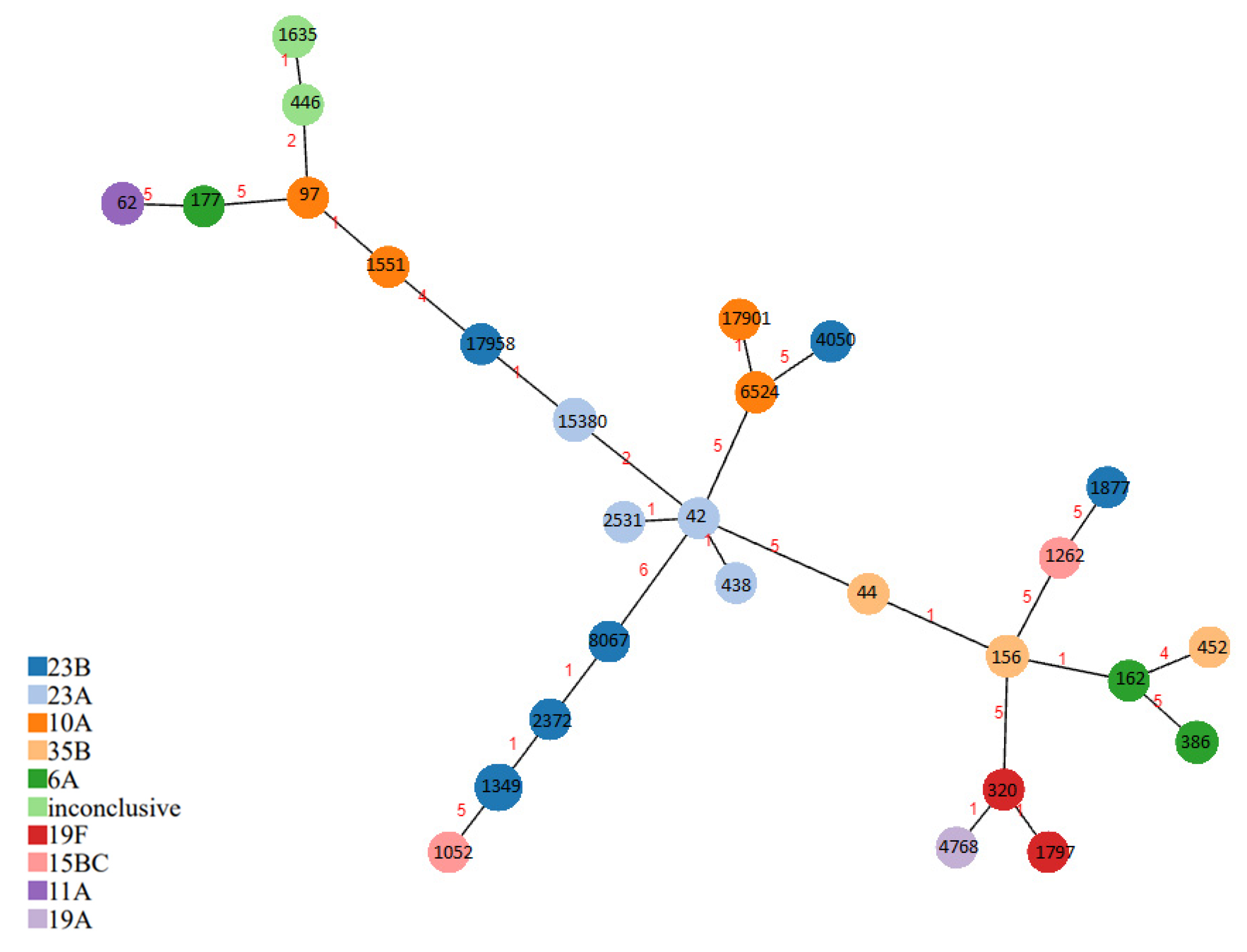
2.2. Multilocus Sequence Typing (MLST) and PMEN Clones

The 39 nasopharyngeal isolates were assigned to 28 STs, 2 of which were not previously reported in the MLST database (ST17901 and ST17907, Table 1). MLST revealed six clonal groups (CGs). Six STs consisted of more than one isolate, and twenty-two STs were identified with one single isolate. ST1349 (n  =  6) and ST15380 (n  =  3) were most frequently detected. We observed a correlation between sequence type distribution with the serotype.
Notably, 18 strains were closely related to 7 of the 43 Pneumococcal Molecular Epidemiology Network (PMEN) clones (https://www.pneumogen.net/gps/pmen.html, accessed on 3 March 2023). A total of four isolates had identical or a single-locus variant (SLV) compared with the international clone Spain 9V-3. Fourteen strains were double-locus variants (DLVs) of six other PMEN clones (Tennessee23F-4, Taiwan 19F-14, Colombia 23F-26, Netherlands 8-33, Poland 6B-20, and Greece 21-30).
Presumably, capsular switching cases occurred. The Spain 9V-3 ST156 clone with resistance pattern P was similar to the isolates with serotype 35B ST156 and resistance pattern PE and variants with serotype 35B (ST44 PE) and 6A (ST162 S). Similarly, the DLVs of Tennessee 23F-4 presented serotype 23A, the DLVs of Colombia 23F-26 presented serotype 23B, and the DLVs of Poland 6B-20 presented serotype 6A (Table 1).
Resistance to various antimicrobials was significantly associated with PMEN clones (Table 1). The isolates with resistance to erythromycin, clindamycin, and tetracycline were found in CGs related to Tennessee 23F-4 and Taiwan 19F-14 PMEN clones. In CG2 related to the Spain 9V-3 clone, MEGA elements were found. STs that mutually had at least six of the seven allelic variants were composed of clonal groups (CGs). A minimum spanning tree (MST) based on MLST was plotted using PHYLOVIZ 2.0. Figure 2 shows the relatedness of different serotypes with the identified STs.

3. Discussion

This study is the first report describing the genotypic analysis of both antibiotic-susceptible and -resistant pneumococcal carriage nasopharyngeal isolates from one region in Poland 3 years after the introduction of the vaccination program in Poland. Even though PCV vaccination has lowered the carriage of pneumococcal vaccine types, the carriage of non-vaccine types has increased due to serotype replacement [20]. Moreover, S. pneumoniae epidemiology is complex due to the diversity of the circulating genotypes often masked by varying capsular types. It is very likely that within the nasopharyngeal niche, the competition between different pneumococcal serotypes promotes bacterial colonization.
MLST data using housekeeping genes sequencing showed high diversity among the isolates tested (Simpson’s ID 0.970, 95%CI 0.941–0.999). Among the 28 identified STs, 6 clonal groups and 8 singletons were identified. According to the BURST analysis results, 28.2% of nasopharyngeal isolates were closely related to 7 of the 43 PMEN clones. Our results suggest that PMEN clones are not widely distributed within carriage isolates in the post-vaccination era in the region. In the studies carried out in this region before the introduction of vaccination, 58.4% of the isolates detected in children with recurrent respiratory tract infections were related to STs belonging to 17 out of the 43 resistant PMEN clones, i.e., Spain 9V-3, Spain 23F-1, Norway NT-42, and Poland 6B-20, as well as to 5 STs that were single-locus variants of PMEN-resistant clones (England 14–9, Spain 9V-3, Spain 23F-1, Greece 21–30, and Denmark 14–32) [21]. Among the pneumococcal strains isolated from the middle ear fluid taken from children with acute otitis media, 56% of the isolates belonged to the STs of the pandemic PMEN clones or their variants (SLVs and DLVs), including Spain 9V-3-ST156, England 14–9-ST9, Spain 23F-1-ST81, and Sweden 15A-25-ST63-SVL [22]. Most of these pandemic clones or their SLVs or DLVs were also detected in this study; however, they expressed distinct serotypes.
We found the strains grouped in CGs related to PMEN clones, which presented different serotypes (Spain 9V-3 clone to 35B serotype and Tennessee 23F-4 clone to 23A serotype). The competence of S. pneumoniae to switch its polysaccharide capsule causes the replacement of non-vaccine serotypes. An opportunity for the exchange of DNA between the types occurs when multiple serotypes of S. pneumoniae are concurrently carried in the nasopharynx. Serotype replacement facilitates the spread of drug resistance among diverse capsular types and allows the evasion of serotype-specific host immune defenses against a limited range of vaccine serotypes [23].
Pneumococcal macrolide resistance was mediated by two major mechanisms: a ribosomal methylase encoded by the erm(B) gene, causing target modification, and drug efflux via the membrane efflux pump encoded by the mef(A/E) gene. In our previous studies, macrolide-resistant pneumococci were mainly associated with the MLSB phenotype, confirmed by the presence of the erm(B) gene, and none of the previously tested strains possessed the M phenotype and the mef gene as a sole determinant of erythromycin resistance [21,24]. In the study by Izdebski et al. [25], 4.7% of the clinical pneumococcal strains resistant to macrolides possessed the mef(E) gene and the M phenotype. In this study, surprisingly, 50% of the strains resistant to erythromycin presented the M phenotype.
Pneumococcal resistance to erythromycin is frequently associated with the insertion of the erm(B)/mef gene (as Tn917 or MEGA) into the transposons of the Tn916 family, including Tn6002, Tn3872, Tn2009, Tn2010, Tn1545, and Tn6003 [17,18,19]. Geographical differences in the prevalence and spread of transposons carrying the erm(B) gene in pneumococci have often been reported. In European countries, Tn6002 and Tn3872 were the most frequent transposons detected in S. pneumoniae strains, whereas Tn917 and Tn2010 were most often observed in China and Japan [21,26,27,28,29]. The presence of Tn916, which is a prototype of the family, was observed in two tetracycline-resistant strains with serotype 10A, comprising 22.2% of resistant strains, significantly more than that observed in our other studies, with approximately 11% of prevalence [21,24].
However, past reports from Poland showed a 38.8% prevalence of Tn916 [25]. In our study, Tn6002 was found to be dominant in the tested population, with 42.9% of the strains resistant to macrolide and/or tetracycline, followed by the MEGA element carrier (35.7% resistant strains). One mef gene on the MEGA element was incorporated into Tn916, forming Tn2009. Surprisingly, Tn3872, Tn2010, and Tn6003/Tn1545 were not detected in any of the tested pneumococci, which were very prevalent transposons from the Tn916 family in our previous studies carried out in the prevaccination era [21,26]. However, the changes in the serotype distribution after the implementation of routine vaccination had an impact on transposon distribution. Connections between serotype 6B and Tn3872 prevalence, as well as Tn6002 and serotype 19F, were observed, while among Tn6003/Tn1545 carriers, most either had serotype 23F or were non-typeable [21]. With vaccination, these serotypes were mostly eliminated in our population. The Tn6002 detected in this study was observed in the strains related to PMEN clones Taiwan 19F-14, Poland 6B-20, and Tennessee 23F-4, albeit with replaced serotypes. This is meaningful in that, in this study, the erm(B) and tet(M) genes carried by the elements belonging to the Tn916 family were mainly detected in the isolates collected from unvaccinated children.
The MEGA element can be integrated into at least four loci in the pneumococcal chromosome or can integrate into orf6 of Tn916 named Tn2009 [28]. In the majority of the strains with the M phenotype in this study, the MEGA element was located in the chromosome backbone, and one strain carried Tn2009.
Pili PI-1 and PI-2 facilitate colonization and growth on mucosal surfaces and were found in the strains belonging to clonal types that are frequently found in the nasopharynx. PI-1 promotes colonization and contributes to epithelial adhesion, which is also a role of PI-2 [5]. Before routine pneumococcal vaccination, the genes of PI-1 and/or PI-2 in pneumococci isolated from children were mainly found in the isolates of vaccine serotypes belonging to multidrug-resistant international clones, including PNSP [5,30,31]. Vaccination decreased the rates of piliated pneumococci in invasive pneumococcal disease [32,33]. However, increased multidrug resistance among non-PCV-13 serotypes and in isolates with pilus islets was reported in a Japanese study [34]. Moreover, piliated non-vaccine-type strains emerged in East Jerusalem possibly due to the intrinsic advantage of PI-1 for colonization [35]. In this study, the vast majority of the isolates carrying PI-1 genes belonged to CGs related to the Spain 9V-3 clone expressing serotype 35B and 6A, and the presence of both PI-1 and PI-2 was identified in CG4, consisting of the isolates related to the Taiwan 19F-14 clone expressing serotype 19F and 19A. It can be expected that piliated pneumococci will become rare if vaccination eradicates the vaccine types in healthy children. Importantly, in the nearest future, the piliated strains of serogroups 23B, 23A, and 35B may be of concern, as they are the possible origins of the emerging clones of piliated non-vaccine pneumococcal serotypes in Poland. Due to the role of PI-1 and PI-2 in colonization promotion and epithelial adhesion contribution, they have often been found in successful pneumococcal clones, i.e., PMEN clones. Therefore, it can be expected that the prolonged colonization of non-vaccine serotypes facilitated by pili exposes the isolates to antibiotic selection [5,35].
This study has a limitation. Our sample size was not sufficiently large to have the power to exemplify the situation in the whole of Poland. Nevertheless, the strength of this study lies in performing repeated observations in the same regions with the use of a comparable methodology. A longitudinal carriage survey performed for the same population should allow for a more accurate assessment of the vaccine impact over time.

4. Materials and Methods

4.1. Bacterial Strains

A total of 39 strains from the previously described collection, isolated from the nasopharynx of 176 healthy children between 1 and 6 years old and belonging to different social groups in Lublin Voivodeship in Poland, were tested [20].

4.2. Detection of Resistance Genes, Transposons, and Pili

Chromosomal DNA was extracted from an overnight culture of the isolates grown on blood agar using a QIAamp DNA Mini Kit (Germantown, MD, USA), following the manufacturer’s recommendations. PCR experiments with the primer pairs and the protocols described previously [23] were used to detect the erythromycin resistance gene mef(E) and the tetracycline resistance gene tet(M). The PCR product of the mef(A/E) gene was digested with BamHI (Fermentas, Thermo Fisher Scientific, Waltham, Massachusetts, USA) to differentiate between the mef(A) and mef(E) gene subclasses. The Tn916 and Tn917 transposon-related genes (int, xis, tnpA, and tnpR), the tetracycline-resistance gene tet(M), and the promoter of the aph3′-III gene were detected with PCR, using the primers described in previous studies [7,11,20,23,33]. The resistance gene combinations related to the different presumed transposons were Tn6002 (erm(B), tet(M), int, and xis); Tn2010 (erm(B), tet(M), int, xis, and mef(E)); Tn3872 (erm(B), tet(M), tnpA, and tnpR); Tn1545; or Tn6003 (erm(B), tet(M), int, xis, and aph3′-III). S. pneumoniae virulence genes (lytA, ply, and cpsA) were detected using published primers [21]. The primers targeting rrgA (3′-end) and rrgB (5′-end) amplified PI-1, while those targeting pitA (3′-end) and pitB (5′-end) amplified PI-2. To confirm the absence of these genes, we used the primers that targeted the flanking regions [30].

4.3. Multilocus Sequence Typing (MLST)

The PCR amplicons of the housekeeping genes aroE, gdh, gki, recP, spi, xpt, and ddl were sequenced [36], and allele sequences of each of the seven loci were identified in the database of the pneumococcal multilocus sequence typing (MLST) website (http://spneumoniae.mlst.net, accessed on 24 April 2023). A unique combination of alleles was identified for each isolate as a sequence type (ST). New allelic numbers or new ST numbers were assigned by the curator of the pneumococcal MLST website. The discriminatory power of the MLST method was calculated using Simpson’s diversity index (SID) with the online tool available at http://www.comparingpartitions.info (accessed on 12 April 2023).
Phylogenic tree visualization was performed with the use of the PHYLOViZ program to generate minimum spanning and neighbor-joining trees (https://online.phyloviz.net/, accessed on 6 March 2023).

4.4. Cluster Analysis and PMEN Database

BURST, an algorithm used to group MLST-type data based on a count of the number of profiles that match each other at specified numbers of loci (https://pubmlst.org/bigsdb?db=pubmlst_spneumoniae_isolates&page=plugin&name=BURST, accessed on 25 February 2023), was used to investigate the relationships between the isolates based on the stringent group definition of six out of the seven shared alleles. Single-locus variants (SLVs) and double-locus variants (DLVs) were considered sufficiently related to be members of the same clonal group of the parental ST clone (based upon related sequence types (BURST analysis)). Furthermore, STs were compared with data in the Pneumococcal Molecular Epidemiology Network (PMEN) database (https://www.pneumogen.net/gps/pmen.html, accessed on 3 March 2023).

4.5. Statistical Analysis

The statistical analysis was performed with GraphPad Prism 9.Ink (GraphPad Software, LCC, San Diego, CA, USA). Categorical variables were analyzed using Fisher’s exact tests. p-values  <  0.05 were considered significant.

5. Conclusions

In conclusion, this study presents a thorough molecular analysis of S. pneumoniae isolates circulating among preschool children in southeastern Poland. The prevalent serotypes were 23B, 23A, 6A, 10A, and 35B. A total of 28.2% of nasopharyngeal isolates were closely related to 7 out of the 43 PMEN clones. The distribution of transposons resulted in clear genetic diversity and different serotype prevalence. This is meaningful in that the elements belonging to the Tn916 family were mainly detected in the isolates collected from unvaccinated children. Such epidemiological studies are necessary to investigate the effect of current pneumococcal conjugate vaccines (PCVs) on serotype distribution and antibiotic resistance among S. pneumoniae isolates, including the increase in non-vaccine serotypes, especially those resistant to antibiotics. The replacement of serotypes after PCVs has been an issue due to geographical discrepancies worldwide. Nasopharyngeal carriage in preschool children seems to be an important reservoir for the selection and dissemination of new drug-resistant pneumococcal clones in the community after the elimination of vaccine serotypes.

Author Contributions

Conceptualization, I.K.-G., G.M.-N., K.K., A.G. and E.G.; methodology, I.K.-G. and K.K.; validation, I.K.-G., G.M.-N. and E.G.; formal analysis, I.K.-G. and K.K.; investigation, K.K., I.K.-G. and A.G.; resources, I.K.-G. and G.M.-N.; data curation, I.K.-G. and G.M.-N..; writing—original draft preparation, K.K., I.K.-G. and A.G.; writing—review and editing, E.G. and G.M.-N.; visualization, K.K. and I.K.-G.; supervision, G.M.-N. and I.K.-G.; project administration, I.K.-G. and G.M.-N.; funding acquisition, I.K.-G. All authors have read and agreed to the published version of the manuscript.

Funding

This research was funded by the Medical University of Lublin, Poland, with grant number DS 29.

Institutional Review Board Statement

The protocol of the study was reviewed and approved by the Bioethics Committee of the Medical University of Lublin (KE-0254/45/2020) and performed in compliance with the Helsinki Declaration. Written informed consent was obtained from each patient.

Informed Consent Statement

Informed consent was obtained from all subjects involved in this study. Written informed consent has been obtained from the patients to publish this paper.

Data Availability Statement

The data that support the findings of this study are available on request from the last author (I.K.-G.).

Conflicts of Interest

The authors declare no conflict of interest.

References

  1. De Lencastre, H.; Tomasz, A. From Ecological Reservoir to Disease: The Nasopharynx, Day-Care Centres and Drug-Resistant Clones of Streptococcus pneumoniae. J. Antimicrob. Chemother. 2002, 50 (Suppl. S2), 75–81. [Google Scholar] [CrossRef]
  2. Bogaert, D.; De Groot, R.; Hermans, P.W.M. Streptococcus pneumoniae Colonisation: The Key to Pneumococcal Disease. Lancet Infect. Dis. 2004, 4, 144–154. [Google Scholar] [CrossRef]
  3. Mitchell, A.M.; Mitchell, T.J. Streptococcus pneumoniae: Virulence Factors and Variation. Clin. Microbiol. Infect. 2010, 16, 411–418. [Google Scholar] [CrossRef] [PubMed]
  4. Korona-Głowniak, I.; Malm, A. Streptococcus pneumoniae–colonization and pneumococcal disease. Post. Mikrobiol. 2015, 54, 309–319. Available online: https://www.pm.microbiology.pl (accessed on 20 January 2023).
  5. Dzaraly, N.D.; Muthanna, A.; Mohd Desa, M.N.; Taib, N.M.; Masri, S.N.; Rahman, N.I.A.; Suhaili, Z.; Tuan Soh, T.S.; Abdullah, F.H. Pilus Islets and the Clonal Spread of Piliated Streptococcus pneumoniae: A Review. Int. J. Med. Microbiol. 2020, 310, 151449. [Google Scholar] [CrossRef]
  6. 2017 Poland Introduced 10-Valent Pneumococcal Vaccine in the Immunization Schedule. Szczepienia. Info EN. Available online: http://szczepieniainfo.testa.com.pl/en/stories/introduction_of_pcv_2016/ (accessed on 22 August 2018).
  7. Korona-Glowniak, I.; Malm, A. Characteristics of Streptococcus pneumoniae Strains Colonizing Upper Respiratory Tract of Healthy Preschool Children in Poland. Sci. World J. 2012, 2012, 732901. [Google Scholar] [CrossRef] [PubMed]
  8. Korona-Glowniak, I.; Niedzielski, A.; Malm, A. Upper Respiratory Colonization by Streptococcus pneumoniae in Healthy Pre-School Children in South-East Poland. Int. J. Pediatr. Otorhinolaryngol. 2011, 75, 1529–1534. [Google Scholar] [CrossRef] [PubMed]
  9. Niedzielski, A.; Korona-Glowniak, I.; Malm, A. High Prevalence of Streptococcus pneumoniae in Adenoids and Nasopharynx in Preschool Children with Recurrent Upper Respiratory Tract Infections in Poland—Distribution of Serotypes and Drug Resistance Patterns. Med. Sci. Monit. 2013, 19, 54–60. [Google Scholar] [CrossRef]
  10. Korona-Glowniak, I.; Niedzielski, A.; Kosikowska, U.; Grzegorczyk, A.; Malm, A. Nasopharyngeal vs. Adenoid Cultures in Children Undergoing Adenoidectomy: Prevalence of Bacterial Pathogens, Their Interactions and Risk Factors. Epidemiol. Infect. 2015, 143, 821–830. [Google Scholar] [CrossRef] [PubMed]
  11. Wróbel-Pawelczyk, I.; Ronkiewicz, P.; Wanke-Rytt, M.; Rykowska, D.; Górska-Kot, A.; Włodkowska, K.; Topczewska-Cabanek, A.; Jackowska, T.; Chruszcz, J.; Marchut, W.; et al. Pneumococcal Carriage in Unvaccinated Children at the Time of Vaccine Implementation into the National Immunization Program in Poland. Sci. Rep. 2022, 12, 5858. [Google Scholar] [CrossRef] [PubMed]
  12. Wyres, K.L.; Lambertsen, L.M.; Croucher, N.J.; McGee, L.; von Gottberg, A.; Liñares, J.; Jacobs, M.R.; Kristinsson, K.G.; Beall, B.W.; Klugman, K.P.; et al. Pneumococcal Capsular Switching: A Historical Perspective. J. Infect. Dis. 2013, 207, 439–449. [Google Scholar] [CrossRef] [PubMed]
  13. Weinberger, D.M.; Malley, R.; Lipsitch, M. Serotype Replacement in Disease after Pneumococcal Vaccination. Lancet 2011, 378, 1962–1973. [Google Scholar] [CrossRef] [PubMed]
  14. Knutsen, E.; Johnsborg, O.; Quentin, Y.; Claverys, J.-P.; Håvarstein, L.S. BOX Elements Modulate Gene Expression in Streptococcus pneumoniae: Impact on the Fine-Tuning of Competence Development. J. Bacteriol. 2006, 188, 8307–8312. [Google Scholar] [CrossRef]
  15. Yahiaoui, R.Y.; Bootsma, H.J.; den Heijer, C.D.J.; Pluister, G.N.; John Paget, W.; Spreeuwenberg, P.; Trzcinski, K.; Stobberingh, E.E. Distribution of Serotypes and Patterns of Antimicrobial Resistance among Commensal Streptococcus pneumoniae in Nine European Countries. BMC Infect. Dis. 2018, 18, 440. [Google Scholar] [CrossRef]
  16. Harrison, O.B.; Brueggemann, A.B.; Caugant, D.A.; van der Ende, A.; Frosch, M.; Gray, S.; Heuberger, S.; Krizova, P.; Olcen, P.; Slack, M.; et al. Molecular Typing Methods for Outbreak Detection and Surveillance of Invasive Disease Caused by Neisseria Meningitidis, Haemophilus Influenzae and Streptococcus pneumoniae, a Review. Microbiology 2011, 157 Pt 8, 2181–2195. [Google Scholar] [CrossRef] [PubMed]
  17. Rice, L.B. Tn916 Family Conjugative Transposons and Dissemination of Antimicrobial Resistance Determinants. Antimicrob. Agents Chemother. 1998, 42, 1871–1877. [Google Scholar] [CrossRef] [PubMed]
  18. Del Grosso, M.; Camilli, R.; Iannelli, F.; Pozzi, G.; Pantosti, A. The Mef(E)-Carrying Genetic Element (Mega) of Streptococcus pneumoniae: Insertion Sites and Association with Other Genetic Elements. Antimicrob. Agents Chemother. 2006, 50, 3361–3366. [Google Scholar] [CrossRef]
  19. Del Grosso, M.; Northwood, J.G.E.; Farrell, D.J.; Pantosti, A. The Macrolide Resistance Genes Erm(B) and Mef(E) Are Carried by Tn2010 in Dual-Gene Streptococcus pneumoniae Isolates Belonging to Clonal Complex CC271. Antimicrob. Agents Chemother. 2007, 51, 4184–4186. [Google Scholar] [CrossRef]
  20. Kielbik, K.; Pietras, A.; Jablonska, J.; Bakiera, A.; Borek, A.; Niedzielska, G.; Grzegorczyk, M.; Grywalska, E.; Korona-Glowniak, I. Impact of Pneumococcal Vaccination on Nasopharyngeal Carriage of Streptococcus pneumoniae and Microbiota Profiles in Preschool Children in South East Poland. Vaccines 2022, 10, 791. [Google Scholar] [CrossRef]
  21. Korona-Glowniak, I.; Maj, M.; Siwiec, R.; Niedzielski, A.; Malm, A. Molecular Epidemiology of Streptococcus pneumoniae Isolates from Children with Recurrent Upper Respiratory Tract Infections. PLoS ONE 2016, 11, e0158909. [Google Scholar] [CrossRef]
  22. Korona-Glowniak, I.; Zychowski, P.; Siwiec, R.; Mazur, E.; Niedzielska, G.; Malm, A. Resistant Streptococcus pneumoniae Strains in Children with Acute Otitis Media–High Risk of Persistent Colonization after Treatment. BMC Infect. Dis. 2018, 18, 478. [Google Scholar] [CrossRef]
  23. Paton, J.C.; Trappetti, C. Streptococcus pneumoniae Capsular Polysaccharide. Microbiol. Spectr. 2019, 7. [Google Scholar] [CrossRef] [PubMed]
  24. Korona-Glowniak, I.; Siwiec, R.; Malm, A. Resistance Determinants and Their Association with Different Transposons in the Antibiotic-Resistant Streptococcus pneumoniae. Biomed Res. Int. 2015, 2015, 836496. [Google Scholar] [CrossRef] [PubMed]
  25. Izdebski, R.; Sadowy, E.; Fiett, J.; Grzesiowski, P.; Gniadkowski, M.; Hryniewicz, W. Clonal Diversity and Resistance Mechanisms in Tetracycline-Nonsusceptible Streptococcus pneumoniae Isolates in Poland. Antimicrob. Agents Chemother. 2007, 51, 1155–1163. [Google Scholar] [CrossRef] [PubMed]
  26. Akdoğan Kittana, F.N.; Mustak, I.B.; Hascelik, G.; Saricam, S.; Gurler, N.; Diker, K.S. Erythromycin-Resistant Streptococcus pneumoniae: Phenotypes, Genotypes, Transposons and Pneumococcal Vaccine Coverage Rates. J. Med. Microbiol. 2019, 68, 874–881. [Google Scholar] [CrossRef] [PubMed]
  27. Wang, X.; Cong, Z.; Huang, W.; Li, C. Molecular Characterization of Streptococcus pneumoniae Isolated from Pediatric Patients in Shanghai, China. Pediatr. Pulmonol. 2020, 55, 2135–2141. [Google Scholar] [CrossRef]
  28. Chancey, S.T.; Agrawal, S.; Schroeder, M.R.; Farley, M.M.; Tettelin, H.; Stephens, D.S. Composite Mobile Genetic Elements Disseminating Macrolide Resistance in Streptococcus pneumoniae. Front. Microbiol. 2015, 6, 26. [Google Scholar] [CrossRef]
  29. Cochetti, I.; Tili, E.; Vecchi, M.; Manzin, A.; Mingoia, M.; Varaldo, P.E.; Montanari, M.P. New Tn916-Related Elements Causing Erm(B)-Mediated Erythromycin Resistance in Tetracycline-Susceptible Pneumococci. J. Antimicrob. Chemother. 2007, 60, 127–131. [Google Scholar] [CrossRef]
  30. Hjálmarsdóttir, M.; Pétursdóttir, B.; Erlendsdóttir, H.; Haraldsson, G.; Kristinsson, K. Prevalence of Pilus Genes in Pneumococci Isolated from Healthy Preschool Children in Iceland: Association with Vaccine Serotypes and Antibiotic Resistance. J. Antimicrob. Chemother. 2015, 70, 2203–2208. [Google Scholar] [CrossRef]
  31. Mills, R.O.; Abdullah, M.R.; Akwetey, S.A.; Sappor, D.C.; Cole, I.; Baffuor-Asare, M.; Bolivar, J.A.; Gámez, G.; van der Linden, M.P.G.; Hammerschmidt, S. Post-Vaccination Streptococcus pneumoniae Carriage and Virulence Gene Distribution among Children Less Than Five Years of Age, Cape Coast, Ghana. Microorganisms 2020, 8, 1987. [Google Scholar] [CrossRef]
  32. Regev-Yochay, G.; Hanage, W.P.; Trzcinski, K.; Rifas-Shiman, S.L.; Lee, G.; Bessolo, A.; Huang, S.S.; Pelton, S.I.; McAdam, A.J.; Finkelstein, J.A.; et al. Re-Emergence of the Type 1 Pilus among Streptococcus pneumoniae Isolates in Massachusetts, USA. Vaccine 2010, 28, 4842–4846. [Google Scholar] [CrossRef] [PubMed]
  33. Aguiar, S.I.; Melo-Cristino, J.; Ramirez, M. Use of the 13-Valent Conjugate Vaccine Has the Potential to Eliminate Pilus Carrying Isolates as Causes of Invasive Pneumococcal Disease. Vaccine 2012, 30, 5487–5490. [Google Scholar] [CrossRef] [PubMed]
  34. Kawaguchiya, M.; Urushibara, N.; Aung, M.S.; Shinagawa, M.; Takahashi, S.; Kobayashi, N. Serotype distribution, antimicrobial resistance and prevalence of pilus islets in pneumococci following the use of conjugate vaccines. J. Med. Microbiol. 2017, 66, 643–650. [Google Scholar] [CrossRef]
  35. Regev-Yochay, G.; Jaber, H.; Hamdan, A.; Daana, M.; Nammouz, H.; Thalji, A.; Jaar, F.; Abdeen, Z.; Rubin, C.; Huppert, A.; et al. Vaccine escape of piliated Streptococcus pneumoniae strains. Vaccine 2016, 34, 2787–2792. [Google Scholar] [CrossRef]
  36. Enright, M.C.; Spratt, B.G. A Multilocus Sequence Typing Scheme for Streptococcus pneumoniae: Identification of Clones Associated with Serious Invasive Disease. Microbiology 1998, 144 Pt 11, 3049–3060. [Google Scholar] [CrossRef] [PubMed]
Figure 1. Serotype distribution between drug-resistant and sensitive nasopharyngeal S. pneumoniae isolates from healthy children.
Ijms 24 07883 g001
Figure 2. Minimum spanning tree based on allelic profiles for nasopharyngeal S. pneumoniae isolates from healthy children. Colors show the presence of different serotypes (inconclusive—serotype 35F/47F). Circles present the sequence types (STs) with their size corresponding to the number of isolates. Link labels are shown in absolute count of differences.
Ijms 24 07883 g002
Table 1. Antibiotic resistance patterns, capsular types, and molecular characterization of nasopharyngeal S. pneumoniae isolates from healthy children.
Clonal GroupSequence Types (n)Associated PMEN CloneSerotype (n)Resistance PatternResistance Genes (n)Transposon GenesPresumptive TransposonPili
142 * (1) 23AECCTeerm(B), tet(M)Int/xis916Tn6002
438 (1)Tennessee23F-4, DLV23AS
2531 (1)Tennessee23F-4, DLV23AECCTeerm(B), tet(M)Int/xis916, orf20-19(3.7 kb)Tn6002
15380 (3) 23A (3)S (3) PI-1 + PI-2 (1)
17958 (1) 23AS PI-2
244 (1)Spain 9V-3, SVL35BPEmef(E)melMEGAPI-1
156 * (2)Spain 9V-335B (2)PE (2)mef(E) (2)melMEGA (2)PI-1 (2)
162 (1)Spain 9V-3, SVL6AS PI-1
397 (1) 10AS
446 (1)35F/47FS
1551 (1)10AS
1635 (2)35F/47F (2)S (2)
4320 * (1) 19FPECCTeSXTerm(B), tet(M)int/xis916, orf20-19(3.7 kb)Tn6002PI-1 + PI-2
4768 (1) 19APETeSXTmef(E), tet(M)int/xis916, orf20-29(3.7 kb)Tn2009PI-1 + PI-2
17907 (1)Taiwan 19F-14, DLV19FPECCTeSXTerm(B), tet(M)int/xis916, orf20-19(3.7 kb)Tn6002PI-1 + PI-2
51349 (6)Colombia 23F-26, DLV23B (6)SXT (4) S (2) PI-1 (1), PI-2 (1)
2372 * (1) 23BPPI-1 + PI-2
8067 (1) 23BPSXTPI-2
66524 (1) 10ATetet(M)int/xis916, orf20-19(0.8kb)Tn916
17901 (1)10ATetet(M)int/xis916, orf20-19(0.8kb)Tn916
Singletons62 (2)Netherlands 8-33, DLV11A (2)E (2)mef(E) (2)melMEGA(2)PI-2
177 (1) 6ASXT PI-1 + PI-2
386 (2)Poland 6B-20, DLV6A (2)ECCTe (2)erm(B), tet(M) (2)int/xis916, orf20-19(3.7 kb)Tn6002 (2)
452 (1) 35BS
1025 (1) 15BSXT
1262 (1) 15BS PI-2
1877 (1)Greece 21-30, DLV23BS
4050 (1) 23BS
* The central profile is indicated with an asterisk; P, penicillin; E, erythromycin; CC, clindamycin; Te, tetracycline; SXT, cotrimoxazole; S, sensitive to all tested antibiotics; n, number of isolates; SLV, single-locus variant; DLV, double-locus variant.
Disclaimer/Publisher’s Note: The statements, opinions and data contained in all publications are solely those of the individual author(s) and contributor(s) and not of MDPI and/or the editor(s). MDPI and/or the editor(s) disclaim responsibility for any injury to people or property resulting from any ideas, methods, instructions or products referred to in the content.

