Review

Breaking the Chains: Advances in Substance Addiction Research through Single-Cell Sequencing, Epigenetics, and Epitranscriptomic

by
Ana Filošević Vujnović
1,*,
Ivana Stanković Matić
2,
Lara Saftić Martinović
2 and
Sanja Dević Pavlić
2
1
Faculty of Biotechnology and Drug Development, University of Rijeka, Radmile Matejčić 2, 51000 Rijeka, Croatia
2
Faculty of Medicine, University of Rijeka, Braće Branchetta 20, 51000 Rijeka, Croatia
*
Author to whom correspondence should be addressed.
Future Pharmacol. 2024, 4(1), 115-138; https://doi.org/10.3390/futurepharmacol4010009
Submission received: 15 December 2023 / Revised: 18 January 2024 / Accepted: 24 January 2024 / Published: 29 January 2024

Abstract

:
Addiction is a complex brain disease influenced by genetic, environmental, and neurological factors. Psychostimulants, cocaine, and methamphetamine influence different cell types in different brain regions, with a focus on the neurons responsible for rewarding effects in the nucleus accumbens (NAc) and ventral tegmental area (VTA). Known markers for psychostimulant-induced neuronal plasticity in combination with droplet-based high-throughput single-cell sequencing divided the heterogeneity of cell populations in NAc and VTA into clusters, where all cells of the same type do not respond equally to exposure to psychostimulants. To explain psychostimulant-induced neuronal plasticity as changes in the amplitude and phase shifts of gene expression, we focused on epigenetic mechanisms of DNA and chromatin modifications, as well as DNA accessibility. We also comment on epitranscriptomics as a novel approach in the study of messenger RNA posttranslational modification, which regulates translation and potentially localized transcription in synapses in order to address the molecular chains that connect addiction from changes in gene expression to synaptic and, finally, neuronal plasticity.

Graphical Abstract

1. Introduction

Behaviorally, addiction is defined as compulsive drug seeking and use despite negative consequences [1]. The underlying molecular mechanisms primarily involve the brain reward system, associated with dopamine release [2,3,4]. Acute drug exposure disrupts neurotransmitter signaling, localization, metabolism, and synthesis, resulting in neuroadaptive changes that contribute to the development of addiction [5,6]. Prolonged drug exposure induces neuroplastic changes, altering the structure and function of neurons and influencing synaptic connections, thus impacting reward neuronal circuitry and resulting in long-term behavioral changes [7,8].
Drugs of abuse are a diverse group of compounds classified into various categories based on their pharmacological effects and molecular mechanisms of action. We have concentrated on the psychostimulants cocaine (COC) and methamphetamine (METH) because of the severity of the cognitive, behavioral, and physical disorders brought on by the damage to neuronal cells and the metabolic dysfunction in the central nervous system after its consumption [9]. COC and METH are central nervous system stimulants that increase dopamine release in brain regions of the mesolimbic circuit, influencing reward and cognitive functions [10]. Although COC and METH have similar behavioral and physiological effects, there are some major differences. COC is quickly removed and completely metabolized in the body, while METH has a longer duration of action due to slower metabolism and remains unchanged in the brain longer than COC, leading to prolonged stimulant effects [11]. Both COC and METH are highly addictive stimulants that are widely recognized as one of the most abused drugs in the world, which is why this review focuses specifically on these two psychostimulants [12,13]. Key brain regions involved in reward processing, influenced by COC and METH, include the nucleus accumbens (NAc) and ventral tegmental area (VTA). Long-term changes induced by psychostimulant exposure in these regions involve alterations in gene expression and cellular physiology [14,15]. Psychostimulant-induced synaptic plasticity is linked to the known mechanism of phosphorylation of extracellular signal-regulated kinases (ERK1/2) and cyclic adenosine 3′,5′-monophosphate (cAMP)-response element-binding protein (CREB), with cAMP-dependent protein kinase (PKA) also playing a role in drug-induced memory formation [16,17,18,19]. However, the main challenge in addiction research remains the identification of cell types, gene/s, or different molecular markers capable of distinguishing natural from drug-induced alterations in the reward circuitry.
While some people only use drugs for experimentation and never develop an addiction, others develop an addiction after being exposed to psychoactive substances on a regular basis. Understanding the molecular basis for this phenomenon requires a complex interplay of genetic, environmental, and neurobiological factors. Individual variability and the dynamic nature of drug responses both influence the molecular mechanisms involved in the cascade of events that affect neurotransmitter release, receptor activation, and intracellular signaling pathways. Individual vulnerability and susceptibility to addiction were studied in a mouse model, and it was discovered that in wild-type populations, voluntary oral methamphetamine consumption can undergo bidirectional selective breeding, producing two strings with high and low preference for METH [20]. Using a selective breeding approach, candidate genes can be identified in the absence of METH exposure. By using RNA-Seq to selectively breed low- and high-METH-preference mice, it was discovered that the trace amine-associated receptor 1 (Taar1) gene plays an important role [21]. Indeed, it was confirmed that increasing TAAR1 function reduces drug self-administration and intake [22,23,24]. In addition to genetic influence, non-genetic factors such as social isolation or enrichment have been shown to influence drug preference [25,26]. Taken together, the complexity of the risk for addiction development as influenced by personal genetics and environment should be investigated further, because recent studies point to epigenetic changes caused by isolation that affect addiction development.
Previous studies on molecular changes associated with addiction have primarily focused on candidate genes identified by proteomic or genetic techniques using tissue extracts from drug-exposed individuals. With this approach, it is not possible to distinguish in which cells the genetic changes originate, whether they occur at the gene or protein level and whether the observed changes are time-dependent or stable. To explain the complexities of these influences, it is necessary to refine the accepted central dogma, foundational to our understanding of genetic information flow, by applying epigenetics and epitranscriptomics to gene regulation (Figure 1). Environmental factors influence gene expression by activating or deactivating genes via epigenetic modifications such as DNA methylation and histone acetylation [27]. Moreover, recent studies show that environmental factors can lead to the modification of different RNAs, influencing RNA stability, splicing, translation, and other post-transcriptional processes that also have an impact on protein synthesis and/or function [28,29,30].
The primary focus of this review is on recent research using droplet-based single-cell sequencing methods and epigenetic and epitranscriptomic approaches in NAc and VTA brain regions after acute and chronic exposure to the psychostimulants COC and METH, regardless of the route of administration. The single-cell approach confirmed known and discovered new cell types involved in the response to psychostimulants, which were clustered based on changes in gene expression. In the field of epigenetics and epitranscriptomics, our attention has centered on the role of molecular players known as “readers”, “writers”, and “erasers”, which contribute to the regulation of gene expression through modifications of DNA, histone proteins, and RNA. We have specifically addressed two crucial aspects of addiction development and treatment: first, we distinguished between the natural process of neuronal plasticity and psychostimulant-induced neuronal plasticity; second, we analyzed the common mechanisms and genes involved in obesity and addiction. Additionally, we discussed the systemic effects that are a consequence of addiction and psychostimulant use, including impaired oxidative regulation, immune response, and changes in the microbiome profile. These findings provide an alternative perspective on the long-term consequences of withdrawal and relapse, which are critical in addiction treatment.

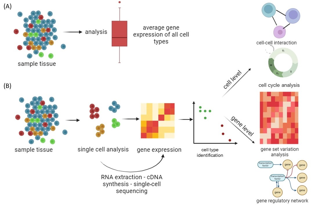
2. Single-Cell Sequencing Techniques

Traditional classification divides cell types based on morphology rather than molecular features [31,32]. Despite the fact that cells have almost identical genotypes, traditional RNA sequencing gives an average expression profile from a population of cells, ignoring cell-to-cell variability and transcriptome information from a subset of active genes [33,34,35]. Single-cell RNA sequencing (scRNA-seq) is used for genetic profiling of cells that require prior separation of one cell type from a cell suspension using a fluorescence-activated cell sorting and flow cytometer [36]. This approach requires a priori knowledge of a cell-type-specific marker, which is dependent on the availability of appropriate antibodies [37]. Great progress in scRNA-seq was made by applying high-throughput droplet-based techniques, which do not require prior separation of the cells from the cell suspension, namely, InDrop [38], Drop-seq [39], and 10× Chromium [40], supporting cost-effective capture and library production for thousands to millions of cells and allowing examination of gene expression heterogeneity among individual cells (Figure 2). Today, we have the possibility of simultaneous profiling of the transcriptome using 10× Chromium and epigenome using Assay for Transposase-Accessible Chromatin sequencing (scATAC-seq). The ATAC-seq technique is used for chromatin accessibility at the single-cell level by measuring the regions of DNA that are accessible to transposase enzymes, providing insights into the epigenetic landscape [41,42]. Combined analysis of scRNA-seq and scATAC-seq data allows the simultaneous exploration of gene expression and chromatin accessibility within individual cells important in the study of addiction development [43,44]. Finally, in the field of neurobiology, the Patch-seq approach stands out as a powerful adaptation of the patch-clamp technique, allowing the simultaneous examination of individual neurons through a combination of electrophysiological, morphological, and transcriptomic analyses [45,46,47]. These novel approaches provide a comprehensive understanding of neuronal properties by providing insights into both functional activities and molecular profiles’ rising direction toward system biology’s understanding of changes induced by drug taking, deprivation, and relapse.

2.1. Application of Single-Cell Sequencing in Addiction Research

By defining region-specific molecular signatures and neuronal circuits involved in addictive behaviors [48,49,50], droplet-based high-throughput scRNA-seq enabled researchers to compare gene expression profiles in different brain regions involved in addiction [51]. At the molecular level, scRNA-seq of individual neurons identifies specific neuronal subtypes associated with reward, reinforcement, and addiction processes [52], whereas transcriptome analysis identifies distinct transcriptional signatures associated with addiction-related behaviors [53]. Understanding how individual cells within a population exhibit different sensitivities or adaptations to drug use is crucial, while interactions between the nervous and immune systems must be integrated in this aspect.

2.1.1. Identifying Cell Populations by Molecular Clustering

scRNA-seq has been shown to be an effective tool for characterizing cellular diversity in brain regions associated with the reward system, such as the NAc and VTA [54,55]. The NAc is a brain region primarily composed of two main types of cells, namely, Medium Spiny Neurons (MSNs) and interneurons. In NAc, clustering analysis revealed nine major cell populations, four neuronal and five non-neuronal [54]. Using this approach, in the NAc, novel subpopulations of interneurons and MSNs were identified [52]. scATAC-seq has been used to map cell-type-specific differences in chromatin accessibility in the NAc, providing insights into the epigenomic landscape of this brain region [55]. Moreover, the characterization of γ-Aminobutyric Acid (GABA) MSNs and the discovery of notable variations in receptor expression patterns and MSN activation within NAc subterritories have highlighted the anatomical and functional heterogeneity of the NAc [56]. The ventral tegmental area (VTA) is best known for containing dopaminergic neurons associated with reward and motivation. scRNA-seq identified in VTA selective markers for dopamine and combinatorial neurons revealed expression profiles for drugs of abuse receptors and population-specific enrichment of genes associated with brain disorders [57]. This comprehensive molecular characterization highlights the heterogeneity of the NAc and VTA cell population, providing a valuable resource for future research into VTA and NAc gene expression and its implications for reward-related behaviors such as addiction.
By applying scRNAseq in the study of acute COC administration in rodents, distinct neuronal clusters within the NAc were discovered. These clusters exhibited known markers associated with two main types of MSNs: dopamine receptor D1–positive (Drd1-MSNs) and dopamine receptor D2–positive (Drd2-MSNs) [58]. The same neuronal substrate that allows drugs of abuse to access Drd1 and Drd2 MSNs in NAc has been confirmed to augment and corrupt a shared pathway that normally serves physiological needs [59]. Nevertheless, the investigation of genetically mediated variations in susceptibility to addiction-like behaviors was restricted by the use of inbred rodent strains. Furthermore, these studies primarily focused on acute drug treatments, ignoring the molecular changes associated with long-term addictive-like behaviors [60].
METH is another commonly abused psychostimulant that induces synaptic plasticity and pathological memory enhancement [61]. Epigenetics plays an important role in regulating METH addiction [62]. METH modulates dopamine (DA), norepinephrine (NE), serotonin, glutamate (Glu), and GABA neurotransmitters in the medial prefrontal cortex (mPFC), the VTA, and the NAc through histone acetylation, methylation, micro RNAs (miRNAs), and ubiquitination. These epigenetic mechanisms do not regulate METH-induced addiction alone but rather collaborate with miRNA regulation of the Ubiquitin proteasome system [62]. Cell clustering was performed on bulk RNA data using single-cell RNA data extracted from astrocytes for analysis of differently expressed genes in METH-exposed mice. The NF-κB signaling pathway, inflammation, and neurodegeneration were among the considerably enriched pathways found in the analysis. Immune infiltration analysis showed that the METH group had significantly lower neutrophil infiltration and significantly higher monocyte, T, and NK cell infiltration [63]. Strong inflammatory responses occur early in METH withdrawal, and they decrease as withdrawal time increases. Cytokines, immune cells, complement, and immunoglobulin form a complex immune network that regulates immune responses after METH withdrawal, shifting the focus of METH addiction research from neurons to non-neuronal cell types [64]. The novel approach in the study of molecular mechanisms is organoids. Brain organoids are generated through the differentiation of pluripotent stem cells within a three-dimensional culture environment, where neurodevelopmental signals are introduced to guide the process. Organoids closely mimic the cellular composition and anatomical structure of the brain [65]. METH treatment of organoids resulted in transcriptional changes in astrocytes and neuronal progenitor cells, among other cell types. Additionally, METH induced novel networks of astrocyte-specific gene expression that control cytokine responses and the inflammasome, resulting in modifications to the RNA and protein levels of neuroinflammatory and cytokine gene expression [66]. After being exposed to METH, neutrospheres made from embryonic rat hippocampal tissue displayed abnormal cell differentiation in both neurons and astrocytes, as well as a decreased capacity for neutrosphere migration coupled with an increase in oxidative stress and apoptosis [67]. Taken together, the scRNA-seq literature overview demonstrated that COC mainly influences neurons in VTA and NAc, while METH has a primary influence on the immune response.

2.1.2. Identifying Transcriptome Mechanism

By analyzing single-cell transcriptional responses in the prefrontal cortex (PFC) cells of mice undergoing COC self-administration, specific cell types that express genes associated with COC addiction were identified, namely, ∆FosB, Methyl CpG binding protein 2 (MeCP2) and brain-derived neurotrophic factor (BDNF) [68]. Following COC administration, ∆FosB was reported to also be expressed in the VTA with dopamine neurons projecting to the NAc [69], MeCP2 was reported to be altered in the NAc [70], and BDNF expression played a role in the VTA-NAc pathway [71]. These genes were selectively expressed in excitatory neurons, inhibitory neurons, and non-neuronal glial cells while showing varied expression levels over the stages of COC addiction [68]. In addition to anatomic location, function and network connectivity have a significant impact on an excitatory neuron’s transcriptional fate. COC, as rewarding stimuli, increases dopamine levels in NAc, which are responsible for COC’s reinforcing effects. Dopamine dynamics is mediated by the activation of dopamine neurons in the VTA, which project to the PFC, hippocampus, and amygdala, separately from NAc cells [53]. In the PFC of mice who self-administrated COC, some of the relevant genes had a specific expression pattern in different PFC cell clusters. Excitatory neurons were the predominant host for the expression of Drd1, Drd2, G-protein-coupled receptors Gpr88 and Gpr6, and Regulator of G-protein signaling 9 (Rgs9), though Drd1, Gpr88, and Rgs9 were also detected in other cell types. Endothelial cells and microglia expressed the adenosine A2A receptor (Adora2a) the most, while inhibitory and excitatory neurons expressed it the least. According to computational analysis of publicly available scRNA-seq data, excitatory neurons in the PFC were the primary site of transcriptional reprogramming of relevant genes. This kind of transcriptional analysis of the addiction gene signature at cell-type-specific levels in the PFC may be useful in developing novel pharmacological strategies to combat addiction at the cellular level [53]. Addiction has a significant impact on highly regulated gene networks in the Dorsum striatum and NAc as well as COC-related gene regulation. While these two brain regions exhibit distinct transcriptomes and addiction-related changes in gene expression, a subset of differentially expressed transcripts was identified that are regulated similarly by COC and METH [72]. Further analysis supports the hypothesis that COC-responsive genes participate in the vulnerability to substance use disorders, as a convergence of up-regulated differentially expressed transcripts was found in individuals with confirmed addiction and genes associated with risk-taking behavior. It has been demonstrated that a significant amount of the striatal molecular pathology underlying COC addiction in humans can be replicated in mice that self-administer COC. This significant finding validates the use of mice models in the pathophysiological study of addiction. In particular, studies conducted on mice link aberrant gene expression patterns in humans with addiction to Drd1 MSNs. The study highlights that this connection was made using a human cohort that included a sizable number of Black individuals, who have not been adequately represented in previous transcriptional studies of addiction, even though there has been recent evidence that they are more susceptible to overdose deaths related to COC [72].
Reactive oxygen species and oxidative stress in striatal regions are known to be elevated by COC, and this effect is further compounded by elevated glutamate release and excessive dopamine levels. The potential involvement of both cell types in the regulation of conserved gene networks was revealed by integrating human transcriptomes with the Drd1 and Drd2 MSN transcriptome data from mice. Numerous transcription factors that are predicted to be upstream of the abnormalities in gene expression linked to addiction were identified through this analysis. The majority are shared between the NAc and dorsum striatum. Notably, several of these predicted upstream transcription factors have been implicated previously in COC addiction in rodent models; in particular, Activator protein 1 (AP-1) (FOS/JUN) family, EGR family, NFκB, E2F, and several nuclear hormone receptors cells [72]. Simultaneously, a number of other transcriptional regulators that had not been previously linked were discovered to have mechanistic roles in COC addiction. Our knowledge of how COC and other addictive drugs cause transcriptomic plasticity in brain regions related to addiction has improved due to the use of animal models. Self-administration procedures have, however, clearly demonstrated that abused drugs, such as COC, act as reinforcers in animal models. Nevertheless, it is still unclear whether the COC-related molecular changes investigated in rodent models accurately reflect transcriptomic changes in human addiction. Preclinical animal models have been shown to be valid and valuable in providing important insight and mechanistic information across transcriptional networks, biological pathways, and even neuronal cell types in the striatum that lead to the harmful neuroadaptations of addiction, according to previous studies. To capture the unique molecular features of addiction, one must carefully consider treatment paradigms and experimental endpoints in these animal models. This is because chronic drug use will present differently at the transcriptional level depending on early versus late withdrawal times or relapse. Future evolution of single-cell and spatial sequencing technologies promises more direct insight into subtype-specific transcriptome alterations in human addiction cells [72].
In response to acute and chronic METH, mice hippocampi exhibit significant volumetric atrophy compared to controls. The genes involved in cytoskeleton organization and phagocytosis were downregulated in the acute METH-treated group compared to the control group. In the group receiving long-term METH treatment, genes linked to synaptic transmission, neuron differentiation regulation, and neurogenesis regulation were downregulated [73]. Drd1 overstimulation after METH exposure induces metabolic changes and transcriptional pathways, switching gene expression and neuronal phenotype underlying addictive behavior [74]. PKA phosphorylates voltage-dependent ion channels, GLUT receptors, transcription factors, and epigenetic enzymes involved in synaptic plasticity as naturally occurring in normal striatum. When the Drd1 is activated, PKA activates mitogen-activated protein kinases (MAPKs) and extracellular signal-regulated kinases 1/2 (ERK1/2) [75]. Nuclear receptors, CREB, Elk-1, and H3 histones are all phosphorylated by ERK1/2 upon translocation to the nucleus, where they control the expression of certain genes [76]. The DA- and cAMP-regulated phosphoprotein (DARPP-32) is a key substrate of DRD1/PKA signaling in the striatum [77].
METH consumption results in oxidative stress in the DA terminals due to excess of free DA undergoing oxidative metabolism and autoxidation, intracellularly and extracellularly. Along with hydrogen peroxide and reactive oxygen species (ROS) production, toxic DA metabolites like quinones and 3,4-Dihydroxyphenylacetaldehyde promote structural modifications of proteins. METH influences other cell organelles, endoplasmic reticulum, and mitochondria, leading to neurotoxic effects, while ROS accumulation leads to misfolded and insoluble proteins and severed organelles, decomposed by cell-clearing mechanisms of autophagy and ubiquitin proteasome [78].
ATACseq, a method used for mapping genome-wide chromatin accessibility, has been used for addiction research to explore the open chromatin regions associated with exposure to drugs of abuse [79,80] including METH [78]. Changes in chromatin accessibility interact with DNA and histone modifications that are normally associated with transcription factor binding. Addictive drugs affect transcription factors like ΔFosB, EGRs, and MEF2, which regulate downstream target genes [81].
Patch-sequencing (Patch-seq), a variation of the patch-clamp technique, is a multimodal method that combines individual neuronal morphological, transcriptomic, and electrophysiological characterization. After electrophysiological recordings from individual neurons and morphologic reconstruction of the same cells, its cytoplasm is extracted and prepared for RNA-seq analysis. Approaches using Patch-seq have already made significant contributions to the identification of cell types and subtypes [45,47]. This method was useful in the isolation of DA subtype cells that are influenced by METH consumption. DA neurons of the VTA-NAcLat circuit were identified and its gene expression was defined [82,83].

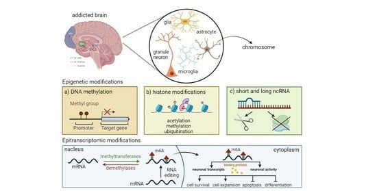
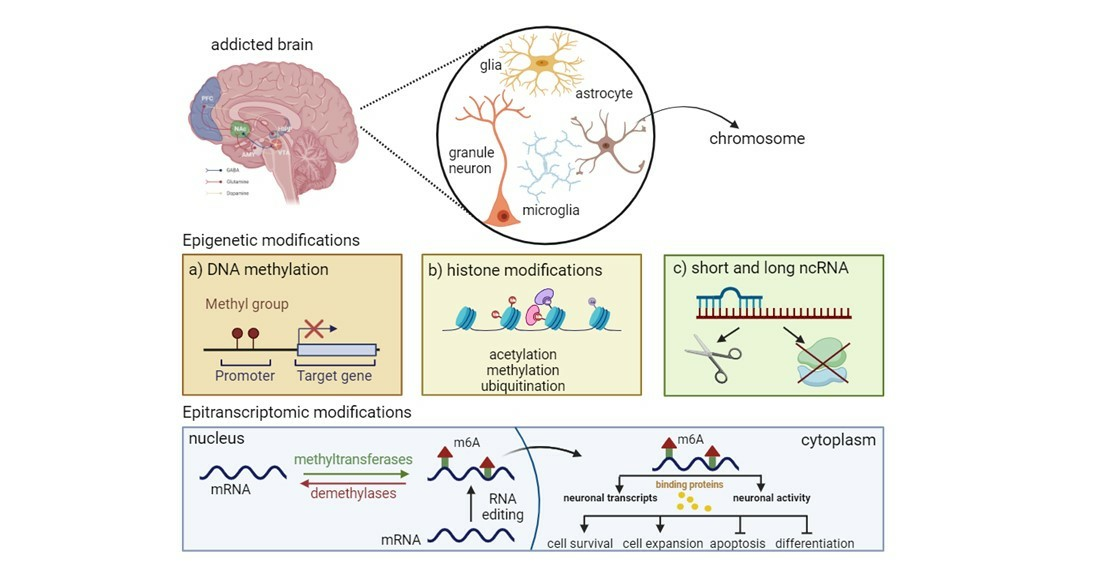
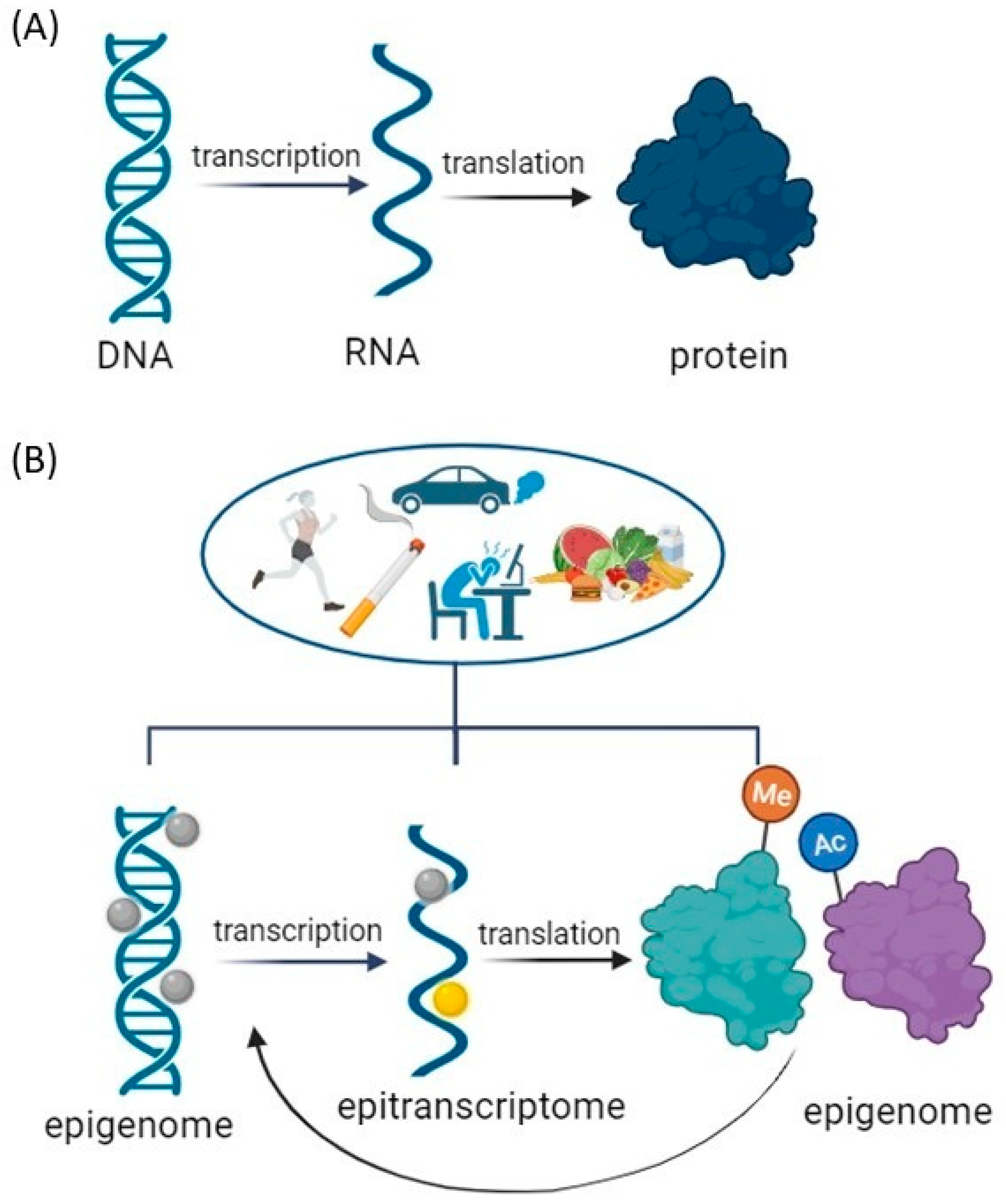
3. Epigenetic Mechanisms and Psychostimulant Addiction

Although single-cell sequencing technologies have transformed our understanding of cellular heterogeneity, single-cell data are challenging for downstream analysis [84]. In addition, we have a process that alters gene activity without changing the DNA sequence, especially influenced by psychostimulants, which lead to neuronal plasticity [85].
Epigenetics refers to changes in the DNA molecule that do not alter the nucleotide sequence but control gene expression by inducing specific chemical modifications that regulate the activation or repression of genes and lead to different expression patterns [86]. The predominant mechanisms of epigenetic modification in eukaryotes include DNA methylation, histone modifications, and non-coding RNA [87]. In contrast to genetic modifications, epigenetic changes are reversible and respond to various environmental factors. DNA methylation is the most extensively studied epigenetic modification. It represents a stable gene silencing mechanism, which, in combination with modifications of histones and other chromatin-associated proteins, plays an important role in the regulation of gene expression and chromatin structure [88]. In mammals, DNA methylation occurs primarily through covalent modification of cytosine residues in CpG dinucleotides—DNA sequences consisting of consecutive cytosine-guanine (CpG) nucleotide pairs. The precise DNA methylation patterns are generated and maintained by the cooperative activity of DNA methyltransferases (DNMTs). DNMT1 ensures the maintenance of existing methylation patterns, while DNMT3A and DNMT3B are responsible for the de novo establishment of new methylation patterns. Moreover, DNMTs are also responsible for the complex process of DNA demethylation together with the enzyme family of ten-eleven translocation (TET). Furthermore, the DNA is tightly wrapped around the histones, thus preventing the expression of specific genes. In particular, the N-terminal tails of histones can undergo post-translational covalent modifications at specific residues, influencing critical cellular processes such as transcription, replication, and DNA repair. Histone modifications primarily modulate the degree of chromatin condensation, thereby regulating gene expression based on the modified residues and the type of modifications [89,90]. Distinct cell types exhibit specific patterns of histone modifications that contribute to cellular identity precisely by regulating gene expression. Enzymes that add or remove covalent modifications to histone proteins dynamically control these patterns. While histone deacetylases (HDACs) and histone demethylases (HDMs) remove acetyl and methyl groups, histone acetyltransferases (HATs) and histone methyltransferases (HMTs) add acetyl and methyl groups, respectively [90,91,92]. Chromatin remodeling, transitioning from the condensed to the transcriptionally active state, controls gene expression: increased chromatin condensation impedes transcriptional and other factors from accessing the DNA molecule, suppressing, or disrupting transcription [90]. Mechanisms involved in chromatin remodeling include changes in histone composition within individual nucleosomes, various histone modifications, and the rearrangement of individual nucleosomes. All these mechanisms together influence chromatin compaction, thereby regulating transcriptional activity [87,89,93]. Changes in the activity of enzymes that modify histones, known as writers, enzymes that remove modifications, known as erasers, and protein complexes that modulate gene expression, known as readers, have been linked to a variety of physiological changes and diseases [94,95]. The number of known post-translational modifications (PTMs) of histones is increasing, supported by the development of technologies for their identification: antibody development, omics approaches in peptide analysis, and mass spectrometry [96,97]. PTMs on individual histones are the focus of the current research, as they have been found to influence neuronal plasticity as the biological basis of learning and memory, but also the development of pathological forms of neuronal plasticity, such as in addiction [98]. However, due to the complexity of the interactions between various epigenetic modifications and their multiplicity, developing an understanding of the mechanisms through which they change gene expression and related phenotypes is still challenging. In mammals, the administration of psychostimulants such as COC or METH causes a significant increase in dopamine signaling that is associated with a combinatorial cascade of histone PTMs. Such modifications in dopamine neurons in the reward-processing part of the brain result in behavioral consequences typical of the rewarding effects of psychostimulants.
The mechanisms of epigenetic regulation by RNA molecules are less explored compared to DNA methylation and histone modifications. Non-coding RNAs (ncRNAs) represent a diverse group of regulatory RNA molecules, some of which already have established functions, while others remain to be characterized [93]. Based on their size, they are divided into two categories: long non-coding RNAs (lncRNAs), which comprise more than 200 nucleotides, and small ncRNAs, which comprise no more than 200 nucleotides [99]. The ncRNAs relevant in the field of epigenetic regulation include lncRNAs, miRNAs (microRNA), siRNAs (small interfering RNA), and piRNAs (PIWI-interacting RNA) [93].

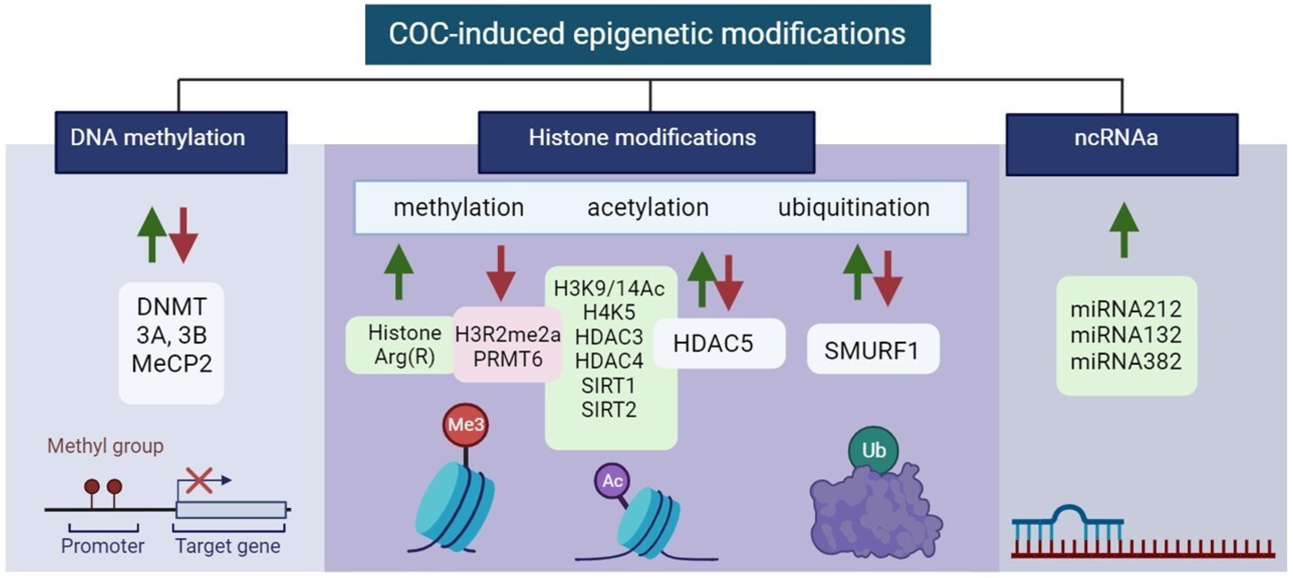
3.1. Cocaine-induced Epigenetic Modifications

Epigenetic modifications in the control of gene expression of specific brain regions in COC addiction are summarized in Table 1 and Figure 3.

3.1.1. DNA Methylation

Environmental stimuli in COC exposure can be translated to changes in gene expression and phenotypes that, through enzymatic modifications to DNA sequences, create long-lasting behavioral phenotypes. These methylated DNA regions are bound by methyl-binding domain-containing proteins such as methyl CpG-binding protein 2 (MeCP2), which is important for recruiting co-repressors such as methyltransferases to the gene promoter [100]. DNA methylation is critical for imprinting, X chromosome inactivation, and cell differentiation. Therefore, it is important that it can be altered at specific loci in germ cells by exposure to environmental factors such as toxins [101] and stress [102], and can therefore be inherited by offspring over multiple generations. Acute cocaine administration increases DNA methylation and DNMT3A and DNMT3B levels, while the binding of MeCP2 to specific gene promoters can decrease gene expression in the NAc [103]. When cocaine is self-administered, MeCP2 is increased in both the striatum and PFC of rats during that response [104,105]. Moreover, MeCP2 can be switched off in the striatum in association with an impaired COC-dependent increase in BDNF levels [104]. It is demonstrated that following repeated COC exposure, including self-administration, in the NAc of mice and rats as well as in the NAc of COC-dependent humans, there is a decrease in protein-R-methyltransferase-6 (PRMT6) and its corresponding histone mark, asymmetric demethylation of R2 on histone H3 (H3R2me2a). First, PRMT6 down-regulation is observed specifically in NAc medium spiny neurons (MSNs) that express dopamine D2 receptors (D2-MSNs), whereas D1-MSNs exhibit the opposite regulation.

3.1.2. Histone Modifications

Histones Methylation

Histones H3 and H4 can have their lysine or arginine residues methylated, with varying effects on transcription. While lysine methylation is connected to both transcriptional activation and repression depending on the methylation site, arginine methylation encourages transcriptional activation [106]. This adaptability could be explained by the fact that, in contrast to acetylation, methylation has no effect on histone charge or histone–DNA interactions directly. H3 methylation increases the gene transcription, while lysine (K) methylation on H3K9Me3 and H3K27Me3 at specific gene regions represses transcription [107]. In addition to lysine (K) methylation, protein-R-methyltransferases (PRMT1 and 6) have the ability to methylate histone arginine (R), producing H3R2Me2 and H4R3Me2 in NAc following repeated cocaine exposure [108].

Histones Acetylation

COC administration leads to significant acetylation of histones H3 and H4 [109]. H3K9, H3K14, H4K5, H4K8, H4K12, and H4K16 have emerged as the most extensively researched acetylation sites in the context of COC exposure [90]. Histone acetylation in the NAc is associated with CREB-binding protein (CBP), a histone acetyltransferase [110]. Repeated exposure to COC leads to a dual effect on histone acetylation, resulting in H3Ac/H4Ac-increased and -decreased acetylation of gene promoters, attributed to the hypoacetylation of histones H3 and H4 [109]. In the VTA during COC withdrawal, there is an observed enrichment of CBP in the promoter region of BDNF leading to an elevated level of histone acetylation, particularly H3K9/14Ac, at the Bdnf promoter [111]. However, because the balance of acetylation and deacetylation is dynamic, increased HDAC5 deacetylase activity lowers COC preference in the Conditioned Place Preference (CPP) [112]. A genetically modified version of HDAC5 that is expressed during COC administration, on the other hand, produces different results in various behavioral measures of COC effects [113,114,115]. Furthermore, not only must the global level of acetylation and deacetylation be considered but also the location along the DNA where chromatin modulation occurs, i.e., the role of the PTM gene, which may act antagonistically on gene programs induced by psychostimulant administration [109]. Acute COC exposure increases global histone H3 phospho-acetylation and histone H4 acetylation in the striatum [116,117]. While long-term COC exposure increases the expression of HDAC5 target genes and decreases HDAC5 activity in the NAc, it also promotes histone acetylation [118]. Ferguson et al. demonstrated that the overexpression of sirtuin (SIR1 and SIR2) in the NAc in response to chronic COC administration enhances the rewarding effects, identifying sirtuins as key mediators of cellular and molecular plasticity [119]. Zell et al. revealed that m-opioid receptors may also regulate VTA neurotransmission by directly engaging glutamatergic neurons [120]. Subclassing of the current dataset indicates that rather than directly modulating DA neurons, these effects might be mediated through GABAergic or glutamate/DA combinatorial populations.

Histones Ubiquitination

The ubiquitin-proteasome system (UPS), one of the epigenetic hallmarks, is a multifaceted network of ubiquitin ligases and proteasome structures that controls synaptic and epigenetic plasticity [121] and is also involved in memory processes and substance use disorders [122]. E3 ubiquitin-protein ligase SMURF1 is a key mediator of neuroadaptations in the nucleus accumbens that follow COC exposure and mediates cue-induced COC seeking during withdrawal [123]. All of these results point to the possibility that the SMURF1–SMAD1/5–RUNX2 pathway functions as a crucial transcriptional regulator that modifies plasticity after COC self-administration. The finding that RUNX2 and SMAD1/5 are upregulated in the NAc following COC self-administration allowed researchers to investigate RUNX2 binding to target genes that had previously been connected to COC plasticity. RUNX2 is found to bind at AP-1 sites and to the promoters of these genes following COC self-administration, indicating that it may be a master regulator of several pathways regulating COC-induced plasticity [124]. There is a critical need for new research to identify novel potential therapeutic targets in order to develop effective treatments for COC use disorder.
In addition to histone acetylation, methylation, and ubiquitination, other histone modifications such as SUMOlation, propionylation, butyrylation, and the recently described dopa-mineylation and serotonylation play important roles in the regulation of psychostimulant-induced behavioral effects [80,97,125].

3.1.3. ncRNAs

The most commonly investigated ncRNAs involved in epigenome modifications in COC addiction are miRNAs. They can regulate COC intake and potentially have an impact on developing compulsive consumption of the drug. The mechanisms that stand by miR-212 influences COC intake were also a point of exploration. The cAMP signaling cascade strongly activates the miR-212/132 gene cluster, with CREB upregulating both miRNAs. It was found that the pathway of miR-212 regulation dramatically boosts CREB signaling in cultured cells and also in the striatum of rats [104]. miR-206 has been shown to negatively regulate BDNF expression, which is known to be important for the motivational effects of COC and other addictive substances [126]. Additionally, it was demonstrated that the activation of miR-382 attenuates COC-induced increases in the transcription factor ΔFosB, which is known to be important in the rewarding effects of addictive drugs by negatively regulating the expression of the dopamine D1 receptor [127].
Table 1. The role of epigenetic modifications in the control of gene expression of specific brain regions in cocaine addiction.
Type of Epigenetic ModificationsLocalization of Modification ↓↑ *Brain RegionNeuronal Cell Type, Pathways, and ReceptorsReference
DNA methylationDNMT ↑↓
3A, 3B ↑↓, MeCP2 ↑↓
NAc, DS, PFCBDNF [100,101,102,103,104]
Histone modificationsmethylationH3R2me2a
PRMT6
Histone Arg(R)
NAc, DS, PFCBDNF ,
D1-MSN , D2-MSN
[100,101,102,103,104]
acetylationH3K9/14Ac ,H4K5 ,
HDAC3 , HDAC4 , HDAC5 , SIRT1 , SIRT2
DS, NAc, mPFC, VTABdnf Creb1
Cbp, Cdk5
DA
Glu/DA ↓↑
GABA
[116,117,118,120,128]
ubiquitinationSMURF1 ↑↓NAcAP1
RUNX2
SMAD1/5
[123,124]
ncRNAsmiRNAsmiRNA212
miRNA132
miRNA382
DS, NAc-MSNAP1
RUNX2
SMAD1/5
[104,126,127]
* ↑ indicates upregulation and ↓ indicates downregulation.

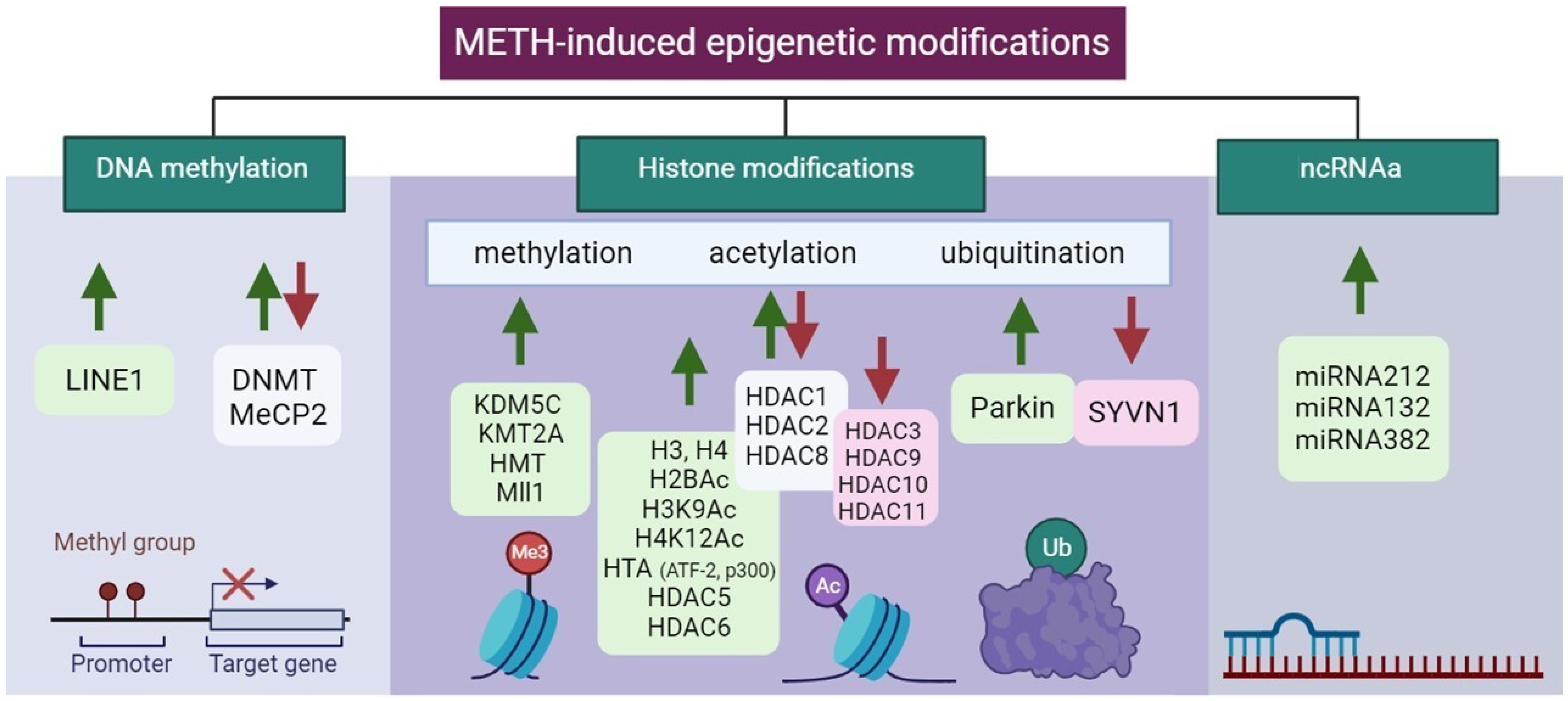
3.2. Methamphetamine-Induced Epigenetic Modifications

Epigenetic modifications in the control of gene expression of specific brain regions in METH addiction are summarized in Table 2 and Figure 4.

3.2.1. DNA Methylation

DNA methylation levels have been found to be changed in METH addicts [129], and moreover, this also occurs in their offspring [130]. BDNF methylation was increased in the PFC of METH-addicted rats and patients but was decreased in the hippocampus of rats [131]. The neurotoxic effects of METH exposure were partly caused by a decrease in BDNF expression [131,132]. According to a report by Moszczynska et al., METH induction leads to impaired cognition and memory, accompanied by an increase in long-interspersed element-1 (LINE-1) activity in both the dentate gyrus of the hippocampus (Hip) and the dorsum striatum (DS) [133]. METH improved spatial memory by downregulating the expressions of MeCP2 and DNMTs and reducing DNA methylation at the promoter of Synaptophysin (Syp) in the Hip. While METH increased the expression of MeCP2 and DNMTs, it also caused DNA hypermethylation at the Syp promoter in the PFC and decreased memory function. The rewarding effect of amphetamine was increased by the specific knockout of MeCP2 in the NAc [134]. By reversing DNA methylation at Sy genes involved in METH-mediated changes in dendritic spines and synaptic transmission, oxytocin (OT) prevented METH-seeking behavior and relapse [135,136,137,138].

3.2.2. Histone Modifications

Histones Methylation

According to recent research, epigenetic mechanisms that mediate drug-induced transcriptional and behavioral changes induced by METH consumption are mostly caused by histone modification [139]. Indeed, it was shown that METH induced H3 methylation through increased tri-methylation of histone H3 at lysine 4 (H3K4me3) at the promoter site of chemokine receptor 2 associated with behavioral sensitization in mice [140]. Iamjan et al. reported that rats given METH and humans with METH dependence, particularly METH-dependent psychosis, exhibit aberrant Brain-derived neurotrophic factor (BDNF) methylation [131]. Salehzadeh et al. also confirmed this hypothesis and showed BDNF hypomethylation [132]. METH consumption also influenced Long INterspersed Element (LINE-1) activity. Systemic METH administration boosts LINE-1 activity in two neurogenic niches of the adult rat brain in a promoter hypomethylation-independent manner. Neurological impairments can be triggered by altered levels of LINE-1 expression, indicating that LINE-1 induction may play a role in the development of cognitive impairments in METH users [133].

Histones Acetylation

Similar to COC, METH induces alterations in the acetylation levels of histones H3 and H4, influencing the expression of various enzymes in different brain regions [141]. In NAc, METH exposure leads to differential acetylation changes on various histone lysine residues by regulating the protein levels of histone deacetylases [142]. Acute METH exposure results in decreased H3 acetylation (H3K9Ac and H3K18Ac) and increased H4 acetylation (H4K5Ac, H4K8Ac, and H4K16Ac) [143]. Chronic METH causes a reduction in histone H4 acetylation (H4K5Ac, H4K12Ac, and H4K16Ac) at glutamate (GLUT) receptor promoters, impacting the expression of alpha-amino-3-hydroxy-5-methyl-4-isoxazole propionic acid (AMPA) and N-methyl-d-aspartate (NMDA) receptors, leading to oxidation, excitotoxicity, and neuroinflammation [144]. However, differences in H3 and H4 acetylation patterns have been observed between single and repeated exposures, which may contribute to the escalating dose cravings observed in the drug-seeking behavior of individuals with METH addiction [18,145]. Additionally, METH increased the Hip’s expressions of H2BAc, H3K9Ac, and H4K12Ac as well as the DS’ expression of H4K12Ac. In the medial prefrontal cortex (mPFC), repeated METH treatment increased H4AC enrichment at the promoters of D1, hypocretin (orexin) receptor 1 (HCRTR1), and NMDA 1, but decreased H3AC enrichment at the promoters of D2, HCRTR1, HCRTR2, histamine receptor H1 (HRH1), HRH3, and NMDA [135]. In histone deacetylases (HDAC 1, 2, 3, 5, 6, 8, 9, and 11), a single treatment of METH reduces the mRNA level of HDAC3 in the NAc [146]. On the other hand, rats’ NAc and mPFC express more HDAC2 when given a large single dose of METH, while their HDAC1 expression is decreased. In mice, a single METH treatment lowers the amount of HDAC8 mRNA in the NAc [147]. Similarly, the HDAC11 mRNA level decreased after acute METH exposure in the NAc in mice. METH induced activation of the D1 receptor (D1R) in the PFC, which affected HDAC1 and HDAC2 levels [135] and the miRNA 181a/d level in the ventral tegmentum area (VTA), increased the expressions of α-adrenergic receptors and the NMDA receptor subunit, and then regulated the function of dopamine receptors (DRs) [146,148]. METH administration increased GluN2B, an NMDA receptor subunit expression and sequential activation of the ERK/CREB)/BDNF pathway. Histone acetylation has consequences at a given location and could be in and of during mediation of the targets in fully different neurons dopaminergic, glutamatergic, and GABAergic neurons (DA, Glu, and GABA). Therefore, enzymes controlling the transfer of acetyl groups are involved in METH addiction as well. ATF-2, a member of the ATF/CREB family, enhances CREB-dependent transcription and possesses intrinsic HAT activity on histone H4 [143]. METH increases P300, one type of HAT that regulates Glu release induced by METH in human primary astrocytes [149]. Another novel molecular mechanism that may affect METH-induced behavioral sensitivity is histone methylation. Methyltransferases (HMTs/KMTs) and demethylases (HDMs/KDMs)—such as KMT2A, an enzyme involved in H3K4me3—were linked to METH addiction. METH increased this enzyme, which is necessary for the development and maintenance of METH-associated memory. KDM5C is linked to METH because it demethylates H3K4. Using small interfering (siRNAs) in the NAc, some studies report decreased expressions of histone methyltransferase (HMT), histone lysine demethylase KDM5C, and mixed lineage leukemia 1 (Mll1).

Histones Ubiquitination

Neurotransmitter excitation and synaptic plasticity in brain disorders associated with drug addiction have been connected to the UPS, an enzymatic complex that controls proteolysis and turnover. Both pre- and post-synaptic neurons in the DA circuitry are impacted by UPS inhibition. Through endocytic internalization and degradation, the UPS reduces D1/D2-like DRs and Alphaamino-3-hydroxy-5-methyl-4-isoxazolepropionic acid (AMPA) [150]. The UPS regulates the presynaptic release of Glu via both D1-like and D2-like receptors, which controls DA transmission [151], also influencing postsynaptic plasticity [152,153]. As a result, the DA and Glu signaling pathways interact with UPS substrates [121]. METH, despite being an extremely powerful DA releaser, impairs UPS activity, which is largely due to dopamine. As a result, pre- and post-synaptic neurons in the dopamine circuitry are extremely vulnerable to UPS inhibition [150]. Parkin promotes the ubiquitination of substrate proteins, which aids in their degradation. Sharma et al. showed that studying the neuronal substrates underlying “resilience” or vulnerability to METH use disorder can be facilitated by using rats with excess or deficit PARKIN. Additionally, PARKIN may be a novel therapeutic target for the treatment of METH use disorder [154]. After METH withdrawal, protein ubiquitination and E3 ubiquitin ligases are increased in the central amygdala. Research conducted by Cates et al. showed that ubiquitination of the central amygdala is linked to METH craving behavior [155]. Synovial apoptosis inhibitor 1 (SYVN1) is an E3 ubiquitin ligase from the endoplasmic reticulum (ER). SYVN1 knockdown has been linked to METH-CPP by increasing GABAA1 in the DS [156]. MiRNA-181a also regulates METH addiction via the ERAD pathway [136].

3.3. ncRNAs

Similarly, as in COC addiction, epigenetic modifications in METH addiction involving ncRNA predominantly include miRNAs. Using the KEGG pathway analysis, miRNA-regulated genes were found to be involved in vesicular transport, METH addiction, the cyclic guanosine monophosphate cGMP-protein kinases G (PKG) signaling pathway, the dopaminergic synapse, and the GABAergic synapse [157]. In genome-wide transcriptional profiling, the expression of multiple miRNAs is increased in the central amygdala alongside molecules related to METH addiction [155]. METH increased the levels of miRNAs 237, 296, and 501 in the NAc. MiRNAs in the NAc regulate Wnt signaling and axon guidance genes [158]. MiR-128 influenced METH-induced behavioral sensitization by altering synaptic plasticity-related molecules in the NAc [159]. METH-induced locomotor sensitization is disrupted by Ago2-dependent miRNAs in the NAc. These Ago2/miR-3068-5p effects occur in conjunction with the glutamate receptor, GluN1/Grin1 [138]. METH can increase the expression of miRNAs in the striatum, which harmed motor coordination and reduced striatal volume and dendritic length [160].
Table 2. The role of epigenetic modifications in the control of gene expression of specific brain regions in methamphetamine addiction. Table produced according to Wang et al. [62].
Type of Epigenetic ModificationsLocalization of Modification ↓↑ *Brain RegionNeuronal Cell Type, Pathways, and ReceptorsReference
DNA methylationDNMT ↑↓,
MeCP2 ↑↓, LINE1 ↑
DS, Hip, NAc, PFC, mPFCBDNF ↑, OT ↓, K+ channel ↑↓, Syp ↑↓, Glu ↑,
GluA1,2 ↑, LINE-1 ↑, NR4A1, GABA ↓
[131,132,133,161,162]
Histone modificationsmethylationKDM5C ↑, KMT2A ↑,
HMT ↑, Mll1 ↑
DS, Hip, NAc, PFC, mPFCBDNF ↑, OT ↓, K+ channel ↑↓, Syp ↑↓, Glu ↑,
GluA1,2 ↑, LINE-1 ↑, NR4A1, GABA ↓
[131,132,133,161,162]
acetylationH3 ↑, H4 ↑, H2Bac ↑, H3K9Ac ↑, H4K12Ac ↑
HTA (ATF-2 ↑, p300 ↑)
HDAC1 ↑↓, 2 ↑↓, 3 ↓, 5 ↑, 6 ↑, 8 ↑↓, 9 ↓, 10 ↓, 11 ↓
DS, Hip, mPFC, NAcD1 ↑, D2 ↑, HCRTR1 ↓↑, HCRTR2 ↑, HRH1, 3 ↑, NMDA ↑
Glu ↑
α-adrenergic receptors ↑,
BDNF ↑, NMDA ↑, D1 ↑, D2 ↓, CREB ↓
[135,141,163]
ubiquitinationParkin ↑, SYVN1 ↓BLA, CeA, DSD1, 2 ↑, NMDA ↑, AMPA ↑, GABAAα1 ↑[62,154,164]
ncRNAsmiRNAsmiRNA128 ↑, 237 ↑, 296 ↑, 501 ↑ 31-3p ↑, 34a-5p ↑, 183–5p ↑, 9a-5p ↑, 369–3p ↑, 29a ↑, 181a/d ↑DS, Hip, NAc, VTAPKG ↑, PI3K ↑, Wnt ↑, Ago2 ↑, BDNF ↑,
GluN1 ↑
[137,138,157]
* ↑ indicates upregulation and ↓ indicates downregulation.

4. Epitranscriptomics and Psychostimulant Addiction

Epigenetic modifications to DNA and histones regulate gene transcription, whereas epitranscriptomic post-transcriptional RNA modifications influence gene expression [165]. Although various types of RNA are involved in these changes, we have concentrated on messenger RNA (mRNA) modifications. The most common mRNA modifications are N1-methyladenosine (m1A), N6-methyladenosine (m6A), 5-methylcytosine (m5C), pseudouridine (Ψ), and others [166]. m6A modification is one of the most abundant and reversible epitranscriptomic modifications, mediated by a set of proteins, which include methyltransferases (‘writers’), demethylases (‘erasers’), and m6A-binding proteins (‘readers’) [167]. m6A methylation is associated with the control of mRNA metabolism, splicing, export, stability, translation, and degradation [168,169]. Out of all tissues in the body, the brain has the highest abundance of m6A methylation, which is developmentally decreasing [168]. In the brain, m6A methylation regulates neuronal transcripts and neuronal activity. Aside from its role in neuronal development [30], m6A modification is essential for the process of axon regeneration [170]. Methyltransferases like-3 (METTL3) and like-14 (METTL14) [171,172,173], along with other proteins needed for m6A deposition, such as Wilms’ tumor 1-associating protein (WTAP) [174] and RNA binding protein 15 (Rbm15) [175], form a stable protein complex that catalyzes m6A modification. Because they deposit RNA methylation modifications, these methyltransferases are collectively known as “writers”. Fat mass obesity-associated protein (FTO) and alkB homologue 5 (ALKBH5) are two demethylases from the family of α-ketoglutarate dependent dioxygenases that can reverse m6A modification because it is dynamically regulated [176,177]. Demethylases are known as “erasers” because they remove RNA methylation modifications. Posttranscriptional, site-specific adenosine-to-inosine base conversions, known as RNA editing, contribute to gene expression diversity and are catalyzed by Adenosine deaminases acting on RNA (ADARs) [178]. Pseudouridine Synthase 7 (PUS7) is one of the major mRNA-modifying enzymes leading to pseudouridine (Ψ), a ubiquitous RNA modification [179]. Both ADAR and PUS7 can lead to further mRNA modifications. m6A modifications are in direct connection to the so-called m6A “reader” proteins, which recognize the modified site. The proteins with YTH domains, which can specifically bind m6A through their YTH domain, are the most well-studied m6A readers. Fragile X mental retardation protein (FMRP) was also reported to be m6A reader and plays critical roles in synaptic plasticity and neuronal development. The identification of methylated nucleosides (m6A, m5C, m1A) is performed using immunoprecipitation and their variants using antibodies against methylated nucleosides or associated proteins methyltransferases, demethylases, and binding proteins. After fragmenting the RNA, fragments containing modified nucleosides are enriched before sequencing. The immunoprecipitate is analyzed using next-generation sequencing (NGS) to identify and map the modification [180].
The connection between epigenetic regulation and m6A RNA modification was associated with histone H3 trimethylation at Lys36 (H3K36me3), a marker for transcription elongation, which guides m6A deposition globally connected through METTL14 (DOI: 10.1038/s41586-019-1016-7). m6A modifications are mainly associated with neuronal plasticity in the brain, which is a consequence of learning and memory, and most of the literature is based on this line of research together with neurodegenerative disorders [176,181,182]. Indeed, deficiency in m6A-dependent pathways significantly impairs neuronal function including dopamine signaling and dopamine-dependent learning.
Lowering neuronal m6A by overexpressing FTO or by adding m6A inhibitor led to the induction of N-methyl-d-aspartate (NMDA) receptor 1 expression, elevated oxidative stress, and Ca2+ influx, resulting in dopaminergic neuron apoptosis [183]. In addition, it was shown that the overexpression of FTO delays the dephosphorylation of CREB, increases the expression of the CREB, and targets neuropeptide receptor 1 (NPY1R) and BDNF known to regulate food intake and energy homeostasis [184]. FTO affects dopamine (D2)-dependent responses to reward learning in meso-striato-prefrontal regions, suggesting a mechanism by which genetic predisposition alters reward processing not only in obesity but also in other disorders with altered D2R-dependent impulse control, such as addiction [185]. Other than that, FTO was reported to be part of the regulation of BDNF processing [186]. FTO demethylase, but not METTL3 and METTL14, was downregulated in the hippocampus following COC-induced CPP, leading to a higher level of m6A [187]. Mice lacking the FTO gene exhibited lower body weight and decreased anxiety- and depression-like behaviors, mediated by changes in the gut microbiota [188].

5. Challenges of the Rewarding Molecular Pathway

Disrupting natural reward pathways, such as with food overeating leading to obesity and addictive drug usage leading to addiction, share many molecular regulatory sites in the central nervous system. Both influence neurotransmitters, leading to the dysregulation of the reward circuitry in the brain, and they have been studied as a complex interplay of genetic and environmental factors influencing their development. This review emphasizes the importance of the FTO gene, which, together with CREB and BDNF, regulates synaptic plasticity, but is also associated with cellular metabolism, oxidative stress in the brain, and the cycle of inflammation. Based on this, antioxidant and anti-inflammatory synthetic molecules have been implicated in the treatment of drug addiction. Future sc-RNA-seq studies as well as epigenetic and epitranscriptomic studies should focus on NAc D1R and D2R neurons, as these encode positive valence and reward responses to drugs of abuse and food and have a projection to the VTA.

6. Conclusions

In this review article, we covered single-cell sequencing research as well as epigenetic and epitranscriptomics methods applied to the brain regions VTA, NAc, and DS after both short-term and long-term exposure to the psychostimulants COC and METH. The single-cell method, which clustered cell types based on gene expression variations, was used to validate both known and unknown cell types implicated in the psychostimulant response. Research in the fields of epigenetics and epitranscriptomics has focused on the role of molecular actors, also called “readers”, “writers”, and “erasers”, which alter DNA, histone proteins, and RNA to help regulate gene expression.

Author Contributions

Conceptualization, A.F.V.; writing—original draft preparation, A.F.V., L.S.M., I.S.M. and S.D.P.; writing—review and editing, A.F.V., L.S.M., I.S.M. and S.D.P.; visualization, L.S.M. and A.F.V.; supervision, A.F.V. All authors have read and agreed to the published version of the manuscript.

Funding

The authors declare that no funds, grants, or other support were received during the preparation of this manuscript.

Institutional Review Board Statement

Not applicable.

Informed Consent Statement

Not applicable.

Data Availability Statement

Not applicable.

Conflicts of Interest

The authors declare no conflict of interest.

References

  1. Steketee, J.D.; Kalivas, P.W. Drug Wanting: Behavioral Sensitization and Relapse to Drug-Seeking Behavior. Pharmacol. Rev. 2011, 63, 348–365. [Google Scholar] [CrossRef]
  2. Robbins, T.W.; Everitt, B.J. Neurobehavioural mechanisms of reward and motivation. Curr. Opin. Neurobiol. 1996, 6, 228–236. [Google Scholar] [CrossRef]
  3. Robinson, T.E.; Berridge, K.C. The incentive sensitization theory of addiction: Some current issues. Philos. Trans. R. Soc. B Biol. Sci. 2008, 363, 3137. [Google Scholar] [CrossRef] [PubMed]
  4. Lewis, R.G.; Florio, E.; Punzo, D.; Borrelli, E. The Brain’s Reward System in Health and Disease. Adv. Exp. Med. Biol. 2021, 1344, 57–69. [Google Scholar] [CrossRef] [PubMed]
  5. Rezayof, A.; Ghasemzadeh, Z.; Sahafi, O.H. Addictive drugs modify neurogenesis, synaptogenesis and synaptic plasticity to impair memory formation through neurotransmitter imbalances and signaling dysfunction. Neurochem. Int. 2023, 169, 105572. [Google Scholar] [CrossRef]
  6. Peters, K.Z.; Cheer, J.F.; Tonini, R. Modulating the Neuromodulators: Dopamine, Serotonin, and the Endocannabinoid System. Trends Neurosci. 2021, 44, 464–477. [Google Scholar] [CrossRef]
  7. Lüscher, C.; Malenka, R.C. Drug-evoked synaptic plasticity in addiction: From molecular changes to circuit remodeling. Neuron 2011, 69, 650–663. [Google Scholar] [CrossRef]
  8. Kutlu, M.G.; Gould, T.J. Effects of drugs of abuse on hippocampal plasticity and hippocampus-dependent learning and memory: Contributions to development and maintenance of addiction. Learn. Mem. 2016, 23, 515–533. [Google Scholar] [CrossRef] [PubMed]
  9. Anghel, D.-M.C.; Nițescu, G.V.; Tiron, A.-T.; Guțu, C.M.; Baconi, D.L. Understanding the Mechanisms of Action and Effects of Drugs of Abuse. Molecules 2023, 28, 4969. [Google Scholar] [CrossRef]
  10. Perry, A.N.; Westenbroek, C.; Becker, J.B. The development of a preference for cocaine over food identifies individual rats with addiction-like behaviors. PLoS ONE 2013, 8, e79465. [Google Scholar] [CrossRef]
  11. Chiu, V.M.; Schenk, J.O. Mechanism of action of methamphetamine within the catecholamine and serotonin areas of the central nervous system. Curr. Drug Abus. Rev. 2012, 5, 227–242. [Google Scholar] [CrossRef] [PubMed]
  12. Chomchai, C.; Chomchai, S. Global patterns of methamphetamine use. Curr. Opin. Psychiatry 2015, 28, 269–274. [Google Scholar] [CrossRef] [PubMed]
  13. Gangu, K.; Bobba, A.; Basida, S.D.; Avula, S.; Chela, H.; Singh, S. Trends of Cocaine Use and Manifestations in Hospitalized Patients: A Cross-Sectional Study. Cureus 2022, 14, e22090. [Google Scholar] [CrossRef]
  14. Chen, G.; Lai, S.; Bao, G.; Ke, J.; Meng, X.; Lu, S.; Wu, X.; Xu, H.; Wu, F.; Xu, Y.; et al. Distinct reward processing by subregions of the nucleus accumbens. Cell Rep. 2023, 42, 112069. [Google Scholar] [CrossRef] [PubMed]
  15. Becker-Krail, D.D.; Walker, W.H.; Nelson, R.J. The Ventral Tegmental Area and Nucleus Accumbens as Circadian Oscillators: Implications for Drug Abuse and Substance Use Disorders. Front. Physiol. 2022, 13, 886704. Available online: https://www.frontiersin.org/articles/10.3389/fphys.2022.886704 (accessed on 11 December 2023). [CrossRef] [PubMed]
  16. Lee, A.M.; Messing, R.O. Protein Kinases and Addiction. Ann. N. Y. Acad. Sci. 2008, 1141, 22–57. [Google Scholar] [CrossRef] [PubMed]
  17. Amaral, I.M.; Scheffauer, L.; Hofer, A.; El Rawas, R. Protein kinases in natural versus drug reward. Pharmacol. Biochem. Behav. 2022, 221, 173472. [Google Scholar] [CrossRef]
  18. Shi, X.; McGinty, J.F. Repeated amphetamine treatment increases phosphorylation of extracellular signal-regulated kinase, protein kinase B, and cyclase response element-binding protein in the rat striatum. J. Neurochem. 2007, 103, 706–713. [Google Scholar] [CrossRef]
  19. Jia, W.; Kawahata, I.; Cheng, A.; Fukunaga, K. The Role of CaMKII and ERK Signaling in Addiction. Int. J. Mol. Sci. 2021, 22, 3189. [Google Scholar] [CrossRef]
  20. Wheeler, J.M.; Reed, C.; Burkhart-Kasch, S.; Li, N.; Cunningham, C.L.; Janowsky, A.; Franken, F.H.; Wiren, K.M.; Hashimoto, J.G.; Scibelli, A.C.; et al. Genetically correlated effects of selective breeding for high and low methamphetamine consumption. Genes Brain Behav. 2009, 8, 758–771. [Google Scholar] [CrossRef]
  21. Hitzemann, R.; Iancu, O.D.; Reed, C.; Baba, H.; Lockwood, D.R.; Phillips, T.J. Regional Analysis of the Brain Transcriptome in Mice Bred for High and Low Methamphetamine Consumption. Brain Sci. 2019, 9, 155. [Google Scholar] [CrossRef]
  22. Phillips, T.J.; Roy, T.; Aldrich, S.J.; Baba, H.; Erk, J.; Mootz, J.R.K.; Reed, C.; Chesler, E.J. Confirmation of a Causal Taar1 Allelic Variant in Addiction-Relevant Methamphetamine Behaviors. Front. Psychiatry 2021, 12, 725839. Available online: https://www.frontiersin.org/articles/10.3389/fpsyt.2021.725839 (accessed on 18 January 2024). [CrossRef]
  23. Shabani, S.; Houlton, S.; Ghimire, B.; Tonello, D.; Reed, C.; Baba, H.; Aldrich, S.; Phillips, T.J. Robust aversive effects of trace amine-associated receptor 1 activation in mice. Neuropsychopharmacology 2023, 48, 1–9. [Google Scholar] [CrossRef]
  24. Stafford, A.M.; Reed, C.; Baba, H.; Walter, N.A.; Mootz, J.R.; Williams, R.W.; Neve, K.A.; Fedorov, L.M.; Janowsky, A.J.; Phillips, T.J.; et al. Taar1 gene variants have a causal role in methamphetamine intake and response and interact with Oprm1. eLife 2019, 8, e46472. [Google Scholar] [CrossRef] [PubMed]
  25. Althobaiti, Y.S.; Almalki, A.H. Effects of environmental enrichment on reinstatement of methamphetamine-induced conditioned place preference. Behav. Brain Res. 2020, 379, 112372. [Google Scholar] [CrossRef] [PubMed]
  26. Stafford, A.M.; Reed, C.; Phillips, T.J. Non-genetic factors that influence methamphetamine intake in a genetic model of differential methamphetamine consumption. Psychopharmacology 2020, 237, 3315–3336. [Google Scholar] [CrossRef] [PubMed]
  27. Gibney, E.R.; Nolan, C.M. Epigenetics and gene expression. Heredity 2010, 105, 4–13. [Google Scholar] [CrossRef] [PubMed]
  28. Nachtergaele, S.; He, C. The emerging biology of RNA post-transcriptional modifications. RNA Biol. 2017, 14, 156–163. [Google Scholar] [CrossRef] [PubMed]
  29. Roundtree, I.A.; Evans, M.E.; Pan, T.; He, C. Dynamic RNA Modifications in Gene Expression Regulation. Cell 2017, 169, 1187–1200. [Google Scholar] [CrossRef] [PubMed]
  30. Lence, T.; Akhtar, J.; Bayer, M.; Schmid, K.; Spindler, L.; Ho, C.H.; Kreim, N.; Andrade-Navarro, M.A.; Poeck, B.; Helm, M.; et al. m6A modulates neuronal functions and sex determination in Drosophila. Nature 2016, 540, 242–247. [Google Scholar] [CrossRef]
  31. Zeng, H.; Sanes, J.R. Neuronal cell-type classification: Challenges, opportunities and the path forward. Nat. Rev. Neurosci. 2017, 18, 530–546. [Google Scholar] [CrossRef] [PubMed]
  32. Arendt, D. The evolution of cell types in animals: Emerging principles from molecular studies. Nat. Rev. Genet. 2008, 9, 868–882. [Google Scholar] [CrossRef] [PubMed]
  33. Zeng, H. What is a cell type and how to define it? Cell 2022, 185, 2739–2755. [Google Scholar] [CrossRef] [PubMed]
  34. Miller, J.A.; Gouwens, N.W.; Tasic, B.; Collman, F.; van Velthoven, C.T.; Bakken, T.E.; Hawrylycz, M.J.; Zeng, H.; Lein, E.S.; Bernard, A. Common cell type nomenclature for the mammalian brain. eLife 2020, 9, e59928. [Google Scholar] [CrossRef] [PubMed]
  35. Gupta, A.D.; Asan, L.; John, J.; Beretta, C.; Kuner, T.; Knabbe, J. Accurate classification of major brain cell types using in vivo imaging and neural network processing. PLoS Biol. 2023, 21, e3002357. [Google Scholar] [CrossRef]
  36. O’Donnell, E.A.; Ernst, D.N.; Hingorani, R. Multiparameter flow cytometry: Advances in high resolution analysis. Immune Netw. 2013, 13, 43–54. [Google Scholar] [CrossRef] [PubMed]
  37. Baron, C.S.; Barve, A.; Muraro, M.J.; van der Linden, R.; Dharmadhikari, G.; Lyubimova, A.; de Koning, E.J.; van Oudenaarden, A. Cell Type Purification by Single-Cell Transcriptome-Trained Sorting. Cell 2019, 179, 527–542.e19. [Google Scholar] [CrossRef]
  38. Klein, A.M.; Mazutis, L.; Akartuna, I.; Tallapragada, N.; Veres, A.; Li, V.; Peshkin, L.; Weitz, D.A.; Kirschner, M.W. Droplet barcoding for single-cell transcriptomics applied to embryonic stem cells. Cell 2015, 161, 1187–1201. [Google Scholar] [CrossRef]
  39. Macosko, E.Z.; Basu, A.; Satija, R.; Nemesh, J.; Shekhar, K.; Goldman, M.; Tirosh, I.; Bialas, A.R.; Kamitaki, N.; Martersteck, E.M.; et al. Highly Parallel Genome-wide Expression Profiling of Individual Cells Using Nanoliter Droplets. Cell 2015, 161, 1202–1214. [Google Scholar] [CrossRef]
  40. Zheng, G.X.Y.; Terry, J.M.; Belgrader, P.; Ryvkin, P.; Bent, Z.W.; Wilson, R.; Ziraldo, S.B.; Wheeler, T.D.; McDermott, G.P.; Zhu, J.; et al. Massively parallel digital transcriptional profiling of single cells. Nat. Commun. 2017, 8, 14049. [Google Scholar] [CrossRef]
  41. Buenrostro, J.D.; Wu, B.; Chang, H.Y.; Greenleaf, W.J. ATAC-seq: A Method for Assaying Chromatin Accessibility Genome-Wide. Curr. Protoc. Mol. Biol. 2015, 109, 21.29.1–21.29.9. [Google Scholar] [CrossRef]
  42. Grandi, F.C.; Modi, H.; Kampman, L.; Corces, M.R. Chromatin accessibility profiling by ATAC-seq. Nat. Protoc. 2022, 17, 1518–1552. [Google Scholar] [CrossRef]
  43. Jansen, C.; Ramirez, R.N.; El-Ali, N.C.; Gomez-Cabrero, D.; Tegner, J.; Merkenschlager, M.; Conesa, A.; Mortazavi, A. Building gene regulatory networks from scATAC-seq and scRNA-seq using Linked Self Organizing Maps. PLoS Comput. Biol. 2019, 15, e1006555. [Google Scholar] [CrossRef]
  44. Zhang, Z.; Yang, C.; Zhang, X. scDART: Integrating unmatched scRNA-seq and scATAC-seq data and learning cross-modality relationship simultaneously. Genome Biol. 2022, 23, 139. [Google Scholar] [CrossRef]
  45. Lipovsek, M.; Bardy, C.; Cadwell, C.R.; Hadley, K.; Kobak, D.; Tripathy, S.J. Patch-seq: Past, Present, and Future. J. Neurosci. Off. J. Soc. Neurosci. 2021, 41, 937–946. [Google Scholar] [CrossRef]
  46. Lee, B.R.; Budzillo, A.; Hadley, K.; Miller, J.A.; Jarsky, T.; Baker, K.; Hill, D.; Kim, L.; Mann, R.; Ng, L.; et al. Scaled, high fidelity electrophysiological, morphological, and transcriptomic cell characterization. eLife 2021, 10, e65482. [Google Scholar] [CrossRef] [PubMed]
  47. van den Hurk, M.; Erwin, J.A.; Yeo, G.W.; Gage, F.H.; Bardy, C. Patch-Seq Protocol to Analyze the Electrophysiology, Morphology and Transcriptome of Whole Single Neurons Derived From Human Pluripotent Stem Cells. Front. Mol. Neurosci. 2018, 11, 261. Available online: https://www.frontiersin.org/articles/10.3389/fnmol.2018.00261 (accessed on 11 December 2023). [CrossRef] [PubMed]
  48. Koob, G.F.; Volkow, N.D. Neurobiology of addiction: A neurocircuitry analysis. Lancet Psychiatry 2016, 3, 760–773. [Google Scholar] [CrossRef] [PubMed]
  49. Uhl, G.R.; Koob, G.F.; Cable, J. The neurobiology of addiction. Ann. N. Y. Acad. Sci. 2019, 1451, 5–28. [Google Scholar] [CrossRef] [PubMed]
  50. Poisson, C.L.; Engel, L.; Saunders, B.T. Dopamine Circuit Mechanisms of Addiction-Like Behaviors. Front. Neural Circuits 2021, 15, 125. Available online: https://www.frontiersin.org/articles/10.3389/fncir.2021.752420 (accessed on 11 December 2023). [CrossRef] [PubMed]
  51. Srinivasan, C.; Phan, B.N.; Lawler, A.J.; Ramamurthy, E.; Kleyman, M.; Brown, A.R.; Kaplow, I.M.; Wirthlin, M.E.; Pfenning, A.R. Addiction-Associated Genetic Variants Implicate Brain Cell Type- and Region-Specific Cis-Regulatory Elements in Addiction Neurobiology. J. Neurosci. Off. J. Soc. Neurosci. 2021, 41, 9008–9030. [Google Scholar] [CrossRef]
  52. Tran, M.N.; Maynard, K.R.; Spangler, A.; Huuki, L.A.; Montgomery, K.D.; Sadashivaiah, V.; Tippani, M.; Barry, B.K.; Hancock, D.B.; Hicks, S.C.; et al. Single-nucleus transcriptome analysis reveals cell-type-specific molecular signatures across reward circuitry in the human brain. Neuron 2021, 109, 3088–3103.e5. [Google Scholar] [CrossRef]
  53. Navandar, M.; Martín-García, E.; Maldonado, R.; Lutz, B.; Gerber, S.; de Azua, I.R. Transcriptional signatures in prefrontal cortex confer vulnerability versus resilience to food and cocaine addiction-like behavior. Sci. Rep. 2021, 11, 9076. [Google Scholar] [CrossRef]
  54. Chen, R.; Blosser, T.R.; Djekidel, M.N.; Hao, J.; Bhattacherjee, A.; Chen, W.; Tuesta, L.M.; Zhuang, X.; Zhang, Y. Decoding molecular and cellular heterogeneity of mouse nucleus accumbens. Nat. Neurosci. 2021, 24, 1757–1771. [Google Scholar] [CrossRef]
  55. Bhatia, P.; Yang, L.; Luo, J.X.J.; Xu, M.; Renthal, W. Epigenomic profiling of mouse nucleus accumbens at single-cell resolution. Mol. Cell. Neurosci. 2023, 126, 103857. [Google Scholar] [CrossRef]
  56. Gangarossa, G.; Espallergues, J.; D’Exaerde, A.d.K.; El Mestikawy, S.; Gerfen, C.R.; Hervé, D.; Girault, J.-A.; Valjent, E. Distribution and compartmental organization of GABAergic medium-sized spiny neurons in the mouse nucleus accumbens. Front. Neural Circuits 2013, 7, 22. [Google Scholar] [CrossRef] [PubMed]
  57. Phillips, R.A.; Tuscher, J.J.; Black, S.L.; Andraka, E.; Fitzgerald, N.D.; Ianov, L.; Day, J.J. An atlas of transcriptionally defined cell populations in the rat ventral tegmental area. Cell Rep. 2022, 39, 110616. [Google Scholar] [CrossRef] [PubMed]
  58. Savell, K.E.; Tuscher, J.J.; Zipperly, M.E.; Duke, C.G.; Phillips, R.A.; Bauman, A.J.; Thukral, S.; Sultan, F.A.; Goska, N.A.; Ianov, L.; et al. A dopamine-induced gene expression signature regulates neuronal function and cocaine response. Sci. Adv. 2020, 6, eaba4221. [Google Scholar] [CrossRef] [PubMed]
  59. Tan, B.; Browne, C.J.; Nöbauer, T.; Vaziri, A.; Friedman, J.M.; Nestler, E.J. Drugs of abuse hijack a mesolimbic pathway that processes homeostatic need. bioRxiv 2023, 2023.09.03.556059. [Google Scholar] [CrossRef]
  60. Zhou, J.L.; de Guglielmo, G.; Ho, A.J.; Kallupi, M.; Pokhrel, N.; Li, H.-R.; Chitre, A.S.; Munro, D.; Mohammadi, P.; Carrette, L.L.G.; et al. Single-nucleus genomics in outbred rats with divergent cocaine addiction-like behaviors reveals changes in amygdala GABAergic inhibition. Nat. Neurosci. 2023, 26, 1868–1879. [Google Scholar] [CrossRef] [PubMed]
  61. Čechová, B.; Šlamberová, R. Methamphetamine, neurotransmitters and neurodevelopment. Physiol. Res. 2021, 70, S301–S315. [Google Scholar] [CrossRef] [PubMed]
  62. Wang, H.; Dong, X.; Awan, M.U.N.; Bai, J. Epigenetic mechanisms involved in methamphetamine addiction. Front. Pharmacol. 2022, 13, 984997. Available online: https://www.frontiersin.org/articles/10.3389/fphar.2022.984997 (accessed on 11 December 2023). [CrossRef] [PubMed]
  63. Li, K.; Ling, H.; Wang, X.; Xie, Q.; Gu, C.; Luo, W.; Qiu, P. The role of NF-κB signaling pathway in reactive astrocytes among neurodegeneration after methamphetamine exposure by integrated bioinformatics. Prog. Neuropsychopharmacol. Biol. Psychiatry 2024, 129, 110909. [Google Scholar] [CrossRef]
  64. Re, G.-F.; Jia, J.; Xu, Y.; Zhang, Z.; Xie, Z.-R.; Kong, D.; Lu, D.; Li, Y.; Peng, Q.-Y.; Yu, J.; et al. Dynamics and correlations in multiplex immune profiling reveal persistent immune inflammation in male drug users after withdrawal. Int. Immunopharmacol. 2022, 107, 108696. [Google Scholar] [CrossRef] [PubMed]
  65. Chen, H.; Jin, X.; Li, T.; Ye, Z. Brain organoids: Establishment and application. Front. Cell Dev. Biol. 2022, 10, 1029873. Available online: https://www.frontiersin.org/articles/10.3389/fcell.2022.1029873 (accessed on 12 December 2023). [CrossRef]
  66. Dang, J.; Tiwari, S.K.; Agrawal, K.; Hui, H.; Qin, Y.; Rana, T.M. Glial cell diversity and methamphetamine-induced neuroinflammation in human cerebral organoids. Mol. Psychiatry 2021, 26, 1194–1207. [Google Scholar] [CrossRef] [PubMed]
  67. Wang, S.; Wang, L.; Bu, Q.; Wei, Q.; Jiang, L.; Dai, Y.; Zhang, N.; Kuang, W.; Zhao, Y.; Cen, X. Methamphetamine exposure drives cell cycle exit and aberrant differentiation in rat hippocampal-derived neurospheres. Front. Pharmacol. 2023, 14, 1242109. Available online: https://www.frontiersin.org/articles/10.3389/fphar.2023.1242109 (accessed on 13 December 2023). [CrossRef]
  68. Bhattacherjee, A.; Djekidel, M.N.; Chen, R.; Chen, W.; Tuesta, L.M.; Zhang, Y. Cell type-specific transcriptional programs in mouse prefrontal cortex during adolescence and addiction. Nat. Commun. 2019, 10, 4169. [Google Scholar] [CrossRef]
  69. Perrotti, L.I.; Bolaños, C.A.; Choi, K.; Russo, S.J.; Edwards, S.; Ulery, P.G.; Wallace, D.L.; Self, D.W.; Nestler, E.J.; Barrot, M. DeltaFosB accumulates in a GABAergic cell population in the posterior tail of the ventral tegmental area after psychostimulant treatment. Eur. J. Neurosci. 2005, 21, 2817–2824. [Google Scholar] [CrossRef]
  70. Vaillancourt, K.; Ernst, C.; Mash, D.; Turecki, G. DNA Methylation Dynamics and Cocaine in the Brain: Progress and Prospects. Genes 2017, 8, 138. [Google Scholar] [CrossRef]
  71. Eisch, A.J.; Bolaños, C.A.; de Wit, J.; Simonak, R.D.; Pudiak, C.M.; Barrot, M.; Verhaagen, J.; Nestler, E.J. Brain-derived neurotrophic factor in the ventral midbrain-nucleus accumbens pathway: A role in depression. Biol. Psychiatry 2003, 54, 994–1005. [Google Scholar] [CrossRef] [PubMed]
  72. Mews, P.; Cunningham, A.M.; Scarpa, J.; Ramakrishnan, A.; Hicks, E.M.; Bolnick, S.; Garamszegi, S.; Shen, L.; Mash, D.C.; Nestler, E.J. Convergent abnormalities in striatal gene networks in human cocaine use disorder and mouse cocaine administration models. Sci. Adv. 2023, 9, eadd8946. [Google Scholar] [CrossRef] [PubMed]
  73. Choi, M.R.; Chun, J.-W.; Kwak, S.M.; Bang, S.H.; Jin, Y.-B.; Lee, Y.; Kim, H.-N.; Chang, K.-T.; Chai, Y.G.; Lee, S.-R.; et al. Effects of acute and chronic methamphetamine administration on cynomolgus monkey hippocampus structure and cellular transcriptome. Toxicol. Appl. Pharmacol. 2018, 355, 68–79. [Google Scholar] [CrossRef] [PubMed]
  74. Ujike, H.; Onoue, T.; Akiyama, K.; Hamamura, T.; Otsuki, S. Effects of selective D-1 and D-2 dopamine antagonists on development of methamphetamine-induced behavioral sensitization. Psychopharmacology 1989, 98, 89–92. [Google Scholar] [CrossRef] [PubMed]
  75. Gerfen, C.R.; Miyachi, S.; Paletzki, R.; Brown, P. D1 dopamine receptor supersensitivity in the dopamine-depleted striatum results from a switch in the regulation of ERK1/2/MAP kinase. J. Neurosci. Off. J. Soc. Neurosci. 2002, 22, 5042–5054. [Google Scholar] [CrossRef] [PubMed]
  76. Mizoguchi, H.; Yamada, K.; Mizuno, M.; Mizuno, T.; Nitta, A.; Noda, Y.; Nabeshima, T. Regulations of methamphetamine reward by extracellular signal-regulated kinase 1/2/ets-like gene-1 signaling pathway via the activation of dopamine receptors. Mol. Pharmacol. 2004, 65, 1293–1301. [Google Scholar] [CrossRef] [PubMed]
  77. Valjent, E.; Pascoli, V.; Svenningsson, P.; Paul, S.; Enslen, H.; Corvol, J.-C.; Stipanovich, A.; Caboche, J.; Lombroso, P.J.; Nairn, A.C.; et al. Regulation of a protein phosphatase cascade allows convergent dopamine and glutamate signals to activate ERK in the striatum. Proc. Natl. Acad. Sci. USA 2005, 102, 491–496. [Google Scholar] [CrossRef]
  78. Limanaqi, F.; Gambardella, S.; Biagioni, F.; Busceti, C.L.; Fornai, F. Epigenetic Effects Induced by Methamphetamine and Methamphetamine-Dependent Oxidative Stress. Oxid. Med. Cell. Longev. 2018, 2018, e4982453. [Google Scholar] [CrossRef]
  79. Mews, P.; Walker, D.M.; Nestler, E.J. Epigenetic Priming in Drug Addiction. Cold Spring Harb. Symp. Quant. Biol. 2018, 83, 131–139. [Google Scholar] [CrossRef]
  80. Stewart, A.F.; Fulton, S.L.; Maze, I. Epigenetics of Drug Addiction. Cold Spring Harb. Perspect. Med. 2021, 11, a040253. [Google Scholar] [CrossRef]
  81. Robison, A.J.; Nestler, E.J. Transcriptional and epigenetic mechanisms of addiction. Nat. Rev. Neurosci. 2011, 12, 623–637. [Google Scholar] [CrossRef] [PubMed]
  82. Liu, E.; Pang, K.; Liu, M.; Tan, X.; Hang, Z.; Mu, S.; Han, W.; Yue, Q.; Comai, S.; Sun, J. KCNQ3 Normalizes Hyperactivity of VTA-NAcLat Circuit and Attenuates Methamphetamine Addiction in Mic. 2022. Available online: https://www.researchsquare.com/article/rs-1977142/v1 (accessed on 11 December 2023).
  83. Liu, E.; Pang, K.; Liu, M.; Tan, X.; Hang, Z.; Mu, S.; Han, W.; Yue, Q.; Comai, S.; Sun, J. Activation of Kv7 channels normalizes hyperactivity of the VTA-NAcLat circuit and attenuates methamphetamine-induced conditioned place preference and sensitization in mice. Mol. Psychiatry 2023, 1–12. [Google Scholar] [CrossRef] [PubMed]
  84. Lähnemann, D.; Köster, J.; Szczurek, E.; McCarthy, D.J.; Hicks, S.C.; Robinson, M.D.; Vallejos, C.A.; Campbell, K.R.; Beerenwinkel, N.; Mahfouz, A.; et al. Eleven grand challenges in single-cell data science. Genome Biol. 2020, 21, 31. [Google Scholar] [CrossRef]
  85. Weinhold, B. Epigenetics: The Science of Change. Environ. Health Perspect. 2006, 114, A160–A167. [Google Scholar] [CrossRef]
  86. Cavalli, G.; Heard, E. Advances in epigenetics link genetics to the environment and disease. Nature 2019, 571, 489–499. [Google Scholar] [CrossRef]
  87. Fitz-James, M.H.; Cavalli, G. Molecular mechanisms of transgenerational epigenetic inheritance. Nat. Rev. Genet. 2022, 23, 325–341. [Google Scholar] [CrossRef]
  88. Li, S.; Tollefsbol, T.O. DNA methylation methods: Global DNA methylation and methylomic analyses. Methods 2021, 187, 28–43. [Google Scholar] [CrossRef]
  89. Geng, H.; Chen, H.; Wang, H.; Wang, L. The Histone Modifications of Neuronal Plasticity. Neural Plast. 2021, 2021, 6690523. [Google Scholar] [CrossRef]
  90. Kouzarides, T. Chromatin modifications and their function. Cell 2007, 128, 693–705. [Google Scholar] [CrossRef] [PubMed]
  91. Haberland, M.; Montgomery, R.L.; Olson, E.N. The many roles of histone deacetylases in development and physiology: Implications for disease and therapy. Nat. Rev. Genet. 2009, 10, 32–42. [Google Scholar] [CrossRef]
  92. Shi, Y. Histone lysine demethylases: Emerging roles in development, physiology and disease. Nat. Rev. Genet. 2007, 8, 829–833. [Google Scholar] [CrossRef] [PubMed]
  93. Bure, I.V.; Nemtsova, M.V.; Kuznetsova, E.B. Histone Modifications and Non-Coding RNAs: Mutual Epigenetic Regulation and Role in Pathogenesis. Int. J. Mol. Sci. 2022, 23, 5801. [Google Scholar] [CrossRef] [PubMed]
  94. Farrelly, L.A.; Zheng, S.; Schrode, N.; Topol, A.; Bhanu, N.V.; Bastle, R.M.; Ramakrishnan, A.; Chan, J.C.; Cetin, B.; Flaherty, E.; et al. Chromatin profiling in human neurons reveals aberrant roles for histone acetylation and BET family proteins in schizophrenia. Nat. Commun. 2022, 13, 2195. [Google Scholar] [CrossRef] [PubMed]
  95. Farrelly, L.A.; Thompson, R.E.; Zhao, S.; Lepack, A.E.; Lyu, Y.; Bhanu, N.V.; Zhang, B.; Loh, Y.-H.E.; Ramakrishnan, A.; Vadodaria, K.C.; et al. Histone serotonylation is a permissive modification that enhances TFIID binding to H3K4me3. Nature 2019, 567, 535–539. [Google Scholar] [CrossRef]
  96. Zhao, Y.; Garcia, B.A. Comprehensive Catalog of Currently Documented Histone Modifications. Cold Spring Harb. Perspect. Biol. 2015, 7, a025064. [Google Scholar] [CrossRef] [PubMed]
  97. Chan, J.C.; Maze, I. Nothing Is Yet Set in (Hi)stone: Novel Post-Translational Modifications Regulating Chromatin Function. Trends Biochem. Sci. 2020, 45, 829–844. [Google Scholar] [CrossRef] [PubMed]
  98. Liu, S.-S.; Wu, F.; Jin, Y.-M.; Chang, W.-Q.; Xu, T.-M. HDAC11: A rising star in epigenetics. Biomed. Pharmacother. Biomedecine Pharmacother. 2020, 131, 110607. [Google Scholar] [CrossRef]
  99. Mattick, J.S.; Makunin, I.V. Non-coding RNA. Hum. Mol. Genet. 2006, 15, R17–R29. [Google Scholar] [CrossRef]
  100. Suzuki, M.M.; Bird, A. DNA methylation landscapes: Provocative insights from epigenomics. Nat. Rev. Genet. 2008, 9, 465–476. [Google Scholar] [CrossRef]
  101. Guerrero-Bosagna, C.; Settles, M.; Lucker, B.; Skinner, M.K. Epigenetic transgenerational actions of vinclozolin on promoter regions of the sperm epigenome. PLoS ONE 2010, 5, e13100. [Google Scholar] [CrossRef]
  102. Franklin, T.B.; Russig, H.; Weiss, I.C.; Gräff, J.; Linder, N.; Michalon, A.; Vizi, S.; Mansuy, I.M. Epigenetic transmission of the impact of early stress across generations. Biol. Psychiatry 2010, 68, 408–415. [Google Scholar] [CrossRef] [PubMed]
  103. Anier, K.; Malinovskaja, K.; Aonurm-Helm, A.; Zharkovsky, A.; Kalda, A. DNA methylation regulates cocaine-induced behavioral sensitization in mice. Neuropsychopharmacol. Off. Publ. Am. Coll. Neuropsychopharmacol. 2010, 35, 2450–2461. [Google Scholar] [CrossRef] [PubMed]
  104. Im, H.-I.; Hollander, J.A.; Bali, P.; Kenny, P.J. MeCP2 controls BDNF expression and cocaine intake through homeostatic interactions with microRNA-212. Nat. Neurosci. 2010, 13, 1120–1127. [Google Scholar] [CrossRef] [PubMed]
  105. Bodetto, S.P.; Romieu, P.; Sartori, M.; Tesone-Coelho, C.; Majchrzak, M.; Barbelivien, A.; Zwiller, J.; Anglard, P. Differential regulation of MeCP2 and PP1 in passive or voluntary administration of cocaine or food. Int. J. Neuropsychopharmacol. 2014, 17, 2031–2044. [Google Scholar] [CrossRef] [PubMed]
  106. Greer, E.L.; Shi, Y. Histone methylation: A dynamic mark in health, disease and inheritance. Nat. Rev. Genet. 2012, 13, 343–357. [Google Scholar] [CrossRef] [PubMed]
  107. Feng, J.; Wilkinson, M.; Liu, X.; Purushothaman, I.; Ferguson, D.; Vialou, V.; Maze, I.; Shao, N.; Kennedy, P.; Koo, J.; et al. Chronic cocaine-regulated epigenomic changes in mouse nucleus accumbens. Genome Biol. 2014, 15, R65. [Google Scholar] [CrossRef]
  108. Damez-Werno, D.M.; Sun, H.; Scobie, K.N.; Shao, N.; Rabkin, J.; Dias, C.; Calipari, E.S.; Maze, I.; Pena, C.J.; Walker, D.M.; et al. Histone arginine methylation in cocaine action in the nucleus accumbens. Proc. Natl. Acad. Sci. USA 2016, 113, 9623–9628. [Google Scholar] [CrossRef]
  109. Renthal, W.; Kumar, A.; Xiao, G.; Wilkinson, M.; Covington, H.E.; Maze, I.; Sikder, D.; Robison, A.J.; LaPlant, Q.; Dietz, D.M.; et al. Genome Wide Analysis of Chromatin Regulation by Cocaine Reveals a Novel Role for Sirtuins. Neuron 2009, 62, 335–348. [Google Scholar] [CrossRef]
  110. Chrivia, J.C.; Kwok, R.P.S.; Lamb, N.; Hagiwara, M.; Montminy, M.R.; Goodman, R.H. Phosphorylated CREB binds specifically to the nuclear protein CBP. Nature 1993, 365, 855–859. [Google Scholar] [CrossRef]
  111. Schmidt, H.D.; Sangrey, G.R.; Darnell, S.B.; Schassburger, R.L.; Cha, J.J.; Pierce, R.C.; Sadri-Vakili, G. Increased brain-derived neurotrophic factor (BDNF) expression in the ventral tegmental area during cocaine abstinence is associated with increased histone acetylation at BDNF exon I-containing promoters. J. Neurochem. 2012, 120, 202–209. [Google Scholar] [CrossRef]
  112. Taniguchi, M.; Carreira, M.B.; Smith, L.N.; Zirlin, B.C.; Neve, R.L.; Cowan, C.W. Histone deacetylase 5 limits cocaine reward through cAMP-induced nuclear import. Neuron 2012, 73, 108–120. [Google Scholar] [CrossRef]
  113. Host, L.; Dietrich, J.-B.; Carouge, D.; Aunis, D.; Zwiller, J. Cocaine self-administration alters the expression of chromatin-remodelling proteins; modulation by histone deacetylase inhibition. J. Psychopharmacol. Oxf. Engl. 2011, 25, 222–229. [Google Scholar] [CrossRef]
  114. Smith, A.C.W.; Kenny, P.J. HDAC5 Regulates the Formation of Drug Memories. Trends Mol. Med. 2018, 24, 106–108. [Google Scholar] [CrossRef]
  115. Taniguchi, M.; Carreira, M.B.; Cooper, Y.A.; Bobadilla, A.-C.; Heinsbroek, J.A.; Koike, N.; Larson, E.B.; Balmuth, E.A.; Hughes, B.W.; Penrod, R.D.; et al. HDAC5 and its target gene, NPAS4, function in the nucleus accumbens to regulate cocaine conditioned behaviors. Neuron 2017, 96, 130–144.e6. [Google Scholar] [CrossRef]
  116. Brami-Cherrier, K.; Valjent, E.; Hervé, D.; Darragh, J.; Corvol, J.-C.; Pages, C.; Simon, A.J.; Girault, J.-A.; Caboche, J. Parsing Molecular and Behavioral Effects of Cocaine in Mitogen- and Stress-Activated Protein Kinase-1-Deficient Mice. J. Neurosci. 2005, 25, 11444–11454. [Google Scholar] [CrossRef] [PubMed]
  117. Kumar, A.; Choi, K.-H.; Renthal, W.; Tsankova, N.M.; Theobald, D.E.; Truong, H.-T.; Russo, S.J.; LaPlant, Q.; Sasaki, T.S.; Whistler, K.N.; et al. Chromatin remodeling is a key mechanism underlying cocaine-induced plasticity in striatum. Neuron 2005, 48, 303–314. [Google Scholar] [CrossRef] [PubMed]
  118. Renthal, W.; Maze, I.; Krishnan, V.; Covington, H.E.; Xiao, G.; Kumar, A.; Russo, S.J.; Graham, A.; Tsankova, N.; Kippin, T.E.; et al. Histone deacetylase 5 epigenetically controls behavioral adaptations to chronic emotional stimuli. Neuron 2007, 56, 517–529. [Google Scholar] [CrossRef]
  119. Ferguson, D.; Koo, J.W.; Feng, J.; Heller, E.; Rabkin, J.; Heshmati, M.; Renthal, W.; Neve, R.; Liu, X.; Shao, N.; et al. Essential role of SIRT1 signaling in the nucleus accumbens in cocaine and morphine action. J. Neurosci. Off. J. Soc. Neurosci. 2013, 33, 16088–16098. [Google Scholar] [CrossRef]
  120. Zell, V.; Steinkellner, T.; Hollon, N.G.; Warlow, S.M.; Souter, E.; Faget, L.; Hunker, A.C.; Jin, X.; Zweifel, L.S.; Hnasko, T.S. VTA Glutamate Neuron Activity Drives Positive Reinforcement Absent Dopamine Co-release. Neuron 2020, 107, 864–873.e4. [Google Scholar] [CrossRef] [PubMed]
  121. Bach, S.V.; Hegde, A.N. The proteasome and epigenetics: Zooming in on histone modifications. Biomol. Concepts 2016, 7, 215–227. [Google Scholar] [CrossRef]
  122. Jarome, T.J.; Helmstetter, F.J. The ubiquitin–proteasome system as a critical regulator of synaptic plasticity and long-term memory formation. Neurobiol. Learn. Mem. 2013, 105, 107–116. [Google Scholar] [CrossRef]
  123. Werner, C.T.; Viswanathan, R.; Martin, J.A.; Gobira, P.H.; Mitra, S.; Thomas, S.A.; Wang, Z.-J.; Liu, J.-F.; Stewart, A.F.; Neve, R.L.; et al. E3 Ubiquitin-Protein Ligase SMURF1 in the Nucleus Accumbens Mediates Cocaine Seeking. Biol. Psychiatry 2018, 84, 881–892. [Google Scholar] [CrossRef]
  124. Zhang, Y. Transcriptional regulation by histone ubiquitination and deubiquitination. Genes Dev. 2003, 17, 2733–2740. [Google Scholar] [CrossRef]
  125. Lepack, A.E.; Werner, C.T.; Stewart, A.F.; Fulton, S.L.; Zhong, P.; Farrelly, L.A.; Smith, A.C.W.; Ramakrishnan, A.; Lyu, Y.; Bastle, R.M.; et al. Dopaminylation of histone H3 in ventral tegmental area regulates cocaine seeking. Science 2020, 368, 197–201. [Google Scholar] [CrossRef]
  126. Tapocik, J.D.; Barbier, E.; Flanigan, M.; Solomon, M.; Pincus, A.; Pilling, A.; Sun, H.; Schank, J.R.; King, C.; Heilig, M. microRNA-206 in rat medial prefrontal cortex regulates BDNF expression and alcohol drinking. J. Neurosci. Off. J. Soc. Neurosci. 2014, 34, 4581–4588. [Google Scholar] [CrossRef]
  127. Li, J.; Li, J.; Liu, X.; Qin, S.; Guan, Y.; Liu, Y.; Cheng, Y.; Chen, X.; Li, W.; Wang, S.; et al. MicroRNA expression profile and functional analysis reveal that miR-382 is a critical novel gene of alcohol addiction. EMBO Mol. Med. 2013, 5, 1402–1414. [Google Scholar] [CrossRef] [PubMed]
  128. Yang, X.-J.; Seto, E. HATs and HDACs: From structure, function and regulation to novel strategies for therapy and prevention. Oncogene 2007, 26, 5310–5318. [Google Scholar] [CrossRef] [PubMed]
  129. Liu, L.; Luo, T.; Dong, H.; Zhang, C.; Liu, T.; Zhang, X.; Hao, W. Genome-Wide DNA Methylation Analysis in Male Methamphetamine Users With Different Addiction Qualities. Front. Psychiatry 2020, 11, 588229. [Google Scholar] [CrossRef] [PubMed]
  130. Asaoka, Y.; Won, M.; Morita, T.; Ishikawa, E.; Lee, Y.-A.; Goto, Y. Monoamine and genome-wide DNA methylation investigation in behavioral addiction. Sci. Rep. 2020, 10, 11760. [Google Scholar] [CrossRef] [PubMed]
  131. Iamjan, S.-A.; Thanoi, S.; Watiktinkorn, P.; Fachim, H.; Dalton, C.F.; Nudmamud-Thanoi, S.; Reynolds, G.P. Changes of BDNF exon IV DNA methylation are associated with methamphetamine dependence. Epigenomics 2021, 13, 953–965. [Google Scholar] [CrossRef]
  132. Salehzadeh, S.A.; Mohammadian, A.; Salimi, F. Effect of chronic methamphetamine injection on levels of BDNF mRNA and its CpG island methylation in prefrontal cortex of rats. Asian J. Psychiatry 2020, 48, 101884. [Google Scholar] [CrossRef]
  133. Moszczynska, A.; Flack, A.; Qiu, P.; Muotri, A.R.; Killinger, B.A. Neurotoxic Methamphetamine Doses Increase LINE-1 Expression in the Neurogenic Zones of the Adult Rat Brain. Sci. Rep. 2015, 5, 14356. [Google Scholar] [CrossRef]
  134. Walker, D.M.; Nestler, E.J. Neuroepigenetics and addiction. Handb. Clin. Neurol. 2018, 148, 747–765. [Google Scholar] [CrossRef]
  135. Li, H.; Chen, J.-A.; Ding, Q.-Z.; Lu, G.-Y.; Wu, N.; Su, R.-B.; Li, F.; Li, J. Behavioral sensitization induced by methamphetamine causes differential alterations in gene expression and histone acetylation of the prefrontal cortex in rats. BMC Neurosci. 2021, 22, 24. [Google Scholar] [CrossRef]
  136. Wang, Y.; Wei, T.; Zhao, W.; Ren, Z.; Wang, Y.; Zhou, Y.; Song, X.; Zhou, R.; Zhang, X.; Jiao, D. MicroRNA-181a Is Involved in Methamphetamine Addiction through the ERAD Pathway. Front. Mol. Neurosci. 2021, 14, 667725. Available online: https://www.frontiersin.org/articles/10.3389/fnmol.2021.667725 (accessed on 12 December 2023). [CrossRef] [PubMed]
  137. Qian, H.; Shang, Q.; Liang, M.; Gao, B.; Xiao, J.; Wang, J.; Li, A.; Yang, C.; Yin, J.; Chen, G.; et al. MicroRNA-31-3p/RhoA signaling in the dorsal hippocampus modulates methamphetamine-induced conditioned place preference in mice. Psychopharmacology 2021, 238, 3207–3219. [Google Scholar] [CrossRef] [PubMed]
  138. Liu, D.; Liang, M.; Zhu, L.; Zhou, T.-T.; Wang, Y.; Wang, R.; Wu, F.-F.; Goh, E.L.K.; Chen, T. Potential Ago2/miR-3068-5p Cascades in the Nucleus Accumbens Contribute to Methamphetamine-Induced Locomotor Sensitization of Mice. Front. Pharmacol. 2021, 12, 708034. Available online: https://www.frontiersin.org/articles/10.3389/fphar.2021.708034 (accessed on 12 December 2023). [CrossRef] [PubMed]
  139. Renthal, W.; Nestler, E.J. Epigenetic mechanisms in drug addiction. Trends Mol. Med. 2008, 14, 341–350. [Google Scholar] [CrossRef] [PubMed]
  140. Ikegami, D.; Narita, M.; Imai, S.; Miyashita, K.; Tamura, R.; Narita, M.; Takagi, S.; Yokomizo, A.; Takeshima, H.; Ando, T.; et al. Epigenetic modulation at the CCR2 gene correlates with the maintenance of behavioral sensitization to methamphetamine. Addict. Biol. 2010, 15, 358–361. [Google Scholar] [CrossRef] [PubMed]
  141. Cadet, J.L.; Jayanthi, S. Epigenetic Landscape of Methamphetamine Use Disorder. Curr. Neuropharmacol. 2021, 19, 2060–2066. [Google Scholar] [CrossRef] [PubMed]
  142. Jayanthi, S.; McCoy, M.T.; Cadet, J.L. Epigenetic Regulatory Dynamics in Models of Methamphetamine-Use Disorder. Genes 2021, 12, 1614. [Google Scholar] [CrossRef] [PubMed]
  143. Martin, T.A.; Jayanthi, S.; McCoy, M.T.; Brannock, C.; Ladenheim, B.; Garrett, T.; Lehrmann, E.; Becker, K.G.; Cadet, J.L. Methamphetamine Causes Differential Alterations in Gene Expression and Patterns of Histone Acetylation/Hypoacetylation in the Rat Nucleus Accumbens. PLoS ONE 2012, 7, e34236. [Google Scholar] [CrossRef] [PubMed]
  144. Limanaqi, F.; Busceti, C.L.; Celli, R.; Biagioni, F.; Fornai, F. Autophagy as a gateway for the effects of methamphetamine: From neurotransmitter release and synaptic plasticity to psychiatric and neurodegenerative disorders. Prog. Neurobiol. 2021, 204, 102112. [Google Scholar] [CrossRef]
  145. González, B.; Torres, O.V.; Jayanthi, S.; Gomez, N.; Sosa, M.H.; Bernardi, A.; Urbano, F.J.; García-Rill, E.; Cadet, J.-L.; Bisagno, V. The effects of single-dose injections of modafinil and methamphetamine on epigenetic and functional markers in the mouse medial prefrontal cortex: Potential role of dopamine receptors. Prog. Neuropsychopharmacol. Biol. Psychiatry 2019, 88, 222–234. [Google Scholar] [CrossRef]
  146. González-Rojo, S.; Lombó, M.; Fernández-Díez, C.; Herráez, M.P. Male exposure to bisphenol a impairs spermatogenesis and triggers histone hyperacetylation in zebrafish testes. Environ. Pollut. 2019, 248, 368–379. [Google Scholar] [CrossRef]
  147. Torres, C.M.; Biran, A.; Burney, M.J.; Patel, H.; Henser-Brownhill, T.; Cohen, A.-H.S.; Li, Y.; Ben-Hamo, R.; Nye, E.; Spencer-Dene, B.; et al. The linker histone H1.0 generates epigenetic and functional intratumor heterogeneity. Science 2016, 353, aaf1644. [Google Scholar] [CrossRef]
  148. Jayanthi, S.; Ladenheim, B.; Sullivan, P.; McCoy, M.T.; Krasnova, I.N.; Goldstein, D.S.; Cadet, J.L. Biochemical Neuroadaptations in the Rat Striatal Dopaminergic System after Prolonged Exposure to Methamphetamine Self-Administration. Int. J. Mol. Sci. 2022, 23, 10092. [Google Scholar] [CrossRef]
  149. Doke, M.; Pendyala, G.; Samikkannu, T. Psychostimulants and opioids differentially influence the epigenetic modification of histone acetyltransferase and histone deacetylase in astrocytes. PLoS ONE 2021, 16, e0252895. [Google Scholar] [CrossRef]
  150. Limanaqi, F.; Biagioni, F.; Busceti, C.L.; Ryskalin, L.; Fornai, F. The effects of proteasome on baseline and methamphetamine-dependent dopamine transmission. Neurosci. Biobehav. Rev. 2019, 102, 308–317. [Google Scholar] [CrossRef] [PubMed]
  151. Briones-Lizardi, L.J.; Cortés, H.; Avalos-Fuentes, J.A.; Paz-Bermúdez, F.J.; Aceves, J.; Erlij, D.; Florán, B. Presynaptic control of [3H]-glutamate release by dopamine receptor subtypes in the rat substantia nigra. Central role of D1 and D3 receptors. Neuroscience 2019, 406, 563–579. [Google Scholar] [CrossRef]
  152. Dong, C.; Bach, S.V.; Haynes, K.A.; Hegde, A.N. Proteasome modulates positive and negative translational regulators in long-term synaptic plasticity. J. Neurosci. Off. J. Soc. Neurosci. 2014, 34, 3171–3182. [Google Scholar] [CrossRef] [PubMed]
  153. Hegde, A.N. Proteolysis, synaptic plasticity and memory. Neurobiol. Learn. Mem. 2017, 138, 98–110. [Google Scholar] [CrossRef] [PubMed]
  154. Sharma, A.; Harutyunyan, A.; Schneider, B.L.; Moszczynska, A. Parkin regulates drug-taking behavior in rat model of methamphetamine use disorder. Transl. Psychiatry 2021, 11, 293. [Google Scholar] [CrossRef]
  155. Cates, H.M.; Li, X.; Purushothaman, I.; Kennedy, P.J.; Shen, L.; Shaham, Y.; Nestler, E.J. Genome-wide transcriptional profiling of central amygdala and orbitofrontal cortex during incubation of methamphetamine craving. Neuropsychopharmacol. Off. Publ. Am. Coll. Neuropsychopharmacol. 2018, 43, 2426–2434. [Google Scholar] [CrossRef] [PubMed]
  156. Jiao, D.-L.; Chen, Y.; Liu, Y.; Ju, Y.-Y.; Long, J.-D.; Du, J.; Yu, C.-X.; Wang, Y.-J.; Zhao, M.; Liu, J.-G. SYVN1, an ERAD E3 Ubiquitin Ligase, Is Involved in GABAAα1 Degradation Associated with Methamphetamine-Induced Conditioned Place Preference. Front. Mol. Neurosci. 2017, 10, 313. [Google Scholar] [CrossRef] [PubMed]
  157. Li, H.; Li, C.; Zhou, Y.; Luo, C.; Ou, J.; Li, J.; Mo, Z. Expression of microRNAs in the serum exosomes of methamphetamine-dependent rats vs. ketamine-dependent rats. Exp. Ther. Med. 2018, 15, 3369. [Google Scholar] [CrossRef] [PubMed]
  158. Yang, J.; Li, L.; Hong, S.; Zhang, D.; Zhou, Y. Methamphetamine leads to the alterations of microRNA profiles in the nucleus accumbens of rats. Pharm. Biol. 2020, 58, 797–805. [Google Scholar] [CrossRef] [PubMed]
  159. Li, J.; Zhu, L.; Su, H.; Liu, D.; Yan, Z.; Ni, T.; Wei, H.; Goh, E.L.; Chen, T. Regulation of miR-128 in the nucleus accumbens affects methamphetamine-induced behavioral sensitization by modulating proteins involved in neuroplasticity. Addict. Biol. 2021, 26, e12881. [Google Scholar] [CrossRef]
  160. Chavoshi, H.; Boroujeni, M.E.; Abdollahifar, M.-A.; Amini, A.; Tehrani, A.M.; Moghaddam, M.H.; Norozian, M.; Farahani, R.M.; Aliaghaei, A. From dysregulated microRNAs to structural alterations in the striatal region of METH-injected rats. J. Chem. Neuroanat. 2020, 109, 101854. [Google Scholar] [CrossRef]
  161. Werner, C.T.; Altshuler, R.D.; Shaham, Y.; Li, X. Epigenetic Mechanisms in Drug Relapse. Biol. Psychiatry 2021, 89, 331–338. [Google Scholar] [CrossRef]
  162. Kaplan, G.; Xu, H.; Abreu, K.; Feng, J. DNA Epigenetics in Addiction Susceptibility. Front. Genet. 2022, 13, 806685. [Google Scholar] [CrossRef]
  163. Gonzalez, W.G.; Zhang, H.; Harutyunyan, A.; Lois, C. Persistence of neuronal representations through time and damage in the hippocampus. Science 2019, 365, 821–825. [Google Scholar] [CrossRef]
  164. Peeler, J.C.; Schedin-Weiss, S.; Soula, M.; Kazmi, M.A.; Sakmar, T.P. Isopeptide and ester bond ubiquitination both regulate degradation of the human dopamine receptor 4. J. Biol. Chem. 2017, 292, 21623–21630. [Google Scholar] [CrossRef]
  165. Kan, R.L.; Chen, J.; Sallam, T. Crosstalk between epitranscriptomic and epigenetic mechanisms in gene regulation. Trends Genet. TIG 2022, 38, 182–193. [Google Scholar] [CrossRef]
  166. Flamand, M.N.; Meyer, K.D. The epitranscriptome and synaptic plasticity. Curr. Opin. Neurobiol. 2019, 59, 41–48. [Google Scholar] [CrossRef] [PubMed]
  167. Angelova, M.T.; Dimitrova, D.G.; Dinges, N.; Lence, T.; Worpenberg, L.; Carré, C.; Roignant, J.-Y. The Emerging Field of Epitranscriptomics in Neurodevelopmental and Neuronal Disorders. Front. Bioeng. Biotechnol. 2018, 6, 46. [Google Scholar] [CrossRef] [PubMed]
  168. Chokkalla, A.K.; Mehta, S.L.; Vemuganti, R. Epitranscriptomic regulation by m6A RNA methylation in brain development and diseases. J. Cereb. Blood Flow Metab. Off. J. Int. Soc. Cereb. Blood Flow Metab. 2020, 40, 2331–2349. [Google Scholar] [CrossRef] [PubMed]
  169. Engel, M.; Chen, A. The emerging role of mRNA methylation in normal and pathological behavior. Genes Brain Behav. 2018, 17, e12428. [Google Scholar] [CrossRef]
  170. Weng, Y.-L.; Wang, X.; An, R.; Cassin, J.; Vissers, C.; Liu, Y.; Liu, Y.; Xu, T.; Wang, X.; Wong, S.Z.H.; et al. Epitranscriptomic m6A Regulation of Axon Regeneration in the Adult Mammalian Nervous System. Neuron 2018, 97, 313–325.e6. [Google Scholar] [CrossRef] [PubMed]
  171. Śledź, P.; Jinek, M. Structural insights into the molecular mechanism of the m6A writer complex. eLife 2016, 5, e18434. [Google Scholar] [CrossRef]
  172. Wang, P.; Doxtader, K.A.; Nam, Y. Structural Basis for Cooperative Function of Mettl3 and Mettl14 Methyltransferases. Mol. Cell 2016, 63, 306–317. [Google Scholar] [CrossRef]
  173. Schöller, E.; Weichmann, F.; Treiber, T.; Ringle, S.; Treiber, N.; Flatley, A.; Feederle, R.; Bruckmann, A.; Meister, G. Interactions, localization, and phosphorylation of the m6A generating METTL3–METTL14–WTAP complex. RNA 2018, 24, 499–512. [Google Scholar] [CrossRef]
  174. Ping, X.-L.; Sun, B.-F.; Wang, L.; Xiao, W.; Yang, X.; Wang, W.-J.; Adhikari, S.; Shi, Y.; Lv, Y.; Chen, Y.-S.; et al. Mammalian WTAP is a regulatory subunit of the RNA N6-methyladenosine methyltransferase. Cell Res. 2014, 24, 177–189. [Google Scholar] [CrossRef]
  175. Patil, D.P.; Chen, C.-K.; Pickering, B.F.; Chow, A.; Jackson, C.; Guttman, M.; Jaffrey, S.R. m6A RNA methylation promotes XIST-mediated transcriptional repression. Nature 2016, 537, 369–373. [Google Scholar] [CrossRef] [PubMed]
  176. Jia, G.; Fu, Y.; Zhao, X.; Dai, Q.; Zheng, G.; Yang, Y.; Yi, C.; Lindahl, T.; Pan, T.; Yang, Y.-G.; et al. N6-Methyladenosine in nuclear RNA is a major substrate of the obesity-associated FTO. Nat. Chem. Biol. 2011, 7, 885–887. [Google Scholar] [CrossRef] [PubMed]
  177. Zheng, G.; Dahl, J.A.; Niu, Y.; Fedorcsak, P.; Huang, C.-M.; Li, C.J.; Vågbø, C.B.; Shi, Y.; Wang, W.-L.; Song, S.-H.; et al. ALKBH5 Is a Mammalian RNA Demethylase that Impacts RNA Metabolism and Mouse Fertility. Mol. Cell 2013, 49, 18–29. [Google Scholar] [CrossRef] [PubMed]
  178. Vitali, P.; Basyuk, E.; Le Meur, E.; Bertrand, E.; Muscatelli, F.; Cavaillé, J.; Huttenhofer, A. ADAR2-mediated editing of RNA substrates in the nucleolus is inhibited by C/D small nucleolar RNAs. J. Cell Biol. 2005, 169, 745–753. [Google Scholar] [CrossRef] [PubMed]
  179. Purchal, M.K.; Eyler, D.E.; Tardu, M.; Franco, M.K.; Korn, M.M.; Khan, T.; McNassor, R.; Giles, R.; Lev, K.; Sharma, H.; et al. Pseudouridine synthase 7 is an opportunistic enzyme that binds and modifies substrates with diverse sequences and structures. Proc. Natl. Acad. Sci. USA 2022, 119, e2109708119. [Google Scholar] [CrossRef] [PubMed]
  180. Akhtar, J.; Lugoboni, M.; Junion, G. m6A RNA modification in transcription regulation. Transcription 2021, 12, 266–276. [Google Scholar] [CrossRef] [PubMed]
  181. Livneh, I.; Moshitch-Moshkovitz, S.; Amariglio, N.; Rechavi, G.; Dominissini, D. The m6A epitranscriptome: Transcriptome plasticity in brain development and function. Nat. Rev. Neurosci. 2020, 21, 36–51. [Google Scholar] [CrossRef]
  182. Widagdo, J.; Wong, J.J.-L.; Anggono, V. The m6A-epitranscriptome in brain plasticity, learning and memory. Semin. Cell Dev. Biol. 2022, 125, 110–121. [Google Scholar] [CrossRef]
  183. Chen, X.; Yu, C.; Guo, M.; Zheng, X.; Ali, S.; Huang, H.; Zhang, L.; Wang, S.; Huang, Y.; Qie, S.; et al. Down-Regulation of m6A mRNA Methylation Is Involved in Dopaminergic Neuronal Death. ACS Chem. Neurosci. 2019, 10, 2355–2363. [Google Scholar] [CrossRef] [PubMed]
  184. Lin, L.; Hales, C.M.; Garber, K.; Jin, P. Fat mass and obesity-associated (FTO) protein interacts with CaMKII and modulates the activity of CREB signaling pathway. Hum. Mol. Genet. 2014, 23, 3299–3306. [Google Scholar] [CrossRef]
  185. Sevgi, M.; Rigoux, L.; Kühn, A.B.; Mauer, J.; Schilbach, L.; Hess, M.E.; Gruendler, T.O.; Ullsperger, M.; Stephan, K.E.; Brüning, J.C.; et al. An Obesity-Predisposing Variant of the FTO Gene Regulates D2R-Dependent Reward Learning. J. Neurosci. 2015, 35, 12584–12592. [Google Scholar] [CrossRef] [PubMed]
  186. Spychala, A.; Rüther, U. FTO affects hippocampal function by regulation of BDNF processing. PLoS ONE 2019, 14, e0211937. [Google Scholar] [CrossRef] [PubMed]
  187. Xue, A.; Huang, Y.; Li, M.; Wei, Q.; Bu, Q. Comprehensive Analysis of Differential m6A RNA Methylomes in the Hippocampus of Cocaine-Conditioned Mice. Mol. Neurobiol. 2021, 58, 3759–3768. [Google Scholar] [CrossRef]
  188. Sun, L.; Ma, L.; Zhang, H.; Cao, Y.; Wang, C.; Hou, N.; Huang, N.; von Deneen, K.M.; Zhao, C.; Shi, Y.; et al. Fto Deficiency Reduces Anxiety- and Depression-Like Behaviors in Mice via Alterations in Gut Microbiota. Theranostics 2019, 9, 721–733. [Google Scholar] [CrossRef]
Figure 1. Modifications to the Central Dogma of Molecular Biology: Gene Regulation Modulation by Environmental Factors. (A) Central Dogma: a theory stating that genetic information flows only in one direction, from DNA to RNA to protein, or RNA directly to protein. (B) Environmental Modulation of Central Dogma: Impact on DNA Unfolding, Transcription, and Translation Processes. Epigenome modification = DNA methylation or acetylation and post-translational modifications of histone tails that include phosphorylation, ubiquitination, acetylation, and methylation. Epitranscriptome modifications = RNA modifications primary messenger RNA (mRNA) modifications: N1-methyladenosine (m1A), N6–methyladenosine (m6A), 5–methylcytosine (m5C), pseudouridine (Ψ), and others. Created with BioRender.com (accessed on 11 December 2023).
Futurepharmacol 04 00009 g001
Figure 2. Genomics, transcriptomics, and epigenomics technologies focused on the characterization of individual cells from tissue samples. (A) Conventional massive population sequencing provides average expression signals for different cells, ignoring cell-to-cell variability. (B) By applying high-throughput droplet-based scRNA-seq to complex cell populations, it is possible to uncover different cell types and interactions between cells, follow the development of distinct cell lineages, track changes in gene expression, and identify regulatory relationships between genes. Created with BioRender.com (accessed on 11 December 2023).
Futurepharmacol 04 00009 g002
Figure 3. Illustration of aberrant alterations in COC-induced epigenetic modifications. Green and red arrows indicate upregulation and downregulation, respectively. Created with BioRender.com (accessed on 13 December 2023).
Futurepharmacol 04 00009 g003
Figure 4. Illustration of aberrant alterations in METH-induced epigenetic modifications. Green and red arrows indicate upregulation and downregulation, respectively. Created with BioRender.com (accessed on 13 December 2023).
Futurepharmacol 04 00009 g004
Disclaimer/Publisher’s Note: The statements, opinions and data contained in all publications are solely those of the individual author(s) and contributor(s) and not of MDPI and/or the editor(s). MDPI and/or the editor(s) disclaim responsibility for any injury to people or property resulting from any ideas, methods, instructions or products referred to in the content.

