
Photocatalytic and Antimicrobial Activity of TiO2 Films Deposited on Fiber-Cement Surfaces
 ,
,  , and
, and
Abstract
:1. Introduction
2. Results and Discussion
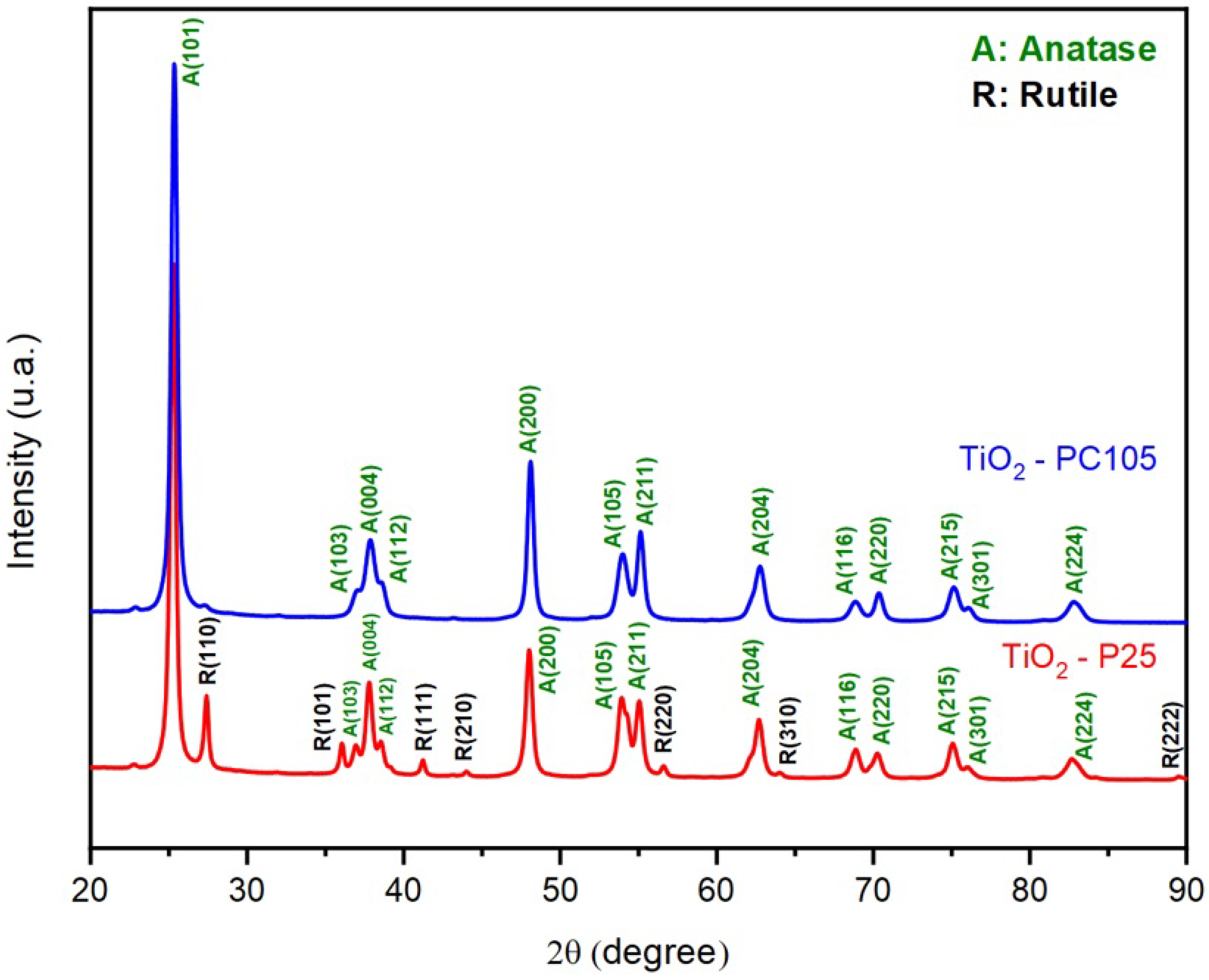
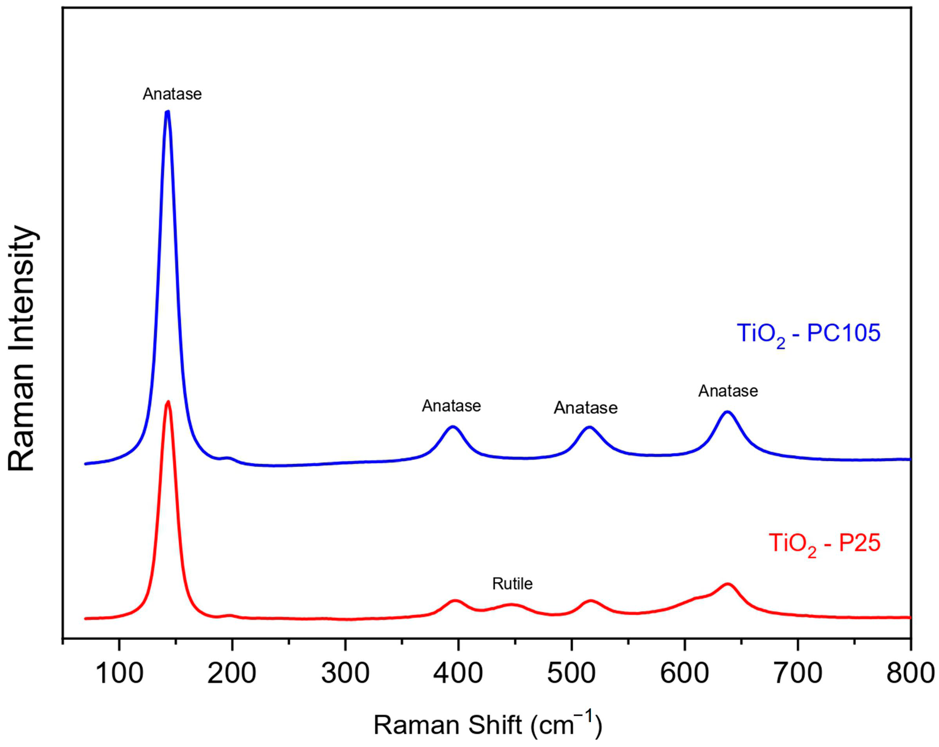
2.1. Characterization of TiO2 Powders
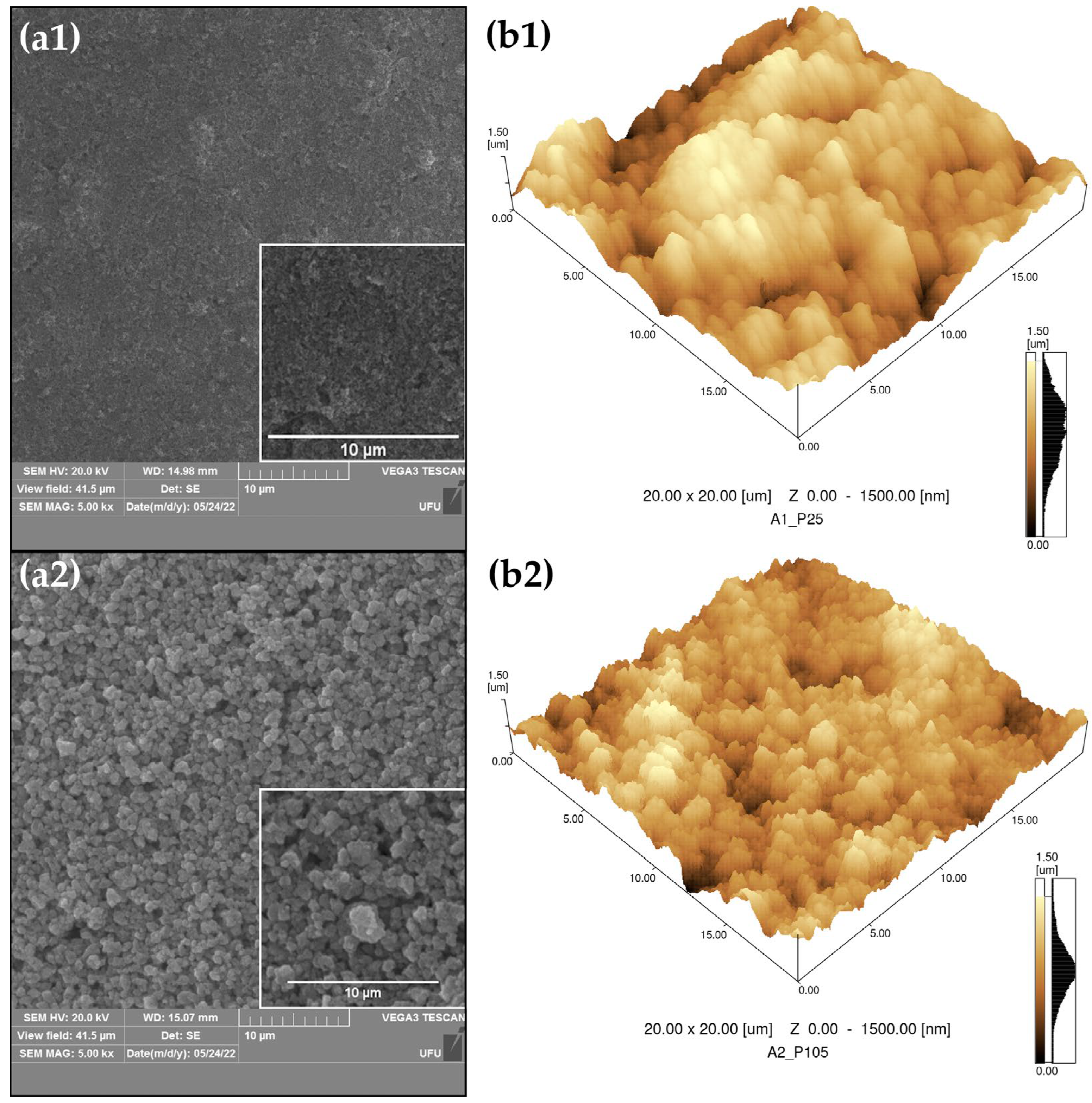
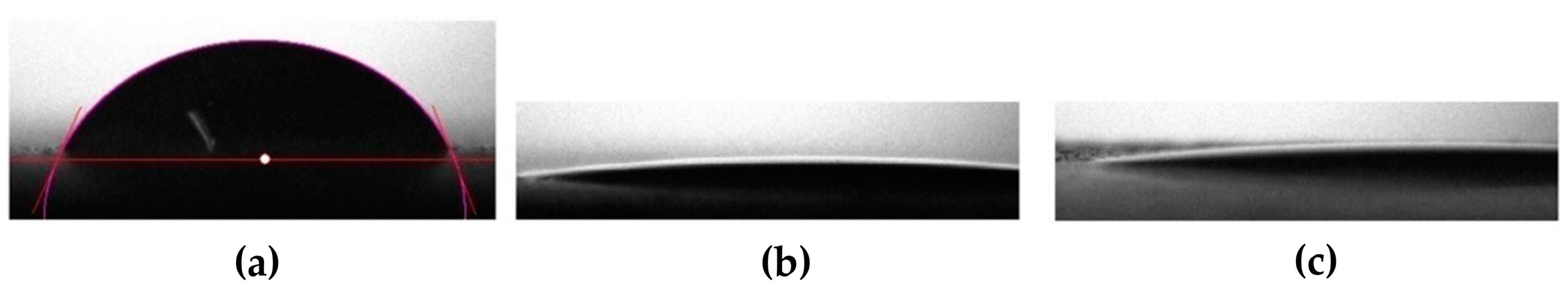
2.2. Characterization of TiO2 Films
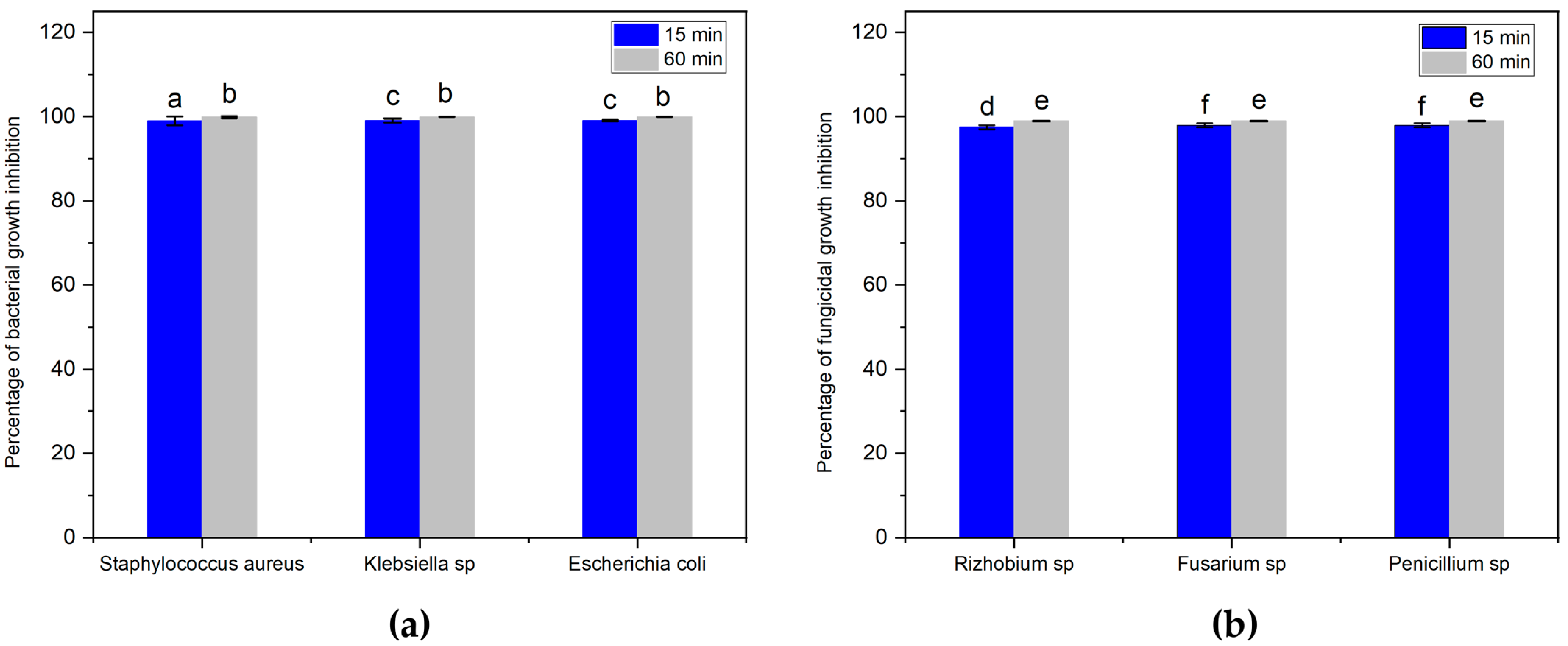
2.3. Antimicrobial Evaluation
3. Conclusions
4. Materials and Methods
4.1. Characterization of the Raw Material
4.2. The Production and Deposition of TiO2 Films
4.3. Characterization of TiO2 Films
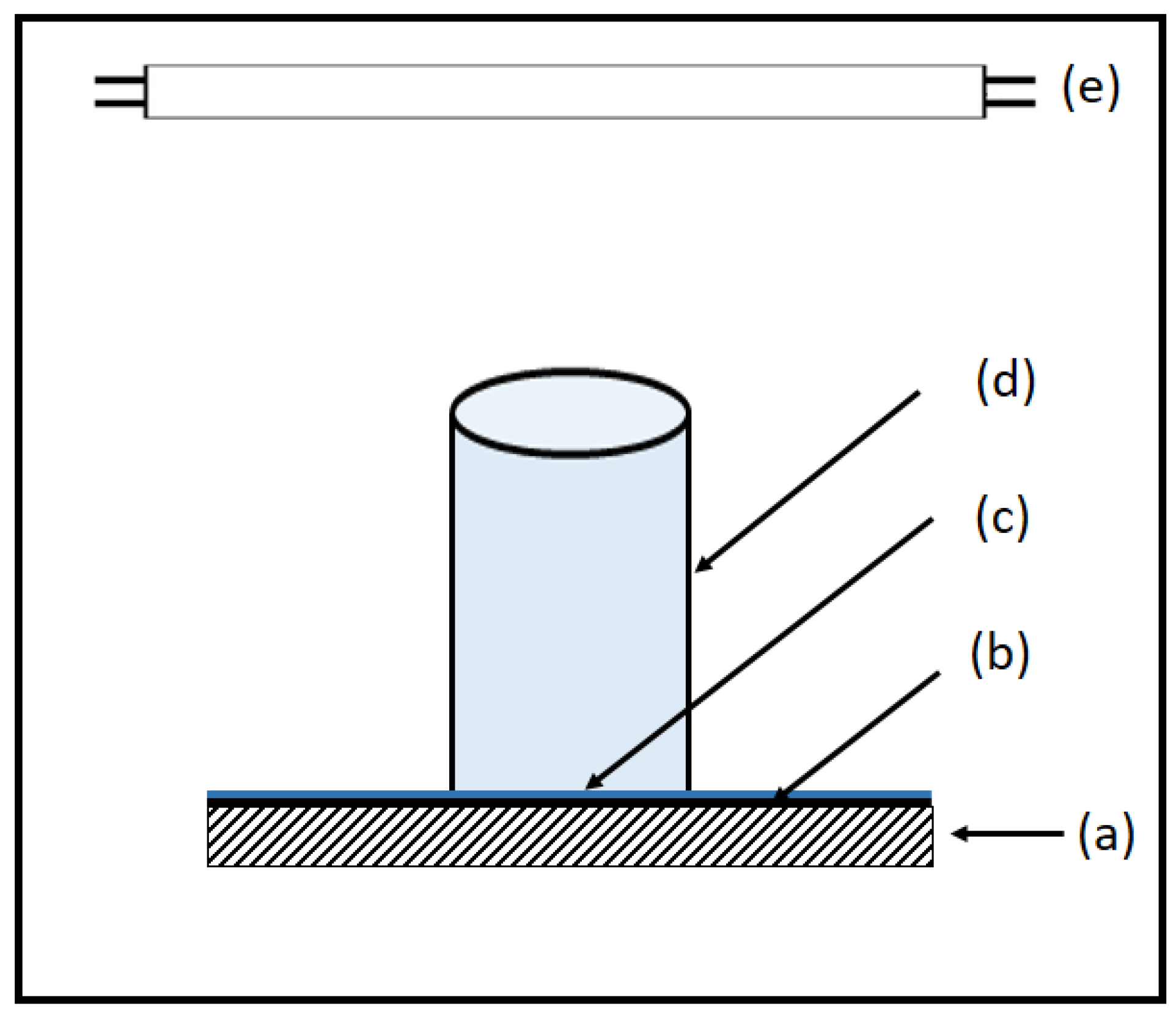
4.4. Photocatalytic Properties
4.5. Antimicrobial Tests
Author Contributions
Funding
Data Availability Statement
Acknowledgments
Conflicts of Interest
References
- Thomas, R.E.; Thomas, B.C.; Conly, J.; Lorenzetti, D. Cleaning and disinfecting surfaces in hospitals and long-term care facilities for reducing hospital- and facility-acquired bacterial and viral infections: A systematic review. J. Hosp. Infect. 2022, 122, 9–16. [Google Scholar] [CrossRef] [PubMed]
- Rudhart, S.A.; Gunther, F.; Dapper, L.; Stuck, B.A.; Hoch, S. UV-C Light-Based Surface Disinfection: Analysis of Its Virucidal Efficacy Using a Bacteriophage Model. Int. J. Environ. Res. Public Health 2022, 19, 3246. [Google Scholar] [CrossRef] [PubMed]
- Margarucci, L.M.; Spica, V.R.; Protano, C.; Gianfranceschi, G.; Giuliano, M.; Di Onofrio, V.; Mucci, N.; Valerianl, F.; Vitali, M.; Romano, F. Potential antimicrobial effects of photocatalytic nanothecnologies in hospital setting. Ann. Ig. Med. Prev. Comunita 2019, 31, 461–473. [Google Scholar] [CrossRef]
- Mitra, A.; Morfill, G.E.; Shimizu, T.; Steffes, B.; Isbary, G.; Schmidt, H.U.; Li, Y.F.; Zimmermann, J.L. Applications in plasma medicine: A SWOT approach. Compos. Interfaces 2012, 19, 231–239. [Google Scholar] [CrossRef]
- Soltan, D.G.; Das Neves, P.; Olvera, A.; Savastano Junior, H.; Li, V.C. Introducing a curauá fiber reinforced cement-based composite with strain-hardening behavior. Ind. Crops Prod. 2017, 103, 1–12. [Google Scholar] [CrossRef]
- Baroud, M.; Dandache, I.; Araj, G.F.; Wakim, R.; Kanj, S.; Kanafani, Z.; Khairallah, M.; Sabra, A.; Shehab, M.; Dbaibo, G.; et al. As underlying mechanisms of carbapenem resistance in extended-spectrum β-lactamase-producing Klebsiella pneumoniae and Escherichia coli isolates at a tertiary care centre in Lebanon: Role of OXA-48 and NDM-1 carbapenemases. Int. J. Antimicrob. Agents 2013, 41, 75–79. [Google Scholar] [CrossRef]
- Ozturk, I.; Tunçel, A.; Ince, M.; Ocakoglu, K.; Hoşgör-limoncu, M.; Yurt, F. Antibacterial properties of subphthalocyanine and subphthalocyanine-TiO2 nanoparticles on Staphylococcus aureus and Escherichia coli. J. Porphyr. Phthalocyanines 2018, 22, 1099–1105. [Google Scholar] [CrossRef]
- Jiang, X.; Lv, B.; Wang, Y.; Shen, Q.; Wang, X. Bactericidal mechanisms and effector targets of TiO2 and Ag-TiO2 against Staphylococcus aureus. J. Med. Microbiol. 2017, 66, 440–446. [Google Scholar] [CrossRef]
- Lockhart, S.R.; Guarner, J. Emerging and reemerging fungal infections. Semin. Diagn. Pathol. 2019, 36, 177–181. [Google Scholar] [CrossRef]
- Richardson, M.; Lass-flor, C. Changing epidemiology of systemic fungal infections. Clin. Microbiol. Infect. 2008, 14, 5–24. [Google Scholar] [CrossRef]
- Kirk, P.M.; Cannon, P.F.; Minter, D.W.; Stalpers, J.A. Ainsworth and Bisbys Dictionary of the Fungi, 10th ed.; CABI: Wallingford, UK, 2008. [Google Scholar] [CrossRef]
- Padmanabhan, N.T.; John, H. Titanium dioxide based self-cleaning smart surfaces: A short review. J. Environ. Chem. Eng. 2020, 8, 104211. [Google Scholar] [CrossRef]
- Fujishima, A.; Rao, T.N.; Tryk, D.A. Titanium dioxide photocatalysis. J. Photochem. Photobiol. C Photochem. Rev. 2000, 1, 1–21. [Google Scholar] [CrossRef]
- Reza, K.M.; Kurny, A.; Gulshan, F. Parameters affecting the photocatalytic degradation of dyes using TiO2: A review. Appl. Water Sci. 2017, 7, 1569–1578. [Google Scholar] [CrossRef]
- Colmenares, J.C.; Luque, R. Heterogeneous photocatalytic nanomaterials: Prospects and challenges in selective transformations of biomass-derived compounds. Chem. Soc. Rev. 2014, 43, 765–778. [Google Scholar] [CrossRef]
- Kotzias, D.; Binas, V.; Kiriakidis, G. Smart Surfaces: Heterogeneous Photo-Catalysis on TiO2 Based Coatings for De-pollution Purposes in Indoor and Outdoor Environments. Top. Catal. 2020, 63, 875–881. [Google Scholar] [CrossRef]
- Kumaravel, V.; Nair, K.M.; Mathew, S.; Bartlett, J.; Kennedy, J.E.; Manning, H.G.; Whelan, B.J.; Leyland, N.S.; Pillai, S.C. Antimicrobial TiO2 nanocomposite coatings for surfaces, dental and orthopaedic implants. Chem. Eng. J. 2021, 416, 129071. [Google Scholar] [CrossRef]
- Amézaga-Madrid, P.; Nevárez-Moorillón, G.V.; Orrantia-Borunda, E.; Miki-Yoshida, M. Photoinduced bactericidal activity against Pseudomonas aeruginosa by TiO2 based thin films. FEMS Microbiol. Lett. 2002, 211, 183–188. [Google Scholar] [CrossRef]
- Prakash, J.; Cho, J.; Mishra, Y.K. Photocatalytic TiO2 nanomaterials as potential antimicrobial and antiviral agents: Scope against blocking the SARS-COV-2 spread. Micro Nano Eng. 2022, 14, 100100. [Google Scholar] [CrossRef]
- Chung, C.-J.; Lin, H.-I.; Tsou, H.-K.; Shi, Z.-Y.; He, J.-L. An antimicrobial TiO2 coating for reducing hospital-acquired infection. J. Biomed. Mater. Res. Part B Appl. Biomater. 2008, 85, 220–224. [Google Scholar] [CrossRef]
- FU, G.; Vary, P.S.; Lin, C.T. Anatase TiO2 Nanocomposites for Antimicrobial Coatings. J. Phys. Chem. B 2005, 109, 8889–8898. [Google Scholar] [CrossRef]
- Lukong, V.T.; Mouchou, R.T.; Enebe, G.C.; Ukoba, K.; Jen, T.C. Deposition and characterization of self-cleaning TiO2 thin films for photovoltaic application. Mater. Today: Proc. 2022, 62, S63–S72. [Google Scholar] [CrossRef]
- Xi, R.; Wang, Y.; Wang, X.; Lv, J.; Li, X.; Li, T.; Zhang, X.; Du, X. Ultrafine nano-TiO2 loaded on dendritic porous silica nanoparticles for robust transparent antifogging self-cleaning nanocoatings. Ceram. Int. 2020, 46, 23651–23661. [Google Scholar] [CrossRef]
- Xi, B.; Verma, L.K.; Li, J.; Bhatia, C.S.; Danner, A.J.; Yang, H.; Zeng, H.C. TiO2 Thin Films Prepared Via Adsorptive Self-Assembly for Self-Cleaning Applications. ACS Appl. Mater. Interfaces 2012, 4, 1093–1102. [Google Scholar] [CrossRef]
- Wu, H. Nanometer TiO2 Film-Based Solar Thin Film Manufacturing Technology and Performance Research. Adv. Mater. Sci. Eng. 2022, 2022, 8328378. [Google Scholar] [CrossRef]
- Ducman, V.; Petrovic, V.; Skapin, S.D. Photo-catalytic efficiency of laboratory made and commercially available ceramic building products. Ceram. Int. 2013, 39, 2981–2987. [Google Scholar] [CrossRef]
- Zhang, P.; Tian, J.; Xu, R.; Ma, G. Hydrophilicity, photocatalytic activity and stability of tetraethyl orthosilicate modified TiO2 film on glazed ceramic surface. Appl. Surf. Sci. 2013, 266, 141–147. [Google Scholar] [CrossRef]
- Kim, M.-S.; Liu, G.; Cho, H.-K.; Kim, B.-W. Application of a hybrid system comprising carbon-doped TiO2 film and a ceramic media-packed biofilter for enhanced removal of gaseous styrene. J. Hazard. Mater. 2011, 190, 537–543. [Google Scholar] [CrossRef]
- Custódia, P.; Lied, E.B.; Da Silva, A.V.; Frare, L.M.; Bittencourt, P.R.S.; De Oliveira Basso, R.L.; De Oliveira Tavares, F.; Ferandin Honório, J.; Trevisan, A.P. TiO2 coated fiber cement composites: Effect of the load of TiO2 particles on photocatalytic degradation of H2S. Constr. Build. Mater. 2020, 262, 120379. [Google Scholar] [CrossRef]
- Wang, J.; Lu, C.H.; Xiong, J. Self-Cleaning and Depollution of Fiber Reinforced Cement Materials Modified by Neutral TiO2/SiO2 Hydrosol Photoactive Coatings. Appl. Surf. Sci. 2014, 298, 19–25. [Google Scholar] [CrossRef]
- Guo, X.; Rao, L.; Wang, P.; Wang, C.; Ao, Y.; Jiang, T.; Wang, W. Photocatalytic Properties of P25-Doped TiO2 Composite Film Synthesized via Sol–Gel Method on Cement Substrate. J. Environ. Sci. 2018, 66, 71–80. [Google Scholar] [CrossRef]
- Yuenyongsuwan, J.; O’Rear, E.A.; Pongprayoon, T. Admicellar Polymerization of a Co-Fluoropolymer Ultrathin Film on TiO2 Nanoparticles for Use as a Photocatalytic Cement. Surf. Interfaces 2021, 25, 101172. [Google Scholar] [CrossRef]
- Dantas, S.R.A.; Lima, F.J.N.; Romano, R.C.O.; Pileggi, R.; Loh, K. Evaluation of rheological properties of mortar with TiO2 addition. Ambiente Construído 2021, 21, 7–21. [Google Scholar] [CrossRef]
- Patterson, A.L. X-Ray Diffraction Procedures for Polycrystalline and Amorphous Materials. J. Am. Chem. Soc. 1955, 77, 2030–2031. [Google Scholar] [CrossRef]
- Neto, W.P.F.; Silvério, H.A.; Dantas, N.O.; Pasquini, D. Extraction and characterization of cellulose nanocrystals from agro-industrial residue—Soy hulls. Ind. Crops Prod. 2013, 42, 480–488. [Google Scholar] [CrossRef]
- Spurr, R.A.; Myers, H. Quantitative Analysis of Anatase-Rutile Mixtures with an X-Ray Diffractometer. Anal. Chem. 1957, 29, 760–762. [Google Scholar] [CrossRef]
- Brunauer, S.; Emmett, P.H.; Teller, E. Adsorption of Gases in Multimolecular Layers. J. Am. Chem. Soc. 1938, 60, 1938. [Google Scholar] [CrossRef]
- Folli, A.; Pochard, I.; Nonat, A.; Jakobsen, U.H.; Shepherd, A.M.; Macphee, D.E. Engineering Photocatalytic Cements: Understanding TiO2 Surface Chemistry to Control and Modulate Photocatalytic Performances. J. Am. Ceram. Soc. 2010, 93, 3360–3369. [Google Scholar] [CrossRef]
- Uddin, M.J.; Cesano, F.; Chowdhury, A.R.; Trad, T.; Cravanzola, S.; Martra, G.; Mino, L.; Zecchina, A.; Scarano, D. Surface Structure and Phase Composition of TiO2 P25 Particles After Thermal Treatments and HF Etching. Front. Mater. 2020, 7, 192. [Google Scholar] [CrossRef]
- Challagulla, S.; Tarafder, K.; Ganesan, R.; Roy, S. Structure sensitive photocatalytic reduction of nitroarenes over TiO2. Sci. Rep. 2017, 7, 8783. [Google Scholar] [CrossRef]
- Cho, H.-W.; Liao, K.-L.; Yang, J.-A.; Wu, J.-J. Revelation of rutile phase by Raman scattering for enhanced photoelectrochemical performance of hydrothermally-grown anatase TiO2 film. Appl. Surf. Sci. 2018, 440, 125–132. [Google Scholar] [CrossRef]
- Hardcastle, F.D. Raman Spectroscopy of Titania (TiO2) Nanotubular WaterSplitting Catalysts. J. Ark. Acad. Sci. 2011, 65, 9. [Google Scholar] [CrossRef]
- Aguilar, T.; Navas, J.; De Los Santos, D.M.; Sánchez-Coronilla, A.; Fernández-Lorenzo, C.; Alcantara, R.; Gallardo, J.J.; Blanco, G.; Martin-Calleja, J. TiO2 and pyrochlore Tm2Ti2O7 based semiconductor as a photoelectrode for dye-sensitized solar cells. J. Phys. D Appl. Phys. 2015, 48, 145102. [Google Scholar] [CrossRef]
- Tulli, F.; Morales, J.M.N.; Salas, E.E.; Vieyra, F.E.M.; Borsarelli, C.D. Photocatalytic Efficiency Tuning by the Surface Roughness of TiO2 Coatings on Glass Prepared by the Doctor Blade Method. Photochem. Photobiol. 2021, 97, 22–31. [Google Scholar] [CrossRef]
- Sarker, S.; Nath, N.C.; Rahman, M.; Lim, S.S.; Ahammad, A.J.; Choi, W.Y.; Lee, J.J. TiO2 Paste Formulation for Crack-Free Mesoporous Nanocrystalline Film of Dye-Sensitized Solar Cells. J. Nanosci. Nanotechnol. 2012, 12, 5361–5366. [Google Scholar] [CrossRef]
- Chen, Y.; Dionysiou, D.D. Bimodal mesoporous TiO2–P25 composite thick films with high photocatalytic activity and improved structural integrity. Appl. Catal. B Environ. 2008, 80, 147–155. [Google Scholar] [CrossRef]
- Patrocinio, A.O.T.; Paula, L.F.; Paniago, R.M.; Freitag, J.; Bahnemann, D.W. Layer-by-Layer TiO2/WO3 Thin Films as Efficient Photocatalytic Self-Cleaning Surfaces. ACS Appl. Mater. Interfaces 2014, 6, 16859–16866. [Google Scholar] [CrossRef] [PubMed]
- Kuroiwa, A.; Nomura, Y.; Ochiai, T.; Sudo, T.; Nomoto, R.; Hayakawa, T.; Kanzaki, H.; Nakamura, Y.; Hanada, N. Antibacterial, Hydrophilic Effect and Mechanical Properties of Orthodontic Resin Coated with UV-Responsive Photocatalyst. Materials 2018, 11, 889. [Google Scholar] [CrossRef]
- Borrás, A.; López, C.; Rico, V.; Gracia, F.; González-Elipe, A.R.; Richter, E.; Battiston, G.; Gerbasi, R.; McSporran, N.; Sauthier, G.; et al. Effect of Visible and UV Illumination on the Water Contact Angle of TiO2 Thin Films with Incorporated Nitrogen. J. Phys. Chem. C 2007, 111, 1801–1808. [Google Scholar] [CrossRef]
- Dulian, P.; Nachit, W.; Jaglarz, J.; Zięba, P.; Kanak, J.; Żukowski, W. Photocatalytic methylene blue degradation on multilayer transparent TiO2 coatings. Opt. Mater. 2019, 90, 264–272. [Google Scholar] [CrossRef]
- Lin, Y.; Qian, Q.; Chen, Z.; Dinh Tuan, P.; Feng, D. Fabrication of high specific surface area TiO2 nanopowders by anodization of porous titanium. Electrochem. Commun. 2022, 136, 107234. [Google Scholar] [CrossRef]
- Supphasrirongjaroen, P.; Praserthdam, P.; Panpranot, J.; Na-Ranong, D.; Mekasuwandumrong, O. Effect of quenching medium on photocatalytic activity of nano-TiO2 prepared by solvothermal method. Chem. Eng. J. 2008, 138, 622–627. [Google Scholar] [CrossRef]
- Liao, D.L.; Liao, B.Q. Shape, size and photocatalytic activity control of TiO2 nanoparticles with surfactants. J. Photochem. Photobiol. A Chem. 2007, 187, 363–369. [Google Scholar] [CrossRef]
- Hou, H.; Shang, M.; Wang, L.; Li, W.; Tang, B.; Yang, W. Efficient Photocatalytic Activities of TiO2 Hollow Fibers with Mixed Phases and Mesoporous Walls. Sci. Rep. 2015, 5, 15228. [Google Scholar] [CrossRef] [PubMed]
- Zhang, S.; Yang, D.; Jing, D.; Liu, H.; Liu, L.; Jia, Y.; Gao, M.; Guo, L.; Huo, Z. Enhanced photodynamic therapy of mixed phase TiO2(B)/anatase nanofibers for killing of HeLa cells. Nano Res. 2014, 7, 1659–1669. [Google Scholar] [CrossRef]
- Huang, D.; Liu, H.; Bian, J.; Li, T.; Huang, B.; Niu, Q. High Specific Surface Area TiO2 Nanospheres for Hydrogen Production and Photocatalytic Activity. J. Nanosci. Nanotechnol. 2020, 20, 3217–3224. [Google Scholar] [CrossRef] [PubMed]
- LI, D.; ZHANG, W. Influence of PECVD-TiO2 film morphology and topographyon the spectroscopic ellipsometry data fitting process. Mod. Phys. Lett. B 2020, 34, 2050228. [Google Scholar] [CrossRef]
- Maeng, W.Y.; Yoon, J.H.; Kim, D.J. Effect of process conditions (withdrawal rate and coating repetition) on morphological characteristics of sol–gel TiO2 film during dip coating. J. Coat. Technol. Res. 2020, 17, 1171–1193. [Google Scholar] [CrossRef]
- Xiao-Hua, L.; Shu-Qin, Y.; Yu, Z.; Zhi-An, W.; Ning-Kang, H. Effects of Different Dispersion Methods on the Microscopical Morphology of TiO2 Film. Chin. Phys. Lett. 2007, 24, 3567–3569. [Google Scholar] [CrossRef]
- Meek, S.J.; Pitman, C.L.; Miller, A.J.M. Deducing Reaction Mechanism: A Guide for Students, Researchers, and Instructors. J. Chem. Educ. 2016, 93, 275–286. [Google Scholar] [CrossRef]
- Gaya, U.I. Heterogeneous Photocatalysis Using Inorganic Semiconductor Solids; Springer Science & Business Media: Berlin/Heidelberg, Germany, 2014. [Google Scholar] [CrossRef]
- Bogdan, J.; Zarzyńska, J.; Pławińska-Czarnak, J. Comparison of Infectious Agents Susceptibility to Photocatalytic Effects of Nanosized Titanium and Zinc Oxides: A Practical Approach. Nanoscale Res. Lett. 2015, 10, 1–15. [Google Scholar] [CrossRef]
- Kim, B.; Kim, D.; Cho, D.; Cho, S. Bactericidal effect of TiO2 photocatalyst on selected food-borne pathogenic bactéria. Chemosphere 2003, 52, 277–281. [Google Scholar] [CrossRef] [PubMed]
- Jacoby, W.A.; Maness, P.C.; Wolfrum, E.J.; Blake, D.M.; Fennell, J.A. Mineralization of bacterial cell mass on a photocatalytic surface in air. Environ. Sci. Technol 1998, 32, 2650–2653. [Google Scholar] [CrossRef]
- Sunada, K.; Kikuchi, Y.; Hashimoto, K.; Fujishima, A. Bactericidal and detoxification effects of TiO2 thin film photocatalysts. Environ. Sci. Technol 1998, 32, 726–728. [Google Scholar] [CrossRef]
- Liu, J.-W.; Chang, H.-H. Bactericidal Effects and Mechanisms of Visible Light-Responsive Titanium Dioxide Photocatalysts on Pathogenic Bacteria. Arch. Immunol. Ther. Exp. 2012, 60, 267–275. [Google Scholar] [CrossRef]
- Yu, J.C.; Ho, W.; Yu, J.; Yip, H.; Wong, P.K.; Zhao, J. Efficient Visible-Light-Induced Photocatalytic Disinfection on Sulfur-Doped Nanocrystalline Titania. Environ. Sci. Technol. 2005, 39, 1175–1179. [Google Scholar] [CrossRef]
- Eezzeldin, H.M.; Badi, S.; Youssef, B.A. The Antibiotic Resistance and Multidrug Resistance Pattern of Uropathogenic Escherichia coli at Soba University Hospital: A Descriptive Retrospective Survey. Sudan J. Med. Sci. 2022, 17, 56–69. [Google Scholar] [CrossRef]
- Taba, A.; Laupland, K.B. Update on Staphylococcus aureus bacteraemia. Curr. Opin. Crit. Care 2022, 28, 495–504. [Google Scholar] [CrossRef]
- Meyer, G.; Picoli, S.U. Fenótipos de betalactamases em Klebsiella pneumoniae de hospital de emergência de Porto Alegre. J. Bras. Patol. Med. Lab. 2011, 47, 24–31. [Google Scholar] [CrossRef]
- Podgórska-Kryszczuk, I.; Solarska, E.; Kordowska-Wiater, M. Reduction of the Fusarium Mycotoxins: Deoxynivalenol, Nivalenol and Zearalenone by Selected Non-Conventional Yeast Strains in Wheat Grains and Bread. Molecules 2022, 27, 1578. [Google Scholar] [CrossRef]
- Long, H.; Pu, L.; Xu, W.; Nan, M.; Oyom, W.; Prusky, D.; Bi, Y.; Xue, H. Inactivation of Penicillium expansum spores in apple juice by contact glow discharge electrolysis and its related mechanism. Innov. Food Sci. Emerg. Technol. 2022, 80, 103100. [Google Scholar] [CrossRef]
- Golshan, V.; Mirjalili, F.; Fakharpour, M. Self-Cleaning Surfaces with Superhydrophobicity of Ag–TiO2 Nanofilms on the Floor Ceramic Tiles. Glass Phys. Chem. 2022, 48, 35–42. [Google Scholar] [CrossRef]
- Kisand, V.; Visnapuu, M.; Rosenberg, M.; Danilian, D.; Vlassov, S.; Kook, M.; Lange, S.; Pärna, R.; Ivask, A. Antimicrobial Activity of Commercial Photocatalytic SaniTise™ Window Glass. Catalysts 2022, 12, 197. [Google Scholar] [CrossRef]
- Hegyi, A.; Grebenisan, E.; Lazarescu, A.-V.; Stoian, V.; Szilagyi, H. Influence of TiO2 Nanoparticles on the Resistance of Cementitious Composite Materials to the Action of Fungal Species. Materials 2021, 14, 4442. [Google Scholar] [CrossRef]
- Foster, H.A.; Sheel, D.W.; Sheel, P.; Evans, P.; Varghese, S.; Rutschke, N.; Yates, H.M. Antimicrobial activity of titania/silver and titania/copper films prepared by CVD. J. Photochem. Photobiol. A Chem. 2010, 216, 283–289. [Google Scholar] [CrossRef]
- Ditta, I.B.; Steele, A.; Liptrot, C.; Tobin, J.; Tyler, H.; Yates, H.M.; Sheel, D.W.; Foster, H.A. Photocatalytic antimicrobial activity of thin surface films of TiO2, CuO and TiO2/CuO dual layers on Escherichia coli and bacteriophage T4. Appl. Microbiol. Biotechnol. 2008, 79, 127–133. [Google Scholar] [CrossRef]
- Evans, P.; Sheel, D.W. Photoactive and antibacterial TiO2 thin films on stainless steel. Surf. Coat. Technol. 2007, 201, 9319–9324. [Google Scholar] [CrossRef]
- ISO 10678:2010; Fine Ceramics (Advanced Ceramics, Advanced Technical Ceramics): Determination of Photocatalytic Activity of Surfaces in an Aqueous Medium by Degradation of Methylene Blue. International Organization for Standardization ISO: Geneve, Switzerland, 2010. Available online: https://www.iso.org/standard/46019.html (accessed on 15 March 2023).


| Material | Specific Surface Area –(m2/g) | Average Crystallite Diameter–(nm) | Percentage of Phases–(%) | ||
|---|---|---|---|---|---|
| Anatase | Rutile | Anatase | Rutile | ||
| TiO2-P25 | 49.32 | 23.0 | 41.0 | 80.0 | 20.0 |
| TiO2-PC105 | 78.56 | 18.0 | - | 100.0 | - |
| Substrate | Contact Angle | Reference | |
|---|---|---|---|
| Before UV-A Irradiation | After UV-A Irradiation | ||
| Plate with acrylic paint | 65 ± 3 | 65 ± 3 | -- |
| TiO2-P25 | 9 ± 1 | <6 | -- |
| TiO2-PC105 | 9 ± 1 | <6 | -- |
| Orthodontic Resin coated with TiO2 | 80 ± 5 | 34 ± 1 | [48] |
| Self-assembled TiO2 thin films on FTO glass | 12 ± 2 | <5 | [47] |
| TiO2 thin films on FTO | 70 ± 5 | <10 | [49] |
| Parameter | Film | |
|---|---|---|
| TiO2-P25 | TiO2-PC105 | |
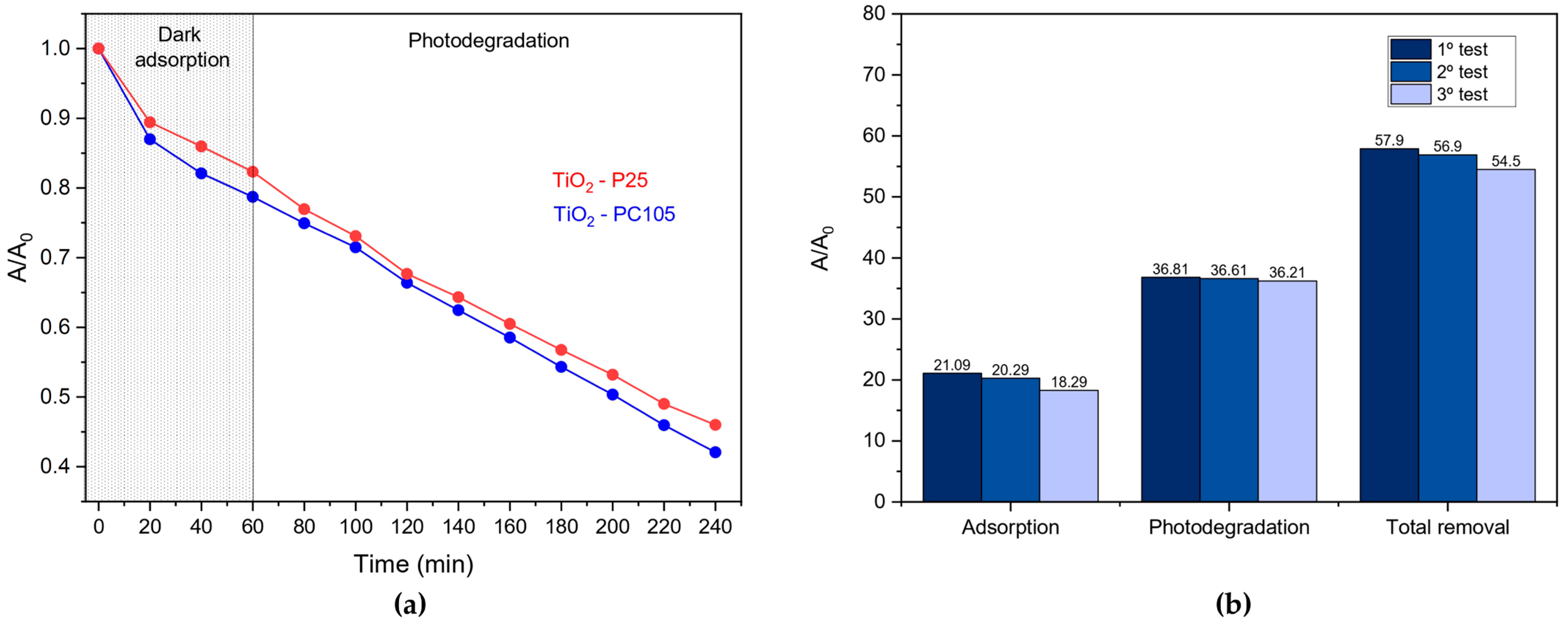
| Velocity constant–kobs (min−1) | 3.21 × 10⁻³ | 3.48 × 10⁻³ |
| Degradation rate–v (mol/s) | 2.14 × 10⁻¹¹ | 2.32 × 10⁻¹¹ |
| Source Intensity–I0 (Einstein/s) | 2.45 × 10⁻⁸ | 2.45 × 10⁻⁸ |
| Adsorption in the dark–(%) | 17.7 ± 0.5 | 21.3 ± 0.5 |
| Photodegradation after 3 h irradiation–(%) | 36.3 ± 0.5 | 36.6 ± 0.5 |
| Total removal–(%) | 54.0 ± 0.5 | 57.9 ± 0.5 |
| Photonic efficiency–ξ (%) | 0.08 | 0.1 |
| References | Light Source | Substrate | Microorganisms | Main Results |
|---|---|---|---|---|
| [73] | Visible light | Floor ceramic tiles | Escherichia coli, Staphylococcus aureus, | S. aureus ATCC 6538 and E. coli ATCC 259 were reduced by 99 and 95%, respectively |
| [74] | UV-A/Dark | Glass | Escherichia coli, Staphylococcus aureus | >2.8 log decrease in E. coli and >2.5 log decrease in S. aureus viable cell counts after 4 h |
| [75] | UV-A | Cementitious composite | Penicillium notatum, Aspergillus niger | 50.4% Penicillium notatum inhibition after 3 days of exposure |
| [76] | UV-A/Dark | Glass | Escherichia coli, Staphylococcus aureus, Pseudomonas aeruginosa | Near 100% killing of Pseudomonas aeruginosa over 24 h of irradiation |
| [77] | UV-A | Glass | Escherichia coli, Bacteriophage T4 | Near 100% killing of bacteriophage T4 after 2 h of irradiation |
| [20] | -- | Stainless steel | Escherichia coli, Staphylococcus aureus, | >5log reduction of Staphylococcus aureus after 40 h of exposure |
| [78] | UV-A | Stainless steel | Escherichia coli | Near 100% killing of E. Coli in less than 3 h |
Disclaimer/Publisher’s Note: The statements, opinions and data contained in all publications are solely those of the individual author(s) and contributor(s) and not of MDPI and/or the editor(s). MDPI and/or the editor(s) disclaim responsibility for any injury to people or property resulting from any ideas, methods, instructions or products referred to in the content. |
© 2023 by the authors. Licensee MDPI, Basel, Switzerland. This article is an open access article distributed under the terms and conditions of the Creative Commons Attribution (CC BY) license (https://creativecommons.org/licenses/by/4.0/).
Share and Cite
Rosa, R.H.; Silva, R.S.; Nascimento, L.L.; Okura, M.H.; Patrocinio, A.O.T.; Rossignolo, J.A. Photocatalytic and Antimicrobial Activity of TiO2 Films Deposited on Fiber-Cement Surfaces. Catalysts 2023, 13, 861. https://doi.org/10.3390/catal13050861
Rosa RH, Silva RS, Nascimento LL, Okura MH, Patrocinio AOT, Rossignolo JA. Photocatalytic and Antimicrobial Activity of TiO2 Films Deposited on Fiber-Cement Surfaces. Catalysts. 2023; 13(5):861. https://doi.org/10.3390/catal13050861
Chicago/Turabian StyleRosa, Robson H., Ricardo S. Silva, Lucas L. Nascimento, Monica H. Okura, Antonio Otavio T. Patrocinio, and João A. Rossignolo. 2023. "Photocatalytic and Antimicrobial Activity of TiO2 Films Deposited on Fiber-Cement Surfaces" Catalysts 13, no. 5: 861. https://doi.org/10.3390/catal13050861