
Essential Oil Analysis and Antimicrobial Evaluation of Three Aromatic Plant Species Growing in Saudi Arabia


 , , ,
, , ,  and
and
Abstract
:1. Introduction
2. Results and Discussion
2.1. Essential Oil Compositions
2.2. Antimicrobial Activity of the Essential Oils
2.3. Antimicrobial Activity of L. Pubescens and Carvacrol
3. Materials and Methods
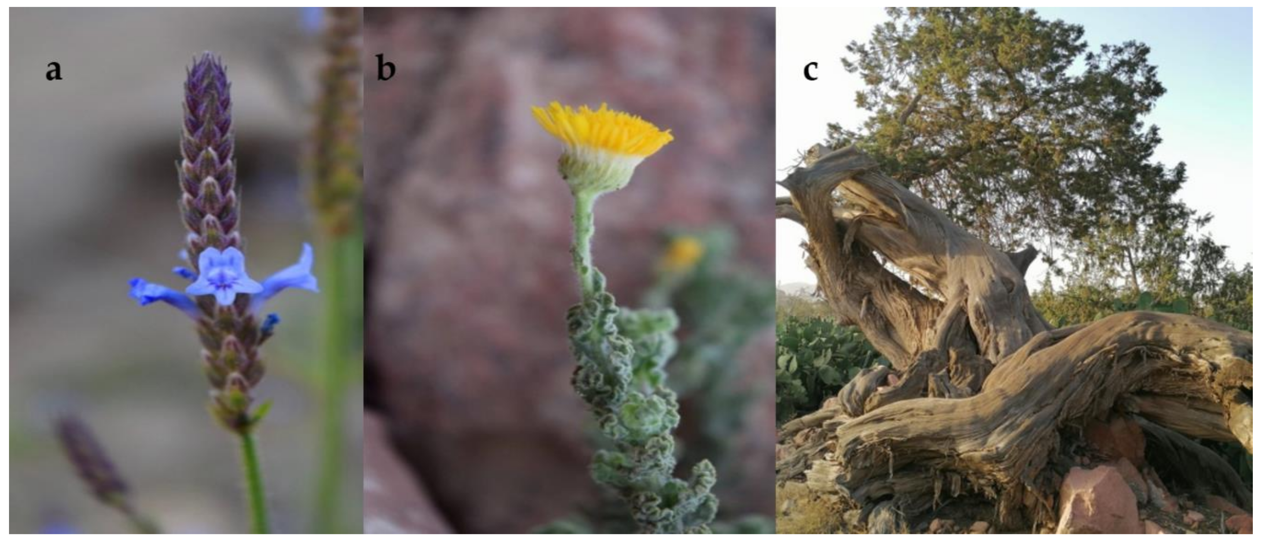
3.1. Plant Material
3.2. Chemicals and Reagents
3.3. Essential Oil Extraction
3.4. Gas Chromatography-Mass Spectrometry Analyses and Peak Identification
3.5. Diffusion Assay on Agar Plates
3.6. Microdilution of Broth Assay
3.7. Determination of the Minimum Inhibitory Concentration (MIC) and Minimum Bactericidal Concentration (MBC)
4. Conclusions
Author Contributions
Funding
Institutional Review Board Statement
Informed Consent Statement
Data Availability Statement
Conflicts of Interest
Sample Availability
References
- Islam, W.; Qasim, M.; Noman, A.; Tayyab, M.; Chen, S.; Wang, L. Management of Tobacco Mosaic Virus through Natural Metabolites. Rec. Nat. Prod. 2018, 12, 403–415. [Google Scholar] [CrossRef]
- Jiang, Z.; Jiang, H.; Xie, P. Antifungal activities against Sclerotinia sclerotiorum by Cinnamomum cassia oil and its main components. J. Essent. Oil Res. 2013, 25, 444–451. [Google Scholar] [CrossRef]
- Della Pepa, T.; Elshafie, H.S.; Capasso, R.; De Feo, V.; Camele, I.; Nazzaro, F.; Scognamiglio, M.R.; Caputo, L. Antimicrobial and Phytotoxic Activity of Origanum heracleoticum and O. majorana Essential Oils Growing in Cilento (Southern Italy). Molecules 2019, 24, 2576. [Google Scholar] [CrossRef] [Green Version]
- Yan, G.; Zhu, B.-R.; Tian, F.-L.; Hui, X.; Li, H.; Li, Y.-M.; Gao, W.-Y. Inhibitory Activity of Plant Essential Oils against E. coli 1-Deoxy-d-xylulose-5-phosphate reductoisomerase. Molecules 2019, 24, 2518. [Google Scholar] [CrossRef] [PubMed] [Green Version]
- Soliman, S.; Alsaadi, A.; Youssef, E.; Khitrov, G.; Noreddin, A.; Husseiny, M.; Ibrahim, A. Calli Essential Oils Synergize with Lawsone against Multidrug Resistant Pathogens. Molecules 2017, 22, 2223. [Google Scholar] [CrossRef] [Green Version]
- Oliva, A.; Costantini, S.; De Angelis, M.; Garzoli, S.; Božović, M.; Mascellino, M.; Vullo, V.; Ragno, R. High Potency of Melaleuca alternifolia Essential Oil against Multi-Drug Resistant Gram-Negative Bacteria and Methicillin-Resistant Staphylococcus aureus. Molecules 2018, 23, 2584. [Google Scholar] [CrossRef] [Green Version]
- Ferri, M.; Ranucci, E.; Romagnoli, P.; Giaccone, V. Antimicrobial resistance: A global emerging threat to public health systems. Crit. Rev. Food Sci. Nutr. 2017, 57, 2857–2876. [Google Scholar] [CrossRef]
- Obaidat, R.M.; Bader, A.; Al-Rajab, W.; Abu Sheikha, G.; Obaidat, A.A. Preparation of Mucoadhesive Oral Patches Containing Tetracycline Hydrochloride and Carvacrol for Treatment of Local Mouth Bacterial Infections and Candidiasis. Sci. Pharm. 2011, 79, 197–212. [Google Scholar] [CrossRef] [Green Version]
- Omran, Z.; Bader, A.; Porta, A.; Vandamme, T.; Anton, N.; Alehaideb, Z.; El-Said, H.; Faidah, H.; Essa, A.; Vassallo, A.; et al. Evaluation of Antimicrobial Activity of Triphala Constituents and Nanoformulation. Evid.-Based Complement. Altern. Med. 2020, 2020, 6976973. [Google Scholar] [CrossRef] [PubMed]
- Gemeda, N.; Tadele, A.; Lemma, H.; Girma, B.; Addis, G.; Tesfaye, B.; Abebe, A.; Gemechu, W.; Yirsaw, K.; Teka, F.; et al. Development, Characterization, and Evaluation of Novel Broad-Spectrum Antimicrobial Topical Formulations from Cymbopogon martini (Roxb.) W. Watson Essential Oil. Evid.-Based Complement. Altern. Med. 2018, 2018, 9812093. [Google Scholar] [CrossRef] [Green Version]
- Komeh-Nkrumah, S.A.; Nanjundaiah, S.M.; Rajaiah, R.; Yu, H.; Moudgil, K.D. Topical Dermal Application of Essential Oils Attenuates the Severity of Adjuvant Arthritis in Lewis Rats. Phyther. Res. 2012, 26, 54–59. [Google Scholar] [CrossRef]
- Amin, B.; Hosseinzadeh, H. Black Cumin (Nigella sativa) and Its Active Constituent, Thymoquinone: An Overview on the Analgesic and Anti-inflammatory Effects. Planta Med. 2015, 82, 8–16. [Google Scholar] [CrossRef] [Green Version]
- Ali, B.H.; Blunden, G. Pharmacological and toxicological properties of Nigella sativa. Phyther. Res. 2003, 17, 299–305. [Google Scholar] [CrossRef]
- Bedini, S.; Flamini, G.; Ascrizzi, R.; Venturi, F.; Ferroni, G.; Bader, A.; Girardi, J.; Conti, B. Essential oils sensory quality and their bioactivity against the mosquito Aedes albopictus. Sci. Rep. 2018, 8, 17857. [Google Scholar] [CrossRef] [PubMed] [Green Version]
- Abdalla, A.N.; Shaheen, U.; Abdallah, Q.M.A.; Flamini, G.; Bkhaitan, M.M.; Abdelhady, M.I.S.; Ascrizzi, R.; Bader, A. Proapoptotic Activity of Achillea membranacea Essential Oil and Its Major Constituent 1,8-Cineole against A2780 Ovarian Cancer Cells. Molecules 2020, 25, 1582. [Google Scholar] [CrossRef] [Green Version]
- Burits, M.; Bucar, F. Antioxidant activity of Nigella sativa essential oil. Phyther. Res. 2000, 14, 323–328. [Google Scholar] [CrossRef]
- Giuliani, A.; Pirri, G.; Nicoletto, S. Antimicrobial peptides: An overview of a promising class of therapeutics. Open Life Sci. 2007, 2, 1–33. [Google Scholar] [CrossRef]
- Nostro, A.; Cannatelli, M.A.; Morelli, I.; Cioni, P.L.; Bader, A.; Marino, A.; Alonzo, V. Preservative properties of Calamintha officinalis essential oil with and without EDTA. Lett. Appl. Microbiol. 2002, 35, 385–389. [Google Scholar] [CrossRef]
- Bader, A.; Caponi, C.; Cioni, P.L.; Flamini, G.; Morelli, I. Acorenone in the essential oil of flowering aerial parts of Seseli tortuosum L. Flavour Fragr. J. 2003, 18, 57–58. [Google Scholar] [CrossRef]
- Bader, A.; Cioni, P.L.; De Tommasi, N.; Flamini, G. Essential Oil Compositions of Two Populations of Salvia samuelssonii Growing in Different Biogeographical Regions of Jordan. Nat. Prod. Commun. 2014, 9. [Google Scholar] [CrossRef] [Green Version]
- Ascrizzi, R.; Flamini, G. Leek or Garlic? A Chemical Evaluation of Elephant Garlic Volatiles. Molecules 2020, 25, 2082. [Google Scholar] [CrossRef] [PubMed]
- Bader, A.; Flamini, G.; Cioni, P.L.; Morelli, I. The Composition of the Root Oil of Salvadora persica L. J. Essent. Oil Res. 2002, 14, 128–129. [Google Scholar] [CrossRef]
- Fico, G.; Bader, A.; Flamini, G.; Cioni, P.L.; Morelli, I. Essential Oil of Nigella damascena L. (Ranunculaceae) Seeds. J. Essent. Oil Res. 2003, 15, 57–58. [Google Scholar] [CrossRef]
- Naik, D.G.; Dandge, C.N.; Rupanar, S.V. Chemical Examination and Evaluation of Antioxidant and Antimicrobial Activities of Essential Oil from Gymnema sylvestre R. Br. Leaves. J. Essent. Oil Res. 2011, 23, 12–19. [Google Scholar] [CrossRef]
- Iannuzzi, A.M.; Camero, C.M.; D’Ambola, M.; D’Angelo, V.; Amira, S.; Bader, A.; Braca, A.; De Tommasi, N.; Germanò, M.P. Antiangiogenic Iridoids from Stachys ocymastrum and Premna resinosa. Planta Med. 2019, 85, 1034–1039. [Google Scholar] [CrossRef] [PubMed] [Green Version]
- Bader, A.; Abdallah, Q.; Abdelhady, M.; De Tommasi, N.; Malafronte, N.; Shaheen, U.; Bkhaitan, M.; Cotugno, R. Cytotoxicity of Some Plants of the Asteraceae Family: Antiproliferative Activity of Psiadia punctulata Root Sesquiterpenes. Rec. Nat. Prod. 2019, 13, 307–315. [Google Scholar] [CrossRef]
- Dal Piaz, F.; Bader, A.; Malafronte, N.; D’Ambola, M.; Petrone, A.M.; Porta, A.; Ben Hadda, T.; De Tommasi, N.; Bisio, A.; Severino, L. Phytochemistry of compounds isolated from the leaf-surface extract of Psiadia punctulata (DC.) Vatke growing in Saudi Arabia. Phytochemistry 2018, 155, 191–202. [Google Scholar] [CrossRef] [PubMed]
- Ali-Shtayeh, M.S.; Abu-Zaitoun, S.Y.; Dudai, N.; Jamous, R.M. Downy Lavender Oil: A Promising Source of Antimicrobial, Antiobesity, and Anti-Alzheimer’s Disease Agents. Evid.-Based Complement. Altern. Med. 2020, 2020, 5679408. [Google Scholar] [CrossRef] [PubMed] [Green Version]
- Al-Badani, R.N.; Da Silva, J.K.R.; Setzer, W.N.; Awadh Ali, N.A.; Muharam, B.A.; Al-Fahad, A.J.A. Variations in Essential Oil Compositions of Lavandula pubescens (Lamiaceae) Aerial Parts Growing Wild in Yemen. Chem. Biodivers. 2017, 14, e1600286. [Google Scholar] [CrossRef]
- Al-Badani, R.N.; Da Silva, J.K.R.; Mansi, I.; Muharam, B.A.; Setzer, W.N.; Awadh Ali, N.A. Chemical Composition and Biological Activity of Lavandula pubescens Essential Oil from Yemen. J. Essent. Oil Bear. Plants 2017, 20, 509–515. [Google Scholar] [CrossRef]
- Shehata, I.A. Essential oils of Lavandula species growing in Yemen. Bull. Fac. Pharm. (Cairo Univ.) 2001, 39, 233–238. [Google Scholar]
- Albalawi, M.A.D.; Bashir, N.A.O.; Tawfik, A. Anticancer and Antifolate Activities of Extracts of Six Saudi Arabian Wild Plants Used in Folk Medicine. J. Life Sci. 2015, 10. [Google Scholar]
- Hassan, S.A.; Al-Thobaiti, A.T.I. morphological nutlet characteristics of some lamiaceae taxa in Saudi Arabia and their taxonomic significance. Pak. J. Bot. 2015, 47, 1969–1977. [Google Scholar]
- Alghamdi, S.B.; Abdelshafeek, K.A. Evaluation of Antimicrobial and Cytotoxic Activities of Different Extracts of Lavandula pubescens growing in Albaha Region, KSA. Int. J. Biol. Pharm. Allied Sci. 2019, 8, 1338–1353. [Google Scholar]
- Shahat, E.A.; Bakr, R.O.; Eldahshan, O.A.; Ayoub, N.A. Chemical Composition and Biological Activities of the Essential Oil from Leaves and Flowers of Pulicaria incisa sub. candolleana (Family Asteraceae). Chem. Biodivers. 2017, 14, e1600156. [Google Scholar] [CrossRef]
- El-Shahaby, O.; El-Zayat, M.; Rabei, R.; Aldesuquy, H.S. Phytochemical constituents, antioxidant activity and antimicrobial potential of Pulicaria incisa (lam.) DC as a folk medicinal plant. Prog. Chem. Biochem. Res. 2019, 2, 22–227. [Google Scholar]
- Leangapichart, T.; Gautret, P.; Griffiths, K.; Belhouchat, K.; Memish, Z.; Raoult, D.; Rolain, J.-M. Acquisition of a High Diversity of Bacteria during the Hajj Pilgrimage, Including Acinetobacter baumannii with bla OXA-72 and Escherichia coli with bla NDM-5 Carbapenemase Genes. Antimicrob. Agents Chemother. 2016, 60, 5942–5948. [Google Scholar] [CrossRef] [Green Version]
- Al Johani, S.M.; Akhter, J.; Balkhy, H.; El-Saed, A.; Younan, M.; Memish, Z. Prevalence of antimicrobial resistance among gram-negative isolates in an adult intensive care unit at a tertiary care center in Saudi Arabia. Ann. Saudi Med. 2010, 30, 364–369. [Google Scholar] [CrossRef] [PubMed] [Green Version]
- Aprotosoaie, A.C.; Gille, E.; Trifan, A.; Luca, V.S.; Miron, A. Essential oils of Lavandula genus: A systematic review of their chemistry. Phytochem. Rev. 2017, 16, 761–799. [Google Scholar] [CrossRef]
- Almadiy, A.A. Chemical composition, insecticidal and biochemical effects of two plant oils and their major fractions against Aedes aegypti, the common vector of dengue fever. Heliyon 2020, 6, e04915. [Google Scholar] [CrossRef]
- Adams, R.P. Juniperus procera of East Africa: Volatile leaf oil composition and putative relationship to J. excelsa. Biochem. Syst. Ecol. 1990, 18, 207–210. [Google Scholar] [CrossRef]
- Adams, R.P. Systematics of multi-seeded eastern hemisphere Juniperus based on leaf essential oils and RAPD DNA fingerprinting. Biochem. Syst. Ecol. 1999, 27, 709–725. [Google Scholar] [CrossRef]
- Burits, M.; Asres, K.; Bucar, F. The antioxidant activity of the essential oils of Artemisia afra, Artemisia abyssinica and Juniperus procera. Phyther. Res. 2001, 15, 103–108. [Google Scholar] [CrossRef]
- Adams, R.P. Identification of Essential oil Components by Gas Chromatography/Quadrupole Mass Spectroscopy; Allured Publishing Corporation: Carol Stream, IL, USA, 1995; ISBN 1932633219. [Google Scholar]
- Linstrom, P.J.; Mallard, W.J. NIST Chemistry WebBook; NIST Standard Reference Database Number 69; National Institute of Standards and Technology: Gaithersburg MD, USA. [CrossRef]
- Haseeb, A.; Faidah, H.S.; Bakhsh, A.R.; Al Malki, W.H.; Elrggal, M.E.; Saleem, F.; ur Rahman, S.; Khan, T.M.; Hassali, M.A. Antimicrobial resistance among pilgrims: A retrospective study from two hospitals in Makkah, Saudi Arabia. Int. J. Infect. Dis. 2016, 47, 92–94. [Google Scholar] [CrossRef] [PubMed] [Green Version]
- Al-Tawfiq, J.A.; Memish, Z.A. Potential risk for drug resistance globalization at the Hajj. Clin. Microbiol. Infect. 2015, 21, 109–114. [Google Scholar] [CrossRef] [Green Version]
- Samadi, N.; Zaree, R.; Bakhtiar, H.; Salehnia, A.; Azimi, S. Comparative Antibacterial Efficacy of Endemic Satureja Khuzistanica Jamzad Essential Oil, Sodium Hypochlorite and Chlorhexidine Gluconate Solutions as Root Canal Irrigations. Dent. Res. J. (Isfahan). 2011, 8, 28–32. [Google Scholar]
- Miladi, H.; Zmantar, T.; Kouidhi, B.; Chaabouni, Y.; Mahdouani, K.; Bakhrouf, A.; Chaieb, K. Use of carvacrol, thymol, and eugenol for biofilm eradication and resistance modifying susceptibility of Salmonella enterica serovar Typhimurium strains to nalidixic acid. Microb. Pathog. 2017, 104, 56–63. [Google Scholar] [CrossRef] [PubMed]
- National Institute of Standards and Technology (NIST). NIST/EPA/NIH Mass Spectral Library; The NIST Mass Spectrometry Data Center: Gaithersburg, MD, USA, 2014. [Google Scholar]
- Ansari, A.; Abbas, Z.; Saggu, S.; Rehman, H.; Moawed, M. Growth responses of Lavandula pubescens to temperature regimes of Tabuk, Saudi Arabia. J. Med. Plants Stud. 2014, 2, 38–41. [Google Scholar]
- El-Juhany, L.; Aref, I.; Al-Ghamdi, M. Effects of Different Pretreatments on Seed Germination and Early Establishment of the Seedlings of Juniperus procera Trees. World Appl. Sci. J. 2009, 7, 616–624. [Google Scholar]


| Constituents. | l.r.i. a | l.r.i. b | Relative Abundance (%) | |||||
|---|---|---|---|---|---|---|---|---|
| L. pubescens | P. incisa | J. procera | J. procera | J. procera | J. procera | |||
| Aerial Parts | Aerial Parts | Leaves | Stems | Unripe Fruits | Ripe Fruits | |||
| santolina triene | 910 | 908 | - c | - | - | - | 0.1 | - |
| tricyclene | 928 | 926 | - | - | - | 0.4 | 0.1 | - |
| α-thujene | 933 | 931 | - | 0.1 | - | 0.3 | - | - |
| α-pinene * | 941 | 939 | - | 1 | 33.9 | 62.5 | 31.4 | 31.3 |
| α-fenchene | 954 | 951 | - | - | 1.6 | 0.5 | 1.2 | 1.1 |
| camphene * | 955 | 953 | - | 0.2 | 0.6 | 0.7 | 0.3 | 0.5 |
| thuja-2,4 (10)-diene | 959 | 957 | - | - | - | 0.3 | - | - |
| sabinene * | 977 | 976 | - | - | - | 0.1 | 0.2 | - |
| β-pinene * | 982 | 980 | - | - | 4.6 | 3.4 | 3.6 | 3.3 |
| 2,3-dehydro-1,8-cineole | 992 | 991 | - | 2.3 | - | - | - | - |
| myrcene * | 993 | 991 | 3.5- | - | 3.7 | 3.3 | 4.1 | 4.2 |
| cis-dehydroxylinalool oxide | 1002 | 999 | - | 0.2 | - | - | - | - |
| α-phellandrene * | 1006 | 1005 | 0.2 | |||||
| δ-3-carene * | 1013 | 1011 | 0.3 | - | 30.3 | 7.3 | 26.8 | 25.8 |
| α-terpinene * | 1020 | 1018 | 0.2 | |||||
| p-cymene * | 1028 | 1027 | 0.3 | 0.5 | 0.5 | 0.4 | 0.2 | 0.2 |
| limonene * | 1032 | 1031 | 0.2 | 0.2 | 2.8 | 2.3 | 2.1 | 2.4 |
| (Z)-β-ocimene * | 1042 | 1040 | 4.1 | - | - | - | - | - |
| (E)-β-ocimene * | 1052 | 1050 | 0.4 | - | - | - | - | - |
| γ-terpinene * | 1063 | 1062 | - | 0.2 | - | - | 0.1 | 0.3 |
| cis-linalool oxide (furanoid) * | 1076 | 1074 | - | 1.6 | - | - | - | - |
| terpinolene * | 1089 | 1088 | 6.1 | - | 3.8 | 1.6 | 5.8 | 4.9 |
| trans-linalool oxide (furanoid) * | 1090 | 1088 | - | 1.0 | - | - | - | - |
| p-cymenene | 1091 | 1089 | - | - | - | 0.7 | - | - |
| linalool * | 1101 | 1098 | - | 33.0 | 0.3 | 0.4 | 0.1 | - |
| isopentyl-2-methylbutanoate * | 1102 | 1099 | - | 2.6 | - | - | - | - |
| α-cyclocitral | 1117 | 1116 | - | 0.4 | - | - | - | - |
| α-isophorone * | 1120 | 1118 | - | 0.3 | - | - | - | - |
| chrysanthenone | 1126 | 1123 | - | 10.3 | - | - | - | - |
| α-campholenal | 1127 | 1125 | - | - | - | 0.6 | - | - |
| trans-pinocarveol * | 1141 | 1139 | - | - | - | 1.1 | - | - |
| camphor * | 1145 | 1144 | - | - | - | - | - | 0.2 |
| trans-pinocamphone | 1162 | 1160 | - | - | - | 0.2 | - | - |
| cis-chrysanthenol | 1163 | 1162 | - | 8.0 | - | - | - | - |
| pinocarvone | 1164 | 1162 | - | - | - | 0.4 | - | - |
| borneol * | 1168 | 1165 | - | - | - | 0.1 | - | - |
| 4-terpineol * | 1179 | 1177 | - | 0.9 | - | 0.3 | - | - |
| p-cymen-8-ol * | 1185 | 1183 | 0.7 | - | - | 0.2 | - | - |
| α-terpineol * | 1191 | 1190 | 0.1 | 0.7 | 0.5 | 1.2 | 0.3 | 0.2 |
| myrtenal * | 1194 | 1193 | - | - | - | 0.5 | - | - |
| myrtenol * | 1195 | 1194 | - | - | - | 0.3 | - | - |
| verbenone * | 1207 | 1204 | - | 0.3 | - | 0.2 | - | - |
| 8,9-dehydrothymol | 1221 | 1221 | - | 0.2 | - | - | - | - |
| methylcarvacrol * | 1244 | 1244 | 13.4 | - | - | - | - | - |
| cis-chrysanthenyl acetate | 1264 | 1262 | - | 1.3 | - | - | - | - |
| isopiperitenone | 1271 | 1272 | - | 1.8 | - | - | - | - |
| bornyl acetate * | 1287 | 1285 | - | - | 0.5 | 0.7 | 0.7 | 0.1 |
| p-menth-1-en-9-ol | 1294 | 1291 | 0.2 | - | - | - | - | - |
| carvacrol * | 1298 | 1298 | 55.7 | - | - | - | - | - |
| eugenol * | 1358 | 1356 | - | 8.9 | - | - | - | - |
| (E)-β-damascenone | 1382 | 1380 | - | 0.3 | - | - | - | - |
| β-bourbonene | 1385 | 1384 | - | - | 2.0 | - | - | - |
| β-elemene | 1392 | 1391 | - | - | 0.4 | - | 0.2 | 0.2 |
| (E)-jasmone | 1393 | 1390 | - | 0.4 | - | - | - | - |
| (Z)-jasmone * | 1395 | 1394 | - | 5.7 | - | - | - | - |
| β-caryophyllene * | 1419 | 1418 | 3.8 | 1.1 | 2.9 | 1.6 | 5.9 | 6.4 |
| dimethoxy-p-cymene | 1424 | 1423 | - | 0.2 | - | - | - | - |
| α-humulene * | 1455 | 1454 | 0.1 | - | 3.2 | 1.5 | 6.7 | 6.9 |
| (E)-β-farnesene | 1459 | 1458 | - | - | - | 0.5 | - | - |
| γ-muurolene | 1478 | 1477 | - | - | - | 0.4 | - | 0.1 |
| germacrene D | 1482 | 1480 | - | - | 1.6 | 0.2 | 6.3 | 5.8 |
| thymyl isobutyrate | 1490 | 1489 | - | 2.1 | - | - | - | - |
| neryl isobutyrate * | 1492 | 1491 | - | 0.3 | - | - | - | - |
| valencene * | 1493 | 1491 | - | - | - | 0.1 | - | - |
| viridiflorene | 1495 | 1493 | - | - | - | - | - | 0.1 |
| α-muurolene | 1499 | 1499 | - | - | - | - | - | 0.1 |
| germacrene A | 1505 | 1503 | - | - | - | - | - | - |
| α-bulnesene | 1507 | 1505 | - | - | - | - | 0.2 | 0.2 |
| β-bisabolene | 1508 | 1509 | 9.1 | 2 | - | 9.1 | - | - |
| trans-γ-cadinene | 1514 | 1513 | - | 0.7 | - | - | - | 0.2 |
| δ-cadinene | 1524 | 1524 | - | - | 0.2 | 0.4 | 0.3 | 0.4 |
| elemol | 1550 | 1549 | - | - | 3.0 | 0.1 | 1.2 | 1.3 |
| germacrene B | 1557 | 1556 | 0.1 | - | - | - | - | 0.3 |
| germacrene-D-4-ol | 1574 | 1574 | - | - | - | - | 0.1 | 0.2 |
| caryophyllene oxide * | 1582 | 1581 | 1.1 | 1.9 | 0.5 | 0.6 | - | 0.3 |
| cedrol * | 1601 | 1599 | - | - | - | 1.7 | - | - |
| humulene epoxide II | 1607 | 1606 | - | - | 0.4 | 0.5 | - | 0.3 |
| 10-epi-γ-eudesmol | 1619 | 1619 | - | - | - | - | - | 0.2 |
| γ-eudesmol | 1632 | 1630 | - | - | 0.5 | - | 0.1 | 0.3 |
| T-cadinol | 1641 | 1640 | - | 1.4 | - | - | 0.1 | 0.3 |
| β-eudesmol | 1650 | 1649 | - | 1.1 | - | - | 0.1 | 0.3 |
| α-cadinol | 1652 | 1653 | - | 0.3 | - | - | - | - |
| α-eudesmol | 1653 | 1652 | - | - | - | - | 0.4 | 1.2 |
| Monoterpene-hydrocarbons | 15.3 | 2.2 | 81.8 | 83.8 | 76.0 | 74.0 | ||
| Oxygenated-monoterpenes | 70.1 | 64.2 | 1.3 | 6.0 | 1.1 | 0.5 | ||
| Sesquiterpene-hydrocarbons | 13.1 | 3.8 | 10.3 | 4.7 | 19.6 | 20.7 | ||
| Oxygenated-sesquiterpenes | 1.2 | 4.7 | 4.4 | 2.9 | 2.0 | 4.4 | ||
| Phenylpropanoids | - | 8.9 | - | - | - | - | ||
| Apocarotenes | - | 1.0 | - | - | - | - | ||
| Non-terpene-derivatives | - | 8.7 | - | - | - | - | ||
| Total-identified (%) | 99.6 | 93.5 | 97.8 | 97.4 | 98.7 | 99.6 | ||
| Extraction yield (% w/w) | 1.09 | 1.14 | 0.51 | <0.1 | 2.7 | 2.39 | ||
| Microbial Strains | L. pubescens Aerial Parts EO | P. incisa Aerial Parts EO | J. procera Leaves EO |
|---|---|---|---|
| Enterococcusfaecalis ATCC * 51299 | 12 mm | R | R |
| Enterococcusfaecalis(VRE) ATCC 51299 | R | R | R |
| Staphylococcus aureus ATCC 25923 | R | R | R |
| Staphylococcus aureus (MRSA) ATCC 43300 | R | R | R |
| Staphylococcus epidermidis ATCC 12228 | 10 mm | R | R |
| Salmonella typhimurium ATCC 700720 | 13 mm | R | R |
| Klebsiella pneumonia (ESBL) ATCC 14028 | R | R | R |
| Klebsiella pneumonia (CRE) ATCC 1705 | R | R | R |
| Acinetobacter baumannii (CRE) ATCC 19605 | 15 mm | R | R |
| Shigella sonnei ATCC 25931 | 11 mm | R | R |
| Pseudomonas aeruginosa ATCC 15442 | R | R | R |
| Proteus mirabilis ATCC 3071 | R | R | R |
| Escherichia coli ATCC 35218 | R | R | R |
| Tested ATCC Strains | Diameter of Zone of Inhibition (mm) | ||||||
|---|---|---|---|---|---|---|---|
| Carvacrol 300 µg/well | L. pubescens EO 200 µg/well | L. pubescens EO 300 µg/well | Ciprofloxacin 30 µg/well | Amikacin 30 µg (Disc) | Vancomycin 30 µg (Disc) | DMSO | |
| Enterococcus faecalis ATCC 51299 | 12 ± 0.00 | 12 ± 0.00 | 14 ± 0.00 | - | - | 18.5 ± 0.71 | R |
| Staphylococcus epidermidis ATCC 12228 | 15 ± 0.00 | 10 ± 0.00 | 15 ± 0.00 | - | - | 28 ± 0.00 | R |
| Salmonella typhimurium ATCC 700720 | 20 ± 0.00 | 13 ± 0.00 | 19 ± 0.00 | 29 ± 0.00 | 20 ± 0.00 | - | R |
| Acinetobacter baumannii (CRE) ATCC 1605 | 15 ± 0.00 | 15 ± 0.00 | 24 ± 0.00 | 9 ± 0.00 | 15 ± 0.00 | - | R |
| Shigella sonnei ATCC 25931 | 15 ± 0.00 | 11 ± 0.00 | 16 ± 0.00 | 30 ± 0.00 | R | - | R |
| Tested ATCC Strains | Carvacrol | L. pubescens EO | Ciprofloxacin | Vancomycin | ||||
|---|---|---|---|---|---|---|---|---|
| MIC | MBC | MIC | MBC | MIC | MBC | MIC | MBC | |
| Enterococcus faecalis ATCC 51299 | 500 ± 0.00 | 1000 ± 0.00 | 312 ± 0.00 | 625 ± 0.00 | - | - | 0.06 ± 0.00 | 0.12 ± 0.00 |
| Staphylococcus epidermidis ATCC 12228 | 500 ± 0.00 | 1000 ± 0.00 | 312 ± 0.00 | 625 ± 0.00 | - | - | 0.06 ± 0.00 | 0.12 ± 0.00 |
| Salmonella typhimurium ATCC 700720 | 250 ± 0.00 | 500 ± 0.00 | 78 ± 0.00 | 156 ± 0.00 | 0.70 ± 0.17 | 1.4 ± 0.65 | - | - |
| Acinetobacter baumannii ATCC 1605 | 250 ± 0.00 | 500 ± 0.00 | 78 ± 0.00 | 156 ± 0.00 | 15 ± 0.00 | 30 ± 0.00 | - | - |
| Shigella sonnei ATCC 25931 | 250 ± 0.00 | 500 ± 0.00 | 78 ± 0.00 | 156 ± 0.00 | 0.70 ± 0.17 | 1.4 ± 0.65 | - | - |
Publisher’s Note: MDPI stays neutral with regard to jurisdictional claims in published maps and institutional affiliations. |
© 2021 by the authors. Licensee MDPI, Basel, Switzerland. This article is an open access article distributed under the terms and conditions of the Creative Commons Attribution (CC BY) license (http://creativecommons.org/licenses/by/4.0/).
Share and Cite
El-Said, H.; Ashgar, S.S.; Bader, A.; AlQathama, A.; Halwani, M.; Ascrizzi, R.; Flamini, G. Essential Oil Analysis and Antimicrobial Evaluation of Three Aromatic Plant Species Growing in Saudi Arabia. Molecules 2021, 26, 959. https://doi.org/10.3390/molecules26040959
El-Said H, Ashgar SS, Bader A, AlQathama A, Halwani M, Ascrizzi R, Flamini G. Essential Oil Analysis and Antimicrobial Evaluation of Three Aromatic Plant Species Growing in Saudi Arabia. Molecules. 2021; 26(4):959. https://doi.org/10.3390/molecules26040959
Chicago/Turabian StyleEl-Said, Hamdi, Sami S. Ashgar, Ammar Bader, Aljawharah AlQathama, Majed Halwani, Roberta Ascrizzi, and Guido Flamini. 2021. "Essential Oil Analysis and Antimicrobial Evaluation of Three Aromatic Plant Species Growing in Saudi Arabia" Molecules 26, no. 4: 959. https://doi.org/10.3390/molecules26040959