
Cytogenetic analyses in Trinomys (Echimyidae, Rodentia), with description of new karyotypes
- Published
- Accepted
- Received
- Academic Editor
- Marcial Escudero
- Subject Areas
- Genetics, Zoology
- Keywords
- Fluorescent in situ hybridization (FISH), Spiny rats, Chromosome banding, Mitochondrial cytochrome b gene, Principal Coordinate Analysis (PCoA)
- Copyright
- © 2018 Araújo et al.
- Licence
- This is an open access article distributed under the terms of the Creative Commons Attribution License, which permits unrestricted use, distribution, reproduction and adaptation in any medium and for any purpose provided that it is properly attributed. For attribution, the original author(s), title, publication source (PeerJ) and either DOI or URL of the article must be cited.
- Cite this article
- 2018. Cytogenetic analyses in Trinomys (Echimyidae, Rodentia), with description of new karyotypes. PeerJ 6:e5316 https://doi.org/10.7717/peerj.5316
Abstract
Trinomys Thomas (1921) is a terrestrial genus of spiny rats endemic to the Brazilian areas of Atlantic Forest and the transitional areas of Cerrado and Caatinga. Although most species have been already karyotyped, the available cytogenetic information is mostly restricted to diploid and fundamental numbers. We analyzed the chromosomes of two Trinomys species: Trinomys moojeni (2n = 56, FN = 106) and Trinomys setosus setosus (2n = 56, FN = 106 and 2n = 56, FN = 108). Our analyses included GTG- and CBG-banding, silver-staining of the nucleolar organizer regions, and chromosome mapping of telomeres and 45S rDNA by fluorescent in situ hybridization (FISH). Comparative GTG- and CBG-banding suggested that the interspecific variation may be due to rearrangements such as pericentric inversions, centromere repositioning, and heterochromatin variation. We report two new karyotypes for T. s. setosus and describe for the first time the banding patterns of the two Trinomys species.
Introduction
Spiny rats (family Echimyidae) are the most diverse group of South American hystricognath rodents. There are 22 extant genera and around 90 species found from Central America to Northern Argentina, where they have radiated across multiple biomes, including a vast array of ecomorphological adaptations, encompassing arboreal, semi-fossorial, terrestrial, and semi-aquatic lifestyles (Emmons, Leite & Patton, 2015). The great variation in their life history, adaptations, and morphotypes also extends to their karyotypes. Their diploid numbers (2n) range from 2n = 14 in Proechimys gr. longicaudatus (Amaral et al., 2013) to 2n = 118 in the arboreal species Dactylomys boliviensis (Dunnum, Salazar-Bravo & Yates, 2001), which has the highest 2n known among mammals. This variation results from the presence of B-chromosomes (Yonenaga-Yassuda et al., 1985; Fagundes, Camacho & Yonenaga-Yassuda, 2004), multiple sex chromosome systems (Amaral et al., 2013; Costa et al., 2016), and several rearrangements, including inversions, fusions/fissions, and constitutive heterochromatin variation. The great karyotypic variability observed in Echimyidae represents an opportunity to elucidate mechanisms of chromosome evolution and their role during speciation and diversification.
Within Echimyidae, the Atlantic spiny rats of the genus Trinomys Thomas, 1921, allocated within Euryzygomatomyinae (Lara & Patton, 2000; Fabre et al., 2017), are amongst the most taxonomically complex genera. Trinomys comprises ten extant species endemic to Brazilian areas of Atlantic Forest and transitional areas of Cerrado and Caatinga (Pessôa et al., 2015). Most species have few morphological synapomorphies, with many primitive and few derived features (Dalapicolla & Leite, 2015), which led different authors to consider several of them as subspecies in different taxonomic arrangements (Lara, Patton & Da Silva, 1996; Lara & Patton, 2000; Pessôa et al., 2015). Three species, Trinomys eliasi, Trinomys moojeni, and Trinomys yonenagae, are considered near threatened or endangered due to forest fragmentation and habitat destruction (http://www.iucnredlist.org).
As for most rodents, Trinomys presents a confusing taxonomic history. Until 1996, it was considered a subgenus of Proechimys due to craniodental and body similarities (Moojen, 1948; Lara, Patton & Da Silva, 1996). Trinomys was then raised to a generic level after studies including biogeographic data, dental characters, and mitochondrial DNA sequence-based phylogenies (Lara, Patton & Da Silva, 1996; Lara & Patton, 2000; Carvalho & Salles, 2004). More recently molecular phylogenetic studies with mitochondrial and nuclear sequences strongly supported Trinomys as a sister taxon to Clyomys and Euryzygomatomys, excluding its relationship with Proechimys (Fabre et al., 2012; Fabre et al., 2017; Upham & Patterson, 2012).
| Speciesa | 2n/FN | Banding patterns/FISH | References |
|---|---|---|---|
| Trinomys albispinus | |||
| T. a. albispinus | 60/116 | Ag-RONs | Souza, Corrêa & Pessôa (2006) |
| T. a. minor | 60/116 | GTG, CBG, RBG, Ag-NORs | Leal-Mesquita et al. (1992) |
| Trinomys dimidiatus | 60/116 | – | Pessôa et al. (2005) |
| Trinomys eliasi | 58/112 | – | Pessôa et al. (2005) |
| Trinomys gratiosus | |||
| T. g. gratiosus | – | – | – |
| T. g. bonafidei | 56/108 | – | Pessôa et al. (2005) |
| T. g. panema | – | – | – |
| Trinomys iheringi | 61–65/116 | GTG, CBG, RBG, Ag-NORs, FISH with telomeric and rDNA probes |
Yonenaga-Yassuda et al. (1985) Fagundes, Camacho & Yonenaga-Yassuda (2004) |
| Trinomys mirapitanga | – | – | – |
| Trinomys moojeni | 56/106 | – | Corrêa et al. (2005) |
| Trinomys paratus | 58/112 | CBG | Lazar et al. (2017) |
| Trinomys setosus | |||
| T. s. elegans | 56/104 | – | Corrêa et al. (2005) |
| T. s. setosus | – | – | – |
| Trinomys yonenagae | 54/104 | GTG, CBG, RBG, Ag-NORs | Leal-Mesquita et al. (1992) |
The karyotypes of all recognized species of Trinomys have already been described, with the exception of Trinomys mirapitanga (Table 1). Nevertheless, most reported cytogenetic data are restricted to the description of the 2n and fundamental numbers (FN), without information on banding patterns or FISH. The 2n ranges from 2n = 54 in T. yonenagae to 2 n = 60 in Trinomys albispinus, Trinomys dimidiatus, and Trinomys iheringi (Table 1). Some specimens of T. iheringi presented a higher 2n due to the presence of minute supernumerary chromosomes (Yonenaga-Yassuda et al., 1985; Fagundes, Camacho & Yonenaga-Yassuda, 2004). Comparisons of the GTG-banded chromosomes of T. iheringi, T. albispinus minor (2n = 60, FN = 116), and T. yonenagae (2n = 54, FN = 104), the only Trinomys species analyzed after banding, revealed very conserved karyotypes (Leal-Mesquita et al., 1992). Closely related species of Trinomys seem to share the same karyotype, as is the case of the sister taxa Trinomys paratus and T. eliasi (both with 2n = 58, FN = 112), and of T. dimidiatus and T. iheringi (both with 2n = 60, FN = 116). In fact, it has been suggested that the divergence time among Trinomys species was not sufficient to produce great karyotypic changes (Souza, Corrêa & Pessôa, 2006; Lazar et al., 2017).
We comparatively analyzed the karyotypes of T. moojeni and T. s. setosus, including GTG- and CBG-banding, silver staining of the nucleolar organizer regions (Ag-NORs), and FISH with telomeric and 45S rDNA probes. Two new karyotypes are described for T. s. setosus and this is the first description of banding patterns for both species.
Material and Methods
We studied five specimens of Trinomys, collected in the state of Minas Gerais, southeastern Brazil (Table 2), under the permits provided by the Instituto Chico Mendes de Conservação da Biodiversidade (ICMBio; permit number 22279-1 to Beatriz Dias Amaro) and the Instituto Brasileiro do Meio Ambiente e dos Recursos Naturais Renováveis (SISBIO-IBAMA; permit numbers 12989-2 and 36574-1, conceded to Adriano Pereira Paglia and Fabíola Keesen Ferreira, respectively). The conducted research was approved by the Ethics Committee in Animal Experimentation (CEUA) of Universidade Federal de Minas Gerais (approval number: 211/2013). Voucher specimens were deposited in the Museu de Ciências Naturais da Pontifícia Universidade Católica de Minas Gerais (MCN-M, Minas Gerais, Brazil) or in the Centro de Coleções Taxonômicas da Universidade Federal de Minas Gerais (CCT-UFMG, Minas Gerais, Brazil). Morphological identification was based on skull, dental and skin characters described by Moojen (1948), Iack-Ximenes (2005), Dalapicolla & Leite (2015) and Pessôa et al. (2015). The morphological diagnosis of each specimen is given in Supplemental Table S1. We plotted the sampling sites of the specimens used in this study against the known range of Trinomys species using QGIS 2.18.16 (Fig. 1; QGIS Development Team, 2018). Spatial datasets containing the known range of Trinomys species were obtained from IUCN (http://www.iucnredlist.org).
| Species | 2n | FN | Collection sites | Deposit numbers (sex) | GenBank accession numbers |
|---|---|---|---|---|---|
| Trinomys moojeni | 56 | 106 | Serra do Caraça Private Reserve/MG (20°05′S, 43°29′W) | MCN-M 2816 (F) | KX650080.1 |
| Trinomys setosus setosus | 56 | 106 | Serro/MG (18°36′S, 43°22′W) | UFMG 6024 (F) | KX655539.1 |
| 56 | 108 | Morro do Pilar/MG (19°15′S, 43°19′W) São Gonçalo do Rio Abaixo/MG (19°49′S, 43°21′W) |
MCN-M 3296 (F)/ MCN-M 3297 (F) MCN-M 2587 (M) |
MG214347/ MG214348
MG214349 |
Notes:
- 2n
-
diploid number
- FN
-
fundamental number
- M
-
male
- F
-
female
- MCN-M
-
Museu de Ciências Naturais—Pontifícia Universidade Católica (PUC, Minas Gerais, Brazil)
- UFMG
-
Centro de Coleções Taxonômicas—Universidade Federal de Minas Gerais (CCT-UFMG, Minas Gerais, Brazil)
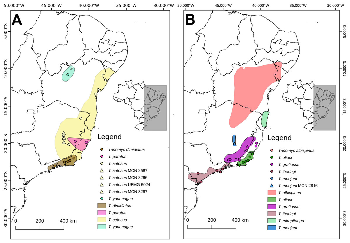
Figure 1: Sample sites of the specimens used in this study against the known range of Trinomys species.
For detailed references on spatial data downloaded from IUCN for each species, see Supplemental Information 1. Distribution of (A) T. dimidiatus, T. paratus, T. setosus, and T, yonenagae, and (B) T. albispinus, T. eliasi, T. gratiosus, T. iheringi, T. mirapitanga, and T. moojeni. Triangles represent our specimens and circles indicate the specimens whose mitochondrial sequences were retrieved from GenBank.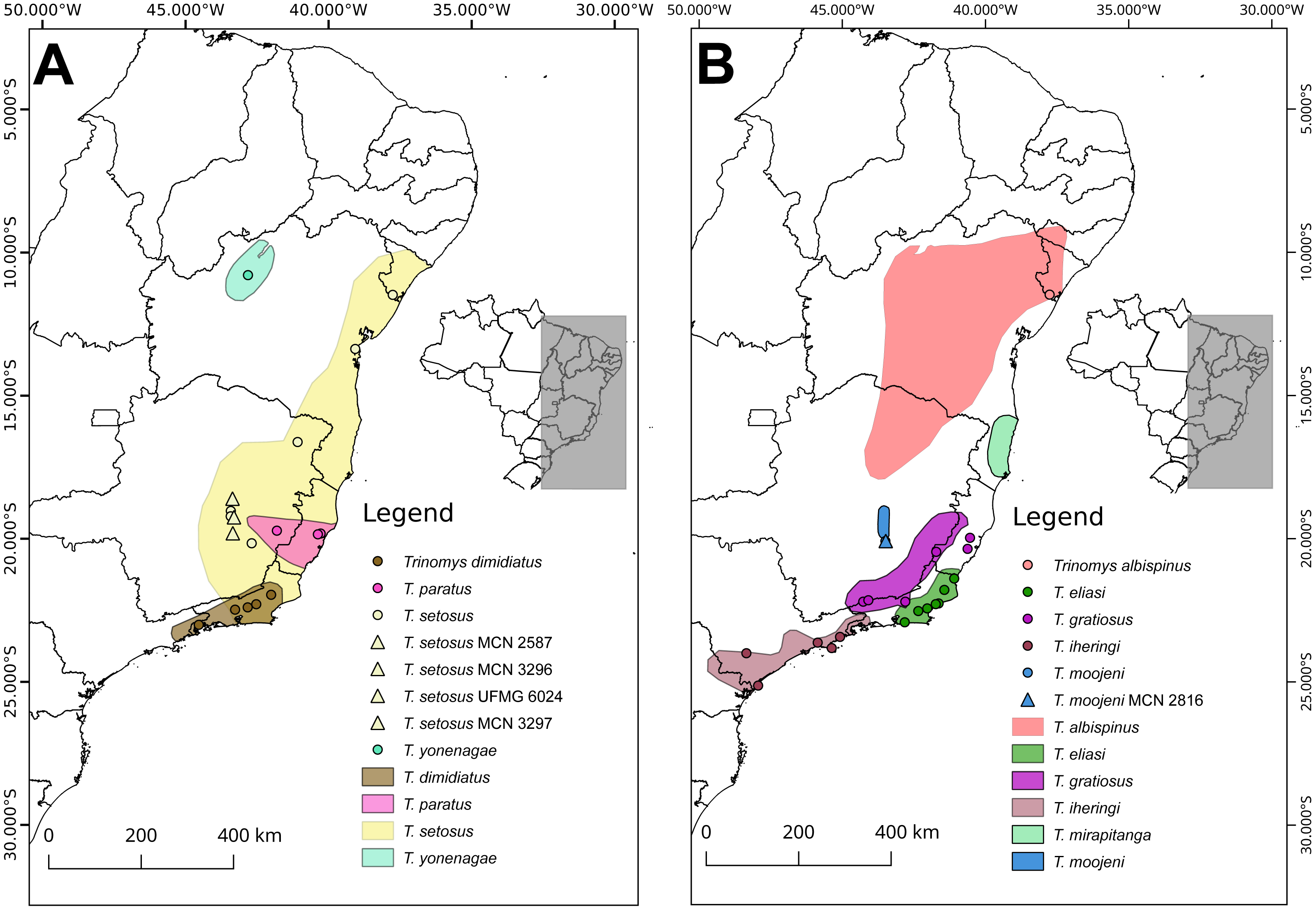
Chromosome preparations were obtained directly from bone marrow (Ford & Hamerton, 1956). GTG- and CBG-banding patterns and silver-staining of the nucleolar organizer regions (Ag-NORs) were performed according to Seabright (1971), Sumner (1972), and Howel & Black (1980), respectively. FISH with a biotinylated telomeric sequence (Invitrogen, Carlsabad, CA, USA) and with the R2 45S rDNA probe labeled by nick translation with digoxigenin-11-dUTP (DIG-Nick Translation mix; Roche Applied Science, Penzberg, Germany), followed Araújo et al. (2017); Araújo et al. (2014), respectively. Immunodetection was carried out with neutravidin and antidigoxigenin, both conjugated with rhodamine (Roche Applied Science). The analyses and image acquisition were performed under a Zeiss Axioimager 2 epifluorescence microscope using the AxioVision software (Zeiss, Oberkochen, Germany), Adobe Photoshop CS3 Extended was used for image edition. For each specimen, at least 20 metaphases of each experiment were analyzed.
Ordination and phylogenetic methods were employed in order to check the assignment of MCN-M 2587, MCN-M 3296, and MCN-M 3297 to T. setosus. In order to do this, we sequenced the 401 bp-long segment of the mitochondrial cytochrome b (cytb) of each specimen and included sequences from nine Trinomys species retrieved from GenBank in a phylogenetic analysis. The sequences of the specimens MCN-M2816 (T. moojeni) and UFMG 6024 (T. s. setosus) were previously deposited in GenBank after assembly of their mitochondrial genomes (Araújo et al., 2016). Euryzygomatomys spinosus, Fischer, 1814, was used as outgroup. Total genomic DNA of each Trinomys specimen was extracted from liver and their cytb was amplified by polymerase chain reaction (PCR) with primers MVZ 05 and MVZ 04 (Smith & Patton, 1993). The PCR products were purified using the Wizard SV Gel and PCR Clean-up System kit (Promega, Madison, WI, USA) and sequenced on the ABI3130 platform (Myleus Biotechnology). The GenBank accession numbers of the sequences generated in this study, as well as those included in the analyses are presented in the Supplemental Information 1.
The sequences obtained and those from GenBank were aligned using the Muscle (Edgar, 2004) algorithm. MEGA 7 (Kumar, Stecher & Tamura, 2016) was used to build a Kimura-2-parameter corrected distance matrix in which our ordination analysis was based. Ordination methods were used since they are useful tools to perform dimensionality reduction and to represent the distance between sequences in a coordinate (Cartesian) space where the distances are preserved (Higgins, 1992; Ramette, 2007; Zhang et al., 2011). Principal Coordinate Analysis (PCoA) was used to explore the similarity among our specimen’s sequences and other Trinomys species. The analysis was conducted in R (R Core Team, 2017) using the “pcoa” function in package APE (Paradis, Claude & Strimmer, 2004) and Lingoes procedure for correcting for negative eigenvalues.
Phylogenetic relatedness was used as a way of determining the most probable identity of the subject sequences. Thus, two methods of phylogenetic reconstruction were employed: maximum likelihood (ML) and bayesian inference (BI), which were carried out in RaxML 8 (Stamatakis, 2014) and MrBayes 3.2 (Ronquist et al., 2012), respectively. ML search comprised optimizations over 100 randomized maximum parsimony starting trees using the rapid hill-climbing algorithm under the GTRGAMMA model. As a measure of branch support, information on frequencies of 1,000 replicates of non-parametric bootstrap were annotated on the best-scoring ML tree. Bayesian inference comprised two independent runs composed of four chains each. A reversible jump MCMC sampling was used in order to explore different substitution schemes. Parameters and trees were sampled every 1,000 generations along a total of 20 million generations. After discarding a quarter of samples as burn-in, parameters and trees were summarized and the following metrics were used to assess MCMC convergence: standard deviation of split frequencies, effective sample size and potential scale reduction factor for each parameter.
Results
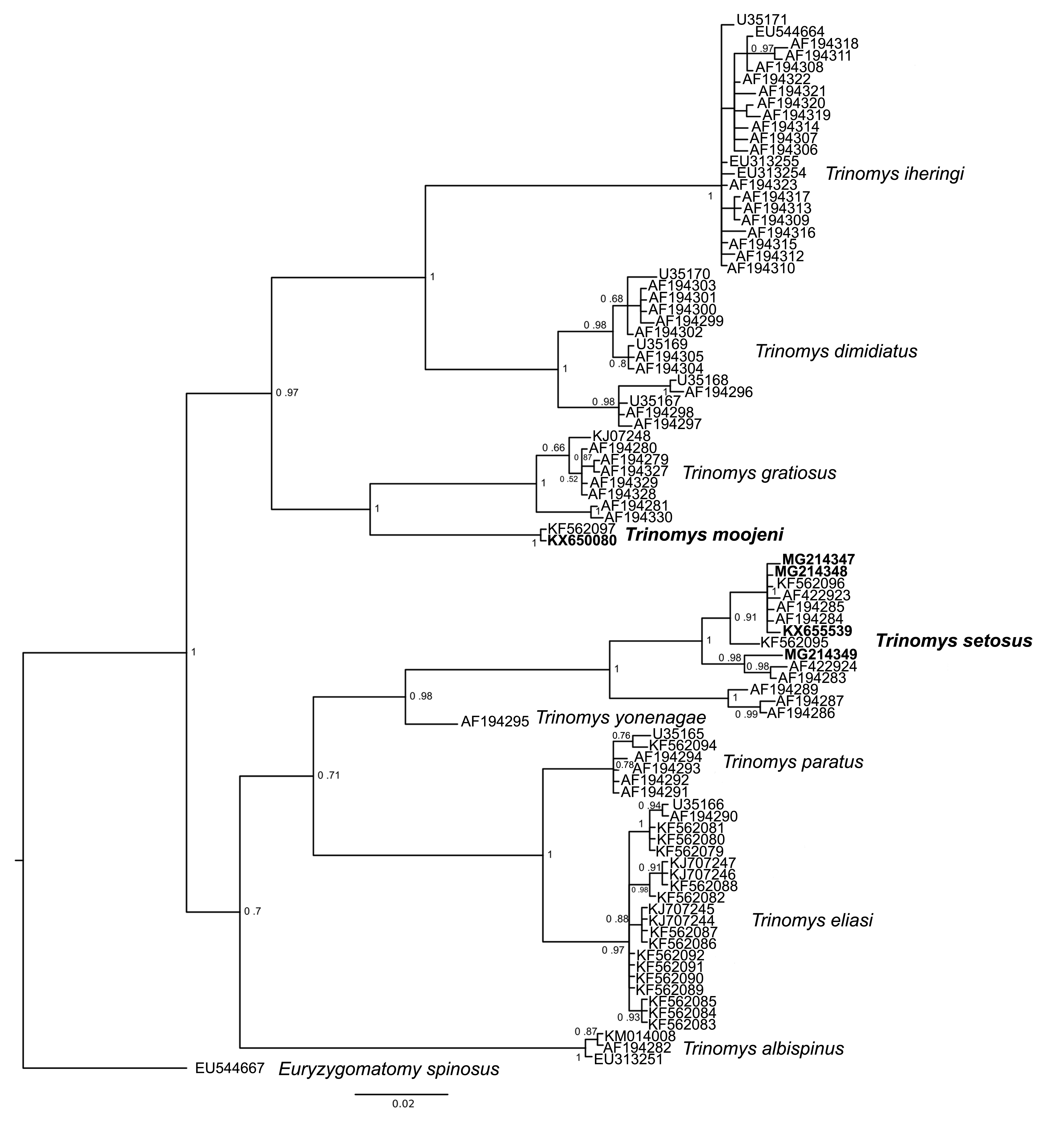
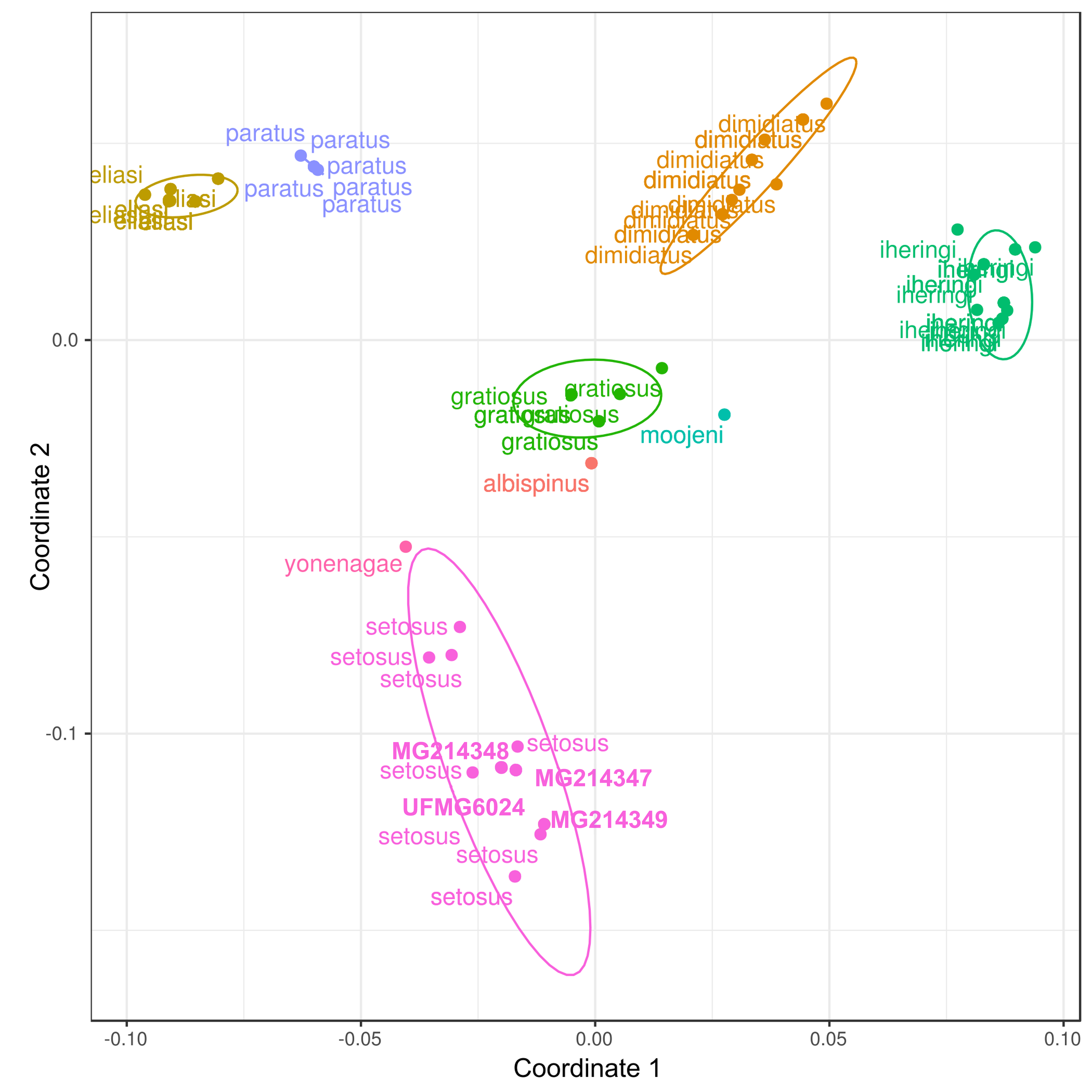
Bayesian inference and ML trees recovered Trinomys as monophyletic and grouped MCN-M 2816 within T. moojeni and the specimens UFMG 6024, MCN-M 2587, MCN-M 3296, and MCN-M 3297 within T. setosus (Fig. 2; Figs. S1 and S2). This was further supported by the PCoA results, that showed samples of the same species clustering together on the graph (Fig. S3). Morphological characters analyses corroborated the phylogeny and allowed assigning the T. setosus specimens as T. s. setosus (Table S1).
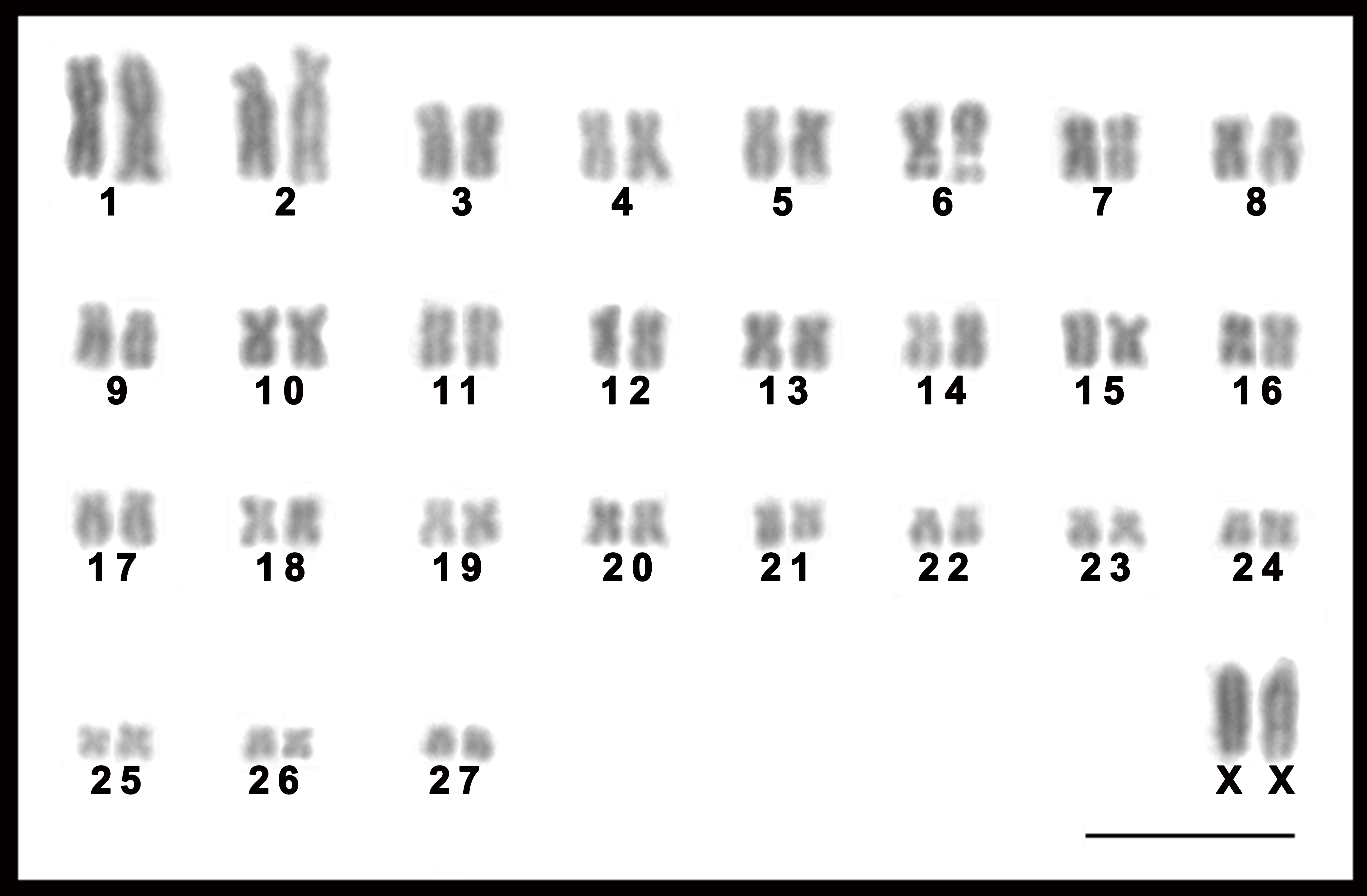
The female T. moojeni had a complement with 2n = 56 and FN = 106, similar to that described by Corrêa et al. (2005), composed of 26 pairs of biarmed (pairs 1–26) and one pair of acrocentric (pair 27) autosomes, and submetacentric X chromosomes (Fig. 3). The autosomes of the female T. s. setosus (2n = 56, FN = 106) collected in Serro included 26 biarmed pairs decreasing in size from large to small (pairs 1–25 and 27) and a small acrocentric pair (pair 26). The X chromosomes were large acrocentrics (Fig. 4A). The other three specimens of T. s. setosus had karyotypes with 2n = 56 and FN = 108 (Fig. 4C and Fig. S4), similar to the other cytotype of T. s. setosus, but with pair 26 as a biarmed element. Their X chromosome was a large acrocentric and the Y was a small acrocentric (Fig. 4 and Fig. S4).
After GTG-banding, it was possible to identify all chromosomes of each species (Figs. 3A and 4) and to verify that the complements of T. s. setosus with FN = 106 and FN = 108 (Fig. 5) differed in relation to pair 26, which was acrocentric or biarmed, in the animals with FN = 106 and FN = 108, respectively. CBG-banding revealed weak centromeric constitutive heterochromatin in pairs 1–5, 9, 10, 15, 17–27, and the X chromosome of T. moojeni (Fig. 3B); pairs 1, 11, 15, 16, 19–27, and the X chromosome of T. s. setosus (2n = 56, FN = 106; Fig. 4B); and pairs 1, 6, 8, 16, 18–27, and the sex chromosomes of T. s. setosus (2n = 56, FN = 108; Fig. 4D). Both species had a large interstitial secondary constriction on the long arm of pair 6, which bears the NORs (Fig. 6). Hybridization with the telomeric probe showed signals only at the termini of all chromosomes of the two species studied (Figs. 6C, 6F and 6I).

Figure 2: Collapsed bayesian inference tree based on a 401-bp fragment of the cytochrome b gene from species of Trinomys.
E. spinosus was used as outgroup. Collection sites: (A) Serra do Caraça Private Reserve/MG, (B) Morro do Pilar/MG, (C) Serro/MG, (D) São Gonçalo do Rio Abaixo/MG. Numbers represent Bayesian posterior probabilities ≥0.95. Specimens included in this study are in bold. See Fig. S1 for specimens details of the collapsed branches. Scale bar represents the number of substitutions per site.
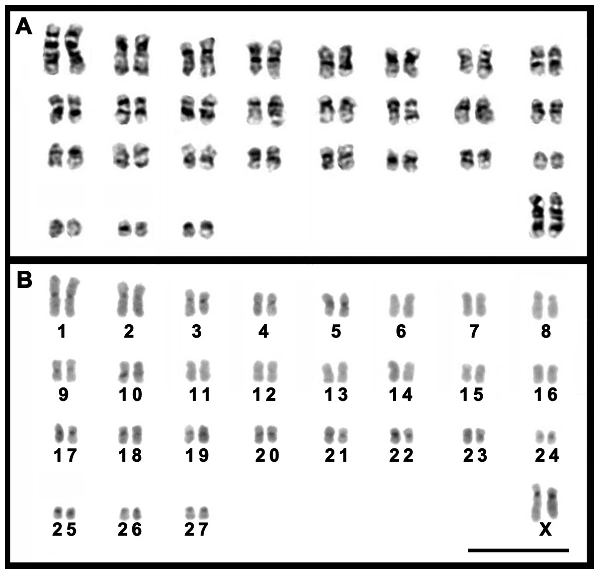
Figure 3: Karyotypes of a female T. moojeni (2n = 56, FN = 106).
Karyotypes of a female T. moojeni (2n = 56, FN = 106) from Serra do Caraça Private Reserve, Minas Gerais State, after (A) GTG- and (B) CBG-banding. Scale bar = 10 µm.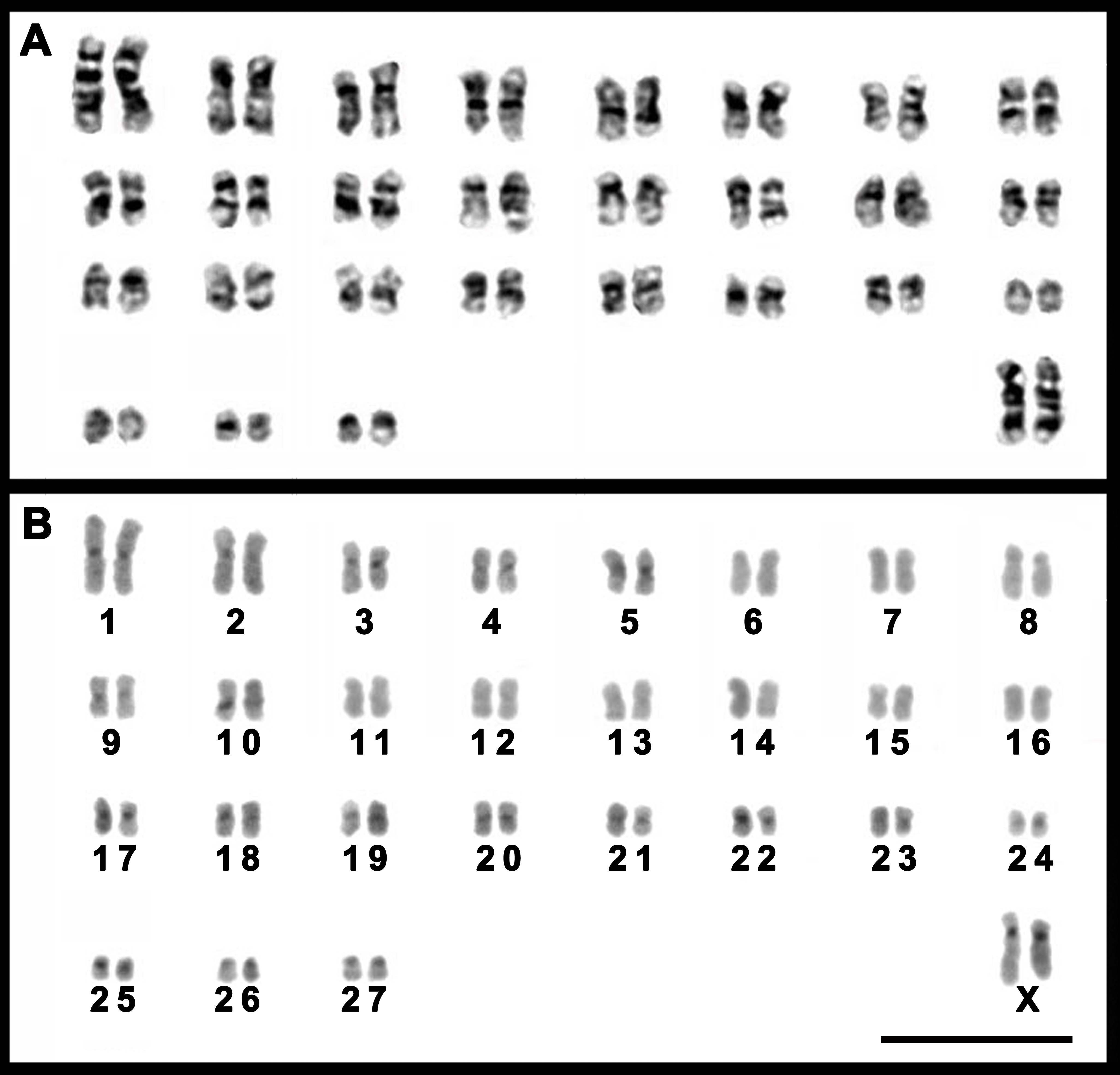
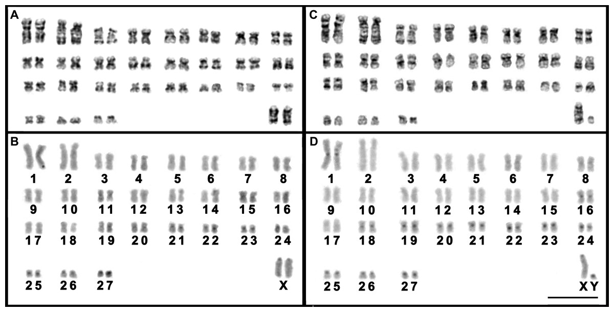
Figure 4: Karyotypes of T. s. setosus.
A female with 2n = 56, FN = 106 (A and B) from Serro, Minas Gerais state, and of a male with 2n = 56, FN = 108 (C and D) from São Gonçalo do Rio Abaixo, Minas Gerais state, after GTG- (A and C) and CBG-banding (B and D). Scale bar = 10 µm.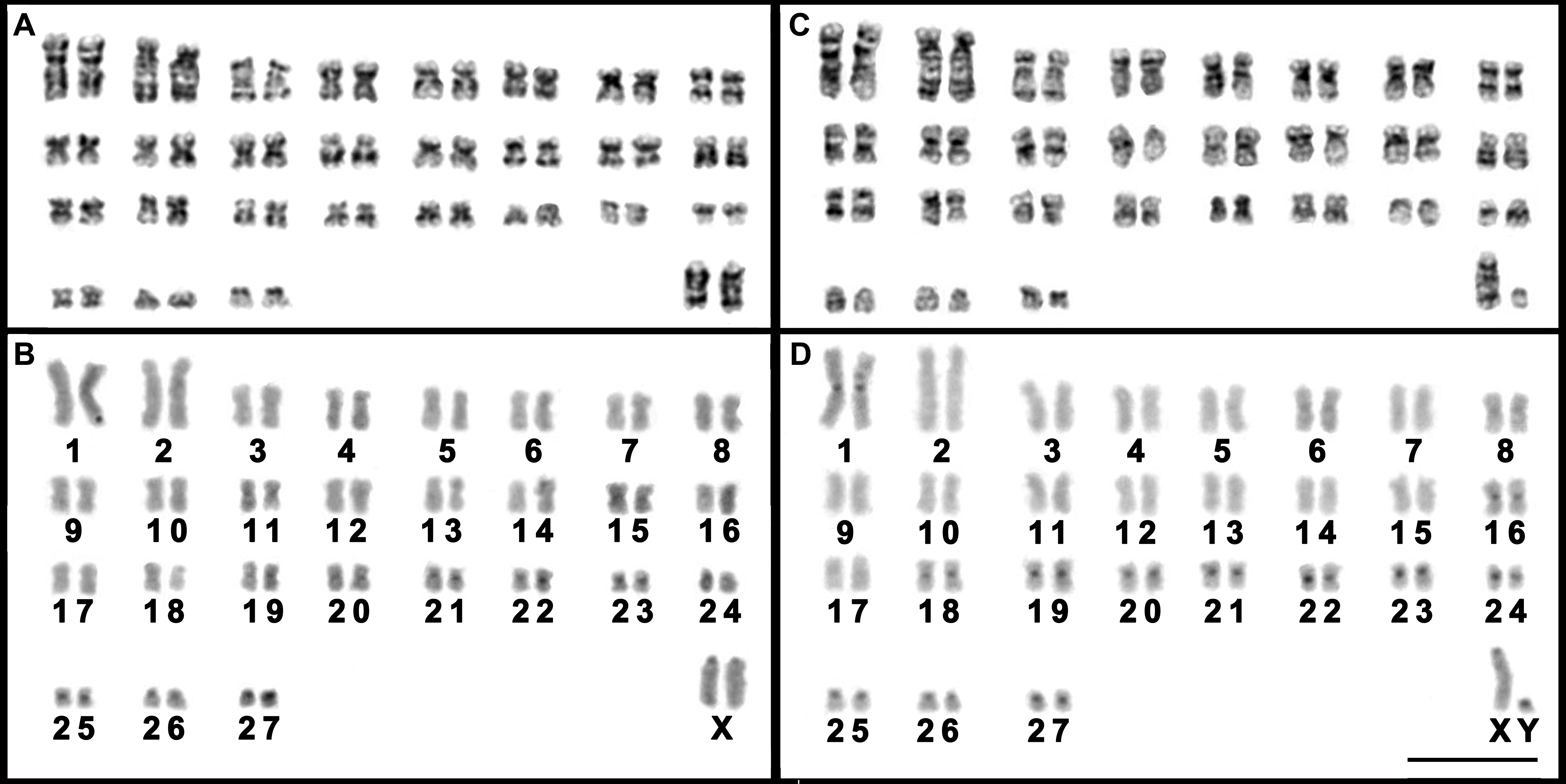
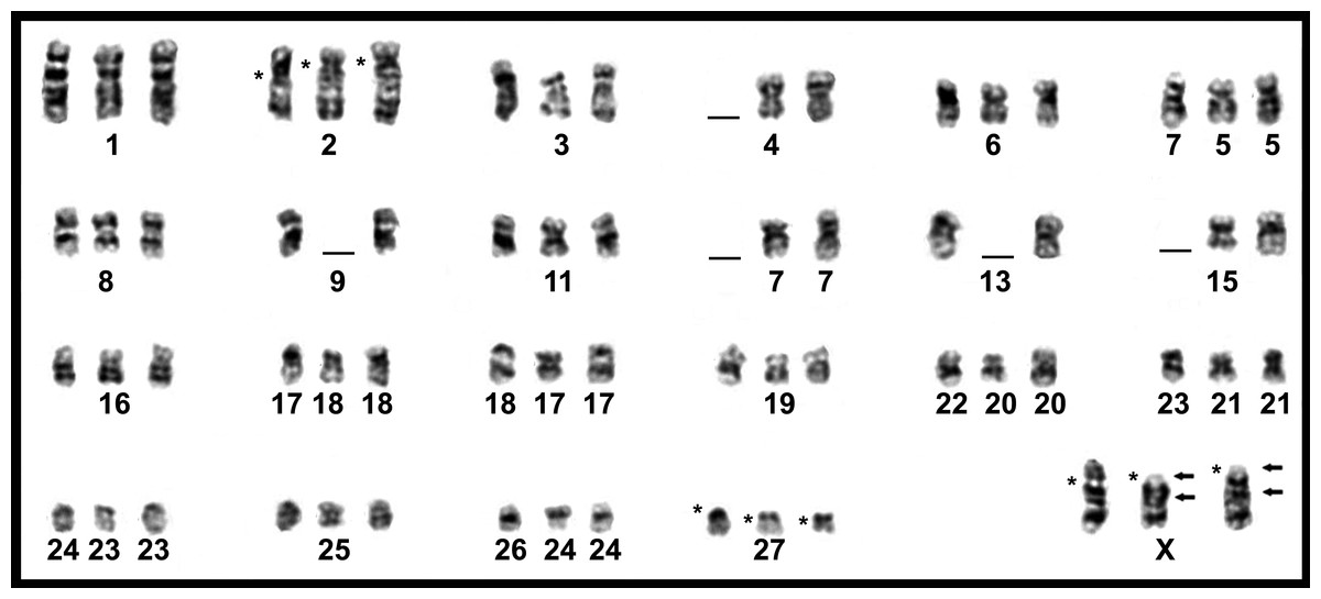
Figure 5: Comparison of GTG-banded chromosomes of Trinomys species
Chromosomes from the left to the right: T. moojeni (2n = 56, FN = 106), T. s. setosus (2n = 56, FN = 106), and T. s. setosus (2n = 56, FN = 108). * = centromere position. The arrows indicate possible inversion sites.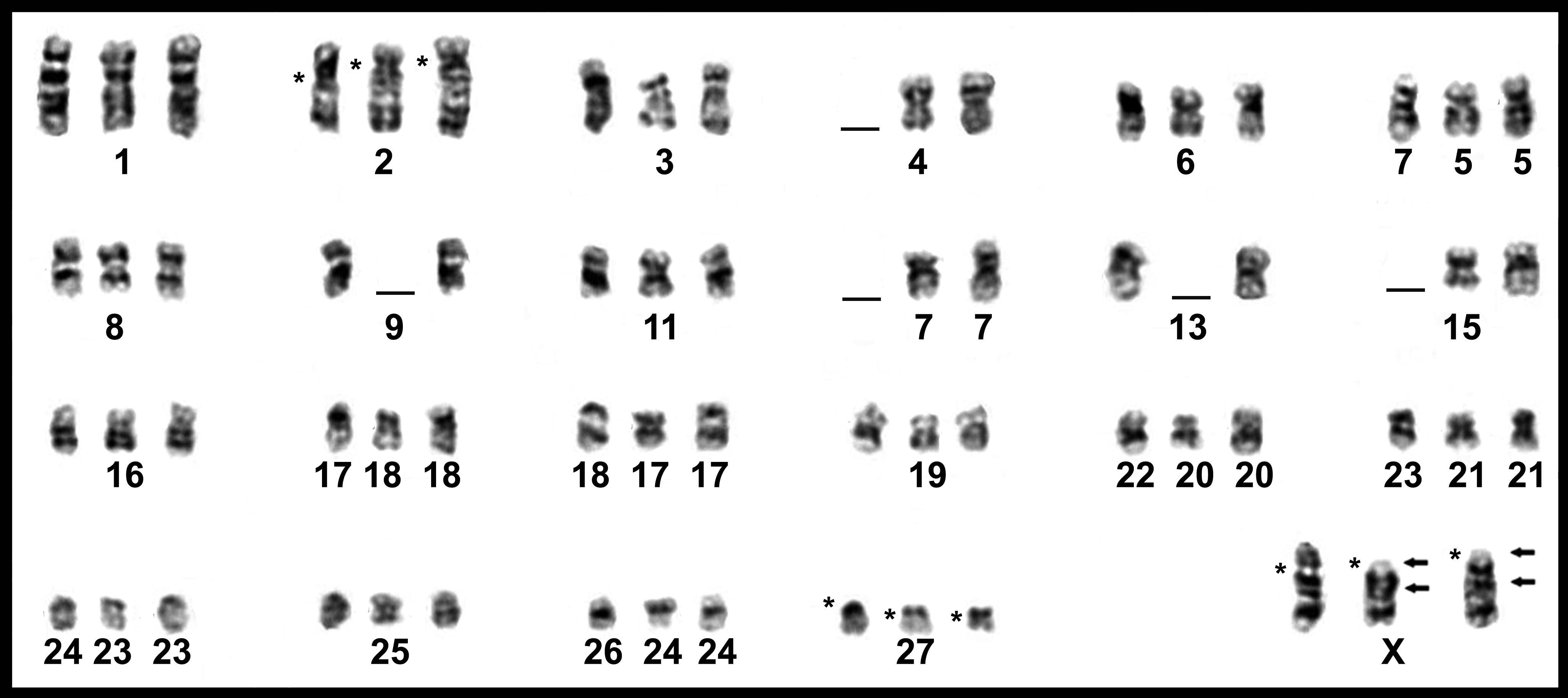
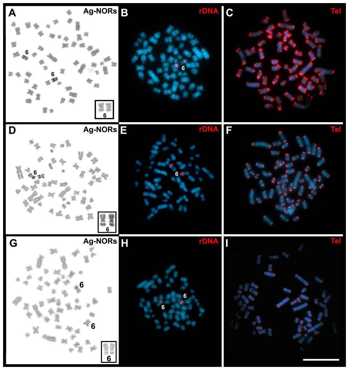
Figure 6: Cells of Trinomys species after Ag-NOR, FISH with the 45S rDNA, and telomeric probe (Tel).
Metaphases of (A–C) a female T. moojeni (2n = 56, FN = 106), (D–F) a female T. s. setosus (2n = 56, FN = 106), and (G–I) a male T. s. setosus (2n = 56, FN = 108). In the insets (A, D, and G), pair 6 after Giemsa staining. Note the secondary constrictions. Scale bar = 10 µm.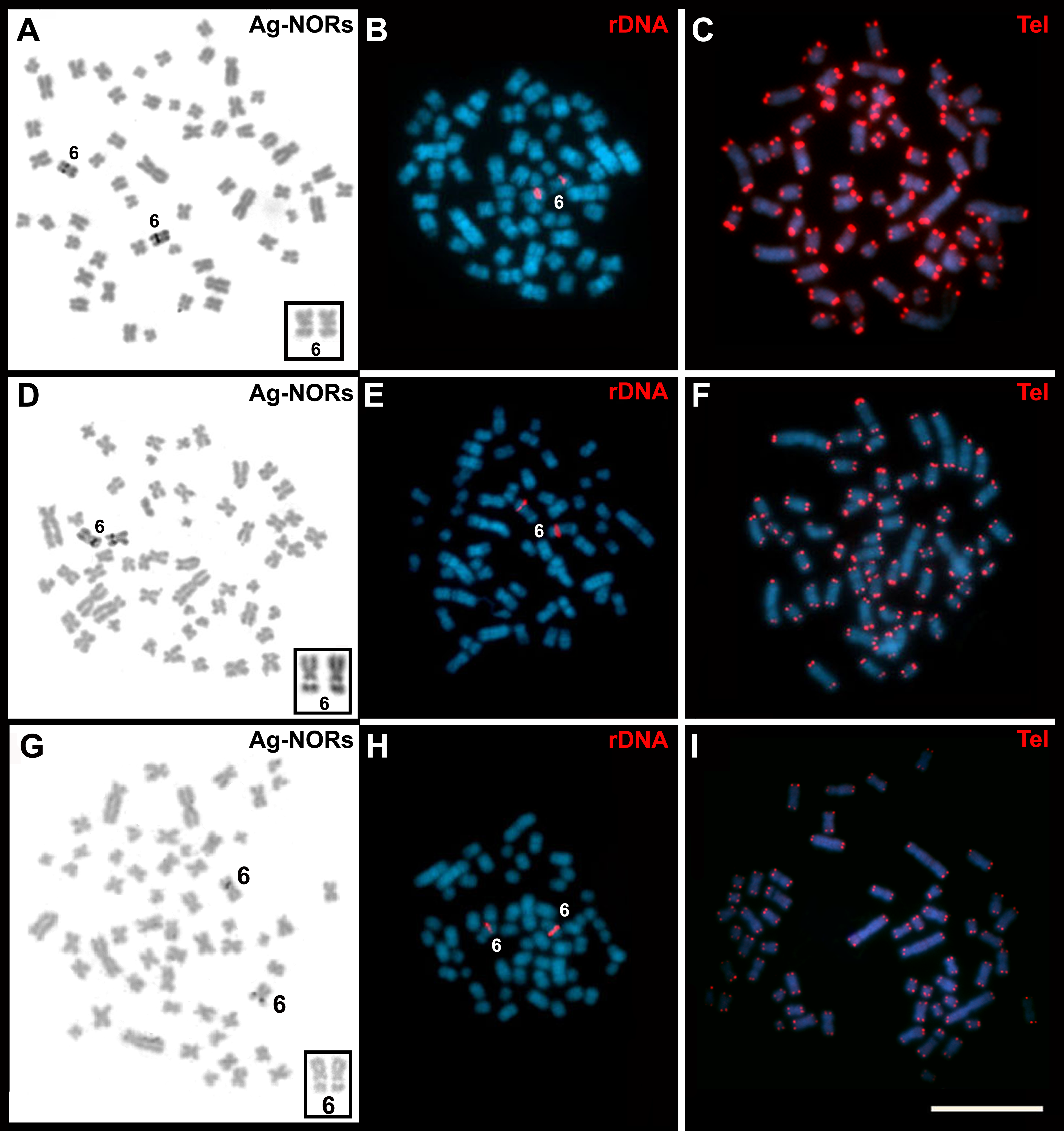
Discussion
The interspecific grouping of Trinomys, recovered by the phylogenetic analyses, was congruent with previous studies (Lara & Patton, 2000; Tavares, Pessôa & Seuánez, 2015; Lazar et al., 2017). Our phylogenetic analysis is also supported by the specimens’ distribution (Fig. 1), morphology (Supplemental Table S1) and karyotypes. The collecting locality of T. moojeni (MCN-M 2816), Serra do Caraça Private Reserve, is the same of the specimens studied by Cordeiro-Júnior & Talamoni (2006) and the karyotype was similar to that described for this species (Corrêa et al., 2005; Fig. 3). T. s. setosus, in turn, which occurs from the coastal area of the Brazilian states of Sergipe, Bahia, and Espírito Santo to the interior of Minas Gerais (Pessôa et al., 2015), were collected in three municipalities of Minas Gerais.
A comparison of the GTG-banded chromosomes of T. moojeni and T. s. setosus (2n = 56, FN = 106) evidenced very similar karyotypes. They mainly differed on their pairs 2, 27, and X chromosomes, possibly due to inversions and/or centromere repositioning (Fig. 5). In order to establish the exact mechanisms involved, further experiments including FISH with specific sequences from the regions of interest are necessary.
The karyotypes described herein for T. s. setosus differed in 2n, FN, and/or the sex chromosome morphology from those already published for this genus (Yonenaga-Yassuda et al., 1985; Leal-Mesquita et al., 1992; Corrêa et al., 2005; Pessôa et al., 2005; Souza, Corrêa & Pessôa, 2006; Lazar et al., 2017; Table 1). Trinomys gratiosus bonafidei also has 2n = 56 and FN = 108, but differently from our specimens, has a metacentric Y chromosome (Pessôa et al., 2005). The most recent revision on Trinomys divided T. setosus into the subspecies T. s. setosus and T. s. elegans (Pessôa et al., 2015). The diploid number was reported only for T. s. elegans and without banding patterns (2n = 56, FN = 104; Corrêa et al., 2005). Pessôa et al. (2015) mentioned that the karyotype of T. s. setosus from Almenara, Minas Gerais state, has 2n = 56 and FN = 108, but no figure was provided. Our T. s. setosus had karyotypes with FN = 106 and 108 and differed from that described by Corrêa et al. (2005) by the presence of additional short arms on pair 27 and pairs 26 and 27 of our specimens, respectively. These differences may be real or may reflect variations in chromosome condensation between both samples, as poorly elongated small chromosomes could prevent the detection of short arms. If real, these differences between T. s. setosus and T. s. elegans may be correlated with their subspecies allocation or may be due to interpopulational variation, as seems to be the case of T. s. setosus. Our phylogenetic analyses did not distinguish between T. s. setosus and T. s. elegans (Fig. 2, Figs. S1 and S2), but the morphological analysis allowed to recognize these subspecific taxa. The karyotype information was also relevant in species identification, revealing karyotypes that differed from those of other species of the genus.
The T. s. setosus karyotypes described herein also differed from others previously reported for Trinomys in the morphology of the X chromosome. With the exception of T. setosus and T. yonenagae, which presented acrocentric X chromosomes, all Trinomys species had a submetacentric X (T. albispinus, T. dimidiatus, T. eliasi, T. gratiosus, T. iheringi, T. moojeni, and T. paratus). Based on our cytochrome b phylogeny (Fig. 2), we suggest that a pericentric inversion or a centromere shift on the X chromosome occurred in the lineage that gave rise to T. setosus and T. yonenagae. The change in X chromosome morphology in the common ancestor of both species may be related to karyotype differentiation from other taxa and reproductive isolation. It has been suggested that chromosome rearrangements may affect chromatin structure (Johnson & Lachance, 2012) and, consequently, play a role in hybrid incompatibility. The change in gene expression after chromosome rearrangements was also suggested to contribute to the speciation process (Potter et al., 2017).
Our specimens and all the other species of the genus analyzed after CBG-banding, (T. albispinus minor, T. iheringi, T. paratus, and T. yonenagae) exhibited faint heterochromatic blocks on centromeric regions, mainly located on the smallest autosomes and the sex chromosomes (Yonenaga-Yassuda et al., 1985; Leal-Mesquita et al., 1992; Fagundes, Camacho & Yonenaga-Yassuda, 2004; Lazar et al., 2017). Interestingly, both cytotypes of T. s. setosus differed in heterochromatin distribution (Fig. 4), which may be involved in the chromosome evolution of this taxon.
T. moojeni and T. s. setosus had a large interstitial secondary constriction on the long arm of pair 6, which bears the NORs (Fig. 6). A chromosome pair with a large secondary constriction bearing the single NOR is a marker of echimyids, as already reported for T. iheringi and other Echimyidae genera (Fagundes, Camacho & Yonenaga-Yassuda, 2004; Silva et al., 2012; Araújo et al., 2014). The comparison of the GTG-banded karyotypes suggests that the NOR-bearing chromosome is the same in our specimens and in T. albispinus, T. iheringi, and T. yonenagae (Leal-Mesquita et al., 1992; Fagundes, Camacho & Yonenaga-Yassuda, 2004) and is probably conserved in the genus.
Hybridization with the telomeric probe showed signals only at the extremities of all chromosomes (Figs. 6C, 6F, and 6I). This pattern of hybridization was similar to that described for T. iheringi (Fagundes, Camacho & Yonenaga-Yassuda, 2004). Bolzán (2017) suggested that the absence of interstitial telomeric sequences indicates the evolutionary status of the chromosomes of a species. Accordingly, species without or with only a few interstitial telomeric sequences would have more conserved chromosomes, as seems to be the case of Trinomys.
Conclusions
In summary, based on the available data, it is clear that the Trinomys species present conserved karyotypes with small variation in diploid numbers (2n = 54 to 2n = 61–65) and mostly composed of biarmed autosomes. The X chromosomes are usually large submetacentrics and all the species analyzed presented one marker chromosome pair with a secondary constriction corresponding to the NOR, which is also typical for the other echimyid genera. The great conservation extends to the GTG- and CBG-banding patterns in the few species which had these patterns described. As previously proposed by Leal-Mesquita et al. (1992), pericentric inversions, centromere repositioning, and other minor rearrangements seem to be responsible for the chromosome evolution in this genus. Further analyses, including a robust phylogenetic hypothesis, cytogenetic studies with high resolution banding patterns and molecular data of a larger array of Trinomys species, are needed to improve our understanding of the chromosome evolution and genome organization of this genus. It should be stressed that Trinomys species, especially those from Minas Gerais, need more thorough morphological and molecular analyses, as their cytogenetic information alone is insufficient for taxonomic identification. In fact, several different species present very similar karyotypes (Lazar et al., 2017).
Supplemental Information
Bayesian inference tree based on a 401-bp fragment of the cytochrome b gene from species of Trinomys
Euryzygomatomys spinosus was used as outgroup. Numbers indicate Bayesian posterior probabilities ≥0.95. Specimens included in this study are in bold. Scale bar represents the number of substitutions per site.
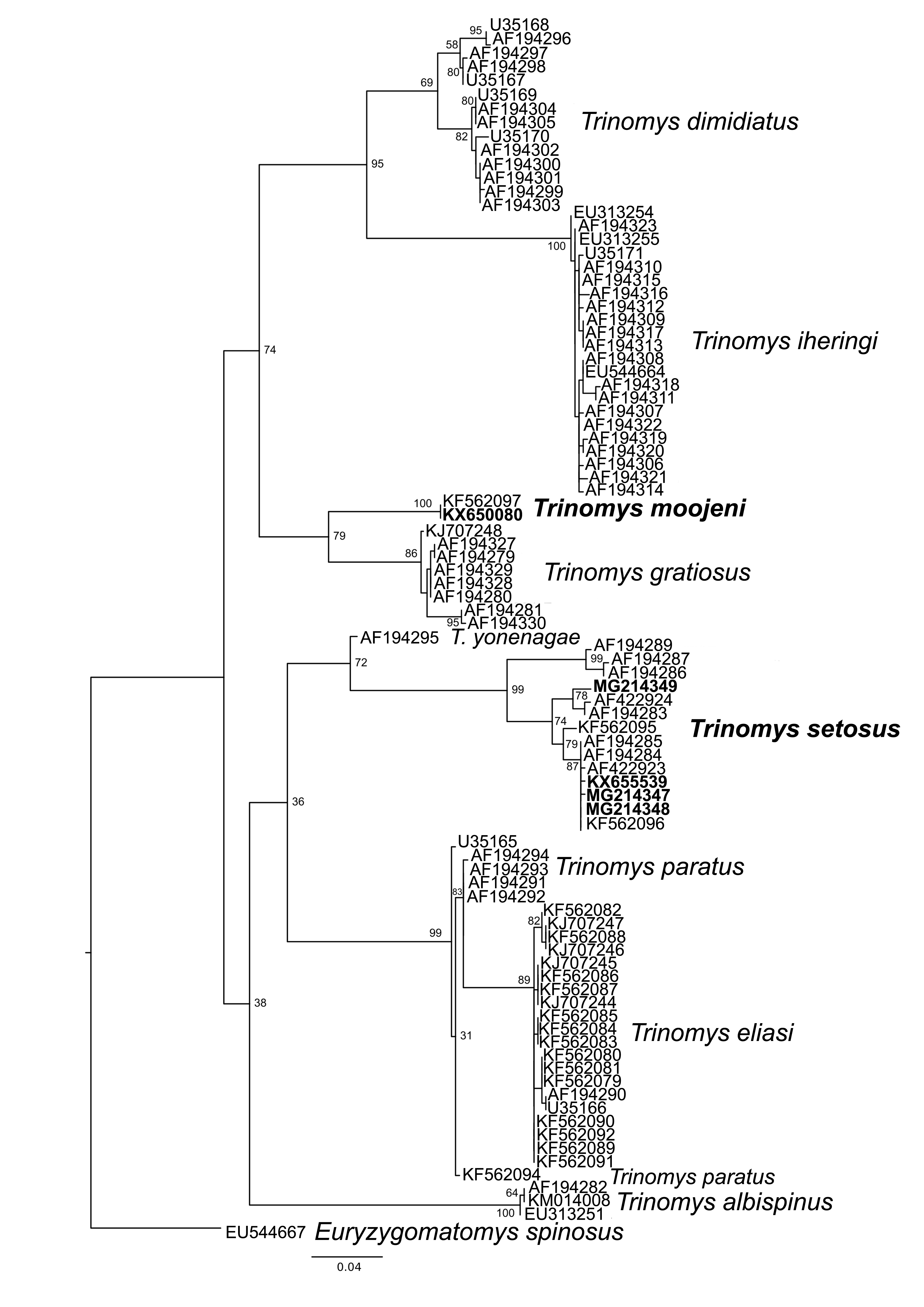
Maximum likelihood tree based on a 401-bp fragment of the cytochrome b gene from species of Trinomys plus Euryzygomatomys spinosus as outgroup
Numbers represent bootstrap support. Specimens included in this study are in bold. Scale bar indicates the number of substitutions per site.
Two-dimensional PCoA ordination diagram of Trinomys species
Two-dimensional PCoA ordination diagram of Trinomys species, showing the similarity (or dissimilarity) between individuals. The specimens included in the present study are indicated by their deposit numbers.
Karyotype of a female Trinomys setosus setosus (2n = 56, FN = 108)
Karyotype of a female Trinomys setosus setosus (2n = 56, FN = 108), from Morro do Pilar, Minas Gerais state, after Giemsa staining. Scale bar = 10 µm.