
Integrative taxonomy of the genus Pseudostegana (Diptera, Drosophilidae) from China, with descriptions of eleven new species
- Published
- Accepted
- Received
- Academic Editor
- John Ringo
- Subject Areas
- Biodiversity, Entomology, Taxonomy, Zoology
- Keywords
- Taxonomy, Drosophilidae, Biodiversity, DNA, Morphology
- Copyright
- © 2018 Lu et al.
- Licence
- This is an open access article distributed under the terms of the Creative Commons Attribution License, which permits unrestricted use, distribution, reproduction and adaptation in any medium and for any purpose provided that it is properly attributed. For attribution, the original author(s), title, publication source (PeerJ) and either DOI or URL of the article must be cited.
- Cite this article
- 2018. Integrative taxonomy of the genus Pseudostegana (Diptera, Drosophilidae) from China, with descriptions of eleven new species. PeerJ 6:e5160 https://doi.org/10.7717/peerj.5160
Abstract
The genus Pseudostegana (Okada, 1978) currently contains thirty-nine described species. A number of Pseudostegana were collected from the fieldwork in southwestern China from 2010 to 2017. Eleven new species were discovered and are described from southwestern China: Pseudostegana alpina Zhang & Chen, sp. nov.; Pseudostegana amnicola Zhang & Chen, sp. nov.; Pseudostegana amoena Zhang & Chen, sp. nov.; Pseudostegana mailangang Zhang & Chen, sp. nov.; Pseudostegana meiduo Zhang & Chen, sp. nov.; Pseudostegana meiji Zhang & Chen, sp. nov.; Pseudostegana mystica Zhang & Chen, sp. nov.; Pseudostegana stictiptrata Zhang & Chen, sp. nov.; Pseudostegana stigmatptera Zhang & Chen, sp. nov.; Pseudostegana ximalaya Zhang & Chen, sp. nov. and Pseudostegana zhuoma Zhang & Chen, sp. nov. A key to all Chinese Pseudostegana species based on morphological characters is provided. Two mitochondrial loci (COI and ND2) and one nuclear locus (28S rRNA) were sequenced for the Pseudostegana specimens, and Bayesian and RAxML concatenated analyses were run. Molecular species delimitation is performed using the distance-based automatic barcode gap discovery (ABGD) method. Molecular data support the morphological characteristics observed among these Chinese species and confirm the new species as being distinctly different.
Introduction
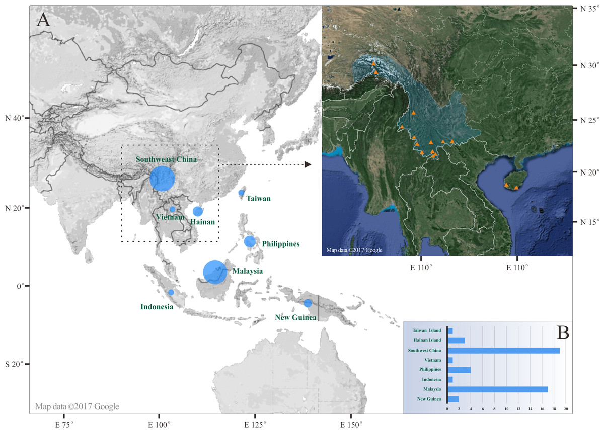
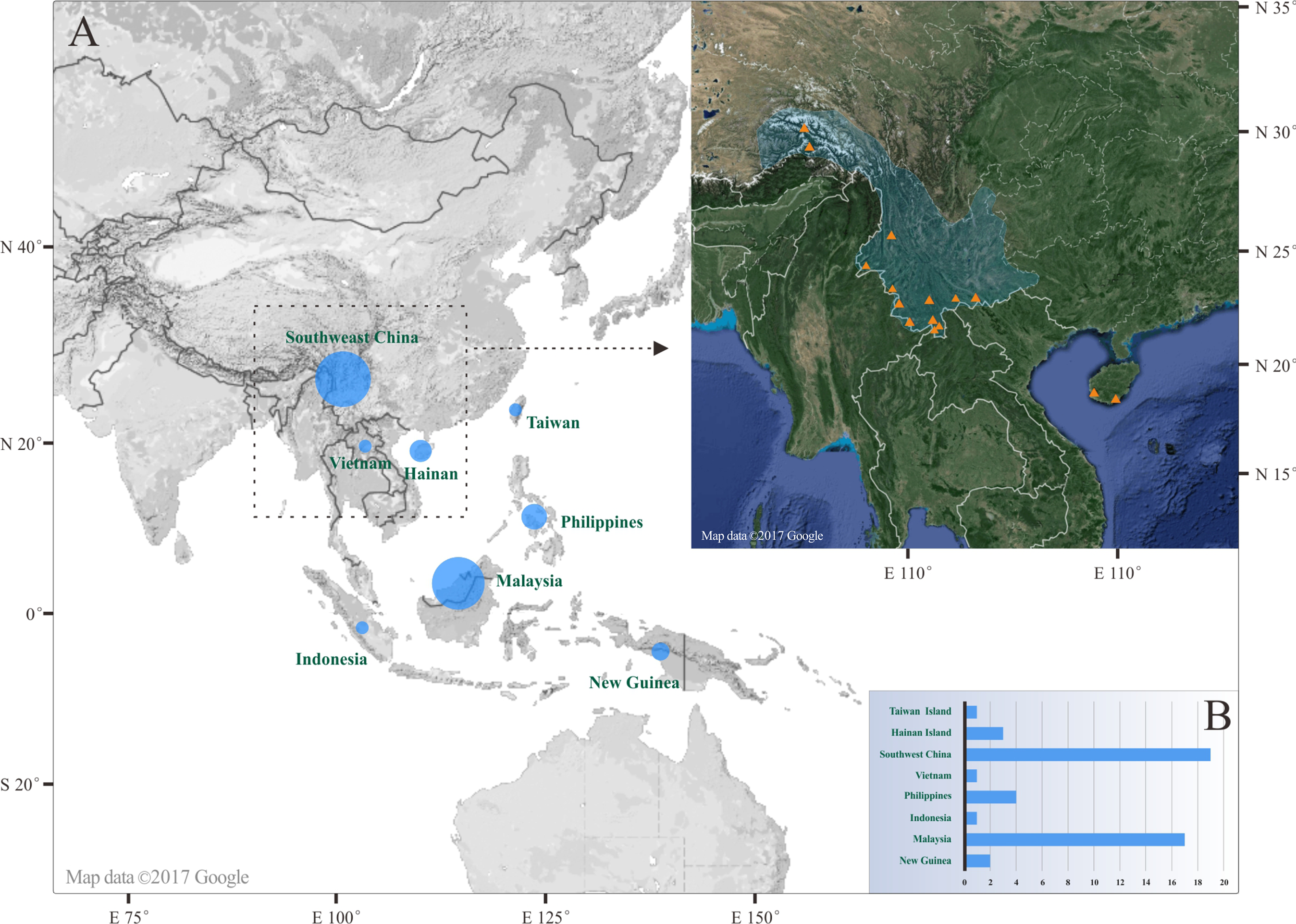
The genus Pseudostegana (Okada, 1978) is widely distributed in tropical area from Oriental to Australasian regions. Adult flies of this genus are yellow to black in body color, with a body length of about two to four mm. They are usually found resting on fallen logs, tussocks or fruits beside a stream, flapping their wings slowly like butterflies (Li, Gao & Chen, 2010). Currently, a total of 39 Pseudostegana species have been described (Chen, Toda & Wang, 2005; Li, Gao & Chen, 2010): 17 spp. from Malaysia, 12 spp. from China, five spp. from the Philippines, three spp. from Papua New Guinea, two spp. from Indonesia and one sp. from Vietnam. Chen, Toda & Wang (2005) revised the Pseudostegana taxonomy using morphological characters, and proposed six species groups (the atrofrons, javana, latiparma, grandipalpis, fleximediata and zonaria groups) based on wing patterns and male genitalia. Among these six species groups, the javana, latiparma, fleximediata and zonaria groups have been found in Southern China (Chen, Toda & Wang, 2005; Li, Gao & Chen, 2010).
There are few phylogenetic studies concerning the Pseudostegana genus (Okada, 1978; Li et al., 2013). Okada (1978) conducted a taxometric analysis on subgeneric relationships of the genus Stegana using 11 morphological characters and recognized a clade composed of the genera Pseudostegana and Parastegana Okada, 1971, which was placed as the sister group to the rest of the genus Stegana. Li et al. (2013) reconstructed a molecular phylogeny of the East Asian species in the Stegana genus group and confirmed the sister relationship of Parastegana and Pseudostegana. The phylogeny of species groups within the genus is still little known.
The COI gene is a commonly used marker for the DNA barcode identification and is potentially useful for species discovery and identification (Hebert et al., 2003; Robe, Machado & Bartholomei-Santos, 2012; Madeira et al., 2016; Melbourne et al., 2017). Additional genetic markers on mitochondrial (ND2, 16S, COII) and nuclear (28S, ITS1, ITS2) genome have been used alongside the COI fragment for species discrimination and phylogenetic analysis (Li et al., 2013; Boykin et al., 2014; Jürgenstein, Kurina & Põldmaa, 2015; Madeira et al., 2016; Zhang, Li & Chen, 2016; Wong et al., 2017; Yusseff-Vanegas & Agnarsson, 2017).
Our paper addresses the study of the Pseudostegana in Southwest China. Considering the recent diversity and the phylogenetic reorganization of Steganinae subfamily, we used an integrative taxonomy approach to delimit new species from the Pseudostegana genus. This improves the knowledge of global and East Asia biodiversity, and could give insights to the origin of the Pseudostegana genus.
Materials and Methods
Sample collection and morphological treatment
This study is based on material collected at several sites in China from 2010 to 2017 (Fig. 1). Specimens were collected by net-sweeping from tree trunks and then fixed in 75% ethanol. Male genitalia were removed from the abdomen, treated with 8% KOH for about 3–5 min and observed under the stereomicroscope (CX31; Olympus, Tokyo, Japan) for identification and drawing. Digital images were taken using a MD50 camera (Mshot, Guangzhou, China) mounted on the Olympus CX31 stereomicroscope. Some specimen was damaged during the extraction of the abdominal tissue, and thus we draw the morphological structure form the original abdomen. The female individuals were identified based on the description in Chen, Toda & Wang (2005) and Li, Gao & Chen (2010). The definitions of measurement abbreviations were followed as Zhang & Toda (1992) and Chen & Toda (2001) and listed in Table S1. The type specimens were deposited in the following institutions: Kunming Institute of Zoology (KIZ), Chinese Academy of Sciences, Kunming, China; Department of Entomology, South China Agricultural University (SCAU), Guangzhou, China.
The electronic version of this article in portable document format will represent a published work according to the International Commission on Zoological Nomenclature (ICZN), and hence the new names contained in the electronic version are effectively published under that code from the electronic edition alone. This published work and the nomenclatural acts it contains have been registered in ZooBank, the online registration system for the ICZN. The ZooBank LSIDs (Life Science Identifiers) can be resolved and the associated information viewed through any standard web browser by appending the LSID to the prefix http://zoobank.org/. The LSID for this publication is: urn:lsid:zoobank.org:pub:91F288F7-0083-4EBD-9B49-DF51048E9519. The online version of this work is archived and available from the following digital repositories: PeerJ, PubMed Central and CLOCKSS.
DNA extraction, PCR and sequencing
Detailed information on samples used for molecular study is given in Table 1. Two Parastegana species, Parastegana femorata Duda, 1923 and Parastegana brevivena Chen & Zhang, 2007, are chosen as the outgroups in this study. Genomic DNA was extracted from the abdominal tissue of a single fly after the dissection of the genitalia using the TIANamp Genomic DNA kit (TIANGEN™, Beijing, China). DNA fragments of COI, ND2 and 28S were sequenced for each of the selected samples (Table 1). Target fragments were amplified and sequenced using published primers (Table S2). PCR products were sequenced at BGI (Beijing, China) by the ABI 3730 Genetic Analyzer (Applied Biosystems, Foster City, CA, USA) and were performed forward and reversed strand sequencing.
| Genus | Species group | Species | Collection site | Latitude (°N) | Longitude (°E) | COI | ND2 | 28S |
|---|---|---|---|---|---|---|---|---|
| Parastegana | femorata Duda, 1923 | Menglun, Mengla, Yunnan | 21.6833 | 101.4167 | KJ813937 | KJ813971 | KJ813904 | |
| brevivena (Chen & Watabe, 2007)—1 | Hesong, Menghai, Yunnan | 21.8166 | 100.1000 | KJ813938 | KJ813972 | KJ813905 | ||
| brevivena (Chen & Watabe, 2007)—2 | Wuliangshan, Jingdong, Yunnan | 24.5333 | 101.0167 | KP981251 | KY542068 | KY542041 | ||
| brevivena (Chen & Watabe, 2007)—3 | Wuliangshan, Jingdong, Yunnan | 24.5333 | 101.0167 | KP981252 | KP981250 | KY542040 | ||
| Pseudostegana | fleximediata | meiduo Zhang & Chen, sp. nov. | Beibeng, Motuo, Xizang | 29.3167 | 95.3333 | KJ813939 | KJ813973 | KJ813906 |
| javana | xanthoptera Chen & Wang, 2005—1 | Wangtianshu, Mengla, Yunnan | 21.4667 | 101.6333 | HQ842765* | HQ842786* | HQ842738* | |
| xanthoptera Chen & Wang, 2005—2 | Wangtianshu, Mengla, Yunnan | 21.4667 | 101.6333 | KJ813940 | KJ813974 | KJ813907 | ||
| xanthoptera Chen & Wang, 2005—3 | Yixiang, Pu’er, Yunnan | 22.7833 | 101.0333 | KJ813941 | – | – | ||
| meiji Zhang & Chen, sp. nov. —1 | Muyiji Park, Ximeng, Yunnan | 22.6208 | 99.5950 | KJ813942 | KJ813975 | KJ813908 | ||
| meiji Zhang & Chen, sp. nov. —2 | Muyiji Park, Ximeng, Yunnan | 22.6208 | 99.5950 | KY542055 | KY542069 | KY542042 | ||
| meiji Zhang & Chen, sp. nov. —3 | Mengdong, Cangyuan, Yunnan | 23.1689 | 99.2311 | KY542056 | KY542070 | KY542043 | ||
| stictiptrata Zhang & Chen, sp. nov. —1 | Hesong, Menghai, Yunnan | 21.8166 | 100.1000 | KJ813943 | KJ813976 | KJ813909 | ||
| stictiptrata Zhang & Chen, sp. nov. —2 | Huanglianshan, Lvchun, Yunnan | 22.8903 | 102.3167 | KY542057 | KY542071 | KY542044 | ||
| stigmatptera Zhang & Chen, sp. nov. —1 | Hesong, Menghai, Yunnan | 21.8166 | 100.1000 | KJ813944 | KJ813977 | KJ813910 | ||
| stigmatptera Zhang & Chen, sp. nov. —2 | Hesong, Menghai, Yunnan | 21.8166 | 100.1000 | KJ813945 | KJ813978 | KJ813911 | ||
| stigmatptera Zhang & Chen, sp. nov. —3 | Baihualing, Baoshan, Yunnan | 25.4500 | 98.8667 | KJ813946 | – | – | ||
| latiparma | acutifoliolata Li, Gao & Chen, 2010 | Diaoluoshan, Lingshui, Hainan | 18.6597 | 109.9025 | KJ813947 | KJ813979 | KJ813912 | |
| angustifasciata Chen & Wang, 2005—1 | Wangtianshu, Mengla, Yunnan | 21.4667 | 101.6333 | HQ842766* | HQ842787* | HQ842739* | ||
| angustifasciata Chen & Wang, 2005—2 | Wangtianshu, Mengla, Yunnan | 21.4667 | 101.6333 | KJ813948 | KJ813980 | KJ813913 | ||
| bifasciata Chen & Wang, 2005—1 | Wangtianshu, Mengla, Yunnan | 21.4667 | 101.6333 | KJ813949 | KJ813981 | KJ813914 | ||
| bifasciata Chen & Wang, 2005—2 | Wangtianshu, Mengla, Yunnan | 21.4667 | 101.6333 | KJ813950 | KJ813982 | KJ813915 | ||
| bilobata Li, Gao & Chen, 2010—1 | Yixiang, Pu’er, Yunnan | 22.7833 | 101.0333 | KJ813951 | KJ813983 | KJ813916 | ||
| bilobata Li, Gao & Chen, 2010—2 | Muyiji Park, Ximeng, Yunnan | 22.6208 | 99.5950 | KY542058 | KY542072 | KY542045 | ||
| minutipalpata Li, Gao & Chen, 2010—1 | Wangtianshu, Mengla, Yunnan | 21.4667 | 101.6333 | KJ813952 | – | KJ813917 | ||
| minutipalpata Li, Gao & Chen, 2010—2 | Wangtianshu, Mengla, Yunnan | 21.4667 | 101.6333 | KJ813953 | KJ813984 | KJ813918 | ||
| minutipalpata Li, Gao & Chen, 2010—3 | Muyiji Park, Ximeng, Yunnan | 22.6208 | 99.5950 | KY542059 | KY542073 | KY542046 | ||
| pallidemaculata Chen & Wang, 2005 | Fenshuiling, Jinping, Yunnan | 22.9012 | 103.2302 | KJ813954 | KJ813985 | KJ813919 | ||
| alpina Zhang & Chen, sp. nov. | Hesong, Menghai, Yunnan | 21.8166 | 100.1000 | KJ813955 | KJ813986 | KJ813920 | ||
| amoena Zhang & Chen, sp. nov.—1 | Hesong, Menghai, Yunnan | 21.8166 | 100.1000 | KJ813956 | KJ813987 | KJ813921 | ||
| amoena Zhang & Chen, sp. nov.—2 | Hesong, Menghai, Yunnan | 21.8166 | 100.1000 | KJ813957 | KJ813988 | KJ813922 | ||
| amoena Zhang & Chen, sp. nov.—3 | Hesong, Menghai, Yunnan | 21.8166 | 100.1000 | KJ813958 | KJ813989 | KJ813923 | ||
| ximalaya Zhang & Chen, sp. nov. | Beibeng, Motuo, Xizang | 29.3167 | 95.3333 | KJ813959 | KJ813990 | KJ813924 | ||
| zhuoma Zhang & Chen, sp. nov.—1 | Tongmai, Bomi, Xizang | 30.1000 | 95.0833 | KJ813960 | KJ813991 | KJ813925 | ||
| zhuoma Zhang & Chen, sp. nov.—2 | Tongmai, Bomi, Xizang | 30.1000 | 95.0833 | KJ813961 | KJ813992 | KJ813926 | ||
| zonaria | insularis Li, Gao & Chen, 2010 | Jianfengling, Ledong, Hainan | 18.6800 | 108.8700 | KJ813963 | KJ813994 | KJ813928 | |
| nitidifrons Chen & Wang, 2005—1 | Wangtianshu, Mengla, Yunnan | 21.4667 | 101.6333 | KJ813962 | KJ813993 | KJ813927 | ||
| nitidifrons Chen & Wang, 2005—2 | Menglun, Mengla, Yunnan | 21.6833 | 101.4167 | HQ842767* | HQ842788* | HQ842740* | ||
| nitidifrons Chen & Wang, 2005—3 | Yixiang, Pu’er, Yunnan | 22.7833 | 101.0333 | KJ813964 | KJ813995 | KJ813929 | ||
| nitidifrons Chen & Wang, 2005—4 | Mengyuan, Mengla, Yunnan | 21.7914 | 101.3831 | KY542063 | KY542076 | KY542049 | ||
| silvana Li, Gao & Chen, 2010—1 | Diaoluoshan, Lingshui, Hainan | 18.6597 | 109.9025 | KJ813965 | KJ813996 | KJ813930 | ||
| silvana Li, Gao & Chen, 2010—2 | Diaoluoshan, Lingshui, Hainan | 18.6597 | 109.9025 | – | KJ813997 | KJ813931 | ||
| amnicola Zhang & Chen, sp. nov.—1 | Beibeng, Motuo, Xizang | 29.3167 | 95.3333 | KJ813966 | KJ813998 | KJ813932 | ||
| amnicola Zhang & Chen, sp. nov.—2 | Hesong, Menghai, Yunnan | 21.8166 | 100.1000 | KJ813967 | KJ813999 | KJ813933 | ||
| amnicola Zhang & Chen, sp. nov.—3 | Baihualing, Baoshan, Yunnan | 25.4500 | 98.8667 | KJ813968 | KJ814000 | KJ813934 | ||
| amnicola Zhang & Chen, sp. nov.—4 | Mengdong, Cangyuan, Yunnan | 23.1689 | 99.2311 | KY542064 | KY542077 | KY542050 | ||
| mailangang Zhang & Chen, sp. nov.—1 | Wangtianshu, Mengla, Yunnan | 21.4667 | 101.6333 | KJ813969 | KJ814001 | KJ813935 | ||
| mailangang Zhang & Chen, sp. nov.—2 | Guanlei, Mengla, Yunnan | 21.6353 | 101.1722 | KY542066 | KY542080 | KY542053 | ||
| mystica Zhang & Chen, sp. nov. | Beibeng, Motuo, Xizang | 29.3167 | 95.3333 | MH251912 | – | – |
Note:
Alignment and phylogenetic reconstruction
The obtained mitochondrial sequences were aligned with the ClustalW method implemented in MEGA 6.0 (Tamura et al., 2013) and they were translated into amino acid sequences to avoid the nuclear paralogous copies (Numts). In addition, the ratio between the number of synonymous substitutions per nonsynonymous sites (dN) and the number of synonymous substitutions per synonymous sites (dS) was assessed in MEGA 6.0 (Tamura et al., 2013) using Nei–Gojobori method (Nei & Gojobori, 1986). The “Q-INS-I” method in the online MAFFT software (http://mafft.cbrc.jp/alignment/server/) was applied for the alignment of 28S data set.
Phylogenetic analyses were performed using maximum likelihood (ML) and Bayesian inference (BI) methods based on the combined data set of COI, ND2 and 28S segments (only the specimens with all three genes were employed). The combined alignment was partitioned into seven blocks, including six blocks for the first, second and third codon positions for the two mitochondrial coding genes, and one rRNA gene fragment. Then the partitioning schemes were searched for under PartitionFinder 1.1.1 (Lanfear et al., 2012) using the “greedy” algorithm with the Akaike’s Information Criteria (AIC) and corresponding optimal models were selected under the AIC using jModelTest v2.1.3 (Guindon & Gascuel, 2003; Darriba et al., 2012). The best partition scheme and substitution models were selected for BI or ML analysis: COI codon position 1 and 2 + ND2 codon position 1 and 2 + 28S − TIM+I+G; COI codon position 3 + ND2 codon position 3 − GTR+G+I. BI was accessed in MrBayes 3.2.1 (Huelsenbeck & Ronquist, 2001; Ronquist & Huelsenbeck, 2003) and run on the CIPRES science gateway (http://www.phylo.org). Two independent runs with 20,000,000 generations were implemented in parallel and a sampling frequency of every 2,000 generations was employed. For each run the 4,000 early-phase samples were discarded as burn-in and the two runs were combined using LogCombiner (Drummond & Rambaut, 2007) to estimate a consensus tree. ML analysis was performed with RAxML GUI 1.3 (Silvestro & Michalak, 2012) with 20 random addition replicates. Of the models selected, the GTR+G+I model was used for the ML analysis. Reliability of the ML tree was assessed by thorough analysis for 1,000 bootstrap replications. A calculated posterior probability in the Bayesian tree ≥0.95 or a bootstrap support in the ML tree ≥70 was considered to indicate strong support for a given clade (Hillis & Bull, 1993; Erixon et al., 2003).
Species delimitation
Pairwise genetic distances (Kimura-2-prameter) between taxa were calculated using MEGA 6.0 (Tamura et al., 2013). DNA-based species delimitations were tested using separate data sets of COI, ND2 and 28S with the automatic barcoding gap discovery (ABGD) method. The ABGD analyses were performed at the web interface (http://wwwabi.snv.jussieu.fr/public/abgd/, web version April 11, 2013), with a prior P that ranges from 0.005 to 0.1, and the simple distance model. This method statistically infers the DNA barcode gap in a single locus alignment, partitioning the data based on this gap in putative species (Puillandre et al., 2012).
Results
Phylogenetic analysis
The GenBank accession numbers for the obtained DNA sequences are shown in Table 1. The final sequence alignments length of COI, ND2 and 28S were 666 (252 variable, 225 parsimony informative), 1,047 (579 variable, 469 parsimony informative) and 1,002 (182 variable, 110 parsimony informative) bases long, respectively. Moreover, no strong evidence of Numts was found throughout our mitochondrial data, since neither stop-codons nor frameshifts were detected within the COI and ND2 sequences. In addition, the dN/dS ratio analyses did not detect any recent COI or ND2 pseudogene.
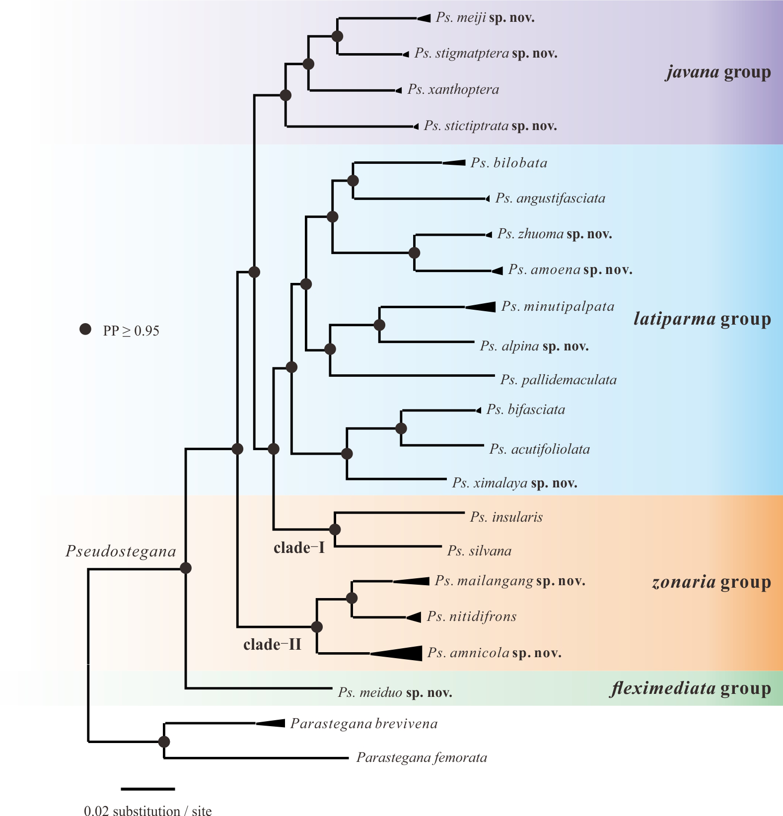
In general, the ML (Fig. 2) and Bayesian trees (Fig. S1) were similar in their topologies, especially at the terminal branches. Based on the phylogeny, Pseudostegana were recovered as a monophyletic genus with strong support. Within Pseudostegana, the monophyly of the fleximediata, latiparma and javana group all received strong support. The zonaria group was separated in two clades: (I) Pseudostegana insularis + Pseudostegana silvana and (II) Pseudostegana nitidifrons + Pseudostegana amnicola sp. nov. + Pseudostegana mailangang sp. nov. The monophyletic status of zonaria group is not supported by the Bayesian and ML analyses, as Pseudostegana insularis and Pseudostegana silvana were phylogenetically more closely related to the latiparma group (Fig. 3 and Fig. S1). At the species level, the phylogeny of the combined data set yielded 13 monophyletic clades and seven singletons with strong support.

Figure 2: Maximum likelihood tree of the genus Pseudostegana inferred from the COI, ND2 and 28S sequences.
The vertical bars represent the operational taxonomic units.
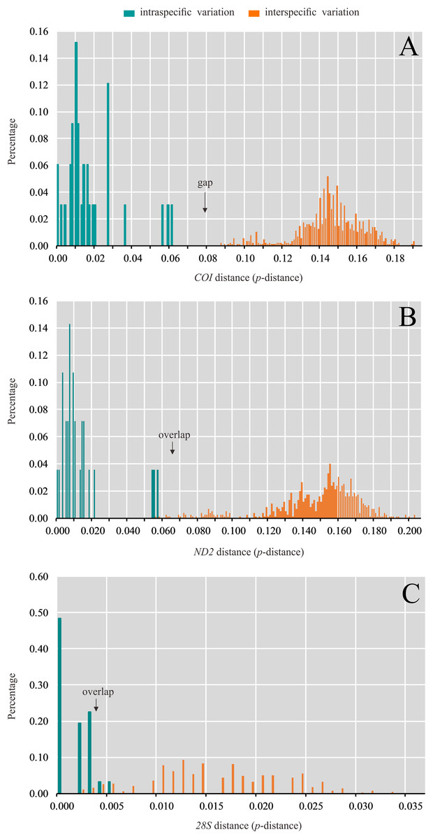
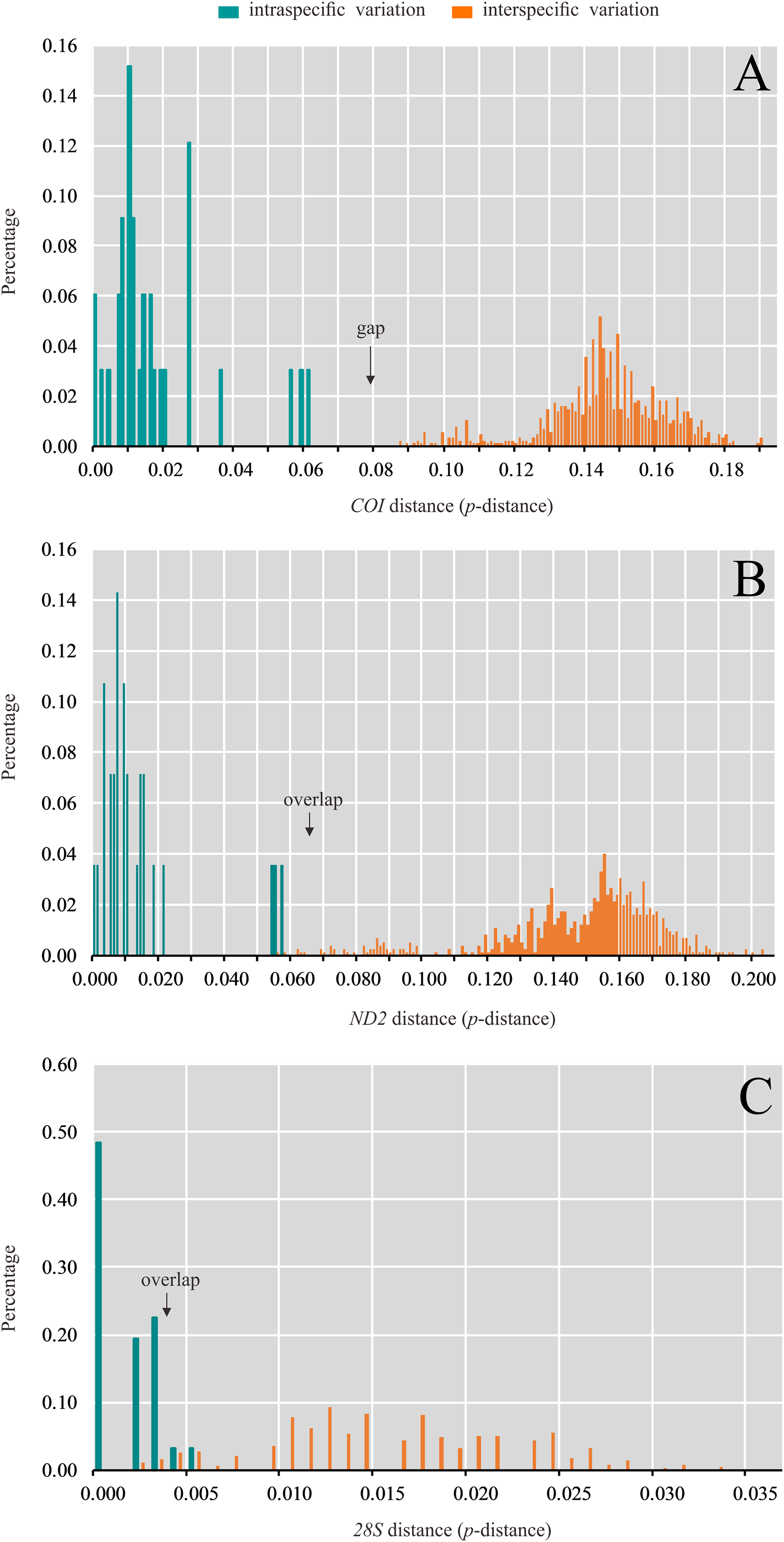
Figure 3: Distribution of intra- and interspecific pairwise genetic distance (p-distance) based on (A) COI, (B) ND2 and (C) 28S data set.
Species delimitation
The values of genetic variation (K2P distance) across taxonomic level are summarized (Table S3, S4 and S5). Intraspecific genetic variation calculated using COI ranged from 0.0% to 6.0% and the maximum intraspecific variation was detected in P. amnicola. In most cases, small intraspecific distances (<1%) were observed. The interspecific genetic variation ranged from 8.6% to 19.0% and the minimal interspecific genetic variation exceeded the maximum intraspecific genetic variation (Fig. 3A; Table S3). Intraspecific genetic variation calculated using ND2 ranged from 0.0% to 5.6% and the maximum intraspecific variation was also detected in P. amnicola. The interspecific genetic variation ranged from 5.6% to 20.2%. Thus the intraspecific and interspecific genetic variation slightly overlapped (Fig. 3B; Table S4). Compared to the mitochondrial genes, genetic variation for the nuclear gene 28S was small. The interspecific genetic variation ranged from 0.0% to 0.5%, while interspecific genetic variation ranged from 0.1% to 3.3%. The intraspecific and interspecific genetic variation largely overlapped (Fig. 3C; Table S5).
Species delimitation with the ABGD method based on the COI and ND2 resulted in 22 (Table S6) and 20 (Table S7) molecular operational taxonomic units (MOTUs), respectively. These two mitochondrial fragment were largely congruent in most of the MOTUs, while the analysis based on COI fragment divided P. amnicola to two groups (Fig. 2). The analysis based on the 28S data set yielded the lowest number of MOTUs (Table S8).
Systematic accounts
Genus Pseudostegana Okada
Stegana (Pseudostegana) Okada, 1978: 392; Okada, 1982: 39. Type species: Stegana (Parastegana) grandipalpis Takada & Momma, 1975.
Pseudostegana: Sidorenko, 2002: 14 (as a genus); Chen, Toda & Wang, 2005: 407; Li, Gao & Chen, 2010: 1402.
Diagnosis: Arista with one ventral branch except for terminal fork; subvibrissa longer than half the length of vibrissa; first tarsomere of foreleg with five or six black, short and thick setae basally; aedeagus lacking outer membrane (modified from Chen, Toda & Wang, 2005).
Description: Male and female (Figs. 4–9). Head: Eye brownish red. Ocellar triangle sometimes broadly or narrowly elongated to anterior margin of frons. Frons mostly glabrous, lacking minute, interfrontal setulae. Anterior reclinate orbital seta minute; posterior reclinate orbital seta situated nearer to proclinate seta than to inner vertical. Arista with long, dorsal branches. Clypeus brown to black. Subvibrissa mostly longer than half the length of vibrissa. Palpus slender in female, variable in male. Thorax: Mesonotum and scutellum dorsally convex. Katepisternal setae two or three; medial one shortest. Scutellum usually pale at tip; subscutellum swollen. Wing: Basal medial-cubital crossvein absent. Costal vein extending beyond tip of R4+5 vein, with five to seven peg-like spinules on ventral surface between veins R2+3 and R4+5. R2+3 vein slightly curved to costa at tip; M vein strongly convergent with R4+5 vein. Halter: stalk grayish; knob white. Legs: Mostly yellow, slender; mid tibia basally without strong, postero-dorsal setae. Abdomen: Sternites usually yellow to brown. Male terminalia: Epandrium broad, sometimes slightly constricted mid-dorsally, pubescent except for anterior margin. Surstylus separated from epandrium, mostly lacking pubescence, with several setae on outer and inner surfaces. Cercus separated from epandrium, pubescent and setigerous. Hypandrium broad, large, laterally mostly with one pair of paramedian setae, mid-anteriorly connected with apical part of aedeagal apodeme by aedeagal guide. Paramere with two long sensilla distally and several, small sensilla. Gonopods forming postero-median lobe, baso-laterally contiguous to parameres. Aedeagus usually with one pair of flap-like, serrated processes basally. Aedeagal apodeme long, rod-shaped, basally laterally flattened.
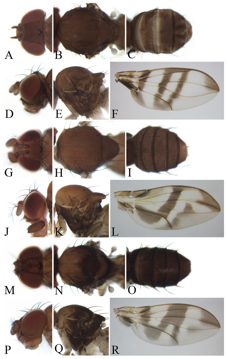
Figure 4: Morphological structures of female; frons, palpus, mesonotum, pleura, wing and abdominal tergites.
(A–F) Pseudostegana meiduo sp. nov. Morphological structures of male frons, palpus, mesonotum, pleura, wing and abdominal tergites: (G–L) Pseudostegana meiji sp. nov.; (M–R) Pseudostegana stictiptrata sp. nov. Photo credit: Yuan Zhang.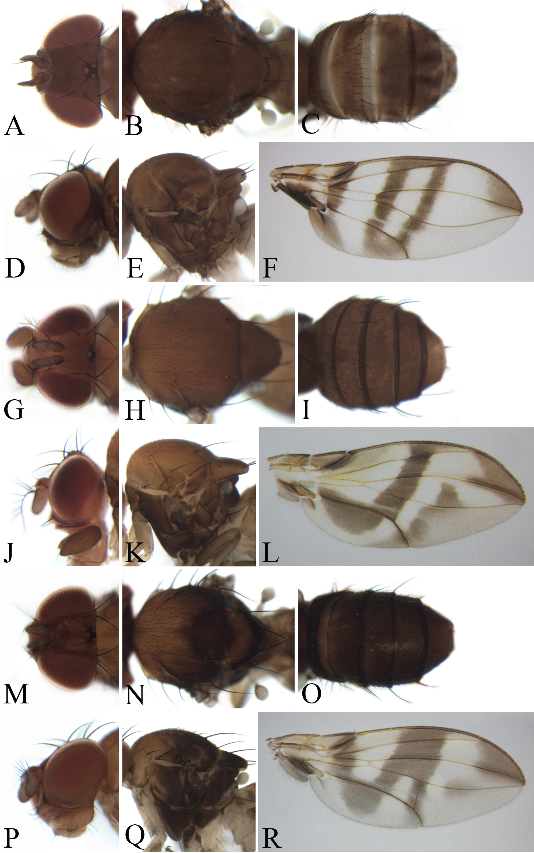
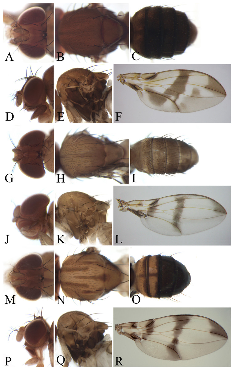
Figure 5: Morphological structures of male frons, palpus, mesonotum, pleura, wing and abdominal tergites.
(A–F) Pseudostegana stigmatptera sp. nov.; (G–L) Pseudostegana alpina sp. nov.; (M–R) Pseudostegana amoena sp. nov. Photo credit: Yuan Zhang.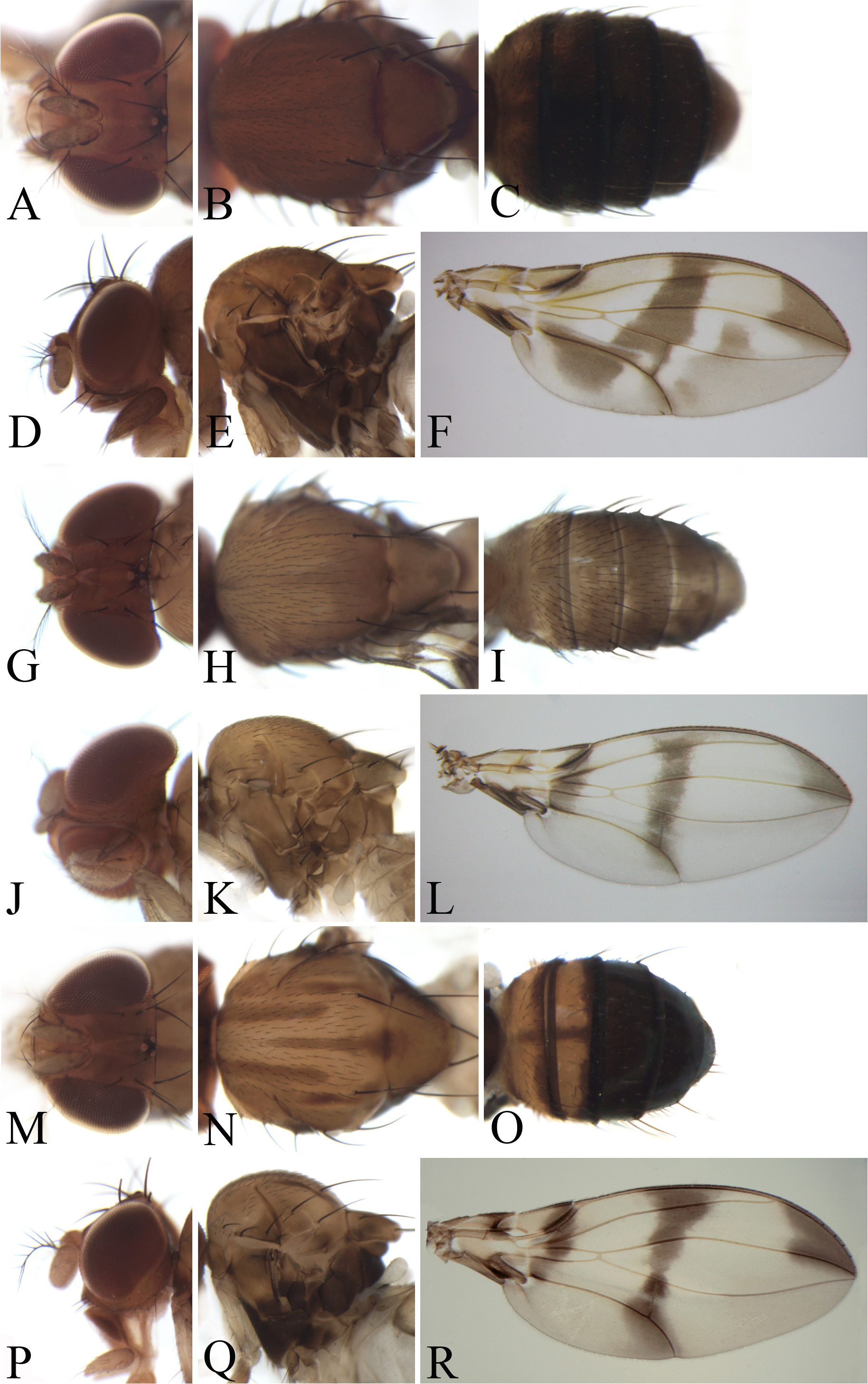

Figure 6: Morphological structures of male frons, palpus, mesonotum, pleura, wing and abdominal tergites.
(A–F) Pseudostegana ximalaya sp. nov.; (G–L) Pseudostegana zhuoma sp. nov.; (M–R) Pseudostegana amnicola sp. nov. Photo credit: Yuan Zhang.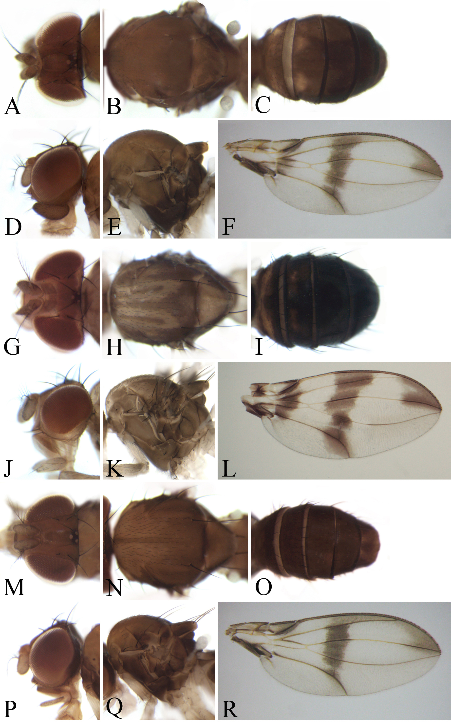
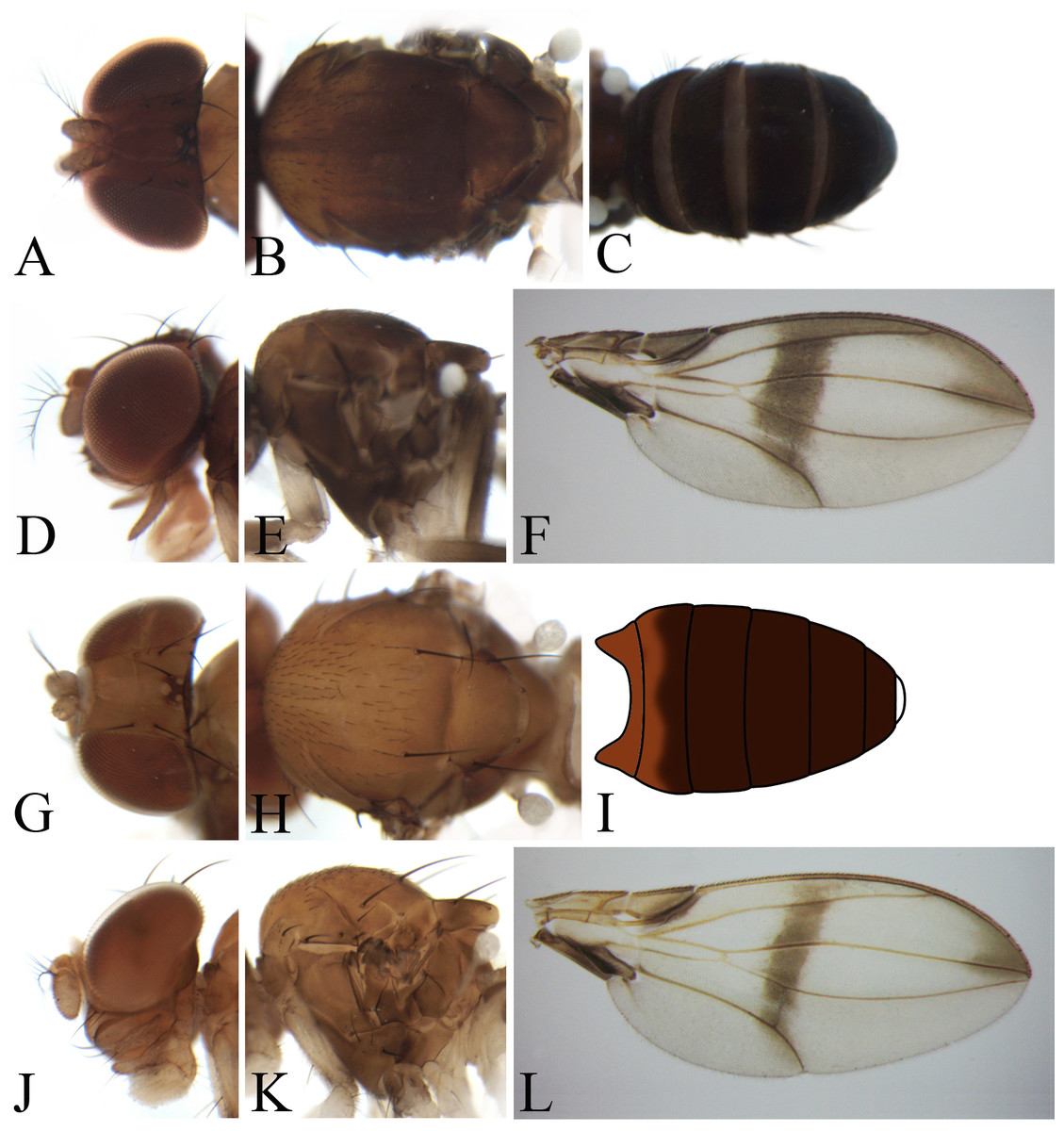
Figure 7: Morphological structures of male frons, palpus, mesonotum, pleura, wing and abdominal tergites.
(A–F) Pseudostegana mailangang sp. nov.; (G–L) Pseudostegana mystica sp. nov. Photo credit: Yuan Zhang.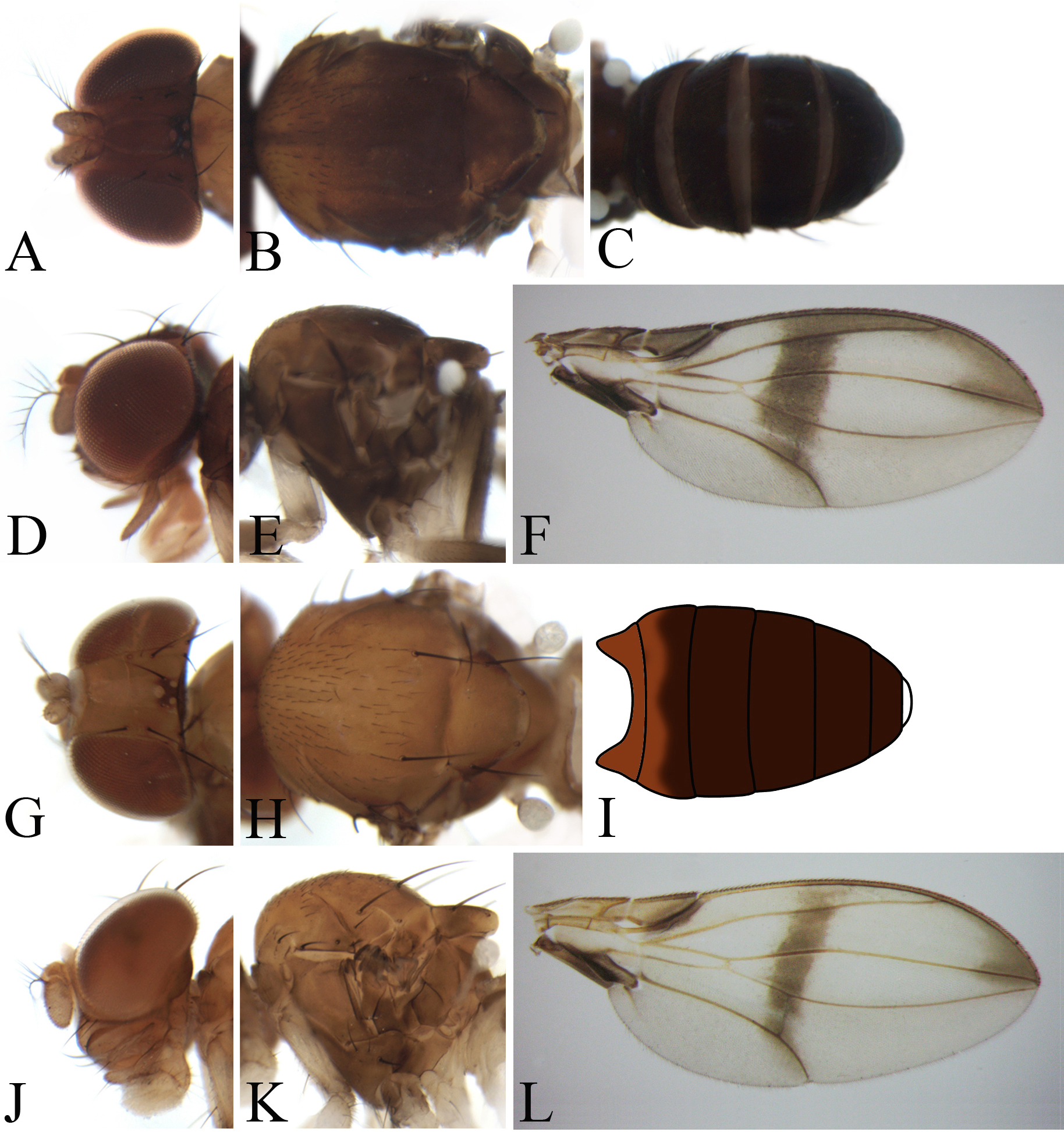
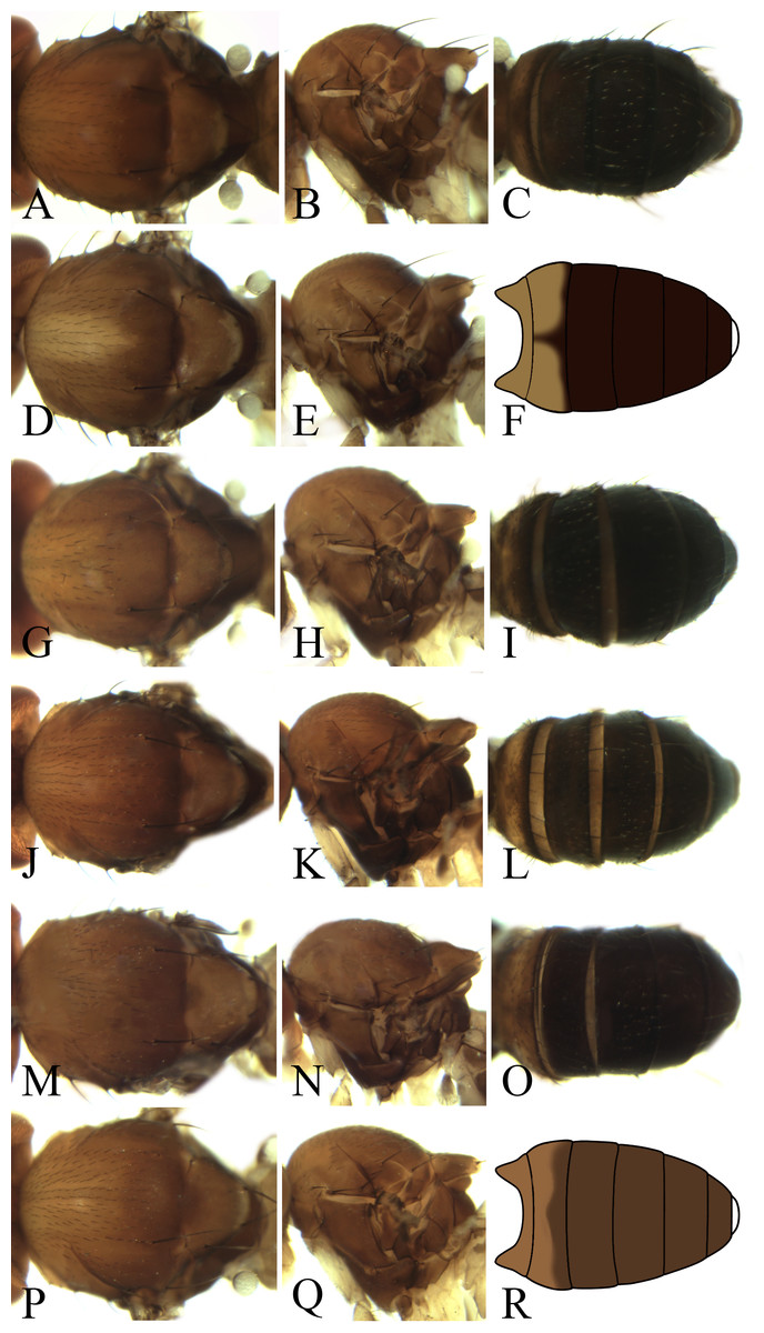
Figure 8: Morphological structures of male mesonotum, pleura and abdominal tergites.
(A–C) Pseudostegana acutifoliolata Li, Gao & Chen, 2010; (D–F) Pseudostegana angustifasciata Chen & Wang, 2005; (G–I) P. bifasciata Chen & Wang, 2005; (J–L) Pseudostegana bilobate Li, Gao & Chen, 2010; (M–O). Pseudostegana minutipalpata Li, Gao & Chen, 2010; (P–R) Pseudostegana pallidemaculata Chen & Wang, 2005. Photo credit: Yuan Zhang.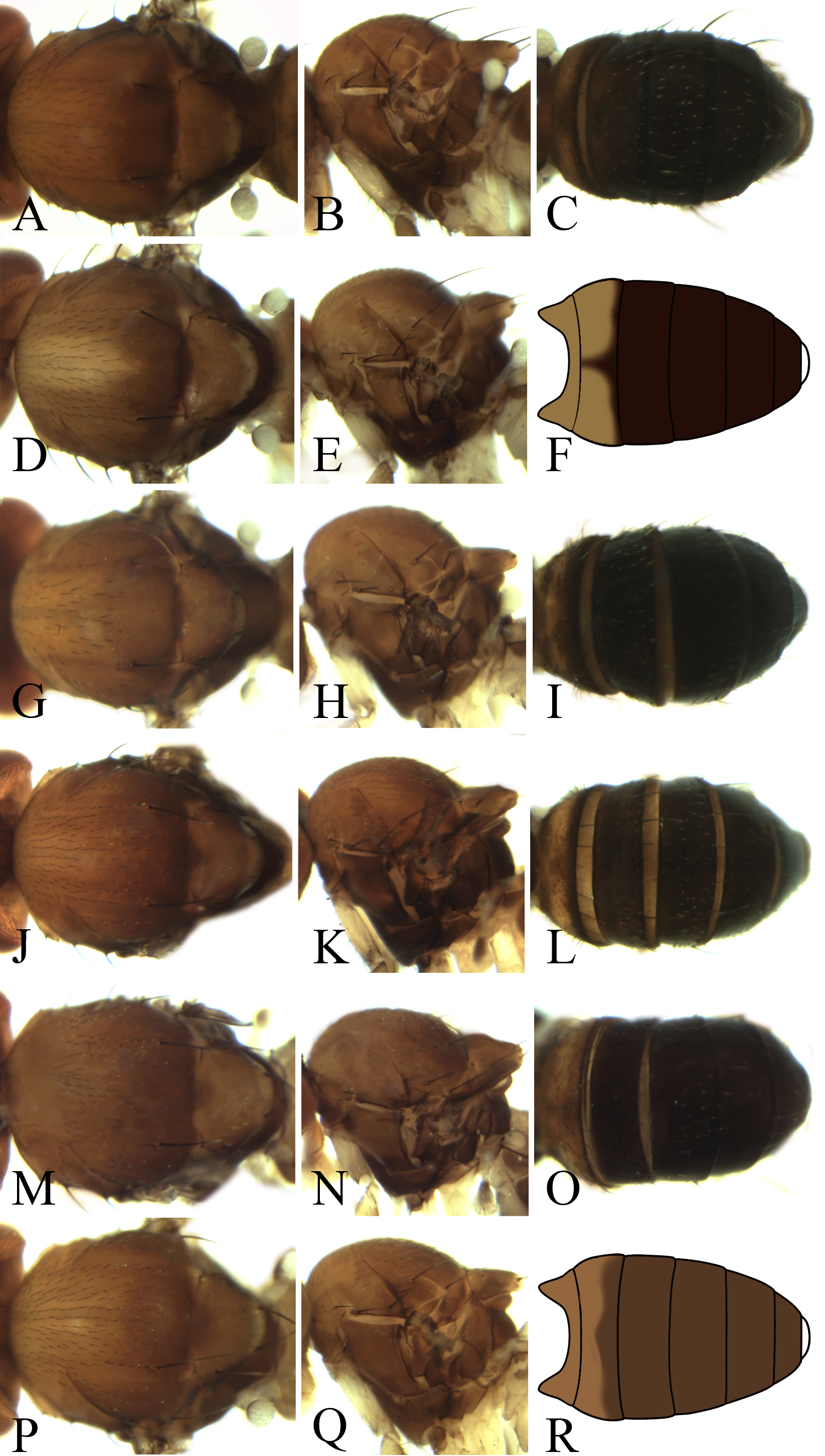
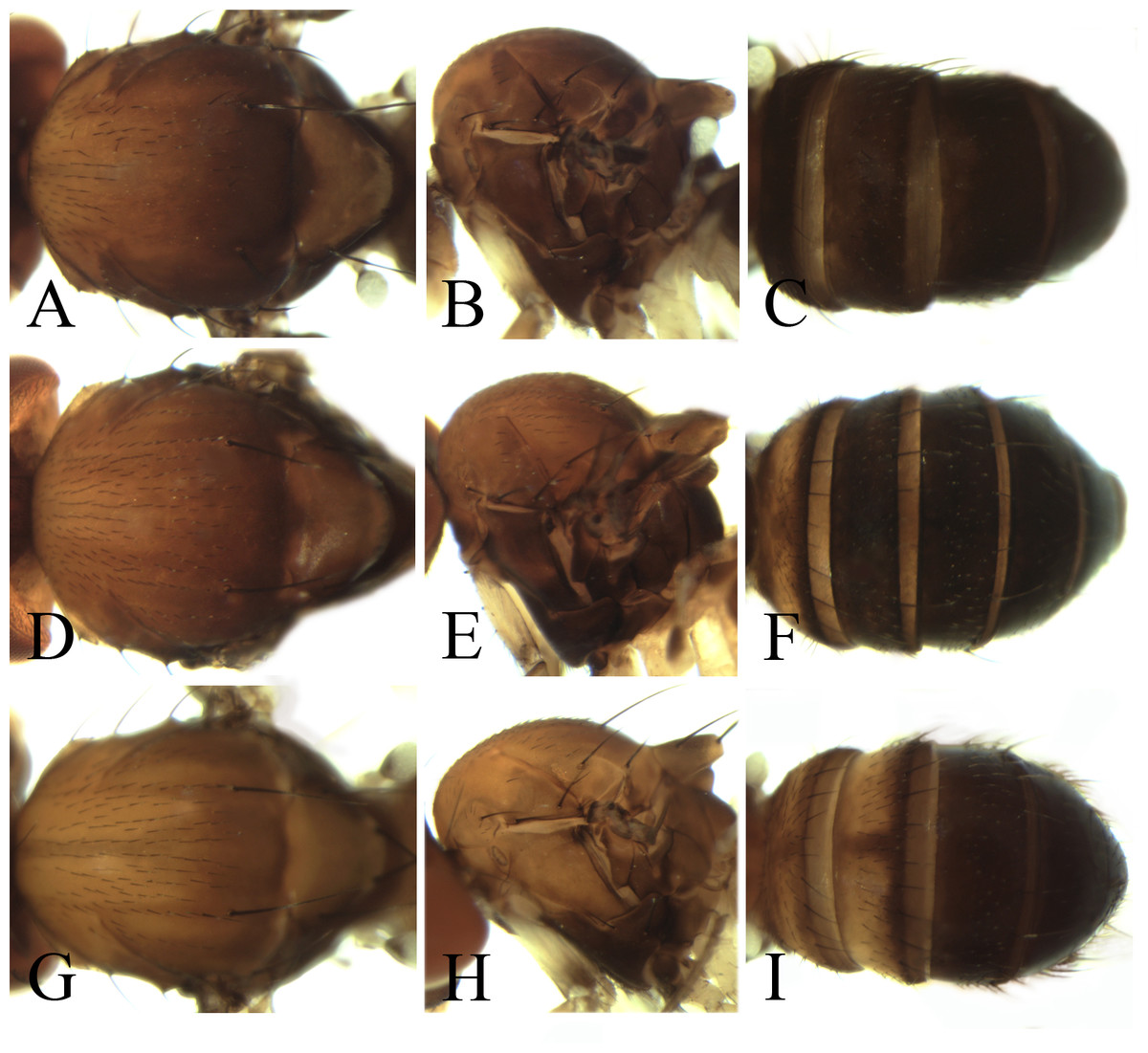
Figure 9: Morphological structures of male mesonotum, pleura and abdominal tergites.
(A–C) Pseudostegana insularis Li, Gao & Chen, 2010; (D–F) Pseudostegana nitidifrons Chen & Wang, 2005; (G–I) Pseudostegana silvana Li, Gao & Chen, 2010. Photo credit: Yuan Zhang.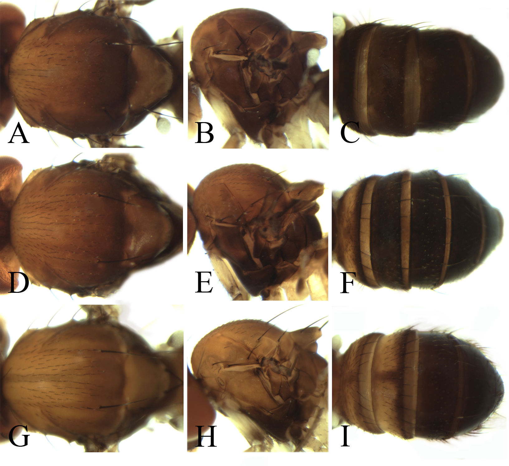
The fleximediata species group
Diagnosis: Ocellar triangle not elongated (Fig. 4A); M vein strongly curved after dm-cu crossvein (Fig. 4F); medial, dark color band much narrower than a half distance between r-m and dm-cu crossveins (Fig. 4F); seventh tergite with slender processes laterally in male.
Pseudostegana meiduo Zhang & Chen, sp. nov.
urn:lsid:zoobank.org:act:CD5E03C7-3130-4FF6-9A1B-C4FE6690B7ED
Diagnosis: This species obviously differs from the other fleximediata species by the wing having an annular patch medially (Fig. 4F), and distinct cross band subbasally (Fig. 4F).
Description: Female. Head: Ocellar triangle not elongated (Fig. 4A). Front brownish (Fig. 4A). Face grayish brown medially, dark brown laterally, black on lower corners. Clypeus and gena dark brown. Palpus brown, slightly broad (Fig. 4D). Thorax: Mesonotum and scutellum nearly brown (Fig. 4B). Pleura dark brown (Fig. 4E). Legs: Yellow, brown on forefemur and hind tibia, brown to dark brown on mid- and hindleg femora. Abdomen: All abdominal tergites nearly brown (Fig. 4C). Sternites yellow.
Measurements: BL = 2.40 mm in holotype, ThL = 1.25 mm, WL = 2.63 mm, WW = 1.10 mm, arb = 8/1, avd = 1.17, adf = 1.61, flw = 1.82, FW/HW = 0.43, ch/o = 0.09, prorb = 1.10, rcorb = 1.08, vb = 0.48, dcl = 0.29, sctl = 1.44, sterno = 0.70, orbito = 0.79, dcp = 0.13, sctlp = 1.11.
Type specimen: Holotype female (SCAU, no. 124842), CHINA: Beibeng, Motuo, Xizang, 29°19′N, 95°20′E, 1,000 m, 2.x.2010, ex. fallen logs, L Wu.
Etymology: The name means “flower” in Tibetan.
Distribution: China (Xizang).
The javana species group
Diagnosis: Wing with basal and medial cross bands fused posteriorly, forming V-shaped pattern (Figs. 4L, 4R and 5F).
Pseudostegana meiji Zhang & Chen, sp. nov.
urn:lsid:zoobank.org:act:0E0F9E07-C2E7-48F6-B021-F699E21F88CC
Diagnosis: This species is similar to Pseudostegana stigmatptera sp. nov. by the wing patches (Fig. 4L) and male terminalia (Fig. 10), but can be distinguished by the surstylus: in this species, the surstylus is strongly protruded on posterior corner in lateral view (Fig. 10A), broadened, and approximately one and half times as high as wide (Fig. 10B); compare with Pseudostegana stigmatptera sp. nov. The 5.8% COI interspecific genetic distance to P. yiqini is one of the smallest interspecific distances ascertained within this subgenus.

Figure 10: Pseudostegana meiji sp. nov., male terminalia.
(A) Epandrium (epan), cercus (cerc) and surstylus (sur) (lateral view); (B) surstylus (ventral view); (C) hypandrium (hypd), paramere (pm), gonopods (gon), aedeagus (aed) and aedeagal apodeme (aed a) (ventral view); (D) ditto (lateral view). Scale bars = 0.1 mm. Drawing credit: Yuan Zhang.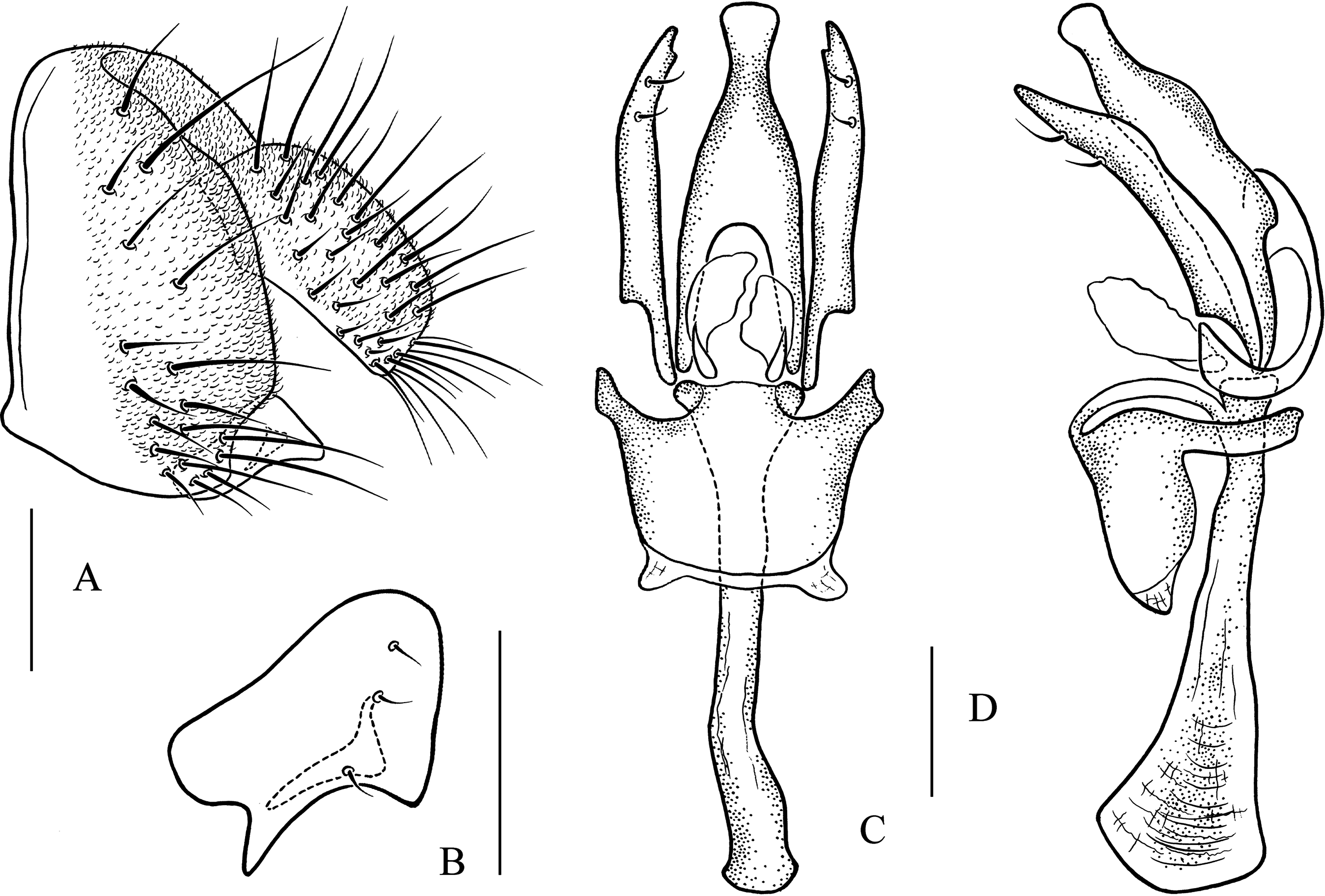
Description: Male and female. Head: Ocellar triangle not elongated (Fig. 4G). Frons brown in male (Fig. 4G), yellowish brown in female. Face and gena yellow. Clypeus yellow medially, black laterally. Palpus brown in both sexes, broad, large in male, medially about one-third as wide as long, slender in female (Fig. 4J). Thorax: Mesonotum yellow in male (Fig. 4H), brownish yellow in female; scutellum brown (Fig. 4H). Pleura dark brown in male (Fig. 4K), dark brown to black in female. Legs: Mostly yellow; brown to dark on mid- and hindleg femora. Abdomen: All tergites brown (male) to dark brown (female), yellow latterly (Fig. 4I). Sternites brownish yellow in male, brown in female. Male terminalia: Epandrium with ∼18 setae (Fig. 10A). Hypandrial paramedian setae absent (Fig. 10C). Paramere shallowly bifurcated apically (Fig. 10C). Aedeagus round apically, with membranous processes basally (Figs. 10C and 10D).
Measurements: BL = 2.85 mm in holotype (range in five males and five females paratypes, 2.63–3.01 mm in males, 2.53–2.93 mm in females), ThL = 1.25 mm (1.26–1.43 mm in males, 1.00–1.46 mm in females), WL = 2.43 mm (2.47–2.67 mm in males, 2.07–2.63 mm in females), WW = 1.06 mm (1.10–1.20 mm in males, 1.00–1.20 mm in females), arb = 7/1 (6–8/1), avd = 0.72 (0.86–1.00), adf = 2.63 (1.83–2.24), flw = 2.37 (1.70–2.07), FW/HW = 0.39 (0.39–0.55), ch/o = 0.09 (0.06–0.09), prorb = 0.85 (0.79–0.96), rcorb = 0.93 (0.86–1.21), vb = 0.73 (0.64–0.95), dcl = 0.30 (0.31–0.35), sctl = 1.13 (0.89–1.05), sterno = 0.48 (0.60–1.00), orbito = 0.98 (1.00–1.03), dcp = 0.17 (0.18–0.16), sctlp = 1.24 (0.94–1.21).
Type specimens: Holotype male (SCAU, no. 122031), CHINA: Muyiji Park, Ximeng, Yunnan, 22°37′N, 99°36′E, 1,100 m, 2.iv.2011, ex. fallen logs, YR Su. Paratypes: CHINA: two females (SCAU, nos. 122032, 33), same data as holotype; 10 males, 15 females (five males and five females in KIZ, nos. 0088165–74; the rest in SCAU, nos. 124844–58), Muyiji, Ximeng, Yunnan, 22°37′N, 99°36′E, 1,200 m, 29.iv. −3.v.2016, ex. tussock, J Huang, YQ Liu, YL Wang, L Zhu; eight males, 14 females (SCAU, nos. 124859–80), Mengdong, Cangyuan, Yunnan, 23°10′N, 99°14′E, 1,320 m, 7.v.2016, ex. tussock, J Huang, YQ Liu, YL Wang, L Zhu; one male, (SCAU, no. 124881), Botanic Garden, Ruili, Yunnan, 24°1′N, 97°51′E, 1,174 m, 22.viii.2016, ex. tussock, L Gong; one male, two females (SCAU, nos. 111353–55), Santaishan, Mangshi, Yunnan, 24°19′N, 98°20′E, 900 m, 4.xi.2017, ex. tussock, HW Chen, L Gong, BX Li; one male, one female (SCAU, nos. 111356, 57), Moli Forest Park, Ruili, Yunnan, 24°7′N, 97°59′E, 920 m, 5.xi.2017, L Gong, BX Li.
Etymology: The name means “great deity” in the language of the Wa nationality in Yunnan Province.
Distribution: China (Yunnan).
Pseudostegana stictiptrata Zhang & Chen, sp. nov.
urn:lsid:zoobank.org:act:88C101ED-F994-4E0A-8CCB-B2F60378412F
Diagnosis: This species differs from the other javana species by the wing pattern (Fig. 4R), and the epandrium being roundly protruded ventrally, with dense setae (Fig. 11A).
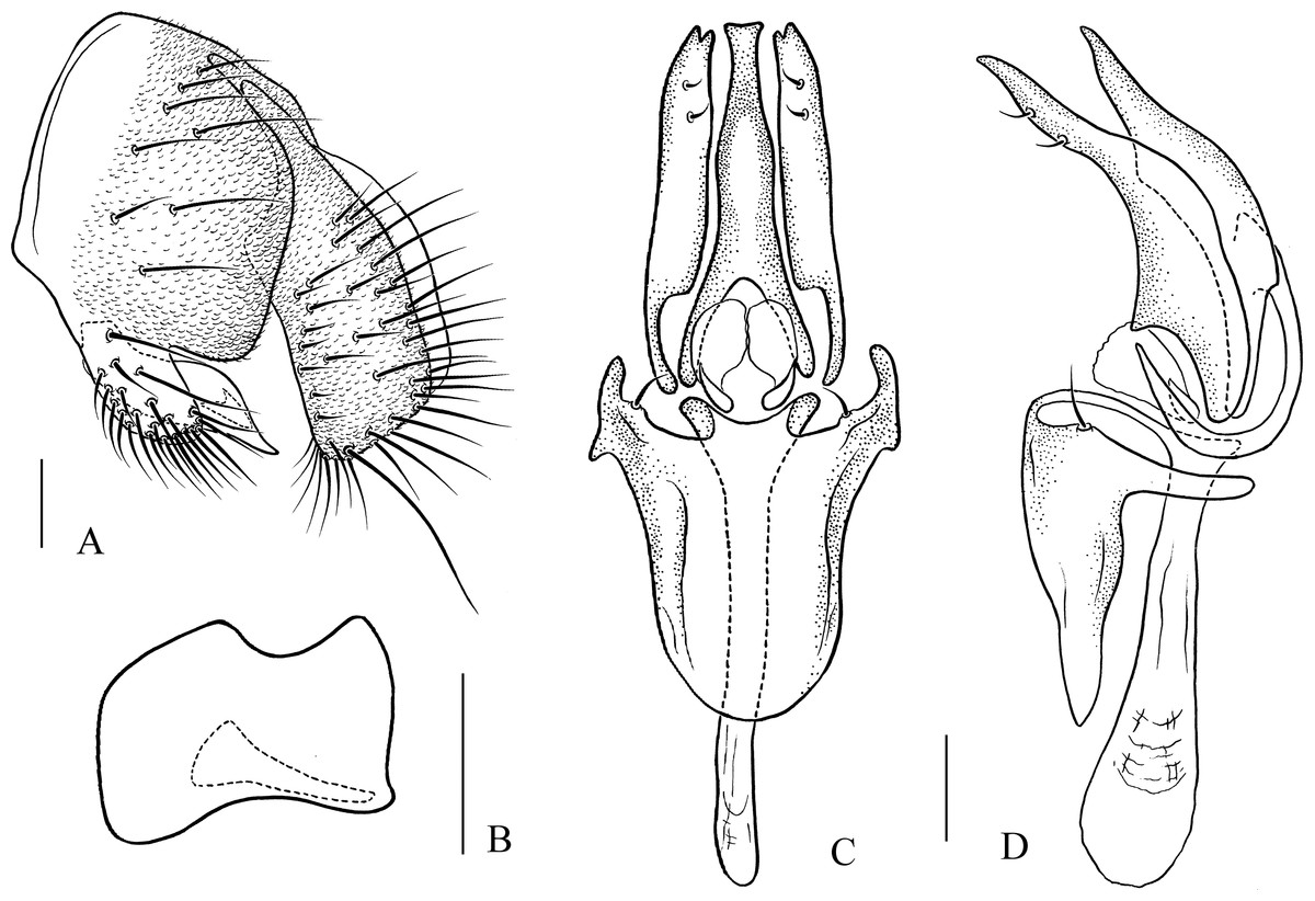
Figure 11: Pseudostegana stictiptrata sp. nov., male terminalia.
(A) Epandrium, cercus and surstylus; (B) surstylus (ventral view); (C) hypandrium, paramere, gonopods, aedeagus and aedeagal apodeme (ventral view); (D) ditto (lateral view). Scale bars = 0.1 mm. Drawing credit: Yuan Zhang.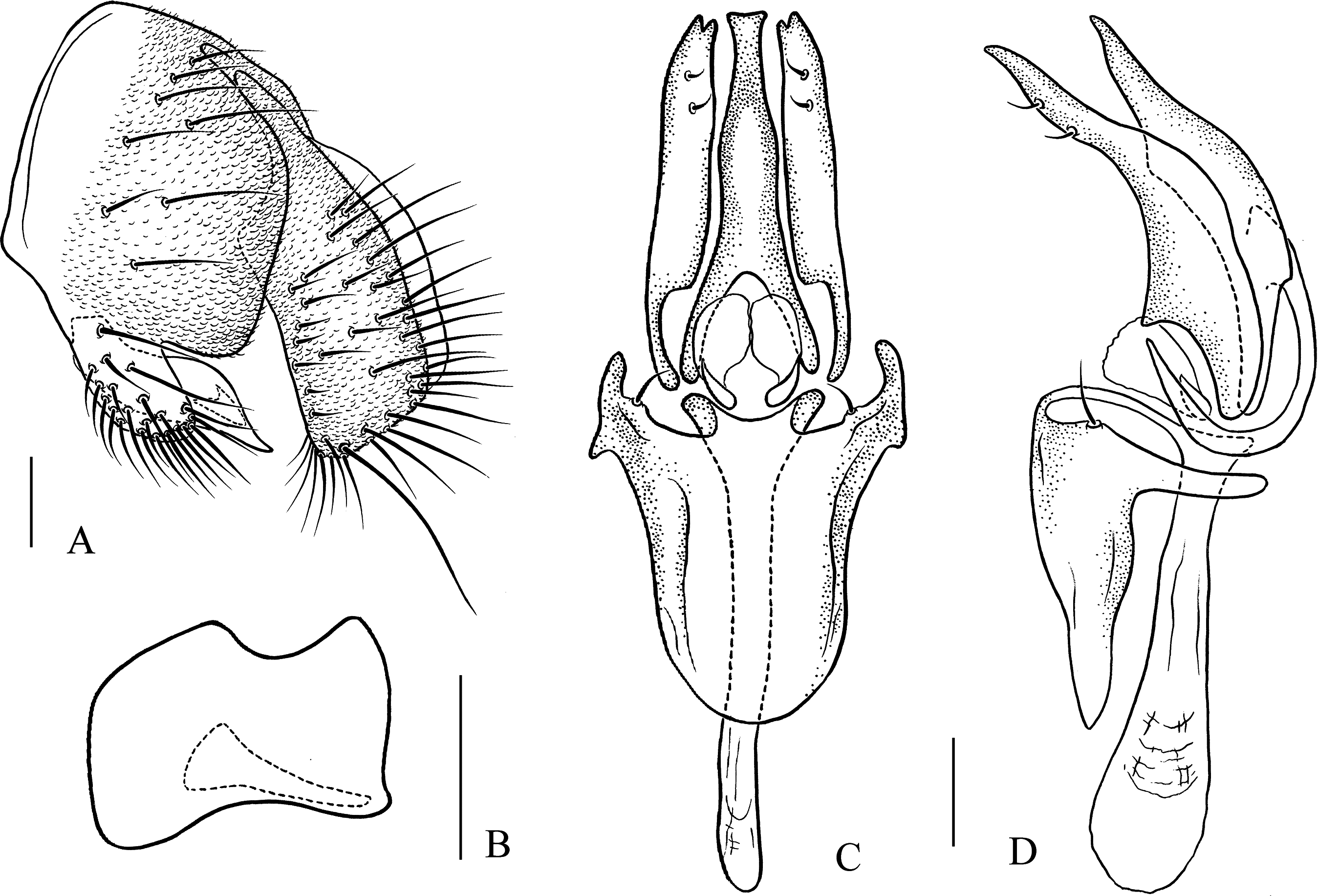
Description: Male. Head: Ocellar triangle elongated (Fig. 4M). Frons yellow to yellowish brown (Fig. 4M). Face brown. Clypeus brown medially, black laterally. Gena orange brown. Palpus yellow, broad, large in male, medially about one-third as wide as long (Fig. 4P). Thorax: Mesonotum orange–yellow on anterior one-third to half, black on posterior half to one-third (Fig. 4N); scutellum and pleura black (Fig. 4N). Pleura dark brown to black (Fig. 4Q). Legs: Mostly yellow, dark brown distally on all femora. Abdomen: All tergites dark brown to black (Fig. 4O). Sternites yellow on second and third, brownish fourth to sixth. Male terminalia: Epandrium with numerous setae (Fig. 11A). Cercus slightly protruded ventrally (Fig. 11A). Surstylus strongly protruded on posterior corner in lateral view (Fig. 11A), broadened, approximately one and a half times as high as wide (Fig. 11B). Hypandrium with one pair of paramedian setae (Figs. 11C and 11D). Paramere shallowly bifurcated apically (Figs. 11C and 11D). Aedeagus apically slightly concave in ventral view, with membranous processes basally (Figs. 11C and 11D).
Measurements: BL = 3.39 mm in holotype (range in four males paratypes: 2.40–2.83 mm), ThL = 1.52 mm (1.25–1.39 mm), WL = 3.03 mm (2.57–2.89 mm), WW = 1.36 mm (1.12–1.28 mm), arb = 8/1 (6–8/1), avd = 0.69 (0.81–0.87), adf = 2.08 (1.83–2.33), flw = 1.86 (1.52–2.00), FW/HW = 0.46 (0.35–0.42), ch/o = 0.08 (0.06–0.10), prorb = 0.78 (0.84–1.04), rcorb = 0.94 (0.80–0.87), vb = 0.56 (0.80–1.08), dcl = 0.29 (0.25–0.33), sctl = 0.94 (0.98–1.04), sterno = 0.71 (0.79–0.87), orbito = 1.20 (0.96–1.11), dcp = 0.17 (0.22), sctlp = 1.28 (1.11–1.25).
Type specimens: Holotype male (SCAU, no. 122034), CHINA: Hesong, Menghai, Yunnan, 21°49′N, 100°06′E, 1,700 m, 12.v.2012, ex. stone with moss, HW Chen. Paratypes: CHINA: two males (SCAU, nos. 122035, 36), same data as holotype; two males (SCAU, nos. 124882, 83), Yakou, Huanglianshan, Lvchun, Yunnan, 22°50′N, 102°17′E, 1,900 m, 31.x.2016, ex. tussock, HW Chen; one male (KIZ, 0090510), Daweishan, Pingbian, Yunnan, 22°92′N, 103°68′E, 1,700–1,900 m, 12.viii.2017, ex. tussock, HW Chen.
Etymology: A combination of the Greek words: “stict” + “pteron,” referring to the wing having pattern.
Distribution: China (Yunnan).
Pseudostegana stigmatptera Zhang & Chen, sp. nov.
urn:lsid:zoobank.org:act:72488FCE-4052-403B-A450-3A9312B4BB2F
Diagnosis: Surstylus strongly protruded and pointed on anterior corner, broadened, approximately two times as high as wide (Fig. 12B).
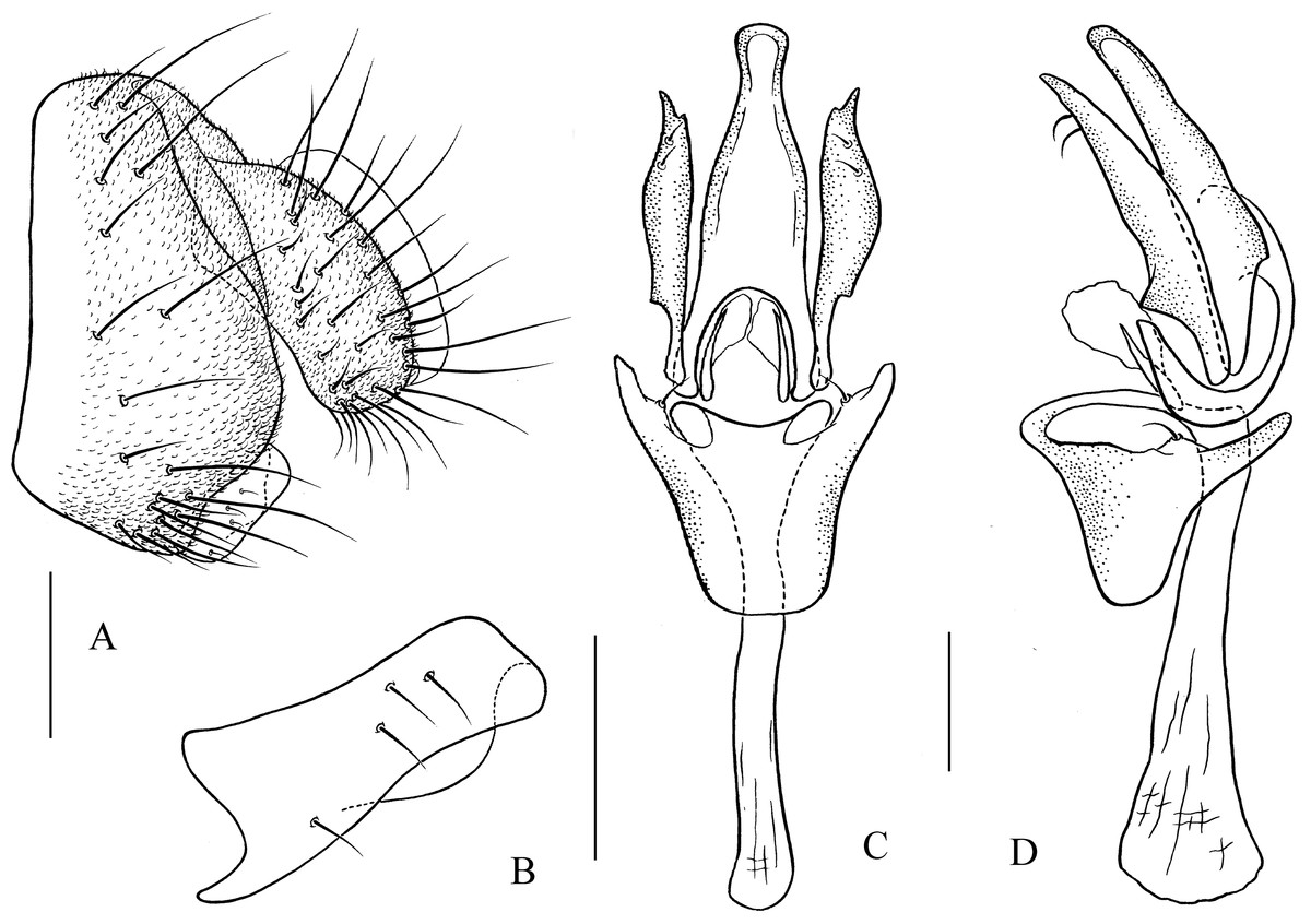
Figure 12: Pseudostegana stigmatprata sp. nov., male terminalia.
(A) Epandrium, cercus and surstylus; (B) surstylus; (C) hypandrium, paramere, gonopods and aedeagus (ventral view); (D) ditto (lateral view). Scale bars = 0.1 mm. Drawing credit: Yuan Zhang.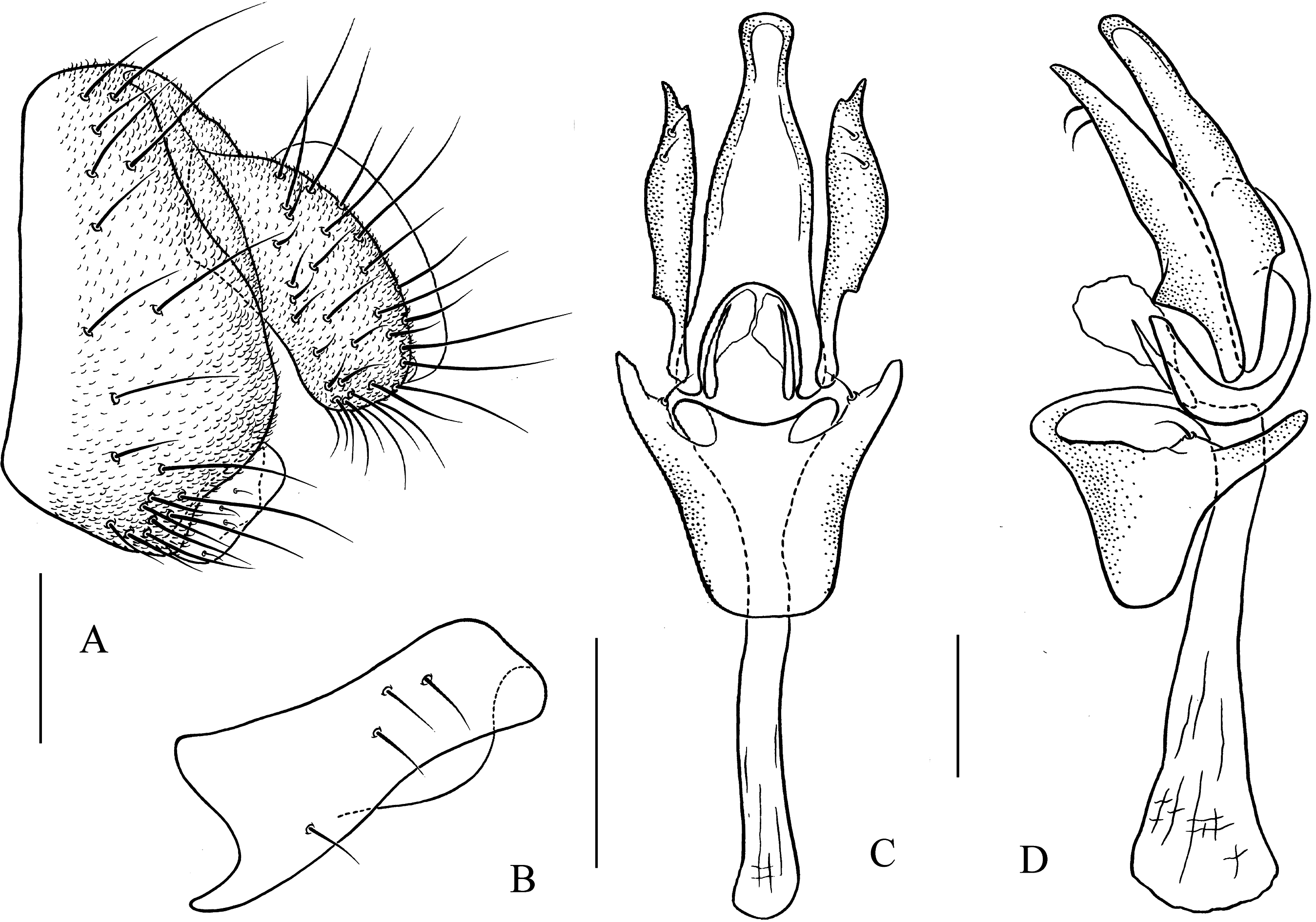
Description: Male and female. Head: Ocellar triangle not elongated (Fig. 5A). Frons brown (Fig. 5A). Face and gena yellow. Clypeus dark brown. Palpus brown yellow, broad, large in male, medially one-third as wide as long (Fig. 5D). Thorax: Mesonotum yellowish brown; scutellum brown, yellow at tip (Fig. 5B). Pleura dark brown (Fig. 5E). Legs: Mostly yellow, brown on all femora. Abdomen: All tergites black, sometimes laterally yellow on second to fourth (Fig. 5C). Sternites brown in male, dark brown in female. Male terminalia: Epandrium with ∼19 setae (Fig. 12A). Hypandrial paramedian setae present (Figs. 12C and 12D). Paramere shallowly bifurcated apically (Figs. 12C and 12D). Aedeagus apically round, with membranous processes basally (Figs. 12C and 12D).
Measurements: BL = 2.97 mm in holotype (range in five males and five females paratypes, 2.41–2.97 mm in males, 2.88–3.23 mm in females), ThL = 1.37 mm (1.15–1.42 in males, 1.42–1.50 mm in females), WL = 2.80 mm (2.41–2.79 mm in males, 2.73–2.89 mm in females), WW = 1.17 mm (1.13–1.30 mm in males, 1.23–1.35 mm in females), arb = 7/1 (6–7/1), avd = 0.97 (0.72–1.09), adf = 1.61 (1.67–1.96), flw = 2.03 (1.46–1.97), FW/HW = 0.47 (0.46–0.52), ch/o = 0.04 (0.06–0.09), prorb = 0.77 (0.74–0.91), rcorb = 0.96 (0.82–1.06), vb = 0.71 (0.73–0.89), dcl = 0.29 (0.21–0.30), sctl = 0.84 (1.02–1.11), sterno = 0.41 (0.43–0.71), orbito = 1.05 (0.87–1.11), dcp = 0.18 (0.16–0.26), sctlp = 1.05 (0.95–1.22).
Type specimens: Holotype male (SCAU, no. 122037), CHINA: Hesong, Menghai, Yunnan, 1,700–1,900 m, 16.iv.2010, ex. tussock, JJ Gao. Paratypes: CHINA: 25 males, 28 females (five males, five females in KIZ, nos. 0088175–82, 0090501–02; 20 males, 23 females in SCAU, nos. 122038–122080), ex. tree trunks, tussock and stone with moss, JJ Gao, YR Su, L Wang, L Wu, the rest same data as holotype; 26 males, 24 females (SCAU, nos. 122081–122130), 26.iii.2011, 7.iv.2011, 11.v.2012, ex. tree trunks, tussock and stone with moss, HW Chen, JM Lu, ZF Shao, YR Su, SJ Yan, same data as holotype; three males, three females (SCAU, nos. 122131–36), Baihualing, Baoshan, Yunnan, 25°18′N, 98°48′E, 1,400 m, 22.vi.2013, 24.viii.2013, ex. tree tussock, KY An, QS Gao, K Liu, JJ Liu.
Etymology: A combination of the Greek words: “stigma” + “pteron” meaning spotted wing.
Distribution: China (Yunnan).
The latiparma species group
Diagnosis: Wing subbasally with distinct cross band (Figs. 5L, 5R, 6F and 6L).
Pseudostegana alpina Zhang & Chen, sp. nov.
urn:lsid:zoobank.org:act:31D32207-72FD-41A4-8971-8349404EB97C
Diagnosis: This species is related to Pseudostegana minutipalpata Li, Gao & Chen, 2010 from Yunnan by the wing pattern (Fig. 5L), and periphallic organs (Fig. 13A), but can be distinguished by the surstylus and aedeagus; in this species, the surstylus is protruded on anterior corner (Fig. 13B); aedeagus apically acute and subapically so strongly, dorsally protruded in lateral view (Fig. 13D).

Figure 13: Pseudostegana alpina sp. nov., male terminalia.
(A) Epandrium, cercus and surstylus; (B) surstylus; (C) hypandrium, paramere, gonopods, aedeagus and aedeagal apodeme (ventral view); (D) ditto (lateral view). Scale bars = 0.1 mm. Drawing credit: Yuan Zhang.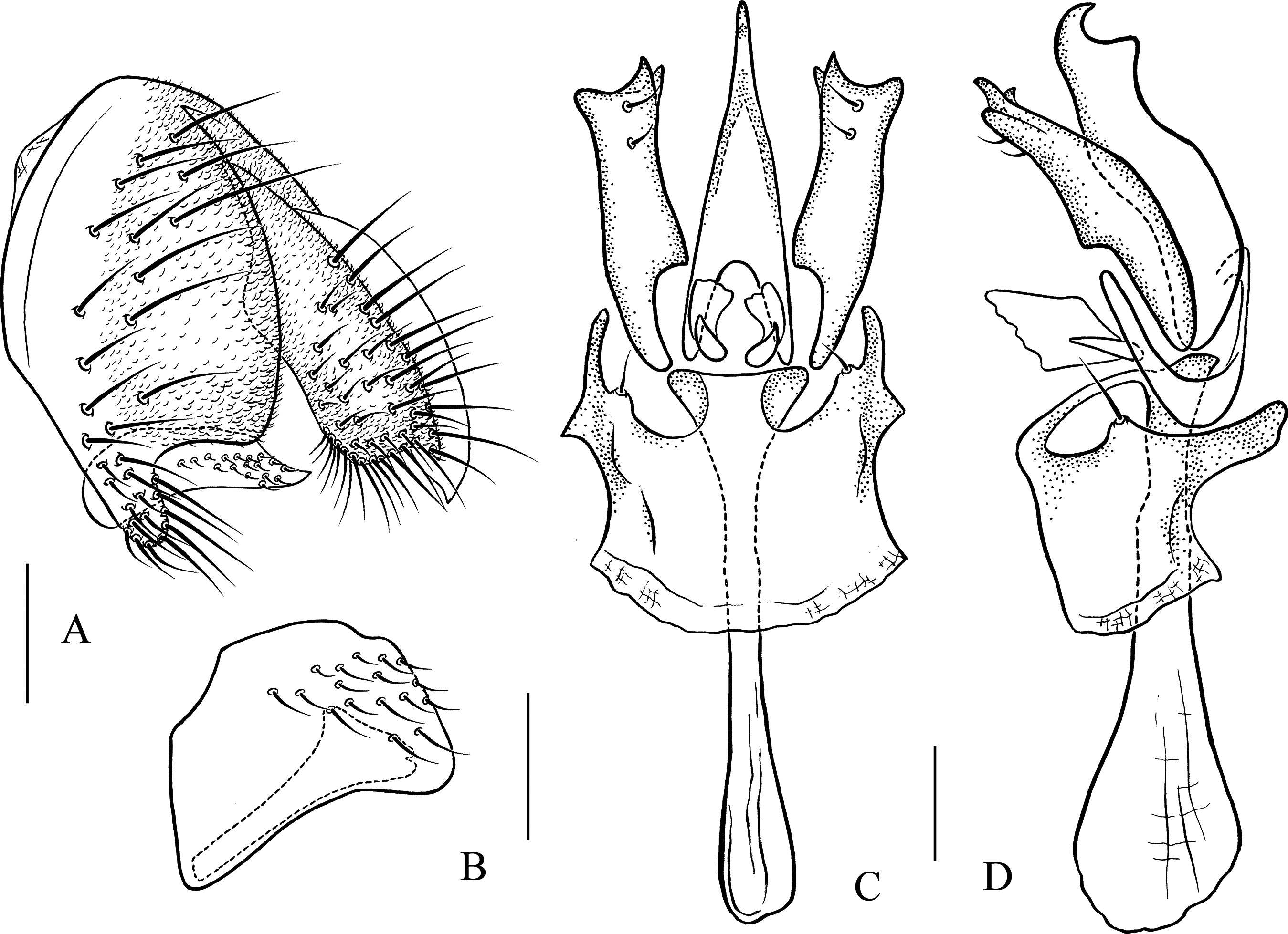
Description: Male. Head: Ocellar triangle not elongated (Fig. 5G). Frons brown (Fig. 5G). Face and gena brown. Clypeus brown medially, black laterally. Palpus brown, slender (Fig. 5J). Thorax: Mesonotum medially yellow and with a thin brownish yellow, longitudinal stripe, laterally brownish yellow; scutellum brownish yellow (Fig. 5H). Pleura slightly glossy, brownish yellow above, dark brown below (Fig. 5K). Legs: Yellow, dark brown on all knees. Abdomen: All tergites brown, yellow on first to third medially (Fig. 5I). Sternites yellow. Male terminalia: Epandrium roundly protruded on ventral margin, with numerous setae (Fig. 13A). Surstylus slightly protruded on posterior corner (Fig. 13B). Cercus ventrally elongated in lateral view (Fig. 13A). Hypandrium with one pair of paramedian setae (Figs. 13C and 13D). Paramere acute, with one small projection apically, subapically broadened (Figs. 13C and 13D) Aedeagus apically slightly pointed in ventral view (Fig. 13C), basally with nearly membranous processes (Figs. 13C and 13D).
Measurements: BL = 2.89 mm in holotype, ThL = 1.24 mm, WL = 2.68 mm, WW = 1.23 mm, arb = 7/1, avd = 0.97, adf = 1.84, flw = 1.64, FW/HW = 0.41, ch/o = 0.08, prorb = 1.15, rcorb = 1.22, vb = 0.95, dcl = damage, sctl = 1.25, sterno = 0.69, orbito = 1.11, dcp = damage, sctlp = 1.05.
Type specimen: Holotype male (SCAU, no. 122158), CHINA: Hesong, Menghai, Yunnan, 1,800 m, 12.v.2012, ex. stone with moss, HW Chen.
Etymology: From the Latin word: alpinus, means high mountain, referring to the type locality.
Distribution: China (Yunnan).
Pseudostegana amoena Zhang & Chen, sp. nov.
urn:lsid:zoobank.org:act:D3696645-FA69-4B4B-A5D8-D6FCF6F90764
Diagnosis: This species is related to Pseudostegana zhuoma sp. nov. by the wing pattern (Figs. 5R and 6L) and male terminalia (Figs. 14 and 16); they can be distinguished by the yellow mesonotum, with five brown, longitudinal stripes (Fig. 5N); scutellum yellowish brown, yellow at tip (Fig. 5N); pleura brownish yellow anteriorly, black posteriorly (Fig. 5Q); aedeagus slightly smooth ventrally (Fig. 14C); compare with Pseudostegana zhuoma sp. nov.
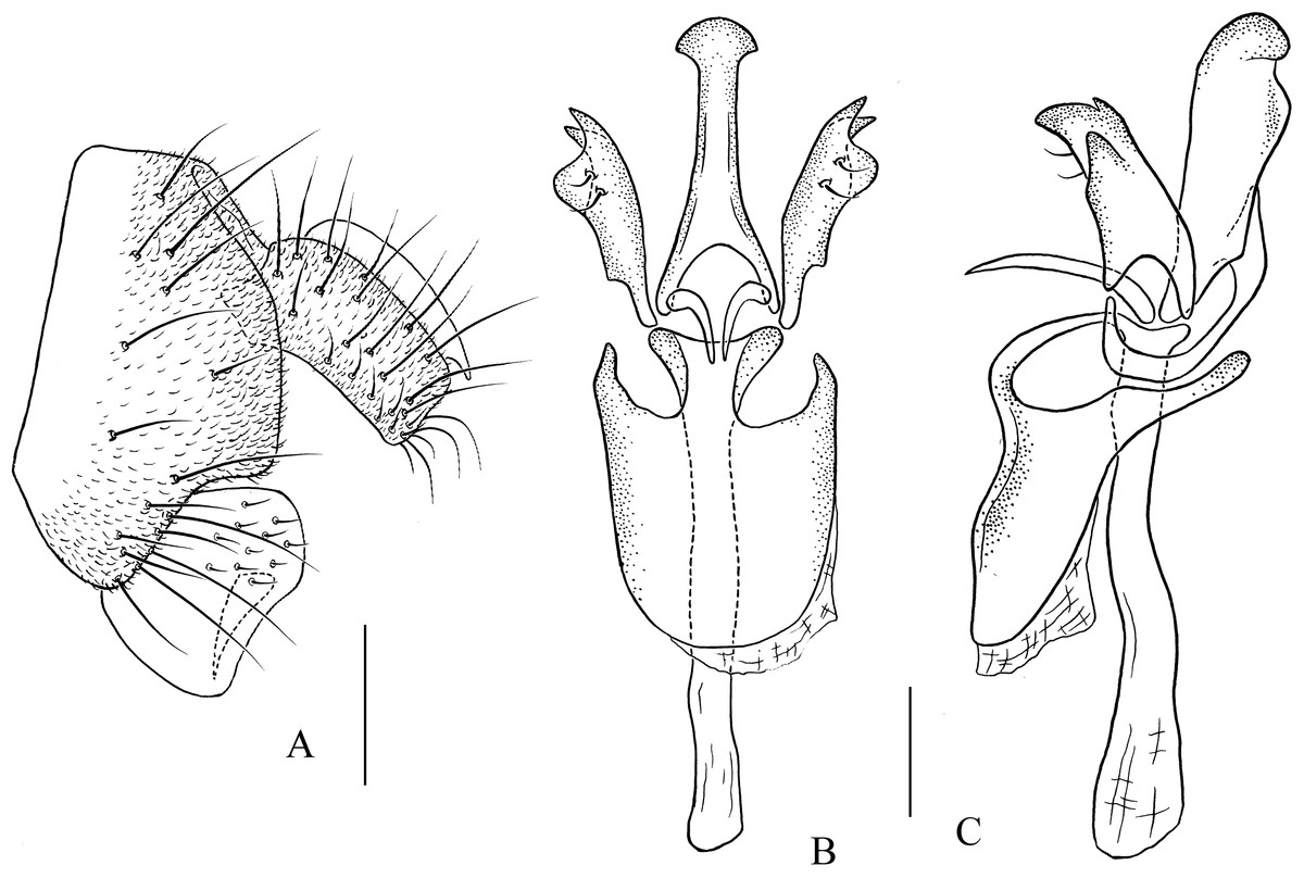
Figure 14: Pseudostegana amoena sp. nov., male terminalia.
(A) Epandrium, cercus and surstylus; (B) hypandrium, paramere, gonopods, aedeagus and aedeagal apodeme (ventral view); (C) ditto (lateral view). Scale bars = 0.1 mm. Drawing credit: Yuan Zhang.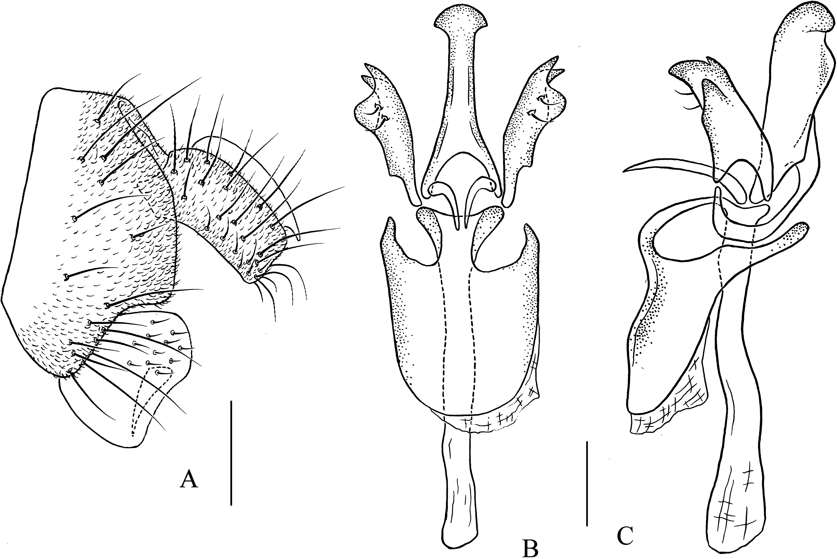
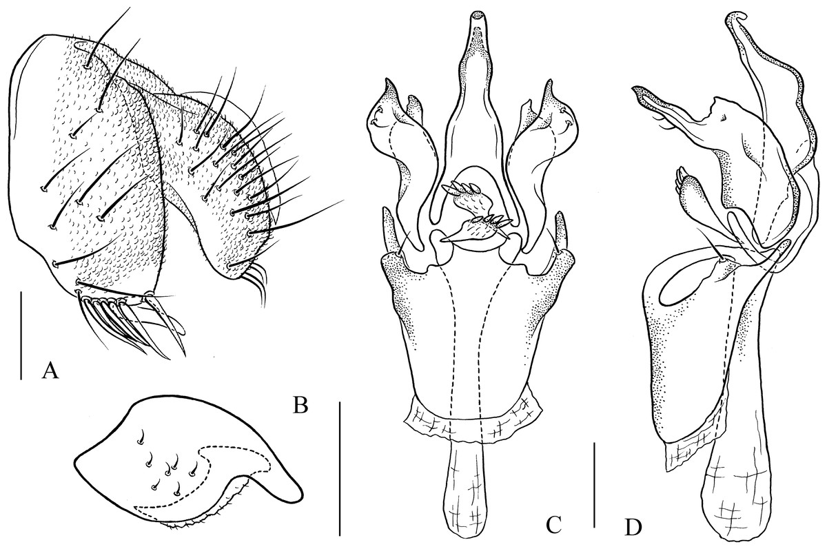
Figure 15: Pseudostegana ximalaya sp. nov., male terminalia.
(A) Epandrium, cercus and surstylus; (B) surstylus; (C) hypandrium, paramere, gonopods, aedeagus and aedeagal apodeme (ventral view); (D) ditto (lateral view). Scale bars = 0.1 mm. Drawing credit: Yuan Zhang.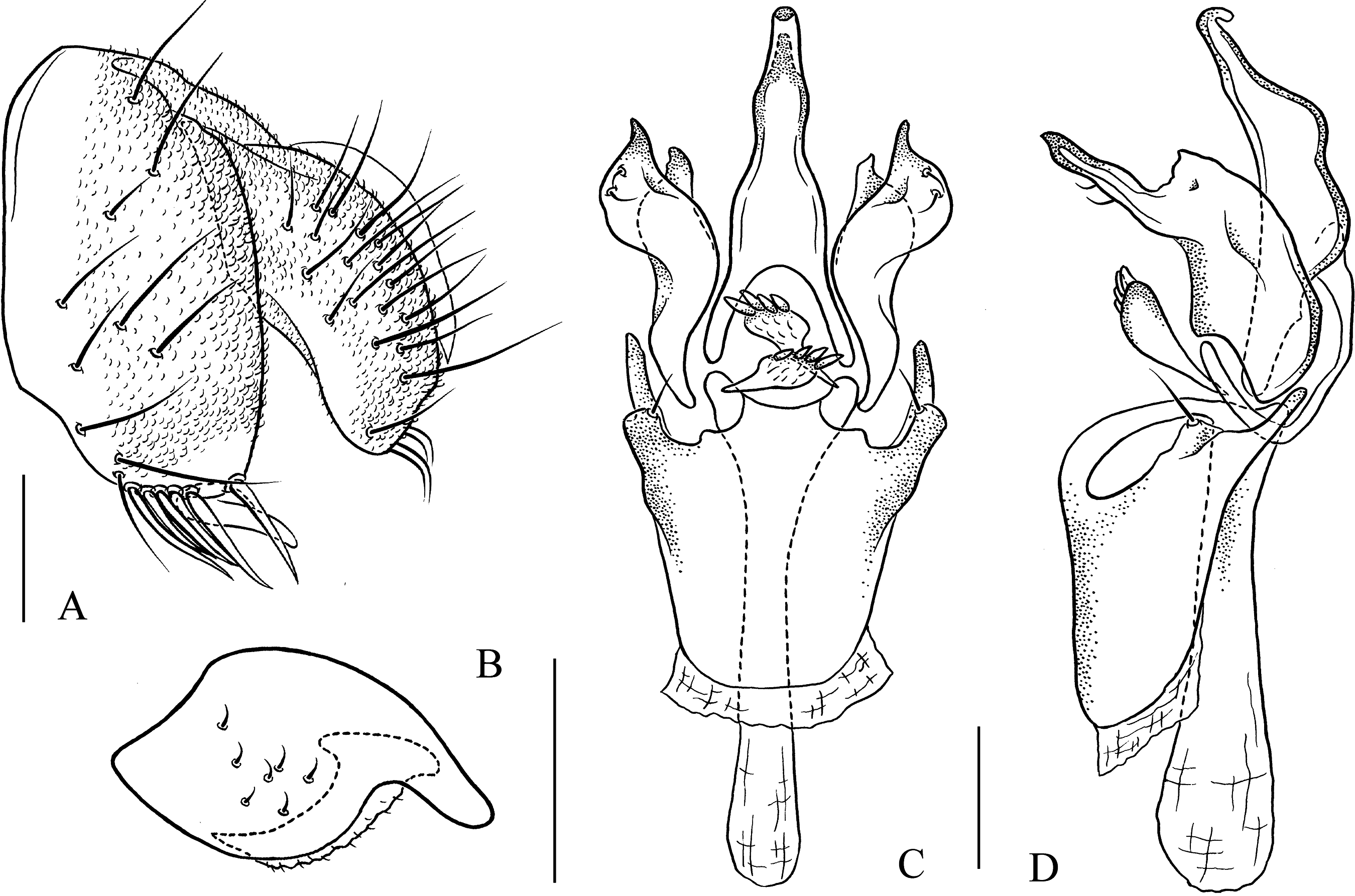

Figure 16: Pseudostegana zhuoma sp. nov., male terminalia.
(A) Epandrium, cercus and surstylus; (B) hypandrium, paramere, gonopods, aedeagus and aedeagal apodeme (ventral view); (C) ditto (lateral view). Scale bars = 0.1 mm. Drawing credit: Yuan Zhang.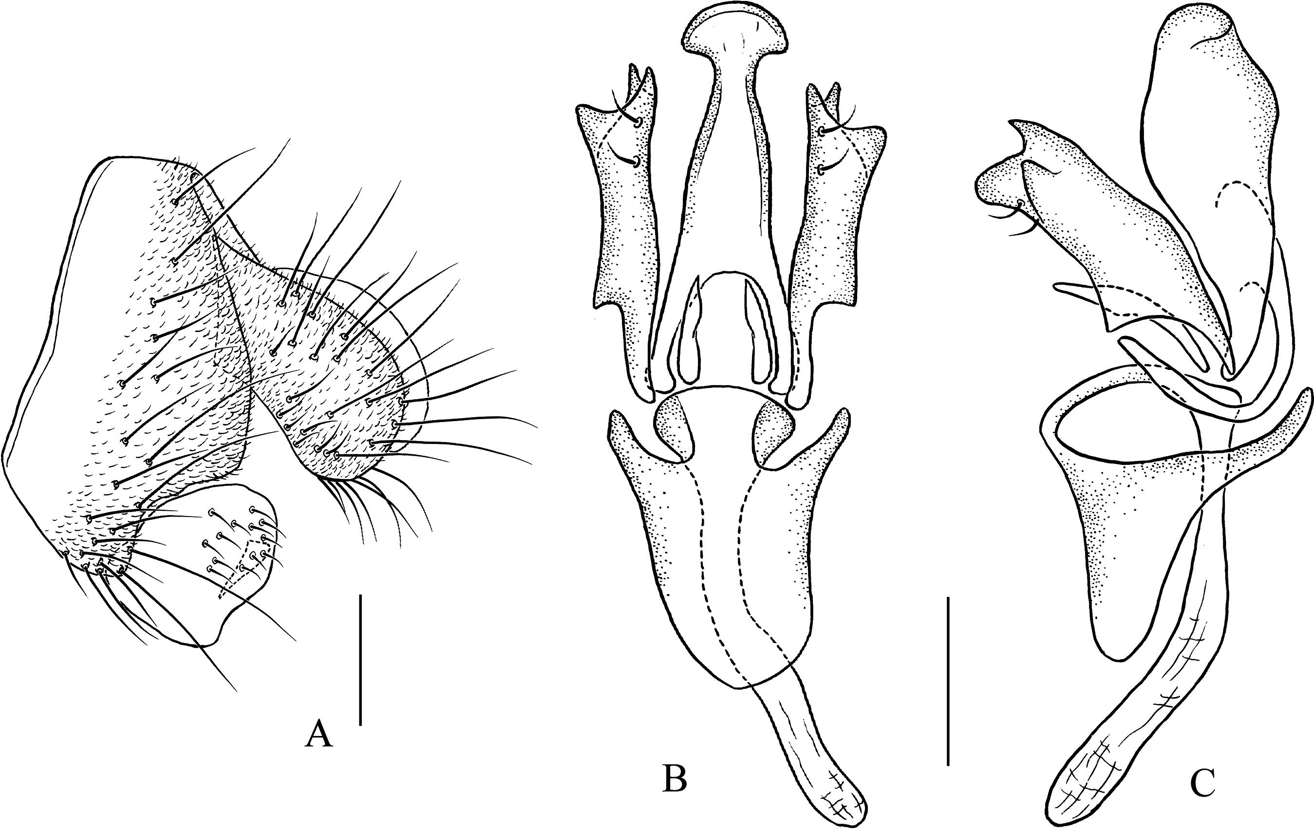
Description: Male and female. Head: Ocellar triangle narrowly elongated, yellow (Fig. 5M). Frons brown (Fig. 5M). Face yellowish brown. Clypeus brown to dark brown. Gena brown. Palpus brownish yellow to brown, slightly broad in male, medially a quarter as wide as long (Fig. 5P), yellow and slender female. Legs: Yellow. Abdomen: Tergites glossy, yellow on second and third tergites, black along lateral and posterior margins, with one brownish longitudinal stripe medially, the rest black (Fig. 5O). Sternites brown to dark brown. Male terminalia: Epandrium broadened on posterior margin, roundly protruded on ventral margin (Fig. 14A). Surstylus broadened, with several setae on outer surfaces (Fig. 14A). Hypandrial paramedian setae absent (Figs. 14B and 14C). Paramere round apically, brodened subapically, with one acute, dorsal projection apically (Figs. 14B and 14C). Aedeagus broadways expanded apically, slightly dorsad protruded submedially in lateral view, with rod-like processes basally (Figs. 14B and 14C).
Measurements: BL = 3.05 mm in holotype (range in five males and five females paratypes: 2.41–2.95 mm in males, 2.97–3.43 mm in females), ThL = 1.47 mm (0.96–1.25 mm in males, 1.25–1.51 mm in females), WL = 3.13 mm (2.30–2.93 mm in males, 2.97–3.07 mm in females), WW = 1.43 mm (1.03–1.37 mm in males, 1.33–1.45 mm in females), arb = 6/1 (5–7/1), avd = 1.60 (0.85–1.67), adf = 1.73 (1.47–1.92), flw = 0.93 (0.99–2.09), FW/HW = 0.47 (0.36–0.48), ch/o = 0.05 (0.04–0.09), prorb = 0.72 (0.72–0.93), rcorb = 0.93 (0.92–1.06), vb = 0.71 (0.40–0.78), dcl = 0.24 (0.25–0.40), sctl = 0.99 (0.90–1.28), sterno = 0.61 (0.44–0.68), orbito = 1.20 (1.00–1.44), dcp = 0.20 (0.16–0.22), sctlp = 1.17 (1.05–1.30).
Type specimens: Holotype male (SCAU, no. 122235), CHINA: Hesong, Menghai, Yunnan, 1,600–1,900 m, 17.iv.2010, ex. tussock, JJ Gao. Paratypes: CHINA: seven males, five females (three males and two females in KIZ, nos. 0090548–552; four males and three females in SCAU, nos. 122236–42), ex. tussock and tree trunks, 16,17.iv.2010, JJ Gao, YR Su, L Wang, L Wu, same data as holotype; three males, one female (SCAU, nos. 122243–46), 7.iv.2011, JM Lu, SJ Yan, ZF Shao, YR Su, same data as holotype; five males, one female (SCAU, nos. 122247–52), 12.v.2012, HW Chen, same data as holotype.
Etymology: From the Latin world: amoenus, meaning delighted.
Distribution: China (Yunnan).
Pseudostegana ximalaya Zhang & Chen, sp. nov.
urn:lsid:zoobank.org:act:3AE261B2-3982-42B3-ABC6-B62141244BD9
Diagnosis: This species differs from the other species of the latiparma group by having the epandrium with six strong prensisetae on ventral margin (Fig. 15A).
Description: Male and female. Head: Ocellar triangle brown on posterior three-quarter, dark brown on anterior a quarter (Fig. 6A). Frons brown (Fig. 6A). Face brown above, dark brown below. Clypeus black. Gena brown. Palpus dark brown, broadened in male, medially one-third as wide as long in male (Fig. 6D), brown in female. Thorax: Mesonotum brownish yellow anteriorly, yellowish brown to brown posteriorly (Fig. 6B); scutellum yellowish brown, yellow at tip (Fig. 6B). Pleura glossy, yellow on anterior one-third, dark brown on posterior two-thirds (Fig. 6E). Legs: Yellow. Abdomen: Tergites glossy, brownish yellow on second and third in male but only on second in female, the rest black (Fig. 6C). Sternites brownish to dark brown. Male terminalia: Surstylus strongly protruded on postero-ventral corners (Figs. 15A and 15B). Hypandrium with one pair of paramedian setae (Figs. 15C and 15D). Paramere dorsally protruded submedially, slender distally (Figs. 15C and 15D). Aedeagus dorsally curved apically in lateral view (Fig. 15D), with four finger-like processes basally (Figs. 15C and 15D).
Measurements: BL = 2.72 mm in holotype (range in three females paratypes: 2.25–2.83 mm), ThL = 1.08 mm (0.89–1.27 mm), WL = 2.09 mm (1.77–2.32 mm), WW = 0.95 mm (0.81–0.95 mm), arb = 6/1 (5–6/1), avd = 1.05 (0.93–0.98), adf = 1.64 (1.59–1.78), flw = 2.03 (1.52–1.66), FW/HW = 0.39 (0.40–0.46), ch/o = 0.05 (0.06–0.09), prorb = 0.80 (1.05–1.11), rcorb = 0.97 (0.90–1.01), vb = 0.72 (0.50–0.87), dcl = damaged (damaged), sctl = 1.13 (1.00–1.08), sterno = 0.62 (0.36–0.75), orbito = 1.02 (1.00–1.11), dcp = damaged (damaged), sctlp = 1.17 (1.11–1.26).
Type specimens: Holotype male (SCAU, no. 122256), CHINA: Beibeng, Motuo, Xizang, 800 m, 29.ix.2010, ex. tussock JJ Gao. Paratypes: CHINA: three females (SCAU, nos. 122257–59), ex. fallen logs, L Wang, L Wu, same data as holotype.
Etymology: The name means “Snow Country” in Tibetan, referring to the type locality.
Distribution: China (Xizang).
Pseudostegana zhuoma Zhang & Chen, sp. nov.
urn:lsid:zoobank.org:act:835597CE-9D32-4928-96BC-CC3EEBBA760D
Diagnosis: This species is distinguished from Pseudostegana amoena sp. nov. by having the mesonotum brown, submedially with two pairs of yellow longitudinal stripes not reaching the scutellum (Fig. 6H); the pleura yellow on anterior quarter, brown on posterior 3/4 (Fig. 6K); the aedeagus ventrally protruded submedially in lateral view (Fig. 16C).
Description: Male and female. Head: Ocellar triangle narrowly elongated, yellow (Fig. 6G). Frons brown (Fig. 6G). Face brownish. Clypeus dark brown. Gena brown. Palpus yellow, slightly broadened in male, medially a quarter as wide as long (Fig. 6J), brown and slender in female. Thorax: Scutellum brown, yellow at tip (Fig. 6H). Legs: Yellow, brown on femur of foreleg, and knees of mid- and hindlegs. Abdomen: Tergites glossy, brown to dark brown, submedially with two yellow patches each on second and third tergites (Fig. 6I). Sternites brownish yellow. Male terminalia: Epandrium broadened on posterior margin, roundly protruded on ventral margin (Fig. 16A). Surstylus broadened, with several setae on outer surfaces (Fig. 16A). Hypandrial paramedian setae absent (Figs. 16B and 16C). Paramere round apically, with one acute dorsal projection; broadened subapically (Figs. 16B and 16C). Aedeagus broadways expanded apically, slightly dorsad protruded submedially in lateral view, with rod-like processes basally (Figs. 16B and 16C).
Measurements: BL = 2.90 mm in holotype (range in two females paratypes: 3.63–3.73 mm), ThL = 1.24 mm (1.49–1.50 mm), WL = 2.95 mm (3.21–3.37 mm), WW = 1.35 mm (1.61), arb = 6/1 (6/1), avd = 0.94 (0.90–1.08), adf = 1.41 (1.76–1.79), flw = 1.82 (2.05–2.10), FW/HW = 0.48 (0.59–0.60), ch/o = 0.07 (0.06–0.10), prorb = 0.87 (0.84–0.90), rcorb = 0.98 (0.87–0.88), vb = 0.78 (0.69–0.85), dcl = 0.28 (0.26–0.36), sctl = 0.83 (1.03–1.05), sterno = 0.70 (0.43–0.66), orbito = 0.90 (0.78–0.88), dcp = 0.13 (0.17–0.25), sctlp = 0.82 (0.75–0.97).
Type specimens: Holotype male (SCAU, no. 122253), CHINA: Tongmai, Bomi, Xizang, 30°06′N, 95°05′E, 2,000 m, 9.x.2010, ex. tussock, JJ Gao. Paratypes: CHINA: two females (SCAU, nos. 122254, 55), same data as holotype.
Etymology: The name means “fairy maiden” in the Tibetan, referring to the type locality.
Distribution: China (Xizang).
The zonaria species group
Diagnosis: Wing: Medial band at least as broad as length of dm-cu crossvein; r-m crossvein clear; M vein gently curved to R4+5 vein after dm-cu crossvein (Figs. 6R and 7L).
Pseudostegana amnicola Zhang & Chen, sp. nov.
urn:lsid:zoobank.org:act:17602786-17CA-4FD6-A6DB-5722E9FC5D72
Diagnosis: This species is similar to Pseudostegana latifasciata Chen, Toda & Wang, 2005 from Vietnam by the wing patches (Fig. 6R) and periphallic organs, but can be distinguished by the following characters: surstylus protruded on postero-ventral corner (Figs. 17A and 17B); aedeagus expanded apically (Fig. 17D). In Pseudostegana latifasciata: surstylus protruded on antero- and postero-ventral corner (Chen, Toda & Wang, 2005: Fig. 145); aedeagus slightly pointed apically in ventral view (Chen, Toda & Wang, 2005: Fig. 147).
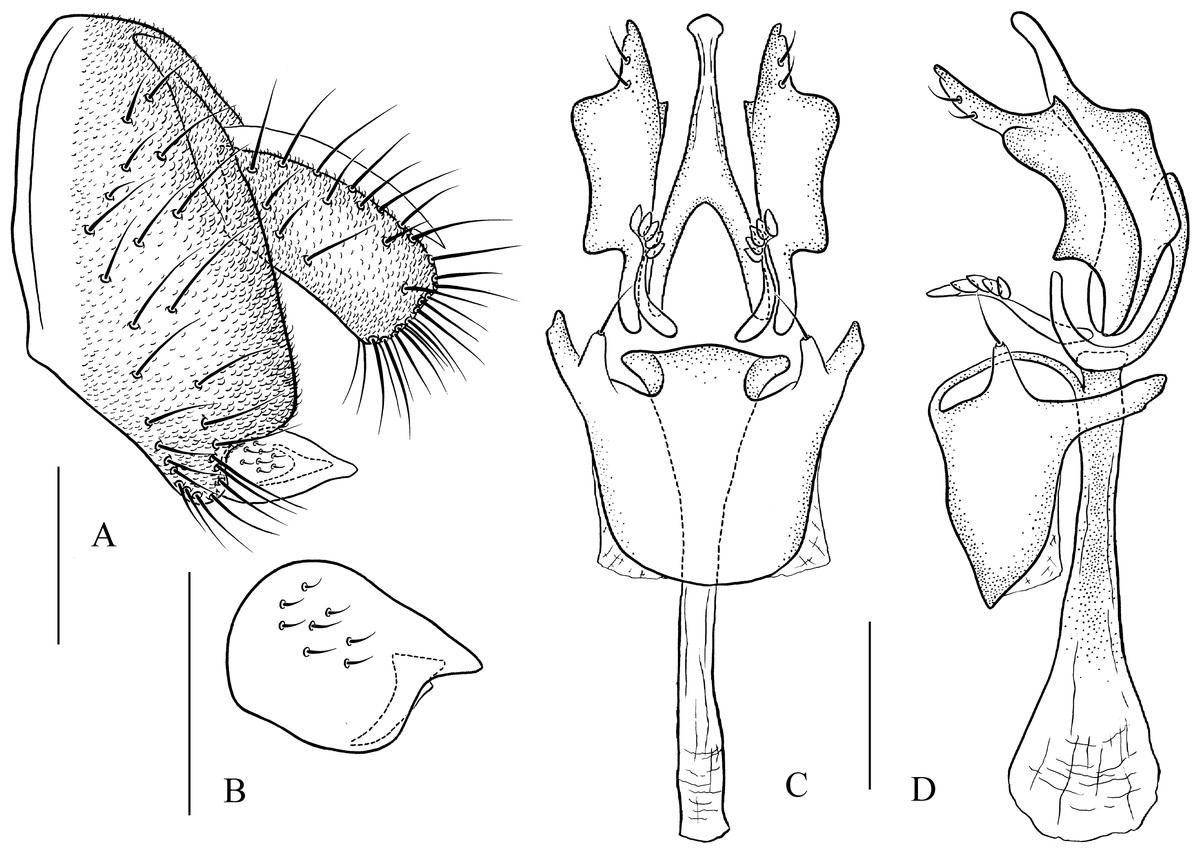
Figure 17: Pseudostegana amnicola sp. nov., male terminalia.
(A) Epandrium, cercus and surstylus; (B) surstylus; (C) hypandrium, paramere, gonopods, aedeagus and aedeagal apodeme (ventral view); (D) ditto (lateral view). Scale bars = 0.1 mm. Drawing credit: Yuan Zhang.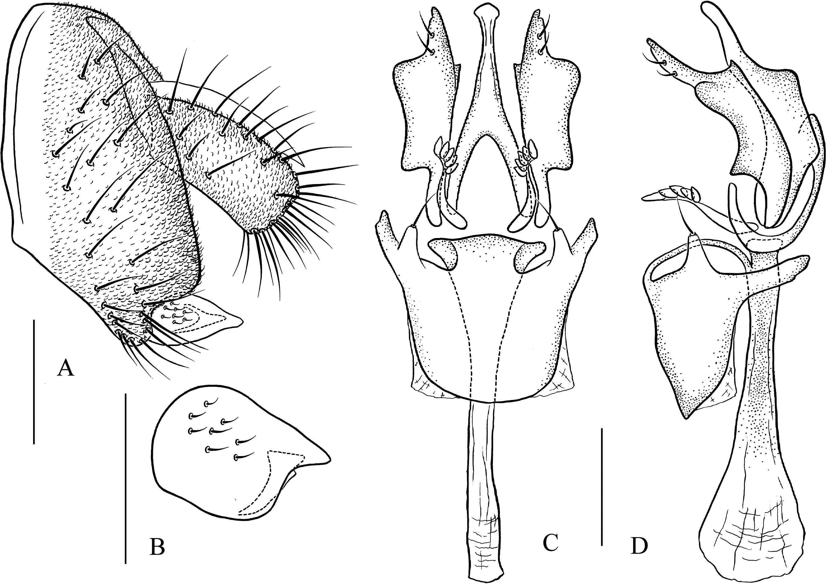
Description: Male and female. Head: Ocellar triangle elongated (Fig. 6M). Frons brown (Fig. 6M). Face and gena brown. Clypeus black. Palpus brown, slender (Fig. 6P). Thorax: Mesonotum glossy, yellow on anterior half, brown (male, Fig. 6N) to black (female) on posterior half; scutellum brown in male (Fig. 6N), dark brown in female, pale at tip (Fig. 6N). Pleura glossy, brownish yellow on anterior half, brown (male, Fig. 6Q) to black (female) on posterior half. Legs: Yellow, black on mid- and hindlegs tibiae. Abdomen: All tergites glossy, dark brown in male (Fig. 6O), black in female. Sternites yellow in both sexes. Male terminalia: Epandrium roundly protruded on ventral margin, with numerous setae (Fig. 17A). Hypandrium with one pair of paramedian setae sublaterally (Figs. 17C and 17D). Paramere broadened subapically (Fig. 17C). Aedeagus with four finger-like processes basally (Figs. 17C and 17D).
Measurements: BL = 2.84 mm in holotype (range in four males and one females paratypes: 2.48–2.90 mm in males, 2.88 mm in females), ThL = 1.21 mm (0.97–1.30 mm in males, 1.25 mm in females), WL = 2.49 mm (2.15–2.67 mm in males, 2.53 mm in females), WW = 1.07 mm (0.97–1.23 mm in males, 1.05 mm in females), arb = 7/1 (5–8/1), avd = 0.97 (0.96–1.08), adf = 1.84 (1.50–1.90), flw = 1.75 (1.32–1.93), FW/HW = 0.48 (0.43–0.61), ch/o = 0.09 (0.05–0.07), prorb = 0.74 (0.85–1.00), rcorb = 0.91 (0.86–1.02), vb = 0.87 (1.00–1.42), dcl = damaged (damaged), sctl = 0.92 (1.08–1.18), sterno = 0.63 (0.61–0.74), orbito = 1.14 (1.11–1.23), dcp = 1.17 (1.20), sctlp = 1.02 (1.00–1.42).
Type specimens: Holotype male (SCAU, no. 122260), CHINA: Beibeng, Motuo, Xizang, 1,000 m, 1.x.2010, ex. fallen logs, JJ Gao. Paratypes: CHINA: one female (SCAU, no. 122261), same data as holotype; two males (SCAU, nos. 122262, 63), Baihualing, Baoshan, Yunnan, 25°18′N, 98°48′E, 1,400 m, 13.vi.2011, ex. tussock, JJ Gao, K Liu; one male (SCAU, no. 122159), Hesong, Menghai, Yunnan, 1,600 m, 13.v.2012, ex. tussock, HW Chen; one male (SCAU, no. 124884), Mengdong, Cangyuan, Yunnan, 23°10′N, 99°14′E, 1,320 m, 6.v.2016, ex. tussock, YQ Liu; two females (SCAU, nos. 111358, 59), Husa, Longchuan, Yunnan, 1,230 m, 20.viii.2016, ex. tussock, HW Chen, YQ Liu; one male (SCAU, no. 124842), Xincheng, Yingjiang, Yunnan, 960 m, 18,viii.2016, ex. tussock, L Gong.
Etymology: From the Latin word amnicola, meaning riverain.
Distribution: China (Yunnan).
Pseudostegana mailangang Zhang & Chen, sp. nov.
urn:lsid:zoobank.org:act:048491D3-80F6-4F0C-A8EE-809113596DA9
Diagnosis: Mesonotum orange yellow on anterior half, dark brown to black on posterior half (Fig. 7B); paramere not broadened subapically in ventral view (Fig. 18B).
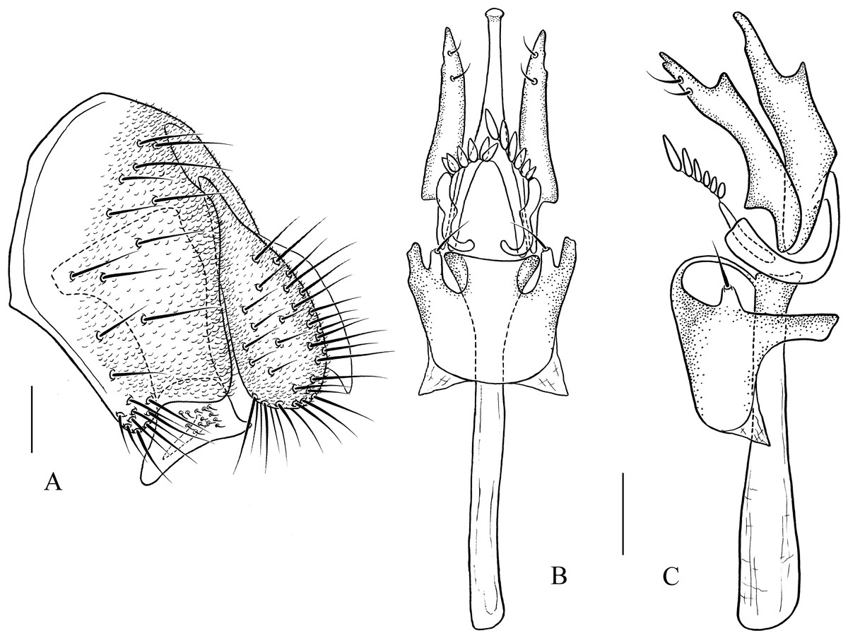
Figure 18: Pseudostegana mailangang sp. nov., male terminalia.
(A) Epandrium, cercus and surstylus; (B) hypandrium, paramere, gonopods, aedeagus and aedeagal apodeme (ventral view); (C) ditto (lateral view). Scale bars = 0.1 mm. Drawing credit: Yuan Zhang.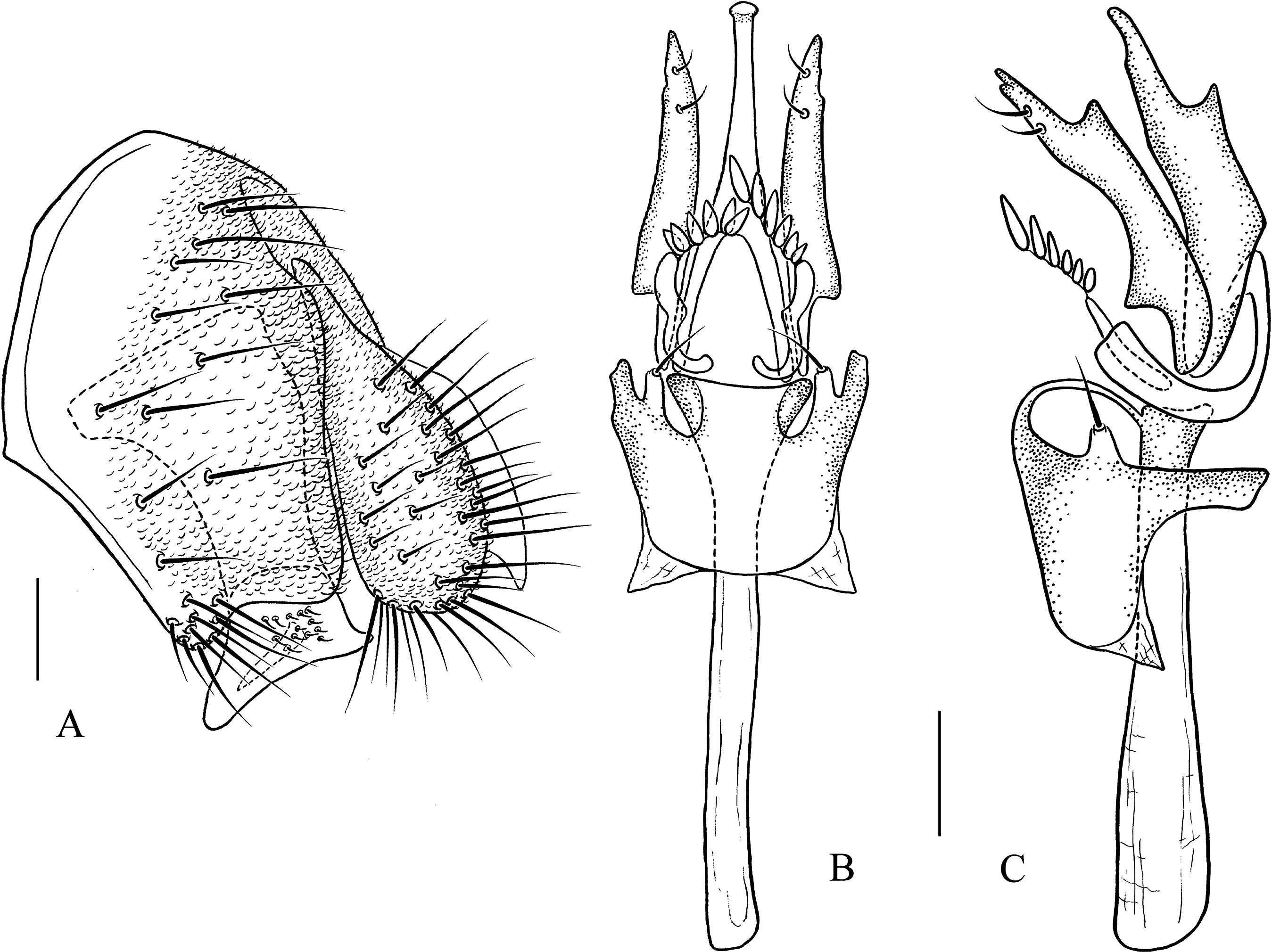
Description: Male and female. Head: Ocellar triangle elongated, dark brown (Fig. 7A). Frons, face and gena dark brown (Fig. 7A). Clypeus black. Palpus grayish brown, slender (Fig. 7D). Thorax: Mesonotum orange yellow on anterior half, dark brown to black on posterior half (Fig. 7B); scutellum dark brown, yellow at tip (Fig. 7B). Pleura glossy, mostly dark brown (Fig. 7E). Legs: Yellow, dark brown on femora and tibiae mid- and hindlegs. Abdomen: All tergites glossy, black (Fig. 7C). Sternites brown to dark. Male terminalia: Epandrium roundly protruded on ventral margin, with numerous setae (Fig. 18A). Surstylus protruded on antero- and postero-ventral corners (Fig. 18A); paramere smoothly narrowed in ventral view (Fig. 18B). Hypandrium with one pair of paramedian setae sublaterally (Figs. 18B and 18C). Aedeagus acutely protruded on distal one-third in lateral view (Fig. 18C), and with four finger-like processes basally (Figs. 18B and 18C).
Measurements: BL = 2.83 mm in holotype (range in five males and three females paratypes: 2.62–2.83 mm in males, 3.12–3.21 mm in females), ThL = 0.97 mm (0.70–1.05 mm in males, 1.19–1.25 mm in females), WL = 1.79 mm (1.87–2.08 mm in males, 2.19–2.33 mm in females), WW = 0.84 mm (0.83–0.93 mm in males, 1.03–1.07 mm in females), arb = 7/1 (6–9/1), avd = 0.94 (0.88–1.13), adf = 1.78 (0.88–2.38), flw = 1.43 (1.13–1.95), FW/HW = 0.40 (0.40–0.45), ch/o = 0.05 (0.04–0.07), prorb = 1.08 (0.81–1.12), rcorb = 0.98 (0.82–0.95), vb = 1.04 (0.69–1.14), dcl = damage (0.29–0.38), sctl = 1.25 (0.91–1.25), sterno = 0.57 (0.43–0.62), orbito = 1.07 (1.06–1.29), dcp = 0.25 (0.19–0.24), sctlp = 1.31 (1.13–1.73).
Type specimens: Holotype male (SCAU, no. 122264), CHINA: Wangtianshu, Mengla, Yunnan, 21°28′N, 101°38′E, 600 m, 31.ix.2011, ex. fallen logs, HW Chen. Paratypes: CHINA: seven males, three females (two males and one female in KIZ, nos. 0090507–09; five males and two females in SCAU, nos. 122265–71), HW Chen, JJ Gao, same data as holotype; one male (SCAU, no. 124885), Guanlei, Mengla, Yunnan, 21°38′N, 101°10′E, 620 m, 23.iv.2016, ex. tussock, YQ Liu.
Etymology: The name means “tall tree” in the language of the Dai nationality in Yunnan Province.
Distribution: China (Yunnan).
Pseudostegana mystica Zhang & Chen, sp. nov.
urn:lsid:zoobank.org:act:7754C40D-A608-48F3-AACF-431F3E5F8D9D
Diagnosis: This species is similar to Pseudostegana insularis Li, Gao & Chen, 2010 from Hainan, China by the wing patches (Fig. 7L) and periphallic organs (Fig. 19A), but can be distinguished by the aedeagus being broadened apically (Fig. 19B).
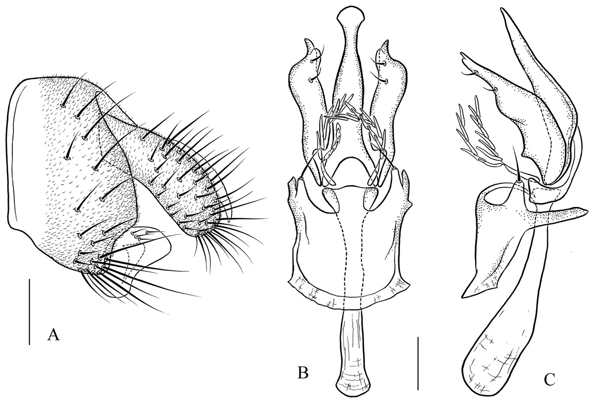
Figure 19: Pseudostegana mystica sp. nov., male terminalia.
(A) Epandrium, cercus and surstylus; (B) hypandrium, paramere, gonopods, aedeagus and aedeagal apodeme (ventral view); (C) ditto (lateral view). Scale bars = 0.1 mm. Drawing credit: Yuan Zhang.
Description: Male. Head: Ocellar triangle elongated, yellowish (Fig. 7G). Frons and gena yellow (Fig. 7G). Face mostly yellow, brown on lower margin. Clypeus brown. Palpus yellow, slender (Fig. 7J). Thorax: Mesonotum mostly yellow; scutellum brown, yellow at tip (Fig. 7H). Pleura glossy, yellow on anterior two-thirds, brown on posterior one-third (Fig. 7K). Legs: Mostly yellow. Abdomen: All tergites glossy, mostly black (Fig. 7I). Sternites yellow. Male terminalia: Epandrium slightly roundly protruded on ventral margin, with numerous setae (Fig. 19A). Surstylus protruded on antero- and postero-ventral corners (Fig. 19A). Hypandrium with one pair of paramedian setae sublaterally (Figs. 19B and 19C). Aedeagus smoothly narrowed on distal two-thirds, with seven finger-like process basally (Figs. 19B and 19C).
Measurements: BL = 2.40 mm in holotype, ThL = 1.25 mm, WL = 2.63 mm, WW = 1.10 mm, arb = 7/1, avd = 0.87, adf = 1.88, flw = 2.11, FW/HW = 0.39, ch/o = 0.07, prorb = 0.83, rcorb = 0.83, vb = 0.72, dcl = 0.27, sctl = damaged, sterno = 0.53, orbito = 1.26, dcp = 0.19, sctlp = 1.00.
Type specimen: Holotype male (SCAU, no. 122272), CHINA: Beibeng, Motuo, Xizang, 1,000 m, 1.x.2010, ex. fallen logs, JJ Gao.
Etymology: From the Latin word silvanus, meaning forest deity.
Distribution: China (Xizang).
Key to all Chinese species of the genus Pseudostegana
3. Ocellar triangle not elongated (Fig. 4A); M vein strongly curved after dm-cu crossvein (Fig. 4F); wing with annular patch medially (Fig. 4F) (fleximediata group) Pseudostegana meiduo sp. nov.
– Ocellar triangle elongated (Figs. 5A, 5G and 6A); M vein gently curved after dm-cu crossvein; wing with only one medial band (Figs. 5L, 5R, 6F and 6L) (latiparma group)7
4. Epandrium strongly roundly protruded ventrally, with dense setae (Fig. 11A); aedeagus apically slightly concave in ventral view (Fig. 11C) Pseudostegana stictiptrata sp. nov.
– Epandrium slightly protruded ventrally (Figs. 10A and 12A); aedeagus apically slightly protruded in ventral view (Figs. 10C and 12C)5
6. Surstylus strongly protruded on posterior corner in lateral view, broadened, approximately 1.5 times as high as wide (Figs. 10A and 10B)Pseudostegana meiji sp. nov.
– Surstylus strongly protruded and pointed on anterior corner, broadened, approximately two times as high as wide (Figs. 12A and 12B) Pseudostegana stigmatptera sp. nov.
7. R-m crossvein clouded; medial, dark-color band much broadened and with one distinct, protruded part submedially (Li, Gao & Chen, 2010: Fig. 15) Pseudostegana bilobata
8. Palpus expanded, medially one-third to half as wide as long (Fig. 6D)9
– Palpus slender, rod-shaped11
9. Epandrium with six strong prensisetae on ventral margin (Fig. 15A)Pseudostegana ximalaya sp. nov.
– Epandrium without strong setae on ventral margin10
10. Paramere apically shallowly bifurcated, subapically lacking small projection, dorsomedially slightly roundly protruded in lateral view (Chen, Toda & Wang, 2005: Fig. 93) Pseudostegana bifasciata
– Paramere apically not bifurcated, subapically with one small, acute projection, dorsomedially triangularly protruded in lateral view (Li, Gao & Chen, 2010: Figs. 3D and 3E) Pseudostegana acutifoliolata
12. Mesonotum yellow, with five brown longitudinal stripes (Fig. 5N); abdominal second and third tergites mostly yellow (Fig. 5O) Pseudostegana amoena sp. nov.
– Mesonotum brown, submedially with two pairs of yellow longitudinal stripes (Fig. 6H); abdominal second and third tergites each with two yellow patches submedially (Fig. 6I) Pseudostegana zhuoma sp. nov.
13. Paramere subapically with one acute, dorsal projection (Fig. 13A)14
– Paramere subapically without projection.15
14. Mesonotum antero-medially yellow and with thin brownish yellow, longitudinal stripe, posteriorly and laterally brownish yellow (Fig. 5H); aedeagus apically pointed in ventral view (Fig. 13D) Pseudostegana alpina sp. nov.
– Mesonotum yellow on anterior one-third, brown to black on posterior two-thirds (Fig. 8M); aedeagus hammer-shaped in lateral view (Li, Gao & Chen, 2010: Fig. 7E) Pseudostegana minutipalpata
15. Paramere subapically triangularly expanded in ventral view; basal process of aedeagus membranous, lacking finger-like processes (Chen, Toda & Wang, 2005: Figs. 92 and 93) Pseudostegana angustifasciata
– Paramere subapically roundly expanded in ventral view; basal process of aedeagus with finger-like processes (Chen, Toda & Wang, 2005: Figs. 105 and 106) Pseudostegana pallidimaculata
16. Palpus expanded, medially wider than one-third of length17
– Palpus slender, rod-like.19
17. Paramere apically bifurcated (Li, Gao & Chen, 2010: Fig. 8E) Pseudostegana insularis
– Paramere apically not bifurcated (Li, Gao & Chen, 2010: Fig. 9E)18
18. Palpus black, yellow at tip (Li, Gao & Chen, 2010: Fig. 1L); aedeagus strongly narrowed on distal one-third Pseudostegana silvana
– Palpus yellow; aedeagus smoothly narrowed on distal half Pseudostegana latipalpis
19. Paramere with one nearly triangular process on distal one-third in lateral view20
– Paramere distally lacking process in lateral view21
20. Paramere not broadened subapically in ventral view (Fig. 18B); aedeagus triangularly protruded on distal one-third in lateral view (Fig. 18C) Pseudostegana mailangang sp. nov.
– Paramere broadened subapically in ventral view (Fig. 17C); aedeagus slightly roundly protruded on distal one-third in lateral view (Fig. 18D) Pseudostegana amnicola sp. nov.
21. Paramere not bifurcated apically (Chen, Toda & Wang, 2005: Fig. 144); aedeagus slightly pointed in ventral view (Chen, Toda & Wang, 2005: Fig. 143) Pseudostegana dolichopoda
– Paramere shallowly bifurcated apically (Figs. 19B and 19C); aedeagus apically slightly expanded22
22. Aedeagus apically heart-shaped in ventral view, protruded on distal one-third in lateral view (Chen, Toda & Wang, 2005: Figs. 149 and 150) Pseudostegana nitidifrons
– Aedeagus apically roundly expanded in ventral view, nearly straight in lateral view (Figs. 19B and 19C) Pseudostegana mystica sp. nov.
Discussion
DNA sequence data may process effective species boundary information, which provides a useful tool for taxonomic studies (Hamilton et al., 2014; Kekkonen et al., 2015). The integration of DNA sequence data and the traditional morphological characters increases the ease and reliability of both species identification and species discovery (Vogler, 2006; Cardoso, Serrano & Vogler, 2009; Kekkonen & Hebert, 2014; Roberts et al., 2016).
We proposed the Pseudostegana species based on morphological variation and then clarified their status by DNA data. Although some new species are described (e.g., Pseudostegana alpina sp. nov. and Pseudostegana meiduo sp. nov.) based on few observed specimens, both morphological and molecular analysis support our taxonomic hypothesis. The new species, Pseudostegana alpina sp. nov., Pseudostegana amoena sp. nov., Pseudostegana mailangang sp. nov., Pseudostegana meiduo sp. nov., Pseudostegana meiji sp. nov., Pseudostegana stictiptrata sp. nov., Pseudostegana stigmatptera sp. nov., Pseudostegana ximalaya sp. nov. and Pseudostegana zhuoma sp. nov., which we described here, were recovered as distinct entities in phylogenetic trees and the ABGD analyses. The minimal interspecific genetic divergences between these new species and their close relatives all exceed 3%. DNA data support the morphological characteristics observed among these nine Chinese species and confirms the new species as being distinctly different.
Pseudostegana mystica sp. nov. from Xizang is similar to Pseudostegana insularis from Hainan in the wing patches, but they can be morphologically distinguished by the aedeagus, which is apically expanded in ventral view in the former one (Li, Gao & Chen, 2010). Although we only amplified the COI region for Pseudostegana mystica sp. nov., the COI marker still provided reliable evidence for its new species status, as high genetic divergences (>14%; Table S3) were observed between Pseudostegana mystica sp. nov. and the other Pseudostegana species.
High intraspecific variation is sometimes observed among Drosophilidae species, for example, estimates of COI intraspecific divergence were 8%, 9% and 11% for three Drosophila species (Yassin et al., 2010), and 4.4% for the Leucophenga euryphylla (Huang et al., 2013). Interestingly, the Yunnan lineage and Xizang lineage of Pseudostegana amnicola sp. nov. show relatively large sequence divergences (5.5% for COI, 5.1% for ND2). Moreover, the ABGD analysis based on COI data separated them to two MOTUs. Although no obvious morphological variation was observed, genetic diversification seems to have occurred between these two geographic lineages. The prominent genetic divergence between lineages probably indicate the presence of unrecognized cryptic species. More samples from different populations and genders should be sampled in order to assess if they belong to cryptic species or not.
The COI and ND2 genes are useful for species identification or discrimination of Drosophilidae species (He et al., 2009; Zhao, Gao & Chen, 2009; Lu et al., 2011; Lu, Li & Chen, 2011; Zhao et al., 2013; Shao et al., 2014; Zhang, Li & Chen, 2016). Our results show that COI barcoding successfully distinguished all the studied Pseudostegana species and helped to discover the potential new species. The power of the ND2 fragment to discriminate the studied specimens of Pseudostegana at the species level is similar to that of the COI fragment. In several studies, the nuclear 28S gene was successful for identification in insect at the species level (Monteiro et al., 2000; Monaghan et al., 2005; Asokan et al., 2013; Resch et al., 2014). In contrast to COI and ND2, the 28S fragment showed less resolution at species level for the studied Pseudostegana. This can possibly be attributed to the conservation of the 28S gene such that closely related species showed little variation in sequences.
Chen, Toda & Wang (2005) examined 32 Pseudostegana species, and proposed six species groups (the atrofrons, javana, latiparma, grandipalpis, fleximediata and zonaria groups) on the basis of morphological features. In our phylogeny, Pseudostegana meiduo sp. nov. is placed at the basal position within Pseudostegana and recovered as a distinct clade from the other three species groups (the latiparma, java and zonaria groups). Morphologically, Pseudostegana meiduo sp. nov. is classified in the fleximediata group as it is congruent with the description of the species group, e.g., ocellar triangle is not elongated and M vein is strongly curved after dm-cu crossvein (Fig. 4F in Chen, Toda & Wang, 2005). However, its wing pattern differs from the other fleximediata group species by having distinct cross band at the basal position of the wing. Unfortunately, no other fleximediata group species was included in the present phylogenetic analyses for testing its phylogenetic status. We temporarily place Pseudostegana mediuo sp. nov. in the fleximediata group, but its taxonomic status should be evaluated by additional sampling. Phylogenetic analyses recovered the monophyly of the latiparma and javana groups, but the zonaria group was found to be paraphyletic as clade I (Pseudostegana insularis + Pseudostegana silvana) were separated from clade II (Pseudostegana nitidifrons + Pseudostegana amnicola sp. nov. + Pseudostegana mailangang sp. nov.). The latter clade is morphologically different from the former concerning the shape of the palpus. The palpus of Pseudostegana latifasciata, Pseudostegana nitidifrons, Pseudostegana amnicola sp. nov. and Pseudostegana mailangang sp. nov. are slender and rod-like shape, while the palpus of the other zonaria group species are expanded (Chen, Toda & Wang, 2005).
Yunnan and the adjacent area (Southwest China) are located at the junction of the Himalaya, Mountains of Southwest China and Indo-Burma biodiversity hotspots (Myers et al., 2000). In recent years, fieldwork by members of our laboratory have revealed a hidden diversity of Pseudostegana species in this area, where previously only eight species were reported (Chen, Toda & Wang, 2005; Li, Gao & Chen, 2010). Southwest China total contains 38% (19 out of 50; including new species described in this study) of Pseudostegana species, and all of them are endemic to this region. Southwest China seems to be an important center of diversification of Pseudostegana species. Moreover, the short, relatively ancient branches of the phylogenetic trees suggest that adaptive radiation probably occurred during the early evolutionary history of Pseudostegana. Although we provided a molecular phylogeny for the Chinese Pseudostegana, additional research on systematics including other species groups and species in Southwest Asia, will be needed to better understand the origin and diversification of this genus.