
Research Article | DOI: https://doi.org/10.58489/2836-8657/005
*Corresponding Author: Biswas S.
Citation: Biswas S., (2022). Histopathological Characteristics of Breast Lesions in a Tertiary Health Institution in Southwest Nigeria; A 9-year Review. Journal of Surgery and Postoperative Care. 1(1). DOI: 10.58489/2836-8657/005
Copyright: © 2022 Biswas S., this is an open access article distributed under the Creative Commons Attribution License, which permits unrestricted use, distribution, and reproduction in any medium, provided the original work is properly cited.
Received: 12 December 2022 | Accepted: 20 December 2022 | Published: 28 December 2022
Keywords: breast lesion, histopathology, Ogbomoso, Nigeria
Background: The breast is affected by several diseases, including developmental, inflammatory, and neoplastic. While benign breast neoplasms are usually more common, breast cancer is the most common nonskin cancer and the leading cause of cancer-related deaths in women after lung cancer in the world.
Objective: The review aimed to assess the histopathological diagnosis of breast lesions over a period of 9years
Methods: This was a retrospective review of eight hundred and fourteen histopathologic cases of breast lesions accessed through laboratory records of all breast lesions cases seen. The demographic data were obtained and the patient’s histology slides were reviewed. The tumors were classified according to the WHO International Classification of breast tumors and Malignant breast tumors were graded according to the Nottingham grading system. Data were analyzed using SPSS version 25.
Results: The majority of breast cancer cases were seen in patients below 40 years, malignant cases were predominant in older age groups (40 – 59 years; 57.4%, 60 years and above; 23.3%) and malignancy was significantly associated with age group, p-value < 0.05. The prevailing neoplasm was Benign type (68.3%) while malignant neoplasm accounted for 30.2% and invasive ductal carcinoma (89.1%) was most prevalent. Moreso, the majority were in grade 2; 152 (76.8%) using the Nottigham grading system. However, the commonest benign neoplasm was fibroadenoma (53.7%) followed by fibrocystic change (20.1%).
Conclusion: Breast lesions are common in this environment. Benign breast neoplasms are commoner than malignant diseases. One in three of every sub-Saharan woman diagnosed with malignant breast lesion may not survive five years post-diagnosis. The prognosis of breast malignancies in sub-Saharan is poor due to late Presentation, ignorance and poverty.
Breast cancer is the most common cancer type in women in sub-Saharan Africa, affecting 129,000 women who were newly diagnosed in 2020. In high-income countries, breast cancer has a good prognosis, but in sub-Saharan Africa, survival is considerably lower. In this region, estimates for 5-year survival are near 50%, which implies that 1 in 2 women diagnosed with the disease has died within 5 years after diagnosis, compared with fractions in the USA of 1 in 5 for Black women and 1 in 10 for White women [1].
The annual number of women diagnosed with breast cancer in sub-Saharan Africa is projected to nearly double by 2040, due to population aging and expansion. This increase will even be larger if lifestyle and fertility changes are factored in. Although not fully understood, changing demography (increasing older population), improving socioeconomic conditions with consequent westernization of lifestyles, delayed childbearing age, low parity, increasing breast health awareness, improving diagnostic capacity, and reporting have been proposed as the rationale for the rising incidence of breast cancer in Nigeria [2]. Breast cancer is thus rightfully considered a public health problem with several screening modalities to detect early breast cancer [2].
Pain, palpable lump, nipple discharge, or lumpiness, singly or in combination represent the common presentation of breast diseases, while self or clinically detected mass is the most common presentation of breast cancer [1-3]. Hence, a breast lump in a female of any age causes significant anxiety, warranting hospital visits to confirm or exclude breast cancer.
This study reviews the demographic and histopathological characteristics of breast lesions seen at a pathology laboratory of a district hospital in South West Nigeria and compares the findings with similar studies in Nigeria and elsewhere [2-4].
Study area
The study was carried out at the department of Morbid Anatomy and Histopathology, Ladoke Akintola University Teaching Hospital located in Ogbomoso North Local Government Oyo State Nigeria. This tertiary health serves Ogbomoso, Oyo town, Oke-Ogun, and the environs.
Study design
This was a retrospective review of histopathologic cases of breast lesions diagnosed at the histopathology department.
Duration of Review
The review was done over a 9-year period, from January 2012 to December 2021.
Request forms and duplicate copies of all histologically diagnosed breast lesions during the study period were retrieved, the demographic and histopathologic data were collated. The corresponding slides were then retrieved and reviewed. However, where such slides were missing or faded, new slides were made from formalin-fixed, paraffin-embedded blocks. Where the above data or slides or blocks could not be found, those were excluded from the study. The tumors were classified according to the World Health Organization International Classification of breast tumors [5]. Malignant breast tumors were graded according to the Nottingham grading system [6].
Data analysis
All data were computed and analyzed using SPSS version 25. Continuous variables were summarized using range and mean ± standard deviation, while categorical variables presented as percentage frequencies were determined using descriptive statistics. Data were displayed using tables and charts.
A total number of 814 breast samples were received in the histology department of LAUTECH Teaching Hospital Ogbomoso.
Table I: Twenty-two (2.7%) of the diagnosed patients were male and seven hundred and ninety-two (792: 97.3%) were female and the majority of patients were below 40 years (Male; 72.7%, Female; 58.6%) with most predominant between the ages of 20-39years of age (15:68.2% in male and 382: 48.2% in female) and the mean age was 36.80 ± 15.7 years.
Table I: Demographic profile of breast cancer patients
Age group | Male n (%) | Female n (%) | Total n (%) |
< 20> 20 – 39 years 40 – 59 years 60 years and above | 1 (4.5) 15 (68.2) 4 (18.2) 2 (9.1) | 82 (10.4) 382 (48.2) 245 (30.9) 83 (10.5) | 83 (10.2) 397 (48.8) 249 (30.6) 85 (10.4) |
Total | 22 (2.7) | 792 (97.3) | 814 |
Mean age | 32.82 ± 15.0 | 36.91 ± 15.7 | 36.80 ± 15.7 |
Table II: The prevailing neoplasm was Benign type (68.3%) while malignant neoplasm accounted for 30.2% of the reviewed record data. Other nonneoplastic lesions were only 1.5%. (Table II) Moreso, using the Nottigham grading system grade 1; 9 (4.5%), grade 2; 152 (76.8%), grade 3; 35 (17.7%), and grade 4; 2 (1%).
Table II: Pathological characteristics of breast lesion
Characteristic | Frequency (%) |
Nature of lesion Nonneoplastic lesion Benign neoplasm Malignant neoplasm |
12 (1.5) 556 (68.3) 246 (30.2) |
Grade Nottigham 1 2 3 4 |
9 (4.5) 152 (76.8) 35 (17.7) 2 (1) |
Histological diagnosis revealed that the predominant malignant breast disease was invasive ductal carcinoma (89.1%), and other forms of carcinoma were seen in a few percentages. Figure I.
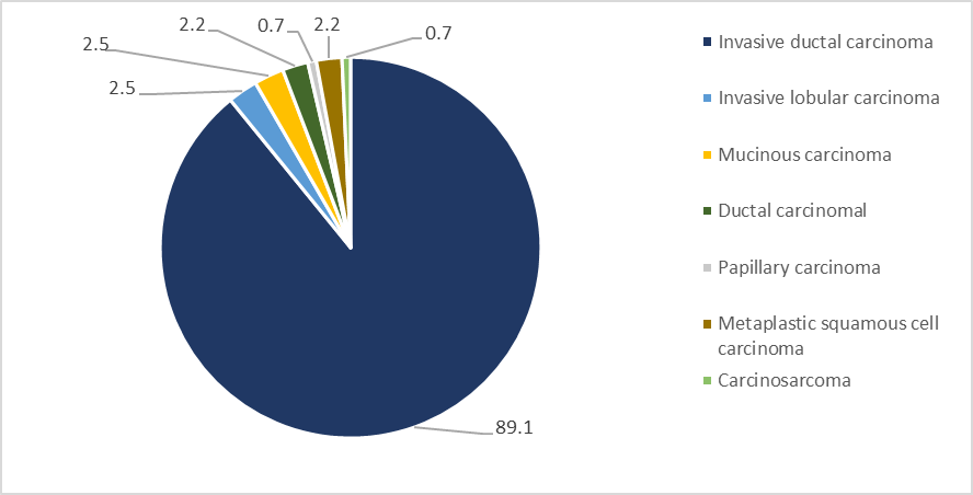
Figure I: Percentage distribution of malignant neoplasms
Table III: The most frequently reported benign neoplasm was fibroadenoma 305(53.7%) followed by fibrocystic change 118(20.1%).
Table III: Frequency distribution of benign neoplasms
Histologic categories and specific subtypes | Frequency (%) |
Nonproliferative epithelial lesions Fibrocystic change Tubular adenoma Lactational adenoma |
118 (20.1) 14 (2.5) 6 (1.1) |
Proliferative epithelial lesions without atypia Intraductal papilloma Sclerosing adenoma Flat epithelial hyperplasia |
8 (1.4) 8 (1.4) 2 (0.4) |
Proliferative epithelial lesions with atypia Atypical ductal hyperplasia |
8 (1.4) |
Fibroepithelial tumors Fibroadenoma Benign phyllodes tumor |
305 (53.7) 16 (2.8) |
Mesenchymal tumors Lipoma Dermatofibroma |
2 (0.4) 1 (0.2) |
Others Dermoid cysts Mastitis Fat necrosis Dermatofibroma Stromal fibrosis Neurofibroma Adenomyoepithelial adenosis |
3 (0.5) 6 (1.1) 6 (1.1) 2 (0.4) 6 (1.1) 1 (0.2) 2 (0.4) |
Normal residual breast tissue | 15 (2.6) |
Tumor of the male breast Gynaecomastia |
19 (3.3) |
Total | 568 (100) |
Table IV: The majority of breast cancer cases were seen in patients below 40 years, malignant cases were predominant in older age groups (40 – 59 years; 57.4%, 60 years and above; 23.3%) and malignancy was significantly associated with age group, p-value < 0>
Table IV: Breast cancer category by age group of patients.
Age group | Benign n (%) | Malignant n (%) | ꭕ2 (p-value) |
< 20> 20 – 39 years 40 – 59 years 60 years and above | 83 (15.5) 342 (63.7) 92 (17.1) 20 (3.7) | 0 (0.0) 52 (19.3) 155 (57.4) 63 (23.3) | 276.7 (*<0> |
*ꭕ2: Chi-square statistic, p-value < 0>
Trend analysis across all age groups showed an increased number of breast cancer cases between 2012 and 2016, a dip between 2017 and 2018, and an upward trend between 2019 and 2021. About one-third (33.5%) of all breast tumour samples were malignant and malignancy increased from 26.3% in 2012 to 42.6% in 2021 (Figure II).
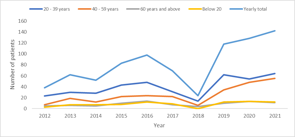
Figure II: Trend of breast cancer cases between 2012 and 2021
From figure III, it was observed that benign lesions are more prevalent than malignant neoplasms without any regular or consistent pattern on yearly basis.
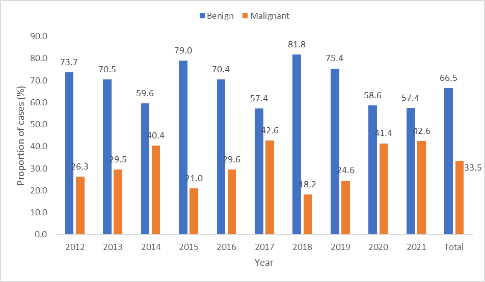
Figure III: Annual percentage of breast lesions.
The investigation of breast lesions accounted for 11.5% of the workload in our Pathology unit. This proportion is higher than the 8.1%, 7.1%, and 3.5% reported by Eke et al [7]. in Makurdi, Isah et al [8]. in Sokoto, and Yusufu et al [9]. in Zaria, respectively, but compares favorably with the 13% reported by Ezike et al.2 Federal Capital Territory Abuja. Our figure is, however, lower than 26% reported by Nwafor and Keshinro in Lagos [10].
Male breast lesions are relatively rare with male breast cancer (MBC), accounting for ˂0.02% of all cases [1,2,4]. This study recorded only 19 (3.3%) cases of breast diseases in males, with a striking female-to-male ratio of 33:1.0. Our male affectation rate of 3.3% is congruent with reported rates of 2.0%–3.5% [9,11,12] within Nigeria but was higher than 1.4% reported by Eke et al in Abuja, and 1.3% reported by Ogbuanya et al [13]. However, our finding is consistent with most other series [9,12], gynecomastia (100% n = 19) was the most common lesion seen in males in our study. It is noteworthy that Ibrahim et al [14]. in Kano, and Ogbuanya et al [13]. In Abakaliki, Nigeria, documented MBC as the most common breast disease in males. However, this was contrary to the findings in our study as all the breast lesions received over the period of study among males were all benign.
Fibroadenoma was the single most common entity (37.5%) and the most common BBN (53.7%) in this work. All other recent works [7,9,11] in Nigeria recorded fibroadenoma as the most frequent BBN except Jeje et al [15]. who reported fibrocystic change as the most common BBN. The documented percentage proportion of fibroadenoma in selected works across Nigeria ranges
from 43.1% to 76.1% [9,12,16], epithelial calcifications, papillary apocrine change, or cysts larger than 0.3 cm [2].
Fibrocystic change, an entity that comprises a group of non-proliferative morphologic changes, is the second most common BBN in our study like other recorded works17,18 in Nigeria, but the report by Jeje et al [15]. highlighted above where it was the most common BBN. It could mimic carcinoma clinically and radiologically due to calcifications and inflammatory changes caused by some of its morphologic elements.2 Other BBNs were relatively rare, individually or as a group in our review, with a frequency similar to other reports [16,18,19].
Comparative to the rate of 3.3% reported in Calabar [20] and 2.3% in Abakaliki [13], it is noteworthy that proliferative lesions (intraductal papilloma, atypical ductal hyperplasia sclerosing adenosis, and flat epithelial hyperplasia) in this study comprised 2.1% of BBNs. These lesions are believed to carry a relative risk for developing invasive breast carcinoma of 1.5% to 2% [2,21]. As with studies across Nigeria.
Malignant breast neoplasm MBNs accounted for 30.2% of all breast diseases reported in this study. The reported proportions of breast carcinoma in Nigerian studies ranged from 21.3% to 44.3% [7,9,12,16,22]. The higher rates reported by Nwafor and Udo, in Uyo (44.3%),10 Ogbuanya et al., in Abakaliki (43.7%) [13], and Eke et al., in Makurdi (43.6%) [7] are in the minority, and the reason is not immediately apparent. Irabor et al [21].documented an unusually low proportion of 10.6%. This may be due to the design of their study which examined only breast lumps from an arm of their general surgery unit. The carcinomas affected women predominantly in the fifth decade of life at diagnosis. The index age distribution is in conformity with the majority of the documented works in Nigeria [23-25] The peak age of incidence was reported a decade earlier in Makurdi [7] and a decade later in Sokoto [8]. Biologic, genetic, and possibly changing environmental factors may be responsible for the early onset of carcinogenic processes in our women [26].
Invasive ductal carcinoma (89.1%) was the prevalent histologic subtype of breast cancer in our study and this is consistent with previous works around the world [1,2,4,9]. The relatively less aggressive subtypes were fewer with invasive lobular and mucinous carcinoma being a distant second after invasive ductal carcinoma. Breast cancer grade is one of the Category I prognostic factors according to the College of American Pathologists which has demonstrated a reproducible significant correlation
with patients’ outcomes and utility in their clinical management [27].
Grade II cancers were the most common in this index study (76.8%). This finding mirrors the works of Ezike et al [2]. and Mandong et al [17]. who reported prevalent Grade II tumors. This finding, however, contrasts with the reported predominance of Grade III cancers by Gogo-Abite et al [22]. and Bewtra [19], and the findings by Imam et al.28 who reported Grade I tumors as the majority in their study.
Breast lesions are common in this environment. Benign breast neoplasms are commoner than the malignant disease. One in three of every sub-Saharan woman diagnosed of malignant breast lesion may not survive five years post diagnosis. The prognosis of breast malignancies in sub-Saharan is poor due to late Presentation, ignorance and poverty.
Therefore, public enlightenment should be intensified which would encourage early presentation. Government should subsidize treatment and improve health sector