 Journal of Medical Research and Surgery
PROVIDES A UNIQUE PLATFORM TO PUBLISH ORIGINAL RESEARCH AND REMODEL THE KNOWLEDGE IN THE AREA OF MEDICAL AND SURGERY
Journal of Medical Research and Surgery
PROVIDES A UNIQUE PLATFORM TO PUBLISH ORIGINAL RESEARCH AND REMODEL THE KNOWLEDGE IN THE AREA OF MEDICAL AND SURGERY
Journal of Medical Research and Surgery
PROVIDES A UNIQUE PLATFORM TO PUBLISH ORIGINAL RESEARCH AND REMODEL THE KNOWLEDGE IN THE AREA OF MEDICAL AND SURGERY
Journal of Medical Research and Surgery
PROVIDES A UNIQUE PLATFORM TO PUBLISH ORIGINAL RESEARCH AND REMODEL THE KNOWLEDGE IN THE AREA OF MEDICAL AND SURGERY


 Indexed Articles
Select your language of interest to view the total content in your interested language
Imran Qureshi*, Hamed Sultan Albusaidi Hamed, Parmanand Nathani, Fatema Mohammed Khamis Al Sadairi, Ghazala, Sobin Kalathil Kuncheria, Aisha Naseer Al-Azri, Lewallen J. Castrodes, Dick Wittington, Yaqoob Yousef Al-Rahbi
Department of Radiology, Royal Oman Police Hospital, Muskat, Oman.
Correspondence to: Imran Qureshi, Department of Radiology, Royal Oman Police Hospital, Muskat, Oman.
Received date: December 07, 2023; Accepted date: December 15, 2023; Published date: December 22, 2023
Citation: Qureshi I, Hamed HAS, Nathani P. Vesicourachal Diverticulum: A Unique Case Report. J Med Res Surg. 2023;4(6):121-122. doi: 10.52916/jmrs234124
Copyright: ©2023 Qureshi I, et al. This is an open-access article distributed under the terms of the Creative Commons Attribution License, which permits unrestricted
use, distribution and reproduction in any medium, provided the original author and source are credited.
Introduction: The inadequate closure of the urachus, an embryological remnant that connects the bladder to the umbilicus, results in the rare congenital defect known as the vesicourachal diverticulum. Even though it is frequently asymptomatic, it might show up as lower abdomen discomfort, hematuria, frequent urination, and recurrent urinary tract infections.
Case Report: The patient in this case study, a 38-year-old male with hematuria and right renal discomfort, had a tiny vesicourachal diverticulum discovered by accident during a CT scan.
Discussion: Vesicourachal diverticulum is one of the congenital urachal remnant abnormalities, representing a result of the failure of the urachus to close at the urinary bladder, forming an out-pouching of variable length from the anterosuperior aspect of the urinary bladder, which does not communicate with the umbilicus as seen in our case. The location and size of the diverticulum might affect how symptoms appear, and in severe situations, surgery such as diverticulectomy or urachal residual resection may be necessary. For prompt and appropriate care, it is essential to comprehend the clinical presentation, diagnostic techniques, and available treatments.
Conclusion: Though often benign, knowledge of the vesicourachal diverticulum helps prevent additional imaging and trials.
Vesicourachal, Diverticulum, Bladder, Ramnant, Embryological, Allontois.
An embryological remnant of allantois to urachal canal that goes from the bladder to the umbilicus, the place where urine empties prior to birth. The median umbilical ligament, a tiny fibrous chord, is left behind when it normally closes around the 12th week of pregnancy [1]. Since the urachus has no function after birth, a patent urachus after delivery is considered an anomaly in fetal development. A vesicourachal diverticulum is one such anomaly when the urachus fails to close, resulting in a tract that ends blindly. This frequently shows no symptoms at all or may manifest as a UTI. Roughly 5 percent of urachal anomalies are vesicourachal diverticula [2,3].
The incidence of urachal abnormalities is 1 in 5,000 newborns, with approximately 2% of those cases occurring in adults. It affects men twice as frequently as it does women [4]. In Adults and children have different clinical symptoms, which are non-specific. In older age, hematuria, dysuria, and discomfort are the most prevalent symptoms. Unless they have an infection, the majority of people with urachal abnormalities do not exhibit any symptoms. Effective diagnostic methods include Computerized Tomography (CT) scanning and Ultrasound (US). Timely and accurate determination of urachal abnormalities is essential, because urachal abnormalities are prone to infection and cancer[5,6].
This case report describes a 38-year-old man who presented to the emergency department complaining of right renal pain radiating to the right inguinal area and hematuria after previously being fine. He was referred to the radiology department for a pyelogram CT scan. An incidental observation (Figure 1) on CT scan imaging is a small outpouching extending from the anterosuperior portion of the bladder and linking with a cord-like structure going up to the umbilicus, however this small outpouching has no connection with the umbilicus. The appearances suggest a tiny vesicourachal diverticulum.
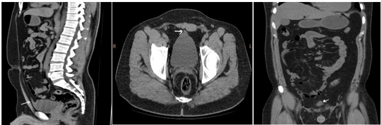 Figure 1: Sagittal, axial, and coronal image of CT pyelogram shows small outpouching which is extending from anterosuperior aspect of the bladder and connecting with cord like structure which is reaching up to umbilicus however this small outpouching have no connection with umbilicus.
The urachus is fibromuscular hollow cordlike structure that extends from the anterosuperior surface of the urinary bladder to the umbilicus and it regresses secondary to fibrosis with age, becoming the median umbilical ligament [1]. When the urachus fails to obliterate completely, the structure that remains is known as a urachal remnant [7].
The incidence of urachal remains is around 1/5000, and they are frequently discovered by accident during cross-sectional imaging. One-third of all people may have urachal remains. Furthermore, urachal remains in children under 6 months of age should be considered physiologic as more than 80% of them dissolve with nonoperative care [4,8].
Vesicourachal diverticulum is one of the congenital urachal remnant abnormalities, representing a result of the failure of the urachus to close at the urinary bladder, forming an out-pouching of variable length from the anterosuperior aspect of the urinary bladder, which does not communicate with the umbilicus [6], as seen in our case.
Urachal remains are divided into four groups: vesicourachal diverticulum, urachal cyst, umbilical-urachal sinus, and patent urachus [8]. When the bladder and umbilicus remain connected, this condition is known as a patent urachus. This causes urine to escape from the umbilicus and is frequently connected to posterior urethral valves [7].
An unconnected blind outpouching from the umbilicus that does not connect to the bladder is referred to as the umbilical-urachal sinus. The proximal and distal extremities of the urachus involute, while the intermediate portion remains intact, resulting in urachal cyst. Similar in principle to the umbilical urachal sinus, the vesicourachal diverticulum has a blind terminating urachus that opens to the bladder, unlike the umbilicus. The rarest form is this one [5,7,8].
This diverticulum can vary in size and can cause a variety of symptoms or consequences depending on its location and size [5]. Vesicourachal diverticulum is asymptomatic in most cases and is usually discovered incidentally at axial CT performed for unrelated reasons, appearing as a midline cystic lesion just above the anterosuperior aspect of the bladder [2].
Recurrent urinary tract infections, pain or discomfort in the lower abdomen, blood in the urine, and urinary frequency can all be symptoms of vesicourachal diverticulum. In some situations, the diverticulum becomes infected or blocked, resulting in more severe symptoms or problems [4].
Treatment options for vesicourachal diverticulum are determined by the severity of symptoms and consequences. In certain cases, onservative treatment with antibiotics and continuous monitoring may suffice [4,5]. However, if the diverticulum is causing considerable symptoms or difficulties, surgical intervention may be required.
Diverticulectomy, which involves removing the diverticulum, and resection of the urachus, which requires removing the complete urachal remnant, are two surgical therapy options [3,7].
The vesicourachal diverticulum is a rare congenital defect related to the urachus. Due to its asymptomatic nature, imaging examinations frequently uncover it by accident. Even though it is usually benign, knowing that it exists is important for any potential consequences and any necessary actions. A better knowledge of this uncommon ailment and its treatment will help to avoid from additional investigation and clinical trials.
The authors affirm that no conflict of interest exist.
No.

