 Journal of Medical Research and Surgery
PROVIDES A UNIQUE PLATFORM TO PUBLISH ORIGINAL RESEARCH AND REMODEL THE KNOWLEDGE IN THE AREA OF MEDICAL AND SURGERY
Journal of Medical Research and Surgery
PROVIDES A UNIQUE PLATFORM TO PUBLISH ORIGINAL RESEARCH AND REMODEL THE KNOWLEDGE IN THE AREA OF MEDICAL AND SURGERY
Journal of Medical Research and Surgery
PROVIDES A UNIQUE PLATFORM TO PUBLISH ORIGINAL RESEARCH AND REMODEL THE KNOWLEDGE IN THE AREA OF MEDICAL AND SURGERY
Journal of Medical Research and Surgery
PROVIDES A UNIQUE PLATFORM TO PUBLISH ORIGINAL RESEARCH AND REMODEL THE KNOWLEDGE IN THE AREA OF MEDICAL AND SURGERY


 Indexed Articles
Select your language of interest to view the total content in your interested language
Dominique Pianel
PhD in semiology, Osteopath, DU in Clinical Anatomy, Paris, France
Correspondence to: Dominique Pianel, PhD in semiology, Osteopath, DU in Clinical Anatomy, Paris, France; E-mail: dpianel.osteo@gmail.com
Received date: June 2, 2020; Accepted date: June 11, 2020; Published date: June 18, 2020
Citation: Pianel D (2020) Synthesis of Antler Antlers, for Research on Neuropeptide Y, in Relation to Calcitonin, at the Ligament Level. J Med Res Surg. 1(4): pp. 1-6
Copyright: ©2020 Pianel D. This is an open-access article distributed under the terms of the Creative Commons Attribution License, which permits unrestricted use,
distribution and reproduction in any medium, provided the original author and source are credited.
The horizontal and multidisciplinary study gives depth to our clinical approach and comparative anatomy can be used to understand the physiological behaviours for this study.
The production of nearly 14 kilos of bone in the antlers, in less than 100 days in the Red Deer is the trigger for the research. Embryology is a relational guide between the mandible, the desmodontal ligament and the stomach which are also under the influence of the functions of the neuropeptide Y which we propose to study.
Powerful orexigenic, because it increases the level of insulin and consequently the reserve, lipido-glucidic, it is its hypotensive natriuretic activity which positions the NPY in the neuropeptides with digestive prevalence. Its involvement in the calcium chain means that NPY will be studied in spectrography and immunofluorescence in our research, at the ligament level to reflect, biomechanics in a sense of functional and chemical stimulation
Neuropeptide Y, Antler, Calcitonin, Comparative anatomy
The growth of antlers in the male elaphus cervus is a seasonal and obsolete phenomenon, causing a significant variation in the weight of the deer head, up to 14 kg.
Having brought together, horizontally, clinical and comparative anatomy, embryology, biophysics, spectrography and protein immunofluorescence which are the main players in this research.
This growth is the result of sexual maturity and can reach 3 to 5 cm/day [1]. Crigel and Co, taking up the work of Haigh et al. [2] , show the cyclical nature broken down into: Osteosynthesis is subjected to androgenic stimulation which confirms the absence of wood in the female.
The 4 growth phases being subject to the growth factor “IGF1, Insulin-Like Grouwth factor 1” and therefore testosterone allows the initiation of the initial phase [3].
The antlers are all different from one individual to another and their difference remains constant each year with the same number of antlers (Figure 1-5).
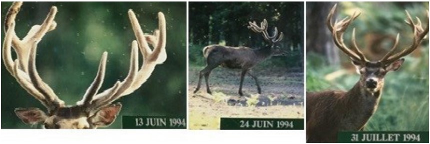 Figure 1: Personal photos from Chambord Castel reserve showing the same deer in the budding phase.
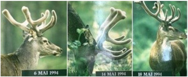 Figure 2: Personal photos from Chambord Castel reserve showing the same deer in the velvet antler.
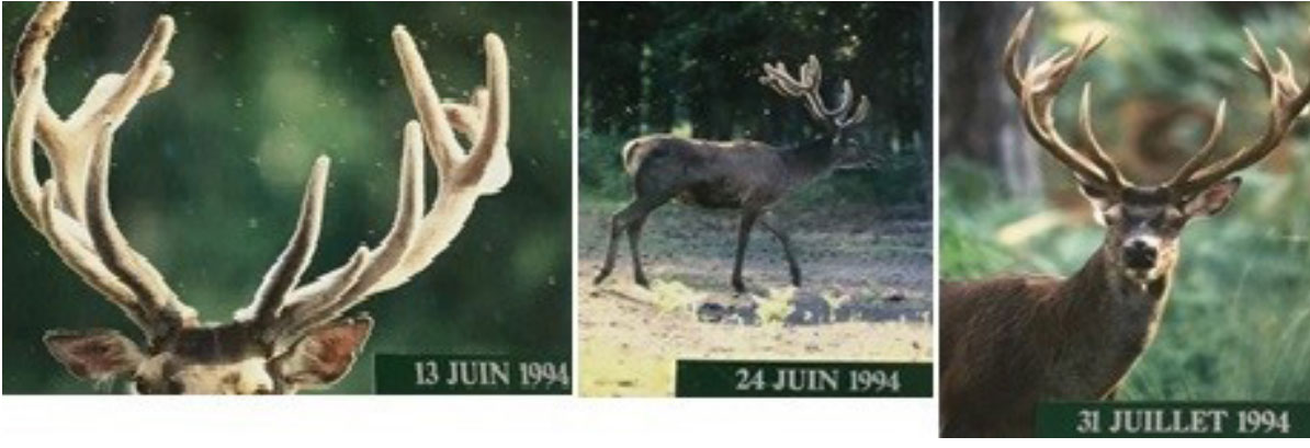 Figure 3: Personal photos from Chambord Castel reserve showing the same deer at the end of growth and loss of velvet.
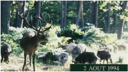 Figure 4: Photos from Chambord Castel reserve showing the same deer in
the antler drying and animal loss.
Figure 4: Photos from Chambord Castel reserve showing the same deer in
the antler drying and animal loss.
There is consistency in this framework of the individual signifier. Lee’s research, taken up by Crigel and Co, shows that the density of Ca++ bone relative to the weight of deer antlers is the least dense in comparison, with the other bone tissues studied [4]. This distinction physically defines resistance by elasticity, compared to the cortical bone of the human which is two to three times less elastic. In their studies they add that the antlers of red deer and the bovine femur have the same resistance tested at 100 to 140 MPa. The fracture threshold is then four to five times higher on the antlers than on the bovine femur, the line of fracture of the antlers increasing by the spiral shape [5]. An anatomical difference on the skulls of the elaphe and the Dama dama, which is smaller than the deer, itself sporting its antlers more vertically than the deer. The plumes have a difference in weight, due to their sizes and density, the flat bones of the deer being lighter. The half antlers of the elaph deer weigh 4.1 kilos measuring 55-90 cm longest, that of the deer dama 2.2 kilos, measuring 32-40 cm longest. A difference increased by more than 42% from two animals of same age (Figure 6-7).
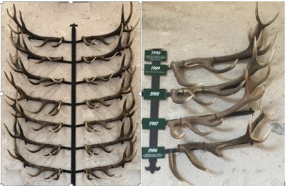 Figure 5: Personal photos from Chambord Castel reserve showing a horde
of males gathered and the difference between their antlers individually.
Figure 5: Personal photos from Chambord Castel reserve showing a horde
of males gathered and the difference between their antlers individually.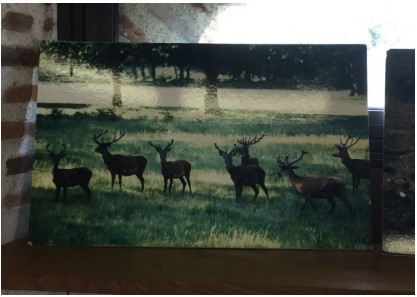 Figure 6: Personal photos of massacre to the same deer from Chambord
Castel reserve showing the individual consistency of the structure of antlers
for each year of the same deer.
Figure 6: Personal photos of massacre to the same deer from Chambord
Castel reserve showing the individual consistency of the structure of antlers
for each year of the same deer.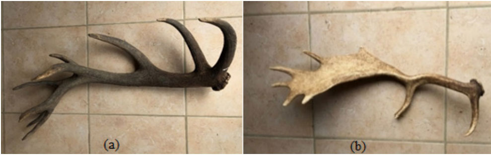 Figure 7: a) Half antler to Elaph deer; b) Half antler to Dama deer.
There is consistency in this framework of the individual signifier. Lee’s research, taken up by Crigel and Co, shows that the density of Ca++ bone relative to the weight of deer antlers is the least The deer dama, on the contrary has a low concavity which responds to the differences in size and shape of the antlers (Figure 8).
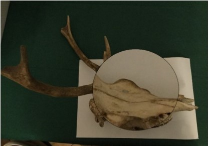 Figure 8: Deer massacre recovered in Boutissaint park (Yonne in French created in 1968), in 2016.
 On the same scale of this deer to the same age, there is an increase of more than 38.54% between the surface of the deer The study of thorny, withers, on the 278 skeletons of the MNHN reserve in Paris, under the direction of Pr A. ABOURACHID, wishing to highlight a biomechanical relationship between the spine and the weight of the antlers, did not was convincing, the standard deviations being too high on the scalar products between lengths and angles of the thorny plants and the zygomatic bone and that of the deer (Figure 9-10).
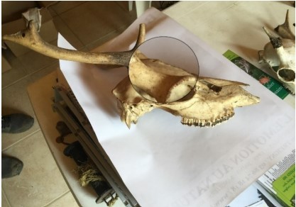 Figure 9: Fallow deer massacre recovered in Boutissaint park (Yonne in
French created in 1968), in 2014.
Figure 9: Fallow deer massacre recovered in Boutissaint park (Yonne in
French created in 1968), in 2014.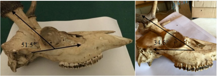 Figure 10: Angle between the antlers and the zygomatic bone is more open for deer antlers with 51.5°, more closed in deer, with 34.6° for the massacres studied
The study of thorny, withers, on the 278 skeletons of the MNHN reserve in Paris, under the direction of Pr A. ABOURACHID, wishing to highlight a biomechanical relationship between the spine and the weight of the antlers, did not was convincing, the standard deviations being too high on the scalar products between lengths and angles of the thorny plants and the measurements related to the sex and to the age of the animals did not allow to draw any relevant correlations (Figure 11-12).
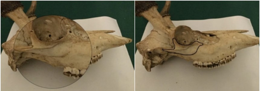 Figure 11: For Red deer, its reported surface is 10.88 cm²
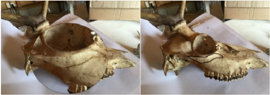 Figure 12: For Dama deer, its reported surface is 6.69 cm².
After having scanned a young male deer, a complete dissection of the spine was carried out at the MNHN, and to show that the diameter of the nuchal ligament in red deer is larger than in other bovids and then in equines. It anastomoses to the supraspinatus ligament up to the sacrococcygeal zone, strengthening the cephalic maintenance of the woods.
It increases in diameter towards its cranial part and measures, for this specimen, more than 42.7 mm in diameter.The nuchal ligament antagonism is the sternozygomatic (Figure 15), it avoids hyperflexion of the cervical spine and presents to describe sternal fibres which are inserted on the ventral and central part of the sternal manubrium [6].
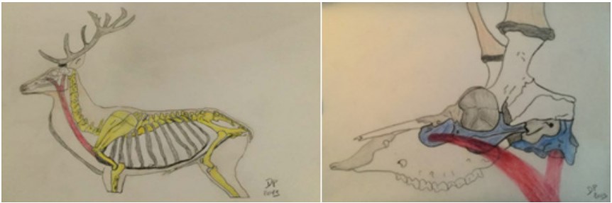 Figure 13: Personal drawings describing the sternozygomatic muscular of the deer studied
The nuchal ligament is continued by the supraspinatus ligament, related to the common embryological evolution of the stomach, cardiac and mandibular buds, it appears that a link between these structures is to be explored. It was within the desmodontal ligament [7] that the work of Doctor Bouchard demonstrated the presence of NPY, although it was on the hypothalus that the greatest concentrations were found [8]. It belongs to the family of eponymous peptide neurotransmitters, its versatile role is oroxigenic, stimulates stomach contraction, is thermoregulatory, vasoconstrictor, anxiolytic and stimulant of the calcium chain [9], this opens the hypothesis of the presence of NPY at the ligament level which has been advanced in 1999 by Yew et al. [10] and by Sengul et al. [11].The latter stressing the embryological relationship from neurulation to the caudal axis reaching future pudendal ganglia in humans. In their work on the spine, Aspden and Porter show that this ligamentous relevance would be verifiable on the whole of the supraspinatus ligament up to the lumbar vertebrae [12]. Discovered and sequenced in 1982 by Tatemoto [13], the NPY sequence in the alligator turns out to be identical to that of humans. The bio-library study of trypsin digested NPY blasts has shown sequence similarities for three positions-except in the rat where the position is different:
The first two sequences are 100% blasted for the mammals listed, and 92% for certain bats, hamsters, and white mice. These results serve as preliminaries for spectrographic analyses at the Monod Institute on different ligament samples. To date our work focuses on the Immunofluorescence in the Biopole of EVMA, to quantify the presence of NPY in these anatomical pieces.
Work carried out since 1986 on calcitonin and NPY has, among other things, made it possible to understand the cholinergic interactions of the brain with NPY in Alzheimer’s disease and its influence on depression and memory stability by Remi Quirion [14] and confirmed by the work of Doctor Gérard Karsenty [15]. It is this set that prompts us to consider chemical analyses on the nuchal ligament. The demonstration of the presence of NPY on the nuchal ligament of several specimens of red deer would be in favour of the hypothesis that the weight of the wood stimulates calcium production. This research will be carried out by immunofluorescence on various ligamentous tissues from a female naturally devoid of wood and a mature male with developed wood.
The author would like to thank the MNHN of Paris for its dissection room. The author posthumously thanks Professor Yves Lignereux professor of human and animal anatomy, director of the MNHN in Toulouse until 2019†, for our collaboration on the evolution of the neurocranium, in April 2012. The author also thanks the heads of the spectrography service at the Monod Institute, in Paris; FICIF, for the removal of anatomical materials; the EVMA Biopole laboratory for research possibilities on NPY in immunofluorescence. The author thanks his wife in the search for publications and her photos of deer in the world.
The author also thanks Professor Rémi Quirion and Professor Pierrette Gaudreau of Montreal for their support in this prospect of NPY research on ligament tissues and Professor William Rostème, who maintains his interest in the continuation of this research.
There are no conflicts of interest.

