Secreted key regulators (Fgf1, Bmp4, Gdf3) are expressed by PAC1-immunopositive retinal ganglion cells in the postnatal rat retina

Submitted: 17 December 2021
Accepted: 2 March 2022
Published: 27 April 2022
Abstract Views: 597
PDF: 480
HTML: 17
Publisher's note
All claims expressed in this article are solely those of the authors and do not necessarily represent those of their affiliated organizations, or those of the publisher, the editors and the reviewers. Any product that may be evaluated in this article or claim that may be made by its manufacturer is not guaranteed or endorsed by the publisher.

Authors

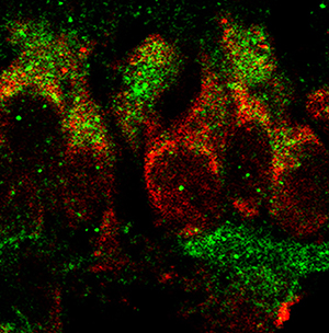
Identified as a member of the secretin/glucagon/VIP superfamily, pituitary adenylate cyclase-activating polypeptide (PACAP1-38) has been recognized as a hormone, neurohormone, transmitter, trophic factor, and known to be involved in diverse and multiple developmental processes. PACAP1-38 was reported to regulate the production of important morphogens (Fgf1, Bmp4, Gdf3) through PAC1-receptor in the newborn rat retina. To follow up, we aimed to reveal the identity of retinal cells responsible for the production and secretion of Fgf1, Bmp4, and Gdf3 in response to PACAP1-38 treatment. Newborn (P1) rats were treated with 100 pmol PACAP1-38 intravitreally. After 24 h, retinas were dissected and processed for immunohistochemistry performed either on flat-mounted retinas or cryosections. Brn3a and PAC1-R double labeling revealed that 90% of retinal ganglion cells (RGCs) expressed PAC1-receptor. We showed that RGCs were Fgf1, Bmp4, and Gdf3-immunopositive and PAC1-R was co-expressed with each protein. To elucidate if RGCs release these secreted regulators, the key components for vesicle release were examined. No labeling was detected for synaptophysin, Exo70, or NESP55 in RGCs but an intense Rab3a-immunoreactivity was detected in their cell bodies. We found that the vast majority of RGCs are responsive to PACAP, which in turn could have a significant impact on their development or/and physiology. Although Fgf1, Bmp4, and Gdf3 were abundantly expressed in PAC1-positive RGCs, the cells lack synaptophysin and Exo70 in the newborn retina, thus unable to release these proteins. These proteins could regulate postnatal RGC development acting through intracrine pathways.

Dimensions

Altmetric

PlumX Metrics

Downloads

Download data is not yet available.

Citations

Reese BE, Colello RJ. Neurogenesis in the retinal ganglion cell layer of the rat. Neuroscience 1992;46:419-29. DOI: https://doi.org/10.1016/0306-4522(92)90062-7
Amini R, Rocha-Martins M, Norden C. Neuronal migration and lamination in the vertebrate retina. Front Neurosci 2017;11:742. DOI: https://doi.org/10.3389/fnins.2017.00742
Guerin MB, McKernan DP, O'Brien CJ, Cotter TG. Retinal ganglion cells: dying to survive. Int J Dev Biol 2006;50:665-74. DOI: https://doi.org/10.1387/ijdb.062159mg
Nguyen-Ba-Charvet KT, Rebsam A. Neurogenesis and specification of retinal ganglion cells. Int J Mol Sci 2020;21:451. DOI: https://doi.org/10.3390/ijms21020451
Du Y, Xiao Q, Yip HK. Regulation of retinal progenitor cell differentiation by bone morphogenetic protein 4 is mediated by the smad/id cascade. Invest Ophthalmol Vis Sci 2010;51:3764-73. DOI: https://doi.org/10.1167/iovs.09-4906
Liu J, Wilson S, Reh T. BMP receptor 1b is required for axon guidance and cell survival in the developing retina. Dev Biol 2003;256:34-48. DOI: https://doi.org/10.1016/S0012-1606(02)00115-X
Casini G. Neuropeptides and retinal development. Arch Ital Biol 2005;143:191-8.
Sherwood NM, Krueckl SL, McRory JE. The origin and function of the pituitary adenylate cyclase-activating polypeptide (PACAP)/glucagon superfamily. Endocr Rev 2000;21:619-70. DOI: https://doi.org/10.1210/edrv.21.6.0414
Vaudry D, Falluel-Morel A, Bourgault S, Basille M, Burel D, Wurtz O, et al. Pituitary adenylate cyclase-activating polypeptide and its receptors: 20 years after the discovery. Pharmacol Rev 2009;61:283-357. DOI: https://doi.org/10.1124/pr.109.001370
Denes V, Geck P, Mester A, Gabriel R. Pituitary adenylate cyclase-activating polypeptide: 30 years in research spotlight and 600 million years in service. J Clin Med 2019;8:1488. DOI: https://doi.org/10.3390/jcm8091488
Njaine B, Rocha-Martins M, Vieira-Vieira CH, De-Melo LD, Linden R, Braas K, et al. Pleiotropic functions of pituitary adenylyl cyclase-activating polypeptide on retinal ontogenesis: involvement of KLF4 in the control of progenitor cell proliferation. J Mol Neurosci 2014;54:430-42. DOI: https://doi.org/10.1007/s12031-014-0299-2
Vaudry D, Gonzalez BJ, Basille M, Fournier A, Vaudry H. Neurotrophic activity of pituitary adenylate cyclase-activating polypeptide on rat cerebellar cortex during development. Proc Natl Acad Sci USA 1999;96:9415-20. DOI: https://doi.org/10.1073/pnas.96.16.9415
Ogata K, Shintani N, Hayata-Takano A, Kamo T, Higashi S, Seiriki K, et al. PACAP enhances axon outgrowth in cultured hippocampal neurons to a comparable extent as BDNF. PLoS One 2015;10:e0120526. DOI: https://doi.org/10.1371/journal.pone.0120526
Cameron DB, Raoult E, Galas L, Jiang Y, Lee K, Hu T, et al. Role of PACAP in controlling granule cell migration. Cerebellum 2009;8:433-40. DOI: https://doi.org/10.1007/s12311-009-0121-9
Girard BM, Keller ET, Schutz KC, May V, Braas KM. Pituitary adenylate cyclase activating polypeptide and PAC1 receptor signaling increase Homer 1a expression in central and peripheral neurons. Regul Pept 2004;123:107-16. DOI: https://doi.org/10.1016/j.regpep.2004.05.024
Dickson L, Finlayson K. VPAC and PAC receptors: From ligands to function. Pharmacol Ther 2009;121:294-316. DOI: https://doi.org/10.1016/j.pharmthera.2008.11.006
Blechman J, Levkowitz G. Alternative splicing of the pituitary adenylate cyclase-activating polypeptide receptor PAC1: Mechanisms of fine tuning of brain activity. Front Endocrinol (Lausanne) 2013;4:55. DOI: https://doi.org/10.3389/fendo.2013.00055
Seki T, Shioda S, Ogino D, Nakai Y, Arimura A, Koide R. Distribution and ultrastructural localization of a receptor for pituitary adenylate cyclase activating polypeptide and its mRNA in the rat retina. Neurosci Lett 1997;238:127-30. DOI: https://doi.org/10.1016/S0304-3940(97)00869-0
Denes V, Czotter N, Lakk M, Berta G, Gabriel R. PAC1-expressing structures of neural retina alter their PAC1 isoform splicing during postnatal development. Cell Tissue Res 2014;355:279-88. DOI: https://doi.org/10.1007/s00441-013-1761-0
Denes V, Hideg O, Nyisztor Z, Lakk M, Godri Z, Berta G, et al. The neuroprotective peptide PACAP1-38 contributes to horizontal cell development in postnatal rat retina. Invest Ophthalmol Vis Sci 2019;60:770-8. DOI: https://doi.org/10.1167/iovs.18-25719
Barnstable CJ, Hofstein R, Akagawa K. A marker of early amacrine cell development in rat retina. Brain Res 1985;352:286-90. DOI: https://doi.org/10.1016/0165-3806(85)90116-6
Perry VH, Walker M. Amacrine cells, displaced amacrine cells and interplexiform cells in the retina of the rat. Proc R Soc Lond B Biol Sci.1980;208:415-31. DOI: https://doi.org/10.1098/rspb.1980.0060
Lakk M, Denes V, Kovacs K, Hideg O, Szabo BF, Gabriel R. Pituitary adenylate cyclase-activating peptide (PACAP), a novel secretagogue, regulates secreted morphogens in newborn rat retina. Invest Ophthalmol Vis Sci 2017;58:565-72. DOI: https://doi.org/10.1167/iovs.16-20566
Schulz S, Rocken C, Mawrin C, Weise W, Hollt V, Schulz S. Immunocytochemical identification of VPAC1, VPAC2, and PAC1 receptors in normal and neoplastic human tissues with subtype-specific antibodies. Clin Cancer Res 2004;10:8235-42. DOI: https://doi.org/10.1158/1078-0432.CCR-04-0939
Catalani E, Tomassini S, Dal Monte M, Bosco L, Casini G. Localization patterns of fibroblast growth factor 1 and its receptors FGFR1 and FGFR2 in postnatal mouse retina. Cell Tissue Res 2009;336:423-38. DOI: https://doi.org/10.1007/s00441-009-0787-9
Yun YR, Won JE, Jeon E, Lee S, Kang W, Jo H, et al. Fibroblast growth factors: biology, function, and application for tissue regeneration. J Tissue Eng 2010;2010:218142. DOI: https://doi.org/10.4061/2010/218142
Bragdon B, Moseychuk O, Saldanha S, King D, Julian J, Nohe A. Bone morphogenetic proteins: a critical review. Cell Signal 2011;23:609-20. DOI: https://doi.org/10.1016/j.cellsig.2010.10.003
McCabe KL, Gunther EC, Reh TA. The development of the pattern of retinal ganglion cells in the chick retina: mechanisms that control differentiation. Development 1999;126:5713-24. DOI: https://doi.org/10.1242/dev.126.24.5713
Russell C. The roles of hedgehogs and fibroblast growth factors in eye development and retinal cell rescue. Vision Res 2003;43:899-912. DOI: https://doi.org/10.1016/S0042-6989(02)00416-9
Zhang SS, Liu MG, Kano A, Zhang C, Fu XY, Barnstable CJ. STAT3 activation in response to growth factors or cytokines participates in retina precursor proliferation. Exp Eye Res 2005;81:103-15. DOI: https://doi.org/10.1016/j.exer.2005.01.016
Eckenstein FP, Kuzis K, Nishi R, Woodward WR, Meshul C, Sherman L, et al. Cellular distribution, subcellular localization and possible functions of basic and acidic fibroblast growth factors. Biochem Pharmacol 1994;47:103-10. DOI: https://doi.org/10.1016/0006-2952(94)90442-1
Forthmann B, Grothe C, Claus P. A nuclear odyssey: fibroblast growth factor-2 (FGF-2) as a regulator of nuclear homeostasis in the nervous system. Cell Mol Life Sci 2015;72:1651-62. DOI: https://doi.org/10.1007/s00018-014-1818-6
Zhan X, Hu X, Friedman S, Maciag T. Analysis of endogenous and exogenous nuclear translocation of fibroblast growth factor-1 in NIH 3T3 cells. Biochem Biophys Res Commun 1992;188:982-91. DOI: https://doi.org/10.1016/0006-291X(92)91328-N
Lin YZ, Yao SY, Hawiger J. Role of the nuclear localization sequence in fibroblast growth factor-1-stimulated mitogenic pathways. J Biol Chem 1996;271:5305-8. DOI: https://doi.org/10.1074/jbc.271.10.5305
Bryant DM, Stow JL. Nuclear translocation of cell-surface receptors: lessons from fibroblast growth factor. Traffic 2005;6:947-54. DOI: https://doi.org/10.1111/j.1600-0854.2005.00332.x
Bouleau S, Grimal H, Rincheval V, Godefroy N, Mignotte B, Vayssiere JL, et al. FGF1 inhibits p53-dependent apoptosis and cell cycle arrest via an intracrine pathway. Oncogene 2005;24:7839-49. DOI: https://doi.org/10.1038/sj.onc.1208932
Trousse F, Esteve P, Bovolenta P. Bmp4 mediates apoptotic cell death in the developing chick eye. J Neurosci 2001;21:1292-301. DOI: https://doi.org/10.1523/JNEUROSCI.21-04-01292.2001
Haynes T, Gutierrez C, Aycinena JC, Tsonis PA, Del Rio-Tsonis K. BMP signaling mediates stem/progenitor cell-induced retina regeneration. Proc Natl Acad Sci USA 2007;104:20380-5. DOI: https://doi.org/10.1073/pnas.0708202104
Maruyama Y, Mikawa S, Hotta Y, Sato K. BMP4 expression in the developing rat retina. Brain Res 2006;1122:116-21. DOI: https://doi.org/10.1016/j.brainres.2006.08.130
Maruyama-Koide Y, Mikawa S, Sasaki T, Sato K. Bone morphogenetic protein-4 and bone morphogenetic protein receptors expressions in the adult rat eye. Eur J Histochem 2017;61:2797. DOI: https://doi.org/10.4081/ejh.2017.2797
Levine AJ, Brivanlou AH. GDF3 at the crossroads of TGF-beta signaling. Cell Cycle. 2006;5:1069-73. DOI: https://doi.org/10.4161/cc.5.10.2771
Grabs D, Bergmann M, Urban M, Post A, Gratzl M. Rab3 proteins and SNAP-25, essential components of the exocytosis machinery in conventional synapses, are absent from ribbon synapses of the mouse retina. Eur J Neurosci 1996;8:162-8. DOI: https://doi.org/10.1111/j.1460-9568.1996.tb01177.x
Binotti B, Jahn R, Chua JJ. Functions of rab proteins at presynaptic sites. Cells 2016;5:7. DOI: https://doi.org/10.3390/cells5010007
Sassoe-Pognetto M, Wassle H. Synaptogenesis in the rat retina: subcellular localization of glycine receptors, GABA(A) receptors, and the anchoring protein gephyrin. J Comp Neurol 1997;381:158-74. DOI: https://doi.org/10.1002/(SICI)1096-9861(19970505)381:2<158::AID-CNE4>3.0.CO;2-2
Dhingra NK, Ramamohan Y, Raju TR. Developmental expression of synaptophysin, synapsin I and syntaxin in the rat retina. Brain Res Dev Brain Res 1997;102:267-73. DOI: https://doi.org/10.1016/S0165-3806(97)00085-0
Fan WJ, Li X, Yao HL, Deng JX, Liu HL, Cui ZJ, et al. Neural differentiation and synaptogenesis in retinal development. Neural Regen Res 2016;11:312-8. DOI: https://doi.org/10.4103/1673-5374.177743
Nag TC, Wadhwa S. Differential expression of syntaxin-1 and synaptophysin in the developing and adult human retina. J Biosci 2001;26:179-91. DOI: https://doi.org/10.1007/BF02703642
Feldman SA, Eiden LE. The chromogranins: their roles in secretion from neuroendocrine cells and as markers for neuroendocrine neoplasia. Endocr Pathol 2003;14:3-23. DOI: https://doi.org/10.1385/EP:14:1:3
Wu B, Guo W. The exocyst at a glance. J Cell Sci 2015;1282957-64. DOI: https://doi.org/10.1242/jcs.156398
Barkefors I, Fuchs PF, Heldin J, Bergstrom T, Forsberg-Nilsson K, Kreuger J. Exocyst complex component 3-like 2 (EXOC3L2) associates with the exocyst complex and mediates directional migration of endothelial cells. J Biol Chem 2011;286:24189-99. DOI: https://doi.org/10.1074/jbc.M110.212209
Re RN, Cook JL. The physiological basis of intracrine stem cell regulation. Am J Physiol Heart Circ Physiol 2008;295:H447-53. DOI: https://doi.org/10.1152/ajpheart.00461.2008
Jouanneau J, Gavrilovic J, Caruelle D, Jaye M, Moens G, Caruelle JP, et al. Secreted or nonsecreted forms of acidic fibroblast growth factor produced by transfected epithelial cells influence cell morphology, motility, and invasive potential. Proc Natl Acad Sci USA 1991;88:2893-7. DOI: https://doi.org/10.1073/pnas.88.7.2893
Redburn DA, Rowe-Rendleman C. Developmental neurotransmitters. Signals for shaping neuronal circuitry. Invest Ophthalmol Vis Sci 1996;37:1479-82.
Fletcher EL, Kalloniatis M. Localisation of amino acid neurotransmitters during postnatal development of the rat retina. J Comp Neurol 1997;380:449-71. DOI: https://doi.org/10.1002/(SICI)1096-9861(19970421)380:4<449::AID-CNE3>3.0.CO;2-1
Johnson J, Tian N, Caywood MS, Reimer RJ, Edwards RH, Copenhagen DR. Vesicular neurotransmitter transporter expression in developing postnatal rodent retina: GABA and glycine precede glutamate. J Neurosci 2003;23:518-29. DOI: https://doi.org/10.1523/JNEUROSCI.23-02-00518.2003
Grunder T, Kohler K, Guenther E. Distribution and developmental regulation of AMPA receptor subunit proteins in rat retina. Invest Ophthalmol Vis Sci 2000;41:3600-6.

Ethics Approval

All procedures were approved by the Ethical Committee of the University of Pécs, Hungary, and the Animal Health and Animal Welfare Directorate of the National Food Chain Safety Office of the Hungarian State (BA/35/51-58/2016)

Rights

TKP2021-EGA-16 has been implemented with the support provided from the National Research, Development and Innovation Fund of Hungary, financed under the TKP2021-EGA funding scheme. The work has also been supported by EFOP 3.6.-2-16-2017-0008 and NKFIH 119 289.

How to Cite

Dénes, V., Kovacs, K., Lukáts, Ákos, Mester, A., Berta, G., Szabó, A., & Gabriel, R. (2022). Secreted key regulators (Fgf1, Bmp4, Gdf3) are expressed by PAC1-immunopositive retinal ganglion cells in the postnatal rat retina. European Journal of Histochemistry, 66(2). https://doi.org/10.4081/ejh.2022.3373