image
Original Research
Genodermatoses in Las Tunas Province, Cuba, 1989–2019
https://doi.org/10.37757/MR2021.V23.N2.10

INTRODUCTION Genodermatoses are a group of genetic diseases that affect the skin and adjoining tissues. They represent 15% of genetic diseases worldwide. Cuba established a National Program for the Diagnosis, Care and Prevention of Genetic Diseases and Congenital Abnormalities in 1980, which was implemented in Las Tunas in 1989. In 2010, a specialized multidisciplinary provincial service for genodermatoses patients was established in Las Tunas province. Several studies in Las Tunas show that genodermatoses represent 22.2% of genetic diseases; the most common are ichthyosis (16.7%), mastocytosis (11.7%), and neurofibromatosis (8.3%). Children aged <12 years are the most affected (61.6%).

OBJECTIVE Describe genodermatoses in Las Tunas Province, Cuba, since the implementation of the National Program for the Diagnosis, Care, and Prevention of Genetic Diseases and Congenital Abnormalities, and after the creation of a specialized multidisciplinary provincial service for genodermatoses patients.

METHODS We conducted an observational, descriptive, retrospective study in 249 patients diagnosed with some type of genodermatosis who received care in Las Tunas during 1989–2019. Variables considered were: type of genodermatosis, complications, deaths and geographic location by municipality. We studied prevalence rates (1989–2019), incidence rates (2010–2019), proportion of complications, survival rates, and types of genodermatosis diagnosed by municipality in two periods (1989–2009 and 2010–2019) one before, and one after the implementation of a targeted multidisciplinary provincial care service.

RESULTS The general prevalence rate of genodermatoses in Las Tunas Province was 46.51 per 100,000 population. The forms with the highest prevalence rates were neurofibromatosis type 1 (13.6 per 100,000 population), classical Ehlers-Danlos syndrome (7.1 per 100,000), ichthyosis vulgaris (5.0 per 100,000) and cutaneous mastocytosis (2.4 per 100,000). The highest incidence rates coincided with the conditions with the highest prevalence: neurofibromatosis type 1 (81.5 per 1000 cases in 2013), classical Ehlers-Danlos syndrome (44.4 per 1000 cases in 2013) and ichthyosis vulgaris (52 per 1000 cases in 2010). From 1989–2009, patients presented a greater frequency of complications, at 40% (22/55) than from 2010–2019 at 21.1% (41/194). Pyodermitis was the most common during the study period (1989–2019), with 29.1% (16/55). Survival was high, at 98.0% (only 5 deaths in 2009, 2010, 2011, 2012, and 2015, and were no deaths during other years) in the study period. The greatest share of genodermatosis cases was registered in the municipality of Majibacoa (0.07%), and consanguinity was found in cases of epidermolysis bullosa, Herlitz type and xeroderma pigmentosum.

CONCLUSION In Las Tunas Province, Cuba, genodermatoses as a whole are not rare diseases. Those with the highest prevalence and incidence rates are neurofibromatosis type 1, classical Ehlers-Danlos syndrome and ichthyosis vulgaris. After implementation of the specialized multidisciplinary provincial clinic for genodermatoses patients within Cuba’s National Program for the Diagnosis, Care, and Prevention of Genetic Diseases and Congenital Abnormalities, in addition to the active screening implemented by this Program, more cases were diagnosed, and a lower proportion of complications and a higher survival rates were recorded.

KEYWORDS Dermatology, skin diseases, genetic, genetic diseases, inborn, neurofibromatoses, Cuba

image

ABSTRACT
INTRODUCTION
Genodermatoses are a group of genetic diseases that affect the skin and adjoining tissues. They represent 15% of genetic diseases worldwide. Cuba established a National Program for the Diagnosis, Care and Prevention of Genetic Diseases and Congenital Abnormalities in 1980, which was implemented in Las Tunas in 1989. In 2010, a specialized multidisciplinary provincial service for genodermatoses patients was established in Las Tunas province. Several studies in Las Tunas show that genodermatoses represent 22.2% of genetic diseases; the most common are ichthyosis (16.7%), mastocytosis (11.7%), and neurofibromatosis (8.3%). Children aged <12 years are the most affected (61.6%).

OBJECTIVE Describe genodermatoses in Las Tunas Province, Cuba, since the implementation of the National Program for the Diagnosis, Care, and Prevention of Genetic Diseases and Congenital Abnormalities, and after the creation of a specialized multidisciplinary provincial service for genodermatoses patients.

METHODS We conducted an observational, descriptive, retrospective study in 249 patients diagnosed with some type of genodermatosis who received care in Las Tunas during 1989–2019. Variables considered were: type of genodermatosis, complications, deaths and geographic location by municipality. We studied prevalence rates (1989–2019), incidence rates (2010–2019), proportion of complications, survival rates, and types of genodermatosis diagnosed by municipality in two periods (1989–2009 and 2010–2019) one before, and one after the implementation of a targeted multidisciplinary provincial care service.

RESULTS The general prevalence rate of genodermatoses in Las Tunas Province was 46.51 per 100,000 population. The forms with the highest prevalence rates were neurofibromatosis type 1 (13.6 per 100,000 population), classical Ehlers-Danlos syndrome (7.1 per 100,000), ichthyosis vulgaris (5.0 per 100,000) and cutaneous mastocytosis (2.4 per 100,000). The highest incidence rates coincided with the conditions with the highest prevalence: neurofibromatosis type 1 (81.5 per 1000 cases in 2013), classical Ehlers-Danlos syndrome (44.4 per 1000 cases in 2013) and ichthyosis vulgaris (52 per 1000 cases in 2010). From 1989–2009, patients presented a greater frequency of complications, at 40% (22/55) than from 2010–2019 at 21.1% (41/194). Pyodermitis was the most common during the study period (1989–2019), with 29.1% (16/55). Survival was high, at 98.0% (only 5 deaths in 2009, 2010, 2011, 2012, and 2015, and were no deaths during other years) in the study period. The greatest share of genodermatosis cases was registered in the municipality of Majibacoa (0.07%), and consanguinity was found in cases of epidermolysis bullosa, Herlitz type and xeroderma pigmentosum.

CONCLUSION In Las Tunas Province, Cuba, genodermatoses as a whole are not rare diseases. Those with the highest prevalence and incidence rates are neurofibromatosis type 1, classical Ehlers-Danlos syndrome and ichthyosis vulgaris. After implementation of the specialized multidisciplinary provincial clinic for genodermatoses patients within Cuba’s National Program for the Diagnosis, Care, and Prevention of Genetic Diseases and Congenital Abnormalities, in addition to the active screening implemented by this Program, more cases were diagnosed, and a lower proportion of complications and a higher survival rates were recorded.

KEYWORDS Dermatology; skin diseases, genetic; genetic diseases, inborn; neurofibromatoses; Cuba

INTRODUCTION
In general, most dermatological diseases are genetic in origin; almost all cases involve a genetic predisposition to skin diseases.[1]

Individual immune response to infectious processes is modulated by epigenetics. The infectious dermatosis that best exemplifies this phenomenon is Hansen’s disease.[2] However, ‘genodermatosis’ refers to the process in which genetic anomalies (generally mutations of a single gene) play a predominant role in the origin of the disease and lead to clinical symptoms, which then manifest in the skin and adjoining tissues (adnexa). Their common characteristic is that they are all inherited.[3]

IMPORTANCE
This study updates understanding of genodermatoses in Las Tunas Province, after the implementation of the National Program for the Diagnosis, Care, and Prevention of Genetic Diseases and Congenital Abnormalities and the specialized multidisciplinary service that provides specialized care to patients affected with these genetic diseases.

Individually, genodermatoses are rare, but together they comprise a large group that is difficult to diagnose, treat and monitor due to their diversity in presentation and inheritance, even within the same disease.[4] Neurofibromatosis, Leopard syndrome, Ehlers-Danlos syndrome and tuberous sclerosis are diagnosed via clinical exam,[5,6] while ichthyosis, Darier disease, epidermolysis bullosa and mastocytosis are confirmed through histopathology.[5] In other cases, such as xeroderma pigmentosum (XP), supplemental studies are not conclusive. To diagnose the aforementioned conditions, Cuba uses histopathology, which is not conclusive, and single gel electrophoresis (SCGE) on isolated lymphocytes from peripheral blood to evaluate DNA’s ability to repair itself after damage is induced by ultraviolet light, a test which is not 100% specific to XP.[7] In general, there are no genetic tests available for mass screening of these conditions.

Genodermatosis lesions can be very visible and have a psychological impact on patients, and can result in social stigma, negatively impacting their quality of life. Many genodermatoses lead to chronic disability, others to death.[4]

It is difficult to gather precise data on the prevalence of genodermatoses (especially in developing countries), due to the diversity of diseases that are grouped under this common name, many of which go undiagnosed. In the most studied diseases, the results vary from one country to the next. Worldwide, the incidence of hypomelanosis of Ito is 1 per 7540–10,000 live births;[8] ectodermal dysplasia, 1 per 100,000 births;[9] oculocutaneous albinism, 1 per 18,000 births;[10] incontinentia pigmenti, 1 per 40,000–50,000 births;[11] tuberous sclerosis, 1–5 per 10,000 live births;[12] epidermolysis bullosa, 1 per 500,000 live births;[13] and Waardenburg syndrome, 1 per 42,000 births.[14]

Few population studies on genodermatoses have been conducted in Cuba. Dorticós conducted research to characterize them in a study involving 10 hospitals in Havana from 1980 to 1986, in which ichthyosis (37.7%) and neurofibromatosis (18.8%) were the most common genodermatoses.[15] Campo Díaz published a study on Ehlers-Danlos syndrome type III conducted in Pinar del Río Province in 2010–2011, finding 100% with manifestations on the skin and 59.3% with associated bleeding disorders.[16] Orraca also made a genetic, clinical and epidemiological characterization of neurofibromatosis type 1 in Pinar del Río Province, finding a prevalence of 1 per 1141 in pediatric patients, above rates reported globally for this disease.[17] One study performed in 2015 in Las Tunas, an eastern Cuban province, suggested that 85% of genetic diseases treated at the Provincial Medical Genetics Department (PMGD) were diagnosed in pediatric patients; of those, 22% were dermatological.[18]

WHO estimates that there are approximately 10,000 genetic diseases affecting 7% of the world population.[19] Genetic diseases make up 25% of pediatric hospital admissions, with 15% of cases affecting the skin and adnexa.[20]

Since 1963, WHO has urged member states to focus on the control and prevention of genetic diseases (through early diagnosis, access to care, genetic counseling, etc.).[21] In developed countries, there are numerous foundations dedicated to the treatment of specific genetic diseases, but there is no unifying vision that involves all levels of health care. In 1980, Cuba’s national health system implemented the National Program for the Diagnosis, Care, and Prevention of Genetic Diseases and Congenital Abnormalities,[22] which we will identify below as NGP (National Genetics Program). This program is supported by a network of Genetics Counseling Service comprising 172 Medical Genetics Municipal Departments (in every municipality in the country), 16 PMGD (located in all Cuban provinces), and the National Medical Genetics Center (CNGM), a lead institution that provides coverage to the entire population. CNGM is a collaborative center focused on genetic developments that help promote health worldwide.[22]

NGP’s work is guided by the Manual of Regulations and Procedures for medical genetics services in Cuba,[23] which offers standards for mass screening for some genetic diseases via specialized screening. NGP not only offers specialized medical care, but also conducts preventive prenatal, perinatal, and postnatal services (at the three levels of prevention: primary, secondary, and tertiary) for early detection of congenital diseases and abnormalities; supports research aimed at designing education strategies for patients, their families, and the general public; and works to promote social inclusion of affected persons and improve their quality of life.[22] Masters and specialists in clinical genetics regularly conduct exchanges for diagnostics and patient follow-up with those in other fields (gynecology and obstetrics, ophthalmology, endocrinology, orthopedics, otorhinolaryngology, neonatology, immunology, molecular biology, dermatology, pediatrics, psychology, neurology, cardiology, oral medicine, pathology and imaging, etc.).[23]

NGP was established in Las Tunas Province in 1989. In 2010, a specialized multidisciplinary provincial service was established to care for genodermatosis patients. This bi-weekly service is managed by specialists in clinical medical genetics and dermatology, the service’s main specialties, with support from psychologists, immunologists, pediatricians and others, depending on patient characteristics and symptoms. From 2010 to 2012, the most common genodermatoses were ichthyosis (16.7%), mastocytosis (11.7%), and neurofibromatosis (8.3%). The most affected were patients <12 years of age (61.6%).[24]

The objective of this study was to describe genodermatoses in Las Tunas Province, Cuba, since the establishment of NGP, and after the creation of a specialized multidisciplinary provincial service for genodermatosis patients.

METHODS
Study type and participants A descriptive observational study was performed, with cross-sectional and retrospective components. It was conducted at the PMGD’s specialized multidisciplinary provincial genodermatoses service in Las Tunas Province, Cuba, and comprised the years 1989–2019. The universe was composed of 942 patients with genetic disease diagnoses, registered in the PMGD database and, for non-probability sampling, 249 patients who met the diagnostic criteria of genodermatoses.

Inclusion criteria Patients seen at the service, who met genodermatosis diagnostic criteria, and who gave written informed consent to participate in the study (in the case of minors, parents or guardians authorized patient participation).

Exclusion criteria Patients seen at the clinic, but whose medical records did not contain all data necessary for the study, or who had psychiatric conditions that would prevent necessary data collection.

Diagnostic criteria Genodermatoses are considered clinical conditions whose primary phenotypic manifestations involve the skin and adnexa and that have a genetic component. Patient genealogy was examined to determine genetic history, inheritance patterns and the diagnosis of new cases in the family since the proband case.[23] In all cases the diagnosis was confirmed by a clinical geneticist.

Supplemental studies were performed to corroborate the diagnosis or presence of complications. These studies were: hematological (liver transaminases, eosinophil count, peripheral blood smears to explore the presence of mast cells in peripheral blood);[25] histopathological (skin biopsy for microscopic diagnosis of aplasia cutis, cutis laxa, cutis verticis gyrata, congenital ectodermal dysplasia, Darier disease, Hailey-Hailey disease, epidermolysis bullosa, hypomelanosis of Ito, ichthyosis, incontenencia pigmenti, mastocytosis, pityriasis rubra pilaris, porokeratosis of Mibelli, palmoplantar keratoderma, Netherton syndrome, Proteus syndrome, and Rothmund-Thomson syndrome);[1,2] imaging (ultrasonography, computed tomography, and magnetic resonance imaging; fundamental in diagnosing neurofibromatosis, tuberous sclerosis and Sturge-Weber syndrome);[23] genetic, to corroborate genodermatosis diagnoses whose clinical presentations were not conclusive on their own or to differentiate them from other clinically similar conditions, such as SGCE on isolated lymphocytes XP diagnosis;[7] indirectly using five markers: four microsatellite markers (IVS27AAAT2.1, IVS38GT53.0, IV27AC28.4 and Mfd15) and one restriction fragment length polymorphism (Rsa I NF1 exon 5) to diagnose neurofibromatosis type 1;[17] and other studies required to diagnose infectious complications, such as bacterial and fungal cultures.[2]

The foundation of all diagnoses in the study were initial clinical exam and personalized physician–patient relationships, the fundamental pillars of diagnostics in general, especially when advanced technology is not available to corroborate the suspected clinical condition.[26]

Variables
Diagnosed genodermatoses (based on diagnostic criteria) The following were considered: Oculocutaneous albinism (OCA) [OMIM 203100 203200 203290 606574 615312 113750 615179 606952 ORPHA 55 GARD 10958], aplasia cutis [OMIM 107600 600360 ORPHA 1114 GARD 5835], cutis laxa [OMIM 123700 6144434 616603 219100 ORPHA 90348 90349 GARD 1639 8480], cutis verticis gyrata (CvsG) [OMIM 123790 ORPHA 671 GARD 1643], autosomal dominant hypohidrotic ectodermal dysplasia (HED-AD) [OMIM 129490 224900 614940 617337 ORPHA 238468 GARD 76], Darier disease (Darier) [OMIM 124200 ORPHA 218 GARD 6243], Hailey-Hailey disease (Hailey-H) [OMIM 169600 ORPHA 2841 GARD 6559], Niemann-Pick disease (Nieman P) [OMIM 257200 607616 257220 607625 ORPHA 77292 77293 646 GARD 7206 10729], generalized epidermolysis bullosa simplex (EB simplex) [OMIM 131900 ORPHA 79399 GARD 2147], junctional epidermolysis bullosa, Herlitz type (EB Herlitz) [OMIM 226700 ORPHANET 79404 GARD 2153], tuberous sclerosis (TS) [OMIM 191100 613254 ORPHA 805 GARD 7830 946], hypomelanosis of Ito (H. Ito) [OMIM 300337], ichthyosis vulgaris (Ichthyosis V) [OMIM 146700] Lamellar ichthyosis (Ichthyosis L) [OMIM 615024 ORPHA 313], incontinencia pigmenti (I.Pigmenti) [OMIM 161000 ORPHA 69087 GARD 3912], cutaneous mastocytosis [OMIM 154800 ORPHA 66646 GARD 7842], neurofibromatosis type 1 (NF-1) [OMIM 162200 ORPHANET 636 GARD 7866], segmental neurofibromatosis (Segmental NF) [OMIM 162200], piebaldism [OMIM 172800 ORPHA 2884 GARD 4344], pityriasis rubra pilaris (PRP) [OMIM 173200 ORPHA 2897 GARD 7401], classic porokeratosis of Mibelli (PM) [OMIM 175800 ORPHA 735 GARD 4438], epidermolytic palmoplantar keratoderma (PPK) [OMIM 144200 600962 ORPHA 2199 496 GARD 2826 5186], Albright syndrome (S. Albright) [OMIM 174800 ORPHA 562 GARD 6995], occipital horn syndrome (OH) [OMIM 304150 ORPHA 198 GARD 4017], classical Ehlers-Danlos syndrome (ED) [OMIM 130000 130010 ORPHA 98249 GARD 6322], LEOPARD syndrome (LEOPARD) [OMIM 151100 611554 613707 ORPHA 500 GARD 1100], Netherton syndrome (Netherton) [OMIM 256500 ORPHANET 634 GARD 7182], Noonan syndrome (Noonan) [OMIM 163950 605275 609942 610733 611553 613224 613706 615355 616559 616564 618499 618624 ORPHA 648 GARD 10955], Proteus syndrome (Proteus) [OMIM 176920 ORPHA 744 GARD 7475], Rothmund-Thomson syndrome (R-T) [OMIM 618625 ORPHANET 2909 GARD 4392], Sturge-Weber syndrome (S-W) [OMIM 185300 ORPHA 3205 GARD 7706], Waardenburg syndrome type I (W-I) [OMIM 193500 ORPHA 3440 GARD 5525] and xeroderma pigmentosum (XP) [OMIM 278700 278720 278730 278740 278760 278780 610651 ORPHA 910 GARD 7910].

New cases New cases diagnosed per year, for each genodermatosis.

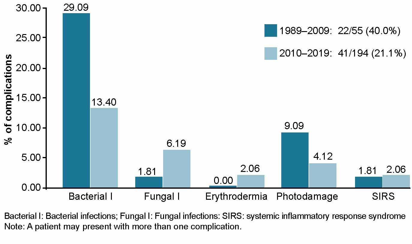
Complications We considered complications secondary to skin abnormalities due to genodermatosis, such as secondary pyoderma (Bacterial I),[2] superficial cutaneous mycoses (Fungal I) and within these, dermatophytosis and mucocutaneous candidiasis, erythroderma, sun damage (sun burns, sun freckles, elastosis, premalignant lesions, skin cancer) and systemic inflammatory response syndrome (SIRS). The number of patients with each complication were considered.

Number of deaths and diagnosis Deceased patients with genodermatosis and genodermatosis diagnosis at death.

Geographic location by municipality All municipalities of the province were considered: Amancio Rodríguez, Colombia, Jesús Menéndez, Jobabo, Las Tunas, Majibacoa, Manatí, and Puerto Padre. This variable provides important epidemiological elements pertaining to inheritance patterns, potential consanguinity and prevalence of specific genodermatoses within particular regions.

Data collection and analysis Information was collected using primary sources consisting of patients of legal age and parents or guardians of patients who were minors, and secondary sources consisting of the patient’s medical record and records from PMGD and specialized multidisciplinary provincial clinic for patients with genodermatoses. Techniques used to obtain information included participatory structured observation, face-to-face medical interviews, and general and more targeted physical examinations, as well as a review of secondary sources. All information was gathered in a Microsoft Excel database and was processed using the SPSS Version 18 for Windows.

Prevalence rates were calculated per 100,000 population, taking into consideration the population of Las Tunas Province in 2019 (535,335 inhabitants), according to the Cuban National Statistics and Information Bureau (ONEI).[27]

The annual incidence rate of genodermatosis was calculated for 2010–2019 (the period following the implementation of the specialized multidisciplinary provincial service for patients with genodermatoses), as was the proportion of complications. Two periods of time were compared, from the start of the National Genetics Program in 1989 to implementation of the specialized multidisciplinary provincial clinic for patients with genodermatoses in 2009, and during the clinic’s operation in 2010–2019. We also calculated the survival index (100 x number of survivors annually/total patients), fatality rate (100 x number of deaths per year/total patients), and number of genodermatoses diagnosed per municipality (number of patients/municipality population) x 100) for each genodermatosis.

To compare results among municipalities, the population of each was taken into consideration (according to ONEI data): 37,695 inhabitants in Amancio; 32,167 in Colombia; 48,208 in Jesús Menéndez; 42,603 in Jobabo; 211,596 in the municipality of Las Tunas; 41,287 in Majibacoa; 29,930 in Manatí, and 91,879 in Puerto Padre.[27]

Ethical considerations The research protocol was approved by the research ethics committee and scientific council at Mártires de las Tunas Provincial Pediatric Hospital and research was conducted in accordance with the Declaration of Helsinki.[28] To participate in the study, written informed consent was obtained from patients (or parents or guardians of patients who were minors). The written information provided explained the importance of participation, the risks and benefits, participants’ rights and study characteristics. Data was coded to protect patient identity.

RESULTS
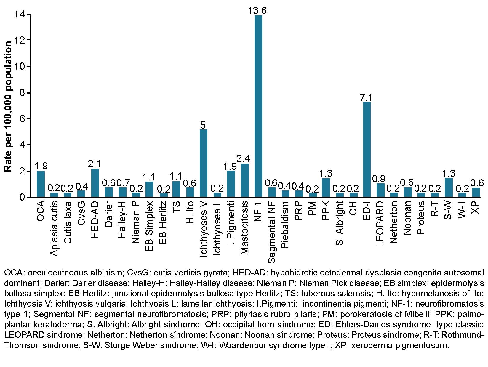
From 1989 through 2019, general prevalence rate of genodermatoses in Las Tunas Province was 46.5 per 100,000 population. Neurofibromatosis type 1 was predominant at 13.6 cases per 100,000 population, followed by classical Ehlers-Danlos syndrome at 7.1 per 100,000, ichthyosis vulgaris at 5 per 100,000 and cutaneous mastocytosis at 2.4 per 100,000 (Figure 1).

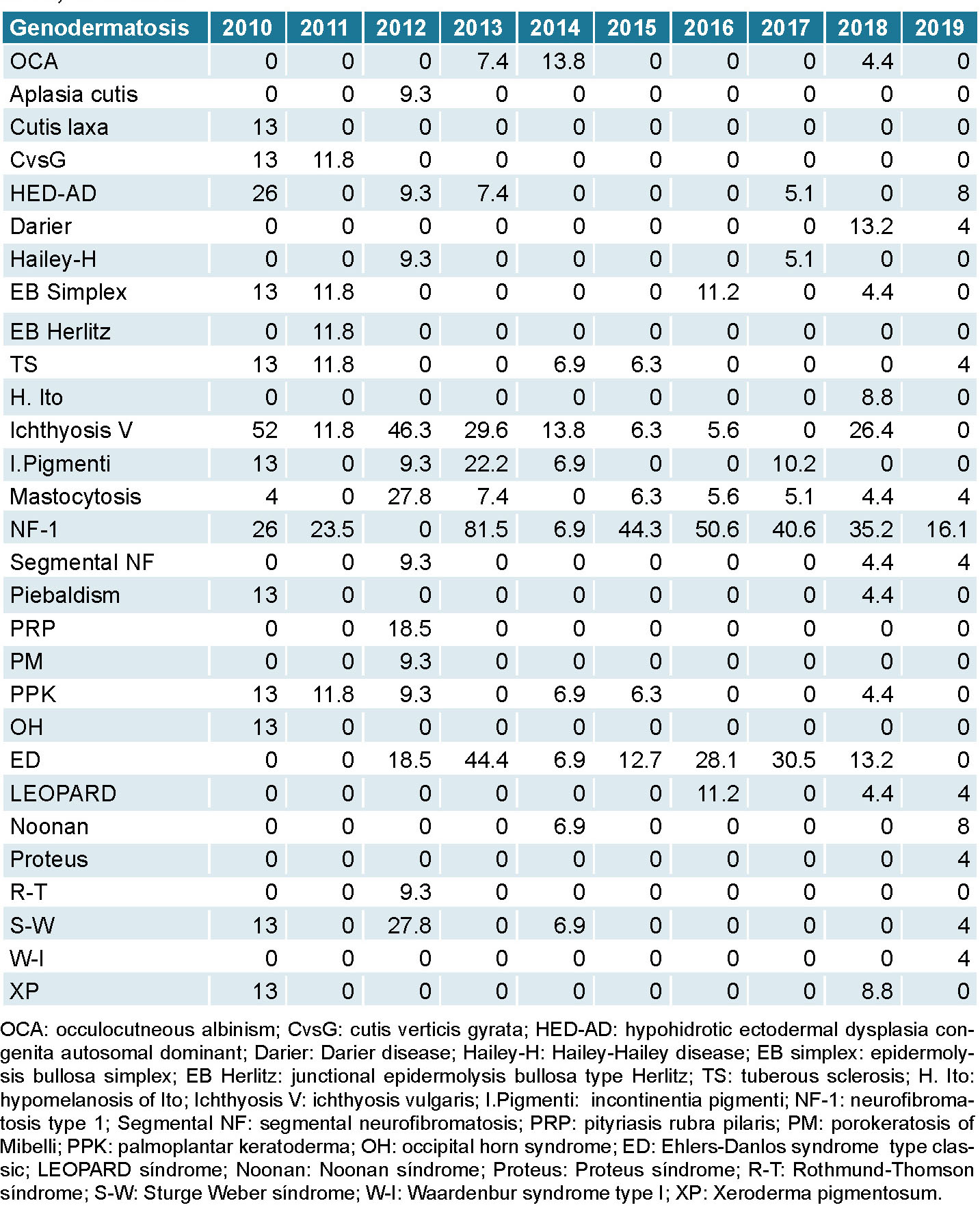
From 2010 through 2019 highest recorded incidence rates were for neurofibromatosis type 1 at 81.5 per 1000 (2013), 50.6 per 1000 (2016), and 40.6 per 1000 (2017). The next highest rates were for ichthyosis vulgaris at 52 per 1000 (2010) and classical Ehlers-Danlos syndrome at 44.4 per 1000 (2013). During this time, all rates decreased and the lowest were observed in 2019, the last year of the study (Table 1).

Figure 1: Genodermatosis prevalence (per 100,000 population). Las Tunas, Cuba, 1989–2019
Figure 1: Genodermatoses prevalence (per 100,000 population). Las Tunas, Cuba, 1989–2019

Complications for 1989–2009 (Figure 2) represented 40% (22 of 55 diagnosed with genodermatosis); higher than during 2010–2019, when it was 21.1% (41 of 194 diagnosed). In both periods, pyodermitis was predominant at 29.1% (16/55) in 1989–2009 and 13.4% (26/194) in 2010–2019, followed by sun damage at 9.1% (5/55) in 1989–2009 and 4.1% (8/194) in 2010–2019.

General survival (1989–2019) was high at 98% (only 5 deaths of 249 diagnosed). Deaths occurred in the years 2009, 2010, 2011, 2012, and 2015 (one in each year). There were no deaths during the other years in the study period.

The municipality with the highest proportion of genodermatosis cases was Majibacoa (0.07%), followed by Las Tunas (0.06%) and Colombia and Jobabo, both at 0.05%. The municipalities with the lowest prevalence were Amancio and Puerto Padre, both at 0.02%. There was consanguinity in the municipality of Manatí, with a case of junctional epidermolysis bullosa, Herlitz type; in the municipality of Colombia there were two cases of XP.

DISCUSSION
The most predominant inheritance patterns of genodermatoses in Las Tunas are those with autosomal dominant Mendelian inheritance, such as neurofibromatosis type 1, classical Ehlers-Danlos syndrome, ichthyosis vulgaris, and cutaneous mastocytosis.[29] In this type of inheritance, traits or conditions are present in all generations, even if the individual is heterozygous; it affects both sexes, with a 50% probability of transmitting the affected allele to their offspring,[29] and accordingly, the proportion of affected individuals in a population will be higher. These conditions often present low fatality, which contributes to increasing prevalence.

According to WHO, rare diseases are those that affect less than 5 persons per 100,000 population.[30,31] Other authors internationally have reported prevalence rate of neurofibromatosis type 1 at 1 per 2500–3000 population,[32] classical Ehlers-Danlos syndrome at 1 per 5000 births,[33,34] ichthyosis vulgaris at 1 per 250–1000 population,[35] and cutaneous mastocytosis at 2 per 300,000 births.[36] Considering the results of our study, these diseases, as a group, and by this definition, are not rare in Las Tunas Province; neither are neurofibromatosis type 1, classical Ehlers-Danlos syndrome, and ichthyosis vulgaris. However, cutaneous mastocytoses have lower rates in the province than those reported at globally.

Table 1: Annual incidence of genodermatoses (per 1000 population). Las Tunas province, Cuba, 2010–2019

The multidisciplinary service offers genetics counseling for families including a discussion of reproductive options. While not all families perceive risk the same way, genetic counseling has increased the birth rate of individuals with conditions that are not lethal or whose life expectancy is higher. High prevalence rates may be due in part to increased treatment effectiveness and genetics counseling availability in the province.

In many genodermatoses, including neurofibromatosis type 1, classical Ehlers-Danlos syndrome, and ichthyosis vulgaris, among others,[5] patients do not manifest signs and symptoms in the first year after birth, which makes it very difficult to determine prevalence rates at birth. However, when incidence rates are studied in relation to specific genodermatoses diagnosed per year, the most commonly-diagnosed genodermatoses in any one year can be identified. For genodermatoses whose manifestations begin in the first year, like congenital ectoder mal dysplasia, oculocutaneous albinism, epidermolysis bullosa, and incontinentia pigmenti,[5] incidence rates coincide with prevalence rates at birth.

The fact that annual rates are higher for genodermatoses that do not present symptoms at birth suggests that diagnosis is based on active case screening, identification of affected members of the same family as the proband case, use of genealogical trees and effectiveness of the risk counseling process. This is possible thanks to NGP working at all three levels of care, both for the patient and their family, facilitating better control of these diseases.[23]

When skin is damaged, as occurs in most genodermatoses, it loses its barrier-protective function, which allows bacteria and fungi to colonize the skin.[37] Comparing the most common complications in both periods, we found fewer during the second period, which coincides with the 2010 implementation of the specialized multidisciplinary provincial clinic for patients with genodermatoses, the multidisciplinary work between the geneticist and dermatologist, and the application of treatments to improve the clinical status of the patient with consequent complication prevention.

General orientations provided by the clinic included hygiene, sun protection, skin self-examination, a balanced diet rich in fruits and vegetables (vitamin therapy), adequate skin hydration and use of emollients for xerodermic or ichthyosiform skin.[2] Preventive therapeutic measures included emollient and antibiotic creams used in combination for diseases with high risk of damage to skin integrity, such as ichthyosis, Darier disease, epidermolysis bullosa and pityriasis rubra,[2] as well as antifungals, since fungal infections such as palmoplantar keratoderma and epidermolytic hyperkeratosis may occur.[2]

For pyodermitis, antibiotics such as penicillins and cephalosporins were employed, as these are effective against Staphylococcus aureus and group A beta-hemolytic Streptococcus, bacteria that commonly colonize the skin and may even lead to systemic complications.[1]

While patient survival was high, five deaths occurred, all due to complications, described as follows:

One patient had junctional Herlitz-type epidermolysis bullosa.[38] This patient presented symptoms of generalized blistering from birth that left atrophic scars, with marked epidermal detachment, presence of the Nikolsky sign (except the palms of the hands and soles of the feet) and nail dystrophy, with no tooth development and madarosis. Skin lesions were potential entry points, which led to symptoms of sepsis, immunodeficiency, delayed psychomotor development, protein-energy undernutrition,[38] and finally, multiorgan failure, with death at 11 months.

Figure 2: Most frequent complications (percentages) in genodermatosis. Las Tunas, Cuba, 1989–2009 and 2010–2019

A patient with occipital horn syndrome presented hypotonia, with frequent symptoms of diarrhea, cutis laxa, joint hypermobility, coarse hair, clotting disorder, and an aortic aneurysm,[39] which led to his death at eight months. This syndrome, formerly known as type IX Ehlers-Danlos syndrome, was excluded from the classification of variants of Ehlers-Danlos syndrome established by an international expert consortium in 2017,[40] and is currently classified in the connective tissue disorder group caused by abnormalities in copper metabolism.

Another patient with a Rothmund–Thomson syndrome presented with typical features of the disease, such as xeroderma, poikiloderma of the face, congenital cataracts, photophobia, delayed physical growth, immunodeficiency and sepsis[41,42] and died at under five months of age.

One patient presented with Netherton syndrome, a rare disease transmitted through autosomal recessive inheritance and defined by the characteristic triad of congenital ichthyosiform erythroderma, hair shaft abnormalities and immunological disorders.[43,44] From birth, this patient had symptoms of erythroderma accompanied by frequent skin infections, leading to repeated admissions to intensive care units and ultimately death from sepsis at two years of age.

Another patient had neurofibromatosis type 1 associated with congenital cardiovascular malformations that led to cardiogenic complications and death. Neurofibromatosis type 1 is a multisystem disease, considered a neuroectodermal syndrome (currently called neurocristopathy), transmitted through autosomal dominance through the nf1 gene located at locus 17q11.2 and affecting mainly the skin and nervous system. It produces melanic manifestations of the skin and tumors, but is often not lethal.[45] In this patient, more than six cafe-au-lait spots and a genetic family history of the disease were identified, but congenital heart disease led to the patient’s death at age two.

The literature considers genodermatoses to be genetic diseases of the skin with high morbidity that, in some cases, present systemic conditions that may be fatal; in most lethal genodermatoses, death often occurs in the first year of life.[5]

This study facilitated identification of geographic distribution (by municipality) and affected families. In general, the number of cases diagnosed was not proportional to the population density of each municipality. The highest proportion of cases was in Majibacoa, which had the fifth highest population density in the province, but a place also characterized by greater involvement of dermatologists in the community. The municipalities with the highest population densities, such as Puerto Padre and Amancio Rodríguez, recorded lower proportions of cases. This may be due to poor screening and less availability of dermatological care. Despite existence of a specialized multidisciplinary provincial clinic for genodermatoses patients that includes both clinical geneticists and dermatologists, the latter are in the highest demand for these patients, and their greater participation in the NGP is needed.[24]

A case of epidermolysis bullosa (Herlitz type) was diagnosed in Manatí municipality. The patient had a family history of the disease, including a maternal uncle who died of it, and parental consanguinity. In Colombia municipality, a case was confirmed of xeroderma pigmentosum with familial ties to an affected family from another province (Ciego de Avila) in which consanguinity was determined. Consanguinity is the genetic union of two people descended from common ancestors, (second-degree cousins or closer). Currently, over 1.2 billion people worldwide are in consanguineous marriages and it is estimated that 10.4% of the world population is married to a biological relative or is the offspring of a consanguineous union.[46] A consanguineous marriage increases the risk of autosomal recessive diseases transmission by 25%–50% because it increases the probability of two affected alleles being combined and thus the birth of an affected child.[46]

CONCLUSIONS
In Las Tunas Province, Cuba, genodermatoses as a whole are not rare diseases according to WHO definitions. Those with the highest prevalence and incidence rates are neurofibromatosis type 1, classical Ehlers-Danlos syndrome and ichthyosis vulgaris. After implementation of the specialized multidisciplinary provincial clinic for genodermatoses patients within Cuba’s NGP, in addition to active screening, more cases were diagnosed, and a lower proportion of complications and a higher survival index were recorded. However, improving the quality of care for patients with genodermatosis is necessary with the implementation of a methodology protocolizing their care and establishing follow-up algorithms.

ACKNOWLEDGMENTS
This study was conducted with support from the National Program for the Diagnosis, Care and Prevention of Genetic Diseases and Congenital Abnormalities. The authors thank the Las Tunas Provincial Medical Genetics Department for its contributions to the study.

References
image
  1. Manzur J. Genodermatosis. In: Dermatología. Havana: Editorial Ciencias Médicas; 2002. p. 48‒67. Spanish.
  2. Arenas R. Dermatología. Atlas, diagnóstico y tratamiento [Internet]. 6th ed. México: McGraw-Hill Interamericana; 2017. Chapter 54, Neurofibromatosis; [cited 2020 May 29]; p. 336‒7. Available at: https://accessmedicina.mhmedical.com/content.aspx?bookid=1538&sectionid=102305123&jumpsectionID=102305146. Spanish.
  3. Morice Picard F. Genética en dermatología [Internet]. Madrid: Elseiver Masson SAS; 2017 [cited 2020 May 29]. p. 51. Available at: https://www.em-consulte.com/es/article/1122358/genetica-en-dermatologia. Spanish.
  4. Penago Homero G. Prevención y manejo de las enfermedades genéticas de la piel en Panamá [master’s thesis] [Internet]. Florida: Atlantic International University; 2006 [cited 2020 May 28]. Available at: http://www.aiu.edu/publications/student/spanish/prevencion-y-manejo-de-las-enfermedades-geneticas-de-la-piel-en-panama.htm. Spanish.
  5. James W, Berger T, Dirk E. Genodermatoses and Congenital Anomalies. Clinical Dermatology. In: Andrews’ Diseases of the Skin [Internet]. 13th ed. New York: Elseiver; 2019 [cited 2020 Jun 3]; p. 547‒86. Available at: https://www.eu.elsevierhealth.com/andrews-diseases-of-the-skin-9780323547536.html  
  6. James W, Berger T, Dirk E. Abnormalities of dermal fibrous and elastic tissue. Clinical Dermatology. In: Andrews’ Diseases of the Skin [Internet]. 13th ed. New York: Elseiver; 2019 [cited 2020 Jun 3]; p. 508‒10. Available at: https://www.eu.elsevierhealth.com/andrews-diseases-of-the-skin-9780323547536.html
  7. Acuña Aguilarte PM. Xeroderma pigmentoso. In: Pediatría. Diagnóstico y tratamiento. Havana: Editorial Ciencias Médicas; 2016. p. 532. Spanish.
  8. Barbel P, Brown S, Peterson K. Identification of hipomelanosis of Ito in pediatric primary care. J Ped Health Care [Internet]. 2015 Nov–Dec [cited 2020 May 29];29(6):551‒4. Available at: https://linkinghub.elsevier.com/retrieve/pii/S0891-5245(15)00004-8
  9. Patterson J. Disorder of epidermal maturation and keratinization. In: Weedon’s Skin Pathology [Internet]. 4th ed. New York: Elseiver; 2015 Jan 12 [cited 2020 Jun 3]; p. 281‒320. Available at: https://www.elsevier.com/books/weedons-skin-pathology/patterson/978-0-7020-5183-8
  10. Fleury de Lemos Pereira D, Lopes Araujo E, Duarte Patuzzo FV. Perfil do paciente albino com visão subnormal e melhoria da acuidade visual com a adaptação de recursos ópticos e/ou eletrônicos. Rev Bras Oftalmol [Internet]. 2016 Nov–Dec [cited 2020 Jun 6];75(6):456–60. Available at: https://www.scielo.br/scielo.php?script=sci_arttext&pid=S0034-72802016000600456. Portuguese, English.
  11. Ocaña Jaramillo S, Del Boz J, Vera Casaño A. Incontinencia pigmenti. Estudio descriptivo de la experiencia en dos centros hospitalarios. An Pediatr [Internet]. 2020 Jan [cited 2020 Jun 1];92(1):3‒12. Available at: https://www.analesdepediatria.org/es-incontinencia-pigmenti-estudio-descriptivo-experiencia-articulo-S1695403319301705?referer=buscador. Spanish.
  12. Arango J, Delgado J, Saldarriaga W. Esclerosis tuberosa, diagnóstico fetal y materno. Rev Chil Obstet Ginecol [Internet]. 2015 Dec [cited 2020 Jun 1];80(6):475–80. Available at: https://scielo.conicyt.cl/scielo.php?script=sci_arttext&pid=S0717-75262015000600007. Spanish.
  13. Prodinger C, Reichelt J, Bauer JW, Laimer M. Epidermolysis bullosa: advances in research and treatment. Exp Dermatol [Internet]. 2019 Oct [cited 2020 Jun 1];28(10):1176–89. Available at: https://doi.org/10.1111/exd.13979  
  14. Pingault V, Ente D, Dastot-Le Moal F, Goossens M, Marlin S, Bondurand N. Review and update of mutations causing Waardenburg syndrome. Hum Mutat [Internet]. 2010 Apr [cited 2020 Jun 1];31(4):391–406. Available at: https://onlinelibrary.wiley.com/doi/abs/10.1002/humu.21211 
  15. Dorticós Balea A, Zaldívar Vaillant T, Falcón Lincheta L. Frecuencia de los distintos tipos de genodermatosis en la Ciudad de la Habana en los años 1980 a 1986.  Digitalización [Internet]. 1989 Jan–Feb [cited 2020 Jun 3];61(1):75–80. Available at: http://files.sld.cu/digitalizacion-bmn/files/2017/01/S0034-7531.1989.0001.0008.pdf. Spanish.
  16. Campo Díaz MC, Hernández González JL, Gato Santiesteban Y, Valdés Sojo C, Fortún Prieto A. Evaluación de la hemostasia en niños con Síndrome de Ehlers-Danlos tipo III. Rev Cubana Hematol Inmunol Hemoter [Internet]. 2014 Apr–Jun [cited 2020 Jun 3];30(2). Available at: http://scielo.sld.cu/scielo.php?script=sci_arttext&pid=S0864-02892014000200007. Spanish.
  17. Orraca Castillo M. Caracterización epidemiológica, clínica y genética de la neurofibromatosis 1 en la provincia Pinar del Río [doctoral thesis] [Internet]. Havana: University of Medical Sciences of Havana; 2017 May 10 [modified 2017 Sep 6; cited 2020 Jun 3]. 200 p. Available at: http://tesis.sld.cu/index.php?P=FullRecord&ID=238&ReturnText=Search+Results&ReturnTo=index.php%3FP%3DAdvancedSearch%26Q%3DY%26FK%3Dneurofibromatosis. Spanish.
  18. Carcasés Carcasés E, Orive Rodríguez NM, Romero Portelles LC, Silva González GK. Enfermedades genéticas más frecuentes en pacientes atendidos en consulta de genética clínica. Rev Zoilo Marinello [Internet]. 2015 Mar [cited 2020 Jun 3];40(3). Available at: http://revzoilomarinello.sld.cu/index.php/zmv/article/view/84. Spanish.
  19. World Health Organization [Internet]. Geneva: World Health Organization; c2020. Genes and human disease. Human Genomics in Global Health; 2017 [updated 2020 Nov 12; cited 2020 May 28]. Available at: http://www.who.int/genomics/public/geneticdiseases/en/index2.html
  20. Lantigua Cruz A. Enfermedades genéticas y defectos congénitos en la infancia. In: Pediatría. Diagnóstico y tratamiento. Havana: Editorial Ciencias Médicas; 2016. p. 97‒114. Spanish.
  21. World Health Organization [Internet]. Geneva: World Health Organization; c2020. Control de las enfermedades genéticas. Informe de la Secretaría. Consejo Ejecutivo; 2005 Apr 21 [cited 2020 May 28]. Available at:   http://apps.who.int/gb/archive/pdf_files/EB116/B116_3-sp.pdf. Spanish.
  22. Roblejo Balbuena H, Marcheco Teruel B. Genetics and genomics medicine in Cuba. Mol Genet Genomic Med [Internet]. 2017 May 21 [cited 2020 May 28];5(3):196‒201. Available at: https://onlinelibrary.wiley.com/doi/full/10.1002/mgg3.299
  23. Marcheco Teruel B, Rojas Betancourt I, Lantigua Cruz A, Benítez Cordero Y, Suárez Besil B. Manual de normas y procedimientos. Servicios de Genética Médica en Cuba. Havana: Editorial Ciencias Médicas; 2017. p. 105‒6. Spanish.
  24. Velázquez Ávila Y, Tamayo Mariño K, Morales Solís M, González Saker AG, García López R. Alteraciones genéticas cutáneas diagnosticadas en la infancia, Las Tunas 2010-2012. Rev Zoilo Marinello [Internet]. 2014 Apr [cited 2020 Jun 3];39(4). Available at: http://revzoilomarinello.sld.cu/index.php/zmv/article/view/261. Spanish.  
  25. Zegpi-Trueba MS, Hasbún Acuña P, Berroeta-Mauriziano D. Mastocitosis cutánea: reporte de un caso. Rev Chil Pediatr [Internet]. 2016 Jun [cited 2020 Jun 6];87(3):204–7. Available at: https://scielo.conicyt.cl/scielo.php?script=sci_arttext&pid=S0370-41062016000300010. Spanish.
  26. De León Ojeda NE. La genética clínica, de la observación a su aplicación. Rev Cubana Pediatr [Internet]. 2014 Apr–Jun [cited 2020 Jun 6];86(1):1–4. Available at: http://scielo.sld.cu/pdf/ped/v86n1/ped01114.pdf. Spanish.
  27. National Bureau of Statistics (CU) [Internet]. Havana: National Bureau of Statistics (CU); c2021. Provincia Las Tunas; 2019 [cited 2020 Jun 3]. Available at: http://www.onei.gob.cu/sites/default/files/anuario_est_provincial/las_tunasok.pdf. Spanish.
  28. Lizaraso Caparó F, Benavides Zúñiga A. Ética Médica. Horiz Med Lima [Internet]. 2018 Oct–Dec [cited 2020 Jun 3];18(4):4–8. Available at:  http://www.scielo.org.pe/scielo.php?script=sci_arttext&pid=S1727-558X2018000400001. Spanish.
  29. Lieberman M, Ricer R, editors. Bioquímica, biología molecular y genética [Internet]. 6th ed. New York: Lippincott W & W; 2015 [cited 2020 Jun 4]. Available at: https://www.ovid.com/product-details.13900.html
  30. López Lambán A, Calatayud Madonado V, Casado de Frías E, Tobajas Asensio LM. La OMS ha identificado 5.000 enfermedades raras, sin contar las ultra raras. Redacción Médica [Internet].  2018 Mar 26 [cited 2020 Jun 4];Sanidad privada:[about 3 p.]. Available at: https://www.redaccionmedica.com/secciones/privada/la-oms-ha-identificado-5-000-enfermedades-raras-sin-contar-las-ultra-raras-7846. Spanish.
  31. Carbajal Rodríguez L, Navarrete Martínez JI. Enfermedades raras. Acta Pediatr Mex [Internet]. 2015 [cited 2020 Jun 4];36(5):369‒73. Available at: https://ojs.actapediatrica.org.mx/index.php/APM/article/view/1057. Spanish.
  32. Online Mendelian Inheritance in Man (OMIM) [Internet]. Maryland: John Hopkins University; c1966–2021. Statistics. Neurofibromatosis, Type I; NF1; [published 1986 Jun 2; updated 2020 Apr 14; cited 2020 Jun 4].  Available at: https://omim.org/entry/162200
  33. Ghali N, Sobey G, Burrows N. Ehlers-Danlos syndromes. BMJ [Internet]. 2019 Sep 18 [cited 2020 Jul 1];366:l4966. Available at: https://www.bmj.com/lookup/pmidlookup?view=long&pmid=31533917
  34. Wiesmann T, Jais C. Recomendaciones para la anestesia de pacientes con síndrome de Ehlers-Danlos [Internet]. Nümberg (GR): ORPHAN ANESTHESIA; 2019 Nov [cited 2020 Jul 1]. Available at: https://www.orphananesthesia.eu/en/component/finder/search.html?q=Recomendaciones+sobre+anestesia+para+pacientes+que+sufren+S%C3%ADndrome+de+Ehlers-Danlos&Itemid=137. Spanish.
  35. Vega Almendra N, Aranibar Duran L. Ictiosis hereditaria: desafío diagnóstico y terapéutico. Rev Chil Pediatr [Internet]. 2016 Jun [cited 2020 Jul 29];87(3):213‒23. Available at: https://scielo.conicyt.cl/scielo.php?script=sci_arttext&pid=S0370-41062016000300013. Spanish.
  36. Valdés González JL, Solís Cartas U, Valdés González EM. Mastocitosis cutánea. Un caso singular. Rev Haban Cienc Méd [Internet]. 2016 Jan–Feb [cited 2020 Jul 29];15(1):64‒71. Available at: http://scieloprueba.sld.cu/scielo.php?script=sci_arttext&pid=S1729-519X2016000100008&lng=es. Spanish.
  37. Torres Rives B, Portal Miranda JA, Mederos Benítez Y, Martínez Téllez G, Lantigua Cruz A, Rodríguez Pelier C, et al. Respuesta inmune humoral en pacientes cubanos con genodermatosis atendidos en el hospital pediátrico Juan Manuel Márquez. Rev Cubana  Gen Comunit [Internet]. 2015 [cited 2020 Aug 6];9(3):36–46. Available at: https://www.medigraphic.com/cgi-bin/new/resumen.cgi?IDARTICULO=68815. Spanish.
  38. Online Mendelian Inheritance in Man (OMIM) [Internet]. Maryland: John Hopkins University; c1966–2021. Statistics. Epidermolysis bullosa, junctional, Herlitz type; [published 1986 Jun 3; updated 2018 Feb 7; cited 2020 Jun 6]. Available at: https://omim.org/entry/226700
  39. Orphanet [Internet]. Paris: Institut Nationale de la Santé et de la Recherche Médicale (FR); c2021. Enfermedades raras. Búsqueda. Síndrome del cuerno occipital; [updated 2021 Mar 3; cited 2020 Aug 6]. Available at: https://www.orpha.net/consor/cgi-bin/OC_Exp.php?lng=ES&Expert=198. Spanish.
  40. Reginato A. Ehlers Danlos syndrome. In: Ferri’s Clinical Advisor. New York: Elsevier; 2020 [cited 2020 Aug 6]; p. 482e2–482e12. Available at: https://www.elsevier.com/books/ferris-clinical-advisor-2020/ferri/978-0-323-67254-2
  41. Dos Santos Martins TG, Fontes de Azevedo Costa AL, Gonçalves dos Santos Martins T. Rothmund syndrome. Rev Bras Oftalmol [Internet]. 2015 Nov–Dec [cited 2020 Aug 6];74(6):390‒2. Available at: https://www.researchgate.net/publication/283663906. English, Portuguese.
  42. Online Mendelian Inheritance in Man (OMIM) [Internet]. Maryland: John Hopkins University; c1966–2021. Statistics. Rothmund-Thomson syndrome, Type 1; RTS1; [published 2019 Oct 15; cited 2020 Aug 6]. Available at: https://omim.org/entry/618625
  43. Diez de Medina JC, Antezana EC, Sangueza M. Síndrome de Netherton: comunicación de dos pacientes. Dermatología CMQ [Internet]. 2015 [cited 2020 Aug 6];13(4):314‒7. Available at: http://www.medigraphic.com/pdfs/cosmetica/dcm-2015/dcm154f.pdf. Spanish.
  44. Online Mendelian Inheritance in Man (OMIM) [Internet]. Maryland: John Hopkins University; c1966–2021. Statistics. Netherton syndrome; NETH; [published 1986 Jun 4; updated 2019 Apr 8; cited 2020 Aug 6]. Available at: https://omim.org/entry/256500
  45. Duat Rodríguez A. Características fenotípicas y genéticas en la neurofibromatosis tipo 1 en la edad pediátrica [doctoral thesis] [Internet]. Madrid: Universidad Autónoma de Madrid; 2015 [cited 2020 Aug 8]. 155 p. Available at: https://repositorio.uam.es/bitstream/handle/10486/667203/duat_rodriguez_anna.pdf?sequence=1. Spanish.
  46. Abarca Barriga HH, Chávez Pastor M, Trubnykova M, La Serna-Infantes JE, Poterico JA. Factores de riesgo en las enfermedades genéticas. Acta Méd Perú [Internet]. 2018 Jan [cited 2020 Aug 8];35(1):43‒50. Available at: http://www.scielo.org.pe/scielo.php?script=sci_arttext&pid=S1728-59172018000100007. Spanish.

THE AUTHORS
Yordania Velázquez-Ávila (Corresponding author: yodaniava@ltu.sld.cu), dermatologist with a master’s degree in infectious diseases. Assistant professor and adjunct researcher, Mártires de las Tunas Pediatric Provincial Hospital, Las Tunas, Cuba. https://orcid.org/0000-0002-2846-3432

Carmen Rosa Valenciano-Rodríguez, clinical geneticist with a master’s degree in comprehensive child health. Assistant professor, Provincial Medical Genetics Department, Las Tunas, Cuba. https://orcid.org/0000-0001-6141-7780

Velázquez-Ávila Y, Valenciano-Rodríguez CR. Genodermatoses in Las Tunas Province, Cuba, 1989-2019. MEDICC Rev. 2021 Apr;23(2):34–41. DOI: https://doi.org/10.37757/MR2021.V23.N2.10

image
Loading...
Loading...
Early Online

No new Early Online articles at this time. The most recent articles are listed in the Current Issue Table of Contents. Early Online articles are added as soon as they are available, so please check back later.