Article

Experimental Infection of Pigs with a ST 245 Brachyspira hyodysenteriae Isolated from an Asymptomatic Pig in a Herd with No History of Swine Dysentery

by
José Paulo H. Sato
,
Amanda G. S. Daniel
,
Carlos E. R. Pereira
,
Mariana R. Andrade
,
Ricardo P. Laub
,
Michelle P. Gabardo
,
Luisa V. A. Otoni
,
Nubia R. Macedo
,
Javier A. Barrera-Zarate
and
Roberto M. C. Guedes
*
Department of Veterinary Clinic and Surgery, Veterinary School, Universidade Federal de Minas Gerais, Belo Horizonte 130161-970, Brazil
*
Author to whom correspondence should be addressed.
Vet. Sci. 2022, 9(6), 286; https://doi.org/10.3390/vetsci9060286
Submission received: 1 May 2022 / Revised: 7 June 2022 / Accepted: 8 June 2022 / Published: 10 June 2022
(This article belongs to the Special Issue New Findings on Long-Known Pathogens in Pigs)

Abstract

:
Swine dysentery (SD) is characterized by a severe mucohemorrhagic colitis caused by infection with Brachyspira species. In infected herds the disease causes considerable financial loss due to mortality, slow growth rates, poor feed conversion, and costs of treatment. B. hyodysenteriae is the most common etiological agent of SD and infection is usually associated with disease. However, isolated reports have described low pathogenic strains of B. hyodysenteriae. The aim of this study was to describe an experimental infection trial using a subclinical B. hyodysenteriae isolated from an animal without clinical signs and from a disease-free herd, to evaluate the pathogenicity and clinical pathological characteristics compared to a highly clinical isolate. Forty-eight 5-week-old pigs were divided into three groups: control, clinical and the subclinical isolates. The first detection/isolation of B. hyodysenteriae in samples of the animals challenged with a known clinical B. hyodysenteriae strain (clinical group) occurred 5th day post inoculation. Considering the whole period of the study, 11/16 animals from this group were qPCR positive in fecal samples, and diarrhea was observed in 10/16 pigs. In the subclinical isolate group, one animal had diarrhea. There were SD large intestine lesions in 3 animals at necropsy and positive B. hyodysenteriae isolation in 7/15 samples of the subclinical group. In the control group, no diarrhea, gross/microscopic lesions, or qPCR positivity were observed. Clinical signs, bacterial isolation, macroscopic and histologic lesions were significantly difference among groups, demonstrating low pathogenicity of the subclinical isolate in susceptible pigs.

1. Introduction

Brachyspira hyodysenteriae is the etiological most common agent of swine dysentery (SD), characterized by mucohemorrhagic colitis [1,2]. Clinical signs range from moderate mucoid to bloody diarrhea, with a mortality rate ranging from 30 to 90%. SD gross lesions include multifocal mucosal necrosis and hemorrhage, excess of mucus, associated with fibrinous exudate and thickening of the mucosa of variable intensity. Microscopically, changes in the cecum and colon characterized by goblet cell hyperplasia, hemorrhage, superficial necrosis, and neutrophilic inflammatory infiltrate in the lamina propria [2].
In recent years there has been reported a reemergence of the disease in several countries and the emergence of two new species, B. hampsonii and B. suanatina, with similar pathogenic characteristics of SD [3,4,5,6]. Atypical isolates of B. hyodysenteriae were previously described and characterized as low pathogenic, capable of colonizing but not inducing clinical disease [7,8,9]. Studies evaluating pathogenic and molecular characteristics of atypical strains isolated from B. hyodysenteriae are scarce from herds without clinical disease [10,11].
This study aimed to describe an experimental infection trial in pigs using a subclinical B. hyodysenteriae isolate obtained from an animal without clinical signs and from an apparently healthy herd with no history of SD. The pathogenicity, clinical pathological and molecular findings of this subclinical isolate of B. hyodysenteriae compared to a highly clinical isolate were evaluated.

2. Materials and Methods

2.1. Animals and Experimental Design

This study was approved by the Ethics Committee on Animal Experimentation of the Universidade Federal de Minas Gerais (Approval Number: CEUA #177/2015).
Forty-eight 5-week-old piglets (7.95 ± 1.29 kg/wt) were obtained from a commercial farm with no history of disease associated with Brachyspira spp., Lawsonia intracellularis or Salmonella sp. The animals were randomly divided into three groups (16 animals/group): control (CTRL), clinical isolate (CLIN) and subclinical isolate (SUBCL), which were kept in 4 pens (4 animals/pen) in three separated rooms throughout the experiment.
During a 7-days acclimatation period, the pigs were tested for Brachyspira spp. and Salmonella sp. by culture of fecal samples and L. intracellularis by PCR testing. Room temperature and humidity among room were the same. Feed and water were available ad libitum for all treatments. Parenteral or oral medications were not used throughout the study period. The daily management activities in each group were performed using strict biosecurity protocols, including unique instruments and different personal to avoid cross-contamination.

2.2. Inoculum

Both isolates used in this study were obtained from two different Brazilian pig farms in 2013 and were frozen at −80 °C in the Laboratory of Molecular Pathology at the Veterinary School of the Universidade Federal de Minas Gerais (UFMG). The clinical isolate used in the CLIN group was obtained from a clinically affected pig with mucohemorrhagic diarrhea and colitis from a SD positive herd. The subclinical isolate was obtained from an animal from a herd with no clinical signs or history of SD. No antimicrobials were being used in this herd, so there was no possibility to mask clinical signs of the disease. Phenotypically, both isolates produced strong hemolysis in blood agar. Both isolates were identified as B. hyodysenteriae based on PCR [12] and nox gene sequencing [5]. The multilocus sequence typing (MLST) analysis of these isolates classified both as sequence type (ST) 245 [13].
Isolates were cultured on trypticase soy agar with 5% sheep blood (TSA) containing 12.5 mg/L of rifampicin, 200 mg/L of espectinomicin, 50 mg/L of vancomicin and 12.5 mg/L of colistin [14], under anaerobic conditions with N2 (80%), CO2 (10%) and H2 (10%), at 42 °C and examined for growth at 72 and 96 h.
After growth, the agar plates were washed with PBS and the supernatant was incubated in trypticase soy broth (TSB), enriched with 0.5% glucose, 0.2% NaHCO3, 0.05% L-cysteine-HCl, 1.0% yeast extract, 10% fetal bovine serum and 5% swine fecal extract [15] in a ratio of 1:100 mL (wash:broth) for 24 h at 42 °C in a shaker, followed by inoculation of the animals.

2.3. Animal Inoculation

All pigs were inoculated by intragastric gavage for three consecutive days as previously described [16,17]. Pigs in the CLIN and SUBCL groups received 50 mL of the inoculum at concentrations determined by qPCR of 5.1 × 10⁷, 2.1 × 10⁸ and 2.2 × 10⁸ bacteria/mL (CLIN group) and 1.2 × 10⁸, 2.3 × 10⁸ and 1.3 × 10⁸ bacteria/mL (SUBCL group) in three consecutive days, respectively. The CTRL group was inoculated with 50 mL of sterile TSB. To decrease gastric transit time, feed was removed for 16 h prior to, and returned one hour after inoculation [17].

2.4. Clinical Evaluation and Sample Collection

After inoculation, all animals were observed twice a day for evaluation of clinical signs of diarrhea, and fecal consistency based on the following score: 0 = normal, 1 = semi-solid consistency, 2 = creamy consistency and 3 = watery consistency, with addition of 0.5 for the presence of detectable mucus and/or blood.
The quantitative evaluation of B. hyodysenteriae elimination in the feces were performed by qPCR on samples collected at −7, 5, 7, 11, 15 and 18 days post inoculation (DPI). Brachyspira spp. isolation in selective medium as described previously were analyzed in fecal samples collected at −7, 0, 3, 5, 7, 9, 11, 13, 15 and 18 DPI.

2.5. DNA Extraction and qPCR

DNA from fecal samples were extracted using a commercial kit (QIAamp DNA Stool kit−Qiagen Inc., Toronto, ON, Canada) according to the manufacturer’s instructions. The amount of B. hyodysenteriae DNA was determined by real-time PCR using published primers [17]. One gram of negative feces and the addition a known number of B. hyodysenteriae (101–108 bacteria/gram of feces) was used to determine the standard curve. Threshold values were considered for detection of Brachyspira determined by 103–108 bacteria/gram of feces, using the regression equation from the standard curve (R2 = 0.990).
The reaction was performed in a final volume of 25 μL, consisting of 1x SYBR Green PCR Master Mix, 1x QN ROX Reference Dye (Quantiia SYBR Green PCR Kit, Qiagen Inc., Toronto, ON, Canada), 500 nM of each primer and 5 μL of DNA. The samples were placed in 96-well plates and amplified in the ViiATM 7 Real-Time PCR System (Applied biosystems) thermocycler, with amplification conditions: 2 min at 50 °C, 10 min at 95 °C, 40 cycles of 15 sec at 95 °C and 1 min at 60 °C. All reactions were performed in duplicate, and each reaction included the standard curve and negative control, being analyzed in QuantStudio TM Real-Time PCR v1.2 software (Thermo Fisher Scientific, Waltham, MA, USA).
PCR specificity was tested against Bacteroides fragilis, B. murdochii, B. pilosicoli, Clostridioides difficile, C. perfringens, Enterococcus faecalis, Escherichia coli, L. intracellularis, Pseudomonas aeruginosa and Salmonella sp.

2.6. Necropsy

Euthanasia and necropsy were performed when the animals were clinically debilitated according to the criteria of the CEUA or at the end of the study, on 18 DPI. For macroscopic and microscopic evaluation, segments of the small intestine, large intestine (cecum, proximal and spiral colon) and mesenteric lymph nodes were analyzed and fixed in 10% buffered formalin.

2.7. Macroscopic Evaluation

Macroscopically, cecum and colon were evaluated for the presence of edema, excessive mucus in the lumen, mucosal hemorrhage and fibrinous exudate.

2.8. Histology

All sampled fragments were processed according to routine histological techniques and stained with hematoxylin and eosin (H&E) [18]. The presence of superficial necrosis, hemorrhage, goblet cell hyperplasia, crypt abscesses and neutrophil infiltrate in the lamina propria were evaluated in the cecum and colon, with lesions scored as zero (no lesion) to three (severe diffuse lesion). The final score was determined by the sum of the five parameters evaluated and classified as mild (<5), moderate (5 and <10) and severe (≥10), with a maximum value of 15. All histological sections were evaluated by two pathologists blinded about experimental groups, and the mean of these two evaluations was used in the analyzes.

2.9. Fluorescence In Situ Hybridization (FISH)

Sections of the large intestines were used for FISH, according to Jensen et al. [19] with probes specific to B. hyodysenteriae [20]. Presence of B. hyodysenteriae was classified as mild (+), moderate (++) or intense (+++), according to the ratio of labeled spirochetes.

2.10. Bacterial Isolation

At the end of the study period, fecal samples, small intestinal contents and mucosal scrapings of the cecum and colon were collected for bacterial isolation. For Brachyspira spp., feces and scrap samples from the large intestine were culture as described above. Samples of the small intestine were seeded on blood and MacConkey agar for evaluation of enterotoxigenic E. coli and Rappaport broth and Hectoein agar for Salmonella sp.

2.11. Statistical Analysis

The SPSS software v19.0 (SPSS Inc., Chicago, IL, USA) was utilized to perform all analyses. The presence or absence of mucohemorrhagic diarrhea, macroscopic lesions and histopathological lesion scores among groups were compared using Kruskal–Wallis test, with p-values < 0.05 considered significant.

3. Results

3.1. Clinical Evaluation

During the acclimation period, one animal from the SUBCL group suddenly died and was excluded from the study. At the necropsy, valvular endocarditis was diagnosed. All other animals had normal or semi-solid fecal consistency (score 0 or 1) and all fecal samples were negative in bacterial isolation for Brachyspira spp. or Salmonella sp. and were negative by PCR for L. intracellularis.
Aqueous and/or mucohemorrhagic diarrhea (score ≥ 3) was first observed in the 7th DPI in tree animals (#2, #11 and #12) of the CLIN group. Considering all study period 10/16 animals in this group had diarrhea. In the SUBCL group, only one animal (#25) had mucohemorrhagic diarrhea starting on the 15th DPI. None of the animals in the CTRL group had any clinical signs of diarrhea during the study period (Table 1). Considering days with diarrhea, an animal from the CLIN group had diarrhea for 11 days and in the SUBCL group, a single pig had diarrhea for three days. Three clinically debilitated animals (#2, #6 and #13) from CLIN group were euthanized at 13, 13 and 16 DPI, respectively. Significant differences of clinical signs of diarrhea were observed between the CTRL and the other two groups (SUBCL and CTRL) (Table 2).

3.2. Anatomopathological Analysis

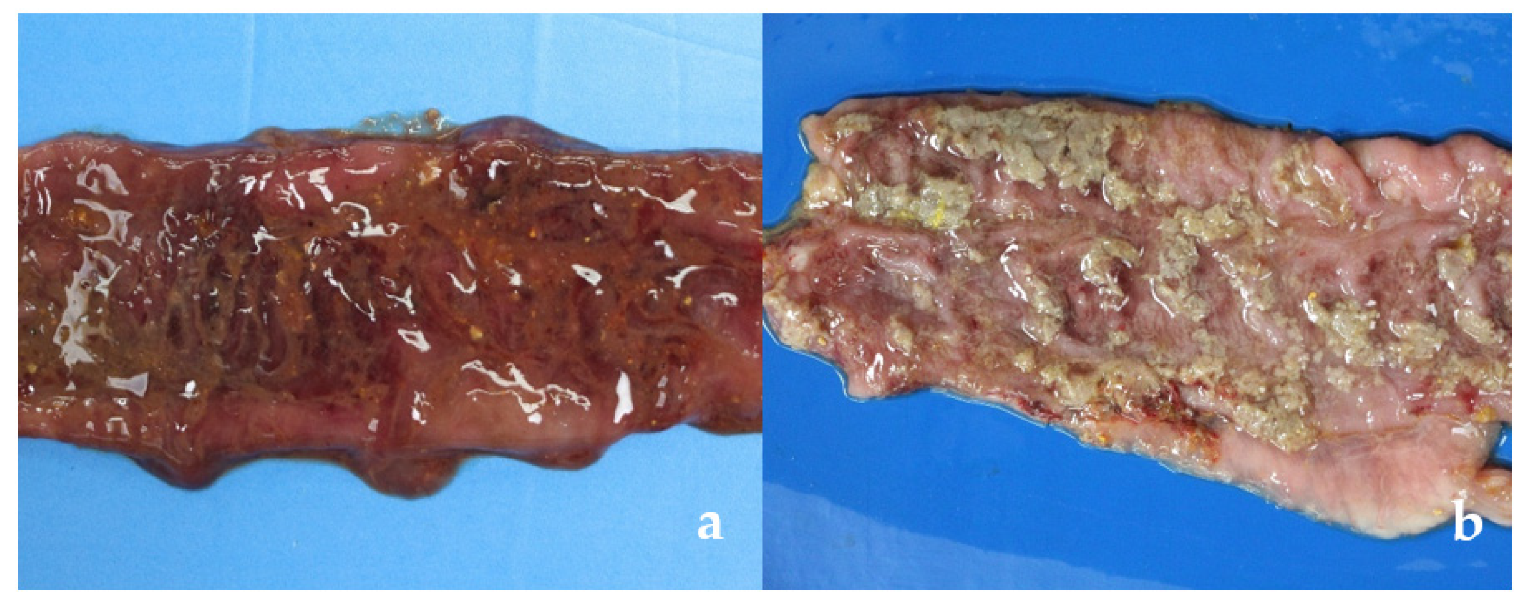
3.2.1. Gross Lesions

Gross pathological findings were compatible with observed clinical signs. Lesions were more frequent in segments of the spiral colon in pigs with aqueous and/or mucohemorrhagic diarrhea (fecal score ≥ 3) from CLIN group (Table 1). Significant differences were observed between the CLIN and the other two groups (SUBCL and CTRL) (Table 2), more severe in the CLIN animals. Nine and four animals of the CLIN and SUBCL groups, respectively, had macroscopic alterations (Table 1) characterized by luminal mucus, mucosal hemorrhage, necrosis and/or fibrous exudate (Figure 1).
One animal of the CLIN group (#16: edema, hemorrhage, thickening and diffuse marked necrosis of the mucosa) and two of the SUBCL group (#24: edema, hemorrhage and thickening of moderate multifocal mucosa and #26: edema and mucosal hyperemia multifocal) had macroscopic lesions, but no clinical signs were observed.

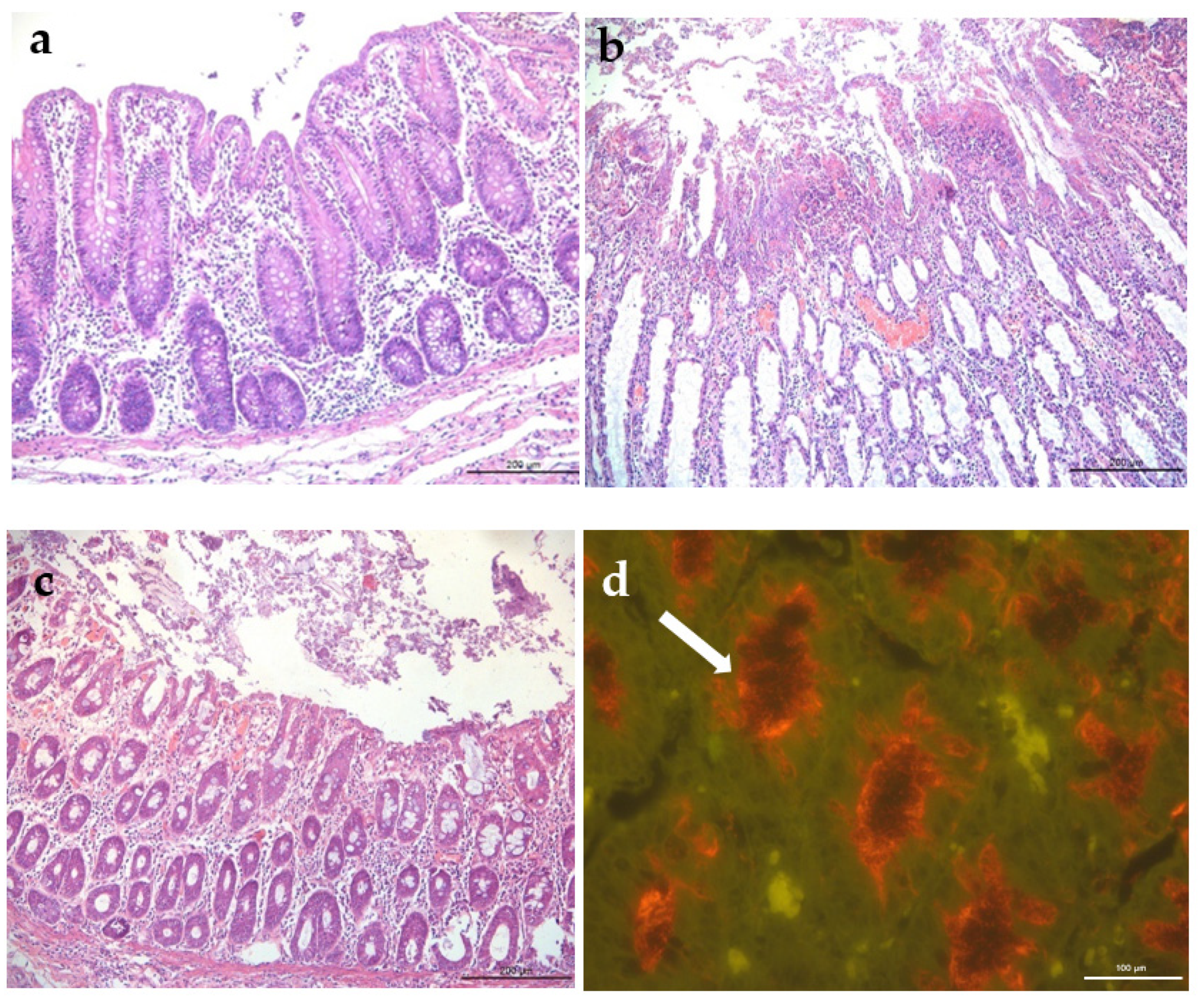
3.2.2. Histopathology and FISH

Histological findings are demonstrated in Table 1. In the CLIN group, all animals had lesions based on the evaluated parameters (superficial necrosis, hemorrhage, goblet cell hyperplasia, crypt abscesses and neutrophils infiltrate in the lamina propria). In this group the lesions were more severe and extensive, 8/16 animals were classified with a score >10 with severe lesions. Animal #25, was the only one with high score (13.5) in the SUBCL group and the only one that showed clinical signs of mucohemorrhagic diarrhea that started at 15 DPI. In the CRTL group, two animals had mild infiltration of inflammatory cell in the large intestine, with a score of 1, and no Brachyspira spp. associated. No significant lesions were observed in the small intestine and mesenteric lymph nodes in any of the experimental animals. Animals from group CLIN had more histological lesions than the other two groups, and pigs from SUBCL had more lesions than the CTRL group (p < 0.05) (Table 2).
FISH assays using probes specific for B. hyodysenteriae were positive in 9 animals of CLIN group, all of these with histologic score ≥9.5 in H&E evaluation. Three animals (#24, #25 and #26) from SUBCL group were positive by FISH with 5, 13.5 and 6.5 histologic scores, respectively (Table 1). All animals from CTRL group were negative. Figure 2 shows histological sections of the large intestine of pigs from the three evaluated groups.

3.3. qPCR

The first detection of B. hyodysenteriae fecal shedding by qPCR was in animal #2 from CLIN group on the 5th DPI. In this group, considering all study period and the last sample collection (18 DPI), 11/16 animals were positive by qPCR.
In the SUBCL group, four animals (#20, #24, #25 and #26) were positive starting at 15 DPI. Animal #20 was negative for all evaluated parameters (clinical signs, gross/microscopic lesions and FISH), but positive for qPCR and bacterial isolation of Brachyspira spp. Animals #24 and #26 had SD mild macroscopic lesions and moderate histology alterations. Only animal #25 was positive for all parameters evaluated, including mucohemorrhagic diarrhea.
The qPCR values ranged from 8.5 × 10³ to 5.9 × 10⁸ and 2.2 × 10² to 2.5 × 10⁷ organisms per gram of feces in the CLIN and SUBCL groups, respectively. The CRTL group were qPCR negative to B. hyodysenteriae in all tested samples.

3.4. Bacterial Isolation

Isolation was the most sensitive technique among all parameters used during this study (Table 1). In the CLIN group, B. hyodysenteriae isolation from feces of animal #2 was the earliest and coincided with qPCR detection. In this group, 14/16 animals had positive B. hyodysenteriae isolation. In the SUBCL group, the first bacterial isolation was obtained from fecal samples of animal #24 at 11 DPI and a total of 7/15 animals had positive isolation on 18 DPI. No growth of B. hyodysenteriae was obtained in the CRTL group.
No other clinical bacteria, particularly enterotoxigenic E. coli and Salmonella sp. were detected in the selective culture media used in samples collected at the time of necropsy.

4. Discussion

Low clinical B. hyodysenteriae imposes a critical epidemiological risk for contamination of negative herds, as healthy replacement animals originated from well managed hog farms with high healthy sanitary status, might carry the infection to not so well managed herds with other sanitary problems, and SD might manifest. So, the understanding of the magnitude and capacity of low clinical and subclinical B. hyodysenteriae isolates to cause SD is imperative. There are some studies describing the detection and isolation of B. hyodysenteriae strains from healthy animals from SD free herds [7,8,9,11,21]. However, clinical-pathological characterization of these subclinical isolates in experimental challenge studies is scarce [7].
In the present study, clinical course of mucohemorrhagic diarrhea started at 7 DPI in animals of the CLIN group, with a morbidity of 62.5% in 18 days of evaluation. These findings were similar with Wilcock et al. (1979) that described clinical signs observed at 7–10 DPI. Three animals of the CLIN group were euthanized due to the debilitating conditions caused of SD, demonstrating high pathogenicity of this isolate when compared to the SUBCL isolate. Lysons et al. [7] using different group of pigs challenged with three subclinical strains of B. hyodysenteriae did not observe clinical signs of SD in two strains used, even with colonization confirmed by qPCR. In the present study, only one animal of the SUBCL group developed mucohemorrhagic diarrhea at 15 DPI, demonstrating the delayed onset of clinical disease and potential reduced pathogenicity. This difference between the manifestation of subclinical B. hyodysenteriae isolates demonstrated the relevance of increasing the knowledge about them and the very likely differences among them.
Necropsy SD findings characterized by mucoid colitis or mucohemorrhagic and fibrous mucus typhlitis observed in the present study are consistent with the literature [17]. The sum of the histology lesion scores in large intestine classified as moderate to severe (≥5) were in accordance with the presence of gross lesions and the proportion of spirochetes labelled by FISH. La et al. [11] reported B. hyodysenteriae recovered from pigs without clinical signs and, on intestinal histological evaluation of three animals, no lesions suggestive of SD were observed. In the present study, spirochetes upon histology evaluation were observed in samples with more pronounced gross and microscopic lesions.
Isolation is considered the gold standard method for Braschyspira spp. diagnosis [22]. In the present study, isolation on selective agar also had the highest diagnostic sensitivity, detecting 14/16 and 7/15 positive samples in CLIN and SUBCL groups, respectively. This finding corroborates the literature [2] demonstrating that isolation is the best method to diagnose spirochetes when compared to clinical signs, qPCR, gross lesions, histology, and FISH. At necropsy, scraping of the colon was the method with highest isolation index, 15 compared to 7 from the cecum (data not shown). The subclinical isolated used in this study demonstrated a reduced ability (at least 50%) to infect and colonize susceptible pigs when compared to a clinical isolate. It is possible that the number of virulence factors harbored in these subclinical strains might be present in lower number. This hypothesis might be possible to be demonstrated in silico evaluation of the whole genome sequence comparing to clinical strains.
The pathogenesis of SD is complex and not fully elucidated, mainly because the disease is multifactorial. The infection depends on others anaerobic bacteria species in the large intestine, which contribute to colonization, induce extensive inflammation and necrosis of the epithelial surface of the cecum and colon [2,23]. Several virulence factors have been described for B. hyodysenteriae, including chemotaxis, motility, adhesion, hemolysin production and lipooligosaccharide (LOS) endotoxic activity [24,25].
B. hyodysenteriae hemolysis is considered one of the main virulence factors [26] and this phenotypic laboratory characteristic is often used to determine the clinical potential of isolates from SD clinical cases [27]. Based on complete genome analysis of B. hyodysenteriae, seven potential hemolysin genes were described [28]. The possible involvement of beta hemolysis in the pathogenesis of the disease has been evaluated in several studies [29,30,31]. However, its evaluation in clinical trials associated with the onset of intestinal lesions is scarce, and its importance in vivo is not fully elucidated. Thomson et al. [8] and Lysons et al. [7] compared virulent and potentially low virulence B. hyodysenteriae strains and reported difference in blood agar growth with poor hemolysis associated to potentially low virulence isolates. In other studies, in vitro hemolytic capacity was evaluated and described differences in hemolytic intensity in different strains of B. hyodysenteriae recovered form cases of SD [32,33]. However, analyzing isolates recovered from apparently healthy farms with no clinical signs of SD, no genetic and phenotypic differences in hemolysis were observed when compared to those isolated from clinically affected animals [11]. In the present study, both the CLIN and SUBCL isolates had the same phenotypic characteristics of strong hemolysis in blood agar and the same ST 245 using the MLST analysis [13]. This study does demonstrate that hemolysis on blood agar and sequence typing are not sufficient to determine the virulence of different typical strongly beta-hemolytic clinical or subclinical isolates of B. hyodysenteriae.
The subclinical isolate used in the present study was obtained from a farm of high healthy status and with no use of antimicrobials that could mask signs of the disease. The in vivo inoculation in pigs of this isolate demonstrated differences in the number of affected animals and beginning of clinical signs, however, it is important to consider the fact that this strain can colonize, induce lesions and clinical signs characteristic of SD in some animals. These findings raise great concern related to the necessity to screen replaced animals while in the quarantine, and the real important of having a quarantine, not only justified by PRRSv, PEDv, TGEv or Mycoplasma hyopneumoniae, but also for B. hyodysenteriae.
Although reported, it is unknown how long subclinical infection may persist in a herd, leading to the assumption that other apparently healthy herds may be similarly infected but remain undiagnosed. This is an important fact, especially when it may happen in nucleus or multiplier breeding herds of high healthy status, as they can carry the spirochete to other farms and spread the disease in conditions where there are healthy and management challenges [11]. Isolates recovered from pigs in apparently healthy multiplier herds has been described in Germany, Swiss and Australia [11,20,34,35], highlighting the epidemiological risk of these herds.
Serological tests were performed to identify herds with SD and could be an option to diagnosed subclinical infected herds, but there are no commercial kits that can be used. Based on LOS, B. hyodysenteriae has 11 serotypes widely distributed in different geographic regions. Genetic variation and marked differences in antigenic proteins are significant limitations for development of a globally applicable serological test [36]. Some studies using recombinant proteins have been carried out to develop a serological test that would be an important tool for detecting B. hyodysenteriae positive herds without clinical signs of SD [21,37].
The hypotheses to explain the presence of the SUBCL isolate in an apparently healthy animal from a herd with no clinical cases of SD but that developed clinical disease in an experimentally inoculated pig in the present study could be the following: (1) The SUBCL isolate had reduced virulence factors compared to typical clinical B. hyodysenteriae isolates, reducing the capability of proliferation and not reaching the infection level enough to cause disease and/or to be detected, and/or lacking virulence factors able to induce typical lesions [11,38,39]. (2) The source herd has a high healthy status with fewer challenges than a commercial herd, a condition that may influence spirochete colonization and disease development, or (3) The pressure of infection of B. hyodysenteriae among the host population on the farm was low and the amount of spirochetes used in the inoculum was sufficient to induce clinical disease in experimentally infected pigs [17]. Other aspects to be considered are the influence of feed ingredients and some substrates that might influence the microbiota and/or the physicochemical environment in the colon, which in turn may influence the ability of B. hyodysenteriae to colonize [39,40]. In addition, both isolates the current study, clinical and subclinical, were classified with the same ST 245; however, they were obtained from different farms, located in different regions (São Paulo and Minas Gerais States), so other unknown factors may be involved the clinical manifestation of SD.

5. Conclusions

This study clearly demonstrated that subclinical B. hyodysenteriae obtained from healthy animals in SD free herds may induce the disease, even with lower severity, when susceptible animals are exposed to high concentrations of the bacteria. Hemolysis or ST characteristics seems not to be definitive markers for pathogenicity and future in silico in association to in vivo studies are required to compared different subclinical and clinical isolates and evaluate possible determinants to disease development. Meanwhile, it is important to be aware about the existence of this subclinical B. hyodysenteriae and have strategies to minimize the chances of contamination of negative herds.

Author Contributions

Conceptualization, J.P.H.S. and R.M.C.G.; methodology, J.P.H.S. and A.G.S.D.; validation, J.P.H.S. and M.P.G.; animal management, J.P.H.S., A.G.S.D., C.E.R.P., M.R.A., R.P.L., M.P.G., L.V.A.O. and J.A.B.-Z.; statistical analysis, N.R.M. and M.R.A.; laboratory analysis, J.P.H.S., L.V.A.O. and J.A.B.-Z.; data curation, J.P.H.S. and R.P.L.; writing—original draft preparation, J.P.H.S.; writing—review and editing, J.P.H.S., A.G.S.D. and R.M.C.G.; supervision, C.E.R.P. and M.P.G.; project administration, R.M.C.G. All authors have read and agreed to the published version of the manuscript.

Funding

This work was conducted with the support of FAPEMIG (Project No. APQ-02908-17), Minas Gerais, Brazil.

Institutional Review Board Statement

The study was conducted according to the Ethical Principles of Animal Experimentation, adopted by the Committee on Ethics in the Use of Animals (CEUA) of the Universidade Federal de Minas Gerais (UFMG) (Approval Number: CEUA #177/2015).

Informed Consent Statement

Not applicable.

Data Availability Statement

The data presented in this study are available by reasonable request from the corresponding author.

Acknowledgments

The authors would like to express their gratitude to FAPEMIG, Capes and CNPq. RMCG has a research fellowship from CNPq.

Conflicts of Interest

The authors declare no conflict of interest. The authors declare that they have no known competing financial interests or personal relationships that could have appeared to influence the work reported in this paper.

References

  1. Wills, R.W. Diarrhea in growing-finishing swine. Vet. Clin. N. Am. Food Anim. Pract. 2000, 16, 135–161. [Google Scholar] [CrossRef]
  2. Hampson, D.J.; Burrougth. Swine Dysentery and Brachyspiral Colits. In Disease of Swine, 11th ed.; Zimmerman, J.J., Karriker, L.A., Ramirez, A., Schwartz, K.J., Stevenson, G.W., Zhang, J., Eds.; Wiley Blackwell: Hoboken, NJ, USA, 2019; pp. 807–834. [Google Scholar]
  3. Burrough, E.R. Swine dysentery—Re-emergence in the United States and Canada. In Proceedings of the 6th International Conference on Colonic Spirochetal Infections in Animals and Humans, University of Surrey, Guildford, UK, 5–6 September 2013; pp. 55–56. [Google Scholar]
  4. Råsbäck, T.; Jansson, D.S.; Johansson, K.; Fellström, C. A novel enteropathogenic, strongly haemolytic spirochaete isolated from pig and mallard, provisionally designated ‘Brachyspira suanatina’ sp. nov. Environ. Microbiol. 2007, 9, 983–991. [Google Scholar] [CrossRef] [PubMed]
  5. Chander, Y.; Primus, A.; Oliveira, S.; Gebhart, C.J. Phenotypic and molecular characterization of a novel strongly hemolytic Brachyspira species, provisionally designated “Brachyspira hampsonii”. J. Vet. Diagn. Investig. 2012, 24, 903–910. [Google Scholar] [CrossRef] [Green Version]
  6. Mushtaq, M.; Zubair, S.; Råsbäck, T.; Bongcam-Rudloff, E.; Jansson, D.S. Brachyspira suanatina sp. nov., an enteropathogenic intestinal spirochaete isolated from pigs and mallards: Genomic and phenotypic characteristics. BMC Microbiol. 2015, 15, 208. [Google Scholar] [CrossRef] [PubMed] [Green Version]
  7. Lysons, R.J.; Lemcke, R.M.; Bew, J.; Burrows, M.R.; Alexander, T.J.I. An avirulent strain of Treponema hyodysenteriae isolated from herds free of swine dysentery. In Proceedings of the 7th International Pig Veterinary Society Congress, Mexico City, Mexico, 26–31 July 1982; p. 40. [Google Scholar]
  8. Thomson, J.R.; Smith, W.J.; Murray, B.P.; Murray, D.; Dick, J.E.; Sumption, K.J. Porcine enteric spirochete infections in the UK: Surveillance data and preliminary investigation of atypical isolates. Anim. Health Res. Rev. 2001, 2, 31–36. [Google Scholar] [CrossRef] [PubMed]
  9. Hampson, D.J.; La, T.; Phillips, N.D. Emergence of Brachyspira species and strains: Reinforcing the need for surveillance. Porc. Health Manag. 2015, 1, 8. [Google Scholar] [CrossRef] [PubMed] [Green Version]
  10. La, T.; Phillips, N.D.; Hampson, D.J. An Investigation into the etiological agents of Swine Dysentery in Australian pig herds. PLoS ONE 2016, 11, e0167424. [Google Scholar] [CrossRef] [PubMed]
  11. La, T.; Rhode, J.; Phillips, N.D.; Hampson, D.J. Comparison of Brachyspira hyodysenteriae isolates recovered from pigs in apparently healthy multiplier herds with isolates from herds with Swine Dysentery. PLoS ONE 2016, 11, e0160362. [Google Scholar] [CrossRef] [PubMed]
  12. La, T.; Phillips, N.D.; Hampson, D.J. Development of a duplex PCR assay for detection of Brachyspira hyodysenteriae and Brachyspira pilosicoli in pig feces. J. Clin. Microbiol. 2003, 41, 3372–3375. [Google Scholar] [CrossRef] [Green Version]
  13. Sato, J.P.H.; Daniel, A.G.S.; Leal, C.A.G.; Barcellos, D.E.S.N.; Guedes, R.M.C. Diversity and potential genetic relationships amongst Brazilian Brachyspira hyodysenteriae isolates from cases of swine dysentery. Vet. Microbiol. 2022, 266, 109369. [Google Scholar] [CrossRef]
  14. Leser, T.D.; Møller, K.; Jensen, T.K.; Jorsal, S.E. Specific detection of Serpulina hyodysenteriae and potentially pathogenic weakly ß-haemolytic porcine intestinal spirochetes by polymerase chain reaction targetting 23S rDNA. Mol. Cell. Probes 1997, 11, 363–372. [Google Scholar] [CrossRef]
  15. Kunkle, R.A.; Harris, D.L.; Kinyon, J.M. Autoclaved liquid medium for propagation of Treponema hyodysenteriae. J. Clin. Microbiol. 1986, 24, 669–671. [Google Scholar] [CrossRef] [PubMed] [Green Version]
  16. Jacobson, M.; Fellström, C.; Lindberg, R.; Wallgren, P.; Jensen-Waern, M. Experimental swine dysentery: Comparison between infection models. J. Med. Microbiol. 2004, 53, 273–280. [Google Scholar] [CrossRef] [PubMed]
  17. Rubin, J.E.; Costa, M.O.; Hill, J.E.; Kittrell, H.E.; Fernando, C.; Huango, Y.; Huango, Y.; O’connor, B.; Harding, J.C. Reproduction of mucohaemorrhagic diarrhea and colitis indistinguishable from Swine Dysentery following experimental inoculation with ‘‘Brachyspira hampsonii’’ Strain 30446. PLoS ONE 2013, 8, e57146. [Google Scholar] [CrossRef]
  18. Luna, L.G. Manual of Histologic Staining Methods of the Armed Forces Institute of Pathology, 3rd ed.; McGraw-Hill: New York, NY, USA, 1968; pp. 24–58. [Google Scholar]
  19. Jensen, T.K.; Boye, M.; Møller, K.; Leser, T.D.; Jorsal, S.E. Association of Serpulina hyodysenteriae with the colonic mucosa in experimental swine dysentery studied by fluorescent in situ hybridization. APMIS 1998, 106, 1061–1068. [Google Scholar] [CrossRef] [PubMed]
  20. Boye, M.; Jensen, T.K.; Møller, K.; Leser, T.D.; Jorsal, S.E. Specific detection of the genus Serpulina, S. hyodysenteriae and S. pilosicoli in porcine intestines by fluorescent rRNA in situ hybridization. Mol. Cell. Probes 1998, 12, 323–330. [Google Scholar] [CrossRef] [PubMed]
  21. Hampson, D.J.; La, T.; Phillips, N.D.; Holyoake, P.K. Brachyspira hyodysenteriae isolated from apparently healthy pig herds following an evaluation of a prototype commercial serological ELISA. Vet. Microbiol. 2016, 191, 15–19. [Google Scholar] [CrossRef]
  22. Råsbäck, T.; Fellström, C.; Gunnarsson, A.; Aspán, A. Comparison of culture and biochemical tests with PCR for detection of Brachyspira hyodysenteriae and Brachyspira pilosicoli. J. Microbiol. Methods 2006, 66, 347–353. [Google Scholar] [CrossRef] [PubMed]
  23. Wilcock, B.P.; Olander, H.J. Studies on the pathogenesis of swine dysentery: I. Characterization of the lesions in colons and colonic segments inoculated with pure cultures or colonic content containing Treponema hyodysenteriae. Vet. Pathol. 1979, 16, 450–465. [Google Scholar] [CrossRef] [PubMed] [Green Version]
  24. Meyer, R.C.; Simon, J.; Byerly, C.S. The etiology of swine dysentery. II. Effect of a known microbial flora, weaning and diet on disease production in gnotobiotic and conventional swine. Vet. Pathol. 1974, 11, 527–534. [Google Scholar] [CrossRef] [PubMed]
  25. Hampson, D.J.; Nagaraja, T.G.; Kennan, R.M.; Rood, J.I. Gram-negative anaerobes. In Pathogenesis of Bacterial Infections in Animals, 4th ed.; Gyles, C.L., Prescott, J.F., Songer, J.G., Thoen, C.O., Eds.; Blackwell Publishing: Ames, IA, USA, 2010; pp. 513–526. [Google Scholar]
  26. Alvarez-Ordóñez, A.; Martínez-Lobo, F.J.; Arguello, H.; Carvajal, A.; Rubio, P. Swine dysentery: Aetiology, pathogenicity, determinants of transmission and the fight against the disease. Int. J. Environ. Res. Public Health 2013, 10, 1927–1947. [Google Scholar] [CrossRef] [PubMed] [Green Version]
  27. Hsu, T.; Hutto, D.L.; Minion, F.C.; Zuerner, R.L.; Wannemuehler, M.J. Cloning of a beta-hemolysin gene of Brachyspira (Serpulina) hyodysenteriae and its expression in Escherichia coli. Infect. Immun. 2001, 69, 706–711. [Google Scholar] [CrossRef] [PubMed] [Green Version]
  28. Burrough, E.R.; Strait, E.L.; Kinyon, J.M.; Bower, L.P.; Madson, D.M.; Wilberts, B.L.; Schwartz, K.J.; Frana, T.S.; Songer, J.G. Comparative virulence of clinical Brachyspira spp. isolates in inoculated pigs. J. Vet. Diagn. Investig. 2012, 24, 1025–1034. [Google Scholar] [CrossRef] [PubMed] [Green Version]
  29. Bellgard, M.; Wanchanthuek, P.; La, T.; Ryan, K.; Moolhuijzen, P.; Albertyn, Z.; Shaban, B.; Motro, Y.; Dunn, D.S.; Schibeci, D.; et al. Genome sequence of the pathogenic intestinal spirochete Brachyspira hyodysenteriae reveals adaptations to its lifestyle in the porcine large intestine. PLoS ONE 2009, 4, e4641. [Google Scholar] [CrossRef] [Green Version]
  30. Ter Huurne, A.A.; Muir, S.; Van Houten, M.; Koopman, M.B.; Kusters, J.G.; Van Der Zeijst, B.A.; Gaastra, W. The role of hemolysin(s) in the pathogenesis of Serpulina hyodysenteriae. Zentralbl. Bakteriol. 1993, 278, 316–325. [Google Scholar] [CrossRef]
  31. Ter Huurne, A.A.; Muir, S.; Van Houten, M.; Van Der Zeijst, B.A.; Gaastra, W.; Kusters, J.G. Characterization of three putative Serpulina hyodysenteriae hemolysins. Microb. Pathog. 1994, 16, 269–282. [Google Scholar] [CrossRef] [PubMed]
  32. Hyatt, D.R.; ter Huurne, A.A.; van der Zeijst, B.A.; Joens, L.A. Reduced virulence of Serpulina hyodysenteriae hemolysin-negative mutants in pigs and their potential to protect pigs against challenge with a virulent strain. Infect. Immun. 1994, 62, 2244–2248. [Google Scholar] [CrossRef] [PubMed] [Green Version]
  33. Mahu, M.; De Pauw, N.; Vande Maele, L.; Verlinden, M.; Boyen, F.; Ducatelle, R.; Haesebrouck, F.; Martel, A.; Pasmans, F. Variation in hemolytic activity of Brachyspira hyodysenteriae strains from pigs. Vet. Res. 2016, 47, 66. [Google Scholar] [CrossRef] [Green Version]
  34. Löbert, S.; Zimmermann, W.; Bürki, S.; Frey, J.; Nathues, H.; Scheer, P.; Zeeh, F. Occurrence of Brachyspira hyodysenteriae in multiplier pig herds in Switzerland. Tierärztl. Prax. 2016, 44, 13–18. [Google Scholar] [CrossRef] [PubMed]
  35. Hampson, D.J.; Cutler, R.; Lee, B.J. Virulent Serpulina hyodysenteriae from a pig in a herd free of clinical swine dysentery. Vet. Rec. 1992, 131, 318–319. [Google Scholar] [CrossRef]
  36. Herbst, W.; Schneider, S.; Baljer, G.; Barth, S.A. An update of Brachyspira hyodysenteriae serotyping. Res. Vet. Sci. 2017, 111, 135–139. [Google Scholar] [CrossRef] [PubMed]
  37. Song, Y.; La, T.; Phillips, N.D.; Hampson, D. Development of a serological ELISA using a recombinant protein to identify pig herds infected with Brachyspira hyodysenteriae. Vet. J. 2015, 206, 365–370. [Google Scholar] [CrossRef] [PubMed] [Green Version]
  38. Achacha, M.; Messier, S.; Mittal, K.R. Development of an experimental model allowing discrimination between virulent and avirulent isolates of Serpulina (Treponema) hyodysenteriae. Can. J. Vet. Res. 1996, 60, 45–49. [Google Scholar]
  39. Siba, P.M.; Pethick, D.W.; Hampson, D.J. Pigs experimentally infected with Serpulina hyodysenteriae can be protected from developing swine dysentery by feeding them a highly digestible diet. Epidemiol. Infect. 1996, 116, 207–216. [Google Scholar] [CrossRef] [PubMed] [Green Version]
  40. Thomsen, L.E.; Knudsen, K.E.B.; Jensen, T.K.; Christensen, A.S.; Møller, K.; Roepstorff, A. The effect of fermentable carbohydrates on experimental swine dysentery and whip worm infections in pigs. Vet. Microbiol. 2007, 119, 152–163. [Google Scholar] [CrossRef]
Figure 1. Gross lesions in the spiral colon. (a,b) Clinical isolate (CLIN group), excessive luminal mucus with diffuse mucosa hemorrhage (a), and superficial fibrinonecrotic exudate (b). (c) Subclinical isolate (SUBCL group), discrete focal increase in luminal mucus and moderate multifocal mucosal hemorrhage. (d) Negative control (CTRL group), no gross lesions.
Vetsci 09 00286 g001aVetsci 09 00286 g001b
Figure 2. Histologic findings in the colon of pigs from the control or inoculated with Brachyspira hyodysenteriae groups. (a) Negative control group (CTRL), no visible lesions (H&E). (b) Clinical group (CLIN), hemorrhage, diffuse severe superficial necrosis and epithelial detachment, severe inflammatory infiltrate in the lamina propria, H&E. (c) Subclinical group (SUBCL), hyperemia, multifocal superficial necrosis with epithelial detachment and moderate inflammatory infiltrate in the lamina propria, H&E. (d) Subclinical isolate, spirochetes (arrow) at the apical border and inside enterocytes, fluorescent hybridization in situ (FISH).
Vetsci 09 00286 g002
Table 1. Fecal score, qPCR, bacterial isolation, gross lesions, microscopic lesion score and fluorescence in situ hybridization (FISH) of experimentally infected pig with clinical (CLIN), subclinical (SUBCL) of Brachyspira hyodysenteriae isolates and negative control (CTRL).
GroupAnimal5 DPI7 DPI11 DPI15 DPI18 DPIB. hyodysenteriae IsolationGross LesionMicroscopic ScoreFISH *
FEqPCRFEqPCRFEqPCRFEqPCRFEqPCR
CLIN11Neg21.5 × 10342.9 × 10742.7 × 10741.4 × 1079 DPIYes13+
228.5 × 1033.52.4 × 10541.2 × 108¥¥¥¥5 DPIYes11.5++
31Neg2Neg2Neg2Neg1Neg18 DPINo3.5-
42Neg2Neg2Neg2Neg2Neg11 DPINo4.5-
52Neg1Neg2Neg2Neg2Neg11 DPINo3-
60Neg19.6 × 10343.4 × 107¥¥¥¥9 DPIYes11++
71Neg2Neg23.5 × 10344.3 × 1073.52.6 × 10715DPIYes10++
81Neg2Neg3Neg31.4 × 10237.4 × 10418 DPIYes9.5++
91Neg2Neg44.4 × 10742.6 × 10642.5 × 10711 DPIYes12.5+++
101Neg2Neg2Neg3Neg3Neg18 DPINo7-
111Neg3Neg3Neg3Neg3Neg11 DPINo4-
121Neg3Neg46.8 × 10747.6 × 10745.9 × 10811 DPIYes14++
130Neg1Neg1Neg42.1 × 107¥¥15 DPIYes14.5++
140Neg1Neg11.5 × 10302.5 × 10321.0 × 104NegNo1-
150Neg1Neg1Neg1Neg11.6 × 105NegNo1-
161Neg2Neg1Neg21.1 × 10327.2 × 10718 DPIYes13.5+++
SUBCL170Neg0Neg1Neg0Neg0NegNegNo0-
180Neg0Neg1Neg1Neg1NegNegNo2.5-
190Neg2Neg1Neg2Neg1NegNegNo7-
200Neg2Neg1Neg27.4 × 10321.5 × 10311 DPINo4-
210Neg0Neg2Neg0Neg0NegNegNo2.5-
220Neg0Neg2Neg2Neg1Neg18 DPINo3-
230Neg0Neg2Neg2Neg1NegNegNo6-
240Neg0Neg1Neg21.6 × 10723.0 × 10411 DPIYes5+++
250Neg0Neg1Neg41.3 × 10742.5 × 10715 DPIYes13.5+++
260Neg0Neg1Neg01.1 × 10422.1 × 10515 DPIYes6.5+
270Neg2Neg1Neg2Neg1NegNegNo0-
280Neg0Neg1Neg1Neg1Neg18 DPINo5-
290Neg0Neg1Neg1Neg1Neg18 DPINo1-
300Neg0Neg0Neg2Neg1NegNegNo3-
311Neg0Neg2Neg2Neg2NegNegYes2.5-
CTRL320Neg0Neg0Neg0Neg0NegNegNo0-
330Neg0Neg0Neg0Neg0NegNegNo0-
340Neg0Neg1Neg0Neg1NegNegNo0-
350Neg0Neg0Neg0Neg0NegNegNo0-
360Neg1Neg0Neg0Neg0NegNegNo0-
370Neg0Neg0Neg0Neg0NegNegNo0-
380Neg0Neg0Neg0Neg0NegNegNo0-
390Neg0Neg1Neg1Neg1NegNegNo0-
400Neg0Neg0Neg1Neg1NegNegNo0-
410Neg0Neg1Neg1Neg1NegNegNo0-
420Neg0Neg0Neg0Neg0NegNegNo1-
430Neg0Neg0Neg0Neg0NegNegNo0-
440Neg1Neg1Neg0Neg1NegNegNo0-
450Neg0Neg0Neg0Neg0NegNegNo1-
461Neg1Neg1Neg1Neg1NegNegNo0-
470Neg0Neg0Neg0Neg0NegNegNo0-
DPI: days post inoculation; FE: fecal score; FISH: fluorescence in situ hybridization; * [-] No fluorescent labelling of B. hyodysenteriae, [+] mild fluorescent labeling of B. hyodysenteriae, [++] moderate fluorescent labeling of B. hyodysenteriae, [+++] severe fluorescent labeling of B. hyodysenteriae; ¥: euthanized animals.
Table 2. Clinical signs, macroscopic and microscopic lesions of swine dysentery after inoculation of Brachyspira hyodysenteriae in swine model.
Clinical Signs *Gross Lesions *Histologic Lesions
GroupAqueous and/or Mucohemorrhagic DiarrheaFirst Observation of DiarrheaTotal Number of Days with DiarrheaExcessive Mucus in the LumenMucosal HemorrhageFibrinous ExudateMean of the Final Score ± SD
Negative0/16 a- **0 a0/16 a0/16 a0/16 a0.02 ± 0.04 a
Subclinical1/15 a15 DPI3 b3/15 a1/15 a1/15 a0.81 ± 0.39 b
Clinical10/16 b7 DPI11 b9/16 b9/16 b8/16 b1.66 ± 0.26 c
* Number of animals affected/total of animals; ** No diarrhea was observed among negative control pigs; a,b,c: different letters indicate statistical differences between groups; DPI: days post inoculation; SD: standard deviation; Kruskal–Wallis test followed by Mann–Whitney test if significant.
Publisher’s Note: MDPI stays neutral with regard to jurisdictional claims in published maps and institutional affiliations.

