
Hepadnavirus Infection in a Cat with Chronic Liver Disease: A Multi-Disciplinary Diagnostic Approach
 , , , , , , , , and
Abstract
:Simple Summary
Abstract
1. Introduction
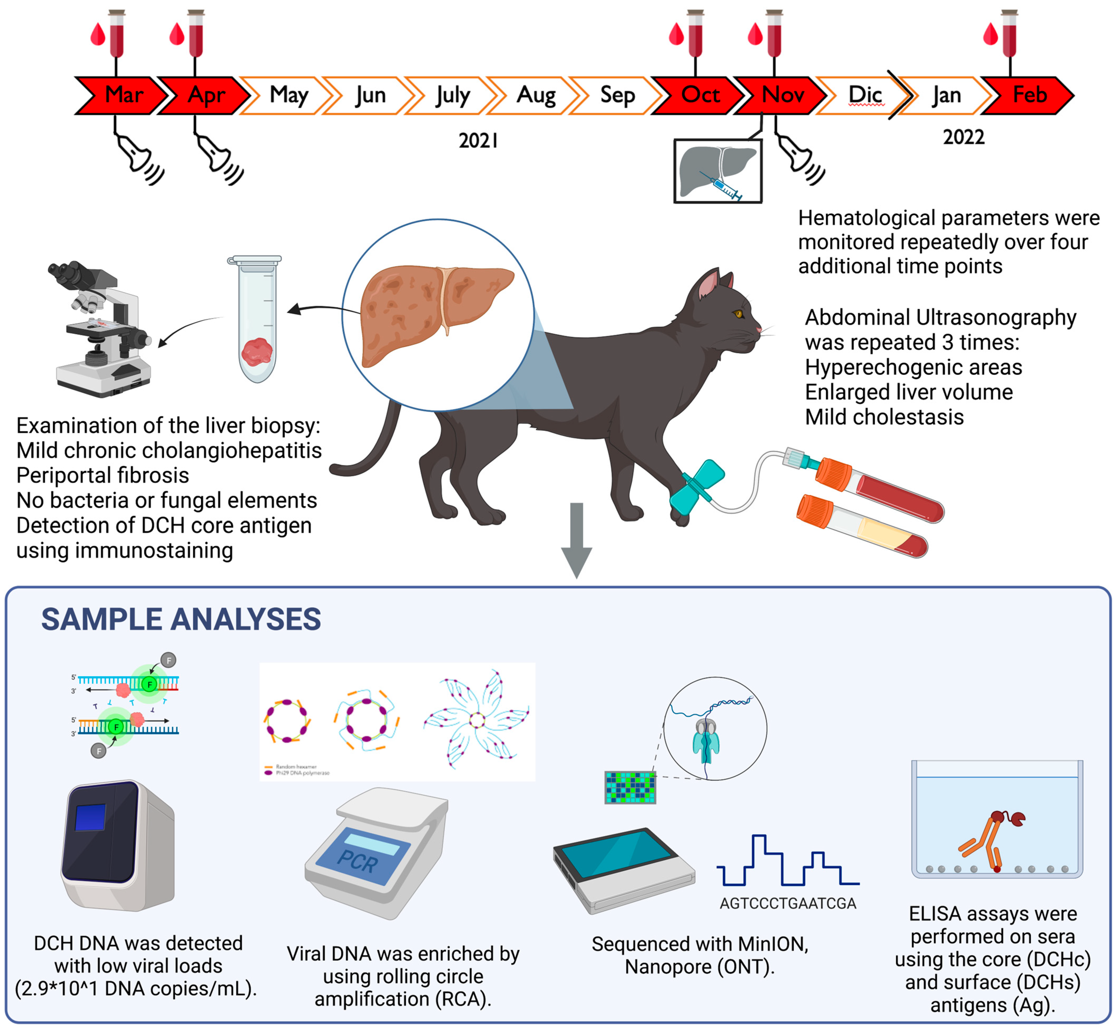
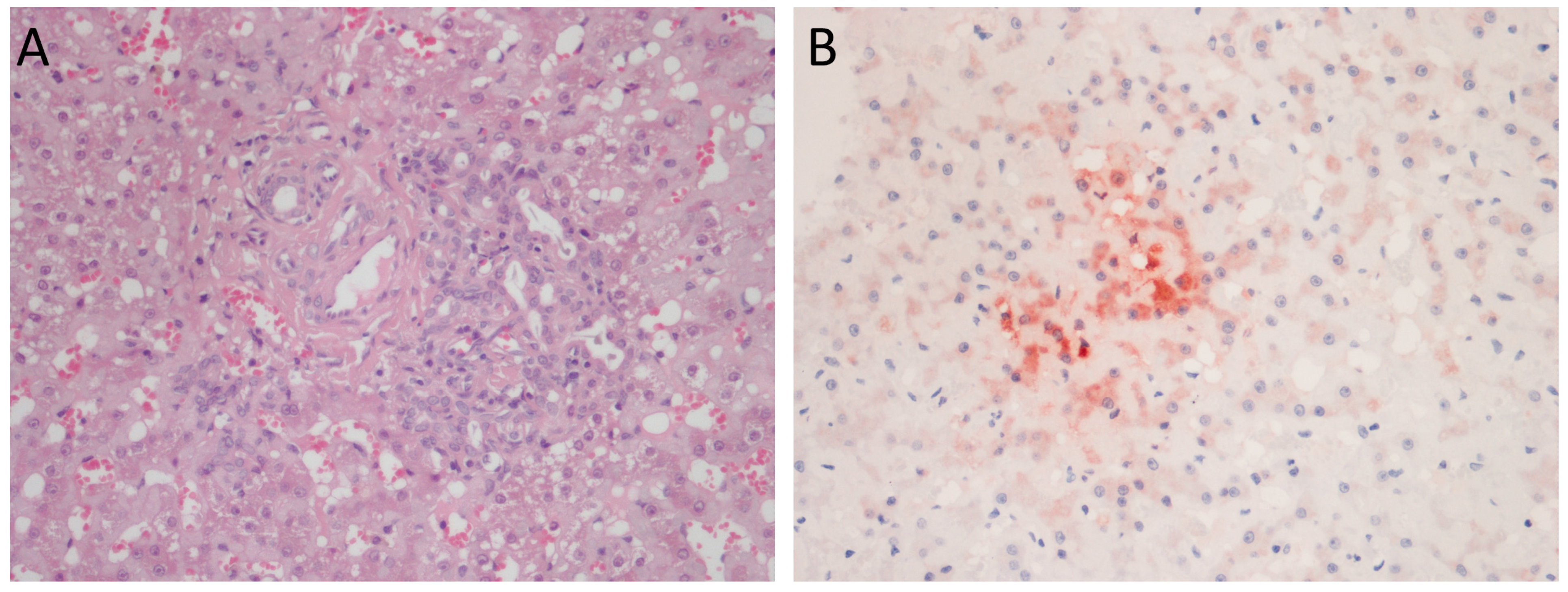
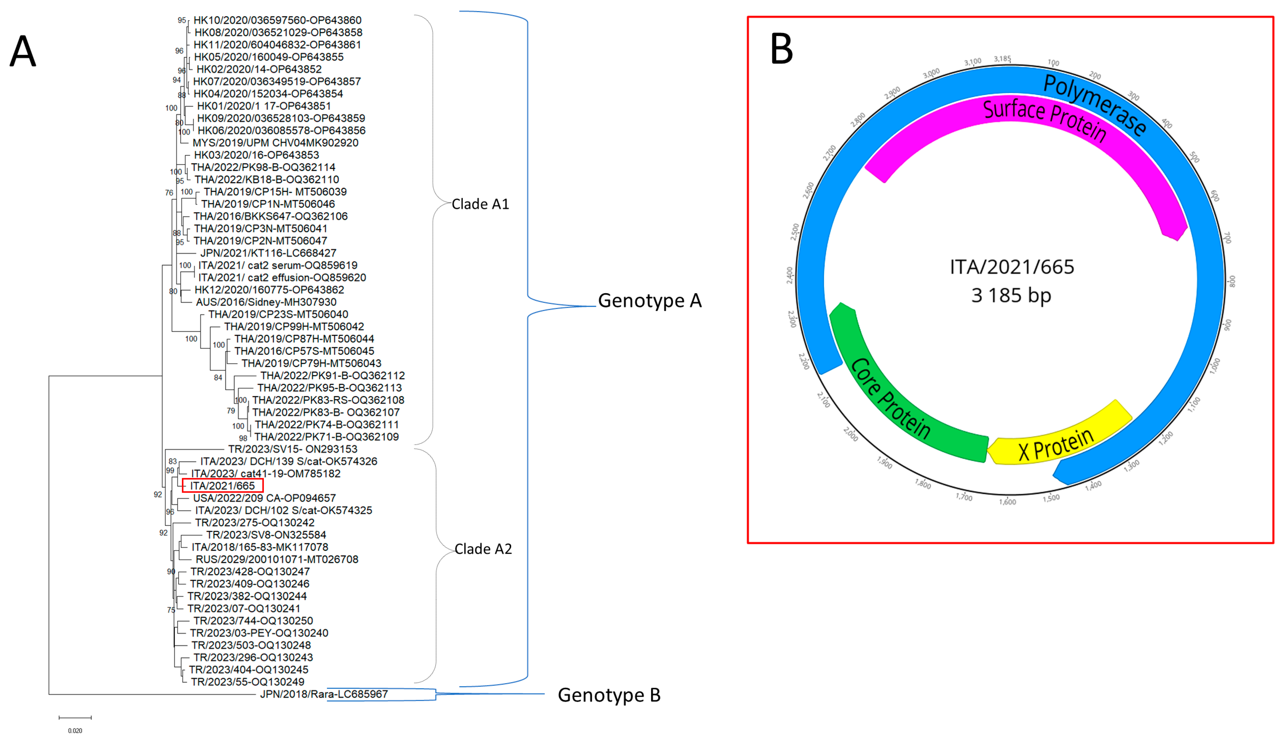
2. Case Description
3. Discussion and Conclusions
Author Contributions
Funding
Institutional Review Board Statement
Informed Consent Statement
Data Availability Statement
Conflicts of Interest
References
- Aghazadeh, M.; Shi, M.; Barrs, V.R.; McLuckie, A.J.; Lindsay, S.A.; Jameson, B.; Hampson, B.; Holmes, E.C.; Beatty, J.A. A Novel Hepadnavirus Identified in an Immunocompromised Domestic Cat in Australia. Viruses 2018, 10, 269. [Google Scholar] [CrossRef] [PubMed]
- Capozza, P.; Carrai, M.; Choi, Y.R.; Tu, T.; Nekouei, O.; Lanave, G.; Martella, V.; Beatty, J.A.; Barrs, V.R. Domestic Cat Hepadnavirus: Molecular Epidemiology and Phylogeny in Cats in Hong Kong. Viruses 2023, 15, 150. [Google Scholar] [CrossRef] [PubMed]
- Shofa, M.; Kaneko, Y.; Takahashi, K.; Okabayashi, T.; Saito, A. Global Prevalence of Domestic Cat Hepadnavirus: An Emerging Threat to Cats’ Health? Front. Microbiol. 2022, 13, 938154. [Google Scholar] [CrossRef] [PubMed]
- Sakamoto, H.; Ito, G.; Goto-Koshino, Y.; Sakamoto, M.; Nishimura, R.; Momoi, Y. Detection of domestic cat hepadnavirus by next-generation sequencing and epidemiological survey in Japan. J. Vet. Med. Sci. 2023, 85, 642–646. [Google Scholar] [CrossRef]
- Ratti, G.; Stranieri, A.; Scavone, D.; Cafiso, A.; Meazzi, S.; Luzzago, C.; Dall’Ara, P.; Tagliasacchi, F.; Cavicchioli, L.; Ferrari, F.; et al. Detection and genetic characterization of domestic cat hepadnavirus in cats with cavitary effusions. Vet. Microbiol. 2023, 284, 109828. [Google Scholar] [CrossRef]
- Capozza, P.; Decaro, N.; Beikpour, F.; Buonavoglia, C.; Martella, V. Emerging Hepatotropic Viruses in Cats: A Brief Review. Viruses 2021, 13, 1162. [Google Scholar] [CrossRef]
- Adiguzel, E.; Erdem-Sahinkesen, E.; Koc, B.T.; Demirden, C.; Oguzoglu, T.C. The detection and full genomic characterization of domestic cat Orthohepadnaviruses from Turkiye. Vet. Med. Sci. 2023, 9, 1965–1972. [Google Scholar] [CrossRef]
- Fruci, P.; Di Profio, F.; Palombieri, A.; Massirio, I.; Lanave, G.; Diakoudi, G.; Pellegrini, F.; Marsilio, F.; Martella, V.; Di Martino, B. Detection of antibodies against domestic cat hepadnavirus using baculovirus-expressed core protein. Transbound. Emerg. Dis. 2022, 69, 2980–2986. [Google Scholar] [CrossRef]
- Pesavento, P.A.; Jackson, K.; Hampson, T.; Munday, J.S.; Barrs, V.R.; Beatty, J.A. A Novel Hepadnavirus is Associated with Chronic Hepatitis and Hepatocellular Carcinoma in Cats. Viruses 2019, 11, 969. [Google Scholar] [CrossRef]
- Lanave, G.; Capozza, P.; Diakoudi, G.; Catella, C.; Catucci, L.; Ghergo, P.; Stasi, F.; Barrs, V.; Beatty, J.; Decaro, N.; et al. Identification of hepadnavirus in the sera of cats. Sci. Rep. 2019, 9, 10668. [Google Scholar] [CrossRef]
- Piewbang, C.; Dankaona, W.; Poonsin, P.; Yostawonkul, J.; Lacharoje, S.; Sirivisoot, S.; Kasantikul, T.; Tummaruk, P.; Techangamsuwan, S. Domestic cat hepadnavirus associated with hepatopathy in cats: A retrospective study. J. Vet. Intern. Med. 2022, 36, 1648–1659. [Google Scholar] [CrossRef] [PubMed]
- Piewbang, C.; Wardhani, S.W.; Chaiyasak, S.; Yostawonkul, J.; Chai-In, P.; Boonrungsiman, S.; Kasantikul, T.; Techangamsuwan, S. Insights into the genetic diversity, recombination, and systemic infections with evidence of intracellular maturation of hepadnavirus in cats. PLoS ONE 2020, 15, e0241212. [Google Scholar] [CrossRef]
- Stone, C.; Petch, R.; Gagne, R.B.; Nehring, M.; Tu, T.; Beatty, J.A.; VandeWoude, S. Prevalence and Genomic Sequence Analysis of Domestic Cat Hepadnavirus in the United States. Viruses 2022, 14, 2091. [Google Scholar] [CrossRef] [PubMed]
- Takahashi, K.; Kaneko, Y.; Shibanai, A.; Yamamoto, S.; Katagiri, A.; Osuga, T.; Inoue, Y.; Kuroda, K.; Tanabe, M.; Okabayashi, T.; et al. Identification of domestic cat hepadnavirus from a cat blood sample in Japan. J. Vet. Med. Sci. 2022, 84, 648–652. [Google Scholar] [CrossRef] [PubMed]
- Anpuanandam, K.; Selvarajah, G.T.; Choy, M.M.K.; Ng, S.W.; Kumar, K.; Ali, R.M.; Rajendran, S.K.; Ho, K.L.; Tan, W.S. Molecular detection and characterisation of Domestic Cat Hepadnavirus (DCH) from blood and liver tissues of cats in Malaysia. BMC Vet. Res. 2021, 17, 9. [Google Scholar] [CrossRef] [PubMed]
- Jeanes, E.C.; Wegg, M.L.; Mitchell, J.A.; Priestnall, S.L.; Fleming, L.; Dawson, C. Comparison of the prevalence of Domestic Cat Hepadnavirus in a population of cats with uveitis and in a healthy blood donor cat population in the United Kingdom. Vet. Ophthalmol. 2022, 25, 165–172. [Google Scholar] [CrossRef] [PubMed]
- Capozza, P.; Lanave, G.; Diakoudi, G.; Stasi, F.; Ghergo, P.; Ricci, D.; Santo, G.; Arena, G.; Grillo, I.; Delle Donne, E.; et al. A longitudinal observational study in two cats naturally-infected with hepadnavirus. Vet. Microbiol. 2021, 254, 108999. [Google Scholar] [CrossRef] [PubMed]
- Scavone, D.; Lauzi, S.; Stranieri, A.; Tramontano, G.; Ratti, G.; Paltrinieri, S. Evaluating the presence of domestic cat hepadnavirus viraemia in cats with biochemical alterations suggestive of liver disease. Vet. Rec. 2022, 191, e1626. [Google Scholar] [CrossRef]
- Seeger, C.; Zoulim, F.; Mason, W.S. Hepadnaviruses. In Fields Virology, 7th ed.; Knipe, D.M., Ed.; Lippincott Williams & Wilkins: Philadelphia, PA, USA, 2013; Volume 1, pp. 2185–2210. [Google Scholar]
- Lee, J.H.; Kim, H.S. Current laboratory tests for diagnosis of hepatitis B virus infection. Int. J. Clin. Pract. 2021, 75, e14812. [Google Scholar] [CrossRef]
- Marsilio, F.; Di Martino, B.; Decaro, N.; Buonavoglia, C. A novel nested PCR for the diagnosis of calicivirus infections in the cat. Vet. Microbiol. 2005, 105, 1–7. [Google Scholar] [CrossRef]
- Decaro, N.; Desario, C.; Lucente, M.S.; Amorisco, F.; Campolo, M.; Elia, G.; Cavalli, A.; Martella, V.; Buonavoglia, C. Specific identification of feline panleukopenia virus and its rapid differentiation from canine parvoviruses using minor groove binder probes. J. Virol. Methods 2008, 147, 67–71. [Google Scholar] [CrossRef] [PubMed]
- Stiles, J.; Bienzle, D.; Render, J.A.; Buyukmihci, N.C.; Johnson, E.C. Use of nested polymerase chain reaction (PCR) for detection of retroviruses from formalin-fixed, paraffin-embedded uveal melanomas in cats. Vet. Ophthalmol. 1999, 2, 113–116. [Google Scholar] [CrossRef]
- Vogtlin, A.; Fraefel, C.; Albini, S.; Leutenegger, C.M.; Schraner, E.; Spiess, B.; Lutz, H.; Ackermann, M. Quantification of feline herpesvirus 1 DNA in ocular fluid samples of clinically diseased cats by real-time TaqMan PCR. J. Clin. Microbiol. 2002, 40, 519–523. [Google Scholar] [CrossRef]
- Gut, M.; Leutenegger, C.M.; Huder, J.B.; Pedersen, N.C.; Lutz, H. One-tube fluorogenic reverse transcription-polymerase chain reaction for the quantitation of feline coronaviruses. J. Virol. Methods 1999, 77, 37–46. [Google Scholar] [CrossRef] [PubMed]
- Quackenbush, S.L.; Dean, G.A.; Mullins, J.I.; Hoover, E.A. Analysis of FeLV-FAIDS provirus burden and productive infection in lymphocyte subsets in vivo. Virology 1996, 223, 1–9. [Google Scholar] [CrossRef] [PubMed]
- Kumar, S.; Stecher, G.; Li, M.; Knyaz, C.; Tamura, K. MEGA X: Molecular Evolutionary Genetics Analysis across Computing Platforms. Mol. Biol. Evol. 2018, 35, 1547–1549. [Google Scholar] [CrossRef] [PubMed]
- Hasegawa, M.; Kishino, H.; Yano, T. Dating of the human-ape splitting by a molecular clock of mitochondrial DNA. J. Mol. Evol. 1985, 22, 160–174. [Google Scholar] [CrossRef]
- Fruci, P.; Palombieri, A.; Sarchese, V.; Aste, G.; Friedrich, K.G.; Martella, V.; Di Martino, B.; Di Profio, F. Serological and Molecular Survey on Domestic Dog Hepadnavirus in Household Dogs, Italy. Animals 2023, 13, 729. [Google Scholar] [CrossRef]
- Jaffey, J.A. Feline cholangitis/cholangiohepatitis complex. J. Small Anim. Pract. 2022, 63, 573–589. [Google Scholar] [CrossRef]
- Weiss, D.J.; Armstrong, P.J.; Gagne, J. Inflammatory liver disease. Semin. Vet. Med. Surg. Small Anim. 1997, 12, 22–27. [Google Scholar] [CrossRef]
- Fuentealba, I.C.; Aburto, E.M. Animal models of copper-associated liver disease. Comp. Hepatol. 2003, 2, 5. [Google Scholar] [CrossRef] [PubMed]
- Coffin, C.S.; Zhou, K.; Terrault, N.A. New and Old Biomarkers for Diagnosis and Management of Chronic Hepatitis B Virus Infection. Gastroenterology 2019, 156, 355–368.e3. [Google Scholar] [CrossRef] [PubMed]


| Sampling Date | RBC 6.0–9.5 × 10⁶/μL | WBC 5.5–12 × 103/μL | PLT 130–400 × 103/μL | CPK 52–295 IU/L | AST 16–46 IU/L | ALT 33–70 IU/L | ALP 14–62 IU/L | GGT 0–1 IU/L | Total Bilirubin 0.14–0.25 mg/dL | DCH DNA | Notes |
|---|---|---|---|---|---|---|---|---|---|---|---|
| 18 March 2021 | 3.51 | 46.7 | 382 | 146 | 26 | 30 | 160 | 6.2 | 4.42 | NT | |
| 9 April 2021 | 5.11 | 60.5 | 397 | 62 | 30 | 101 | 94 | 5.0 | 4.16 | NT | |
| 14 October 2021 | 6.33 | 27.7 | 482 | 68 | 167 | 162 | 63 | 2.8 | 1.87 | NT | |
| 25 November 2021 | 5.78 | 23.8 | 256 | 76 | 20 | 38 | 76 | 4.0 | 1.49 | Pos. | Liver biopsy positive for hepadnavirus (2.9 × 101 DNA copies/mL) |
| 11 February 2022 | 3.68 | 25.7 | 348 | 148 | 183 | 1285 | 93 | 4.1 | 0.31 | Neg. |
Disclaimer/Publisher’s Note: The statements, opinions and data contained in all publications are solely those of the individual author(s) and contributor(s) and not of MDPI and/or the editor(s). MDPI and/or the editor(s) disclaim responsibility for any injury to people or property resulting from any ideas, methods, instructions or products referred to in the content. |
© 2023 by the authors. Licensee MDPI, Basel, Switzerland. This article is an open access article distributed under the terms and conditions of the Creative Commons Attribution (CC BY) license (https://creativecommons.org/licenses/by/4.0/).
Share and Cite
Capozza, P.; Pellegrini, F.; Camero, M.; Diakoudi, G.; Omar, A.H.; Salvaggiulo, A.; Decaro, N.; Elia, G.; Catucci, L.; Di Martino, B.; et al. Hepadnavirus Infection in a Cat with Chronic Liver Disease: A Multi-Disciplinary Diagnostic Approach. Vet. Sci. 2023, 10, 668. https://doi.org/10.3390/vetsci10120668
Capozza P, Pellegrini F, Camero M, Diakoudi G, Omar AH, Salvaggiulo A, Decaro N, Elia G, Catucci L, Di Martino B, et al. Hepadnavirus Infection in a Cat with Chronic Liver Disease: A Multi-Disciplinary Diagnostic Approach. Veterinary Sciences. 2023; 10(12):668. https://doi.org/10.3390/vetsci10120668
Chicago/Turabian StyleCapozza, Paolo, Francesco Pellegrini, Michele Camero, Georgia Diakoudi, Ahmed Hassan Omar, Anna Salvaggiulo, Nicola Decaro, Gabriella Elia, Leonardo Catucci, Barbara Di Martino, and et al. 2023. "Hepadnavirus Infection in a Cat with Chronic Liver Disease: A Multi-Disciplinary Diagnostic Approach" Veterinary Sciences 10, no. 12: 668. https://doi.org/10.3390/vetsci10120668