Article

Genetic Diversity of Plasmodium vivax Field Isolates from the Thai–Myanmar Border during the Period of 2006–2016

by
Abdifatah Abdullahi Jalei
1,
Wanna Chaijaroenkul
2 and
Kesara Na-Bangchang
1,2,3,*
1
Chulabhorn International College of Medicine, Rangsit Campus, Thammasat University, Pathum Thani 12121, Thailand
2
Drug Discovery and Development Center, Rangsit Campus, Thammasat University, Pathum Thani 12121, Thailand
3
Center of Excellence in Pharmacology and Molecular Biology of Malaria and Cholangiocarcinoma, Chulabhorn International College of Medicine, Rangsit Campus, Thammasat University, Pathum Thani 12121, Thailand
*
Author to whom correspondence should be addressed.
Trop. Med. Infect. Dis. 2023, 8(4), 210; https://doi.org/10.3390/tropicalmed8040210
Submission received: 13 February 2023 / Revised: 29 March 2023 / Accepted: 30 March 2023 / Published: 31 March 2023
(This article belongs to the Section Vector-Borne Diseases)

Abstract

:
High levels of genetic variants of Plasmodium vivax have previously been reported in Thailand. Circumsporozoite surface protein (CSP), merozoite surface protein (MSP), and microsatellite markers were used to determine the genetic polymorphisms of P. vivax. This study aimed to investigate the molecular epidemiology of P. vivax populations at the Thai–Myanmar border by genotyping the PvCSP, PvMSP-3α, and PvMSP-3β genes. Four hundred and forty P. vivax clinical isolates were collected from the Mae Sot and Sai Yok districts from 2006–2007 and 2014–2016. Polymerase chain reaction with restriction fragment length polymorphism (RFLP) was used to investigate the genetic polymorphisms of the target genes. Based on PCR band size variations, 14 different PvCSP alleles were identified: eight for VK210 and six for VK247. The VK210 genotype was the dominant variant during both sample collection periods. Based on PCR genotyping, three distinct types (A, B, and C) for both PvMSP-3α and PvMSP-3β were observed. Following RFLP, 28 and 14 allelic variants of PvMSP-3α and 36 and 20 allelic variants of PvMSP-3β with varying frequencies were identified during the first and second periods, respectively. High genetic variants of PvMSP-3 and PvCSP were found in the study area. PvMSP-3β exhibited a higher level of genetic diversity and multiple-genotype infection versus PvMSP-3α.

1. Introduction

Malaria is one of the most important infectious diseases worldwide. Plasmodium vivax, one of the five Plasmodium species, is the most common cause of malarial morbidity in Asia, Central America, and South America [1,2]. About 90% of malaria cases in Thailand are caused by P. vivax [3]. In recent years, the proportion of P. vivax malaria cases in the districts or towns of Thailand that border Myanmar, Malaysia, Cambodia, and Laos has increased [4]. Malaria control depends mostly on antimalarial drugs and insecticides [5], and vaccines are seen as a possible additional way to fight malaria [6]. Thus, the widespread presence of multidrug-resistant parasites has a significant effect on malaria control [7]. Studies on the genetic diversity of the parasite population are important to understand how parasite virulence has evolved over time and the role of parasite diversity in malaria transmission. This information provides insight into how the parasite population may respond to interventions [8,9]. P. vivax exhibited a significantly higher level of genetic polymorphism than P. falciparum [10]. The complex interactions between parasites, parasites and hosts, parasites and vectors, and geographic regions are the major factors that affect the genetics of P. vivax [11]. Microsatellites, as well as genes encoding the circumsporozoite protein (CSP) and merozoite surface protein-3 alpha and beta (MSP-3α and MSP-3β), are among the polymorphic genetic markers used to study the genetic diversity of P. vivax in malaria-endemic areas [12,13]. PvCSP is a highly immunogenic sporozoite surface polypeptide that is considered to be a potential malaria vaccine candidate [14]. Based on variations in the sequences and numbers of peptide repeat motifs in the central repetitive region of PvCSP, three distinct genotypes (VK210, VK247, and P. vivax-like) were found [15]. These genotypes are found worldwide, with VK210 being the most prevalent in many endemic areas [16]. The PvMSP-3 multigene family consists of several related proteins, including PvMSP-3α, PvMSP-3β, and PvMSP-3γ. Of these, PvMSP-3α and PvMSP-3β are highly polymorphic and are regarded as potential vaccine candidates [17]. Understanding their genetic diversity is therefore important, because variations in candidate vaccine antigens can hamper their efficacy [4]. High levels of genetic diversity at these loci have been reported from diverse geographic areas of Thailand; 33 and 20 alleles of PvMSP-3α and 28 and 13 alleles of PvMSP-3β have been reported from the Thai–Myanmar border and the western region of Thailand, respectively [4,18]. Therefore, understanding the genetic diversity of Thai P. vivax, which has been circulating in the country over time, can provide evidence for the association of certain genotypes with disease epidemiology and pathogenesis. The current study aimed to investigate the genetic diversity of PvCSP, PvMSP-3α, and PvMSP-3β in clinical isolates from the Thai–Myanmar border.

2. Materials and Methods

2.1. Ethical Approval

The study protocol was approved by the Ethics Committee of Thammasat University (No. 082/2560). Informed consent was obtained from each participant before sample collection.

2.2. Sample Collection and Plasmodium Diagnosis

A total of 440 P. vivax-positive blood samples were collected during two different periods (2006–2007 and 2015–2016) from patients visiting hospitals or malaria clinics in the provinces of Tak and Kanchanaburi. In the first period, 330 samples were collected from patients in Mae Sot (Tak province) and Sai Yok (Kanchanaburi province), whereas the remaining 110 samples were collected from patients in Mae Sot district during the second period. The Plasmodium species were identified using Giemsa-stained smears. Blood samples were plotted on filter papers and used for molecular analysis.

2.3. Genomic DNA Extraction

Genomic DNA was extracted from dried blood spots using a QIAamp DNA Mini Kit (Qiagen, Valencia, CA, USA) in accordance with the manufacturer’s extraction protocol. The extracted DNA templates were used for parasite genotyping.

2.4. P. vivax Parasite Genotyping

Polymerase chain reaction with restriction fragment length polymorphism (PCR-RFLP) techniques were used to examine allelic variation in the PvCSP, PvMSP-3α, and PvMSP-3β genes using primers described elsewhere [19,20,21]. To distinguish the genotypes of the PvCSP gene, nested PCR products for each sample were separately digested with Bst NI and Alu I enzymes (New England Biolabs Inc., Hitchin, UK), following the manufacturer’s instructions. For PvMSP-3α and PvMSP-3β allelic variant assessments, the second PCR products were digested with Hha I and Pst I enzymes, respectively (New England Biolabs Inc.). The digested fragments were electrophoresed on a 2.0% agarose gel stained with Neogreen (Neo Science, Seoul, Republic of Korea), and restriction banding patterns were used to identify the alleles of each gene.

2.5. Statistical Analysis

Allele frequencies of PvCSP, PvMSP-3α, and PvMSP-3β were calculated as the proportion of alleles observed for each gene. A Chi-square test or Fisher’s exact test was used to compare allele frequency associations between the two periods using SPSS software version 21 (IBM Corp., Armonk, NY, USA). The statistical significance level was set at α < 0.05.

3. Results

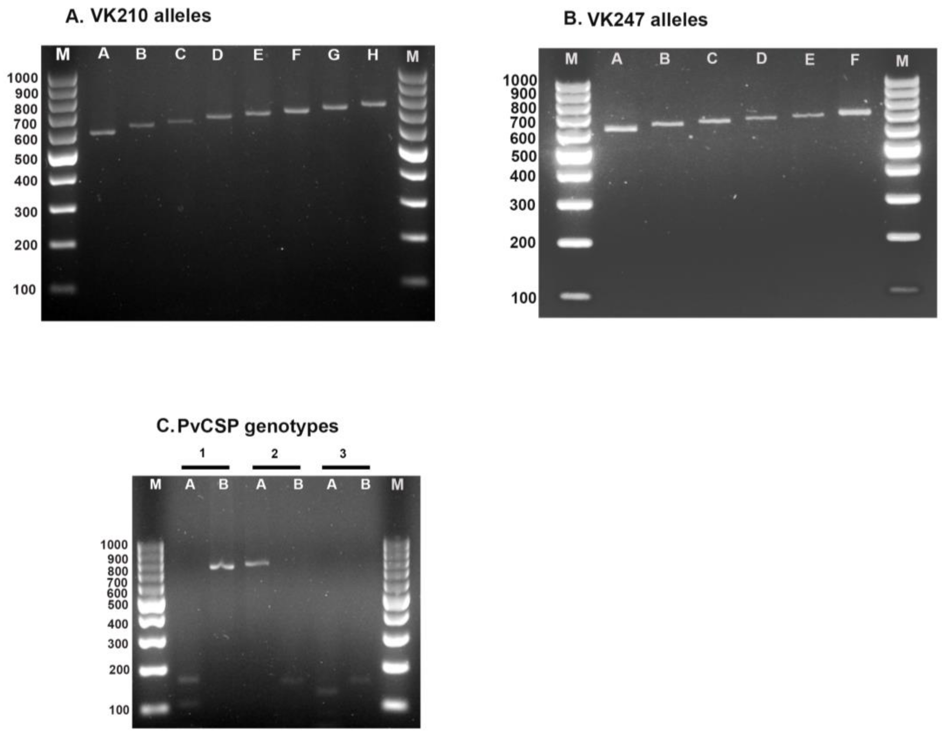
3.1. Genetic Diversity of the PvCSP

The PvCSP gene was successfully genotyped in 417 of 440 P. vivax samples (94.8%). Of these, 315 of 330 (95.5%) and 102 of 110 (92.7%) isolates were from the first and second study periods, respectively. The RFLP of the PvCSP genotypes is shown in Figure 1C. When the two sample collection periods for PvCSP were compared, statistically significant differences were observed (p = 0.012). The results showed that the VK210 type was the most common (94.0% and 87.3% in the first and second sampling periods, respectively; Table 1). Interestingly, the frequency of the VK247 type increased during the second sampling period. Mixed infections with both genotypes, VK210 and VK247, were detected in only two isolates from Sai Yok. According to the size variation in the PCR bands, 14 different alleles (8 for VK210 and 6 for VK247) were found. The variations were arranged in an increasing pattern of 20 base pairs (bp). VK210 had a PCR band size range of 620–800 bp, and VK247 had a range of 620–740 bp (Figure 1A,B).

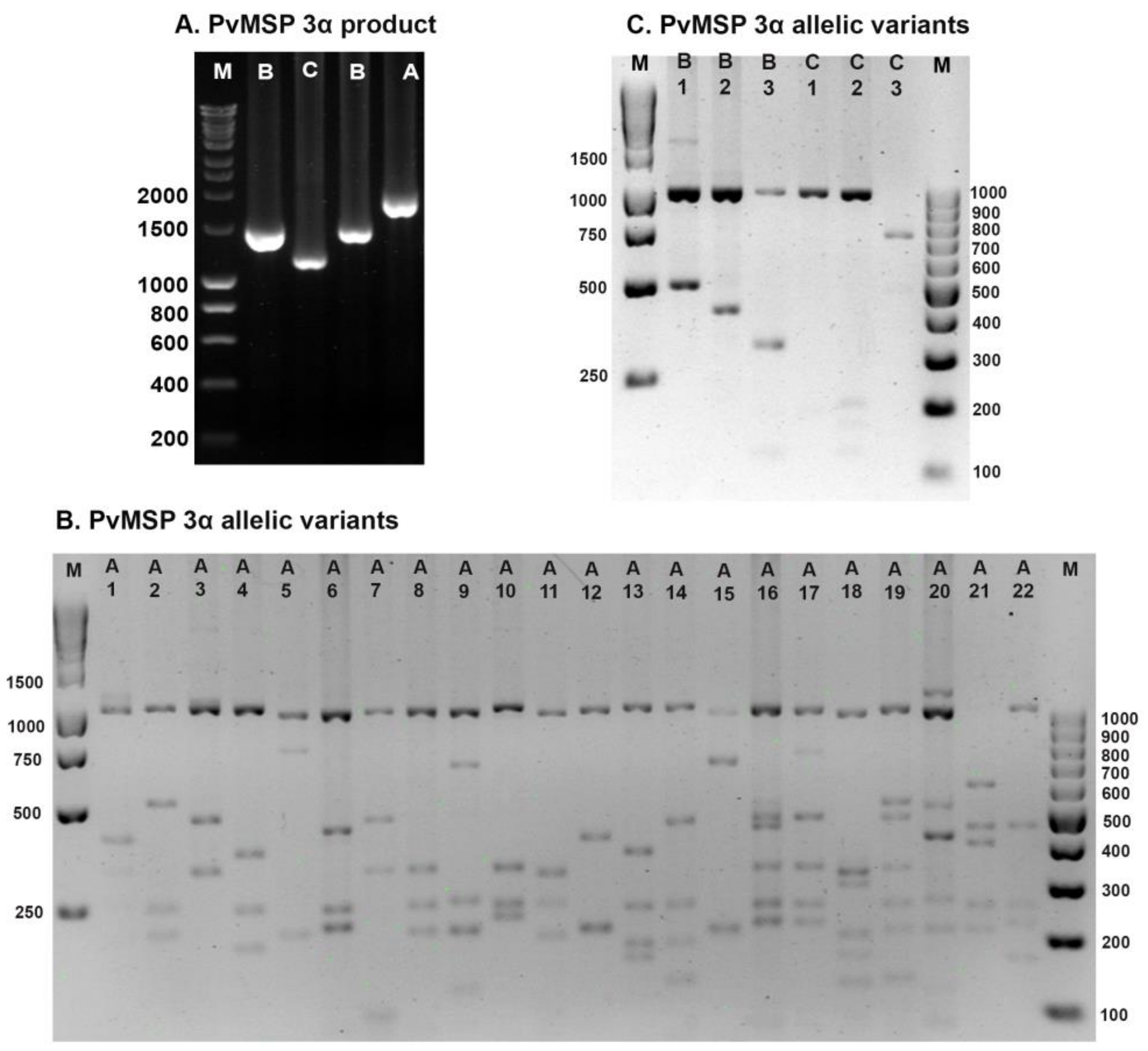
3.2. Genetic Diversity of the PvMSP-3α

The PvMSP-3α was successfully genotyped in 373 (84.8%) of 440 samples. Of these, 277 of 330 (83.9%) isolates were from the first period, and 96 of 110 (87.3%) isolates were from the second period. Based on PCR band size variations for the PvMSP-3α gene, three distinct types, namely, types A (1.9 kb), B (1.5 kb), and C (1.1 kb) (Figure 2A) [18], were detected in the isolates collected during the first period, whereas types A and B were found in the isolates collected during the second period (Table 2). After Hha I digestion, most isolates showed a conserved 1000 bp band with varying patterns of small amplicons (Figure 2B,C). On the basis of these results, 21 allelic variants of type A and 3 allelic variants each of type B and type C were identified in the first sample collection period. Allele A8 was the most common allele in Sai Yok isolates (17.9%), whereas allele C1 was the most prevalent allele in Mae Sot isolates (14.4%). From all the samples analyzed for the first period, multiple-genotype infections were detected in 11 (4.0%) isolates when two different PCR bands were found in a single sample (5 isolates) or when the restriction analysis of a single PCR product exceeded the uncut PCR band size (6 isolates). The isolates from the second period contained 12 allelic variants of type A and 2 allelic variants of type B. Allele A11 was the most abundant allele, accounting for 32.3% of the samples. No mixed genotypes were detected during the second period. The overall differences in allele frequency between the two periods were statistically significant (p < 0.0001). There were significant differences between mixed genotype (p = 0.002) and B (p < 0.001). In addition, Mae Sot isolates from the two different time periods showed a statistically significant difference (p = 0.009).

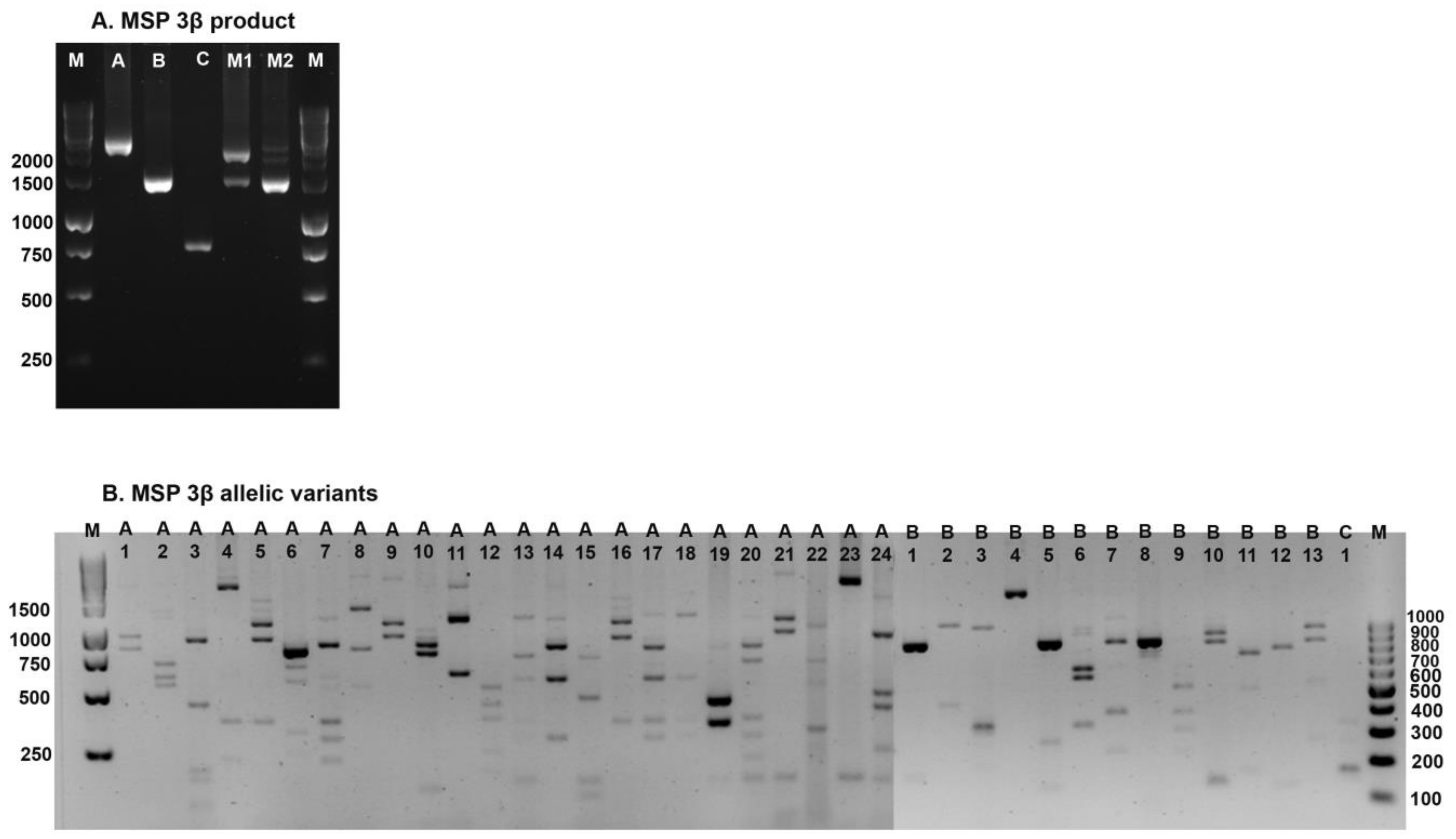
3.3. Genetic Diversity of the PvMSP-3β

The PvMSP-3β was successfully genotyped in 328 (74.5%) of 440 samples. Of these, 248 of 330 (75.2%) isolates were from the first period, and 80 of 110 (72.7%) isolates were from the second period. Based on PCR band size variations, three distinct types were identified in the first period isolates: type A (1.7–2.3 kb), type B (1.4–1.6 kb), and type C (0.780 kb) (Figure 3A) [19]. Only types A and B were observed in the second period. The majority of samples from the first period (63.4%) belonged to type A, whereas in the second period, types A and B were equally prevalent (Table 3). Different banding patterns were observed when PCR products were digested with Pst I enzyme (Figure 3B). The isolates from the second period contained 22 allelic variants of type A, 13 allelic variants of type B, and one allelic variant of type C. Allele A1 (~19.0%) was the predominant allele in Mae Sot and Sai Yok isolates. Different allelic patterns were observed in the two endemic areas during the first period; Sai Yok isolates showed higher genetic diversity than Mae Sot isolates (29 vs. 22 alleles). The isolates from the second period were classified into types A and B with 9 and 10 allelic variants, respectively. Allele B4 was the most prevalent allele, accounting for 27.5% of the isolates. Multiple-genotype infections of the first and second periods were observed in 43 (17.3%) and 11 (13.8%) isolates, respectively. This occurred when more than one PCR product of a different size was found in a single sample (15 and 1 isolates, respectively), or when the summed size of the DNA fragments resulting from the restriction digestion exceeded the size of the PCR product (43 and 11 isolates, respectively). There was no statistically significant difference in the frequency of the PvMSP-3β alleles between the two periods (p = 0.88).

4. Discussion

PvCSP is the main sporozoite surface protein. It is expressed during the pre-erythrocyte phase and is important for sporozoites to move and invade human hepatocytes. This protein is highly immunogenic and is considered to be a prime vaccine candidate [14]. The three PvCSP genotypes are globally distributed, with VK210 being the most common [16]. In this study, VK210 was the most prevalent variant during both study periods (94.0% in the first period and 87.3% in the second period). Our findings showed that these two variants of this gene changed over time. In 2014–2016, 12.7% of the isolates had the VK247 type, up from 5.4% in 2006–2007. A study conducted in Thailand [22] showed that the frequency of the VK247 variant increased over time. This increase may be caused by the varying susceptibilities of mosquito vectors to the VK247 type, host immune selection, or sampling bias [23]. In P. vivax malaria-endemic countries, VK210 and VK247 have been found at different frequencies. Isolates collected from the Thai–Myanmar border revealed a dominance of parasites with the VK210 genotype in all the sample sets analyzed [22], a finding noted in other studies in Thailand (93.4%) [24], Pakistan (85.5%) [25], and Guyana (92.0%) [26]. In contrast to previous studies [27,28], a lower frequency of the VK247 genotype was observed in the isolates under this study. PvCSP allelic dominance appears to be influenced by variations in parasite transmission, vectorial competence, immune responses, and treatment responses in different endemic areas [23,29,30]. Therefore, these factors should be considered when developing a CSP vaccine. Based on size polymorphisms, 14 allelic variants of PvCSP were found, which is consistent with the 17 variants previously observed on the Thai–Myanmar border [22]. However, a study conducted in India reported three variants [31]. Variations in PvCP allelic types may be explained by the influence of the induced immune response on a particular allelic variant that infects a person [32].
PvMSP-3 is a multigene family located on chromosome 10. Among this family, PvMSP-3α and PvMSP-3β have become genetic markers in P. vivax epidemiological studies around the world and are being considered as potential vaccine targets [17]. In this study, three distinct PCR-based types (A, B, and C) were observed. This result is consistent with the allelic variants observed in western and southern Thailand [4], the China–Myanmar border [33], and India [34], and a different band size designated as type D was reported in Pakistan [35]. In Hainan province, China, only types A and B were found [36]. Type A is the most prevalent at each sampling site in the current study. However, it is slightly lower than that found in a previous study of the border areas between Thailand and neighboring countries, which found that type A was 83.3–89.9% more prevalent than the other types. Only 1.8% of the isolates with mixed genotypes were identified using PCR analysis. This proportion is lower than that observed in a recent study carried out in 2014–2016 in diverse geographic areas of Thailand [18]. RFLP of PvMSP-3α revealed 28 allelic variants in the first period, of which 25 were found in isolates from Sai Yok, and 23 were found in isolates from Mae Sot. Moreover, the number of allelic variants between Mae Sot isolates in the two time periods varied considerably (14 alleles in 2014–2016 compared with 23 alleles in 2006–2007). This might be due to the differences in sample sizes between the periods. In contrast to our study, fewer allelic variants were reported in Thailand (12 alleles in 2003 and 14 alleles in 2011) [10,32]. A recent study carried out in Thailand showed 40 PvMSP-3α allelic variants [18]. Other endemic areas, such as India [37] and Myanmar [38], have also reported similar observations of high diversity. The PvMSP-3α genetic diversity in isolates collected in 2006–2007 and 2014–2016 was 10.1% and 14.6%, respectively. An increase in genetic diversity over time was also reported in a study conducted in Thailand [18]. The observed changes in allelic diversity over time can be attributed to the presence of different mosquito species that can transmit specific parasite types in a given area at different times or seasons [39]. Comparing the patterns of allelic variants, at least seven alleles were found to be the same as those found in earlier studies [18,40] conducted in Thailand. This suggests that similar PvMSP-3α alleles are distributed among the parasites in the country.
For the PvMSP-3β gene, three major types (A, B, and C) and 16 isolates of multiple-genotype infections were found. The three types were also found in northwest Thailand and along the Thai–Myanmar border [18,19]. In contrast, a study conducted in southern Thailand found only types A and B [40]. Our study showed that type A was more prevalent in the earlier samples collected from Sai Yok and Mae Sot, whereas in the isolates from Mae Sot in 2014–2016, types A and B were equally prevalent. A report from the Thai border areas [18] showed a higher frequency of type A. In western Thailand, type B was present in 60.4% of the isolates [32]. From all the samples analyzed for PvMSP-3β, only one the type C harboring isolate was found in Mae Sot. This type has been previously reported in Mae Sot isolates [19]. Based on PCR detection, mixed infection in PvMSP-3β was found in 15 samples (6%) in the first period, which is three times higher than that in southern Thailand [40]. However, 19.1% of mixed infections were found on the Thai–Myanmar border [18]. The PCR-RFLP of Pst I showed that 36 alleles of 248 isolates (14.5%) and 20 alleles of 80 isolates (25%) were identified in the first and second periods of our study, respectively. Similar to PvMSP-3α, isolates from the second period showed increased genetic diversity for PvMSP-3β. Our analysis indicated a difference in the PvMSP-3β allelic patterns between Sai Yok (29 alleles) and Mae Sot (22 alleles). The different structures of the parasite populations at these two sites could be due to sampling bias or dissimilar epidemiological settings. Some of our study’s allelic patterns were similar to those found in earlier studies [18,40], indicating that certain parasite genotypes are distributed across many parts of the country. Frequent population movement may contribute to the distribution of various parasite genotypes across the country. Restriction analysis of PvMSP-3β revealed 43 (18.5%) and 10 (12.5%) multiple-genotype infections in the first and second periods, respectively. Relapse and early gametocytogenesis, two biological characteristics of the P. vivax parasite, might be the cause of the infection’s high multiplicity [41].

5. Conclusions

This study found a relatively high degree of genetic polymorphism in PvCSP and PvMSP-3, with low rates of multiple-genotype infections. PvMSP-3 exhibited an increase in allelic variants during the second period; however, PvMSP-3α showed a significant difference in allelic variant frequency between the two periods. The proportion of the two variants of the PvCSP genotype (VK210 and VK247) changed over time, with an increase in the proportion of VK247 genotypes observed during the second sampling period. The results of this study will help in the understanding of the genetic structure of P. vivax and the control of the malarial parasite at the Thai–Myanmar border.

Author Contributions

All authors actively participated in this study. Conceptualization, K.N.-B. and W.C.; methodology, W.C. and A.A.J.; validation, W.C. and A.A.J.; sample collection, K.N.-B. and W.C.; data analysis, W.C. and K.N.-B.; original draft preparation, A.A.J.; manuscript finalization, K.N.-B., funding acquisition, K.N.-B. All authors have read and agreed to the published version of the manuscript.

Funding

This study was funded by Thammasat University (Chulabhorn International College of Medicine, Center of Excellence in Pharmacology and Molecular Biology of Malaria and Cholangiocarcinoma) and the National Research Council of Thailand. Kesara Na-Bangchang is supported by the National Research Council of Thailand under a Research Team Promotion grant (grant number NRCT 820/2563).

Institutional Review Board Statement

The study protocol was approved by the Ethics Committee of Thammasat University (No. 082/2560).

Informed Consent Statement

All participants provided written informed consent for participation in the study.

Data Availability Statement

Data will be made available upon request.

Acknowledgments

We are thankful to the staff of the malaria clinics or hospitals in Sai Yok (Kanchanaburi province) and Mae Sot (Tak province) for their kind assistance during sample collection. We thank Ethan Vindvamara for English editing of the manuscript.

Conflicts of Interest

The authors declare no conflict of interest. The funders had no role in the study design, collection, analysis, interpretation of the data, writing of the manuscript, or decision to publish the results.

References

  1. Greenwood, B.M.; Fidock, D.A.; Kyle, D.E.; Kappe, S.H.; Alonso, P.L.; Collins, F.H.; Duffy, P.E. Malaria: Progress, perils, and prospects for eradication. J. Clin. Investig. 2008, 118, 1266–1276. [Google Scholar] [CrossRef] [PubMed] [Green Version]
  2. Howes, R.E.; Battle, K.E.; Mendis, K.N.; Smith, D.L.; Cibulskis, R.E.; Baird, J.K.; Hay, S.I. Global Epidemiology of Plasmodium vivax. Am. J. Trop. Med. Hyg. 2016, 95, 15. [Google Scholar] [CrossRef] [PubMed] [Green Version]
  3. World Health Organization. World Malaria Report 2020: 20 Years of Global Progress and Challenges; World Health Organization: Geneva, Switzerland, 2020. [Google Scholar]
  4. Suphakhonchuwong, N.; Chaijaroenkul, W.; Rungsihirunrat, K.; Na-Bangchang, K.; Kuesap, J. Evaluation of Plasmodium vivax isolates in Thailand using polymorphic markers Plasmodium merozoite Surface Protein (PvMSP) 1 and PvMSP3. Parasitol. Res. 2018, 117, 3965–3978. [Google Scholar] [CrossRef] [PubMed]
  5. Nadeem, M.F.; Khattak, A.A.; Zeeshan, N.; Zahid, H.; Awan, U.A.; Yaqoob, A.; Ashraf, N.M.; Gul, S.; Alam, S.; Ahmed, W. Surveillance of molecular markers of antimalarial drug resistance in Plasmodium falciparum and Plasmodium vivax in Federally Administered Tribal Area (FATA), Pakistan. Rev. Inst. Med. Trop. Sao Paulo. 2021, 63. [Google Scholar] [CrossRef]
  6. Crompton, P.D.; Pierce, S.K.; Miller, L.H. Advances and challenges in malaria vaccine development. J. Clin. Investig. 2010, 120, 4168–4178. [Google Scholar] [CrossRef]
  7. Barry, A.E.; Arnott, A. Strategies for designing and monitoring malaria vaccines targeting diverse antigens. Front. Immunol. 2014, 5, 359. [Google Scholar] [CrossRef] [Green Version]
  8. Thakur, A.; Alam, M.T.; Sharma, Y.D. Genetic diversity in the C-terminal 42 kDa region of merozoite surface protein-1 of Plasmodium vivax (PvMSP-142) among Indian isolates. Acta Trop. 2008, 108, 58–63. [Google Scholar] [CrossRef]
  9. Mzilahowa, T.; McCall, P.J.; Hastings, I.M. “Sexual” population structure and genetics of the malaria agent P. falciparum. PLoS ONE 2007, 2, e613. [Google Scholar] [CrossRef] [Green Version]
  10. Rungsihirunrat, K.; Chaijaroenkul, W.; Siripoon, N.; Seugorn, A.; Na-Bangchang, K. Genotyping of polymorphic marker (MSP3α and MSP3β) genes of Plasmodium vivax field isolates from malaria endemic of Thailand. Trop. Med. Int. Health 2011, 16, 794–801. [Google Scholar] [CrossRef]
  11. Bahk, Y.Y.; Kim, J.; Ahn, S.K.; Na, B.K.; Chai, J.Y.; Kim, T.S. Genetic Diversity of Plasmodium vivax Causing Epidemic Malaria in the Republic of Korea. Korean J. Parasitol. 2018, 56, 545–552. [Google Scholar] [CrossRef]
  12. Zakeri, S.; Barjesteh, H.; Djadid, N.D. Merozoite surface protein-3α is a reliable marker for population genetic analysis of Plasmodium vivax. Malar. J. 2006, 5, 1–6. [Google Scholar] [CrossRef] [Green Version]
  13. Huang, B.; Huang, S.; Su, X.Z.; Guo, H.; Xu, Y.; Xu, F.; Hu, X.; Yang, Y.; Wang, S.; Lu, F. Genetic diversity of Plasmodium vivax population in Anhui province of China. Malar. J. 2014, 13, 1–11. [Google Scholar] [CrossRef] [Green Version]
  14. López, C.; Yepes-Pérez, Y.; Hincapié-Escobar, N.; Díaz-Arévalo, D.; Patarroyo, M.A. What is known about the immune response induced by Plasmodium vivax malaria vaccine candidates? Front. Immunol. 2017, 8, 126. [Google Scholar] [CrossRef] [Green Version]
  15. Qari, S.H.; Shi, V.-P.; Povoa, M.M.; Alpers, M.P.; Deloron, P.; Murphy, G.S.; Harjosuwarno, S.; Lal, A.A. Global occurrence of Plasmodium vivax-like human malaria parasite. J. Infect. Dis. 1993, 168, 1485–1489. [Google Scholar] [CrossRef]
  16. Kaur, H.; Sehgal, R.; Kumar, A.; Sehgal, A.; Bharti, P.K.; Bansal, D.; Mohapatra, P.K.; Mahanta, J.; Sultan, A.A. Exploration of genetic diversity of Plasmodium vivax circumsporozoite protein (Pvcsp) and Plasmodium vivax sexual stage antigen (Pvs25) among North Indian isolates. Malar. J. 2019, 18, 1–8. [Google Scholar] [CrossRef]
  17. Jiang, J.; Barnwell, J.W.; Meyer, E.V.; Galinski, M.R. Plasmodium vivax merozoite surface protein-3 (PvMSP3): Expression of an 11 member multigene family in blood-stage parasites. PLoS ONE 2013, 8, e63888. [Google Scholar] [CrossRef] [Green Version]
  18. Kuesap, J.; Rungsihirunrat, K.; Chaijaroenkul, W.; Mungthin, M. Genetic diversity of Plasmodium vivax merozoite surface protein-3 alpha and beta from diverse geographic areas of Thailand. Jpn. J. Infect. Dis. 2022, 75, 241–248. [Google Scholar] [CrossRef]
  19. Yang, Z.; Miao, J.; Huang, Y.; Li, X.; Putaporntip, C.; Jongwutiwes, S.; Gao, Q.; Udomsangpetch, R.; Sattabongkot, J.; Cui, L. Genetic structures of geographically distinct Plasmodium vivax populations assessed by PCR/RFLP analysis of the merozoite surface protein 3β gene. Acta Trop. 2006, 100, 205–212. [Google Scholar] [CrossRef] [Green Version]
  20. Bruce, M.C.; Galinski, M.R.; Barnwell, J.W.; Snounou, G.; Day, K.P. Polymorphism at the merozoite surface protein-3alpha locus of Plasmodium vivax: Global and local diversity. Am. J. Trop. Med. Hyg. 1999, 61, 518–525. [Google Scholar] [CrossRef] [Green Version]
  21. Imwong, M.; Pukrittayakamee, S.; Grüner, A.C.; Rénia, L.; Letourneur, F.; Looareesuwan, S.; White, N.J.; Snounou, G. Practical PCR genotyping protocols for Plasmodium vivax using Pvcs and Pvmsp1. Malar. J. 2005, 4, 1–13. [Google Scholar] [CrossRef]
  22. Maneerattanasak, S.; Gosi, P.; Krudsood, S.; Tongshoob, J.; Lanteri, C.A.; Snounou, G.; Khusmith, S. Genetic diversity among Plasmodium vivax isolates along the Thai–Myanmar border of Thailand. Malar. J. 2016, 15, 1–8. [Google Scholar] [CrossRef] [PubMed] [Green Version]
  23. Gonzalez-Ceron, L.; Rodriguez, M.H.; Nettel, J.C.; Villarreal, C.; Kain, K.C.; Hernandez, J.E. Differential susceptibilities of Anopheles albimanus and Anopheles pseudopunctipennis to infections with coindigenous Plasmodium vivax variants VK210 and VK247 in southern Mexico. Infect. Immun. 1999, 67, 410–412. [Google Scholar] [CrossRef] [PubMed] [Green Version]
  24. Kosaisavee, V.; Hastings, I.; Craig, A.; Lek-Uthai, U. The genetic polymorphism of Plasmodium vivax genes in endemic regions of Thailand. Asian Pac. J. Trop. Med. 2011, 4, 931–936. [Google Scholar] [CrossRef] [Green Version]
  25. Raza, A.; Ghanchi, N.K.; Thaver, A.M.; Jafri, S.; Beg, M.A. Genetic diversity of Plasmodium vivax clinical isolates from southern Pakistan using pvcsp and pvmsp1 genetic markers. Malar. J. 2013, 12, 1–5. [Google Scholar] [CrossRef] [PubMed] [Green Version]
  26. Bonilla, J.A.; Validum, L.; Cummings, R.; Palmer, C.J. Genetic diversity of Plasmodium vivax pvcsp and pvmsp1 in Guyana, South America. Am. J. Trop. Med. Hyg. 2006, 75, 830–835. [Google Scholar] [CrossRef]
  27. González-Cerón, L.; Martinez-Barnetche, J.; Montero-Solís, C.; Santillán, F.; Soto, A.M.; Rodríguez, M.H.; Espinosa, B.J.; Chávez, O.A. Molecular epidemiology of Plasmodium vivax in Latin America: Polymorphism and evolutionary relationships of the circumsporozoite gene. Malar. J. 2013, 12, 1–19. [Google Scholar] [CrossRef] [Green Version]
  28. Chenet, S.M.; Tapia, L.L.; Escalante, A.A.; Durand, S.; Lucas, C.; Bacon, D.J. Genetic diversity and population structure of genes encoding vaccine candidate antigens of Plasmodium vivax. Malar. J. 2012, 11, 1–11. [Google Scholar] [CrossRef] [Green Version]
  29. Machado, R.L.; Póvoa, M.M. Distribution of Plasmodium vivax variants (VK210, VK247 and P. vivax-like) in three endemic areas of the Amazon region of Brazil and their correlation with chloroquine treatment. Trans. R. Soc. Trop. Med. Hyg. 2000, 94, 377–381. [Google Scholar] [CrossRef]
  30. Da Silva, A.N.; Santos, C.C.; Lacerda, R.N.; Machado, R.L.; Póvoa, M.M. Susceptibility of Anopheles aquasalis and An. darlingi to Plasmodium vivax VK210 and VK247. Mem. Inst. Oswaldo Cruz. 2006, 101, 547–550. [Google Scholar] [CrossRef]
  31. Kim, J.-R.; Imwong, M.; Nandy, A.; Chotivanich, K.; Nontprasert, A.; Tonomsing, N.; Maji, A.; Addy, M.; Day, N.P.; White, N.J. Genetic diversity of Plasmodium vivax in Kolkata, India. Malar. J. 2006, 5, 1–10. [Google Scholar] [CrossRef] [Green Version]
  32. Cui, L.; Mascorro, C.N.; Fan, Q.; Rzomp, K.A.; Khuntirat, B.; Zhou, G.; Chen, H.; Yan, G.; Sattabongkot, J. Genetic diversity and multiple infections of Plasmodium vivax malaria in Western Thailand. Am. J. Trop. Med. Hyg. 2003, 68, 613–619. [Google Scholar] [CrossRef] [Green Version]
  33. Li, X.; Bai, Y.; Wu, Y.; Zeng, W.; Xiang, Z.; Zhao, H.; Zhao, W.; Chen, X.; Duan, M.; Wang, X. PvMSP-3α and PvMSP-3β genotyping reveals higher genetic diversity in Plasmodium vivax parasites from migrant workers than residents at the China-Myanmar border. Infect. Genet. Evol. 2022, 106, 105387. [Google Scholar] [CrossRef]
  34. Kaul, A.; Bali, P.; Anwar, S.; Sharma, A.K.; Gupta, B.K.; Singh, O.P.; Adak, T.; Sohail, M. Genetic diversity and allelic variation in MSP3α gene of paired clinical Plasmodium vivax isolates from Delhi, India. J. Infect. Public Health. 2019, 12, 576–584. [Google Scholar] [CrossRef]
  35. Khan, S.N.; Khan, A.; Khan, S.; Ayaz, S.; Attaullah, S.; Khan, J.; Khan, M.A.; Ali, I.; Shah, A.H. PCR/RFLP-based analysis of genetically distinct Plasmodium vivax population of Pvmsp-3α and Pvmsp-3β genes in Pakistan. Malar. J. 2014, 13, 1–7. [Google Scholar] [CrossRef] [Green Version]
  36. Li, Y.-C.; Wang, G.-Z.; Meng, F.; Zeng, W.; He, C.-h.; Hu, X.-M.; Wang, S.-Q. Genetic diversity of Plasmodium vivax population before elimination of malaria in Hainan Province, China. Malar. J. 2015, 14, 1–12. [Google Scholar] [CrossRef] [Green Version]
  37. Verma, A.; Joshi, H.; Singh, V.; Anvikar, A.; Valecha, N. Plasmodium vivax msp-3α polymorphisms: Analysis in the Indian subcontinent. Malar. J. 2016, 15, 1–13. [Google Scholar] [CrossRef] [Green Version]
  38. Moon, S.-U.; Lee, H.-W.; Kim, J.-Y.; Na, B.-K.; Cho, S.-H.; Lin, K.; Sohn, W.-M.; Kim, T.-S. High frequency of genetic diversity of Plasmodium vivax field isolates in Myanmar. Acta Trop. 2009, 109, 30–36. [Google Scholar] [CrossRef]
  39. Zakeri, S.; Raeisi, A.; Afsharpad, M.; Kakar, Q.; Ghasemi, F.; Atta, H.; Zamani, G.; Memon, M.S.; Salehi, M.; Djadid, N.D. Molecular characterization of Plasmodium vivax clinical isolates in Pakistan and Iran using pvmsp-1, pvmsp-3α and pvcsp genes as molecular markers. Parasitol. Int. 2010, 59, 15–21. [Google Scholar] [CrossRef]
  40. Thanapongpichat, S.; Khammanee, T.; Sawangjaroen, N.; Buncherd, H.; Tun, A.W. Genetic diversity of Plasmodium vivax in clinical isolates from Southern Thailand using PvMSP1, PvMSP3 (PvMSP3α, PvMSP3β) genes and eight microsatellite markers. Korean J. Parasitol. 2019, 57, 469. [Google Scholar] [CrossRef] [Green Version]
  41. Kibria, M.G.; Elahi, R.; Mohon, A.N.; Khan, W.A.; Haque, R.; Alam, M.S. Genetic diversity of Plasmodium vivax in clinical isolates from Bangladesh. Malar. J. 2015, 14, 1–9. [Google Scholar] [CrossRef] [Green Version]
Figure 1. PCR product size variations and genotypes of PvCSP. Gel images depict: (A) PCR product size variations for VK210; (B) PCR product size variations for VK247; and (C) PvCSP products digested with AluI (A) and BstNI (B) enzymes. Sample No. 1: A cut the product into small fragments, but B did not cut the product (VK210 genotype). Sample No. 2: A did not cut the product into small fragments, but B cut the product (VK247 genotype). Sample No. 3: A and B cut the products into small fragments (mixed genotype). M: Molecular marker (100 bp).
Tropicalmed 08 00210 g001
Figure 2. PCR product size variations and allelic variants of PvMSP-3α. Gel images depict: (A) PCR product size variations; (B) allelic variants for type A; and (C) allelic variants for types B and C. M: Molecular markers (100 bp and 1000 bp).
Tropicalmed 08 00210 g002
Figure 3. PCR product size variations and allelic variants of PvMSP-3β. Gel images depict: (A) PCR product size variations, and (B) allelic variants for types A, B, and C. M: Molecular markers (100 bp and 1000 bp). M1 and M2: Mixed genotypes.
Tropicalmed 08 00210 g003
Table 1. The proportion of PvCSP from the first (2006–2007) and second (2014–2016) periods.
First Period 1Second Period
Sai YokMae SotTotalMae Sot
N %N %N %N %
VK210160 (92.5)136 (95.8)296 (94.0)89 (87.3)
VK24711 (6.4)6 (4.2)17 (5.4)13 (12.7)
Mixed2 (1.2)0 (0.0)2 (0.6)0 (0.0)
Total173 (100.0)142 (100.0)315 (100.0)102 (100.0)
1 Significant difference between first and second period (p = 0.012).
Table 2. The proportion of PvMSP-3α from the first (2006–2007) and second (2014–2016) periods.
First Period 1Second Period
Sai YokMae Sot 3Mae Sot
N%N% N%
Type A (77.3 %)13678.67875Type A (78.1%)7578.1
A184.632.9A344.2
A210.600.0A411.0
A3158.743.8A533.1
A452.921.9A711.0
A584.687.7A81212.5
A652.900.0A1022.1
A710.611.0A113132.3
A83117.91110.6A1255.2
A974.087.7A131212.5
A1021.200.0A1811.0
A1110.61110.6A1911.0
A12116.498.7A2222.1
A1342.321.9Type B (21.9%)2121.9
A1421.221.9B277.3
A1500.032.9B31414.6
A1610.632.9Total96100.0
A17 *21.221.9Pattern (14, 14.6%)14
A182715.676.7
A19 *42.311.0
A2010.600.0
A2100.011.0
Type B (7.9 %)137.698.7
B163.554.8
B263.521.9
B310.621.9
Type C (13%)1911.01716.3
C1179.81514.4
C221.211.0
C300.011.0
M 2 (1.8%)52.90.00.0
Total173100.0104100.0
Pattern (28, 10.1 %)25 23
M: Mixed alleles. * M: Mixed genotype infection after RFLP analysis. 1 The overall statistically significant differences in allele frequencies between the two periods (p < 0.001). 2 Significant difference between first and second period with type M (p = 0.002) and type B (p < 0.001). 3 Significant differences between the Mae Sot isolates from the two time periods (p = 0.009).
Table 3. The proportion of PvMSP-3β from the first (2006–2007) and second (2014–2016) periods.
First PeriodSecond Period
Sai YokMae SotMae Sot
N%N% N%
Type A (63.4%)10263.05566.3Type A (51.2%)4151.2
A13219.41619.3A11417.5
A221.200.0A322.5
A300.044.8A411.3
A431.811.2A7 *810.0
A5 *31.800.0A1022.5
A6 *00.011.2A1122.5
A7 *116.778.4A1211.3
A8 *5300.0A141012.5
A900.011.2A2311.3
A1010.622.4Type B (47.5%)3847.5
A112313.9910.8B122.5
A13 *21.200.0B211.3
A1410.622.4B322.5
A1521.211.2B42227.5
A1642.478.4B522.5
A1731.811.2B611.3
A18 *21.211.2B7 *22.5
A1910.600.0B811.3
A20 *10.600.0B922.5
A21 *31.800.0B1233.8
A22 *31.800.0Type M (1.3%)11.3
A2400.022.4Total80100.0
Type B (30.2%)5230.32327.7Pattern (20, 25%20
B121.233.6
B221.200.0
B342.411.2
B42716.41416.9
B531.800.0
B6 *10.600.0
B7 *21.200.0
B810.600.0
B953.022.4
B10 *00.022.4
B1100.011.2
B1242.400.0
B13 *10.600.0
Type C (0.4 %)00.001.2
C100.011.2
Type M (6 %)116.744.8
Total165100.083100.0
Pattern (36, 14.5%)29 22
M: Mixed alleles. * Mixed genotype infection after RFLP analysis.
Disclaimer/Publisher’s Note: The statements, opinions and data contained in all publications are solely those of the individual author(s) and contributor(s) and not of MDPI and/or the editor(s). MDPI and/or the editor(s) disclaim responsibility for any injury to people or property resulting from any ideas, methods, instructions or products referred to in the content.

