Article

Safety of Oral Carica papaya L. Leaf 10% Ethanolic Extract for Acute and Chronic Toxicity Tests in Sprague Dawley Rats

by
Weerakit Taychaworaditsakul
1,2,
Chalermpong Saenjum
3,
Nongkran Lumjuan
4,
Kriangkrai Chawansuntati
4,
Suphunwadee Sawong
1,2,
Kanjana Jaijoy
5,
Mingkwan Na Takuathung
1,2 and
Seewaboon Sireeratawong
1,2,*
1
Clinical Research Center for Food and Herbal Product Trials and Development (CR-FAH), Faculty of Medicine, Chiang Mai University, Chiang Mai 50200, Thailand
2
Department of Pharmacology, Faculty of Medicine, Chiang Mai University, Chiang Mai 50200, Thailand
3
Department of Pharmaceutical Science, Faculty of Pharmacy, Chiang Mai University, Chiang Mai 50200, Thailand
4
Research Institute for Health Sciences, Chiang Mai University, Chiang Mai 50200, Thailand
5
McCormick Faculty of Nursing, Payap University, Chiang Mai 50000, Thailand
*
Author to whom correspondence should be addressed.
Toxics 2024, 12(3), 198; https://doi.org/10.3390/toxics12030198
Submission received: 27 December 2023 / Revised: 5 February 2024 / Accepted: 28 February 2024 / Published: 1 March 2024
(This article belongs to the Special Issue Toxics Young Investigators Contributions Collection in 2023)

Abstract

:
Carica papaya L. leaves, traditionally utilized in dietary supplements and pharmaceuticals, exhibit a broad spectrum of potentially therapeutic properties, including anti-inflammatory, antimalarial, and wound healing properties. This study examined the acute and chronic toxicity of 10% ethanolic-extracted C. papaya leaf in Sprague Dawley rats. The acute toxicity assessment was a single oral dose of 5000 mg/kg body weight, while the chronic toxicity assessment included daily oral doses of 100, 400, 1000, and 5000 mg/kg over 180 days. Systematic monitoring covered a range of physiological and behavioral parameters, including body and organ weights. End-point evaluations encompassed hematological and biochemical analyses, along with gross and histopathological examinations of internal organs. Findings revealed no acute toxicity in the C. papaya leaf extract group, although a significant decrease in uterine weight was observed without accompanying histopathology abnormalities. In the chronic toxicity assessment, no statistically significant differences between the control and the C. papaya leaf extract groups were detected across multiple measures, including behavioral, physiological, and hematological indices. Importantly, histopathological examination corroborated the absence of any tissue abnormalities. The study results indicate that C. papaya leaf extract exhibited no adverse effects on the rats during the 180-day oral administration period, affirming its potential safety for prolonged usage.

1. Introduction

The Carica papaya L. plant, belonging to the Caricaseae family, is indigenous to Central America and the southern regions of Mexico, but has been widely cultivated in many Asian countries, from the Philippines to Pakistan. This plant is recognized globally for its extensive therapeutic applications which have been delineated in a variety of scientific investigations [1]. The nutritional value of the fruit of C. papaya, known as papaya, is well known globally, yet only minimal attention has been accorded to its leaves, seeds, and roots, which also offer medicinal properties. Previous comprehensive phytochemical investigation has demonstrated that C. papaya leaf extract is rich in bioactive components such as flavonoids, alkaloids, saponins, phytosterols, phenolics, and tannins, highlighting its prospective pharmaceutical utility [2,3,4,5]. Although existing research has predominantly addressed the extract’s effectiveness in treating dengue fever [4], it has also been shown to function as an anti-inflammatory agent, selectively modulating cytokine production [6,7]. Additionally, empirical evidence underscores its multifaceted therapeutic benefits, including anti-bacterial [8], anti-malarial [9], gastroprotective [10], hypoglycemic [11], hypolipidemic [12], anti-oxidant [13], hematological [14], anti-tumor activities [3] and wound-healing capacities [15]. Various extraction methods, ranging from aqueous to ethanol-based processes, have been optimized to facilitate its application across an array of medical conditions [5,16,17]. The existing body of research accentuates the leaves’ prospective utility as a formalized herbal medicine product, indicating not only its potential for disease prevention and health promotion, but also its significant economic value.
Ensuring the safety and proper formulation of medicinal herbs is as critical as establishing their efficacy. As outlined in the World Health Organization (WHO) 2019 Global Report on Traditional and Complementary Medicine, herbal products often undergo stringent safety evaluations similar to those mandated for conventional pharmaceuticals, including post-marketing surveillance. Regulatory standards for traditional herbal medicine frequently incorporate contemporary scientific research on comparable market products [18]. In line with the Principles of Good Laboratory Practice (GLP) of the Organization for Economic Co-operation and Development (OECD), government regulatory agencies often mandate both general and specific animal toxicity studies of varying durations before issuing an herbal medicine registration [18]. Despite the demonstrated effectiveness of C. papaya extracts, questions persist concerning their long-term safety. Acute in vivo toxicity studies have reported that rats administered freeze-dried aqueous extract of C. papaya leaf at dosages ranging from 5 to 2000 mg/kg body weight (BW) exhibit no discernable adverse effects [19]. Short-term studies (up to thirteen weeks) involving daily doses of 2000 mg/kg have reported neither morbidity nor mortality in rats. Data on long-term health implications for chronic users, however, remain conspicuously absent [20].
Toxicological evaluations serve as a cornerstone in pharmacological research and pharmaceutical development, helping to establish a compound’s safety profile before its clinical application in humans [21,22]. Given this imperative, chronic toxicity studies complement acute evaluations to provide a comprehensive assessment of an herbal medicine’s long-term physiological, biochemical, hematological, and pathological impact. This study aims to assess both the acute and chronic toxicity profiles of 10% ethanolic-extracted C. papaya leaf, utilizing Sprague Dawley rats as the experimental model.

2. Materials and Methods

2.1. Chemicals and Reagents

Absolute ethanol, acetic acid, acetonitrile, gallic acid, rutin, quercetin, rosmarinic acid, luteolin, apigenin, and kaempferol were purchased from Sigma-Aldrich Chemical Company (St. Louis, MO, USA). All other chemicals were of analytical grade.

2.2. Plant Material

This study employed naturally cultivated C. papaya (Holland papaya) from Mae On District, Chiang Mai, Thailand. Leaves of C. papaya were harvested from 3- to 4-month-old plants between January and July 2021. The fresh leaves served as the source material for this investigation. Expert botanists authenticated the plant specimens using taxonomic photographs and reference samples deposited in the Plant Museum of the Faculty of Pharmacy at Chiang Mai University (voucher specimen number 0023307). Subsequently, the extracted C. papaya leaves were analyzed using Thai Herbal Pharmacopoeia methods [23], supervised by Associate Professor Dr. Chalermpong Saenjum from the Faculty of Pharmacy at Chiang Mai University.

2.3. Extract Preparation

The leaves of C. papaya were initially cleaned and depurated of their central stems and large branches, followed by triple rinsing with distilled water. The cleansed leaves were then subjected to hot air oven drying at 50 °C for 12 h. Subsequently, the dried material was pulverized using an 80-mesh sieve grinder (Nanotech NT-1000D; Thanapan Ltd., Chiang Mai, Thailand) to achieve a homogenous particle size of less than 177 microns, maximizing the surface area for optimal extraction efficiency. This processed leaf powder was securely stored in a vacuum-sealed bag protected from light and stored at -20 °C, and dry environment until further use. For the extraction process, 8 kg of C. papaya leaf powder were prepared in 60 L of 10% ethanol in water, using high-speed extraction (EURO Best Technology Co., Ltd., Bangkok, Thailand) at 150 rpm and a temperature of 80–90 °C for 3 h. The obtained solution was filtered and then centrifuged to separate the residue, after which extracted twice with 10% ethanol. The separated solution was then weighed, adjusted to a total of 150 kg using 10% v/v ethanol in water solution. The concentration of maltodextrin was 0.5% w/v and homogenized at 5000 rpm. Then, the obtained solution was spray dry (SDE-50 EURO Best Technology Co., Ltd., Bangkok, Thailand), with an inlet temperature of 160 °C, and the flow rate was set at 500 mL/min to yield a granular, water-soluble powder with a 2.81% extraction efficiency.

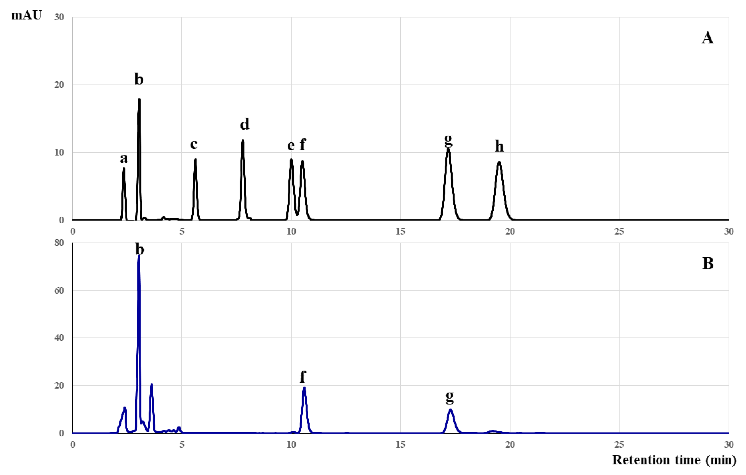
2.4. Phenolic and Flavonoid Content Identification Via High-Performance Liquid Chromatography (HPLC)

The C. papaya leaf extract was subjected to HPLC for quantification and the identification of various phenolic and flavonoid compounds, including, but not limited to, gallic acid, rutin, quercetin, rosmarinic acid, luteolin, apigenin, and kaempferol, under specific analytical conditions following Phromnoi et al. [24]. In brief, chromatographic separation was achieved using a Symmetry-Shield® RP18 column (250 mm × 4.6 mm ID (internal diameter)) supplied by Waters Corporation (Waters Co., Ltd., Milford, MA, USA). The mobile phases were formulated from 0.1% acetic acid in acetonitrile and deionized water mixed in a 30:70 ratio, under isocratic conditions at a flow rate of 1.0 mL/min. Each 10 μL sample was introduced into the column and monitored through a diode array detector at wavelengths of 278 and 325 nm. The amounts of each detected compound in the C. papaya extract were calculated and expressed as mg/g extract.

2.5. The Animal Subjects and Ethical Considerations

This extract was preserved in opaque, airtight containers under cool and dry conditions. Sprague Dawley rats of both sexes, weighing between 180 and 200 g, were employed in this study. The animals were bred by the National Laboratory Animal Center at Mahidol University, Nakhon Pathom, Thailand, and subsequently transferred to the laboratory facilities for acclimatization. Ambient conditions were controlled at a temperature of 25 ± 1 °C and a relative humidity of 60%, with a 12 h light-dark cycle. The rats were provided with ad libitum access to food and water. To facilitate animal welfare, each rat was allowed a minimum acclimatization period of one week prior to the start of the experiment. This study was conducted in compliance with ethical guidelines and received approval from the Research Ethics Committee for Animal Studies of the Faculty of Medicine at Chiang Mai University, Thailand (approval code: 7/2564).

2.6. Hippocratic Assessment

Building on established methods used in previous research [25], this study employed Hippocratic screening to evaluate the safety profile of C. papaya leaf extract. Specifically, two female rats were orally administered a single dose of 2000 mg/kg and observed individually in open fields at 5, 10, 15, 30, 60, 120, and 240 min after administration and 24 h a day for clinical signs and mortality, as measured by motor activity (observing animal movements using infrared (I.R.) beams in a motor activity cage), respiration rate (observing the breathing of an animal in one minute), righting reflex (considering the behavior of an animal that turns and flips over), and screen grip (considering the ability of an animal to cling onto a cage using its forelimbs and hindlimbs). The assessment aimed to identify any potential adverse reactions, e.g., sedation, emesis, muscular spasms, and watery diarrhea.

2.7. Acute Toxicity Evaluation of C. papaya Leaf Extract

In accordance with WHO and OECD Test Guideline 420 [26,27], the rats were randomly assigned to either a treatment group (n = 5) which was administered a single oral dose of 5000 mg/kg of C. papaya leaf extract, or to a control group (n = 5), receiving 2 mL/kg of distilled water by oral gavage. Acute toxicity indicators, e.g., lethargy, emesis, muscle spasms, and watery diarrhea, were initially monitored for a six-hour period and then subsequently on a daily basis for a 14-day period. Additionally, body weight was recorded on days 7 and 14 as were any mortality incidents. At the conclusion of the observation period, the rats were euthanized using intraperitoneal thiopental sodium injection at a dose of 120 mg/kg, after which the animals underwent a physical examination by us checking their vital signs, pulse, and reflexes to confirm death and a comprehensive gross examination of internal organs, including but not limited to the lungs, heart, liver, and kidneys, was performed. All harvested organs were subsequently preserved in 10% formaldehyde for further analysis.

2.8. Assessment of Chronic Toxicity of C. papaya Leaf Extract

The chronic toxicity evaluation was performed in accordance with WHO and OECD Test Guideline 452 [27,28]. A total of 100 rats, half male and half female, were divided into seven groups. Group 1 (n = 10), which served as the control group, received daily doses of 2 mL/kg of distilled water for 14 days. Group 2 (n = 10), designated as the satellite control group, also received 2 mL/kg of distilled water daily for the same period and was observed for an additional 28 days post-treatment (n = 5). Groups 3 to 6 (n = 10 per each) were the treatment groups and were administered doses of 100, 400, 1000, and 5000 mg/kg, respectively, of C. papaya leaf extracts for 180 days. Group 7 (n = 10) functioned as a satellite control, receiving 5000 mg/kg of C. papaya leaf extract daily for 14 days and was observed for an additional 28 days post-treatment (n = 5). All doses were administered by oral gavage.
During the observation periods, behavioral patterns, clinical indicators, and body weights were systematically monitored and documented. Any deceased animals were immediately subjected to a necropsy. On day 180, hematological and biochemical parameters were analyzed from collected blood samples, after which all rats were euthanized with a 120 mg/kg intraperitoneal thiopental sodium injection, and their vital signs, pulse, and reflexes were checked to confirm death and underwent comprehensive macroscopic and microscopic evaluation of a range of internal organs, including the lungs, heart, liver, pancreas, kidneys, stomach, intestines, spleen, adrenal glands, ovaries, uterus, testes, eyes, brain, muscles, and nerves.

2.9. Statistical Analysis Methodology

The results are presented as mean ± S.E.M. For evaluating the data obtained from the acute toxicity study, either a t-test or the Mann–Whitney U test was employed, as appropriate. For chronic toxicity tests, the data underwent an initial analysis using the Shapiro–Wilk test to assess normality. In cases where the Shapiro–Wilk test showed no significant deviation from a normal distribution, an analysis of variance (ANOVA) was conducted, followed by Tukey’s multiple comparison tests. Conversely, if the Shapiro–Wilk test indicated other than a normal distribution, the data were subjected to analysis using the Kruskal–Wallis nonparametric ANOVA test, followed by Dunn’s test. Statistical significance was set at p-values less than 0.05. The statistical analyses were performed using IBM SPSS Statistics, version 22.0 (International Business Machines Corporation, Armonk, NY, USA).

3. Results

3.1. Quality Control and Standard Specification for C. papaya Leaf Extract

Table 1 presents the physical characteristics of desiccated C. papaya leaves, analyzed using randomly selected samples. An analysis of the six parameters presented in Table 1, including acid-insoluble ash, loss on drying, and total ash, provides a description of the qualities and characteristics of the raw materials used for the preparation of the extracts for this experiment.
Remarkably, the C. papaya leaf extract met all regulatory requirements set forth by the Food and Drug Administration (FDA) for food additives. Specifically, the extract was within permissible limits for heavy metals including arsenic, mercury, lead, and cadmium. Additionally, the extract was determined to be free from contamination by key pathogenic microorganisms, including E. coli, S. aureus, Clostridium spp., and Salmonella spp., as shown in Table 2. Additionally, comprehensive pesticide screening revealed no detectable residues of any of the 20 organochlorines, 8 organophosphates, 6 pyrethroids, and 9 carbamates tested, as shown in Table S1. These stringent quality control standards affirm the extract’s safety and suitability for therapeutic application as well as demonstrating the value of further investigation.
In this study, the phenolic and flavonoid constituents of C. papaya leaf extract were comprehensively characterized using HPLC, coupled with ultraviolet (UV) and mass spectrometry detectors. As shown in Figure 1 and Table 3, the extract contained key bioactive compounds, including rutin, quercetin, gallic acid, catechin, apigenin, and kaempferol.

3.2. Preliminary Toxicity Assessment of C. papaya Extract in Female Rats

In accordance with the Hippocratic screening protocol, a preliminary evaluation was conducted to assess the potential toxicity of oral administration of C. papaya leaf extract at a dosage of 2000 mg/kg in female rats. Observations were carried out over a 24 h period and are detailed in Table 4. No adverse behavioral changes or fatalities were observed within this timeframe. Additionally, gross examination of the visceral organs and overall carcass characteristics yield no detachable abnormalities.

3.3. Acute Toxicity Assessment of C. papaya Leaf Extract in Female Rats

An acute evaluation was performed utilizing a single oral dose of 5000 mg/kg of C. papaya leaf extract administered to female rats. During the initial 24 h observation period, no behavioral aberrations were detected in any of the experimental groups or in the control groups. No fatalities were recorded in any of the groups during the study. Table 5 presents body weight measurements on days 7 and 14, revealing no statistically significant divergence from the control group (p < 0.05).
No mortality ensued following administration of a single dose of 5000 mg/kg of the extract, and normal behavioral patterns remained consistent. No pathological changes were noted across the range of physiological parameters, including ocular, dermal, fur, mucosal, respiratory, and circulatory assessments, as well as the autonomic and central nervous systems. At day 14, both body and internal organ weights of the experimental group remained statistically comparable to the control group (Table 6). Notably, although a significant decrease in uterine weight was observed, gross and histopathological examinations confirmed the absence of any discernible abnormalities in uterine size, color or texture (Figure 2).

3.4. Assessment of Chronic Toxicity Induced by C. papaya Leaf Extract

3.4.1. Variation in Body and Organ Weight

Table 7 and Table 8 present longitudinal body weight metrics for female and male rats subjected to a six-month regimen of C. papaya leaf extract administration. On day 90, a rigorous statistical analysis unveiled a notable and statistically significant decrease in the body weight of female rats in the low-dose group (100 mg/kg) as compared to the control group. Similarly, on day 30, a significant reduction in the body weight of male rats was observed in the high-dose satellite group (5000 mg/kg) in comparison to the control group. Conversely, Table 9 and Table 10 depict that there were no discernible changes in the internal organ weights of either female or male rats.

3.4.2. Hematological and Biochemical Assessments

Hematological parameters were rigorously evaluated in rats following oral administration of C. papaya leaf extract as detailed in Table 11 and Table 12. Furthermore, Table 13 and Table 14 show the results of biochemical evaluations. Notably, both sets of evaluations revealed an absence of significant deviations from control values in rats subjected to chronic exposure at doses of 400, 1000, and 5000 mg/kg body weight per day. An exception to this uniformity was a statistically significant decline in alkaline phosphatase (ALP) levels, which was observed exclusively in male rats belonging to the satellite treatment group.

3.4.3. Necropsy and Histopathological Evaluation in Chronic Toxicity Assessment

In response to the observed changes in ALP levels compared to the control group, comprehensive gross examinations were conducted to scrutinize organ appearance and characteristics. The outcomes affirm the absence of any macroscopic abnormalities. In addition, histopathological evaluations conducted on a range of vital organs of rats chronically administered C. papaya leaf extract corroborated these findings, indicating an absence of significant tissue injury (Figure 3 and Figure 4).

4. Discussion

The assessment of the safety of natural products is a critical prerequisite for obtaining regulatory approval, often necessitating a thorough evaluation of diverse scientific documents such as monographs, peer-reviewed articles, and scientific reports. International regulatory bodies like the European Food Safety Authority and the United States Food and Drug Administration place considerable emphasis on toxicological data derived from animal studies as a cornerstone for natural product safety evaluations [29,30]. While medicinal plants offer numerous health benefits, some can pose risks of adverse effects, including diseases and toxicity, necessitating rigorous scientific risk assessment investigations [31,32]. Systematic toxicity studies are pivotal for establishing safe consumption levels in humans, thereby helping to ensure the safe application of plant-derived products [33]. Although studies have explored the long-term toxicity of C. papaya leaf aqueous extracts in non-rodent animals such as rabbits [34], there remains a paucity of such investigations in rodent models. According to the ICH guideline M3(R2), a comprehensive safety profile should include toxicity studies in both rodent and non-rodent animal models [29]. While short-term studies have generally indicated a lack of toxic side effects associated with the use of C. papaya leaf extract [35,36], our study aimed to bridge the existing gap by focusing on its long-term chronic toxicity in rodents. This approach not only aligns with international guidelines but also provides a more robust scientific basis for the safety evaluation of this natural product. Nevertheless, the ethical considerations tied to animal testing in laboratories necessitate unwavering adherence to established protocols throughout the experimental procedure. Furthermore, it is imperative that both laboratory animal care and research activities are conducted within a standardized setting.
Preceding the commencement of clinical trials designed to assess the efficacy of herbal products, it is customary to initiate a preliminary phase involving acute and chronic toxicity tests in animal models. This foundational step is integral to determining optimal dosages and duration of administration, thereby establishing a solid groundwork for subsequent human trials [27]. In the current study, the dosages chosen were guided by existing human usage patterns for C. papaya leaf extract, which has previously been employed as a dietary supplement [37]. In humans, the daily dosage for this extract ranges from 1 to 50 g, which translates into a daily intake of 17 to 833 mg for an average 60 kg male [5,38,39]. To account for differences in metabolic rate among species, a body surface area exponent has been employed to standardize dosages [40]. The human dosages were then converted to rodent-appropriate dosages, resulting in the selection of a range of 103.5–5164.6 mg/kg body weight for our study. Ultimately, we opted for four dosage groups (100, 400, 1000, and 5000 mg/kg body weight) based on our research objectives. In addition, our unpublished data indicate that the range of effective doses of C. papaya leaf extract necessary to modulate platelet counts lies between 100 and 400 mg/kg body weight. For the chronic toxicity evaluation, the present study used doses of 100, 400, 1000, and 5000 mg/kg body weight, thereby encompassing both the effective and the high-dosage ranges to provide a comprehensive toxicity profile.
The quality control of herbal products is imperative for ensuring the standardization and safety of these natural remedies [41]. In accordance with the Thai Herbal Pharmacopoeia 2018, initial quality assessment methodologies for medicinal plant materials involves scrutinizing both the raw materials and the finished herbal products [23]. Good manufacturing practice (GMP) guidelines, as endorsed by the World Health Organization (WHO), advocate the utilization of high-performance liquid chromatography (HPLC) as an instrumental technique to quantify the phenolic and flavonoid content, thereby helping ensure product consistency [42]. In the present investigation, seven batches of C. papaya leaf extract all exhibited uniform concentrations of various bioactive compounds, including rutin, quercetin, gallic acid, catechin, apigenin, and kaempferol (Table S2). These findings align with existing studies, which have identified an array of phytochemicals, e.g., flavonoids and phenolic compounds, saponins, cardiac glycosides, anthraquinones, and alkaloids [43,44]. Rutin, a predominant flavonoid in C. papaya leaves, has traditionally served as a chemical marker for quality control [38,45]. In addition to rutin, gallic acid has also been identified as another phenolic acid compound that offers a reliable indicator for product standardization [46]. Importantly, several of these compounds, particularly rutin, quercetin, and gallic acid, have been reported to exhibit potent anti-oxidant properties [47]. For that reason, our study employed these bioactive constituents, specifically phenolic acids like gallic acid and flavonoids such as rutin and quercetin, as quality control markers to ensure product consistency and efficacy.
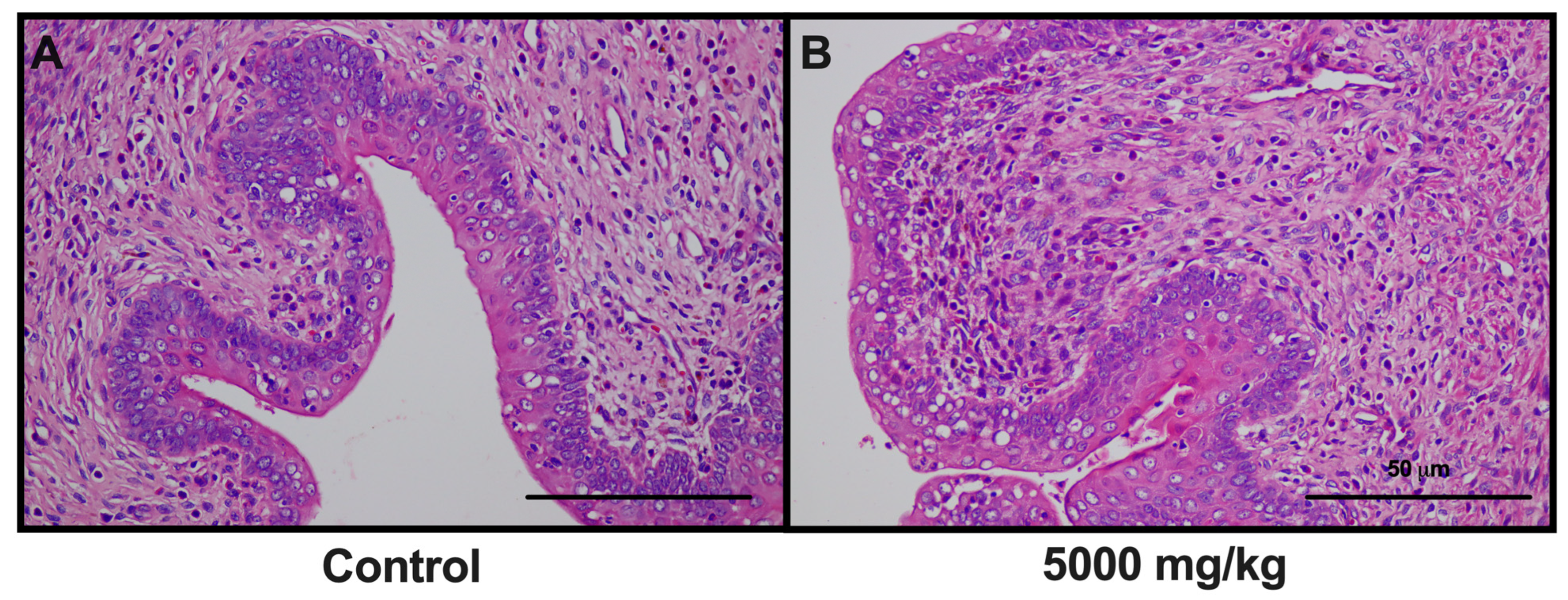
The acute oral toxicity test serves as a critical evaluative method for assessing the immediate adverse effects following a substantial single oral dose of a substance within a 24 h timeframe. In this study, an elevated dose of 5000 mg/kg of C. papaya leaf extract was administered to assess its safety profile. This dosage was selected based on prior research utilizing a 2000 mg/kg dose for Hippocratic screening, a technique employed to uncover noteworthy pharmacological activities in medicinal plants, which yielded no toxic or lethal outcomes [25]. Our findings, presented in Table 6, substantiate that this high-dose administration of 10% ethanolic C. papaya leaf extract results in neither mortality nor significant body weight changes when compared to a control group. Moreover, comprehensive histological examination of various organs—including the brain, lungs, heart, liver, kidney, spleen, gastrointestinal tract, and reproductive organs—revealed no tissue damage. It is noteworthy, however, that the uterus in the high-dose group exhibited a significant decrease in size relative to the control, corroborating previous studies that have highlighted the uterotonic effects of C. papaya leaf extract [48,49]. Importantly, no mortality was observed in the present study, corroborating the existing literature that places the LD50 for C. papaya leaf extract well above 5000 mg/kg [50,51]. Additionally, histopathological analysis revealed no abnormalities in the uterine tissue, as indicated in Figure 2. These results underscore the need for rigorous evaluation of reproductive organs in chronic toxicity studies. Furthermore, given the uterotonic effects observed, caution may be warranted when considering the use of C. papaya leaf extract during pregnancy.
Chronic toxicity studies, typically lasting from six months to two years, are designed to assess the cumulative effects of prolonged exposure to a substance, particularly those adverse effects that may not become immediately apparent. This approach aligns with WHO guidelines which recommend that the duration of substance administration in animal models should mirror the anticipated duration of clinical exposure in humans [27]. A critical parameter for identifying potential toxicity is the assessment of changes in general behavior and body weight. The existing literature suggests that a loss of 10% or more of initial body weight in test animals could be a strong indicator of adverse side effects and may compromise survival [52,53,54]. However, our study revealed significant statistical fluctuations in rat weight. A decline was noted in female rats (low dose, 100 mg/kg) on day 90, and there was a notable reduction in the body weight of male rats (high dose, 5000 mg/kg) on day 30. Importantly, these observed changes, while perceptible, did not surpass a level of concern, specifically remaining below 10% weigh loss. Thus, our findings offer compelling evidence supporting the long-term safety of C. papaya leaf extract administration in a rodent model, thereby laying a foundation for potential translational applications in humans.
Fluctuations in the weight of internal organs are also widely acknowledged as sensitive markers for assessing the effects of drug exposure on organ integrity. Such assessments are integral to toxicological studies, where the organ weights of treated and untreated animals are systematically compared [55]. In the present investigation, gross pathological examinations revealed no significant abnormalities in the internal organs of C. papaya leaf extract-treated groups, i.e., the findings were consistent with those of the control groups. In accordance with the Organization for Economic Co-operation and Development (OECD) Test Guideline 452, the data garnered from this study substantiates the non-toxic profile of C. papaya leaf extract. Nevertheless, caution is advised in interpreting these results. A more in-depth evaluation of hematological, biochemical, and histopathological parameters, particularly in male satellite rats concerning spleen and epididymis, is warranted for a comprehensive safety assessment.
Blood chemistry and hematology analyses can serve as pivotal tools for detecting potential tissue damage or physiological stress, thereby providing valuable insights into the cellular integrity of internal organs. Given the circulatory system’s crucial role in disseminating nutrients and foreign substances throughout the organism, blood serves as a sensitive barometer of both physiological and pathological conditions [56,57]. Specifically, toxic substances can compromise key blood components such as red and white blood cells, platelets, and hemoglobin [58]. In the context of chronic toxicity, our study revealed no significant aberrations in blood parameters in either male or female rats following administration of C. papaya leaf extract. In the context of chronic toxicity, our study revealed no significant aberrations in blood parameters in either male or female rats following administration of C. papaya leaf extract and all values remained within clinically acceptable ranges [59,60,61,62].
To evaluate the potential impact on renal, hepatic, and pancreatic functions, comprehensive clinical blood chemistry assessments were conducted, focusing on key markers such as BUN, creatinine, AST, ALT, ALP, total protein, albumin, bilirubin, and glucose levels. Given the high volume of blood perfusion through the kidneys, this organ is particularly vulnerable to toxic insults. Substances deemed toxic are actively filtered by the kidneys, potentially leading to an accumulation in renal tubules [63], which underscores the relevance of BUN and creatinine as sensitive indicators of renal health [64]. In cases of renal, cortical and/or glomerular injury, an elevation in serum creatinine levels is typically observed [65]. Our study yielded no discernible toxic effects on either BUN or creatinine levels, supporting the assertion that C. papaya leaf extract does not adversely affect renal function. Corroborating these biochemical findings, histological examination of the kidneys from both treated and control groups revealed no discernible morphological differences, substantiating the absence of renal toxicity even after six months of chronic exposure to C. papaya leaf extract.
Liver function tests (LFTs) serve as a valuable diagnostic tool for identifying hepatic impairments. Bilirubin, a catabolic byproduct of hemoglobin, has a known association with hepatic diseases such as jaundice and primary biliary cirrhosis. Elevated bilirubin levels serve as a proxy for the severity of hepatic dysfunction [66]. In our investigation, markers indicative of hepatic synthetic capacity—specifically, bilirubin, total protein, and albumin—demonstrated no abnormal fluctuations [67]. In addition, AST and ALT, markers traditionally used to assess hepatic toxicity, showed no significant variations. These findings suggest that C. papaya leaf extract does not exert direct hepatotoxic effects or induce hepatotoxin-mediated liver injury [68,69]. Notably, a decline in ALP levels was observed, possibly indicating an adaptive or regenerative response of the liver to long-term exposure to the extract; however, these values remained within clinically accepted norms [59,60,61,62,70]. In addition, a satellite-treated group was employed to evaluate any latent or reversible toxic effects [71,72]. All assessed parameters in each of the groups remained within established reference ranges, affirming the extract’s safety profile. Nonetheless, to substantiate these biochemical indicators and further validate the hepatic safety of C. papaya leaf extract, supplemental histopathological assessments are recommended.
Generally, serum glucose levels exceeding 200 mg/dL in rats are indicative of “hyperglycemia” [73,74]. In our study, comparable plasma glucose levels were observed between the C. papaya leaf extract-treated and control groups. This finding suggests the absence of metabolic dysfunction or adverse side effects associated with prolonged use of the extract. The stable blood glucose levels in treated rats provides additional evidence of the absence of pancreatic islet damage.
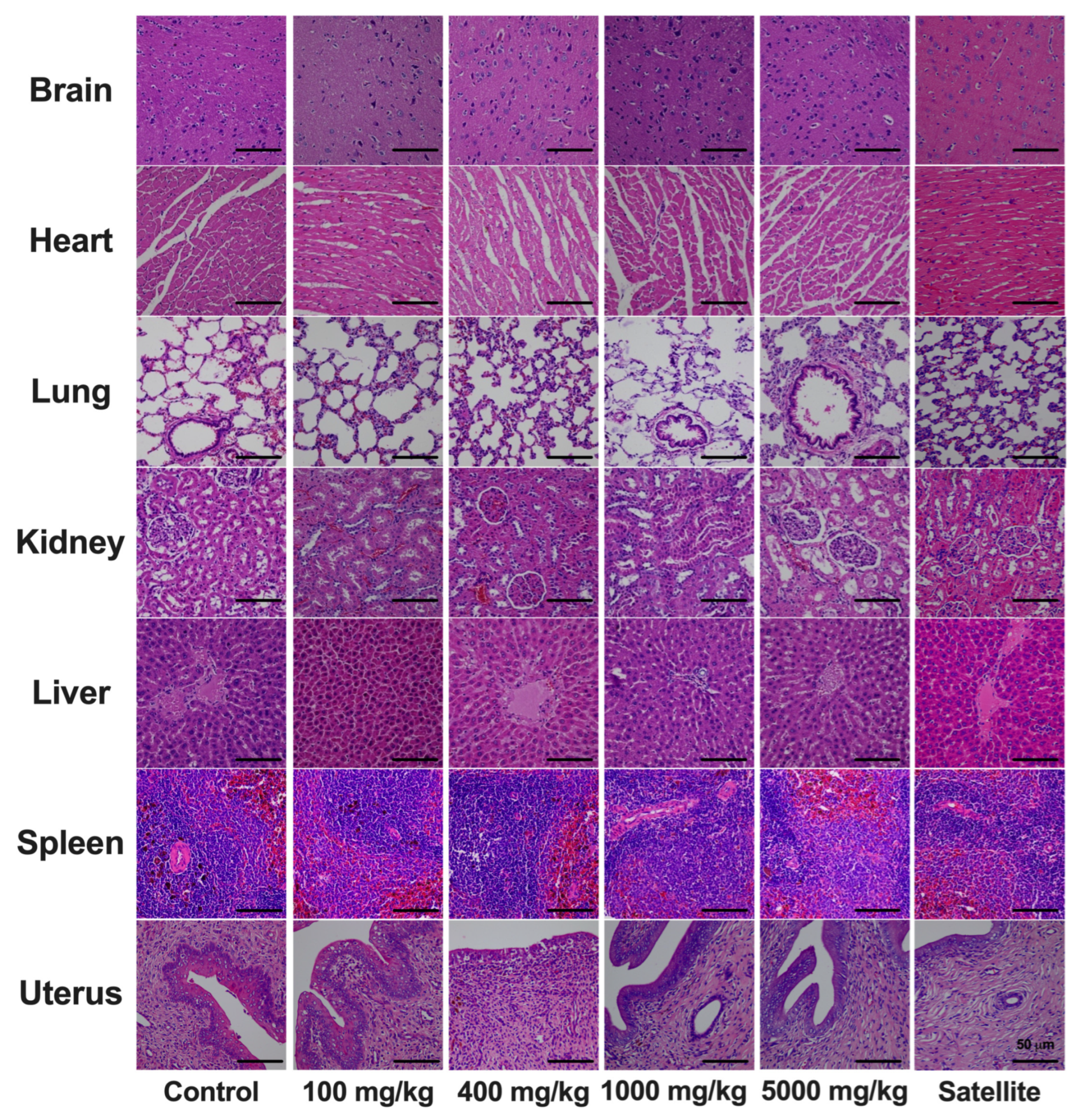
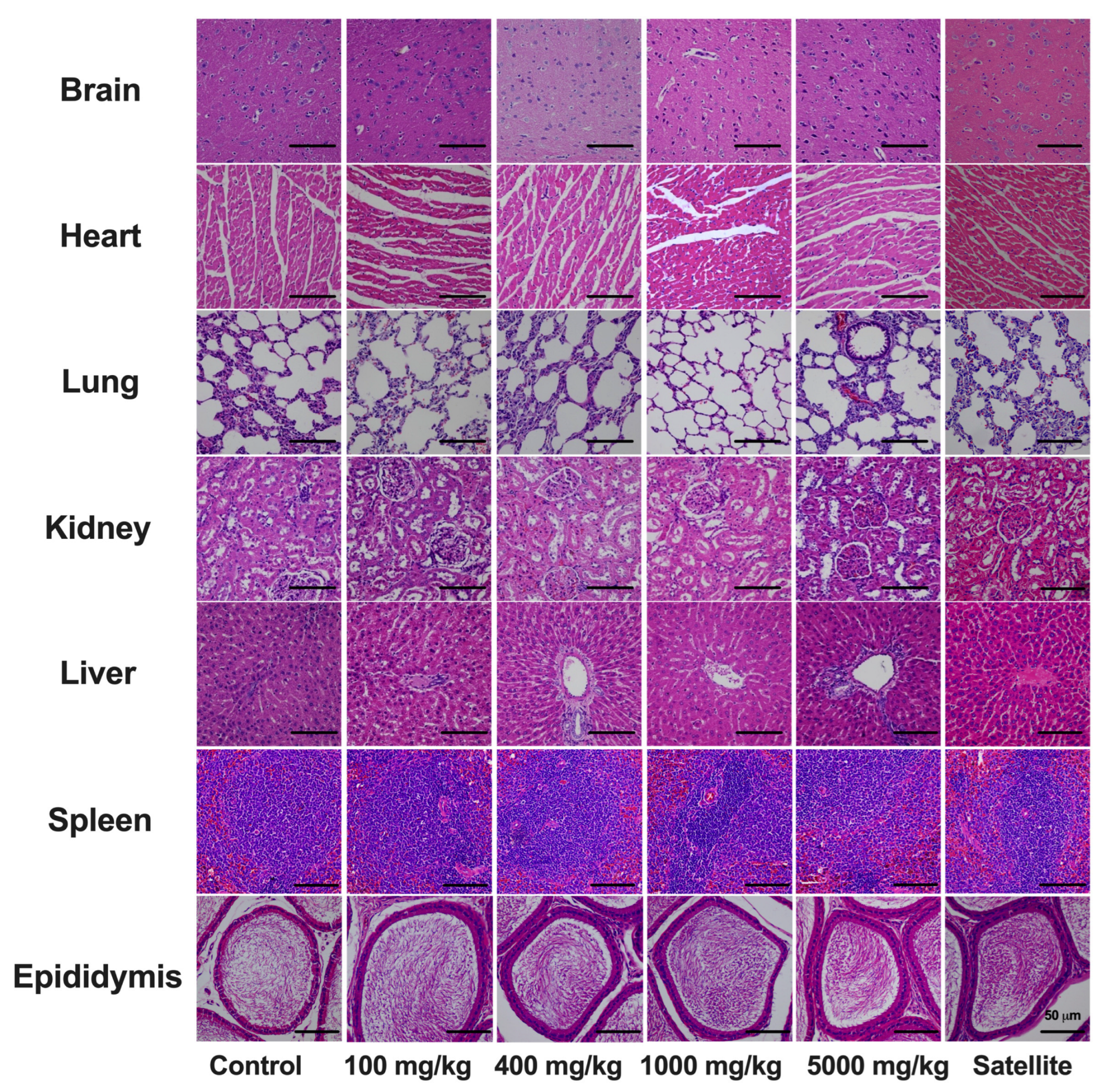
To ascertain the safety profile of C. papaya leaf extract, comprehensive histopathological analyses were conducted on the internal organs of all test animals. Initial gross examination assessed qualitative attributes such as size, coloration, and overall appearance of various organs. These evaluations were followed by hematological tests and in-depth histological analyses. Our findings revealed no signs of toxicity or histopathological abnormalities across all treatment groups, particularly in organs of critical interest such as the liver, epididymis, and spleen in the male satellite group. The highest administered dose of 5000 mg/kg in rats translates to approximately 810 mg/kg of human body weight, according to established conversion factors [75]. This chronic toxicity evaluation substantiates the safety of C. papaya leaf extract for extended use.

5. Conclusions

In summary, the present study provides substantial evidence of the safety of C. papaya leaf extract. Acute toxicity tests using a single oral dose of 5000 mg/kg in rodents revealed no immediate adverse effects. However, notable changes in female reproductive organs, specifically the uterus, warrant further investigation. On the other hand, chronic administration of C. papaya leaf extract at dosages of 100, 400, 1000, and 5000 mg/kg/day over a 180-day period did not yield any observable toxicological effects. These findings strongly suggest that C. papaya leaf extract is amenable to long-term usage without inducing toxicity.

Supplementary Materials

The following supporting information can be downloaded at: https://www.mdpi.com/article/10.3390/toxics12030198/s1, Table S1: Pesticide residue examination results; Table S2: Types and contents of phenolic and flavonoid compounds in the analysis of production in each batch of papaya leaf extract.

Author Contributions

Conceptualization, S.S. (Seewaboon Sireeratawong), N.L., M.N.T. and K.C.; methodology, W.T. and C.S.; software, W.T. and M.N.T.; validation, C.S.; investigation, W.T. and K.J.; resources, S.S. (Seewaboon Sireeratawong), N.L., M.N.T., K.C. and C.S.; writing—original draft preparation and visualization, W.T. and S.S. (Suphunwadee Sawong); writing—review and editing, S.S. (Seewaboon Sireeratawong), N.L., M.N.T., C.S., S.S. (Suphunwadee Sawong) and K.C.; supervision, S.S. (Seewaboon Sireeratawong), N.L. and M.N.T.; project administration and funding acquisition, S.S. (Seewaboon Sireeratawong), N.L. and M.N.T. All authors have read and agreed to the published version of the manuscript.

Funding

This research was funded by the Agricultural Research Development Agency of Thailand (Public Organization) or “ARDA” (Grant numbers: CRP6405030440).

Institutional Review Board Statement

The animal study protocol was approved by the Research Ethics Committee for Animal Studies of the Faculty of Medicine at Chiang Mai University, Thailand (approval code: 7/2564).

Data Availability Statement

Data is available upon request.

Acknowledgments

This research work was partially supported by Chiang Mai University. The authors would like to extend their sincere appreciation to Krit Suksawangwat and Naritsara Sukjit for their invaluable contributions to the rat oral administration procedures. Special thanks are also due to G. Lamar Robert, for his meticulous editing of the manuscript to ensure linguistic accuracy and clarity.

Conflicts of Interest

The authors declare no conflicts of interest.

References

  1. Aravind, G.; Bhowmik, D.; Duraivel, S.; Harish, G. Traditional and medicinal uses of Carica papaya. J. Med. Plant Res. 2013, 1, 7–15. [Google Scholar]
  2. Sagnia, B.; Fedeli, D.; Casetti, R.; Montesano, C.; Falcioni, G.; Colizzi, V. Antioxidant and anti-inflammatory activities of extracts from Cassia alata, Eleusine indica, Eremomastax speciosa, Carica papaya and Polyscias fulva medicinal plants collected in Cameroon. PLoS ONE 2014, 9, e103999. [Google Scholar] [CrossRef]
  3. Otsuki, N.; Dang, N.H.; Kumagai, E.; Kondo, A.; Iwata, S.; Morimoto, C. Aqueous extract of Carica papaya leaves exhibits anti-tumor activity and immunomodulatory effects. J. Ethnopharmacol. 2010, 127, 760–767. [Google Scholar] [CrossRef]
  4. Teh, B.P.; Ahmad, N.B.; Mohamad, S.B.; Tan, T.Y.C.; Mohd Abd Razak, M.R.B.; Afzan, A.B.; Syed Mohamed, A.F.B. Carica papaya Leaf Juice for Dengue: A Scoping Review. Nutrients 2022, 14, 1584. [Google Scholar] [CrossRef]
  5. Lim, X.Y.; Chan, J.S.W.; Japri, N.; Lee, J.C.; Tan, T.Y.C. Carica papaya L. Leaf: A Systematic Scoping Review on Biological Safety and Herb-Drug Interactions. Evid. Based Complement. Alternat. Med. 2021, 2021, 5511221. [Google Scholar] [CrossRef]
  6. Gammulle, A.; Ratnasooriya, W.; Jayakody, J.; Fernando, C.; Kanatiwela, C.; Udagama, P.V. Thrombocytosis and anti-inflammatory properties, and toxicological evaluation of Carica papaya mature leaf concentrate in a murine model. Online Int. J. Med. Plants Res. 2012, 21–30. [Google Scholar]
  7. Owoyele, B.V.; Adebukola, O.M.; Funmilayo, A.A.; Soladoye, A.O. Anti-inflammatory activities of ethanolic extract of Carica papaya leaves. Inflammopharmacology 2008, 16, 168–173. [Google Scholar] [CrossRef] [PubMed]
  8. Baskaran, C.; Velu, S.; Kumaran, K. The efficacy of Carica papaya leaf extract on some bacterial and a fungal strain by well diffusion method. Asian Pac. J. Trop. Dis. 2012, 2, S658–S662. [Google Scholar] [CrossRef]
  9. Teng, W.-C.; Chan, W.; Suwanarusk, R.; Ong, A.; Ho, H.-K.; Russell, B.; Rénia, L.; Koh, H.-L. In vitro antimalarial evaluations and cytotoxicity investigations of Carica papaya leaves and carpaine. Nat. Prod. Commun. 2019, 14, 1934578X1901400110. [Google Scholar] [CrossRef]
  10. Indran, M.; Mahmood, A.; Kuppusamy, U. Protective effect of Carica papaya L leaf extract against alcohol induced acute gastric damage and blood oxidative stress in rats. West. Indian. Med. J. 2008, 57, 323–326. [Google Scholar] [PubMed]
  11. Sobia, K.; Javaid, M.; Ahmad, M.S.; Rehmatullah, Q.; Hina, G.; Iram, B.; Pervaiz, A.; Farhana, B.; Nyla, J.; Gulfraz, M. Assessments of phytochemicals and hypoglycemic activity of leaves extracts of Carica papaya in diabetic mice. Int. J. Pharm. Sci. Rev. Res. 2016, 7, 3658. [Google Scholar]
  12. Sheneni, V.; Shaibu, I.; Okpe, J.; Omada, A. In-vivo biological effect of Carica papaya leaf extracts on P-407 induced hyperlipidemic Wistar rats. MOJ Food Process Technol. 2018, 6, 409–412. [Google Scholar] [CrossRef]
  13. Nariya, A.; Jhala, D. Pharmacognostic study of Carica papaya leaf extract as inhibitors of reactive oxygen species. Int. Res. J. Pharm. 2017, 8, 13–17. [Google Scholar] [CrossRef]
  14. Sathyapalan, D.T.; Padmanabhan, A.; Moni, M.; P-Prabhu, B.; Prasanna, P.; Balachandran, S.; Trikkur, S.P.; Jose, S.; Edathadathil, F.; Anilkumar, J.O.; et al. Efficacy & safety of Carica papaya leaf extract (CPLE) in severe thrombocytopenia (≤30,000/μL) in adult dengue-Results of a pilot study. PLoS ONE 2020, 15, e0228699. [Google Scholar] [CrossRef]
  15. Mahmood, A.; Sidik, K.; Salmah, I. Wound healing activity of Carica papaya L. aqueous leaf extract in rats. Int. J. Mol. Med. 2005, 1, 398–401. [Google Scholar]
  16. Patil, T.; Patil, S.; Patil, A.; Patil, S. Carica papaya leaf extracts—An Ethnomedicinal boon. Int. J. Pharmacogn. Phytochem. Res. 2014, 6, 260–265. [Google Scholar]
  17. Longdet, I.; Adoga, E. Effect of methanolic leaf extract of Carica papaya on plasmodium berghei infection in albino mice. Eur. J. Med. Plants 2017, 20, 1–7. [Google Scholar] [CrossRef]
  18. World Health Organization (WHO). WHO Global Report on Traditional and Complementary Medicine 2019; World Health Organization: Geneva, Switzerland, 2019.
  19. Halim, S.; Abdullah, N.; Afzan, A.; Rashid, B.; Jantan, I.; Ismail, Z. Acute toxicity study of Carica papaya leaf extract in Sprague Dawley rats. J. Med. Plant Res. 2011, 5, 1867–1872. [Google Scholar]
  20. Ismail, Z.; Halim, S.Z.; Abdullah, N.R.; Afzan, A.; Abdul Rashid, B.A.; Jantan, I. Safety Evaluation of Oral Toxicity of Carica papaya Linn. Leaves: A Subchronic Toxicity Study in Sprague Dawley Rats. Evid. Based Complement. Alternat. Med. 2014, 2014, 741470. [Google Scholar] [CrossRef] [PubMed]
  21. Grieshaber, C.K.; Marsoni, S. Relation of preclinical toxicology to findings in early clinical trials. Cancer Treat. Rep. 1986, 70, 65–72. [Google Scholar]
  22. Gad, S.C. Strategies for the Application and Integration of in Vitro Methods to the Development of New Pharmaceuticals and Medical Devices. Toxicol. Methods 1996, 6, 1–12. [Google Scholar] [CrossRef]
  23. Department of Medical Sciences, Ministry of Public Health. Thai Herbal Pharmacopoeia Volume IV; Department of Medical Sciences: Bangkok, Thailand, 2014. [Google Scholar]
  24. Phromnoi, K.; Yodkeeree, S.; Pintha, K.; Mapoung, S.; Suttajit, M.; Saenjum, C.; Dejkriengkraikul, P. Anti-Osteoporosis Effect of Perilla frutescens Leaf Hexane Fraction through Regulating Osteoclast and Osteoblast Differentiation. Molecules 2022, 27, 824. [Google Scholar] [CrossRef]
  25. Wade, A.E. Experiments in the Pharmaceutical Biological Sciences; Malone, M.H., McLaughlin, J.L., Eds.; Biological Sciences Section of the Conference of Teachers of the American Association of Colleges of Pharmacy: Silver Spring, MD, USA, 1973; pp. 120–123. [Google Scholar]
  26. Organisation for Economic Co-operation and Development (OECD). Test No. 420: Acute Oral Toxicity-Fixed Dose Procedure; OECD Publishing: Paris, France, 2002. [Google Scholar]
  27. World Health Organization (WHO). General Guidelines for Methodologies on Research and Evaluation of Traditional Medicine; World Health Organization: Geneva, Switzerland, 2000.
  28. Organisation for Economic Co-operation and Development (OECD). Test No. 452: Chronic Toxicity Studies; OECD Publishing: Paris, France, 2009. [Google Scholar]
  29. European Medicines Agency (EMEA). ICH Topic M3 (R2) Non-Clinical Safety Studies for the Conduct of Human Clinical Trials and Marketing Authorization for Pharmaceuticals; CPMP/ICH/286/95; European Medicines Agency: London, UK, 2009.
  30. Reeve, L.M. Non-clinical Post-Marketing Commitments for newly licenced pharmaceuticals. Regul. Toxicol. Pharmacol. 2009, 55, 181–187. [Google Scholar] [CrossRef] [PubMed]
  31. Subramanian, K.; Sankaramourthy, D.; Gunasekaran, M. Chapter 18-Toxicity Studies Related to Medicinal Plants. In Natural Products and Drug Discovery; Mandal, S.C., Mandal, V., Konishi, T., Eds.; Elsevier: Amsterdam, The Netherlands, 2018; pp. 491–505. [Google Scholar]
  32. Jităreanu, A.; Trifan, A.; Vieriu, M.; Caba, I.-C.; Mârțu, I.; Agoroaei, L. Current Trends in Toxicity Assessment of Herbal Medicines: A Narrative Review. Processes 2023, 11, 83. [Google Scholar] [CrossRef]
  33. Neergheen-Bhujun, V.S. Underestimating the toxicological challenges associated with the use of herbal medicinal products in developing countries. Biomed. Res. Int. 2013, 2013, 804086. [Google Scholar] [CrossRef] [PubMed]
  34. Omonkhua, A.A.; Onoagbe, I.O. Long-term hepatotoxicity and hypoglycaemic study of aqueous extracts of Carica papaya leaves on normal rabbits. Glob. J. Pure Appl. Math. 2011, 17, 241–247. [Google Scholar]
  35. Akinloye, O.O.; Morayo, O.M. Evaluation of andrological indices and testicular histology following chronic administration of aqueous extract of Carica papaya leaf in Wistar rat. Afr. J. Pharmacy. Pharmacol. 2010, 4, 252–255. [Google Scholar]
  36. Afzan, A.; Abdullah, N.R.; Halim, S.Z.; Rashid, B.A.; Semail, R.H.; Abdullah, N.; Jantan, I.; Muhammad, H.; Ismail, Z. Repeated dose 28-days oral toxicity study of Carica papaya L. leaf extract in Sprague Dawley rats. Molecules 2012, 17, 4326–4342. [Google Scholar] [CrossRef] [PubMed]
  37. Rahmat, L.T.; Damon, L.E. The Use of Natural Health Products Especially Papaya Leaf Extract and Dandelion Root Extract in Previously Untreated Chronic Myelomonocytic Leukemia. Case. Rep. Hematol. 2018, 2018, 7267920. [Google Scholar] [CrossRef]
  38. Subenthiran, S.; Choon, T.C.; Cheong, K.C.; Thayan, R.; Teck, M.B.; Muniandy, P.K.; Afzan, A.; Abdullah, N.R.; Ismail, Z. Carica papaya Leaves Juice Significantly Accelerates the Rate of Increase in Platelet Count among Patients with Dengue Fever and Dengue Haemorrhagic Fever. Evid. Based Complement. Alternat. Med. 2013, 2013, 616737. [Google Scholar] [CrossRef] [PubMed]
  39. Gadhwal, A.K.; Ankit, B.S.; Chahar, C.; Tantia, P.; Sirohi, P.; Agrawal, R.P. Effect of Carica papaya Leaf Extract Capsule on Platelet Count in Patients of Dengue Fever with Thrombocytopenia. J. Assoc. Physicians India 2016, 64, 22–26. [Google Scholar]
  40. Nair, A.B.; Jacob, S. A simple practice guide for dose conversion between animals and human. J. Basic. Clin. Pharm. 2016, 7, 27–31. [Google Scholar] [CrossRef]
  41. Balekundri, A.; Mannur, V. Quality control of the traditional herbs and herbal products: A review. Futur. J. Pharm. Sci. 2020, 6, 67. [Google Scholar] [CrossRef]
  42. World Health Organization (WHO). WHO Guidelines on Good Manufacturing Practices (GMP) for Herbal Medicines; World Health Organization: Geneva, Switzerland, 2007.
  43. Canini, A.; Alesiani, D.; D’Arcangelo, G.; Tagliatesta, P. Gas chromatography–mass spectrometry analysis of phenolic compounds from Carica papaya L. leaf. J. Food Compost. Anal. 2007, 20, 584–590. [Google Scholar] [CrossRef]
  44. Santana, L.F.; Inada, A.C.; Espirito Santo, B.L.S.d.; Filiú, W.F.O.; Pott, A.; Alves, F.M.; Guimarães, R.d.C.A.; Freitas, K.d.C.; Hiane, P.A. Nutraceutical Potential of Carica papaya in Metabolic Syndrome. Nutrients 2019, 11, 1608. [Google Scholar] [CrossRef] [PubMed]
  45. Norahmad, N.A.; Mohd Abd Razak, M.R.; Mohmad Misnan, N.; Md Jelas, N.H.; Sastu, U.R.; Muhammad, A.; Ho, T.C.D.; Jusoh, B.; Zolkifli, N.A.; Thayan, R.; et al. Effect of freeze-dried Carica papaya leaf juice on inflammatory cytokines production during dengue virus infection in AG129 mice. BMC Complement. Altern. Med. 2019, 19, 44. [Google Scholar] [CrossRef] [PubMed]
  46. Abdel-Halim, S.; Ibrahim, M.; Abdel Mohsen, M.; Abou-Setta, L.; Sleem, A.; El-Missiry, M. The influence of the extraction method on polyphenols, flavonoids composition and anti-hyperlipidemic properties of papaya leaves (Carica papaya Linn.). Bull. Natl. Res. Cent. 2021, 45, 85. [Google Scholar] [CrossRef]
  47. Vuong, Q.V.; Hirun, S.; Roach, P.D.; Bowyer, M.C.; Phillips, P.A.; Scarlett, C.J. Effect of extraction conditions on total phenolic compounds and antioxidant activities of Carica papaya leaf aqueous extracts. J. Herb. Med. 2013, 3, 104–111. [Google Scholar] [CrossRef]
  48. Setiawan, H.; Wulandari, S.W.; Nurwidyantary, F.E.; Dewantari, I. The effects of Calina papaya leaf ethanol extract on estrus cycle and uterus morphology of wistar rats. Biosaintifika J. Biol. Educ. 2021, 13, 305–312. [Google Scholar] [CrossRef]
  49. Estella, O.U.; Ogoamaka, O.P.; Emmanuel, E.F. Evaluation of the oxytocic and haematogical effects of leaves of Carica papaya Linn (Caricaceae). World J. Adv. Res. Rev. 2020, 6, 212–226. [Google Scholar] [CrossRef]
  50. Nghonjuyi, N.W.; Tiambo, C.K.; Taïwe, G.S.; Toukala, J.P.; Lisita, F.; Juliano, R.S.; Kimbi, H.K. Acute and sub-chronic toxicity studies of three plants used in Cameroonian ethnoveterinary medicine: Aloe vera (L.) Burm. f. (Xanthorrhoeaceae) leaves, Carica papaya L. (Caricaceae) seeds or leaves, and Mimosa pudica L. (Fabaceae) leaves in Kabir chicks. J. Ethnopharmacol. 2016, 178, 40–49. [Google Scholar] [CrossRef] [PubMed]
  51. Ansah, C.; Appiah, J.A.; Mensah, K.; Mante, P. Aqueous leaf extract of Carica papaya (Caricaceae) Linn. causes liver injury and reduced fertility in rats. Int. J. Pharm. Sci. 2016, 8, 261–265. [Google Scholar]
  52. Parasuraman, S. Toxicological screening. J. Pharmacol. Pharmacother. 2011, 2, 74. [Google Scholar] [PubMed]
  53. Sireeratawong, S.; Jaijoy, K.; Panunto, W.; Nanna, U.; Lertprasertsuke, N.; Soonthornchareonnon, N. Acute and chronic toxicity studies of the water extract from dried fruits of Terminalia bellerica (Gaertn.) Roxb. In Spargue-Dawley rats. Afr. J. Tradit. Complement. Altern. Med. 2013, 10, 223–231. [Google Scholar] [CrossRef]
  54. Mehta, T.; Smith, D.L., Jr.; Muhammad, J.; Casazza, K. Impact of weight cycling on risk of morbidity and mortality. Obes. Rev. 2014, 15, 870–881. [Google Scholar] [CrossRef] [PubMed]
  55. Michael, B.; Yano, B.; Sellers, R.S.; Perry, R.; Morton, D.; Roome, N.; Johnson, J.K.; Schafer, K. Evaluation of Organ Weights for Rodent and Non-Rodent Toxicity Studies: A Review of Regulatory Guidelines and a Survey of Current Practices. Toxicol. Pathol. 2007, 35, 742–750. [Google Scholar] [CrossRef]
  56. Liju, V.B.; Jeena, K.; Kuttan, R. Acute and subchronic toxicity as well as mutagenic evaluation of essential oil from turmeric (Curcuma longa L). Food Chem. Toxicol. 2013, 53, 52–61. [Google Scholar] [CrossRef]
  57. Mukinda, J.T.; Eagles, P.F. Acute and sub-chronic oral toxicity profiles of the aqueous extract of Polygala fruticosa in female mice and rats. J. Ethnopharmacol. 2010, 128, 236–240. [Google Scholar] [CrossRef]
  58. Chen, L.; Deng, H.; Cui, H.; Fang, J.; Zuo, Z.; Deng, J.; Li, Y.; Wang, X.; Zhao, L. Inflammatory responses and inflammation-associated diseases in organs. Oncotarget 2018, 9, 7204–7218. [Google Scholar] [CrossRef]
  59. Diferelanko, M.J. The Toxicologist’s Pocket Handbook; Taylor & Francis: Abingdon, UK, 2017. [Google Scholar]
  60. Butadej, D.; Duangchanchot, M.; Inpunkaew, R.; Kengkoom, K. Chemical parameters in healthy Sprague-Dawley and Wistar rats from National Laboratory Animal Center, Mahidol University. In Proceedings of the 49. Kasetsart University Annual Conference, Bangkok, Thailand, 1–4 February 2011. [Google Scholar]
  61. Marzouk, M.; Sayed, A.A.; Soliman, A.M. Hepatoprotective and antioxidant effects of Cichorium endivia L. leaves extract against acetaminophen toxicity on rats. J. Med. Med. Sci. 2011, 2, 1273–1279. [Google Scholar]
  62. Shirazi, M.G.; Toosi, V. Effect of aqueous extract of Rheum ribes on cisplatin-induced nephrotoxicity in rat. J. Pharm. Bioallied Sci. 2013, 5, 81. [Google Scholar]
  63. Akindele, A.J.; Adeneye, A.A.; Salau, O.S.; Sofidiya, M.O.; Benebo, A.S. Dose and time-dependent sub-chronic toxicity study of hydroethanolic leaf extract of Flabellaria paniculata Cav. (Malpighiaceae) in rodents. Front. Pharmacol. 2014, 5, 78. [Google Scholar] [CrossRef] [PubMed]
  64. Edelstein, C.L. Biomarkers of acute kidney injury. Adv. Chronic Kidney Dis. 2008, 15, 222–234. [Google Scholar] [CrossRef] [PubMed]
  65. Ostermann, M.; Joannidis, M. Acute kidney injury 2016: Diagnosis and diagnostic workup. Crit. Care 2016, 20, 299. [Google Scholar] [CrossRef] [PubMed]
  66. Thapa, B.R.; Walia, A. Liver function tests and their interpretation. Indian J. Pediatr. 2007, 74, 663–671. [Google Scholar] [CrossRef] [PubMed]
  67. Rasekh, H.R.; Nazari, P.; Kamli-Nejad, M.; Hosseinzadeh, L. Acute and subchronic oral toxicity of Galega officinalis in rats. J. Ethnopharmacol. 2008, 116, 21–26. [Google Scholar] [CrossRef]
  68. Hasan, K.M.M.; Tamanna, N.; Haque, M.A. Biochemical and histopathological profiling of Wistar rat treated with Brassica napus as a supplementary feed. Food Sci. Hum. Wellness. 2018, 7, 77–82. [Google Scholar] [CrossRef]
  69. Ramaiah, S.K. Preclinical safety assessment: Current gaps, challenges, and approaches in identifying translatable biomarkers of drug-induced liver injury. Clin. Lab. Med. 2011, 31, 161–172. [Google Scholar] [CrossRef]
  70. Auletta, C.S. Acute, subchronic, and chronic toxicology. In Handbook of Toxicology; CRC Press: Boca Raton, FL, USA, 1995; pp. 51–162. [Google Scholar]
  71. Sireeratawong, S.; Jaijoy, K.; Khonsung, P.; Lertprasertsuk, N.; Ingkaninan, K. Acute and chronic toxicities of Bacopa monnieri extract in Sprague-Dawley rats. BMC Complement. Altern. Med. 2016, 16, 249. [Google Scholar] [CrossRef]
  72. Sellers, R.S.; Mortan, D.; Michael, B.; Roome, N.; Johnson, J.K.; Yano, B.L.; Perry, R.; Schafer, K. Society of Toxicologic Pathology position paper: Organ weight recommendations for toxicology studies. Toxicol. Pathol. 2007, 35, 751–755. [Google Scholar] [CrossRef] [PubMed]
  73. Qinna, N.A.; Badwan, A.A. Impact of streptozotocin on altering normal glucose homeostasis during insulin testing in diabetic rats compared to normoglycemic rats. Drug Des. Devel. Ther. 2015, 9, 2515. [Google Scholar] [CrossRef] [PubMed]
  74. Sireeratawong, S.; Lertprasertsuke, N.; Srisawat, U.; Thuppia, A.; Ngamjariyawat, A.; Suwanlikhid, N.; Jaijoy, K. Acute and subchronic toxicity study of the water extract from root of Sida rhombifolia Linn. in rats. Songklanakarin J. Sci. Technol. 2008, 30, 729–737. [Google Scholar]
  75. Food and Drug Administration. Guidance for Industry: Estimating the Maximum Safe Starting dose in Initial Clinical Trials for Therapeutics in Adult Healthy Volunteers; Center for Drug Evaluation and Research (CDER): Hillandale, MD, USA, 2005; Volume 7.
Figure 1. HPLC chromatograms of the reference standards (A) and the C. papaya leaf extract (B). The chromatograms indicate the quantities of key bioactive compounds identified, including gallic acid (a), rutin (b), rosmarinic acid (c), catechin (d), luteolin (e), quercetin (f), apigenin (g), and kaempferol (h).
Toxics 12 00198 g001
Figure 2. Histology of uterine tissue from the acute toxicity testing of C. papaya leaf extract in female rats (hematoxylin and eosin staining, 50×). Control (A); 5000 mg/kg BW (B).
Toxics 12 00198 g002
Figure 3. Histological sections of various tissues (brain, heart, lungs, kidneys, liver, spleen, and uterus) from female rats subjected to six-month chronic toxicity testing with C. papaya leaf extracts. All sections were stained with hematoxylin and eosin and imaged at 50× magnification.
Toxics 12 00198 g003
Figure 4. Histological sections of various tissues (brain, heart, lungs, kidneys, liver, spleen, and epididymis) from male rats subjected to six-month chronic toxicity testing with C. papaya leaf extracts. All sections were stained with hematoxylin and eosin and imaged at 50× magnification.
Toxics 12 00198 g004
Table 1. Physical and chemical properties of C. papaya leaves.
TestsResults
Foreign matter (%w/w)Not found
Ethanol extractive content (%w/w)16.56 ± 0.02
Chloroform-saturated aqueous extractive content (%w/w)31.73 ± 0.05
Loss on drying (%v/w)7.32 ± 0.01
Total ash (%w/w)12.58 ± 0.01
Chemical compositionPhenolic acid, flavonoids
Values are expressed as mean ± S.E.M.
Table 2. Comparison of measured levels of heavy metal and microbe contamination and Food and Drug Administration (FDA) criteria.
Test ItemResultsFDA Criteria
Arsenic and heavy metal (mg/kg)
Arsenic0.356Not more than 2.0
Mercury0.015Not more than 0.5
Lead0.534Not more than 1.0
Cadmium<0.030Not more than 0.3
Microbe contamination
Escherichia coli (MPN/1 g)Less than 3Less than 3
Clostidium spp./0.1 gNot foundNot found
Staphylococcus aureus/0.1 gNot foundNot found
Salmonella spp./25 gNot foundNot found
Table 3. Types and average quantities of phenolic and flavonoid compounds found in the analysis of production batches of C. papaya leaf extract.
CompoundQuantity Detected
(mg/100 g Dry Extract)
Gallic acid873.5 ± 4.9
Rutin1043.0 ± 4.2
Catechin421.5 ± 3.4
Rosmarinic acidn.d.
Luteolinn.d.
Quercetin853.7 ± 3.7
Apigenin105.6 ± 2.8
Kaempferol18.56 ± 1.0
Values are expressed as mean ± S.E.M.; n.d.: not detectable.
Table 4. Hippocratic screening of C. papaya leaf extract in female rats.
Carica papaya L. Extract Hours after Drug Administration
2000 mg/kg123456789101112131415161718192021222324
Decrease in motor activity000000000000000000000000
Decrease in respiratory rate000000000000000000000000
Loss of righting reflex000000000000000000000000
Loss of screen grip000000000000000000000000
Time of death (hr.)------------------------
Decrease in motor activity: 0 = no decrease in motor activity, no change in respiratory rate, no loss of righting reflex, no loss of screen grip; +1 = does not move spontaneously, but when handled will move rapidly; +2 = when handled will move slowly; +3 = when handled will move sluggishly; +4 = when handled will not move at all. Decrease in respiration rate: +1 = 10% decrease in respiratory rate; +2 = 20% decrease in respiratory rate; +3 = 40% decrease in respiratory rate; +4 = 80% decrease in respiratory rate. Loss of righting reflex: +1 = can be placed only on one side; +2 = can be placed on either side equally well; +3 = can be placed on back as well as either side; +4 = cannot be aroused from back position by the hind leg toe pinch. Loss of screen grip: +1 = rat falls off at first shake of screen; +2 = rat falls off when screen has been inverted; +3 = rat falls off when the screen is at a 90° angle; +4 = rat falls off as the screen is tilted to a 45° angle.
Table 5. Body weight of female rats in the acute toxicity test.
GroupBody Weight (g)
Day 0Day 7Day 14
Control181.00 ± 4.00184.00 ± 5.34192.00 ± 3.74
C. papaya L. Leaf Extract 5000 mg/kg180.00 ± 5.70192.00 ± 3.00202.00 ± 4.90
Values are expressed as mean ± S.E.M., n = 5 (female).
Table 6. Organ weight (g) of female rats in the acute toxicity test.
OrgansFemale
ControlCarica papaya L. Extract
(5000 mg/kg)
Brain2.03 ± 0.082.08 ± 0.07
Lung0.96 ± 0.071.10 ± 0.13
Heart0.63 ± 0.060.73 ± 0.12
Liver7.90 ± 0.838.60 ± 1.22
Spleen0.45 ± 0.070.52 ± 0.04
Adrenal gland0.03 ± 0.010.04 ± 0.01
Kidney0.97 ± 0.051.13 ± 0.14
Ovary0.08 ± 0.030.07 ± 0.02
Uterus0.89 ± 0.510.47 ± 0.04 *
Values are expressed as mean ± S.E.M.; n = 5 (female). * Statistically significant difference compared to the control group (p < 0.05).
Table 7. Longitudinal body weight profiles of female rats subjected to a six-month chronic toxicity test with C. papaya leaf extract.
DayControlSatellite ControlCarica papaya L. Extract (mg/kg)
10040010005000Satellite
1180.50 ± 6.60171.00 ± 7.81184.50 ± 6.89189.50 ± 5.55187.00 ± 5.07188.00 ± 5.18156.00 ± 5.10
30242.50 ± 5.64237.00 ± 12.00238.50 ± 5.17247.50 ± 5.12250.00 ± 3.07248.50 ± 2.59247.00 ± 6.44
45264.00 ± 4.27265.00 ± 8.94255.00 ± 3.65265.50 ± 5.03274.00 ± 4.52266.00 ± 5.10264.00 ± 1.00
60263.50 ± 4.22270.00 ± 11.40261.50 ± 3.80275.50 ± 4.62274.00 ± 4.40272.00 ± 4.73273.00 ± 5.39
90283.50 ± 5.11288.00 ± 9.57280.00 ± 4.53 *292.00 ± 4.30291.00 ± 4.27285.00 ± 3.87297.00 ± 6.44
120292.50 ± 4.10301.00 ± 8.86281.50 ± 3.42299.00 ± 5.67299.00 ± 4.27292.50 ± 4.79308.00 ± 11.25
150289.50 ± 5.94290.00 ± 9.35284.00 ± 3.23292.50 ± 5.12294.50 ± 5.08292.50 ± 6.84302.00 ± 11.58
180287.50 ± 5.44284.00 ± 11.55281.50 ± 2.69292.50 ± 5.59295.50 ± 5.65295.00 ± 7.34304.00 ± 14.27
Values are mean ± S.E.M., n = 10, n = 5 (satellite groups). * Statistically significant difference compared to the control group (p < 0.05).
Table 8. Longitudinal body weight profiles of male rats subjected to a six-month chronic toxicity test with C. papaya leaf extract.
DayControlSatellite ControlCarica papaya L. Extract (mg/kg)
10040010005000Satellite
1180.50 ± 6.60184.00 ± 4.85184.50 ± 4.97184.50 ± 4.86182.50 ± 3.59187.00 ± 3.74183.00 ± 4.34
30352.00 ± 8.54336.00 ± 15.28354.50 ± 6.64356.00 ± 4.76359.50 ± 7.32362.50 ± 5.59321.00 ± 18.13 *
45407.50 ± 8.17416.00 ± 7.31401.00 ± 8.56399.50 ± 8.93396.50 ± 11.90408.00 ± 8.47448.00 ± 20.65
60419.00 ± 8.88427.00 ± 3.74413.00 ± 10.39411.50 ± 10.03414.00 ± 12.29422.00 ± 8.57472.00 ± 23.91
90447.00 ± 10.28460.00 ± 10.49442.00 ± 10.47441.00 ± 11.16440.00 ± 15.72443.00 ± 15.04490.00 ± 34.53
120458.00 ± 13.38460.00 ± 11.07454.50 ± 12.50457.50 ± 8.51451.50 ± 13.83466.00 ± 16.63506.00 ± 26.52
150453.50 ± 15.44463.00 ± 8.75449.50 ± 11.77453.50 ± 8.63434.50 ± 15.77474.00 ± 18.77525.40 ± 43.71
180468.00 ± 14.22486.00 ± 13.27466.50 ± 15.22475.00 ± 8.66468.00 ± 23.13488.00 ± 22.86545.00 ± 40.99
Values are mean ± S.E.M., n = 10, n = 5 (satellite groups). * Statistically significant difference compared to the control group (p < 0.05).
Table 9. Organ weights of female rats subjected to a six-month chronic toxicity test with C. papaya leaf extract.
OrganControlSatellite ControlCarica papaya L. Extract (mg/kg)
10040010005000Satellite 5000 mg/kg bw
Brain2.18 ± 0.032.31 ± 0.052.21 ± 0.042.22 ± 0.052.15 ± 0.032.27 ± 0.042.14 ± 0.07
Lung1.69 ± 0.062.55 ± 0.421.91 ± 0.141.97 ± 0.161.75 ± 0.081.76 ± 0.061.90 ± 0.23
Heart0.99 ± 0.041.02 ± 0.040.97 ± 0.030.99 ± 0.020.95 ± 0.021.00 ± 0.031.08 ± 0.03
Liver11.18 ± 0.2511.90 ± 0.4310.94 ± 0.3411.48 ± 0.3811.12 ± 0.3611.82 ± 0.4612.43 ± 0.75
Spleen0.58 ± 0.090.46 ± 0.110.50 ± 0.020.45 ± 0.050.49 ± 0.020.53 ± 0.030.54 ± 0.04
Adrenal gland0.04 ± 0.000.04 ± 0.000.04 ± 0.000.04 ± 0.000.04 ± 0.000.04 ± 0.000.03 ± 0.00
Kidney1.32 ± 0.041.29 ± 0.091.28 ± 0.051.27 ± 0.041.27 ± 0.041.38 ± 0.041.45 ± 0.03
Ovary0.06 ± 0.010.09 ± 0.040.07 ± 0.010.07 ± 0.010.06 ± 0.010.07 ± 0.010.07 ± 0.00
Uterus1.31 ± 0.151.38 ± 0.211.31 ± 0.181.19 ± 0.131.03 ± 0.071.48 ± 0.231.58 ± 0.16
Values are mean ± S.E.M., n = 10, n = 5 (satellite groups).
Table 10. Organ weights of male rats subjected to a six-month chronic toxicity test with C. papaya leaf extract.
OrgansControlSatellite ControlCarica papaya L. Extract (mg/kg)
10040010005000Satellite 5000 mg/kg bw
Brain2.39 ± 0.032.41 ± 0.032.44 ± 0.032.40 ± 0.042.38 ± 0.042.41 ± 0.042.41 ± 0.07
Lung2.80 ± 0.392.43 ± 0.332.19 ± 0.102.31 ± 0.082.32 ± 0.222.41 ± 0.101.94 ± 0.35
Heart1.25 ± 0.041.17 ± 0.041.21 ± 0.061.27 ± 0.031.25 ± 0.051.30 ± 0.061.38 ± 0.10
Liver13.98 ± 0.6313.40 ± 0.5012.49 ± 1.5214.78 ± 0.4613.36 ± 0.8314.44 ± 0.8815.72 ± 1.01
Spleen0.64 ± 0.040.72 ± 0.010.68 ± 0.040.69 ± 0.020.68 ± 0.050.68 ± 0.040.82 ± 0.04
Adrenal gland0.03 ± 0.000.03 ± 0.000.03 ± 0.000.07 ± 0.040.03 ± 0.000.03 ± 0.000.03 ± 0.00
Kidney1.58 ± 0.061.65 ± 0.051.63 ± 0.081.67 ± 0.051.62 ± 0.071.81 ± 0.081.97 ± 0.21
Epididymis0.72 ± 0.030.80 ± 0.040.72 ± 0.020.76 ± 0.030.77 ± 0.040.81 ± 0.010.86 ± 0.04
Testis1.61 ± 0.131.70 ± 0.041.64 ± 0.111.75 ± 0.121.70 ± 0.141.78 ± 0.071.90 ± 0.08
Values are mean ± S.E.M., n = 10, n = 5 (satellite groups).
Table 11. Hematological profile of female rats subjected to six-month chronic toxicity test with C. papaya leaf extract.
Blood ParametersControlSatellite ControlCarica papaya L. Extract (mg/kg)Satellite 5000 mg/kg bw
10040010005000
Red blood cell (×106/μL)8.05 ± 0.127.03 ± 0.407.94 ± 0.157.85 ± 0.077.98 ± 0.127.63 ± 0.137.35 ± 0.32
Hemoglobin (g/dL)14.87 ± 0.1913.08 ± 0.6514.68 ± 0.2414.77 ± 0.1415.10 ± 0.1814.24 ± 0.2413.86 ± 0.42
Hematocrit (%)43.53 ± 0.5440.02 ± 0.9043.38 ± 0.7743.05 ± 0.2944.13 ± 0.6641.70 ± 0.7140.98 ± 1.15
Mean corpuscular volume (fL)54.15 ± 0.3854.40 ± 0.3954.63 ± 0.4454.38 ± 0.3456.45 ± 1.3454.69 ± 0.3755.92 ± 1.32
Mean corpuscular hemoglobin (pg)18.48 ± 0.1718.64 ± 0.2118.51 ± 0.1718.67 ± 0.1618.95 ± 0.1818.67 ± 0.1418.92 ± 0.39
Mean corpuscular hemoglobin concentration (g/dL)34.20 ± 0.1434.28 ± 0.2333.87 ± 0.1434.30 ± 0.1534.24 ± 0.1534.16 ± 0.1633.84 ± 0.13
Platelet (×105/μL)9.66 ± 0.317.89 ± 0.3310.05 ± 1.119.13 ± 0.4610.40 ± 0.7510.15 ± 0.607.20 ± 1.06
White blood cells (×103/μL)3.03 ± 0.253.23 ± 0.683.35 ± 0.453.82 ± 0.233.69 ± 0.414.04 ± 0.703.09 ± 0.29
Neutrophil (×103/μL)0.92 ± 0.081.19 ± 0.250.99 ± 0.201.18 ± 0.101.02 ± 0.141.01 ± 0.181.38 ± 0.42
Lymphocyte (×103/μL)1.63 ± 0.191.76 ± 0.371.98 ± 0.302.28 ± 0.152.24 ± 0.092.43 ± 0.181.53 ± 0.29
Monocyte (×103/μL)0.18 ± 0.040.20 ± 0.050.28 ± 0.070.25 ± 0.030.35 ± 0.070.30 ± 0.110.20 ± 0.05
Eosinophil (×103/μL)0.10 ± 0.020.07 ± 0.020.10 ± 0.020.12 ± 0.010.08 ± 0.020.11 ± 0.030.05 ± 0.01
Basophil (×103/μL)0.00 ± 0.000.00 ± 0.000.00 ± 0.000.00 ± 0.000.00 ± 0.000.00 ± 0.000.00 ± 0.00
Values are mean ± S.E.M., n = 10, n = 5 (satellite groups).
Table 12. Hematology profile of male rats treated with C. papaya leaf extract in the six-month chronic toxicity test.
Blood ParametersControlSatellite ControlCarica papaya L. Extract (mg/kg)Satellite 5000 mg/kg bw
10040010005000
Red blood cell (×106/μL)8.79 ± 0.189.53 ± 0.828.76 ± 0.158.74 ± 0.148.57 ± 0.148.74 ± 0.188.47 ± 0.44
Hemoglobin (g/dL)15.52 ± 0.2916.64 ± 1.1315.44 ± 0.1515.40 ± 0.2115.28 ± 0.1315.23 ± 0.2014.92 ± 0.68
Hematocrit (%)46.27 ± 0.9049.88 ± 4.3746.22 ± 0.5845.77 ± 0.6745.42 ± 0.3845.23 ± 0.8044.16 ± 2.06
Mean corpuscular volume (fL)52.63 ± 0.4052.34 ± 0.7652.84 ± 0.6052.42 ± 0.8052.68 ± 0.5851.79 ± 0.4852.16 ± 0.44
Mean corpuscular hemoglobin (pg)17.66 ± 0.1117.70 ± 0.2317.65 ± 0.2217.64 ± 0.2617.71 ± 0.1817.48 ± 0.2317.64 ± 0.18
Mean corpuscular hemoglobin concentration (g/dL)33.53 ± 0.1433.70 ± 0.3033.42 ± 0.1733.64 ± 0.2333.64 ± 0.0933.70 ± 0.2433.80 ± 0.24
Platelet (×105/μL)8.27 ± 0.297.84 ± 0.368.33 ± 0.158.55 ± 0.298.20 ± 0.318.87 ± 0.287.82 ± 0.76
White blood cells (×103/μL)5.05 ± 0.544.66 ± 0.525.38 ± 0.485.06 ± 0.365.32 ± 0.615.09 ± 0.554.68 ± 0.96
Neutrophil (×103/μL)1.36 ± 0.170.74 ± 0.121.49 ± 0.171.44 ± 0.161.39 ± 0.231.57 ± 0.211.08 ± 0.31
Lymphocyte (×103/μL)3.26 ± 0.372.27 ± 0.423.52 ± 0.323.41 ± 0.213.44 ± 0.363.07 ± 0.333.10 ± 0.66
Monocyte (×103/μL)0.31 ± 0.050.35 ± 0.090.25 ± 0.010.27 ± 0.030.35 ± 0.070.29 ± 0.060.39 ± 0.07
Eosinophil (×103/μL)0.13 ± 0.020.22 ± 0.120.13 ± 0.010.16 ± 0.020.15 ± 0.030.17 ± 0.030.12 ± 0.02
Basophil (×103/μL)0.00 ± 0.000.00 ± 0.000.00 ± 0.000.00 ± 0.000.00 ± 0.000.00 ± 0.000.00 ± 0.00
Values are mean ± S.E.M., n = 10, n = 5 (satellite groups).
Table 13. Biochemical parameters of female rats in the chronic toxicity test.
Blood ParametersControlSatellite ControlCarica papaya L. Extract (mg/kg)Satellite 5000 mg/kg bw
10040010005000
BUN (mg/dL)18.22 ± 1.1217.36 ± 1.4316.11 ± 0.8516.85 ± 0.7017.81 ± 1.1518.51 ± 1.2319.78 ± 2.60
Creatinine (mg/dL)0.75 ± 0.020.74 ± 0.020.74 ± 0.020.74 ± 0.030.75 ± 0.040.75 ± 0.030.74 ± 0.02
Total protein (g/dL)7.24 ± 0.107.38 ± 0.347.27 ± 0.147.15 ± 0.077.08 ± 0.187.23 ± 0.127.46 ± 0.24
Albumin (g/dL)3.73 ± 0.063.86 ± 0.173.62 ± 0.103.67 ± 0.053.62 ± 0.113.68 ± 0.063.72 ± 0.24
Total bilirubin (mg/dL)0.17 ± 0.010.13 ± 0.020.15 ± 0.010.15 ± 0.000.16 ± 0.010.14 ± 0.010.13 ± 0.01
Direct bilirubin (mg/dL)0.08 ± 0.010.07 ± 0.010.08 ± 0.010.07 ± 0.000.07 ± 0.000.07 ± 0.000.08 ± 0.00
AST (U/L)101.10 ± 7.17124.80 ± 25.0690.30 ± 5.8797.00 ± 5.0990.90 ± 6.3889.40 ± 7.18120.40 ± 13.50
ALT (U/L)24.80 ± 2.8929.40 ± 2.8727.00 ± 1.9725.50 ± 1.8827.10 ± 2.2727.00 ± 2.6129.80 ± 3.44
ALP (U/L)23.40 ± 1.3925.20 ± 2.6328.50 ± 4.6326.20 ± 2.6025.40 ± 1.4925.50 ± 1.8626.60 ± 3.58
Glucose (mg/dL)126.58 ± 2.59155.20 ± 11.66149.08 ± 9.54127.50 ± 2.66124.42 ± 3.23130.27 ± 7.90142.80 ± 12.48
BUN, blood urea nitrogen; AST, aspartate aminotransferase; ALT, alanine aminotransferase, ALP alkaline phosphatase. Values are mean ± S.E.M., n = 10, n = 5 (satellite groups).
Table 14. Biochemical parameters of male rats in the chronic toxicity test.
Blood ParametersControlSatellite ControlCarica papaya L. Extract (mg/kg)Satellite 5000 mg/kg bw
10040010005000
BUN (mg/dL)18.13 ± 0.8116.06 ± 0.6717.53 ± 0.8419.10 ± 1.7818.53 ± 0.9716.91 ± 1.1914.20 ± 0.60
Creatinine (mg/dL)0.60 ± 0.020.60 ± 0.030.62 ± 0.020.59 ± 0.010.59 ± 0.010.60 ± 0.010.56 ± 0.03
Total protein (g/dL)6.06 ± 0.135.92 ± 0.116.13 ± 0.096.19 ± 0.116.02 ± 0.056.29 ± 0.105.82 ± 0.11
Albumin (g/dL)2.80 ± 0.042.76 ± 0.022.81 ± 0.062.94 ± 0.042.84 ± 0.042.86 ± 0.062.84 ± 0.07
Total bilirubin (mg/dL)0.11 ± 0.010.11 ± 0.020.12 ± 0.010.11 ± 0.010.11 ± 0.010.11 ± 0.010.10 ± 0.01
Direct bilirubin mg/dL)0.07 ± 0.000.07 ± 0.010.07 ± 0.0100.06 ± 0.000.07 ± 0.010.06 ± 0.000.07 ± 0.01
AST (U/L)129.60 ± 16.26112.00 ± 18.84135.00 ± 10.90143.00 ± 8.67118.60 ± 7.92133.40 ± 6.49135.40 ± 29.38
ALT (U/L)39.70 ± 9.5731.80 ± 5.8635.20 ± 6.0835.40 ± 3.3935.80 ± 4.5933.80 ± 1.7930.00 ± 6.54
ALP (U/L)88.10 ± 6.0763.80 ± 4.2281.20 ± 3.1480.60 ± 5.7275.30 ± 5.7474.80 ± 6.0852.80 ± 3.92 *
Glucose (mg/dL)130.08 ± 7.35122.33 ± 6.49133.17 ± 6.63144.50 ± 6.43133.83 ± 6.79126.91 ± 13.72149.60 ± 15.88
BUN, blood urea nitrogen; AST, aspartate aminotransferase; ALT, alanine aminotransferase, ALP alkaline phosphatase. Values are mean ± S.E.M., n = 10, n = 5 (satellite groups). * Statistically significant difference compared to the control group (p < 0.05).
Disclaimer/Publisher’s Note: The statements, opinions and data contained in all publications are solely those of the individual author(s) and contributor(s) and not of MDPI and/or the editor(s). MDPI and/or the editor(s) disclaim responsibility for any injury to people or property resulting from any ideas, methods, instructions or products referred to in the content.

