
Acetylation of Scaled-Down Chitin Nanofiber Films to Improve Mechanical Properties


Graduate School of Science and Engineering, Kagoshima University, 1-21-40 Korimoto, Kagoshima 890-0065, Japan
*
Author to whom correspondence should be addressed.
Surfaces 2023, 6(3), 249-256; https://doi.org/10.3390/surfaces6030017
Submission received: 12 July 2023
/
Revised: 22 July 2023
/
Accepted: 26 July 2023
/
Published: 27 July 2023
Abstract
:A flexible chitin nanofiber (ChNF) film with a thin fiber morphology, named, scaled-down (SD)-ChNF film, was previously found to be formed via successive partial deacetylation of the parent self-assembled ChNFs, cationization/dispersion via electrostatic repulsion in aqueous acetic acid, and suction filtration/drying. In this study, acetylation of a SD-ChNF film using acetic anhydride in pyridine was carried out to improve the mechanical properties. The FT-IR spectra of the acetylated SD-ChNF films suggested that acetylation progressed from the surface to the interior of the films with the increasing amounts of pyridine and elevating temperatures. The degrees of acetylation (DA) strongly affected the chitin crystallinity and surface morphology of the acetylated SD-ChNF films. Tensile testing of the acetylated SD-ChNF films indicated that the mechanical properties were improved by adjusting the DA values of the films. For example, the acetylated SD-ChNF film with an 1.84 DA value on surface showed values of 44.1 MPa and 24.9% for tensile strength and elongation at break, respectively.
1. Introduction
Polysaccharides are widely present on the earth and act representatively as reservoirs of water/energy and structural materials [1,2,3]. Among such polysaccharides, chitin, which is composed of a β(1→4)-linked N-acetyl-D-glucosamine chain, is a vital biomass resource because it is one of the most abundant polysaccharides, found mainly in the exoskeletons of crustaceans, shellfish, and insects [4,5,6]. However, chitin remains mostly unutilized because of its poor feasibility and processability owing to its robust fibrous chain packing, which is structured by numerous intra- and intermolecular hydrogen bonds. Recently, it has been identified that nanofibrillation, such as the fabrication of nanofibers and nanowhiskers, is one of the useful approaches to increase the functions of chitin [7,8,9,10,11,12,13], based on the remarkable properties of bio-based nanomaterials, such as biocompatibility, high tensile strength and low weight [14,15,16,17,18,19,20].
Based on a bottom-up approach, we previously achieved a facile and efficient procedure to fabricate chitin nanofibers (ChNFs) with a length of several hundred nanometers and a width of approximately 20–60 nm. Regeneration through nanoscale self-assembling from a chitin ion gel with an ionic liquid, namely 1-allyl-3-methylimidazolium bromide (AMIMBr), was successful using methanol [7,10]. This was based on our previous findings that AMIMBr facilely dissolves and swells chitin [21]. The resulting dispersion of ChNF in methanol was subjected to suction filtration to isolate ChNFs, which formed a ChNF film with a heavily entangled nanofiber morphology. Furthermore, self-assembled ChNFs were found to comprise a bundle assembly of thinner fibrils [22]. When treatment of ChNF films with an aqueous NaOH was carried out, amino groups were partially generated on the chitin chains (i.e., partially deacetylated chitin nanofibers (PDA-ChNFs)), leading to the successful disentanglement of the bundles through cationization and electrostatic repulsion in an 1.0 mol/L aqueous acetic acid with ultrasonication, giving rise to individual thin fibril materials, particularly named “scaled-down ChNFs (SD-ChNFs)”. Furthermore, SD-ChNFs were isolated from the resulting dispersion via suction filtration to form a film with a heavily condensed nanofiber morphology, which showed quite a flexible property.
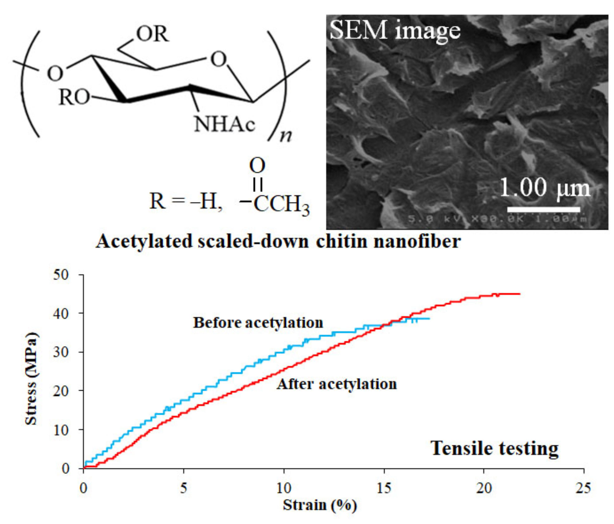
Previously, derivatization and modification on ChNFs have been investigated to improve their properties and introduce new functions [23,24,25,26,27]. For example, acetylation on ChNFs was carried out, which were manufactured through the disentanglement of native chitin microfibrils using a top-down approach [28]. The acetylated ChNFs were well composited with acrylic resin to obtain transparent nanocomposites. In this study, we demonstrated the acetylation of SD-ChNF films using acetic anhydride in pyridine under several conditions to improve the mechanical properties (Figure 1). A reaction using acetic anhydride in pyridine is one of the most well-known acetylation procedures for polysaccharides, such as a chitin powder [29]; pyridine acts as both base and solvent for efficient progress of the reaction. FT-IR analysis of the products indicated that acetylation gradually progressed from the surface to the interior of the films. Consequently, we found that, by adjusting the degrees of acetylation (DAs), the produced films exhibited superior mechanical properties to those of the parent SD-ChNF film. Because the SD-ChNF film, presented in the previous study, has been reported to show excellent flexibility, that was not previously reported for a nanochitin film or sheet [30], the present films will extend the application potentials for ChNF-based functional materials.
2. Materials and Methods
2.1. Materials
The chitin powder from the crab shells was purchased from FUJIFILM Wako Pure Chemical Corporation, Osaka, Japan. An ionic liquid, AMIMBr, was prepared via the reaction of 1-methylimidazole (Sigma-Aldrich, Darmstadt, Germany, >99.0%) with 3-bromo-1-propene (FUJIFILM Wako Pure Chemical Corporation, Osaka, Japan, 97.0%) according to a method adapted from the literature [31]. Authentic chitin acetates with different DA values were prepared according to the method used for AMIMBr with different reaction times adapted from our procedure in the literature [32]. Other reagents and solvents were commercially available and used without further purification.
2.2. Preparation of a Film of Partially Deacetylated Chitin Nanofiber (PDA-ChNF)
A mixture of chitin (0.120 g, 0.59 mmol)/AMIMBr (1.00 g, 4.92 mmol) was allowed to stand for 24 h at room temperature and then heated with stirring for 24 h at 100 °C to obtain a chitin ion gel (10 wt%). This gel was then immersed in methanol (30 mL, FUJIFILM Wako Pure Chemical Corporation, Osaka, Japan, 99.8%) for 72 h at room temperature for regeneration progress, followed by ultrasonication (Branson 1510 (42 kHz, 70 W, Emerson Japan, Ltd., Tokyo, Japan)) for 10 min to produce a self-assembled ChNF dispersion with methanol. The resulting dispersion was subjected to suction filtration to separate the ChNFs, which were washed with methanol and dried under reduced pressure to obtain a self-assembled ChNF film.
A mixture of the resulting film (0.120 g, 0.59 mmol)/aqueous NaOH (30 wt%, 20 mL, FUJIFILM Wako Pure Chemical Corporation, Osaka, Japan, 97%) was heated for 5 h at 80 °C. The film obtained was isolated using suction filtration, immersed in water (30 mL) for 10 min via ultrasonication (Branson 1510 (42 kHz, 70 W)), filtered, and washed with water. The resulting film was dried under reduced pressure to obtain a PDA-ChNF film. 1H NMR (DCl/D2O) δ 2.1–2.3 (CH3), 3.0–4.0 (H2-6), 4.5–5.2 (H1). The degree of deacetylation (DDA) value of the product was 23.0% for the total repeating units (via 1H NMR spectrum in DCl/D2O).
2.3. Preparation of Scaled-Down Chitin Nanofiber (SD-ChNF) Dispersion and Film
A mixture of the PDA-ChNF film (0.120 g)/aqueous acetic acid (1.0 mol/L, 20 mL, FUJIFILM Wako Pure Chemical Corporation, Osaka, Japan, 99.7%) was ultrasonicated using a homogenizer (Branson Advanced-Digital Sonifier 450 (20 kHz, 400 W)) for 10 min at room temperature to produce a SD-ChNF dispersion. After the resulting dispersion was subjected to suction filtration, the residue was washed with water and dried under reduced pressure to obtain a SD-ChNF film.
2.4. Acetylation of SD-ChNF Film
A typical experimental procedure was followed (entry 4, Table 1). A mixture of the SD-ChNF film (0.050 g, 0.26 mmol)/acetic anhydride (1.47 mL, 15.6 mmol, 60 equiv. with a repeating unit, FUJIFILM Wako Pure Chemical Corporation, Osaka, Japan, 97.0%)/pyridine (2.00 mL, 26.0 mmol, 100 equiv. with a repeating unit, FUJIFILM Wako Pure Chemical Corporation, Osaka, Japan, 99.5%) was heated for 1 h at 80 °C and then cooled for 30 min at room temperature. The obtained film was washed with water and dried under reduced pressure to produce an acetylated SD-ChNF (Ac-SD-ChNF) film.
2.5. Measurements
FT-IR spectra were recorded on a PerkinElmer Spectrum Two spectrometer (PerkinElmer Japan Co., Ltd., Yokohama, Japan). The KBr pellet and attenuated total reflection (ATR) methods were used for the evaluation of DAs of the entire material and film surfaces, respectively. The 1H NMR spectra were recorded using an ECX400 instrument (JEOL, Akishima, Tokyo, Japan). Powder X-ray diffraction (XRD) measurements were performed using a PANalytical X’Pert Pro MPD instrument (PANalytical B.V., Almelo, The Netherlands) with Ni-filtered Cu-Kα radiation (λ = 0.15418 nm). The crystalline index (CI) of the chitin crystal was calculated according to a method mentioned in the literature using the following equation [33].
where I110 is the maximum intensity (arbitrary units) of the diffraction (110) at 2 θ = 19.3° and Iam is the intensity of the amorphous diffraction in the same unit at 2 θ = 12.6°.
CI (%) = [(I110 − Iam)/I110] × 100;
SEM images were obtained using a Hitachi S-4100H electron microscope (Hitachi High-Technologies Corporation, Tokyo, Japan). The stress–strain curves were measured using a tensile tester (Little Senstar LSC-1/30, Tokyo Testing Machine, Tokyo, Japan).
3. Results and Discussion
The PDA-ChNF film was first prepared through successive partial deacetylation by treating the self-assembled ChNF film with aqueous NaOH (30 wt%) for 5 h at 80 °C, suction filtration, and drying (DDA value = 23.0%, determined via 1H NMR spectrum in DCl/D2O). The resulting film was then treated with aqueous acetic acid (1.0 mol/L) for 10 min at room temperature using ultrasonication (20 kHz, 400 W) to produce the SD-ChNF/aqueous acetic acid dispersion. The dispersion was then subjected to suction filtration, and the residue was dried under reduced pressure to obtain the SD-ChNF film.
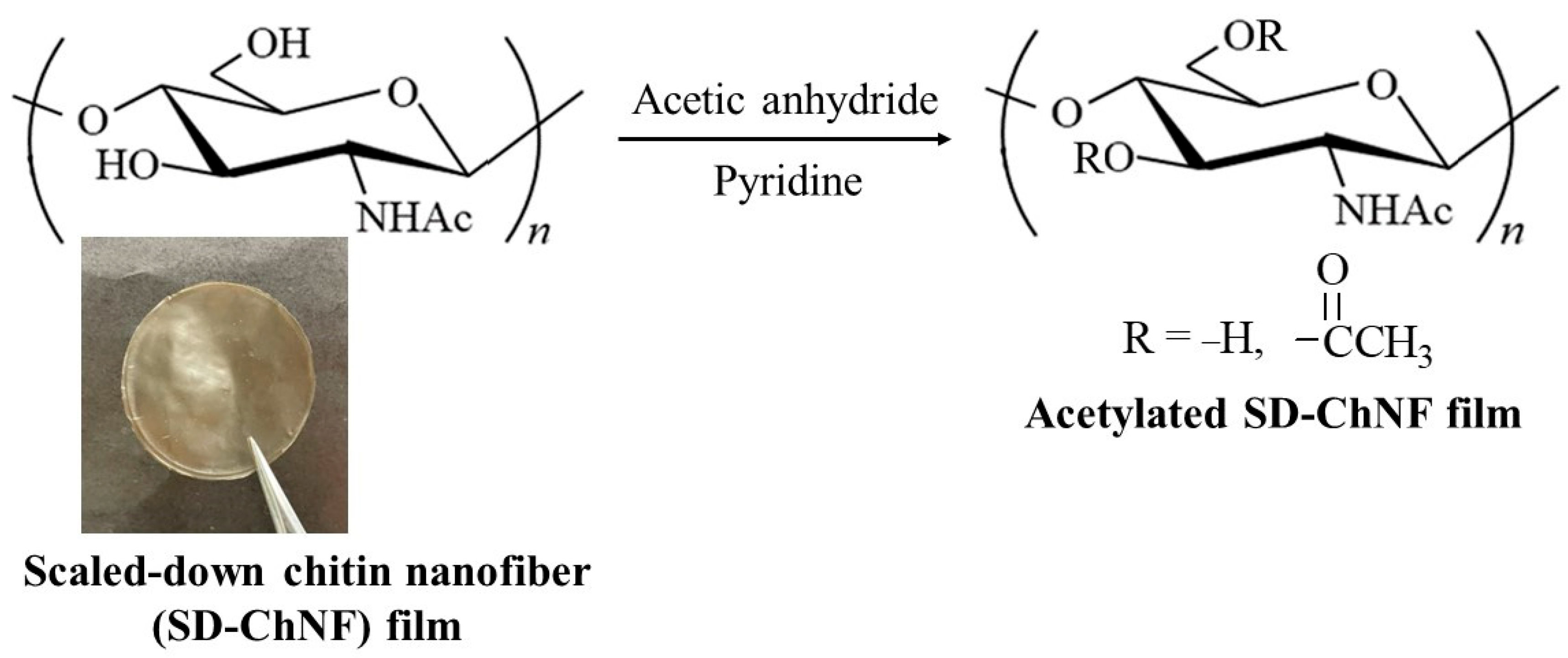
Acetylation of the resulting SD-ChNF film was carried out using acetic anhydride (60 equiv. with a repeating unit) in different amounts of pyridine for 1 h at 50–80 °C, followed by cooling for 30 min at room temperature (Figure 1, entries 1–4, Table 1). The FT-IR spectra of all the newly acetylated products, measured using the KBr pellet method (Figure 2c, entry 4), exhibited the C = O absorptions derived from the ester groups around 1740 cm−1 compared to those of the chitin power (Figure 2a), suggesting progress of acetylation; a similar spectroscopic pattern was also observed in the FT-IR spectrum of chitin powder, measured using the ATR method (Figure 2b) [30] (Found in the Supplementary Materials of this publication). DA values were calculated from the FT-IR spectra using the following equation: R = 0.975–0.003 DA, as previously reported in the literature [34], where R is the absorbance ratio (D1740/D1658), which increased with increasing amounts of pyridine and increasing temperatures for acetylation. The DA values of the film surfaces were also evaluated using FT-IR spectra, measured via the ATR method. To create a calibration curve for evaluation, we prepared authentic chitin acetates with different DA values via the acetylation method using acetic anhydride in AMIMBr with different reaction times, adapted from our procedure in the literature [32]. The exact DA values were then calculated via 1H NMR spectra after acidic hydrolysis and dissolution of the chitin acetates in DCl/D2O (Supplementary Figure S1). Based on the resulting DA values and FT-IR spectra of the chitin acetates, measured using the ATR method, the following calibration curve was obtained: R = 5.5369–9.8114 DA. Based on this equation, the DA values of the film surfaces were estimated from the FT-IR spectra of the acetylated SD-ChNF films measured using the ATR method (Figure 2d, entry 4), which were not mostly affected by the reaction conditions and were close to the quantitative DA value of chitin (~2) as shown in Table 1. These FT-IR results supported the fact that the acetylation reactions occurred almost quantitatively on the surface and progressed from the surface to the interior of the films according to increasing amounts of pyridine and elevating temperatures (from entry 1 to entry 4).
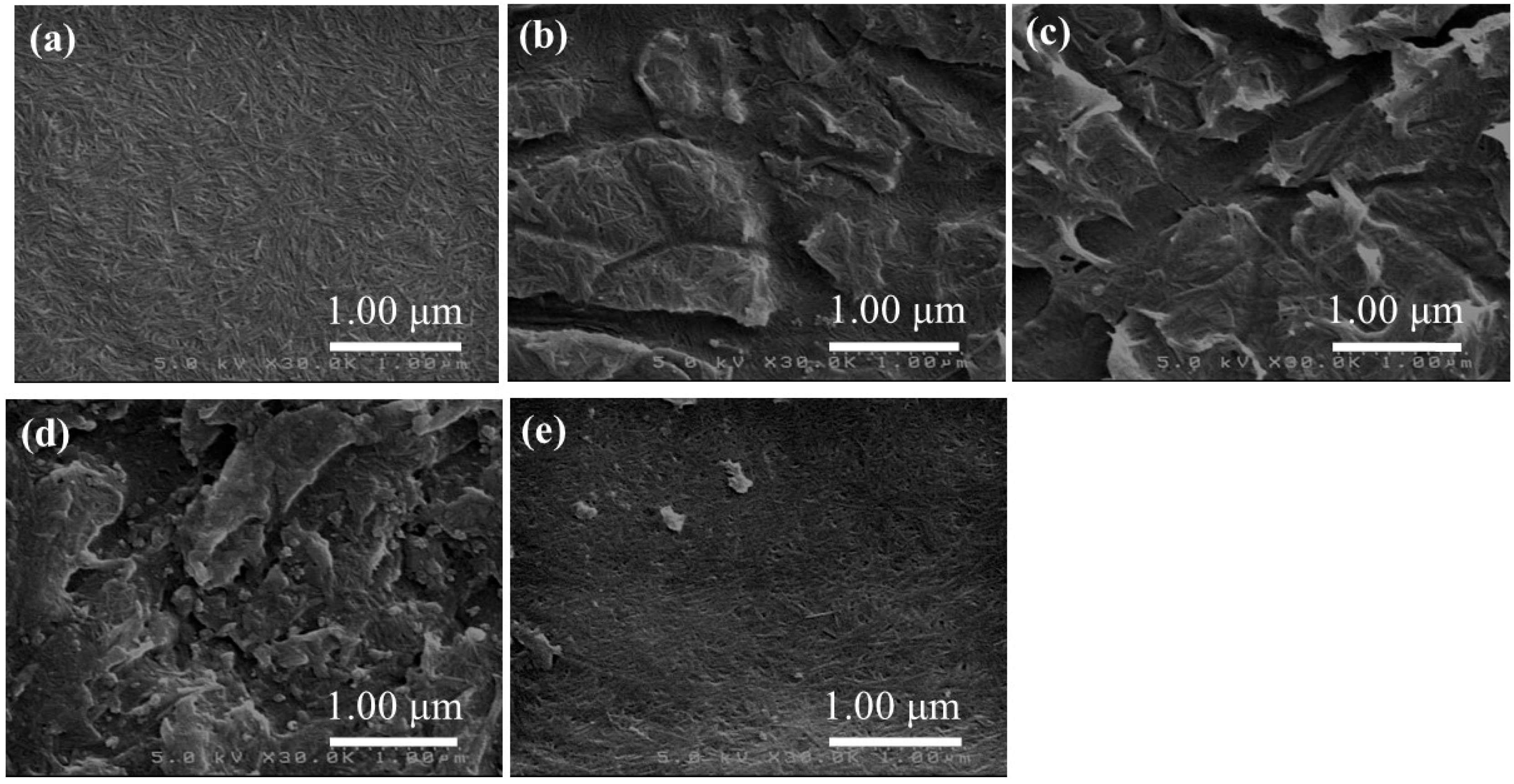
The XRD measurement of the acetylated SD-ChNF films was then performed to evaluate the chitin crystallinity. The CI values in the 110 planes of the chitin crystals (19°), which were calculated based on the XRD results with typical diffraction patterns assigned to the intrinsic crystalline structure of the chitin (Supplementary Figure S2), decreased with increasing DA values of the entire product (Table 1, from FT-IR spectra, measured using the KBr pellet method). These results indicated that the progress of acetylation inside the film gradually disrupted the crystalline structures of the chitin chains. The surface morphologies of the acetylated SD-ChNF films were then evaluated using SEM measurement. The SEM image of the parent SD-ChNF film showed a heavily condensed nanofiber morphology (Figure 3a), as previously reported in the literature [30]. On the other hand, all the SEM images of the acetylated SD-ChNF films did not exhibit clear nanofiber morphologies (Figure 3b–e), owing to the almost quantitative acetylation on the surfaces of the films, regardless of the reaction conditions.
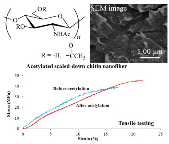
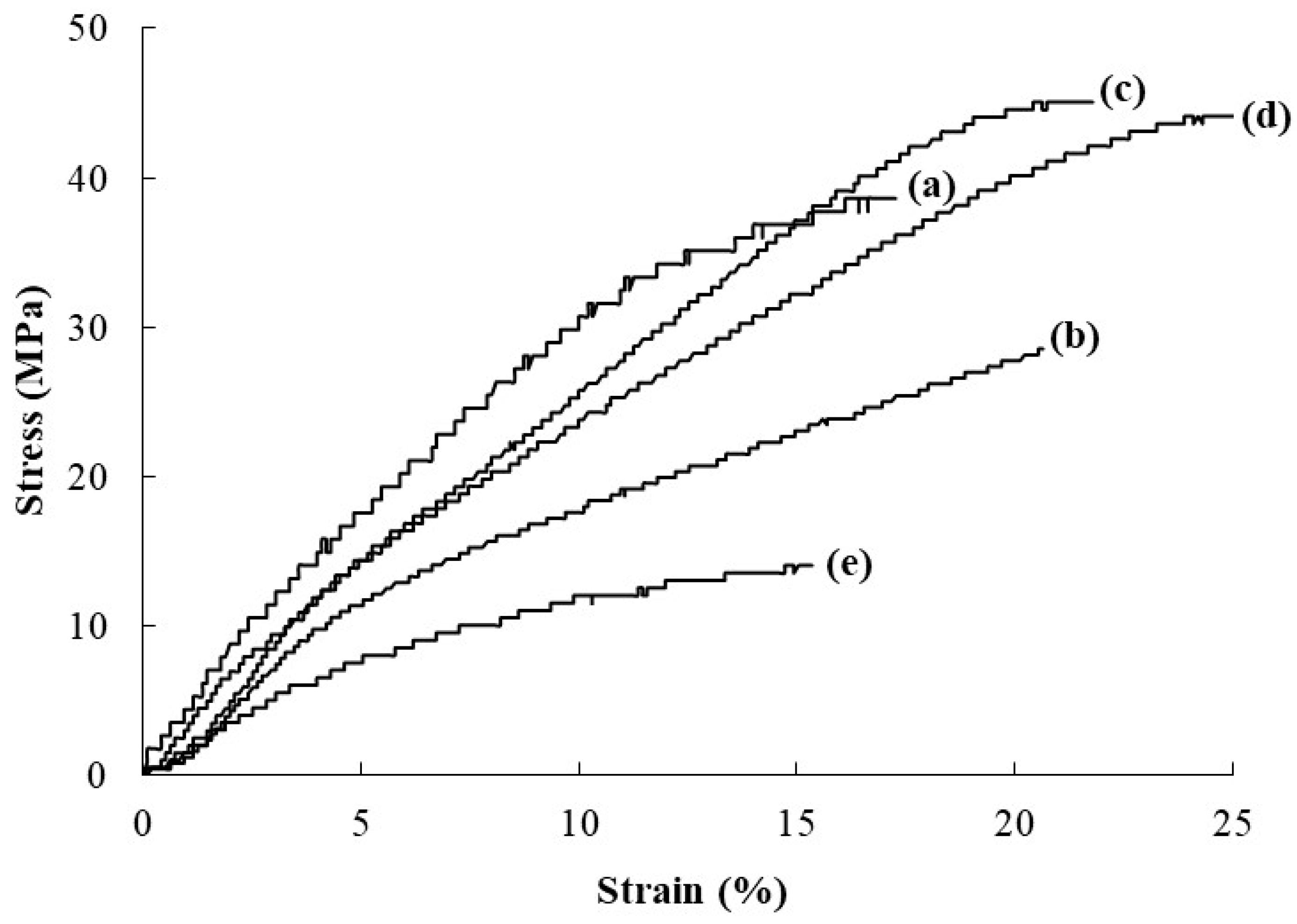
The mechanical properties of the acetylated SD-ChNF films were evaluated through tensile testing. The stress–strain curves of the acetylated SD-ChNF film of entry 1 showed a lower tensile strength than that of the parent SD-ChNF film (Figure 4a,b). With increasing DA values of the entire acetylated SD-ChNF films of entries 2 and 3, both tensile strength and elongation at break increased beyond those of the SD-ChNF film (Figure 4c,d). However, a further increase in the DA value drastically weakened the mechanical properties, as seen in the stress–strain curve of the acetylated SD-ChNF film of entry 4 (Figure 4e). These results strongly suggested that the optimized acetylation of chitin inside the films contributed to the enhancement in the mechanical properties of the SD-ChNF film. Acetylation on the surface probably caused the nanofibers at interfacial area to merge, resulting in a smooth surface of the film (Figure 3) due to an improvement in the mechanical properties. However, further progress of acetylation inside the film reduced the chitin crystallinity, which weakened the mechanical properties.
4. Conclusions
In this study, an improvement in the mechanical properties of the SD-ChNF films was obtained via acetylation along with adjusting the DA values. The acetylation of the SD-ChNF films was carried out using acetic anhydride in pyridine for 1 h at desired temperatures followed by cooling for 30 min at room temperature. The FT-IR spectra of the acetylated SD-ChNF films suggested the progress of acetylation from the surface to the interior of the films in response to increasing amounts of pyridine and elevating temperatures, which caused a disruption of the chitin crystals. The heavily condensed ChNF morphology on the surface of the SD-ChNF film became unclear after acetylation regardless of the DA values, because the surface was almost quantitatively acetylated in all cases. Tensile testing of the SD-ChNF films indicated that their mechanical properties were improved via acetylation with the optimized DA values. The presented ChNF materials with excellent mechanical properties have potentials for practical applications as new bio-based films in green and sustainable fields because of their eco-friendly and non-toxic properties. Furthermore, the present approach will be extended to use other acylation reagents, such as fatty acyl chlorides, to obtain new SD-ChNF materials with additional functions in the future.
Supplementary Materials
The following supporting information can be downloaded at: https://www.mdpi.com/article/10.3390/surfaces6030017/s1, Figure S1: 1H NMR spectrum of sample via acidic hydrolysis and dissolution of chitin acetate, prepared using acetic anhydride in 1-allyl-3-methylimidazolium bromide (AMIMBr), in DCl/D2O, Figure S2: XRD profiles of (a–d) acetylated SD-ChNF films of entries 1–4 and (e) SD-ChNF film.
Author Contributions
J.-i.K. conceived of the project, designed the experiments, directed the research, and wrote the manuscript. C.I. and A.N. performed the experiments. All authors have read and agreed to the published version of the manuscript.
Funding
This research received no external funding.
Institutional Review Board Statement
Not applicable.
Informed Consent Statement
Not applicable.
Data Availability Statement
Not applicable.
Conflicts of Interest
The authors declare no conflict of interest.
References
- Schuerch, C. Polysaccharides. In Encyclopedia of Polymer Science and Engineering, 2nd ed.; Mark, H.F., Bilkales, N., Overberger, C.G., Eds.; John Wiley & Sons: New York, NY, USA, 1986; Volume 13, pp. 87–162. [Google Scholar]
- Kasapis, S.; Norton, I.T.; Ubbink, J.B. Modern Biopolymer Science: Bridging the Divide between Fundamental Treatise and Industrial Application; Academic Press: San Diego, CA, USA, 2009. [Google Scholar]
- Song, E.H.; Shang, J.; Ratner, D.M. 9.08—Polysaccharides. In Polymer Science: A Comprehensive Reference; Matyjaszewski, K., Möller, M., Eds.; Elsevier: Amsterdam, The Netherlands, 2012; pp. 137–155. [Google Scholar]
- Kurita, K. Chitin and chitosan: Functional biopolymers from marine crustaceans. Mar. Biotechnol. 2006, 8, 203–226. [Google Scholar] [CrossRef]
- Rinaudo, M. Chitin and chitosan: Properties and applications. Prog. Polym. Sci. 2006, 31, 603–632. [Google Scholar]
- Pillai, C.K.S.; Paul, W.; Sharma, C.P. Chitin and chitosan polymers: Chemistry, solubility and fiber formation. Prog. Polym. Sci. 2009, 34, 641–678. [Google Scholar] [CrossRef]
- Kadokawa, J.; Takegawa, A.; Mine, S.; Prasad, K. Preparation of chitin nanowhiskers using an ionic liquid and their composite materials with poly(vinyl alcohol). Carbohydr. Polym. 2011, 84, 1408–1412. [Google Scholar] [CrossRef]
- Zeng, J.B.; He, Y.S.; Li, S.L.; Wang, Y.Z. Chitin whiskers: An overview. Biomacromolecules 2012, 13, 1–11. [Google Scholar]
- Ifuku, S. Preparation of chitin nanofibers from crab shell and their applications. Kobunshi Ronbunshu 2012, 69, 460–467. [Google Scholar]
- Tajiri, R.; Setoguchi, T.; Wakizono, S.; Yamamoto, K.; Kadokawa, J. Preparation of self-assembled chitin nanofibers by regeneration from ion gels using calcium halide dihydrate/methanol solutions. J. Biobased Mater. Bioenergy 2013, 7, 655–659. [Google Scholar] [CrossRef]
- Kadokawa, J. Ionic liquid as useful media for dissolution, derivatization, and nanomaterial processing of chitin. Green Sustain. Chem. 2013, 3, 19–25. [Google Scholar]
- Muzzarelli, R.A.A.; El Mehtedi, M.; Mattioli-Belmonte, M. Emerging biomedical applications of nano-chitins and nano-chitosans obtained via advanced eco-friendly technologies from marine resources. Mar. Drugs 2014, 12, 5468–5502. [Google Scholar] [CrossRef] [Green Version]
- Kadokawa, J. Fabrication of nanostructured and microstructured chitin materials through gelation with suitable dispersion media. RSC Adv. 2015, 5, 12736–12746. [Google Scholar] [CrossRef]
- You, J.; Li, M.; Ding, B.; Wu, X.; Li, C. Crab chitin-based 2D soft nanomaterials for fully biobased electric devices. Adv. Mater. 2017, 29, 1606895. [Google Scholar]
- Anraku, M.; Tabuchi, R.; Ifuku, S.; Nagae, T.; Iohara, D.; Tomida, H.; Uekama, K.; Maruyama, T.; Miyamura, S.; Hirayama, F.; et al. An oral absorbent, surface-deacetylated chitin nano-fiber ameliorates renal injury and oxidative stress in 5/6 nephrectomized rats. Carbohydr. Polym. 2017, 161, 21–25. [Google Scholar] [CrossRef]
- Koizumi, R.; Azuma, K.; Izawa, H.; Morimoto, M.; Ochi, K.; Tsuka, T.; Imagawa, T.; Osaki, T.; Ito, N.; Okamoto, Y.; et al. Oral administration of surface-deacetylated chitin nanofibers and chitosan inhibit 5-fluorouracil-induced intestinal mucositis in mice. Int. J. Mol. Sci. 2017, 18, 279. [Google Scholar] [CrossRef] [Green Version]
- Satam, C.C.; Irvin, C.W.; Lang, A.W.; Jallorina, J.C.R.; Shofner, M.L.; Reynolds, J.R.; Meredith, J.C. Spray-coated multilayer cellulose nanocrystal—Chitin nanofiber films for barrier applications. ACS Sustain. Chem. Eng. 2018, 6, 10637–10644. [Google Scholar] [CrossRef]
- Mushi, N.E.; Nishino, T.; Berglund, L.A.; Zhou, Q. Strong and tough chitin film from α-chitin nanofibers prepared by high pressure homogenization and chitosan addition. ACS Sustain. Chem. Eng. 2019, 7, 1692–1697. [Google Scholar] [CrossRef]
- Naghdi, T.; Golmohammadi, H.; Yousefi, H.; Hosseinifard, M.; Kostiv, U.; Horák, D.; Merkoçi, A. Chitin nanofiber paper toward optical (bio)sensing applications. ACS Appl. Mater. Interfaces 2020, 12, 15538–15552. [Google Scholar] [CrossRef]
- Sharma, P.R.; Sharma, S.K.; Lindström, T.; Hsiao, B.S. Water purification: Nanocellulose-enabled membranes for water purification: Perspectives. Adv. Sustain. Syst. 2020, 4, 2070009. [Google Scholar] [CrossRef]
- Prasad, K.; Murakami, M.; Kaneko, Y.; Takada, A.; Nakamura, Y.; Kadokawa, J. Weak gel of chitin with ionic liquid, 1-allyl-3-methylimidazolium bromide. Int. J. Biol. Macromol. 2009, 45, 221–225. [Google Scholar] [CrossRef]
- Kadokawa, J.; Kawano, A.; Yamamoto, K. Fabrication of semi-crystalline film by hexanoylation on self-assembled chitin nanofibers. ChemistrySelect 2019, 4, 797–801. [Google Scholar]
- Ifuku, S.; Saimoto, H. Chitin nanofibers: Preparations, modifications, and applications. Nanoscale 2012, 4, 3308–3318. [Google Scholar] [CrossRef]
- Ifuku, S.; Shervani, Z.; Saimoto, H. Preparation, modification and application of chitin nanofibers. In Nanofibers: Synthesis, Properties, and Applications; Nova Science Publishers, Inc.: Hauppauge, NY, USA, 2012; pp. 167–181. [Google Scholar]
- Ifuku, S. Chitin and chitosan nanofibers: Preparation and chemical modifications. Molecules 2014, 19, 18367–18380. [Google Scholar]
- Ifuku, S. Chitin nanofibers: Preparations, modifications, and applications. In Handbook of Polymer Nanocomposites. Processing, Performance and Application: Volume C: Polymer Nanocomposites of Cellulose Nanoparticles; Springer: Berlin, Germany, 2015; pp. 165–178. [Google Scholar]
- Kadokawa, J. Surface derivatization and grafting on self-assembled chitin nanofibers for modification, functionalization, and application. In Surface Treatment Methods of Natural Fibres and Their Effects on Biocomposites; Elsevier: Amsterdam, The Netherlands, 2022; pp. 187–202. [Google Scholar]
- Ifuku, S.; Morooka, S.; Morimoto, M.; Saimoto, H. Acetylation of chitin nanofibers and their transparent nanocomposite films. Biomacromolecules 2010, 11, 1326–1330. [Google Scholar] [CrossRef]
- Kurita, K.; Ishii, S.; Tomita, K.; Nishimura, S.I.; Shimoda, K. Reactivity characteristics of squid β-chitin as compared with those of shrimp chitin: High potentials of squid chitin as a starting material for facile chemical modifications. J. Polym. Sci. Polym. Chem. 1994, 32, 1027–1032. [Google Scholar] [CrossRef]
- Hashiguchi, T.; Yamamoto, K.; Kadokawa, J. Fabrication of highly flexible nanochitin film and its composite film with anionic polysaccharide. Carbohydr. Polym. 2021, 270, 118369. [Google Scholar]
- Zhao, D.B.; Fei, Z.F.; Geldbach, T.J.; Scopelliti, R.; Laurenczy, G.; Dyson, P.J. Allyl-functionalised ionic liquids: Synthesis, characterisation, and reactivity. Helv. Chim. Acta 2005, 88, 665–675. [Google Scholar] [CrossRef]
- Mine, S.; Izawa, H.; Kaneko, Y.; Kadokawa, J. Acetylation of a-chitin in ionic liquids. Carbohydr. Res. 2009, 344, 2263–2265. [Google Scholar]
- Cárdenas, G.; Cabrera, G.; Taboada, E.; Miranda, S.P. Chitin characterization by SEM, FTIR, XRD, and 13C cross polarization/mass angle spinning NMR. J. Appl. Polym. Sci. 2004, 93, 1876–1885. [Google Scholar] [CrossRef]
- Ando, T.; Kataoka, S. Acylation of chitin with acid anhydrides in trichloroacetic acid system. Kobunshi Ronbunshu 1980, 37, 1–7. [Google Scholar] [CrossRef]
Figure 1.
Acetylation of scaled-down chitin nanofiber (SD-ChNF) film using acetic anhydride in pyridine.
Figure 1.
Acetylation of scaled-down chitin nanofiber (SD-ChNF) film using acetic anhydride in pyridine.

Figure 2.
FT-IR spectra of (a,b) chitin using KBr pellet and ATR methods, respectively, and (c,d) acetylated SD-ChNF film of entry 4 using KBr pellet and ATR methods, respectively.
Figure 2.
FT-IR spectra of (a,b) chitin using KBr pellet and ATR methods, respectively, and (c,d) acetylated SD-ChNF film of entry 4 using KBr pellet and ATR methods, respectively.

Figure 3.
SEM images of the (a) SD-ChNF film and (b–e) acetylated SD-CHNF films of entries 1–4.

Figure 4.
Stress–strain curves of the (a) SD-ChNF film and (b–e) acetylated SD-CHNF films of entries 1–4 under tensile mode.
Figure 4.
Stress–strain curves of the (a) SD-ChNF film and (b–e) acetylated SD-CHNF films of entries 1–4 under tensile mode.


Table 1.
Conditions in the acetylation of the SD-ChNF film and properties of the acetylated SD-ChNF films (a).
Table 1.
Conditions in the acetylation of the SD-ChNF film and properties of the acetylated SD-ChNF films (a).
| Entry | Equiv. of Pyridine (b) | Temp. (°C) | DA (c) (KBr Pellet Method) | DA (c) (ATR Method) | CI (d) | Tensile Strength (MPa) | Elongation at Break (%) |
|---|---|---|---|---|---|---|---|
| SD-ChNF film | - | - | - | - | 95.7 | 38.6 | 17.2 |
| 1 | 0.6 | 50 | 0.81 | 1.82 | 94.7 | 28.5 | 21.4 |
| 2 | 10 | 50 | 0.96 | 1.83 | 93.8 | 45.0 | 21.8 |
| 3 | 10 | 80 | 1.18 | 1.84 | 93.6 | 44.1 | 24.9 |
| 4 | 100 | 80 | 1.22 | 1.86 | 92.7 | 14.0 | 15.4 |
(a) Reaction was carried out using 60 equiv. of acetic anhydride with a repeating unit for 1 h at desired a temperature followed by cooling for 30 min at room temperature. (b) With a repeating unit. (c) Degree of acetylation, determined via FT-IR analysis. (d) Crystalline index of the chitin crystal in the 110 plane, calculated from the XRD result according to the method mentioned in the literature [33].
Disclaimer/Publisher’s Note: The statements, opinions and data contained in all publications are solely those of the individual author(s) and contributor(s) and not of MDPI and/or the editor(s). MDPI and/or the editor(s) disclaim responsibility for any injury to people or property resulting from any ideas, methods, instructions or products referred to in the content. |
© 2023 by the authors. Licensee MDPI, Basel, Switzerland. This article is an open access article distributed under the terms and conditions of the Creative Commons Attribution (CC BY) license (https://creativecommons.org/licenses/by/4.0/).
Share and Cite
MDPI and ACS Style
Kadokawa, J.-i.; Iiyama, C.; Nakashima, A. Acetylation of Scaled-Down Chitin Nanofiber Films to Improve Mechanical Properties. Surfaces 2023, 6, 249-256. https://doi.org/10.3390/surfaces6030017
AMA Style
Kadokawa J-i, Iiyama C, Nakashima A. Acetylation of Scaled-Down Chitin Nanofiber Films to Improve Mechanical Properties. Surfaces. 2023; 6(3):249-256. https://doi.org/10.3390/surfaces6030017
Chicago/Turabian StyleKadokawa, Jun-ichi, Chiharu Iiyama, and Aoi Nakashima. 2023. "Acetylation of Scaled-Down Chitin Nanofiber Films to Improve Mechanical Properties" Surfaces 6, no. 3: 249-256. https://doi.org/10.3390/surfaces6030017