
Green Synthesis of Gold and Silver Nanoparticles Using Invasive Alien Plant Parthenium hysterophorus and Their Antimicrobial and Antioxidant Activities

 , , and
Abstract
:1. Introduction
2. Materials and Methods
2.1. Materials
2.2. Sample Collection and Plant Extraction
2.3. Green Synthesis of AgNPs and AuNPs Using Extracts of P. hysterophorus
2.4. Characterization of Nanoparticles
2.5. Antibacterial and Antifungal Activities
2.6. Antioxidant Activity
2.7. Statistical Analysis
3. Results and Discussion
3.1. Green Synthesis of Silver and Gold Nanoparticles
3.2. Optimization of Silver Nanoparticle Synthesis
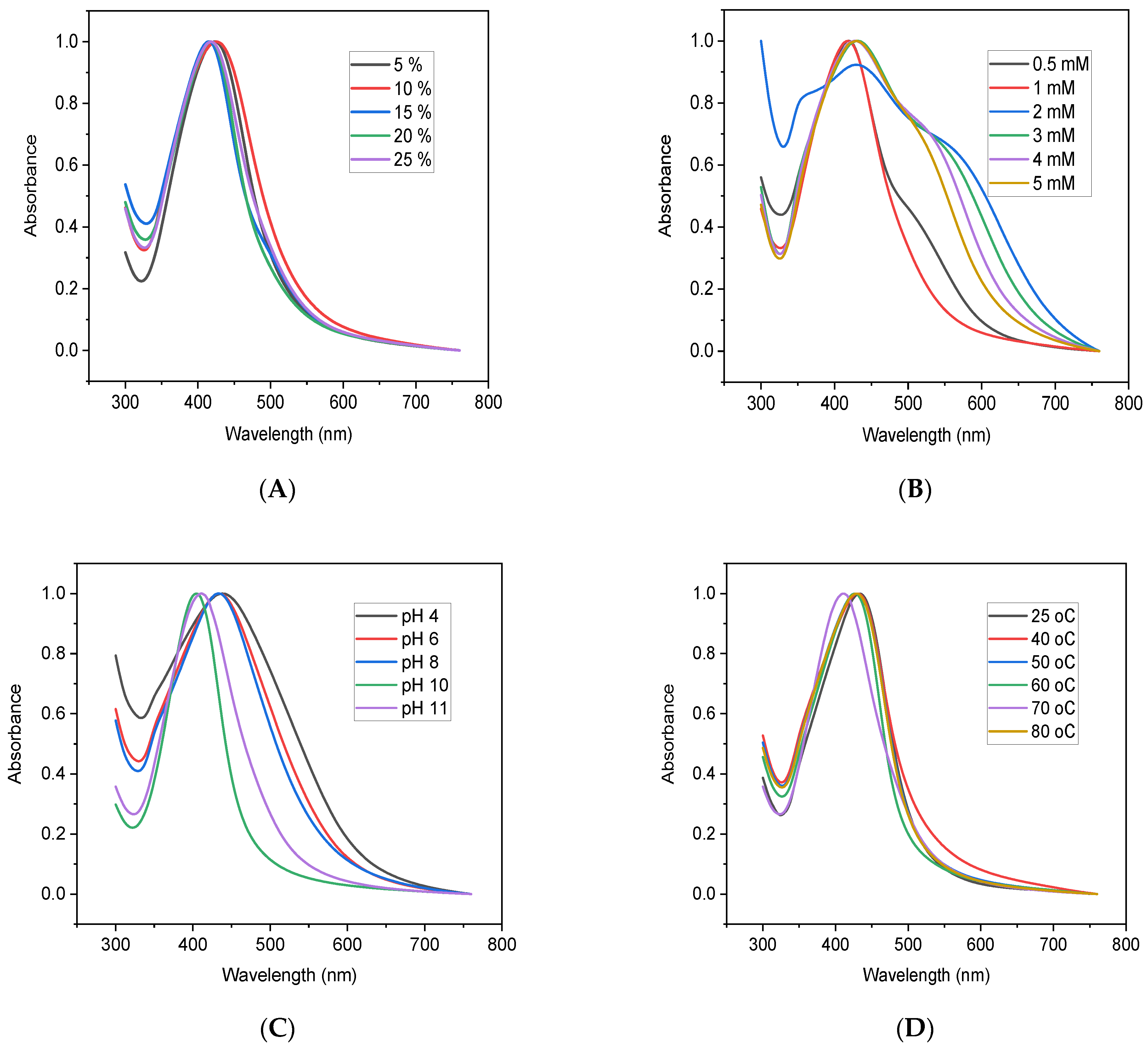
3.3. The Effect of Plant Extract Concentration
3.4. The Effect of AgNO3 Concentration
3.5. The Effects of pH
3.6. The Effect of Temperature
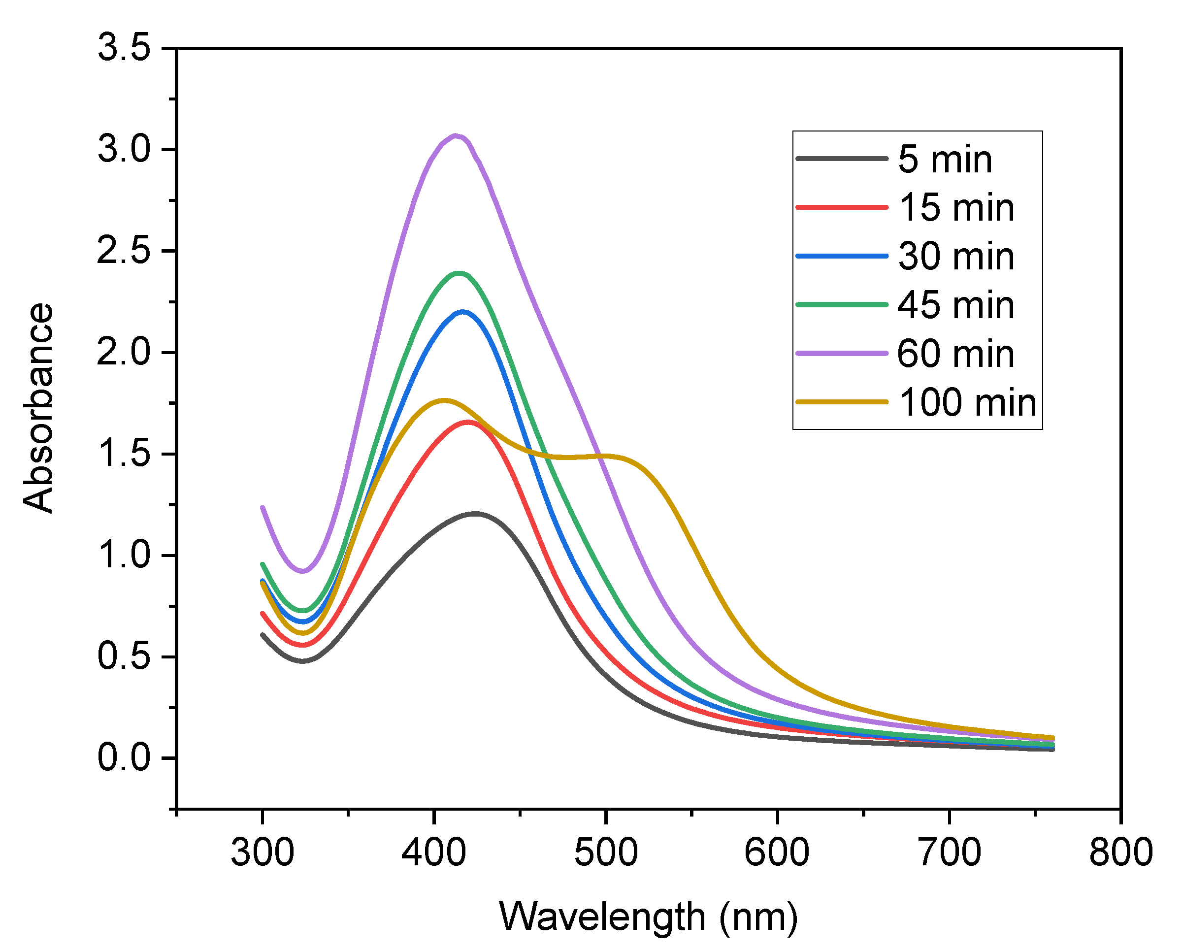
3.7. The Effect of Reaction Time
3.8. Characterizations of the Synthesized Nanoparticles
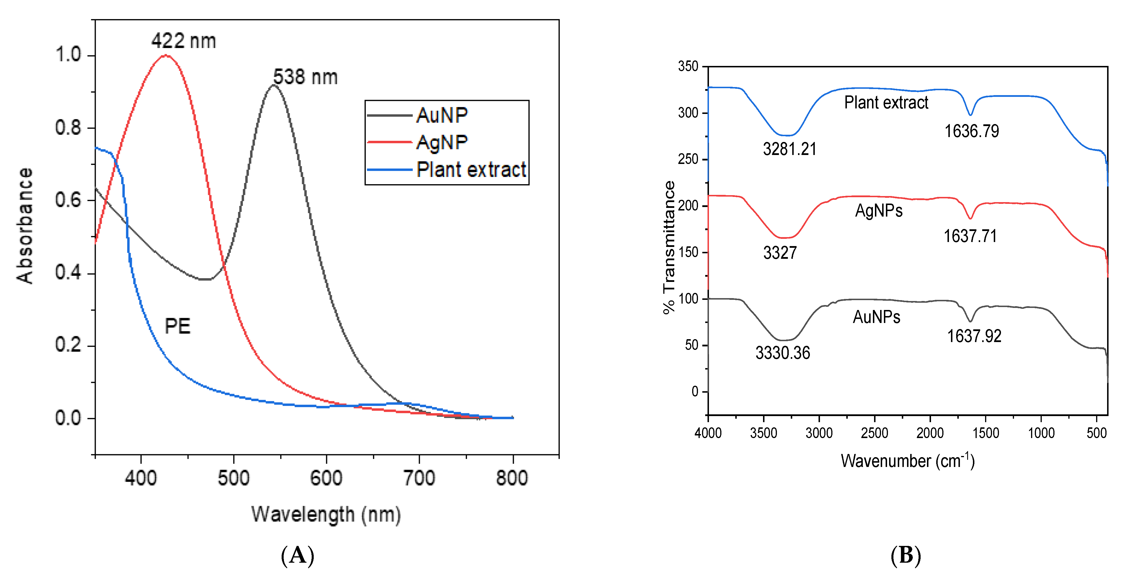
3.8.1. UV–Vis Spectroscopy Analysis
3.8.2. FTIR Spectral Analysis
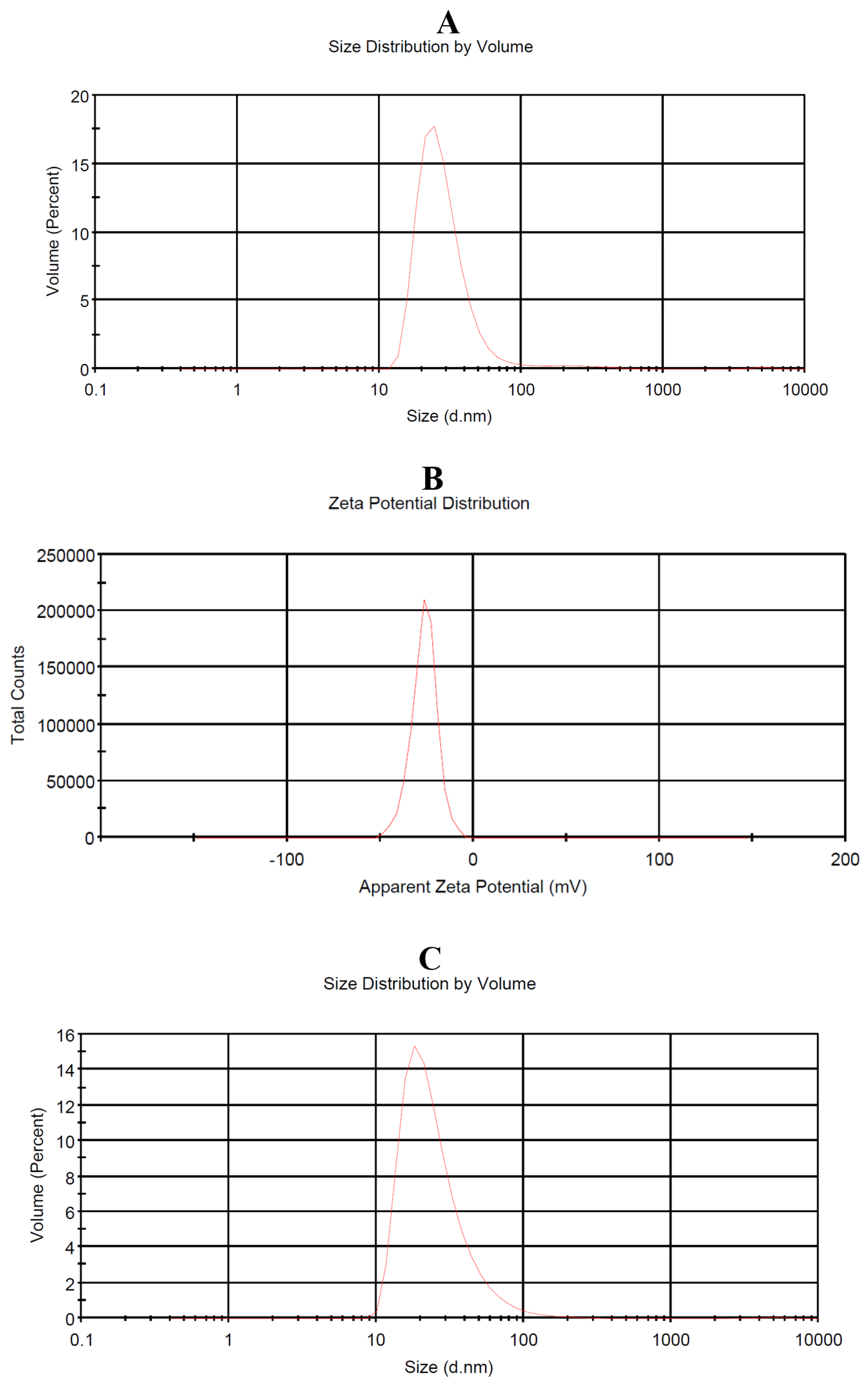
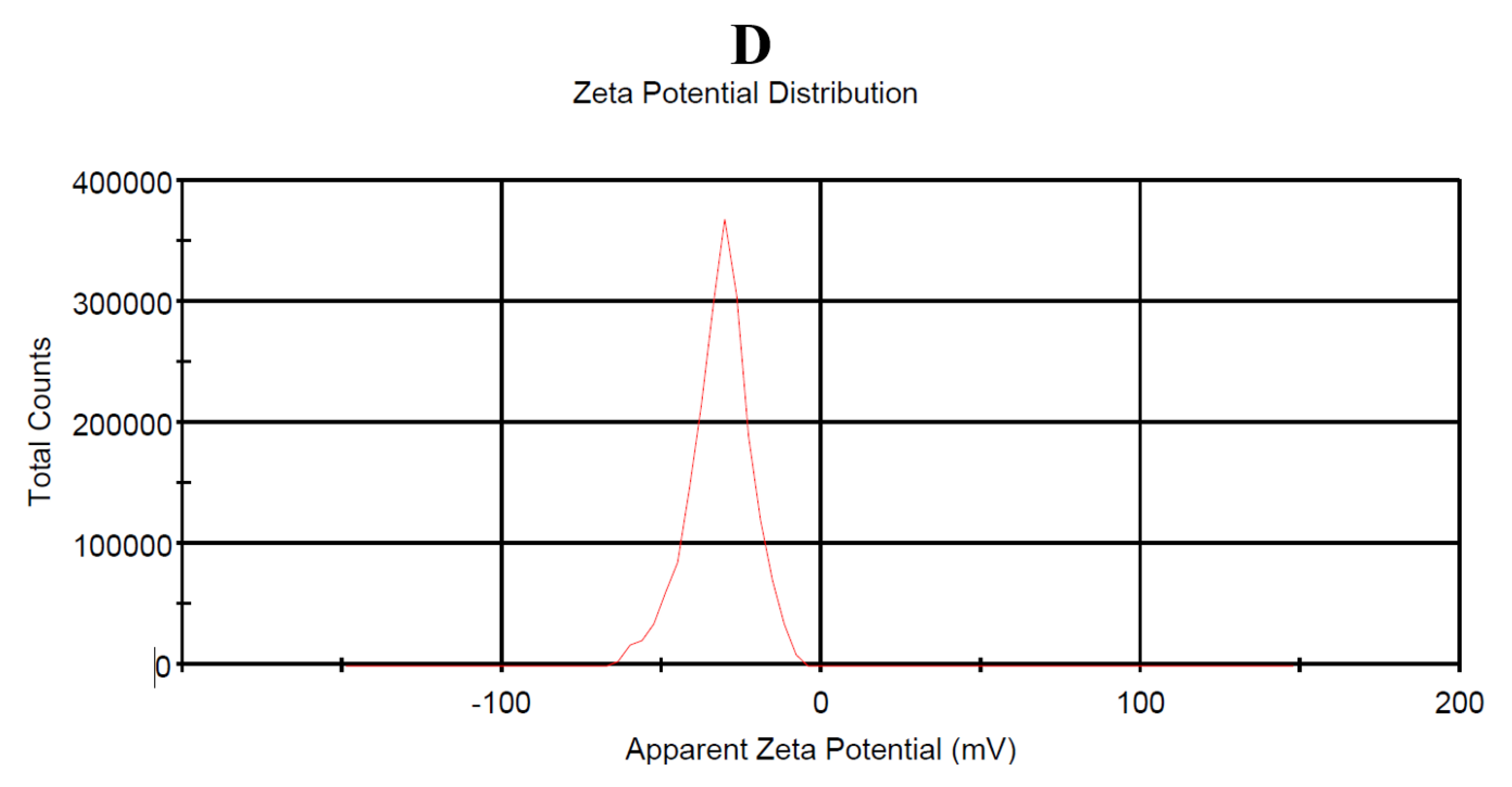
3.8.3. Particle Size and Zeta Potential Measurement
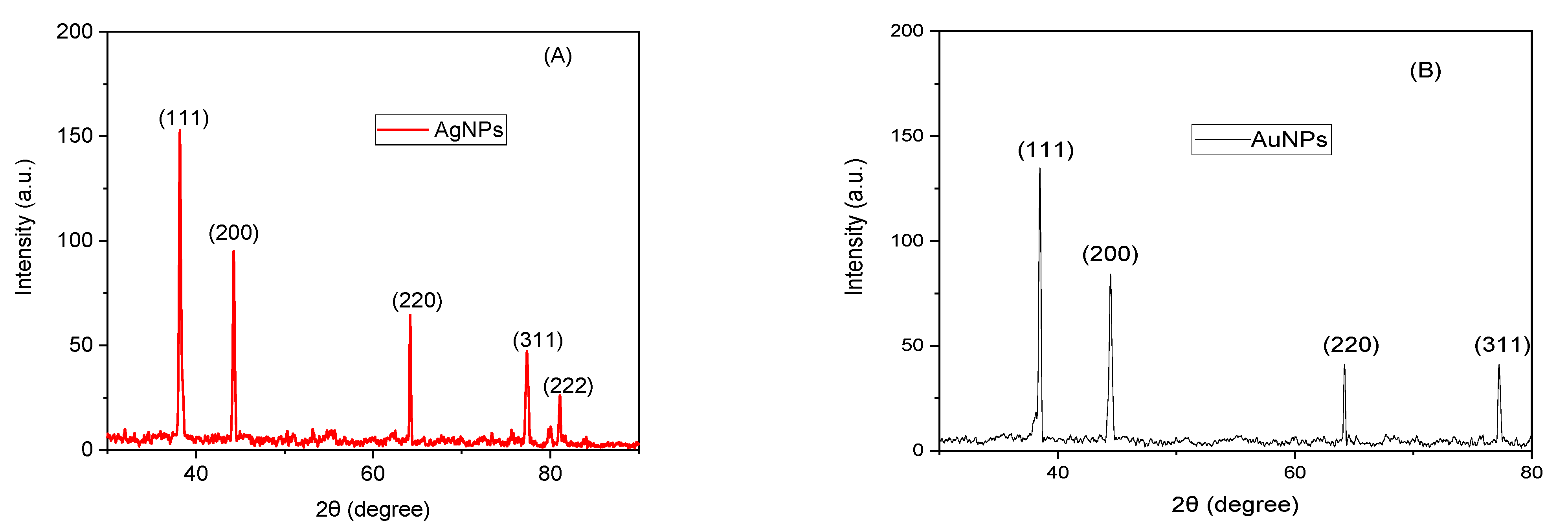
3.8.4. XRD
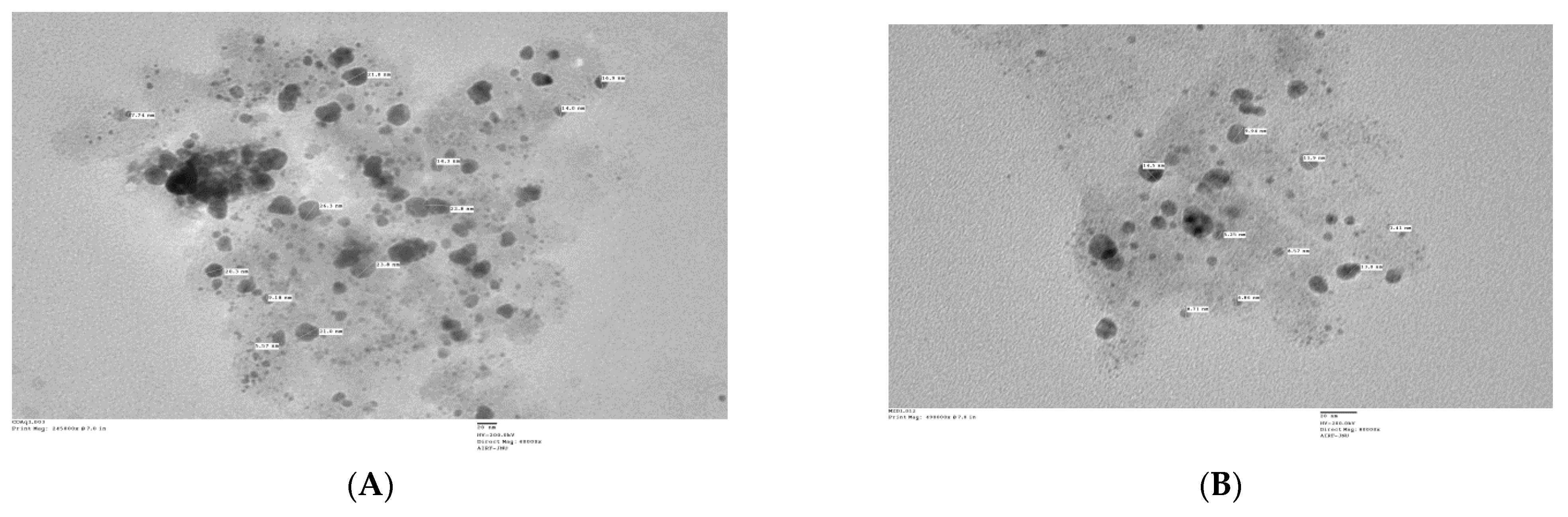
3.8.5. TEM
3.9. Application of the Synthesized Silver and Gold Nanoparticles
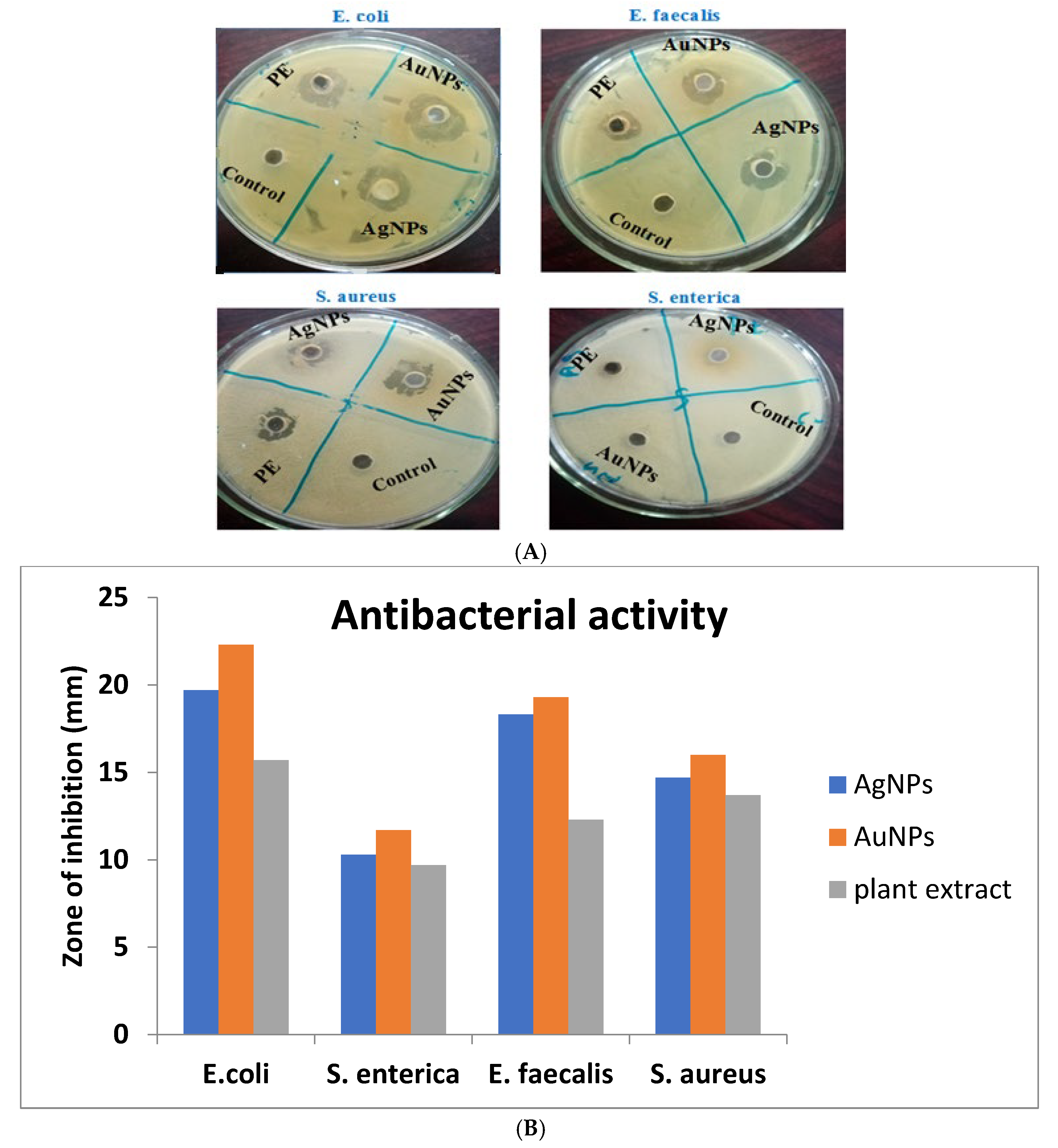
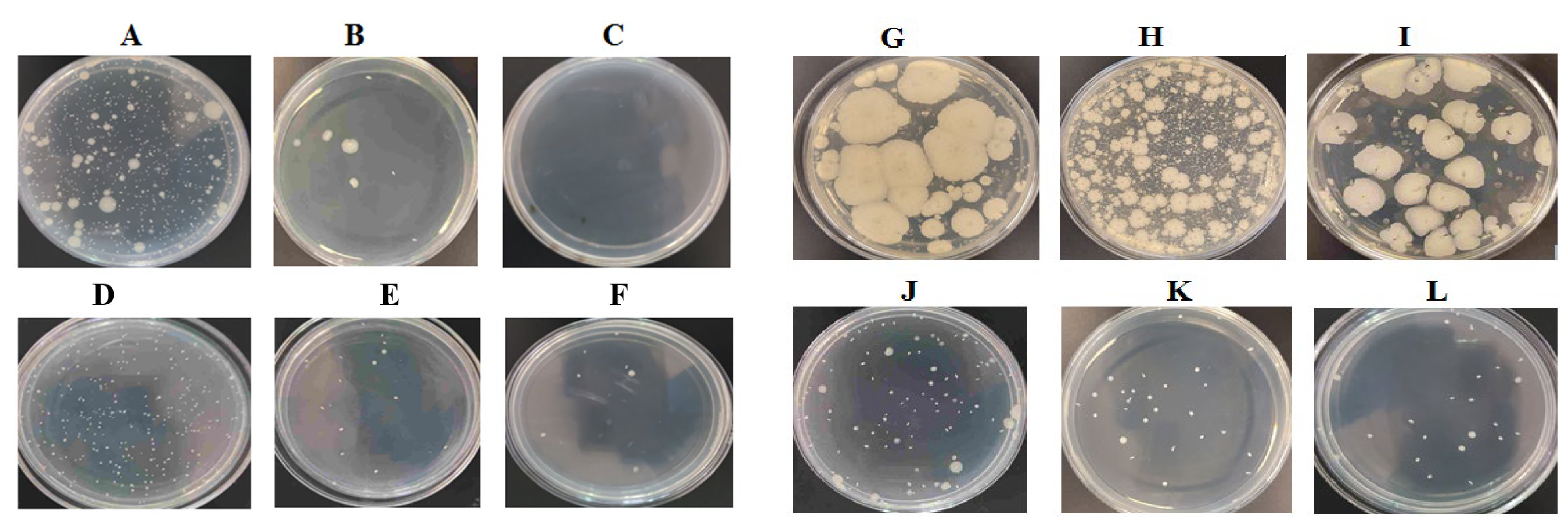
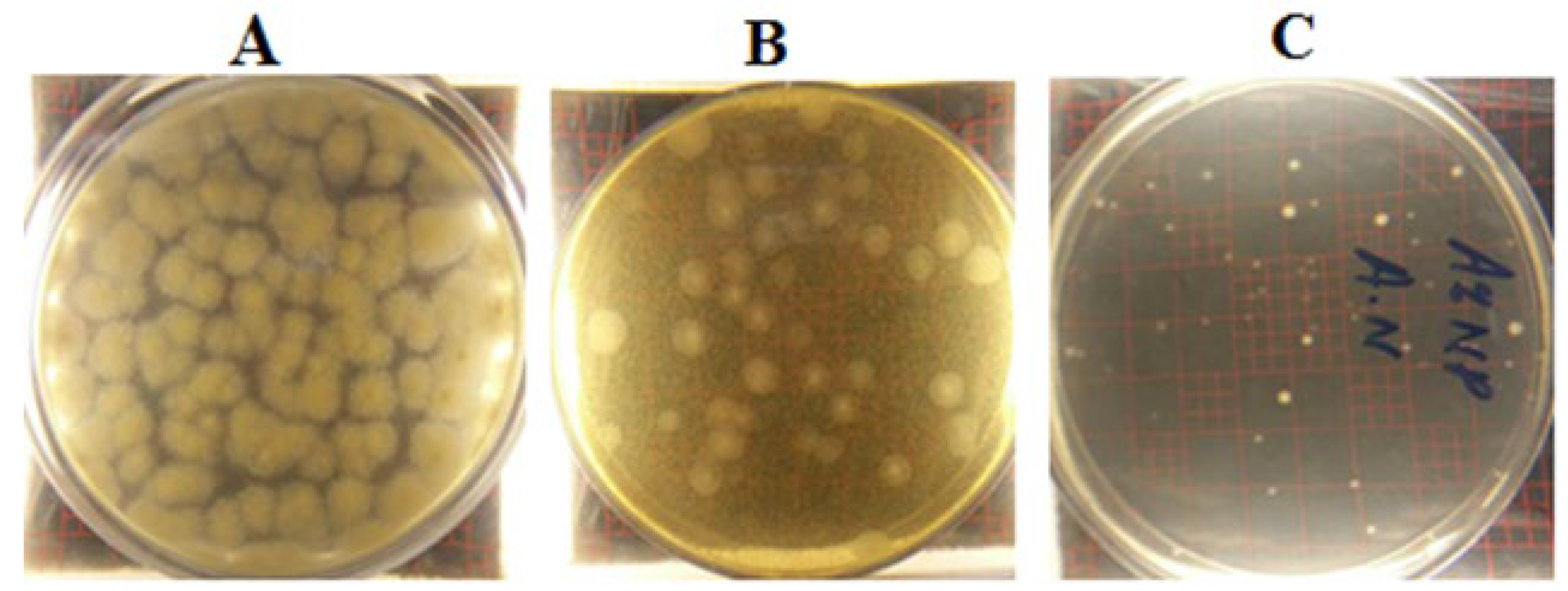
3.9.1. Antibacterial Activity
Method One
Method Two
Antifungal Activity
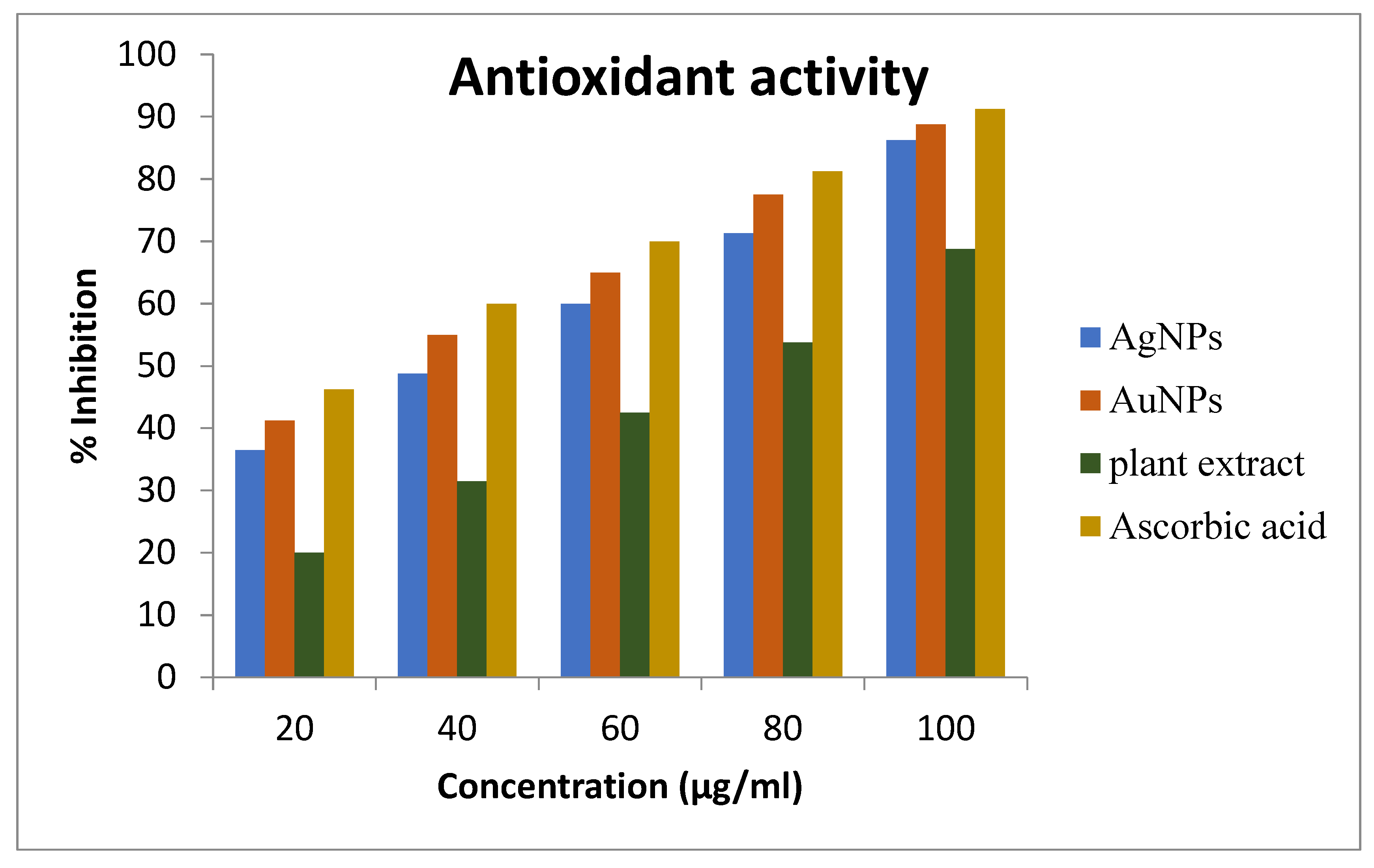
Antioxidant Activity
4. Conclusions
Author Contributions
Funding
Data Availability Statement
Acknowledgments
Conflicts of Interest
Abbreviations
References
- Mohanpuria, P.; Rana, N.K.; Yadav, S.K. Biosynthesis of nanoparticles: Technological concepts and future applications. J. Nanopart Res. 2008, 10, 507–517. [Google Scholar] [CrossRef]
- Bhattacharya, D.; Rajinder, G. Nanotechnology and potential of microorganisms. Crit. Rev. Biotechnol. 2005, 25, 199–204. [Google Scholar] [CrossRef] [PubMed]
- Bachheti, A.; Bachheti, R.K.; Abate, L.; Husen, A. Current status of Aloe-based nanoparticle fabrication, characterization and their application in some cutting-edge areas. S. Afr. J. Bot. 2022, 147, 1058–1069. [Google Scholar] [CrossRef]
- Dhuper, S.; Panda, D.; Nayak, P.L. Green synthesis and characterization of zero valent iron nanoparticles from the leaf extract of Mangifera indica . Nano Trends J. Nanotech. Appl. 2012, 13, 16–22. [Google Scholar]
- Roy, S.; Das, T. Plant Mediated Green Synthesis of Silver Nanoparticles—A Review. Int. J. Plant. Biol. Res. 2015, 3, 1–11. [Google Scholar]
- Suganthy, N.; Ramkumar, V.S.; Pugazhendhi, A.; Benelli, G.; Archunan, G. Biogenic synthesis of gold nanoparticles from Terminalia arjuna bark extract: Assessment of safety aspects and neuroprotective potential via antioxidant, anticholinesterase, and antiamyloidogenic effects. Environ. Sci. Pollut. Res. 2018, 25, 10418–10433. [Google Scholar] [CrossRef]
- Bachheti, R.K.; Abate, L.; Bachheti, A.; Madhusudhan, A.; Husen, A. Algae-, fungi-, and yeast-mediated biological synthesis of nanoparticles and their various biomedical applications. In Handbook of Greener Synthesis of Nanomaterials and Compounds; Elsevier: Amsterdam, The Netherlands, 2021; pp. 701–734. [Google Scholar] [CrossRef]
- Wang, X.; Yuan, L.; Deng, H.; Zhang, Z. Structural characterization and stability study of green synthesized starch stabilized silver nanoparticles loaded with isoorientin. Food Chem. 2021, 338, 127807. [Google Scholar] [CrossRef]
- Manjari, G.; Saran, S.; Arun, T.; Devipriya, S.P.; Vijaya Bhaskara Rao, A. Facile Aglaia elaeagnoidea mediated synthesis of silver and gold nanoparticles: Antioxidant and catalysis properties. J. Clust. Sci. 2017, 28, 2041–2056. [Google Scholar] [CrossRef]
- Husen, A.; Rahman, Q.I.; Iqbal, M.; Yassin, M.O.; Bachheti, R.K. Plant-Mediated Fabrication of Gold Nanoparticles and Their Applications. In Nanomaterials and Plant Potential; Springer: Berlin/Heidelberg, Germany, 2019; pp. 71–110. [Google Scholar] [CrossRef]
- Singh, P.; Ahn, S.; Kang, J.P.; Veronika, S.; Huo, Y.; Singh, H.; Yang, D.C. In vitro anti-inflammatory activity of spherical silver nanoparticles and monodisperse hexagonal gold nanoparticles by fruit extract of Prunus serrulata: A green synthetic approach. Artif. Cells. Nanomed. Biotechnol. 2018, 46, 2022–2032. [Google Scholar]
- Vance, M.E.; Kuiken, T.; Vejerano, E.P.; McGinnis, S.P.; Hochella, M.F., Jr.; Rejeski, D.; Hull, M.S. Nanotechnology in the real world: Redeveloping the nanomaterial consumer products inventory. Beilstein J. Nanotech 2015, 6, 1769–1780. [Google Scholar] [CrossRef] [Green Version]
- Kalishwaralal, K.; Deepak, V.; Pandian, S.R.; Kottaisamy, M.; BarathManiKanth, S.; Kartikeyan, B.; Gurunathan, S. Biosynthesis of silver and gold nanoparticles using Brevibacterium casei . Colloids Surf. B Biointerfaces 2010, 77, 257–262. [Google Scholar] [CrossRef]
- Mamgain, R. Parthenium hysterophorus L. (Asteraceae): A Boon or Curse?—A Review. Orient. J. Chem. 2016, 32, 1283–1294. [Google Scholar]
- McConnachie, A.J.; Strathie, L.W. Current and potential geographical distribution of the invasive plant Parthenium hysterophorus (Asteraceae) in eastern and southern Africa. Weed Res. 2011, 51, 71–84. [Google Scholar] [CrossRef]
- Tamado, T.; Milberg, P. Weed flora in arable fields of eastern Ethiopia with emphasis on the occurrence of Parthenium hysterophorus . Weed Res. 2000, 40, 507–521. [Google Scholar] [CrossRef] [Green Version]
- Guyana, P.; Paraguay, S. Parthenium hysterophorus L. Asteraceae–Parthenium weed. Bull. OEPP/EPPO Bull. 2014, 44, 474–478. [Google Scholar]
- Rawat, Y.S. Sustainable biodiversity stewardship and inclusive development in South Africa: A novel package for a sustainable future. Curr. Opin. Environ. Sustain. 2017, 24, 89–95. [Google Scholar] [CrossRef]
- Rawat, Y.S.; Negi, V.S.; Pant, S.; Bachheti, R.K. Collaborative Adaptive Stewardship for Invasive Alien Plants Management in South Africa. Sustainability 2023, 15, 4833. [Google Scholar] [CrossRef]
- Singh, H.P.; Batish, D.R.; Pandher, J.K.; Kohli, R.K. Assessment of allelopathic properties of Parthenium hysterophorus residues. Agric. Ecosyst. Environ. 2003, 9, 537–541. [Google Scholar] [CrossRef]
- Lakshmi, C.; Srinivas, C.R. Parthenium: A wide angle view. Ind. J. Dermatol. Venereol. Leprol. 2007, 73, 296–306. [Google Scholar]
- Morin, L.; Reid, A.M.; Sims-Chilton, N.M.; Buckley, Y.M.; Dhileepan, K.; Hastwell, G.T.; Nordblom, T.L.; Raghu, S. Review of approaches to evaluate the effectiveness of weed biological control agents. Biol. Control 2009, 5, 1–15. [Google Scholar] [CrossRef]
- Akhtar, N.; Satyam, A.; Anand, V.; Verma, K.K.; Khatri, R.; Sharma, A. Dyes regulation of TH type cytokines in the patients of Parthenium Rajiv induced contact dermatitis. Clin. Chim. Acta 2010, 411, 2024–2028. [Google Scholar] [CrossRef] [PubMed]
- Akter, A.; Zuberi, M.I. Invasive alien species in Northern Bangladesh: Identification, inventory and impacts. Int. J. Biodivers. Conserv. 2009, 15, 129–134. [Google Scholar]
- Marimuthu, K.; Ravi, D. Phytochemical analysis of Parthenium hysterophorus L. leaf. World J. Pharm. Res. 2014, 3, 1066–1074. [Google Scholar]
- Thandapani, K.; Kathiravan, M.; Namasivayam, E.; Padiksan, I.A.; Natesan, G.; Tiwari, M.; Giovanni, B.; Perumal, V. Enhanced larvicidal, antibacterial, and photocatalytic efficacy of TiO2 nanohybrids green synthesized using the aqueous leaf extract of Parthenium hysterophorus. Environ. Sci. Pollut. Res. 2017, 25, 10328–10339. [Google Scholar] [CrossRef]
- Ahsan, A.; Farooq, M.A.; Ahsan, B.A.; Parveen, A. Green synthesis of silver nanoparticles using Parthenium hysterophorus: Optimization, characterization and in vitro therapeutic evaluation. Molecules 2020, 25, 3324. [Google Scholar] [CrossRef]
- Datta, A.; Patra, C.; Bharadwaj, H.; Kaur, S.; Dimri, N.; Khajuria, R. Green synthesis of zinc oxide nanoparticles using parthenium hysterophorus leaf extract and evaluation of their antibacterial properties. J. Biotechnol. Biomater. 2017, 7, 271–276. [Google Scholar] [CrossRef]
- Latha, D.; Prabu, P.; Gnanamoorthy, G.; Sampurnam, S.; Manikandan, R.; Arulvasu, C.; Narayanan, V. Facile Justicia adhatoda leaf extract derived route to silver nanoparticle: Synthesis, characterization and its application in photocatalytic and anticancer activity. Mater. Res. Express. 2019, 6, 045003. [Google Scholar] [CrossRef]
- Khan, S.A.; Shahid, S.; Lee, C.S. Green synthesis of gold and silver nanoparticles using leaf extract of Clerodendrum inerme; characterization, antimicrobial, and antioxidant activities. Biomolecules 2020, 10, 835. [Google Scholar] [CrossRef]
- Balasubramanian, S.; Kala, S.M.J.; Pushparaj, T.L. Biogenic synthesis of gold nanoparticles using Jasminum auriculatum leaf extract and their catalytic, antimicrobial and anticancer activities. J. Drug Deliv. Sci. Technol. 2020, 57, 101620. [Google Scholar] [CrossRef]
- Zangeneh, M.M.; Zangeneh, A. Novel green synthesis of Hibiscus sabdariffa flower extract conjugated gold nanoparticles with excellent anti-acute myeloid leukemia effect in comparison to daunorubicin in a leukemic rodent model. Appl. Organomet. Chem. 2020, 34, e5271. [Google Scholar] [CrossRef]
- Zhang, X.; Qu, Y.; Shen, W.; Wang, J.; Li, H.; Zhang, Z.; Li, S.; Zhou, J. Biogenic synthesis of gold nanoparticles by yeast Magnusiomyces ingens LH-F1 for catalytic reduction of nitrophenols. Colloids Surf. A Physicochem. Eng. Asp. 2016, 497, 280–285. [Google Scholar] [CrossRef]
- Qidwai, A.; Kumar, R.; Dikshit, A. Green synthesis of silver nanoparticles by seed of Phoenix sylvestris L. and their role in the management of cosmetics embarrassment. Green Chem. Lett. Rev. 2018, 11, 176–188. [Google Scholar] [CrossRef] [Green Version]
- Soliman, M.K.; Salem, S.S.; Abu-Elghait, M.; Azab, M.S. Biosynthesis of silver and gold nanoparticles and their efficacy towards antibacterial, antibiofilm, cytotoxicity, and antioxidant activities. Appl. Biochem. Biotechnol. 2023, 195, 1158–1183. [Google Scholar] [CrossRef]
- Roy, K.; Srivastava, A.K.; Ghosh, C.K. Anticoagulant, thrombolytic and antibacterial activities of Euphorbia acruensis latex-mediated bioengineered silver nanoparticles. Green. Process. Synth. 2019, 8, 590–599. [Google Scholar] [CrossRef]
- Chamakura, K.; Perez-Ballestero, R.; Luo, Z.; Bashir, S.; Liu, J. Comparison of bactericidal activities of silver nanoparticles with common chemical disinfectants. Colloids Surf. B Biointerfaces 2011, 84, 88–96. [Google Scholar] [CrossRef]
- Velammal, S.P.; Devi, T.A.; Amaladhas, T.P. Antioxidant, antimicrobial and cytotoxic activities of silver and gold nanoparticles synthesized using Plumbago zeylanica bark. J. Nanostructure Chem. 2016, 6, 247–260. [Google Scholar] [CrossRef] [Green Version]
- Singh, P.; Kim, Y.J.; Yang, D.C. A strategic approach for rapid synthesis of gold and silver nanoparticles by Panax ginseng leaves. Artif. Cells Nanomed. Biotechnol. 2016, 44, 1949–1957. [Google Scholar] [CrossRef] [PubMed] [Green Version]
- Doan, V.D.; Huynh, B.A.; Nguyen, T.D.; Cao, X.T.; Nguyen, V.C.; Nguyen, T.L.; Nguyen, H.T.; Le, V.T. Biosynthesis of silver and gold nanoparticles using aqueous extract of Codonopsis pilosula roots for antibacterial and catalytic applications. J. Nanomater. 2020, 2020, 8492016. [Google Scholar] [CrossRef]
- Velgosová, O.; Mražíková, A.; Marcinčáková, R. Influence of pH on green synthesis of Ag nanoparticles. Mater. Lett. 2016, 180, 336–339. [Google Scholar] [CrossRef]
- Sohrab Nejad, S.; Rassa, M.; Seifi, A. Green synthesis of Ag nanoparticles in montmorillonite. Mater. Lett. 2016, 168, 28–30. [Google Scholar]
- Mat Yusuf, S.N.; Che Mood, C.N.; Ahmad, N.H.; Sandai, D.; Lee, C.K.; Lim, V. Optimization of biogenic synthesis of silver nanoparticles from flavonoid-rich Clinacanthus nutans leaf and stem aqueous extracts. R. Soc. Open Sci. 2020, 7, 200065. [Google Scholar] [CrossRef]
- Shaik, M.R.; Khan, M.; Kuniyil, M.; Al-Warthan, A.; Alkhathlan, H.Z.; Siddiqui, M.R.H.; Adil, S.F. Plant-extract-assisted green synthesis of silver nanoparticles using Origanum vulgare L. extract and their microbicidal activities. Sustainability 2018, 10, 913. [Google Scholar] [CrossRef] [Green Version]
- Patra, J.K.; Baek, K.H. Green synthesis of silver chloride nanoparticles using Prunus persica L. outer peel extract and investigation of antibacterial, anticandidal, antioxidant potential. Green Chem. Lett. Rev. 2016, 9, 132–142. [Google Scholar] [CrossRef] [Green Version]
- Aboelfotoh, E.F.; El-Shenody, R.A.; Ghobara, M.M. Eco-friendly synthesis of silver nanoparticles using green algae (Caulerpa serrulata): Reaction optimization, catalytic and antibacterial activities. Environ. Monit. Assess. 2017, 189, 349. [Google Scholar] [CrossRef]
- Kumar, R.; Ghoshal, G.; Jain, A.; Goyal, M. Rapid Green Synthesis of Silver Nanoparticles (AgNPs) Using (Prunus persica) Plants extract: Exploring its Antimicrobial and Catalytic Activities. J. Nanomed. Nanotechnol. 2017, 8, 452. [Google Scholar]
- Arya, G.; Kumari, R.M.; Gupta, N.; Kumar, A.; Chandra, R.; Nimesh, S. Green synthesis of silver nanoparticles using Prosopis juliflora bark extract: Reaction optimization, antimicrobial and catalytic activities. Artif. Cells Nanomed. Biotechnol. 2018, 46, 985–993. [Google Scholar] [CrossRef] [PubMed] [Green Version]
- Niraimathi, K.; Sudha, V.; Lavanya, R.; Brindha, P. Biosynthesis of silver nanoparticles using Alternanthera sessilis (Linn.) extract and their antimicrobial, antioxidant activities. Colloid. Surf. B 2013, 102, 288–291. [Google Scholar] [CrossRef] [PubMed]
- Krishnaraj, C.; Jagan, E.G.; Rajasekar, S.; Selvakumar, P.; Kalaichelvan, P.T.; Mohan, N.J.C.S.B.B. Synthesis of silver nanoparticles using Acalypha indica leaf extracts and its antibacterial activity against water borne pathogens. Colloids Surf. B Biointerfaces 2010, 76, 50–56. [Google Scholar] [CrossRef] [PubMed]
- Kumar, V.G.; Gokavarapu, S.D.; Rajeswari, A.; Dhas, T.S.; Karthick, V.; Kapadia, Z.; Shrestha, T.; Barathy, I.A.; Roy, A.; Sinha, S. Facile green synthesis of gold nanoparticles using leaf extract of antidiabetic potent Cassia auriculata . Colloids Surf. B Biointerfaces 2011, 87, 159–163. [Google Scholar] [CrossRef]
- Prabhu, S.; Poulose, E.K. Silver nanoparticles: Mechanism of antimicrobial action, synthesis, medical applications, and toxicity effects. Int. Nano Lett. 2012, 2, 32. [Google Scholar] [CrossRef] [Green Version]
- Venu, R.; Ramulu, T.S.; Anandakumar, S.; Rani, V.S.; Kim, C.G. Bio-directed synthesis of platinum nanoparticles using aqueous honey solutions and their catalytic applications. Colloids Surf. A Phys. Eng. Asp. 2011, 384, 733–738. [Google Scholar] [CrossRef]
- Murdock, R.C.; Braydich-Stolle, L.; Schrand, A.M.; Schlager, J.J.; Hussain, S.M. Characterization of nanomaterial dispersion in solution prior to in vitro exposure using dynamic light scattering technique. Toxicol. Sci. 2007, 101, 239–253. [Google Scholar] [CrossRef] [Green Version]
- Moldovan, B.; David, L.; Achim, M.; Clichici, S.; Filip, G.A. A green approach to phyto mediated synthesis of silver nanoparticles using Sambucus nigra L. fruits extract and their antioxidant activity. J. Mol. Liq. 2016, 221, 271–278. [Google Scholar] [CrossRef]
- Borchert, H.; Shevchenko, E.V.; Robert, A.; Mekis, I.; Kornowski, A.; Grübel, G.; Weller, H. Determination of Nanocrystal Sizes: A Comparison of TEM, SAXS, and XRD Studies of Highly Monodisperse CoPt3 Particles. Langmuir 2005, 21, 1931–1936. [Google Scholar] [CrossRef] [PubMed]
- Souza, T.G.; Ciminelli, V.S.; Mohallem, N.D.S. A comparison of TEM and DLS methods to characterize size distribution of ceramic nanoparticles. J. Phys. Conf. Ser. 2016, 733, 012039. [Google Scholar] [CrossRef] [Green Version]
- Ghosh, P.; Han, G.; De, M.; Kim, C.K.; Rotello, V.M. Gold nanoparticles in delivery applications. Adv. Drug Deliv. Rev. 2008, 60, 1307–1315. [Google Scholar] [CrossRef]
- Sathiyaraj, S.; Suriyakala, G.; Gandhi, A.D.; Babujanarthanam, R.; Almaary, K.S.; Chen, T.-W.; Kaviyarasu, K. Biosynthesis, characterization, and antibacterial activity of gold nanoparticles. J. Infect. Public Health 2021, 14, 1842–1847. [Google Scholar] [CrossRef]
- Geethalakshmi, R.; Sarada, D.V.L. Gold and silver nanoparticles from Trianthema decandra: Synthesis, characterization, and antimicrobial properties. Int. J. Nanomed. 2012, 7, 5375. [Google Scholar] [CrossRef] [Green Version]
- Sharad, M.; Prachi, B.; Man Mohan, S. Enhanced antioxidant activity of gold nanoparticles embedded 3, 6-dihydroxyflavone: A combinational study. Appl. Nanosci. 2014, 4, 153–161. [Google Scholar]
- Hristea, E.N.; Caproiu, M.T.; Pencu, G.; Hillebrand, M.; Constantinescu, T.; Balaban, A.T. Reaction of 2, 2-Diphenyl-1-picrylhydrazyl with HO•, O2•–, HO–, and HOO–Radicals and Anions. Int. J. Mol. Sci. 2006, 7, 130–143. [Google Scholar] [CrossRef] [Green Version]
- Donga, S.; Chanda, S. Facile green synthesis of silver nanoparticles using Mangifera indica seed aqueous extract and its antimicrobial, antioxidant and cytotoxic potential (3-in-1 system). Artif. Cells Nanomed. Biotechnol. 2021, 49, 292–302. [Google Scholar] [CrossRef] [PubMed]


| Samples | Polydispersity Index (PDI) | Particle Size (nm) | Zeta Potential (mV) |
|---|---|---|---|
| AgNPs | 0.471 ± 0.003 | 68.12 ± 0.535 | −25.7 ± 0.602 |
| AuNPs | 0.395 ± 0.001 | 53.55 ± 0.483 | −30.4 ± 0.327 |
| No. | Microorganisms | Zone of Inhibition Produced (mm) | ||
|---|---|---|---|---|
| AuNPs | AgNPs | PE | ||
| 1 | E.coli | 22.3 ± 0.58 | 19.7 ± 0.58 | 15.7 ± 1.15 |
| 2 | S. enterica | 11.7 ± 1.53 | 10.3 ± 1.53 | 9.7 ± 1.53 |
| 3 | E. faecalis | 19.3 ± 0.58 | 18.3 ± 2.52 | 12.3 ± 2.08 |
| 4 | S. aureus | 16.0 ± 1.00 | 14.7 ± 0.58 | 13.7 ± 0.58 |
| N Before | AuNPs | AgNPs | |||
|---|---|---|---|---|---|
| N After | % Reduction | N After | % Reduction | ||
| E. coli | 1.1 × 10⁸ | 8.3 × 10⁵ | 99.24 | 1.2 × 10⁶ | 98.91 |
| E. faecalis | 3.0 × 10⁸ | 1.8 × 10⁷ | 94 | 2.7 × 10⁷ | 91 |
| S. aureus | 2.4 × 10⁸ | 4.0 × 10⁷ | 83.33 | 5.6 × 10⁷ | 76.67 |
| S. enterica | 1.8 × 10⁷ | 7.8 × 10⁶ | 56.67 | 8.4 × 10⁶ | 53.33 |
| A. niger | 9.4 × 10⁴ | 4.6 × 10⁴ | 51.06 | 4.9 × 10⁴ | 47.87 |
Disclaimer/Publisher’s Note: The statements, opinions and data contained in all publications are solely those of the individual author(s) and contributor(s) and not of MDPI and/or the editor(s). MDPI and/or the editor(s) disclaim responsibility for any injury to people or property resulting from any ideas, methods, instructions or products referred to in the content. |
© 2023 by the authors. Licensee MDPI, Basel, Switzerland. This article is an open access article distributed under the terms and conditions of the Creative Commons Attribution (CC BY) license (https://creativecommons.org/licenses/by/4.0/).
Share and Cite
Leyu, A.M.; Debebe, S.E.; Bachheti, A.; Rawat, Y.S.; Bachheti, R.K. Green Synthesis of Gold and Silver Nanoparticles Using Invasive Alien Plant Parthenium hysterophorus and Their Antimicrobial and Antioxidant Activities. Sustainability 2023, 15, 9456. https://doi.org/10.3390/su15129456
Leyu AM, Debebe SE, Bachheti A, Rawat YS, Bachheti RK. Green Synthesis of Gold and Silver Nanoparticles Using Invasive Alien Plant Parthenium hysterophorus and Their Antimicrobial and Antioxidant Activities. Sustainability. 2023; 15(12):9456. https://doi.org/10.3390/su15129456
Chicago/Turabian StyleLeyu, Abrha Mengstu, Siraye Esubalew Debebe, Archana Bachheti, Yashwant S. Rawat, and Rakesh Kumar Bachheti. 2023. "Green Synthesis of Gold and Silver Nanoparticles Using Invasive Alien Plant Parthenium hysterophorus and Their Antimicrobial and Antioxidant Activities" Sustainability 15, no. 12: 9456. https://doi.org/10.3390/su15129456