

A Systematic Review and Identification of the Challenges of Deep Learning Techniques for Undersampled Magnetic Resonance Image Reconstruction


Abstract
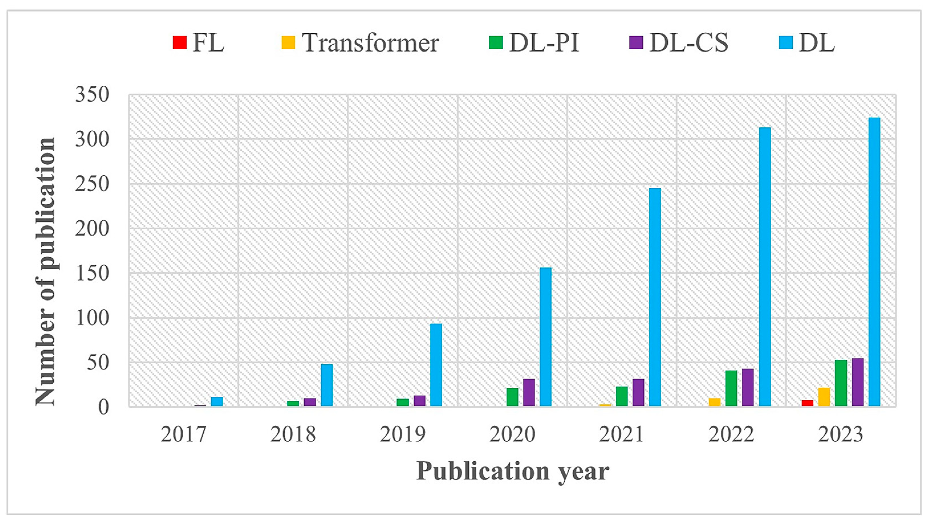
:1. Introduction
- Provide an overview of state-of-the-art DL-based MRI reconstruction techniques, including their advantages and disadvantages.
- Describe the potential of transfer learning (TL), and federated learning (FL) approaches for reducing computation complexity and addressing data scarcity and privacy issues in rapid MRI reconstruction.
- Discuss the advantages and challenges of transformer-based (widely used in natural language processing) networks in image capture, information matching, and reconstruction.
- Review the utilities of DL tools, medical-imaging competitions, and open-source codes in MRI.
- Describe publicly available k-space and image datasets for MRI reconstruction and analysis.
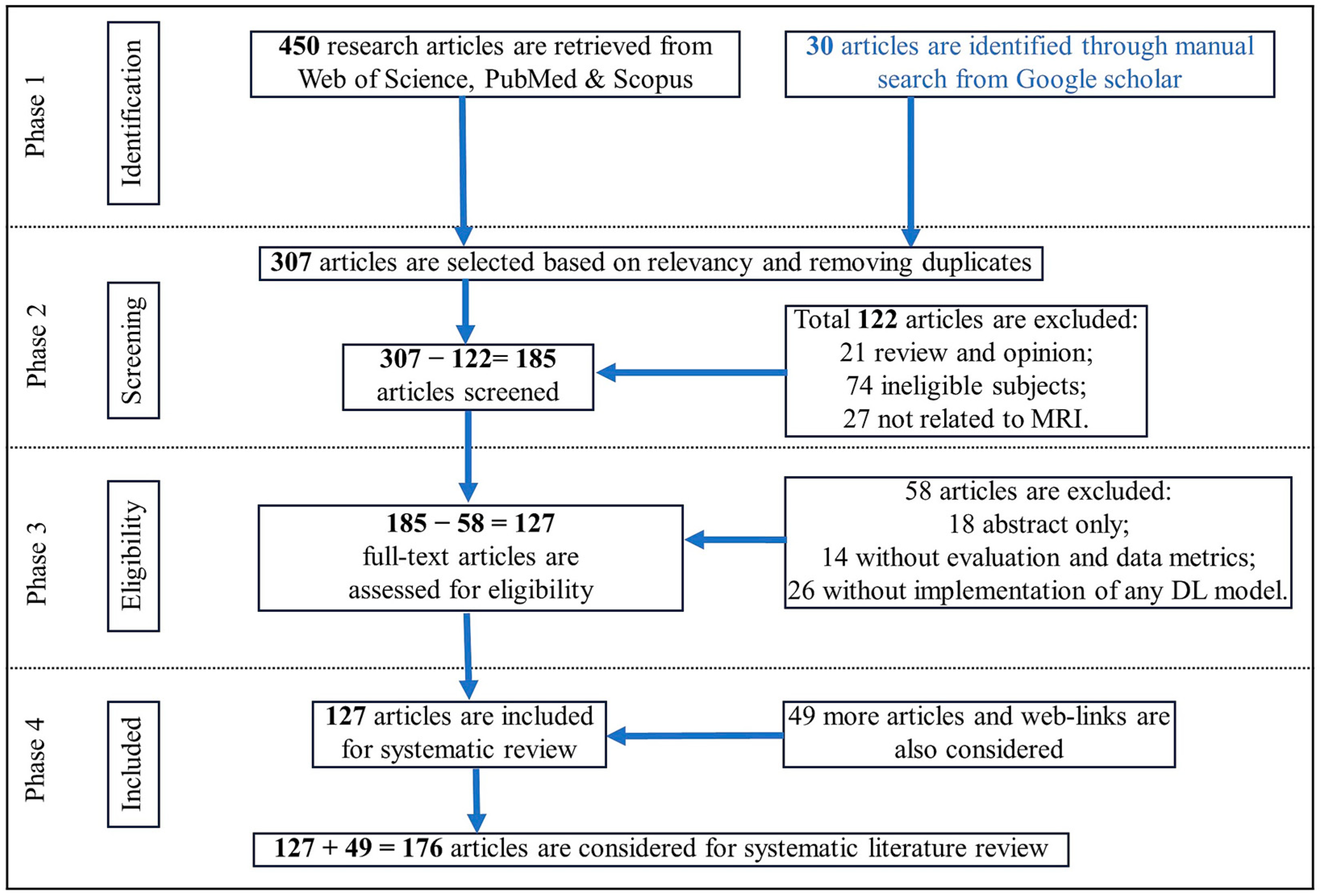
2. Motivation and Methodology
3. DL Frameworks and Tools
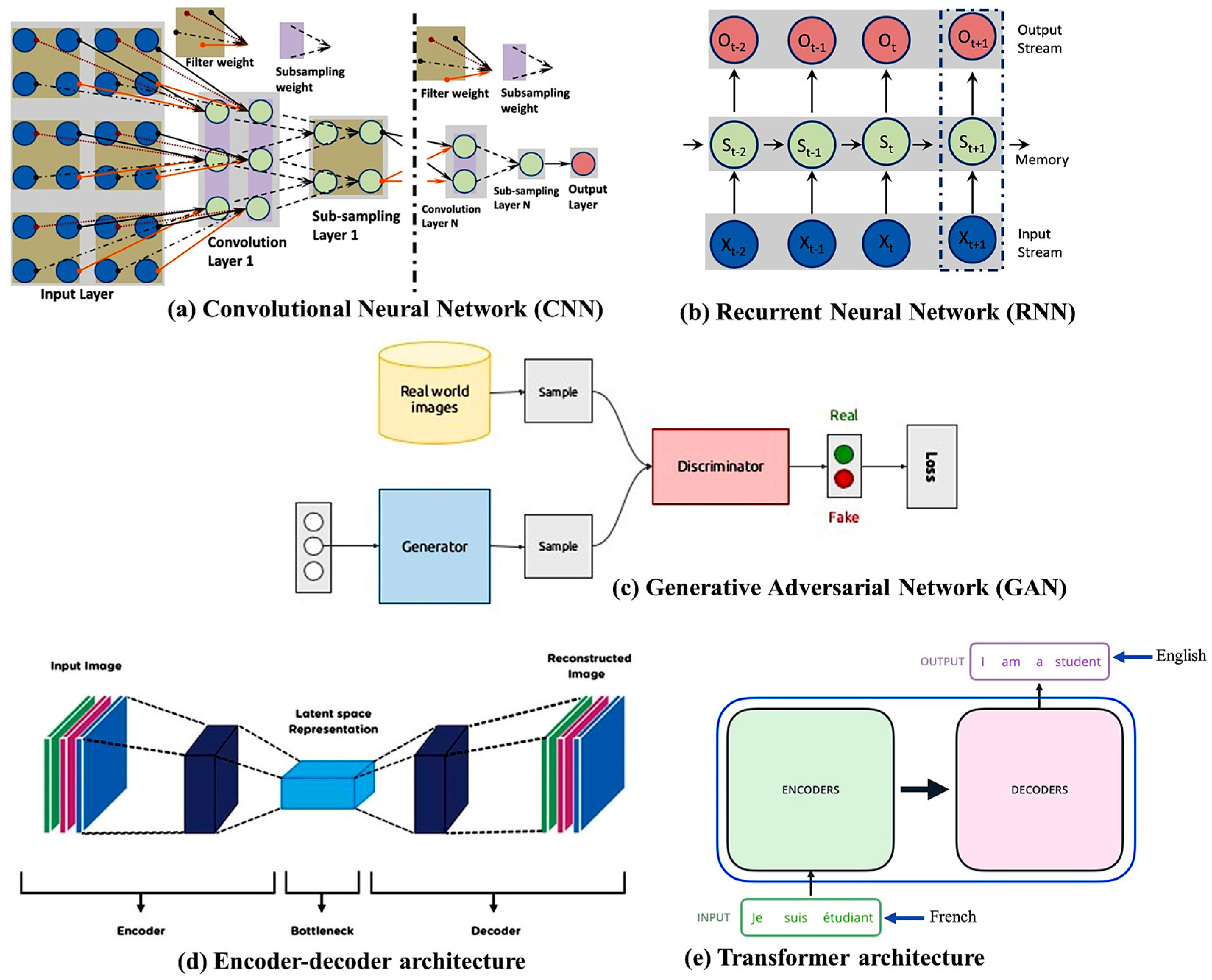
3.1. DL Architectures
3.2. DL Tools
3.3. Network Training Strategies
3.3.1. Supervised and Unsupervised Learning
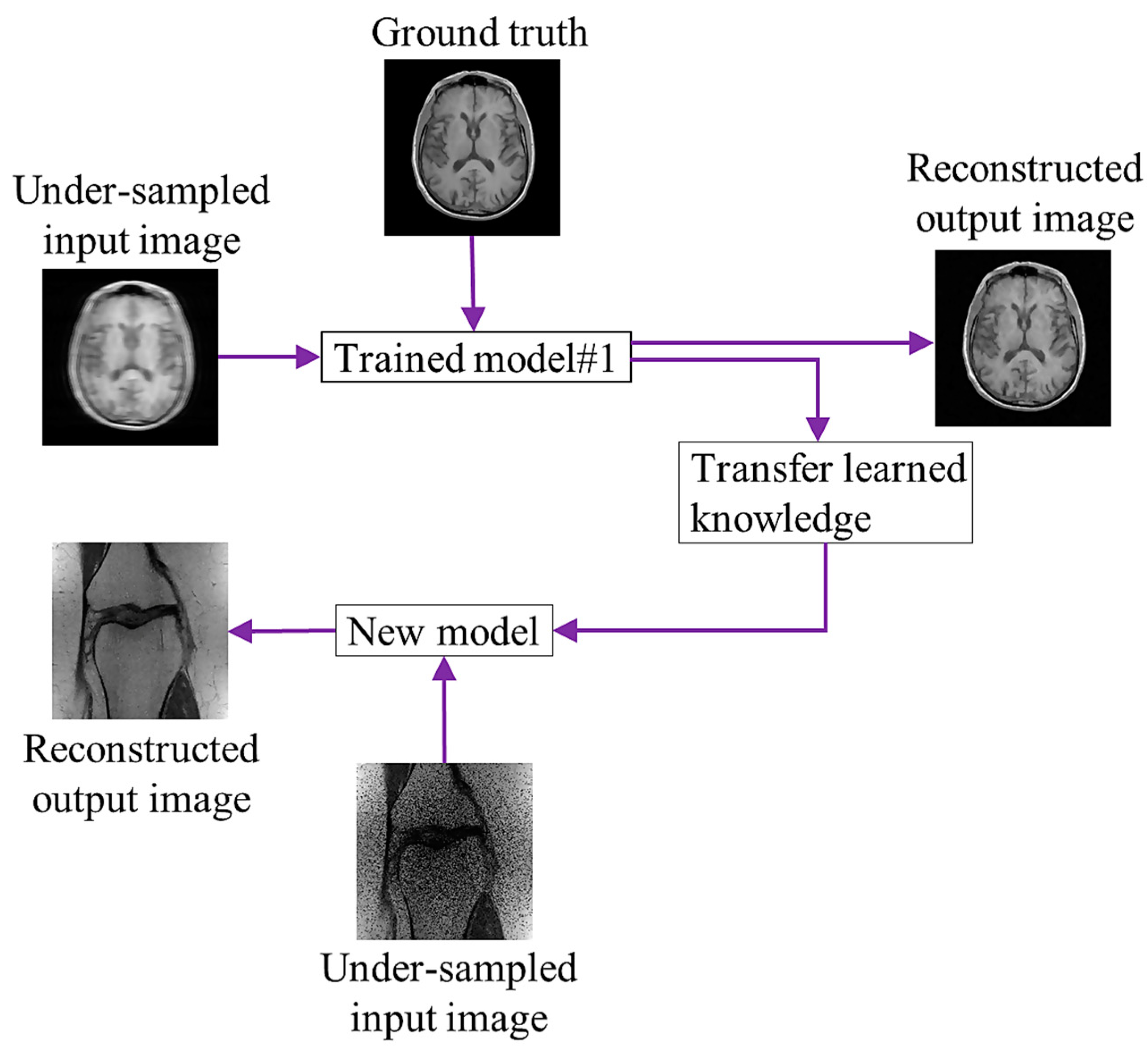
3.3.2. Transfer Learning
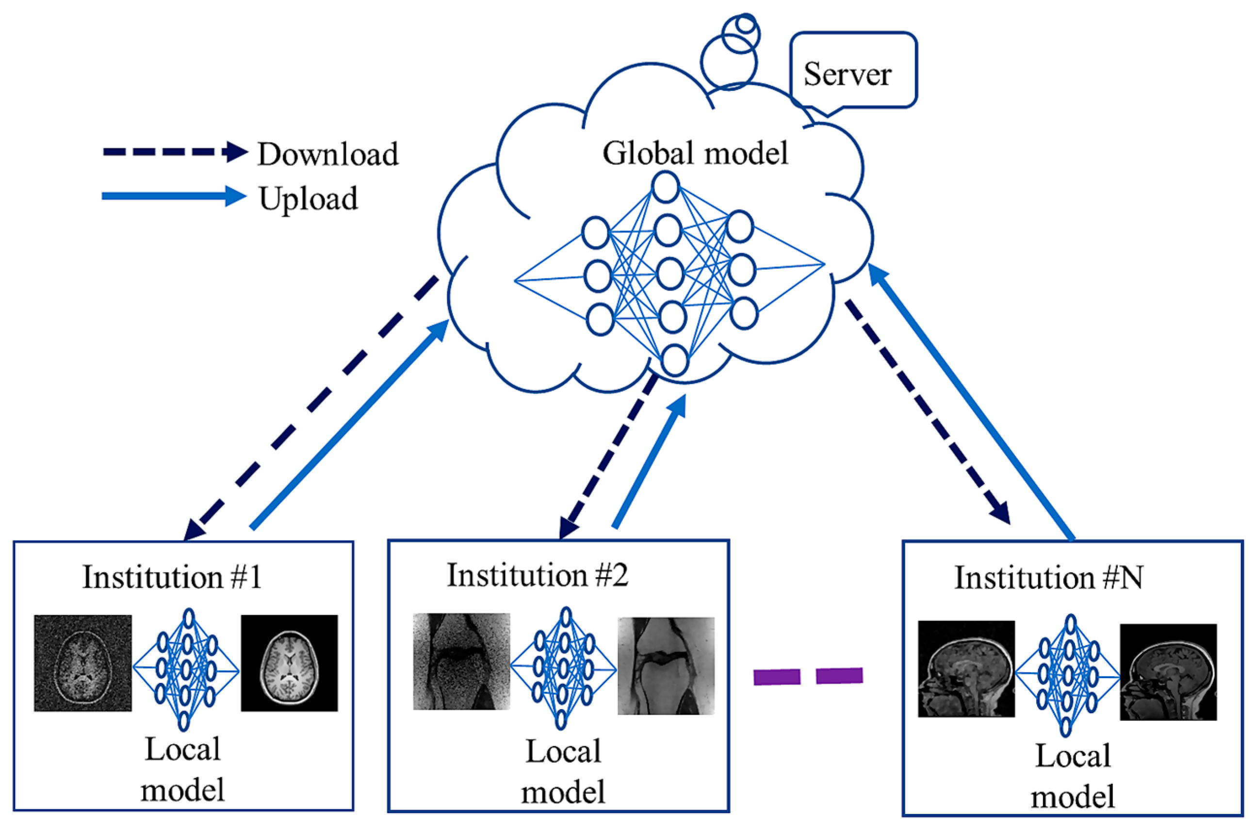
3.3.3. Federated Learning
4. MRI Reconstruction Methods
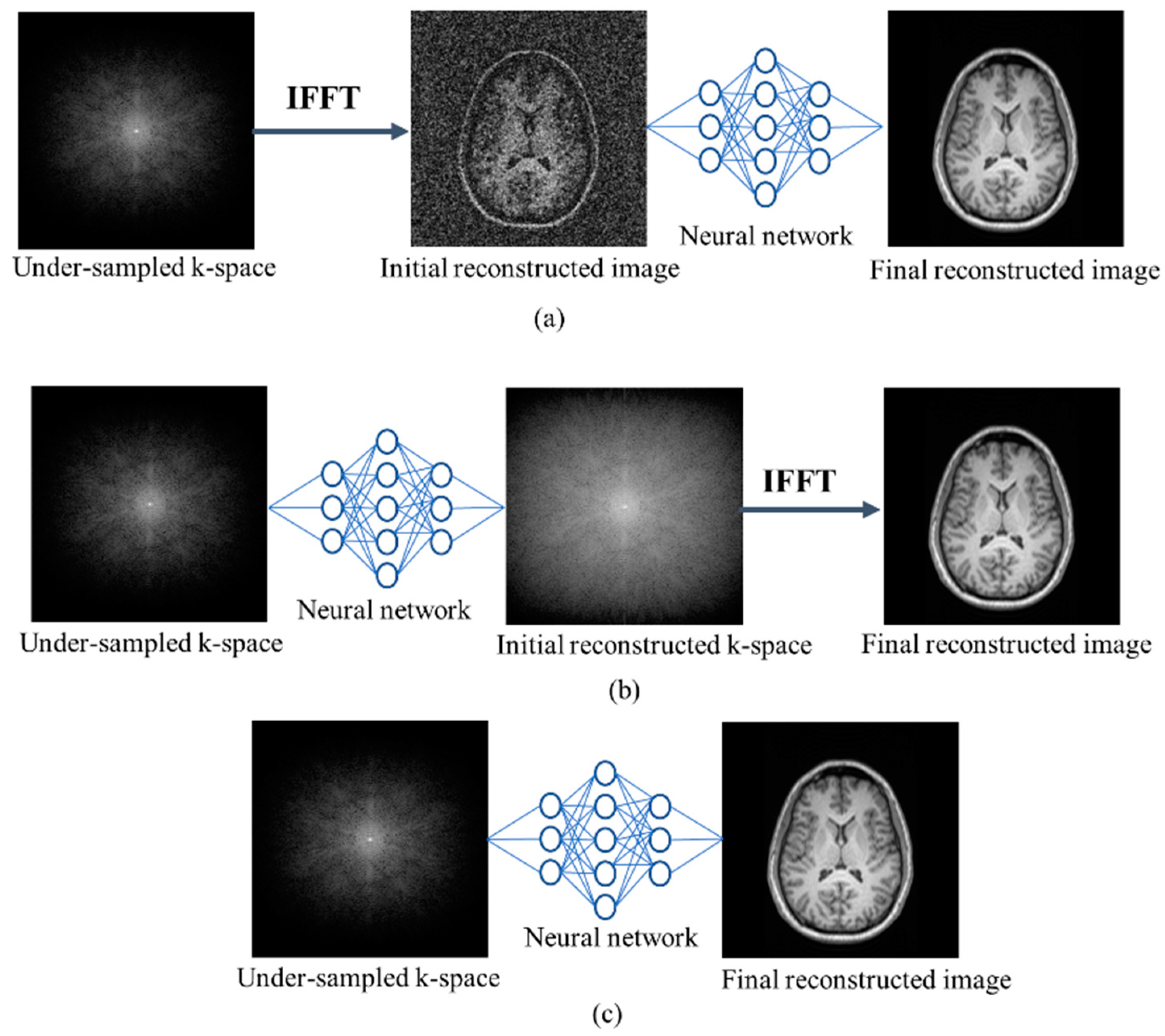
4.1. Single Domain Approach
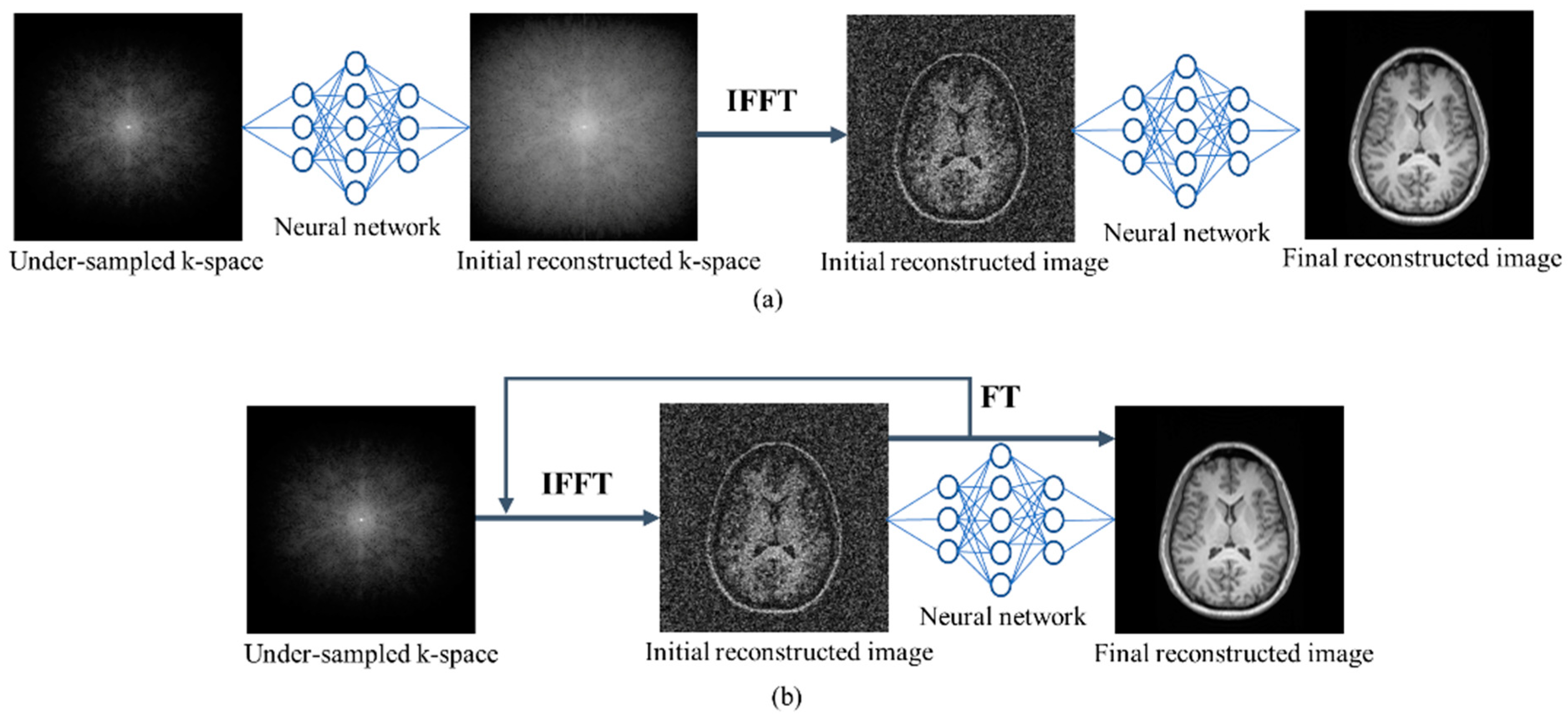
4.2. Multi Domain Approach
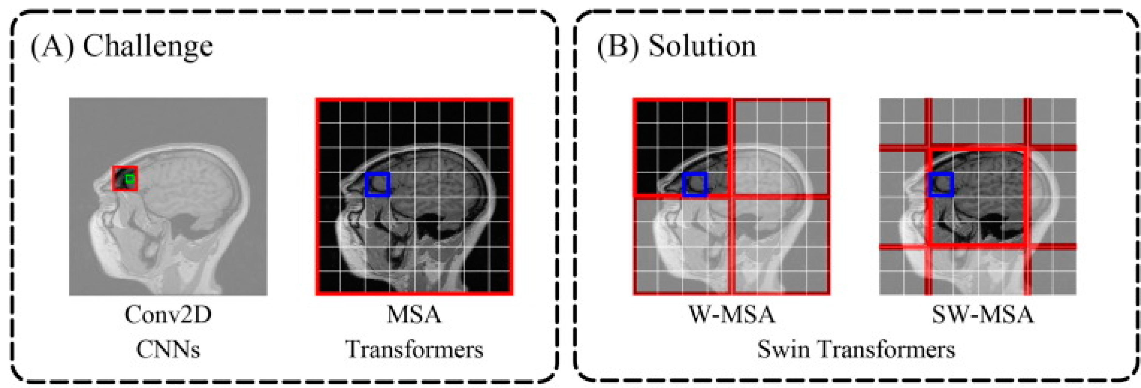
4.3. Transformer-Based Reconstruction
4.4. DL-Based 3D Reconstruction
5. Datasets and Source Codes
5.1. Datasets
5.2. Open-Source Codes
6. Implementation Challenges and Future Perspectives
7. Conclusions
Author Contributions
Funding
Conflicts of Interest
Abbreviations
| 2D | Two-Dimensional |
| 3D | Three-Dimensional |
| 4D | Four-Dimensional |
| ABIDE | Autism Brain Imaging Data Exchange |
| BIDS | Brain Imaging Data Structure |
| Brats | Brain Tumor Segmentation |
| CNN | Convolutional Neural Network |
| CS | Compressed Sensing |
| CS-MRI | Compressed Sensing-Magnetic Resonance Imaging |
| DICOM | Digital Imaging and Communications in Medicine |
| DL | Deep Learning |
| DNN | Deep Neural Network |
| DTI | Diffusion Tensor Imaging |
| FDA-CNN | Fully Dense Attention CNN |
| FL | Federated Learning |
| GAN | Generative Adversarial Network |
| HCP | Human Connectome Project |
| IBSR | Internet Brain Segmentation Repository |
| IFFT | Inverse Fast Fourier Transform |
| ISMRM | International Society For Magnetic Resonance In Medicine |
| LSTM | Long Short-Term Memory |
| MICCAI | Medical Image Computing And Computer-Assisted Intervention |
| MRI | Magnetic Resonance Imaging |
| MSA | Multi-Head Self-Attention |
| NIFTI | Neuroimaging Informatics Technology Initiative |
| NLP | Natural Language Processing |
| NN | Neural Network |
| OASIS | Open Access Series of Imaging Studies |
| PI | Parallel Imaging |
| PRISMA | Preferred Reporting Items for Systematic Reviews and Meta-Analyses |
| RA-CNN | Residual Attention CNN |
| RL | Reinforcement Learning |
| RNN | Recurrent Neural Network |
| Swin | Shifted Windows |
| SW-MSA | Shifted Windows-based Multi-head Self-Attention |
| T | Tesla |
| TL | Transfer Learning |
| VAE | Variational Autoencoder |
References
- Brown, R.W.; Cheng, Y.-C.N.; Haacke, E.M.; Thompson, M.R.; Venkatesan, R. Magnetic Resonance Imaging: Physical Principles and Sequence Design, 2nd ed.; John Wiley & Sons Ltd.: Chichester, UK, 2014; ISBN 9781118633953. [Google Scholar]
- Cercignani, M.; Dowell, N.G.; Tofts, P.S. Quantitative MRI of the Brain: Principles of Physical Measurement; CRC Press: Boca Raton, FL, USA, 2018; Volume 15, ISBN 9781315363578. [Google Scholar]
- Muckley, M.J.; Riemenschneider, B.; Radmanesh, A.; Kim, S.; Jeong, G.; Ko, J.; Jun, Y.; Shin, H.; Hwang, D.; Mostapha, M.; et al. Results of the 2020 FastMRI Challenge for Machine Learning MR Image Reconstruction. IEEE Trans. Med. Imaging 2021, 40, 2306–2317. [Google Scholar] [CrossRef]
- Deshmane, A.; Gulani, V.; Griswold, M.A.; Seiberlich, N. Parallel MR Imaging. J. Magn. Reson. Imaging 2012, 36, 55–72. [Google Scholar] [CrossRef] [PubMed]
- Lustig, M.; Donoho, D. Compressed Sensing MRI. Signal Process. Mag. 2008, 25, 72–82. [Google Scholar] [CrossRef]
- Griswold, M.A.; Jakob, P.M.; Heidemann, R.M.; Nittka, M.; Jellus, V.; Wang, J.; Kiefer, B.; Haase, A. Generalized Autocalibrating Partially Parallel Acquisitions (GRAPPA). Magn. Reson. Med. 2002, 47, 1202–1210. [Google Scholar] [CrossRef] [PubMed]
- Pruessmann, K.P.; Weiger, M.; Scheidegger, M.B.; Boesiger, P. SENSE: Sensitivity Encoding for Fast MRI. Magn. Reson. Med. 1999, 42, 952–962. [Google Scholar] [CrossRef]
- Hu, Z.; Zhao, C.; Zhao, X.; Kong, L.; Yang, J.; Wang, X.; Liao, J.; Zhou, Y. Joint Reconstruction Framework of Compressed Sensing and Nonlinear Parallel Imaging for Dynamic Cardiac Magnetic Resonance Imaging. BMC Med. Imaging 2021, 21, 182. [Google Scholar] [CrossRef]
- Islam, R.; Islam, M.S.; Uddin, M.S. Compressed Sensing in Parallel MRI: A Review. Int. J. Image Graph. 2022, 22, 2250038. [Google Scholar] [CrossRef]
- Lee, J.-G.; Jun, S.; Cho, Y.-W.; Lee, H.; Kim, G.B.; Seo, J.B.; Kim, N. Deep Learning in Medical Imaging: General Overview. Korean J. Radiol. 2017, 18, 570–584. [Google Scholar] [CrossRef]
- Zhang, Y.; Gorriz, J.M.; Dong, Z. Deep Learning in Medical Image Analysis. J. Imaging 2021, 7, 74. [Google Scholar] [CrossRef]
- Hossain, M.B.; Kwon, K.-C.; Shinde, R.K.; Imtiaz, S.M.; Kim, N. A Hybrid Residual Attention Convolutional Neural Network for Compressed Sensing Magnetic Resonance Image Reconstruction. Diagnostics 2023, 13, 1306. [Google Scholar] [CrossRef]
- Badža, M.M.; Barjaktarović, M.C. Classification of Brain Tumors from Mri Images Using a Convolutional Neural Network. Appl. Sci. 2020, 10, 1999. [Google Scholar] [CrossRef]
- Zhao, C.; Xiang, S.; Wang, Y.; Cai, Z.; Shen, J.; Zhou, S.; Zhao, D.; Su, W.; Guo, S.; Li, S. Context-Aware Network Fusing Transformer and V-Net for Semi-Supervised Segmentation of 3D Left Atrium. Expert Syst. Appl. 2023, 214, 119105. [Google Scholar] [CrossRef]
- Kim, S.; Park, S.; Na, B.; Yoon, S. Spiking-YOLO: Spiking Neural Network for Energy-Efficient Object Detection. Proc. AAAI Conf. Artif. Intell. 2020, 34, 11270–11277. [Google Scholar] [CrossRef]
- Ahishakiye, E.; Van Gijzen, M.B.; Tumwiine, J.; Wario, R.; Obungoloch, J. A Survey on Deep Learning in Medical Image Reconstruction. Intell. Med. 2021, 1, 118–127. [Google Scholar] [CrossRef]
- Montalt-Tordera, J.; Muthurangu, V.; Hauptmann, A.; Steeden, J.A. Machine Learning in Magnetic Resonance Imaging: Image Reconstruction. Phys. Medica 2021, 83, 79–87. [Google Scholar] [CrossRef] [PubMed]
- Zhang, H.M.; Dong, B. A Review on Deep Learning in Medical Image Reconstruction. J. Oper. Res. Soc. China 2020, 8, 311–340. [Google Scholar] [CrossRef]
- He, Z.; Quan, C.; Wang, S.; Zhu, Y.; Zhang, M.; Zhu, Y.; Liu, Q. A Comparative Study of Unsupervised Deep Learning Methods for MRI Reconstruction. Investig. Magn. Reson. Imaging 2020, 24, 179. [Google Scholar] [CrossRef]
- Knoll, F.; Hammernik, K.; Zhang, C.; Moeller, S.; Pock, T.; Sodickson, D.K.; Akcakaya, M. Deep-Learning Methods for Parallel Magnetic Resonance Imaging Reconstruction: A Survey of the Current Approaches, Trends, and Issues. IEEE Signal Process. Mag. 2020, 37, 128–140. [Google Scholar] [CrossRef]
- Singh, D.; Monga, A.; de Moura, H.L.; Zhang, X.; Zibetti, M.V.W.; Regatte, R.R. Emerging Trends in Fast MRI Using Deep-Learning Reconstruction on Undersampled k-Space Data: A Systematic Review. Bioengineering 2023, 10, 1012. [Google Scholar] [CrossRef]
- Bakator, M.; Radosav, D. Deep Learning and Medical Diagnosis: A Review of Literature. Multimodal Technol. Interact. 2018, 2, 47. [Google Scholar] [CrossRef]
- O’Shea, K.; Nash, R. An Introduction to Convolutional Neural Networks. arXiv 2015, arXiv:1511.08458. [Google Scholar]
- Khan, A.; Sohail, A.; Zahoora, U.; Qureshi, A.S. A Survey of the Recent Architectures of Deep Convolutional Neural Networks. Artif. Intell. Rev. 2020, 53, 5455–5516. [Google Scholar] [CrossRef]
- Krizhevsky, A.; Sutskever, I.; Hinton, G.E. ImageNet Classification with Deep Convolutional Neural Networks. Adv. Neural Inf. Process. Syst. 2012, 25, 145–151. [Google Scholar] [CrossRef]
- Shafiq, M.; Gu, Z. Deep Residual Learning for Image Recognition: A Survey. Appl. Sci. 2022, 12, 8972. [Google Scholar] [CrossRef]
- Shinde, R.K.; Alam, S.; Hossain, B.; Imtiaz, S.; Kim, J. Squeeze-MNet: Precise Skin Cancer Detection Model for Low Computing IoT Devices Using Transfer Learning. Cancers 2023, 14, 12. [Google Scholar] [CrossRef] [PubMed]
- Ronneberger, O.; Fischer, P.; Brox, T. U-Net: Convolutional Networks for Biomedical Image Segmentation; Lecture Notes in Computer Science (Including Subseries Lecture Notes in Artificial Intelligence and Lecture Notes in Bioinformatics); Springer: Berlin/Heidelberg, Germany, 2015; Volume 9351, pp. 234–241. ISBN 9783319245737. [Google Scholar]
- Ravi, D.; Wong, C.; Deligianni, F.; Berthelot, M.; Andreu-Perez, J.; Lo, B.; Yang, G.-Z. Deep Learning for Health Informatics. IEEE J. Biomed. Health Inform. 2017, 21, 4–21. [Google Scholar] [CrossRef] [PubMed]
- Sherstinsky, A. Fundamentals of Recurrent Neural Network (RNN) and Long Short-Term Memory (LSTM) Network. Phys. D Nonlinear Phenom. 2020, 404, 132306. [Google Scholar] [CrossRef]
- Ramadevi, R.; Marshiana, D.; Bestley, J.S.; Jamuna, R.D. Recurrent Neural Network (RNN) Analysis for Brain Tumor Classification Using Decision Tree Classifiers. J. Crit. Rev. 2020, 7, 2202–2205. [Google Scholar] [CrossRef]
- Alam, M.S.; Kwon, K.-C.; Md Imtiaz, S.; Hossain, M.B.; Kang, B.-G.; Kim, N. TARNet: An Efficient and Lightweight Trajectory-Based Air-Writing Recognition Model Using a CNN and LSTM Network. Hum. Behav. Emerg. Technol. 2022, 2022, 6063779. [Google Scholar] [CrossRef]
- Creswell, A.; White, T.; Dumoulin, V.; Arulkumaran, K.; Sengupta, B.; Bharath, A.A. Generative Adversarial Networks: An Overview. IEEE Signal Process. Mag. 2018, 35, 53–65. [Google Scholar] [CrossRef]
- Yoon, J.; Jordon, J.; Van Der Schaar, M. Supplementary Materials—RadialGAN: Leveraging Multiple Datasets to Improve Target-Specific Predictive Models Using Generative Adversarial Networks. Int. Conf. Mach. Learn. ICML 2018, 13, 9069–9071. [Google Scholar]
- Choi, Y.; Choi, M.; Kim, M.; Ha, J.W.; Kim, S.; Choo, J. StarGAN: Unified Generative Adversarial Networks for Multi-Domain Image-to-Image Translation. In Proceedings of the IEEE/CVF Conference on Computer Vision and Pattern Recognition, Salt Lake City, UT, USA, 18–23 June 2018; pp. 8789–8797. [Google Scholar] [CrossRef]
- Asadi, A.; Safabakhsh, R. The Encoder-Decoder Framework and Its Applications. In Deep Learning: Concepts and Architectures; Springer: Berlin/Heidelberg, Germany, 2020; pp. 133–167. [Google Scholar]
- Zhai, J.; Zhang, S.; Chen, J.; He, Q. Autoencoder and Its Various Variants. In Proceedings of the 2018 IEEE International Conference on Systems, Man, and Cybernetics (SMC), Miyazaki, Japan, 7–10 October 2018; pp. 415–419. [Google Scholar]
- Kingma, D.P.; Welling, M. An Introduction to Variational Autoencoders. Found. Trends Mach. Learn. 2019, 12, 307–392. [Google Scholar] [CrossRef]
- Patwardhan, N.; Marrone, S.; Sansone, C. Transformers in the Real World: A Survey on NLP Applications. Information 2023, 14, 242. [Google Scholar] [CrossRef]
- Carion, N.; Massa, F.; Synnaeve, G.; Usunier, N.; Kirillov, A.; Zagoruyko, S. End-to-End Object Detection with Transformers. In Lecture Notes in Computer Science; Springer: Cham, Switzerland, 2020; Volume 12346, pp. 213–229. [Google Scholar]
- Dosovitskiy, A.; Beyer, L.; Kolesnikov, A.; Weissenborn, D.; Zhai, X.; Unterthiner, T.; Dehghani, M.; Minderer, M.; Heigold, G.; Gelly, S.; et al. An Image Is Worth 16x16 Words: Transformers for Image Recognition at Scale. arXiv 2020, arXiv:2010.11929. [Google Scholar]
- Huang, J.; Wu, Y.; Wu, H.; Yang, G. Fast MRI Reconstruction: How Powerful Transformers Are? In Proceedings of the 2022 44th Annual International Conference of the IEEE Engineering in Medicine & Biology Society (EMBC), Glasgow, UK, 11–15 July 2022; pp. 2066–2070. [Google Scholar]
- Deeplearning4j. Available online: https://deeplearning4j.org/ (accessed on 4 July 2021).
- Julia. Available online: https://julialang.org/ (accessed on 4 July 2021).
- Keras. Available online: https://keras.io/ (accessed on 5 July 2021).
- MatConvNet. Available online: https://www.vlfeat.org/matconvnet/ (accessed on 5 July 2021).
- MS Cognitive Toolkit (CNTK). Available online: https://docs.microsoft.com/en-us/cognitive-toolkit/ (accessed on 5 July 2021).
- Neural Designer. Available online: https://www.neuraldesigner.com/ (accessed on 5 July 2021).
- PyTorch. Available online: https://pytorch.org/ (accessed on 6 July 2021).
- Scikit-Image. Available online: https://scikit-image.org/ (accessed on 6 July 2021).
- Sigpy. Available online: https://sigpy.readthedocs.io/en/latest/ (accessed on 6 July 2021).
- TensorFlow. Available online: https://www.tensorflow.org/ (accessed on 6 July 2021).
- TensorFlow Federated (TFF). Available online: https://www.tensorflow.org/federated (accessed on 15 November 2023).
- PySyft. Available online: https://blog.openmined.org/tag/pysyft/ (accessed on 20 November 2023).
- Substra. Available online: https://www.substra.ai/ (accessed on 10 December 2023).
- Ghahramani, Z. Unsupervised Learning. In Summer School on Machine Learning; Springer: Berlin/Heidelberg, Germany, 2004; pp. 72–112. [Google Scholar]
- Gong, K.; Han, P.; El Fakhri, G.; Ma, C.; Li, Q. Arterial Spin Labeling MR Image Denoising and Reconstruction Using Unsupervised Deep Learning. NMR Biomed. 2022, 35, e4224. [Google Scholar] [CrossRef] [PubMed]
- Aggarwal, H.K.; Pramanik, A.; John, M.; Jacob, M. ENSURE: A General Approach for Unsupervised Training of Deep Image Reconstruction Algorithms. IEEE Trans. Med. Imaging 2023, 42, 1133–1144. [Google Scholar] [CrossRef] [PubMed]
- Wei, R.; Chen, J.; Liang, B.; Chen, X.; Men, K.; Dai, J. Real-time 3D MRI Reconstruction from Cine-MRI Using Unsupervised Network in MRI-guided Radiotherapy for Liver Cancer. Med. Phys. 2023, 50, 3584–3596. [Google Scholar] [CrossRef]
- Yurt, M.; Dalmaz, O.; Dar, S.; Ozbey, M.; Tinaz, B.; Oguz, K.; Cukur, T. Semi-Supervised Learning of MRI Synthesis without Fully-Sampled Ground Truths. IEEE Trans. Med. Imaging 2022, 41, 3895–3906. [Google Scholar] [CrossRef]
- Hu, C.; Li, C.; Wang, H.; Liu, Q.; Zheng, H.; Wang, S. Self-Supervised Learning for MRI Reconstruction with a Parallel Network Training Framework. In Medical Image Computing and Computer Assisted Intervention—MICCAI 2021; Springer: Cham, Switzerland, 2021; pp. 382–391. [Google Scholar]
- Torrey, L.; Shavlik, J. Transfer Learning. In Handbook of Research on Machine Learning Applications and Trends; IGI Global: Hershey, PA, USA, 2010; pp. 242–264. [Google Scholar]
- Dar, S.U.H.; Özbey, M.; Çatlı, A.B.; Çukur, T. A Transfer-Learning Approach for Accelerated MRI Using Deep Neural Networks. Magn. Reson. Med. 2020, 84, 663–685. [Google Scholar] [CrossRef]
- Arshad, M.; Qureshi, M.; Inam, O.; Omer, H. Transfer Learning in Deep Neural Network Based Under-Sampled MR Image Reconstruction. Magn. Reson. Imaging 2021, 76, 96–107. [Google Scholar] [CrossRef]
- Lv, J.; Li, G.; Tong, X.; Chen, W.; Huang, J.; Wang, C.; Yang, G. Transfer Learning Enhanced Generative Adversarial Networks for Multi-Channel MRI Reconstruction. Comput. Biol. Med. 2021, 134, 104504. [Google Scholar] [CrossRef] [PubMed]
- Yaqub, M.; Jinchao, F.; Ahmed, S.; Arshid, K.; Bilal, M.A.; Akhter, M.P.; Zia, M.S. GAN-TL: Generative Adversarial Networks with Transfer Learning for MRI Reconstruction. Appl. Sci. 2022, 12, 8841. [Google Scholar] [CrossRef]
- Park, S.J.; Ahn, C.-B. Blended-Transfer Learning for Compressed-Sensing Cardiac CINE MRI. Investig. Magn. Reson. Imaging 2021, 25, 10. [Google Scholar] [CrossRef]
- Cheng, C.; Lin, D. MRI Reconstruction Based on Transfer Learning Dynamic Dictionary Algorithm. In Proceedings of the 2023 2nd International Conference on Big Data, Information and Computer Network (BDICN), Xishuangbanna, China, 6–8 January 2023; pp. 1–4. [Google Scholar]
- Gulamhussene, G.; Rak, M.; Bashkanov, O.; Joeres, F.; Omari, J.; Pech, M.; Hansen, C. Transfer-Learning Is a Key Ingredient to Fast Deep Learning-Based 4D Liver MRI Reconstruction. Sci. Rep. 2023, 13, 11227. [Google Scholar] [CrossRef] [PubMed]
- Yang, Q.; Liu, Y.; Cheng, Y.; Kang, Y.; Chen, T.; Yu, H. Federated Learning; Synthesis Lectures on Artificial Intelligence and Machine Learning Series; Springer: Cham, Switzerland, 2019; Volume 13, pp. 1–207. [Google Scholar] [CrossRef]
- Li, X.; Gu, Y.; Dvornek, N.; Staib, L.H.; Ventola, P.; Duncan, J.S. Multi-Site FMRI Analysis Using Privacy-Preserving Federated Learning and Domain Adaptation: ABIDE Results. Med. Image Anal. 2020, 65, 101765. [Google Scholar] [CrossRef] [PubMed]
- Guo, P.; Wang, P.; Zhou, J.; Jiang, S.; Patel, V.M. Multi-Institutional Collaborations for Improving Deep Learning-Based Magnetic Resonance Image Reconstruction Using Federated Learning. In Proceedings of the 2021 IEEE/CVF Conference on Computer Vision and Pattern Recognition (CVPR), Nashville, TN, USA, 20–25 June 2021; pp. 2423–2432. [Google Scholar] [CrossRef]
- Feng, C.M.; Yan, Y.; Wang, S.; Xu, Y.; Shao, L.; Fu, H. Specificity-Preserving Federated Learning for MR Image Reconstruction. IEEE Trans. Med. Imaging 2022, 26, 2010–2021. [Google Scholar] [CrossRef] [PubMed]
- Elmas, G.; Dar, S.U.; Korkmaz, Y.; Ceyani, E.; Susam, B.; Ozbey, M.; Avestimehr, S.; Cukur, T. Federated Learning of Generative Image Priors for MRI Reconstruction. IEEE Trans. Med. Imaging 2022, 9, 1996–2009. [Google Scholar] [CrossRef]
- Levac, B.R.; Arvinte, M.; Tamir, J.I. Federated End-to-End Unrolled Models for Magnetic Resonance Image Reconstruction. Bioengineering 2023, 10, 364. [Google Scholar] [CrossRef]
- Feng, C.-M.; Li, B.; Xu, X.; Liu, Y.; Fu, H.; Zuo, W. Learning Federated Visual Prompt in Null Space for MRI Reconstruction. In Proceedings of the IEEE/CVF Conference on Computer Vision and Pattern Recognition (CVPR), Vancouver, BC, Canada, 18–22 June 2023. [Google Scholar]
- Sandhu, S.S.; Gorji, H.T.; Tavakolian, P.; Tavakolian, K.; Akhbardeh, A. Medical Imaging Applications of Federated Learning. Diagnostics 2023, 13, 3140. [Google Scholar] [CrossRef]
- Li, T.; Sahu, A.K.; Talwalkar, A.; Smith, V. Federated Learning: Challenges, Methods, and Future Directions. IEEE Signal Process. Mag. 2020, 37, 50–60. [Google Scholar] [CrossRef]
- Cummings, E.; Macdonald, J.A.; Seiberlich, N. Parallel Imaging. In Advances in Magnetic Resonance Technology and Applications; Elsevier: Amsterdam, The Netherlands, 2022; pp. 129–157. [Google Scholar]
- Li, W.; Feng, X.; An, H.; Ng, X.Y.; Zhang, Y.J. MRI Reconstruction with Interpretable Pixel-Wise Operations Using Reinforcement Learning. Proc. AAAI Conf. Artif. Intell. 2020, 34, 792–799. [Google Scholar] [CrossRef]
- Schlemper, J.; Caballero, J.; Hajnal, J.V.; Price, A.N.; Rueckert, D. A Deep Cascade of Convolutional Neural Networks for Dynamic MR Image Reconstruction. IEEE Trans. Med. Imaging 2018, 37, 491–503. [Google Scholar] [CrossRef]
- Yang, G.; Yu, S.; Dong, H.; Slabaugh, G.; Dragotti, P.L.; Ye, X.; Liu, F.; Arridge, S.; Keegan, J.; Guo, Y.; et al. DAGAN: Deep de-Aliasing Generative Adversarial Networks for Fast Compressed Sensing MRI Reconstruction. IEEE Trans. Med. Imaging 2018, 37, 1310–1321. [Google Scholar] [CrossRef]
- Quan, T.M.; Nguyen-Duc, T.; Jeong, W.K. Compressed Sensing MRI Reconstruction Using a Generative Adversarial Network with a Cyclic Loss. IEEE Trans. Med. Imaging 2018, 37, 1488–1497. [Google Scholar] [CrossRef]
- Chen, Y.; Christodoulou, A.G.; Zhou, Z.; Shi, F.; Xie, Y.; Li, D. MRI Super-Resolution with GAN and 3D Multi-Level DenseNet: Smaller, Faster, and Better. arXiv 2020, arXiv:2003.01217. [Google Scholar]
- Nath, R.; Callahan, S.; Singam, N.; Stoddard, M.; Amini, A.A. Accelerated Phase Contrast Magnetic Resonance Imaging via Deep Learning. In Proceedings of the 2020 IEEE 17th International Symposium on Biomedical Imaging (ISBI), Iowa City, IA, USA, 3–7 April 2020; pp. 834–838. [Google Scholar] [CrossRef]
- Hennig, J. K-Space Sampling Strategies. Eur. Radiol. 1999, 9, 1020–1031. [Google Scholar] [CrossRef]
- Zhu, Y.; Gao, S.; Cheng, L.; Bao, S. Review: K-Space Trajectory Development. In Proceedings of the 2013 IEEE International Conference on Medical Imaging Physics and Engineering, Shenyang, China, 19–20 October 2013; pp. 356–360. [Google Scholar]
- Hossain, M.B.; Kwon, K.-C.; Imtiaz, S.M.; Nam, O.-S.; Jeon, S.-H.; Kim, N. De-Aliasing and Accelerated Sparse Magnetic Resonance Image Reconstruction Using Fully Dense CNN with Attention Gates. Bioengineering 2022, 10, 22. [Google Scholar] [CrossRef]
- Jiang, M.; Zhi, M.; Wei, L.; Yang, X.; Zhang, J.; Li, Y.; Wang, P.; Huang, J.; Yang, G. FA-GAN: Fused Attentive Generative Adversarial Networks for MRI Image Super-Resolution. Comput. Med. Imaging Graph. 2021, 92, 101969. [Google Scholar] [CrossRef]
- Zhang, K.; Hu, H.; Philbrick, K.; Conte, G.M.; Sobek, J.D.; Rouzrokh, P.; Erickson, B.J. SOUP-GAN: Super-Resolution MRI Using Generative Adversarial Networks. Tomography 2022, 8, 905–919. [Google Scholar] [CrossRef]
- Edupuganti, V.; Mardani, M.; Vasanawala, S.; Pauly, J. Uncertainty Quantification in Deep MRI Reconstruction. IEEE Trans. Med. Imaging 2021, 40, 239–250. [Google Scholar] [CrossRef]
- Gao, Z.; Guo, Y.; Zhang, J.; Zeng, T.; Yang, G. Hierarchical Perception Adversarial Learning Framework for Compressed Sensing MRI. IEEE Trans. Med. Imaging 2023, 42, 1859–1874. [Google Scholar] [CrossRef]
- Du, T.; Zhang, Y.; Shi, X.; Chen, S. Multiple Slice K-Space Deep Learning for Magnetic Resonance Imaging Reconstruction. In Proceedings of the 2020 42nd Annual International Conference of the IEEE Engineering in Medicine & Biology Society (EMBC), Montreal, QC, Canada, 20–24 July 2020; pp. 1564–1567. [Google Scholar] [CrossRef]
- Du, T.; Zhang, H.; Li, Y.; Pickup, S.; Rosen, M.; Zhou, R.; Song, H.K.; Fan, Y. Adaptive Convolutional Neural Networks for Accelerating Magnetic Resonance Imaging via K-Space Data Interpolation. Med. Image Anal. 2021, 72, 102098. [Google Scholar] [CrossRef]
- Han, Y.; Sunwoo, L.; Ye, J.C. K-Space Deep Learning for Accelerated MRI. IEEE Trans. Med. Imaging 2020, 39, 377–386. [Google Scholar] [CrossRef]
- Jin, K.H.; Lee, D.; Ye, J.C. A General Framework for Compressed Sensing and Parallel MRI Using Annihilating Filter Based Low-Rank Hankel Matrix. IEEE Trans. Comput. Imaging 2016, 2, 480–495. [Google Scholar] [CrossRef]
- Pineda, L.; Basu, S.; Romero, A.; Calandra, R.; Drozdzal, M. Active MR K-Space Sampling with Reinforcement Learning; Lecture Notes in Computer Science (Including Subseries Lecture Notes in Artificial Intelligence and Lecture Notes in Bioinformatics); Springer: Cham, Switzerland, 2020; Volume 12262, pp. 23–33. ISBN 9783030597122. [Google Scholar]
- Arefeen, Y.; Beker, O.; Cho, J.; Yu, H.; Adalsteinsson, E.; Bilgic, B. Scan-specific Artifact Reduction in K-space (SPARK) Neural Networks Synergize with Physics-based Reconstruction to Accelerate MRI. Magn. Reson. Med. 2022, 87, 764–780. [Google Scholar] [CrossRef]
- Kim, T.H.; Garg, P.; Haldar, J.P. LORAKI: Autocalibrated Recurrent Neural Networks for Autoregressive MRI Reconstruction in k-Space. arXiv 2019, arXiv:1904.09390. [Google Scholar]
- Yiasemis, G.; Sonke, J.-J.; Sánchez, C.; Jonas, T. Recurrent Variational Network: A Deep Learning Inverse Problem Solver Applied to the Task of Accelerated MRI Reconstruction. In Proceedings of the 2022 IEEE/CVF Conference on Computer Vision and Pattern Recognition (CVPR), New Orleans, LA, USA, 21–24 June 2022; pp. 722–731. [Google Scholar]
- Zhu, B.; Liu, J.Z.; Cauley, S.F.; Rosen, B.R.; Rosen, M.S. Image Reconstruction by Domain-Transform Manifold Learning. Nature 2018, 555, 487–492. [Google Scholar] [CrossRef]
- Oh, C.; Kim, D.; Chung, J.-Y.; Han, Y.; Park, H. ETER-Net: End to End MR Image Reconstruction Using Recurrent Neural Network; Lecture Notes in Computer Science (Including Subseries Lecture Notes in Artificial Intelligence and Lecture Notes in Bioinformatics); Springer: Cham, Switzerland, 2018; Volume 11074, pp. 12–20. ISBN 9783030001285. [Google Scholar]
- Eo, T.; Jun, Y.; Kim, T.; Jang, J.; Lee, H.J.; Hwang, D. KIKI-Net: Cross-Domain Convolutional Neural Networks for Reconstructing Undersampled Magnetic Resonance Images. Magn. Reson. Med. 2018, 80, 2188–2201. [Google Scholar] [CrossRef]
- Souza, R.; Lebel, R.M.; Frayne, R. A Hybrid, Dual Domain, Cascade of Convolutional Neural Networks for Magnetic Resonance Image Reconstruction. Proc. Mach. Learn. Res. 2019, 102, 437–446. [Google Scholar]
- Qin, C.; Schlemper, J.; Caballero, J.; Price, A.N.; Hajnal, J.V.; Rueckert, D. Convolutional Recurrent Neural Networks for Dynamic MR Image Reconstruction. IEEE Trans. Med. Imaging 2019, 38, 280–290. [Google Scholar] [CrossRef]
- Souza, R.; Bento, M.; Nogovitsyn, N.; Chung, K.J.; Loos, W.; Lebel, R.M.; Frayne, R. Dual-Domain Cascade of U-Nets for Multi-Channel Magnetic Resonance Image Reconstruction. Magn. Reson. Imaging 2020, 71, 140–153. [Google Scholar] [CrossRef] [PubMed]
- Sun, L.; Wu, Y.; Shu, B.; Ding, X.; Cai, C.; Huang, Y.; Paisley, J. A Dual-Domain Deep Lattice Network for Rapid MRI Reconstruction. Neurocomputing 2020, 397, 94–107. [Google Scholar] [CrossRef]
- Wang, Z.; Jiang, H.; Du, H.; Xu, J.; Qiu, B. IKWI-Net: A Cross-Domain Convolutional Neural Network for Undersampled Magnetic Resonance Image Reconstruction. Magn. Reson. Imaging 2020, 73, 1–10. [Google Scholar] [CrossRef]
- El-Rewaidy, H.; Fahmy, A.S.; Pashakhanloo, F.; Cai, X.; Kucukseymen, S.; Csecs, I.; Neisius, U.; Haji-Valizadeh, H.; Menze, B.; Nezafat, R. Multi-Domain Convolutional Neural Network (MD-CNN) for Radial Reconstruction of Dynamic Cardiac MRI. Magn. Reson. Med. 2021, 85, 1195–1208. [Google Scholar] [CrossRef] [PubMed]
- Ran, M.; Xia, W.; Huang, Y.; Lu, Z.; Bao, P.; Liu, Y.; Sun, H.; Zhou, J.; Zhang, Y. MD-Recon-Net: A Parallel Dual-Domain Convolutional Neural Network for Compressed Sensing MRI. IEEE Trans. Radiat. Plasma Med. Sci. 2021, 5, 120–135. [Google Scholar] [CrossRef]
- Wei, H.; Li, Z.; Wang, S.; Li, R. Undersampled Multi-Contrast MRI Reconstruction Based on Double-Domain Generative Adversarial Network. IEEE J. Biomed. Health Inform. 2022, 26, 4371–4377. [Google Scholar] [CrossRef]
- Cheng, J.; Wang, H.; Ying, L.; Liang, D. Model Learning: Primal Dual Networks for Fast MR Imaging; Lecture Notes in Computer Science (Including Subseries Lecture Notes in Artificial Intelligence and Lecture Notes in Bioinformatics); Springer: Cham, Switzerland, 2019; Volume 11766, pp. 21–29. [Google Scholar] [CrossRef]
- Zhang, X.; Lian, Q.; Yang, Y.; Su, Y. A Deep Unrolling Network Inspired by Total Variation for Compressed Sensing MRI. Digit. Signal Process. 2020, 107, 102856. [Google Scholar] [CrossRef]
- Hosseini, S.A.H.; Yaman, B.; Moeller, S.; Hong, M.; Akcakaya, M. Dense Recurrent Neural Networks for Accelerated MRI: History-Cognizant Unrolling of Optimization Algorithms. IEEE J. Sel. Top. Signal Process. 2020, 14, 1280–1291. [Google Scholar] [CrossRef]
- Jain, P.; Pradeep, C.S.; Sinha, N. The Complex-Valued PD-Net for MRI Reconstruction of Knee Images. In Proceedings of the 2022 44th Annual International Conference of the IEEE Engineering in Medicine & Biology Society (EMBC), Glasgow, UK, 11–15 July 2022; pp. 2093–2096. [Google Scholar]
- Lin, T.; Wang, Y.; Liu, X.; Qiu, X. A Survey of Transformers. AI Open 2022, 3, 111–132. [Google Scholar] [CrossRef]
- Huang, J.; Fang, Y.; Wu, Y.; Wu, H.; Gao, Z.; Li, Y.; Del Ser, J.; Xia, J.; Yang, G. Swin Transformer for Fast MRI. Neurocomputing 2022, 493, 281–304. [Google Scholar] [CrossRef]
- Zhou, B.; Dey, N.; Schlemper, J.; Salehi, S.S.M.; Liu, C.; Duncan, J.S.; Sofka, M. DSFormer: A Dual-Domain Self-Supervised Transformer for Accelerated Multi-Contrast MRI Reconstruction. In Proceedings of the IEEE/CVF Winter Conference on Applications of Computer Vision, Waikoloa, HI, USA, 3–7 January 2023; pp. 4966–4975. [Google Scholar]
- Korkmaz, Y.; Dar, S.U.H.; Yurt, M.; Ozbey, M.; Cukur, T. Unsupervised MRI Reconstruction via Zero-Shot Learned Adversarial Transformers. IEEE Trans. Med. Imaging 2022, 41, 1747–1763. [Google Scholar] [CrossRef] [PubMed]
- Liu, J.; Qin, C.; Yaghoobi, M. High-Fidelity MRI Reconstruction Using Adaptive Spatial Attention Selection and Deep Data Consistency Prior. IEEE Trans. Comput. Imaging 2023, 9, 298–313. [Google Scholar] [CrossRef]
- Huang, J.; Ding, W.; Lv, J.; Yang, J.; Dong, H.; Del Ser, J.; Xia, J.; Ren, T.; Wong, S.T.; Yang, G. Edge-Enhanced Dual Discriminator Generative Adversarial Network for Fast MRI with Parallel Imaging Using Multi-View Information. Appl. Intell. 2022, 52, 14693–14710. [Google Scholar] [CrossRef] [PubMed]
- Lyu, J.; Li, G.; Wang, C.; Qin, C.; Wang, S.; Dou, Q.; Qin, J. Region-Focused Multi-View Transformer-Based Generative Adversarial Network for Cardiac Cine MRI Reconstruction. Med. Image Anal. 2023, 85, 102760. [Google Scholar] [CrossRef] [PubMed]
- Li, Y.; Wang, F.; Hu, X. Deep-Learning-Based 3D Reconstruction: A Review and Applications. Appl. Bionics Biomech. 2022, 2022, 3458717. [Google Scholar] [CrossRef]
- Samavati, T.; Soryani, M. Deep Learning-Based 3D Reconstruction: A Survey. Artif. Intell. Rev. 2023, 56, 9175–9219. [Google Scholar] [CrossRef]
- Kang, S.K.; Shin, S.A.; Seo, S.; Byun, M.S.; Lee, D.Y.; Kim, Y.K.; Lee, D.S.; Lee, J.S. Deep Learning-Based 3D Inpainting of Brain MR Images. Sci. Rep. 2021, 11, 1673. [Google Scholar] [CrossRef] [PubMed]
- Ahn, S.; Wollner, U.; McKinnon, G.; Jansen, I.H.; Brada, R.; Rettmann, D.; Cashen, T.A.; Huston, J.; DeMarco, J.K.; Shih, R.Y.; et al. Deep Learning-Based Reconstruction of Highly Accelerated 3D MRI. arXiv 2022, arXiv:2203.04674. [Google Scholar]
- Jurek, J.; Kociński, M.; Materka, A.; Elgalal, M.; Majos, A. CNN-Based Superresolution Reconstruction of 3D MR Images Using Thick-Slice Scans. Biocybern. Biomed. Eng. 2020, 40, 111–125. [Google Scholar] [CrossRef]
- Küstner, T.; Fuin, N.; Hammernik, K.; Bustin, A.; Qi, H.; Hajhosseiny, R.; Masci, P.G.; Neji, R.; Rueckert, D.; Botnar, R.M.; et al. CINENet: Deep Learning-Based 3D Cardiac CINE MRI Reconstruction with Multi-Coil Complex-Valued 4D Spatio-Temporal Convolutions. Sci. Rep. 2020, 10, 13710. [Google Scholar] [CrossRef]
- Volokitin, A.; Erdil, E.; Karani, N.; Tezcan, K.C.; Chen, X.; Van Gool, L.; Konukoglu, E. Modelling the Distribution of 3D Brain MRI Using a 2D Slice VAE. In Proceedings of the Medical Image Computing and Computer Assisted Intervention–MICCAI 2020: 23rd International Conference, Lima, Peru, 4–8 October 2020; pp. 657–666. [Google Scholar]
- Zhang, H.; Shinomiya, Y.; Yoshida, S. 3D MRI Reconstruction Based on 2D Generative Adversarial Network Super-Resolution. Sensors 2021, 21, 2978. [Google Scholar] [CrossRef] [PubMed]
- Kwon, G.; Han, C.; Kim, D. Generation of 3D Brain MRI Using Auto-Encoding Generative Adversarial Networks. In International Conference on Medical Image Computing and Computer-Assisted Intervention; Springer: Cham, Switzerland, 2019; pp. 118–126. [Google Scholar]
- Nabulsi, Z.; Kosaraju, V.; Chakraborty, S. MRNGAN: Reconstructing 3D MRI Scans Using A Recurrent Generative Model. Available online: https://vineetkosaraju.com/papers/mrngan.pdf (accessed on 5 January 2024).
- Zou, Q.; Miller, Z.; Dzelebdzic, S.; Abadeer, M.; Johnson, K.M.; Hussain, T. Time-Resolved 3D Cardiopulmonary MRI Reconstruction Using Spatial Transformer Network. Math. Biosci. Eng. 2023, 20, 15982–15998. [Google Scholar] [CrossRef]
- Inati, S.J.; Naegele, J.D.; Zwart, N.R.; Roopchansingh, V.; Lizak, M.J.; Hansen, D.C.; Liu, C.Y.; Atkinson, D.; Kellman, P.; Kozerke, S.; et al. ISMRM Raw Data Format: A Proposed Standard for MRI Raw Datasets. Magn. Reson. Med. 2017, 77, 411–421. [Google Scholar] [CrossRef] [PubMed]
- Medical Image Computing and Computer Assisted Intervention (MICCAI). Available online: http://www.miccai.org/ (accessed on 20 October 2021).
- FastMRI Challenge-2020. Available online: https://fastmri.org/ (accessed on 26 October 2021).
- OpenNeuro. Available online: https://openneuro.org/ (accessed on 26 October 2021).
- Alzheimer’s Disease Neuroimaging Initiative (ADNI-3). Available online: http://adni.loni.usc.edu/ (accessed on 27 October 2021).
- Open Access Series of Imaging Studies (OASIS). Available online: https://www.oasis-brains.org/ (accessed on 2 October 2021).
- Human Connectome Project (HCP). Available online: https://www.humanconnectome.org/ (accessed on 3 October 2021).
- Calgary-Campinas-359. Available online: https://miclab.fee.unicamp.br/calgary-campinas-359-updated-05092017 (accessed on 4 October 2021).
- Brain Tumor Segmentation (BRATS). Available online: https://www.med.upenn.edu/sbia/brats2018/registration.html (accessed on 5 October 2021).
- Mridata.Org. Available online: http://mridata.org/ (accessed on 26 November 2021).
- IXT Dataset. Available online: http://brain-development.org/ixi-dataset/ (accessed on 26 November 2021).
- Internet Brain Segmentation Repository (IBSR). Available online: https://www.nitrc.org/projects/ibsr (accessed on 8 May 2023).
- MRI_RL. Available online: https://github.com/wentianli/MRI_RL (accessed on 4 August 2021).
- RefineGAN. Available online: https://github.com/tmquan/RefineGAN (accessed on 5 August 2021).
- Deep-MRI-Reconstruction. Available online: https://github.com/js3611/Deep-MRI-Reconstruction (accessed on 6 August 2021).
- Active-Mri-Acquisition. Available online: https://github.com/facebookresearch/active-mri-acquisition (accessed on 7 August 2021).
- K-Space-Deep-Learning. Available online: https://github.com/hanyoseob/k-space-deep-learning (accessed on 8 August 2021).
- Hybrid-CS-Model-MRI. Available online: https://github.com/rmsouza01/Hybrid-CS-Model-MRI (accessed on 10 August 2021).
- Modl. Available online: https://github.com/hkaggarwal/modl (accessed on 12 August 2021).
- MRI-Reconstruction. Available online: https://github.com/Corey-Zumar/MRI-Reconstruction (accessed on 14 August 2021).
- Mri-Variationalnetwork. Available online: https://github.com/VLOGroup/mri-variationalnetwork (accessed on 15 August 2021).
- Globus. Available online: https://www.globus.org/ (accessed on 8 May 2023).
- Fastmri-Reproducible-Benchmark. Available online: https://github.com/zaccharieramzi/fastmri-reproducible-benchmark (accessed on 22 August 2021).
- Quicksilver. Available online: https://github.com/rkwitt/quicksilver (accessed on 25 August 2021).
- Fully Dense Attention Convolutional Neural Network (FDA-CNN). Available online: https://github.com/biddut2j8/FDA-CNN (accessed on 20 December 2022).
- Residual Attention Convolutional Neural Network (RA-CNN). Available online: https://github.com/biddut2j8/RA-CNN (accessed on 20 January 2023).
- SwinMR. Available online: https://github.com/ayanglab/SwinMR (accessed on 20 December 2022).
- TransferLearning_PIGAN. Available online: https://github.com/ljdream0710/TransferLearning_PIGAN (accessed on 15 December 2022).
- Fed_ABIDE. Available online: https://github.com/xxlya/Fed_ABIDE (accessed on 10 December 2022).
- FedMRI. Available online: https://github.com/chunmeifeng/FedMRI (accessed on 8 December 2022).
- ConvDecoder. Available online: https://github.com/MLI-lab/ConvDecoder (accessed on 20 September 2023).
- MriReconstruction. Available online: https://github.com/amiiiirrrr/MriReconstruction/tree/master (accessed on 20 September 2023).
- Papers with Codes. Available online: https://paperswithcode.com/task/mri-reconstruction/codeless?page=8&q= (accessed on 3 January 2024).
- Slices-to-3d-Brain-Vae. Available online: https://github.com/voanna/slices-to-3d-brain-vae/ (accessed on 10 December 2023).
- 3D Brain Gen. Available online: https://github.com/cyclomon/3dbraingen (accessed on 11 December 2023).
- DL_Motion_Correction. Available online: https://github.com/MRIMoCo/DL_Motion_Correction (accessed on 20 September 2023).
- MRI-Motion-Artifact-Correction-Self-Assisted-Priors. Available online: https://github.com/Yonsei-MILab/MRI-Motion-Artifact-Correction-Self-Assisted-Priors (accessed on 20 September 2023).
- Namer_MRI. Available online: https://github.com/mwhaskell/namer_MRI (accessed on 20 September 2023).
- Lee, D.; Yoo, J.; Tak, S.; Ye, J.C. Deep Residual Learning for Accelerated MRI Using Magnitude and Phase Networks. IEEE Trans. Biomed. Eng. 2018, 65, 1985–1995. [Google Scholar] [CrossRef] [PubMed]
- Ouchi, S.; Ito, S. Efficient Complex-Valued Image Reconstruction for Compressed Sensing MRI Using Single Real-Valued Convolutional Neural Network. Magn. Reson. Imaging 2023, 101, 13–24. [Google Scholar] [CrossRef] [PubMed]
- Cole, E.; Cheng, J.; Pauly, J.; Vasanawala, S. Analysis of Deep Complex-Valued Convolutional Neural Networks for MRI Reconstruction and Phase-Focused Applications. Magn. Reson. Med. 2021, 86, 1093–1109. [Google Scholar] [CrossRef]
- Fliedner, F.P.; Engel, T.B.; El-Ali, H.H.; Hansen, A.E.; Kjaer, A. Diffusion Weighted Magnetic Resonance Imaging (DW-MRI) as a Non-Invasive, Tissue Cellularity Marker to Monitor Cancer Treatment Response. BMC Cancer 2020, 20, 134. [Google Scholar] [CrossRef]
- Zhiye, G.; Jian, L.; Yanli, W.; Mengrui, C.; Duolin, W.; Xu, D.; Jianlin, C. Diffusion Models in Bioinformatics: A New Wave of Deep Learning Revolution in Action. arXiv 2023, arXiv:2302.10907. [Google Scholar]


| Ref. | Tool Name | Description |
|---|---|---|
| [43] | Deeplearning4j | Distributed deep learning library that allows for training models on Java interoperating with the Python environment. |
| [44] | Julia | A flexible and dynamic framework that is more suitable for scientific and numerical computing. |
| [45] | Keras | A Python-based library that is integrated with TensorFlow and used in different ML algorithms. |
| [46] | MatConvNet | A MATLAB toolbox used for image reconstruction, segmentation, and classification by CNN. |
| [47] | MS cognitive toolkit | Describes DNNs as a series of computationally directed graphs, where leaf nodes represent input parameters and other nodes indicate matrix operation. |
| [48] | Neural designer | Data mining tool that was developed by the Artelnics company used in NNs. |
| [49] | PyTorch | Developed by Facebook, works on complex data and is easy to learn. |
| [50] | Scikit-image | Applied for histogram equalization of the input images on various image processing algorithms. |
| [51] | Sigpy | The signal processing package operates on multi-dimensional array plotting and MRI reconstruction. |
| [52] | TensorFlow | Open-source Python framework developed by Google Brain Team that is the most used tool for developing deep learning models. |
| [53] | TensorFlow Federated (TFF) | An open-source framework developed by Google, TFF provides tools for FL. It allows developers to implement federated models and train them across distributed devices. |
| [54] | PySyft | PySyft is a flexible and powerful library for encrypted privacy-preserving ML. It extends PyTorch and TensorFlow to enable the security of FL. |
| [55] | Substra | In 2016, a multi-partner research project developed this FL framework. It concentrates on the medical industry to protect patient privacy and data ownership. It is currently utilized by the pharmaceutical industry for drug discovery. |
| Ref. | Network | DL Tool | Dataset | Domain |
|---|---|---|---|---|
| [146] | RL | PyTorch v0.3.1 | fastMRI knee | Image |
| [147] | GAN | TensorFlow v1.4 | Mridata | Image |
| [148] | RNN | PyTorch v0.4 | Mridata | Dual/cross |
| [149] | RL | PyTorch v0.3 | fastMRI | Sensor |
| [150] | CNN | MatConvNet v1.0-beta24 | Mridata | Sensor |
| [151] | CNN | TensorFlow v1.11 | Calgary-Campinas-359 | Dual/cross |
| [152] | CNN | TensorFlow v1.7 | Private knee and brain data | Iterative |
| [153] | CNN | Keras v2.0.4 | OASIS brain data | Image |
| [154] | VAE | TensorFlow v1.15 | Globus [155] | Iterative |
| [156] | CNN | TensorFlow v2.8 | fastMRI, OASIS | Benchmarking |
| [157] | CNN | PyTorch v0.3 | IBSR-18 | Iterative |
| [158] | Densely attention CNN | TensorFlow v2.4 | Brats, fastMRI, IXI | Image |
| [159] | Residual attention CNN | TensorFlow v2.4 | Calgary-Campinas | Dual/cross |
| [160] | Swin Transformer | PyTorch v1.9 | Calgary-Campinas, Brats | Iterative |
| [161] | TL, GAN | TensorFlow v2.3 | Calgary-Campinas, Mridata | Image |
| [162] | FL | PyTorch v1.1 | ABIDE | - |
| [163] | FL | PyTorch v1.7 | fastMRI, Brats | - |
| [164] | Encoder-decoder | PyTorch v0.2 | FastMRI knee | Image |
| [165] | GAN | TensorFlow v1.7 | Brain data | Image |
| [166] | Encoder-decoder | TensorFlow, PyTorch | IXI, fastMRI | Benchmarking |
| [167] | VAE | TensorFlow v1.14 | HCP | 3D Imaging |
| [168] | VAE-GAN | PyTorch v0.4 | Brats | 3D Imaging |
| [169] | Unet | TensorFlow v2.0 | Private MRI brain data | Motion artifact correction |
| [170] | Stacked Unet | TensorFlow v2.3 | ||
| [171] | CNN | MatConvNet v1.0-beta 19 |
Disclaimer/Publisher’s Note: The statements, opinions and data contained in all publications are solely those of the individual author(s) and contributor(s) and not of MDPI and/or the editor(s). MDPI and/or the editor(s) disclaim responsibility for any injury to people or property resulting from any ideas, methods, instructions or products referred to in the content. |
© 2024 by the authors. Licensee MDPI, Basel, Switzerland. This article is an open access article distributed under the terms and conditions of the Creative Commons Attribution (CC BY) license (https://creativecommons.org/licenses/by/4.0/).
Share and Cite
Hossain, M.B.; Shinde, R.K.; Oh, S.; Kwon, K.-C.; Kim, N. A Systematic Review and Identification of the Challenges of Deep Learning Techniques for Undersampled Magnetic Resonance Image Reconstruction. Sensors 2024, 24, 753. https://doi.org/10.3390/s24030753
Hossain MB, Shinde RK, Oh S, Kwon K-C, Kim N. A Systematic Review and Identification of the Challenges of Deep Learning Techniques for Undersampled Magnetic Resonance Image Reconstruction. Sensors. 2024; 24(3):753. https://doi.org/10.3390/s24030753
Chicago/Turabian StyleHossain, Md. Biddut, Rupali Kiran Shinde, Sukhoon Oh, Ki-Chul Kwon, and Nam Kim. 2024. "A Systematic Review and Identification of the Challenges of Deep Learning Techniques for Undersampled Magnetic Resonance Image Reconstruction" Sensors 24, no. 3: 753. https://doi.org/10.3390/s24030753