

Patient Confidential Data Hiding and Transmission System Using Amplitude Quantization in the Frequency Domain of ECG Signals
Abstract
:1. Introduction
2. Preliminaries
2.1. ECG Signal
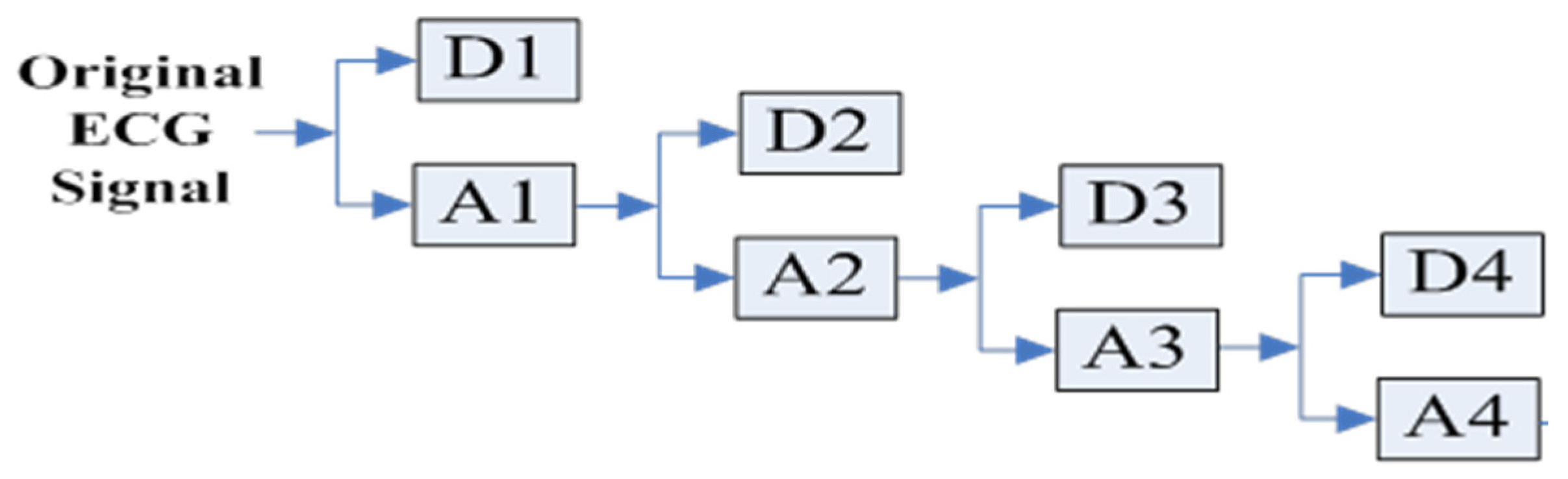
2.2. Discrete Wavelet Transform (DWT)
2.3. Discrete Fourier Transform (DFT)
2.4. Discrete Cosine Transform (DCT)
2.5. Particle Swarm Optimization (PSO)
3. Proposed System
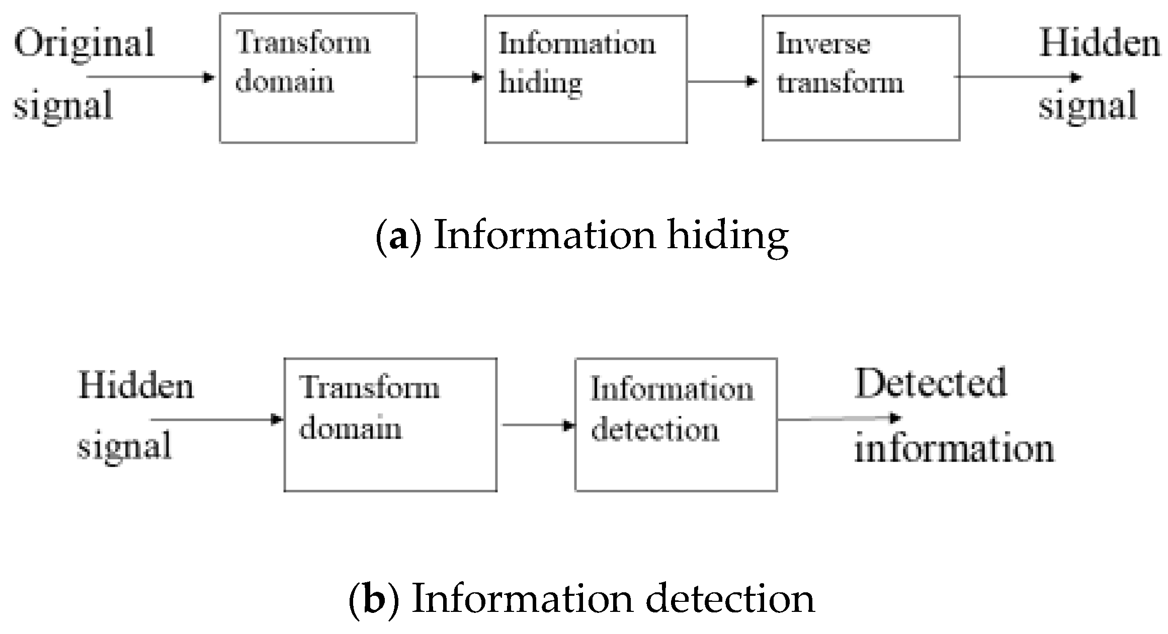
3.1. Information Hiding and Detection
3.2. Enhance Performance by PSO
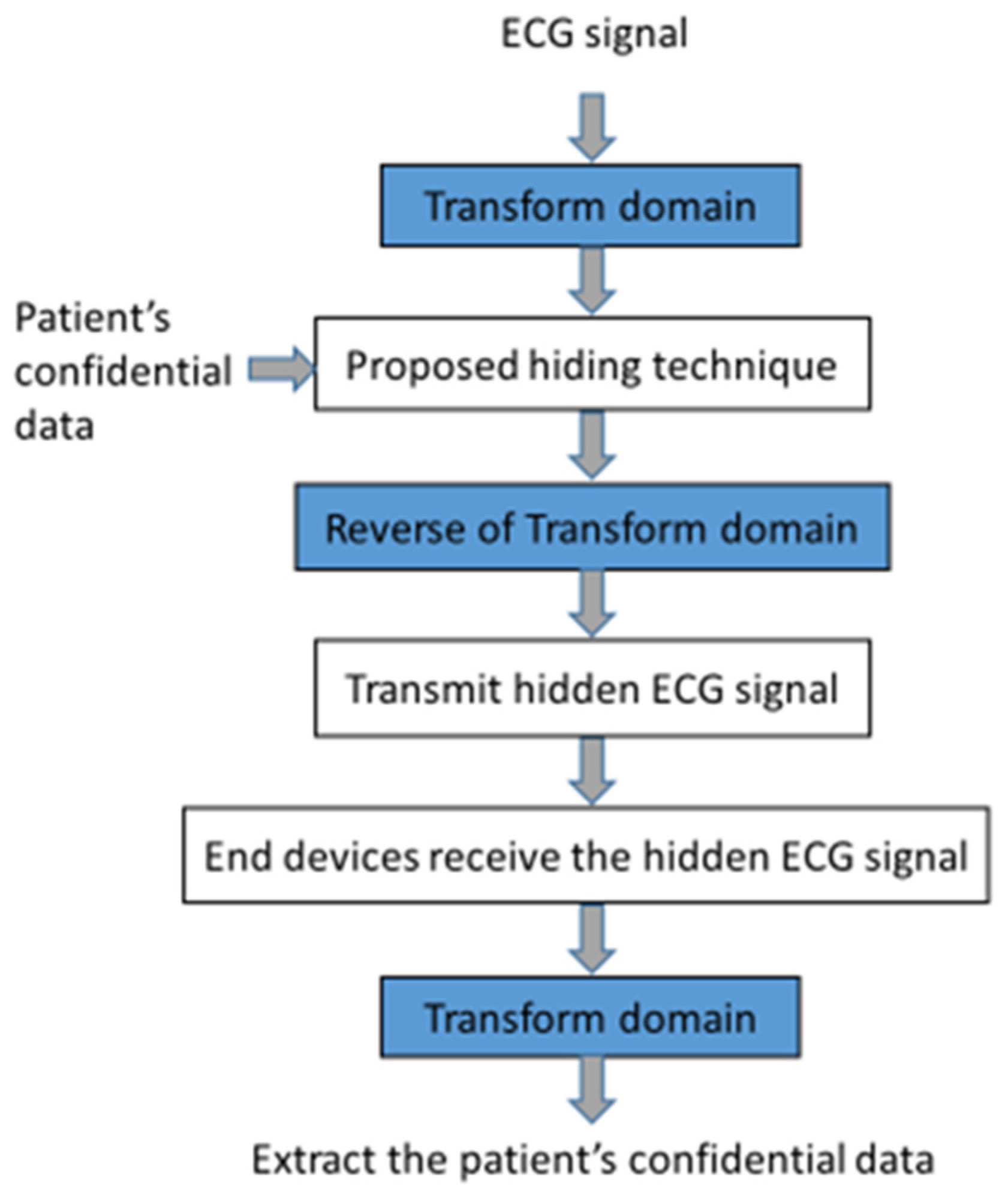
3.3. Flowchart
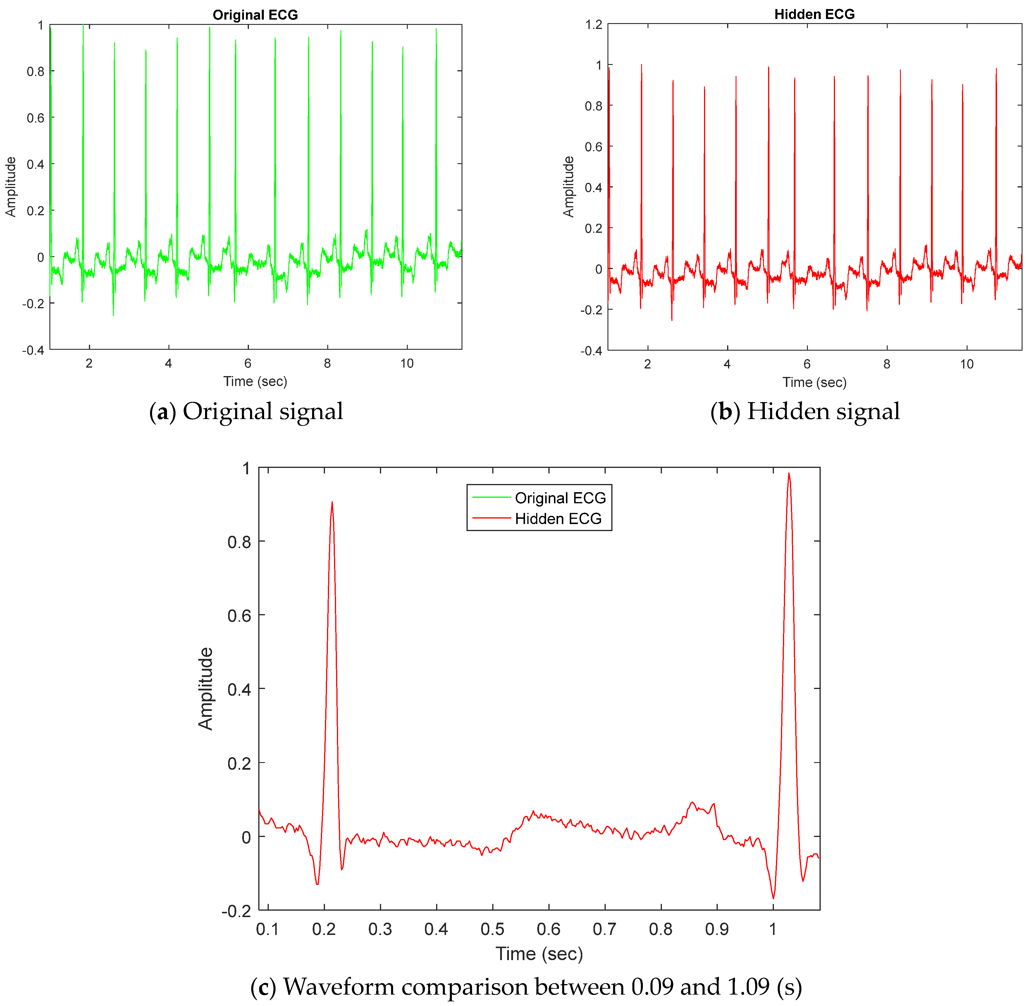
4. Experiments and Discussion
5. Conclusions
Author Contributions
Funding
Institutional Review Board Statement
Informed Consent Statement
Data Availability Statement
Conflicts of Interest
References
- Engin, M.; Çidam, O.; Engin, E.Z. Wavelet transformation based watermarking technique for human electrocardiogram (ECG). J. Med. Syst. 2005, 29, 589–594. [Google Scholar] [CrossRef] [PubMed]
- Zheng, K.M.; Qian, X. Reversible data hiding for electrocardiogram signal based on wavelet transforms. In Proceedings of the 2008 International Conference on Computational Intelligence and Security (CIS2008), Suzhou, China, 13–17 December 2008; pp. 295–299. [Google Scholar]
- Zheng, K.M.; Tang, L.W.; Qian, X. Watermarking technology for electrocardiogram signal certification. Comput. Eng. Appl. 2009, 45, 231–233. [Google Scholar]
- Kaur, S.; Farooq, O.; Singhal, R.; Ahuja, B.S. Digital watermarking of ECG data for secure wireless communication. In Proceedings of the 2010 International Conference on Recent Trends in Information, Telecommunication and Computing (ITC2010), Kochi, India, 12–13 March 2010; pp. 140–144. [Google Scholar]
- Ibaida, A.; Khalil, I.; van Dhiah, A.S. Embedding Patients Confidential Data in ECG Signal for HealthCare Information System. In Proceedings of the 32nd Annual International Conference of the IEEE EMBS, Buenos Aires, Argentina, 31 August–4 September 2010; pp. 3891–3894. [Google Scholar]
- Ibaida, A.; Khalil, I.; van Schyndel, R. A low complexity high capacity ECG signal watermark for wearable sensor-net health monitoring system. In Proceedings of the Computing in Cardiology (CinC), Hangzhou, China, 18–21 September 2011; pp. 393–396. [Google Scholar]
- He, X.; Tseng, K.K.; Huang, H.H.; Chen, S.T.; Tu, S.Y.; Zeng, F.; Pan, J.S. Wavelet-based Quantization Watermarking for ECG Signals. In Proceedings of the 2012 International Conference on Computing, Measurement, Control and Sensor Network (CMCSN), Taiyuan, China, 7–9 July 2012; pp. 233–236. [Google Scholar]
- Guo, Y.; Zhou, D. Single channel surface electromyography blind recognition model based on watermarking. J. Vib. Control. 2012, 18, 42–47. [Google Scholar]
- Dey, N.; Biswas, S.; Roy, A.B.; Das, A.; Chowdhuri, S.S. Analysis of Photoplethysmographic Signals Modified by Reversible Watermarking Technique using Prediction-Error in Wireless Telecardiology. In Proceedings of the First International Conference on Intelligent Infrastructure the 47th Annual National Convention at Computer Society of Inda CSI, Kolkata, India, 1 December 2012; Available online: https://www.academia.edu/64990486/Analysis_of_Photoplethysmographic_Signals_Modified_by_Reversible_Watermarking_Technique_using_Prediction_Error_in_Wireless_Telecardiology (accessed on 15 January 2020).
- Dey, N.; Mukhopadhyay, S.; Das, A.; Chaudhuri, S.S. Analysis of P-QRS-T Components Modified by Blind Watermarking Technique Within the Electrocardiogram Signal for Authentication in Wireless Telecardiology Using DWT. Int. J. Image Graph. Signal Process. 2012, 4, 33–46. [Google Scholar] [CrossRef]
- Tseng, K.-K.; He, X.; Kung, W.-M.; Chen, S.-T.; Liao, M.; Huang, H.-N. Wavelet-Based Watermarking and Compression for ECG Signals with Verification Evaluation. Sensors 2014, 14, 3721–3736. [Google Scholar] [CrossRef] [PubMed]
- Chen, S.-T.; Guo, Y.-J.; Huang, H.-N.; Kung, W.-M.; Tseng, K.-K.; Tu, S.-Y. Hiding Patients Confidential Data in the ECG Signal via Transform-Domain Quantization Scheme. J. Med. Syst. 2014, 38, 54. [Google Scholar] [CrossRef] [PubMed]
- Jero, S.E.; Ramu, P.; Ramakrishna, S.N. ECG steganography using curvelet transform. Biomed. Signal Process. Control. 2015, 22, 161–169. [Google Scholar] [CrossRef]
- Jero, S.E.; Ramu, P. Curvelets-based ECG steganography for data security. Electron. Lett. 2016, 52, 283–285. [Google Scholar] [CrossRef]
- Świerkoszand, A.; Augustyniak, P. Optimizing wavelet ECG watermarking to maintain measurement performance according to industrial standard. Sensors 2018, 18, 3401. [Google Scholar] [CrossRef]
- Goyal, L.M.; Mittal, M.; Kaushik, R.; Verma, A.; Kaur, I.; Roy, S.; Kim, T.-H. Improved ECG watermarking technique using curvelet transform. Sensors 2020, 20, 2941. [Google Scholar] [CrossRef]
- Tripathi, P.M.; Kumar, A.; Komaragiri, R.; Kumar, M. Watermarking of ECG signals compressed using Fourier decomposition method. Multimed. Tools Appl. 2022, 81, 19543–19557. [Google Scholar] [CrossRef]
- Sharma, N.; Anand, A.; Singh, A.K. Optimization based ECG watermarking in RDWT-SVD domain. Multimed. Tools Appl. 2023, 82, 5031–5047. [Google Scholar] [CrossRef]
- PSanivarapu, V.; Rajesh, K.N.V.P.S.; Reddy, N.V.R.; Reddy, N.C.S. Patient data hiding into ECG signal using watermarking in transform domain. Phys. Eng. Sci. Med. 2020, 43, 213–226. [Google Scholar] [CrossRef]
- Sharma, N.K.; Kumar, S.; Kumar, N. HGSmark: An efficient ECG watermarking scheme using hunger games search and Bayesian regularization BPNN. Biomed. Signal Process. Control. 2023, 83, 104633. [Google Scholar] [CrossRef]
- Moody, G.B.; Mark, R.G. The impact of the MIT-BIH arrhythmia database. IEEE Eng. Med. Biol. Mag. 2001, 20, 45–50. [Google Scholar] [CrossRef] [PubMed]
- MIT-BIH Arrhythmia Database Directory, Harvard University-Massachusetts Institute of Technology Division of Health Science and Technology. 1992. Available online: http://www.ph (accessed on 18 January 2020).
- Kothari, V.; Anuradha, J.; Shah, S.; Mittal, P. A survey on particle swarm optimization in feature selection. In Global Trends in Information Systems and Software Applications, Proceedings of the International Conference on Computing and Communication Systems, Vellore, India, 9–11 December 2011; Springer: Berlin/Heidelberg, Germany, 2011; pp. 192–201. [Google Scholar]
- Marini, F.; Walczak, B. Particle swarm optimization (pso). A tutorial. Chemom. Intell. Lab. Syst. 2015, 149, 153–165. [Google Scholar] [CrossRef]
- Mavrovouniotis, M.; Li, C.; Yang, S. A survey of swarm intelligence for dynamic optimization: Algorithms and applications. Swarm Evol. Comput. 2017, 33, 1–17. [Google Scholar] [CrossRef]
- Gad, A.G. Particle Swarm Optimization Algorithm and Its Applications: A Systematic Review. Arch. Comput. Methods Eng. 2022, 29, 2531–2561. [Google Scholar] [CrossRef]
- Zhao, M.; Chen, S.-T.; Tu, S.-Y. Wavelet-Domain Information-Hiding Technology with High-Quality Audio Signals on MEMS Sensors. Sensors 2022, 22, 6548. [Google Scholar] [CrossRef] [PubMed]


| Reference | Domain | Solution. | Fining | Gap. |
|---|---|---|---|---|
| [11] | transform domain | single-coefficient quantization | In case of embedding strength by SNR = 32, most of amplitude simi-larities are 1, root mean square ap-proaches 0. | For increase in embedding strength, there is decrease in the SNR value. |
| [12] | transform domain | single-coefficient quantization | In case of embedding strength by SNR = 32, most of amplitude similarities are 1, root mean square approaches 0. | For increase in embed-ding strength, there is decrease in the SNR value. |
| [13] | transform domain | adaptive selection of watermarking location and threshold selecting for embedding | As the patient data size is increased, the cover signal deteriorates but the Bit Error Rate is zero. The signal deterioration is about 10% when patient data increase 1.5 times. | For increase in watermark size, there is decrease in the PSNR value. |
| [14] | transform domain | adaptive selection of watermarking location and selecting threshold for embedding | As the patient data size is increased, the cover signal deteriorates but the Bit Error Rate is zero. | For increase in watermark size, there is decrease in the PSNR value. |
| [15] | transform domain | adaptive lead-independent beat-to-beat data repository plan | (1) The 11th order Symlet is the best among the wavelets tested (2) The watermark and noise similarity of amplitude and numerical distribution are highly affected | Further watermarking destroys the existing watermark. |
| [16] | transform domain | embed the information of the patient’s data into the ECG signals using curvelet transform | A PSNR value higher than 64 shows the high quality of extracted information. The NC values of all ECG signals are 1 and the SSIM values are close to 1, which indicates high similarity between embedded and extracted information. | Some false positives occur during the watermark embedding process, which not only reduces the quality of the extracted watermark, but also affects the robustness of the image. |
| [17] | transform domain | ECG watermarking and compression in Fourier domain | The improved SNR proves the denoising ability of the watermark signal. | For increase in embedding strength, there is decrease in the SNR value. |
| [18] | transform domain | ECG watermarking using the integration of redundant discrete wavelet transform (RDWT) and singular value decomposition (SVD) | The optimal invisibility and robustness result are more effective. | There does not seem to be any guarantee that the encoded binary bits could be recovered using particle swarm optimization (PSO). |
| [19] | transform domain | embedding factor value is calculated adaptively by harnessing the entropy value of the signal | The embedding factor value is calculated adaptively by harnessing the entropy of the ECG signal. The embedded data can be easily extracted with no distortion. | For increase in embedding strength, there is decrease in the SNR value. |
| [20] | time domain | multiple embedding strength optimized by hunger games search algorithm | maintaining the imperceptibility-robustness trade-off to obtain PSNR = 57.725 dB | Embedded patient information is less hidden and less robust. |
| ID | Method | Domain | Q | Amplitude Similarity | SNR | RMSE | Interval RMSE in ECG | |||
|---|---|---|---|---|---|---|---|---|---|---|
| PR | QRS | ST | QT | |||||||
| Reference [12] | DWT (Level 5) | 400 | 1 | 40.85 | 36.86 | 0 | 0 | 0 | 0 | |
| 1000 | 1 | 35.16 | 67.43 | 0 | 0 | 0 | 0 | |||
| 3000 | 1 | 24.26 | 121.52 | 0 | 0 | 0 | 0 | |||
| DFT | 400 | 1 | 61.13 | 3.75 | 0 | 0 | 0 | 0 | ||
| 1000 | 1 | 54.68 | 8.21 | 0 | 0 | 0 | 0 | |||
| 4000 | 0.99 | 46.12 | 28.36 | 0 | 0 | 0 | 0 | |||
| DCT | 400 | 0.99 | 29.92 | 197.92 | 0 | 0 | 0 | 0 | ||
| 1000 | 0.98 | 20.16 | 435.11 | 0 | 0 | 0 | 0 | |||
| 3000 | 0.81 | 9.21 | 2014.6 | 0 | 0 | 0 | 0 | |||
| Proposed | DWT (Level 5) | 400 | 0.99 | 39.86 | 51.93 | 0 | 0 | 0 | 0 | |
| 1000 | 0.99 | 39.14 | 49.22 | 0 | 0 | 0 | 0 | |||
| 3000 | 0.99 | 38.95 | 49.37 | 0 | 0 | 0 | 0 | |||
| DFT | 400 | 1 | 34.22 | 93.15 | 0 | 0 | 0 | 0 | ||
| 1000 | 1 | 31.81 | 109.74 | 0 | 0 | 0 | 0 | |||
| 3000 | 1 | 31.09 | 105.82 | 0 | 0 | 0 | 0 | |||
| DCT | 400 | 1 | 47.16 | 19.88 | 0 | 0 | 0 | 0 | ||
| 1000 | 1 | 45.94 | 21.90 | 0 | 0 | 0 | 0 | |||
| 3000 | 1 | 45.78 | 23.12 | 0 | 0 | 0 | 0 | |||
| 101 | Reference [12] | DWT (Level 5) | 400 | 1 | 41.78 | 36.39 | 0 | 0.001 | 0.002 | 0 |
| 1000 | 1 | 34.67 | 71.65 | 0 | 0 | 0 | 0 | |||
| 3000 | 1 | 25.39 | 141.47 | 0 | 0 | 0 | 0 | |||
| DFT | 400 | 1 | 60.42 | 4.12 | 0 | 0 | 0 | 0 | ||
| 1000 | 1 | 54.37 | 6.95 | 0 | 0 | 0 | 0 | |||
| 3000 | 1 | 45.58 | 29.73 | 0 | 0 | 0 | 0 | |||
| DCT | 400 | 0.99 | 26.63 | 203.31 | 0 | 0 | 0 | 0 | ||
| 1000 | 0.98 | 18.67 | 441.87 | 0 | 0 | 0 | 0 | |||
| 3000 | 0.78 | 8.32 | 1983.4 | 0 | 0 | 0 | 0 | |||
| Proposed | DWT (Level 5) | 400 | 1 | 33.45 | 92.56 | 0 | 0 | 0 | 0 | |
| 1000 | 1 | 33.16 | 92.13 | 0 | 0 | 0 | 0 | |||
| 3000 | 0.99 | 32.89 | 96.09 | 0 | 0 | 0 | 0 | |||
| DFT | 400 | 0.99 | 32.25 | 107.58 | 0 | 0 | 0 | 0 | ||
| 1000 | 0.99 | 30.82 | 103.14 | 0 | 0 | 0 | 0 | |||
| 3000 | 0.99 | 30.19 | 94.31 | 0 | 0 | 0 | 0 | |||
| DCT | 400 | 1 | 36.46 | 70.15 | 0 | 0 | 0 | 0 | ||
| 1000 | 1 | 35.83 | 68.63 | 0 | 0 | 0 | 0 | |||
| 3000 | 1 | 35.47 | 68.51 | 0 | 0 | 0 | 0 | |||
| 102 | Reference [12] | DWT (Level 5) | 400 | 0.99 | 44.65 | 33.94 | 0 | 0 | 0 | 0 |
| 1000 | 0.99 | 37.35 | 70.16 | 0 | 0 | 0 | 0 | |||
| 3000 | 0.99 | 27.31 | 267.17 | 0 | 0 | 0 | 0 | |||
| DFT | 400 | 1 | 63.18 | 4.12 | 0 | 0 | 0 | 0 | ||
| 1000 | 1 | 56.28 | 7.45 | 0 | 0 | 0 | 0 | |||
| 3000 | 1 | 47.53 | 30.28 | 0 | 0 | 0 | 0 | |||
| DCT | 400 | 0.99 | 28.02 | 200.19 | 0 | 0 | 0 | 0 | ||
| 1000 | 0.99 | 21.45 | 421.37 | 0 | 0 | 0 | 0 | |||
| 3000 | 0.87 | 13.28 | 1890.5 | 0 | 0 | 0 | 0 | |||
| Proposed | DWT (Level 5) | 400 | 1 | 34.28 | 92.78 | 0 | 0 | 0 | 0 | |
| 1000 | 1 | 34.22 | 95.68 | 0 | 0 | 0 | 0 | |||
| 3000 | 1 | 33.42 | 115.24 | 0 | 0 | 0 | 0 | |||
| DFT | 400 | 0.99 | 28.62 | 300.25 | 0 | 0 | 0 | 0 | ||
| 1000 | 0.99 | 26.61 | 334.68 | 0 | 0 | 0 | 0 | |||
| 3000 | 0.99 | 26.60 | 293.24 | 0 | 0 | 0 | 0 | |||
| DCT | 400 | 1 | 29.57 | 171.13 | 0 | 0 | 0 | 0 | ||
| 1000 | 1 | 29.36 | 170.12 | 0 | 0 | 0 | 0 | |||
| 3000 | 1 | 29.24 | 166.77 | 0 | 0 | 0 | 0 | |||
| 103 | Reference [12] | DWT (Level 5) | 400 | 1 | 40.56 | 36.07 | 0 | 0 | 0 | 0 |
| 1000 | 1 | 37.65 | 63.48 | 0 | 0 | 0 | 0 | |||
| 3000 | 1 | 26.35 | 270.75 | 0 | 0 | 0 | 0 | |||
| DFT | 400 | 1 | 63.23 | 4.07 | 0 | 0 | 0 | 0 | ||
| 1000 | 1 | 55.76 | 8.13 | 0 | 0 | 0 | 0 | |||
| 3000 | 0.99 | 47.12 | 30.08 | 0 | 0 | 0 | 0 | |||
| DCT | 400 | 0.99 | 28.23 | 211.17 | 0 | 0 | 0 | 0 | ||
| 1000 | 0.99 | 21.62 | 450.70 | 0 | 0 | 0 | 0 | |||
| 3000 | 0.85 | 10.21 | 1980.2 | 0 | 0 | 0 | 0 | |||
| Proposed | DWT (Level 5) | 400 | 1 | 27.38 | 258.11 | 0 | 0 | 0 | 0 | |
| 1000 | 1 | 26.75 | 238.72 | 0 | 0 | 0 | 0 | |||
| 3000 | 1 | 26.48 | 253.62 | 0 | 0 | 0 | 0 | |||
| DFT | 400 | 0.99 | 28.32 | 155.43 | 0 | 0 | 0 | 0 | ||
| 1000 | 0.99 | 28.54 | 160.41 | 0 | 0 | 0 | 0 | |||
| 3000 | 0.99 | 28.61 | 158.37 | 0 | 0 | 0 | 0 | |||
| DCT | 400 | 1 | 34.17 | 96.24 | 0 | 0 | 0 | 0 | ||
| 1000 | 1 | 34.11 | 95.84 | 0 | 0 | 0 | 0 | |||
| 3000 | 1 | 34.25 | 94.33 | 0 | 0 | 0 | 0 | |||
| 104 | Reference [12] | DWT (Level 5) | 400 | 0.99 | 41.56 | 37.21 | 0.041 | 0 | 0 | 0 |
| 1000 | 0.99 | 37.20 | 68.96 | 0 | 0 | 0 | 0 | |||
| 3000 | 0.99 | 23.67 | 260.17 | 0 | 0 | 0 | 0 | |||
| DFT | 400 | 1 | 62.46 | 4.37 | 0.653 | 0.015 | 0 | 0 | ||
| 1000 | 1 | 56.47 | 8.57 | 0 | 0 | 0 | 0 | |||
| 3000 | 0.99 | 44.45 | 30.16 | 0 | 0 | 0 | 0 | |||
| DCT | 400 | 0.99 | 27.84 | 202.645 | 0 | 0 | 0 | 0 | ||
| 1000 | 0.99 | 21.26 | 431.98 | 0 | 0 | 0 | 0 | |||
| 3000 | 0.86 | 8.41 | 1895.5 | 0 | 0 | 0 | 0 | |||
| Proposed | DWT (Level 5) | 400 | 0.99 | 25.16 | 317.65 | 0 | 0 | 0 | 0 | |
| 1000 | 0.99 | 25.34 | 327.75 | 0 | 0 | 0 | 0 | |||
| 3000 | 0.99 | 25.76 | 322.64 | 0 | 0 | 0 | 0 | |||
| DFT | 400 | 0.99 | 27.18 | 166.73 | 0 | 0 | 0 | 0 | ||
| 1000 | 0.99 | 27.29 | 167.35 | 0 | 0 | 0 | 0 | |||
| 3000 | 0.99 | 27.36 | 166.46 | 0 | 0 | 0 | 0 | |||
| DCT | 400 | 1 | 31.41 | 134.54 | 0 | 0 | 0 | 0 | ||
| 1000 | 1 | 32.26 | 130.17 | 0 | 0 | 0 | 0 | |||
| 3000 | 1 | 32.18 | 128.96 | 0 | 0 | 0 | 0 | |||
| 105 | Reference [12] | DWT (Level 5) | 400 | 1 | 41.96 | 34.54 | 0 | 0 | 0 | 0 |
| 1000 | 1 | 39.15 | 68.28 | 0 | 0 | 0 | 0 | |||
| 3000 | 0.99 | 24.75 | 284.63 | 0 | 0 | 0 | 0 | |||
| DFT | 400 | 1 | 64.46 | 3.70 | 0 | 0 | 0 | 0 | ||
| 1000 | 1 | 58.37 | 7.47 | 0 | 0 | 0 | 0 | |||
| 3000 | 1 | 46.24 | 30.21 | 0 | 0 | 0 | 0 | |||
| DCT | 400 | 0.98 | 29.58 | 205.58 | 0 | 0 | 0 | 0 | ||
| 1000 | 0.99 | 22.76 | 450.82 | 0 | 0 | 0 | 0 | |||
| 3000 | 0.88 | 9.43 | 2092.6 | 0 | 0 | 0 | 0 | |||
| Proposed | DWT (Level 5) | 400 | 1 | 26.83 | 283.36 | 0 | 0 | 0 | 0 | |
| 1000 | 1 | 26.39 | 297.79 | 0 | 0 | 0 | 0 | |||
| 3000 | 1 | 26.56 | 291.29 | 0 | 0 | 0 | 0 | |||
| DFT | 400 | 0.99 | 25.96 | 161.36 | 0 | 0 | 0 | 0 | ||
| 1000 | 0.99 | 25.74 | 162.85 | 0 | 0 | 0 | 0 | |||
| 3000 | 0.99 | 25.36 | 161.46 | 0 | 0 | 0 | 0 | |||
| DCT | 400 | 1 | 46.58 | 31.01 | 0 | 0 | 0 | 0 | ||
| 1000 | 1 | 46.00 | 31.05 | 0 | 0 | 0 | 0 | |||
| 3000 | 1 | 46.22 | 28.54 | 0 | 0 | 0 | 0 | |||
Disclaimer/Publisher’s Note: The statements, opinions and data contained in all publications are solely those of the individual author(s) and contributor(s) and not of MDPI and/or the editor(s). MDPI and/or the editor(s) disclaim responsibility for any injury to people or property resulting from any ideas, methods, instructions or products referred to in the content. |
© 2023 by the authors. Licensee MDPI, Basel, Switzerland. This article is an open access article distributed under the terms and conditions of the Creative Commons Attribution (CC BY) license (https://creativecommons.org/licenses/by/4.0/).
Share and Cite
Chen, S.-T.; Ye, R.-J.; Wu, T.-H.; Cheng, C.-W.; Zhan, P.-Y.; Chen, K.-M.; Zhong, W.-Y. Patient Confidential Data Hiding and Transmission System Using Amplitude Quantization in the Frequency Domain of ECG Signals. Sensors 2023, 23, 9199. https://doi.org/10.3390/s23229199
Chen S-T, Ye R-J, Wu T-H, Cheng C-W, Zhan P-Y, Chen K-M, Zhong W-Y. Patient Confidential Data Hiding and Transmission System Using Amplitude Quantization in the Frequency Domain of ECG Signals. Sensors. 2023; 23(22):9199. https://doi.org/10.3390/s23229199
Chicago/Turabian StyleChen, Shuo-Tsung, Ren-Jie Ye, Tsung-Hsien Wu, Chun-Wen Cheng, Po-You Zhan, Kuan-Ming Chen, and Wan-Yu Zhong. 2023. "Patient Confidential Data Hiding and Transmission System Using Amplitude Quantization in the Frequency Domain of ECG Signals" Sensors 23, no. 22: 9199. https://doi.org/10.3390/s23229199