
Objective Methods of Muscle Tone Diagnosis and Their Application—A Critical Review

1
Faculty of Physical Education and Sport, Charles University, 162 52 Prague, Czech Republic
2
Faculty of Health Sciences, University of Primorska, 6310 Izola, Slovenia
3
Faculty of Science, Humanities and Education, Technical University of Liberec, 461 17 Liberec, Czech Republic
*
Author to whom correspondence should be addressed.
Sensors 2023, 23(16), 7189; https://doi.org/10.3390/s23167189
Submission received: 7 May 2023
/
Revised: 9 August 2023
/
Accepted: 10 August 2023
/
Published: 15 August 2023
(This article belongs to the Section Physical Sensors)
Abstract
:“Muscle tone” is a clinically important and widely used term and palpation is a crucial skill for its diagnosis. However, the term is defined rather vaguely, and palpation is not measurable objectively. Therefore, several methods have been developed to measure muscle tone objectively, in terms of biomechanical properties of the muscle. This article aims to summarize these approaches. Through database searches, we identified those studies related to objective muscle tone measurement in vivo, in situ. Based on them, we described existing methods and devices and compared their reliability. Furthermore, we presented an extensive list of the use of these methods in different fields of research. Although it is believed by some authors that palpation cannot be replaced by a mechanical device, several methods have already proved their utility in muscle biomechanical property diagnosis. There appear to be two issues preventing wider usage of these objective methods in clinical practice. Firstly, a high variability of their reliability, and secondly, a lack of valid mathematical models that would provide the observed mechanical characteristics with a clear physical significance and allow the results to be compared with each other.
1. Introduction
Palpation is one of the oldest examination techniques. It plays an essential role in the diagnosis of soft tissues of the musculoskeletal system, being one of the basic skills of a physiotherapist [1]. Its use is extensive, including the assessment of the consistency, smoothness, displacement, or extensibility of the examined tissue or the temperature and moisture of the body surface. The painfulness of the studied area can also be estimated based on the patient’s reaction.
In the tradition of the so-called Prague School of Rehabilitation, slow, deep, layered palpation with fingertip pads is used to examine the soft tissues of the musculoskeletal system while the patient is completely relaxed [2]. The underlying structures (skin, subcutaneous tissue, adipose tissue, fascia, muscles, etc.) are compressed by creating sufficient pressure on the body surface. Depending on the depth of palpation, the affected tissue layers’ elasticity and resistance to deformation can be assessed. However, the pressure must not be disproportionately large; otherwise, the examiners perceive the mechanical processes on their fingers and not the tissue under examination [3]. Véle [4] recommends palpation at an angle of 60–90° with the surface. This method is often used to diagnose functional disorders of the soft tissues of the musculoskeletal system, especially the so-called muscle tone. Another approach to assess the mechanical properties of skeletal muscle is the so-called tapotement, or tapping perpendicular to the muscle, where the propagation of the wave in the muscle is observed [1]. In order to investigate trigger points, the so-called strumming is used, which elicits a characteristic response in the form of muscle twitching and referred pain [5].
The palpation techniques described above are widely used, but unfortunately, they are also subjective examination methods. This article aims to summarize the possible approaches to objectify the mechanical properties of soft tissues of the human musculoskeletal system, and to provide a selection of research areas in which these objective methods can be used. The basic questions addressed in the text are as follows: What methods can be used to assess the mechanical properties of skeletal muscle in vivo objectively? How can these methods support subjective palpation examinations? In what research areas have they already been used?
2. Muscle Tone
More than a century ago, Foster defined muscle tone as “resistance to passive movements” [6] and this definition is still widely used today [3,7,8,9], serving as the principal criterion for its assessment in clinical practice. Multiple authors have since investigated the physiological nature and components of this phenomenon, inferring that it can be understood as a set of mechanical (rheological) and neurophysiological properties of the skeletal muscle [10,11].
Simons and Mense [11] approach the concept based on the presence of detectable electrical activity in the muscle. That is, based on the detectable EMG (electromyography) signal response of the skeletal muscle. The EMG activity is a manifestation of the excitation of alpha motoneurons, i.e., the tone produced during voluntary but also involuntary (e.g., spasticity, rigidity, spasm) contractions and also during incomplete muscle relaxation (i.e., the measured individual has been instructed to relax but is not able to do so completely). In this case, the muscle’s stiffness is due to the interaction between actin and myosin.
In addition, there is a component of muscle tone unaffected by alpha activity (i.e., EMG-silent), caused by the inherent viscoelastic properties of the tissue. Masi and Hannon [9] describe it as the human resting muscle tone and attribute about 1% of maximal voluntary contraction to it. In addition to the intrinsic mechanical resistance of the muscle, a certain role is attributed to the mutual binding and movement between actin and myosin [9,11]. The latter is also a possible explanation for thixotropy, i.e., the gradual decrease in resistance to movement concerning the preceding movement [11].
Another passive (EMG-silent) component is the stiffness of the surrounding soft tissues, especially fasciae, which also show thixotropy. Muscle contracture due to prolonged stay in a shortened position is a well-known issue, but the inherent variability in the viscoelastic properties of these tissues has not yet been clearly explained. Control by the autonomic nervous system is thought to regulate pressure in small local vessels and plasma washout into the interstitium based on information from receptors, which will affect overall viscosity. At the same time, smooth muscle in the fascia has been demonstrated, implying that the fascia may have a system of tone adjustment, independent of muscle tone. [12] However, when assessing biomechanical properties by palpation or indentation testing, these two components are examined simultaneously. Therefore, some authors [9] suggest using the term “myofascial tone” instead when assessing it.
In addition, there is also contractile muscle activity without electrical activity (in some genetic disorders associated with impaired calcium metabolism in the sarcoplasmic reticulum), which is also the case for myofascial trigger points [11]. Although some studies have shown low EMG activity levels at the trigger point (so-called endplate noise), action potentials are absent in the surrounding rigid core. At the same time, the nature of this particular kind of local hypertonicity is more or less explicitly described in the literature from a physiological point of view. It is assumed that due to the overloading of the muscle, there is a malfunction in the muscle endplate, which continuously flushes out acetylcholine and thus causes a sustained contraction at the site. This contraction is energy depleting; Simons [13] described this as a local energy crisis. Therefore, there is a lack of energy for the reuptake of the sarcoplasmic reticulum, which further promotes contraction. The local ischemia also causes typical soreness.
Partly outside the categories of Simons and Mense [11] lies muscle tone as a measure of “alertness”, i.e., the readiness of a muscle to contract. Latash and Zatsiorski [14] define it as such and describe it as the threshold for eliciting the stretch reflex. This threshold is set from the central nervous system (e.g., by a conscious instruction that the muscle is to be relaxed), and an increase in this threshold may manifest itself, for example, in the inability to achieve a silent EMG when attempting to relax. At the same time, however, it is generally accepted that the gamma system sets muscle excitability through the muscle spindles and from different levels of the central nervous system [3,4,15,16,17]. There is as yet no evidence of EMG manifestations in this context.
Given the above, it is clear that muscle tone is a very complex term and, despite being commonly used in professional literature, it lacks a precise definition [14] and is still a subject of discussion and research, see, e.g., [3,7,8,9,11]. Defining muscle tone as resistance to passive movement is very vague, allowing for ambiguous interpretations [18,19]. Furthermore, the resistance is influenced by other factors besides the muscle and its fascia, such as the joint range of motion, ligament laxity, etc. [3,11,14]. Moreover, while Foster describes it as “independent of all distinct muscular contractions of volitional or other origin” [6], other authors assume that the muscle tone actively contributes to movement [20] and some even differentiate between passive and active muscle tone [9], countering the supposition that the patient has to be completely relaxed for the measurement.
Variability of views and the absence of an explicit definition of muscle tone make it necessary to always specify in what sense this term is understood when speaking of examination of muscle tone. In clinical practice, the healthcare practitioners commonly examine it using palpation, hence the muscle tone in this context refers to the examiner’s perception, i.e., the resistance of the tissue against the palpating finger. Curiously, this criterion for the assessment of muscle tone is cited much less frequently then the already mentioned resistance to passive movement [3,4,9,14].
The expected level of the resistance is not clearly established [21], however, therapists commonly compare this resistance with experience from previous practice. Furthermore, the resistance can be compared, for example, between muscles of one individual or, in the case of long-term therapeutic care, changes in one muscle (site) over time. Above all, however, it is possible to compare one particular muscle (site) before and after therapeutic intervention. Intra-individual comparisons also minimize the influence of the mechanical properties of the surface tissues (skin, fat layer, etc.) on the result, as these are likely to vary only minimally between the individual examinations being compared. It assumes that there is no significant change in body composition due to dehydration, reduction in body fat, etc. Furthermore, if the patient is asked to relax during the test, the voluntary instruction from the central nervous system can be expected to be the same for both examinations being compared. The patient’s ability to relax can then be attributed to the effectiveness of the intervention and should not be considered a confounding factor of the test.
3. Methods
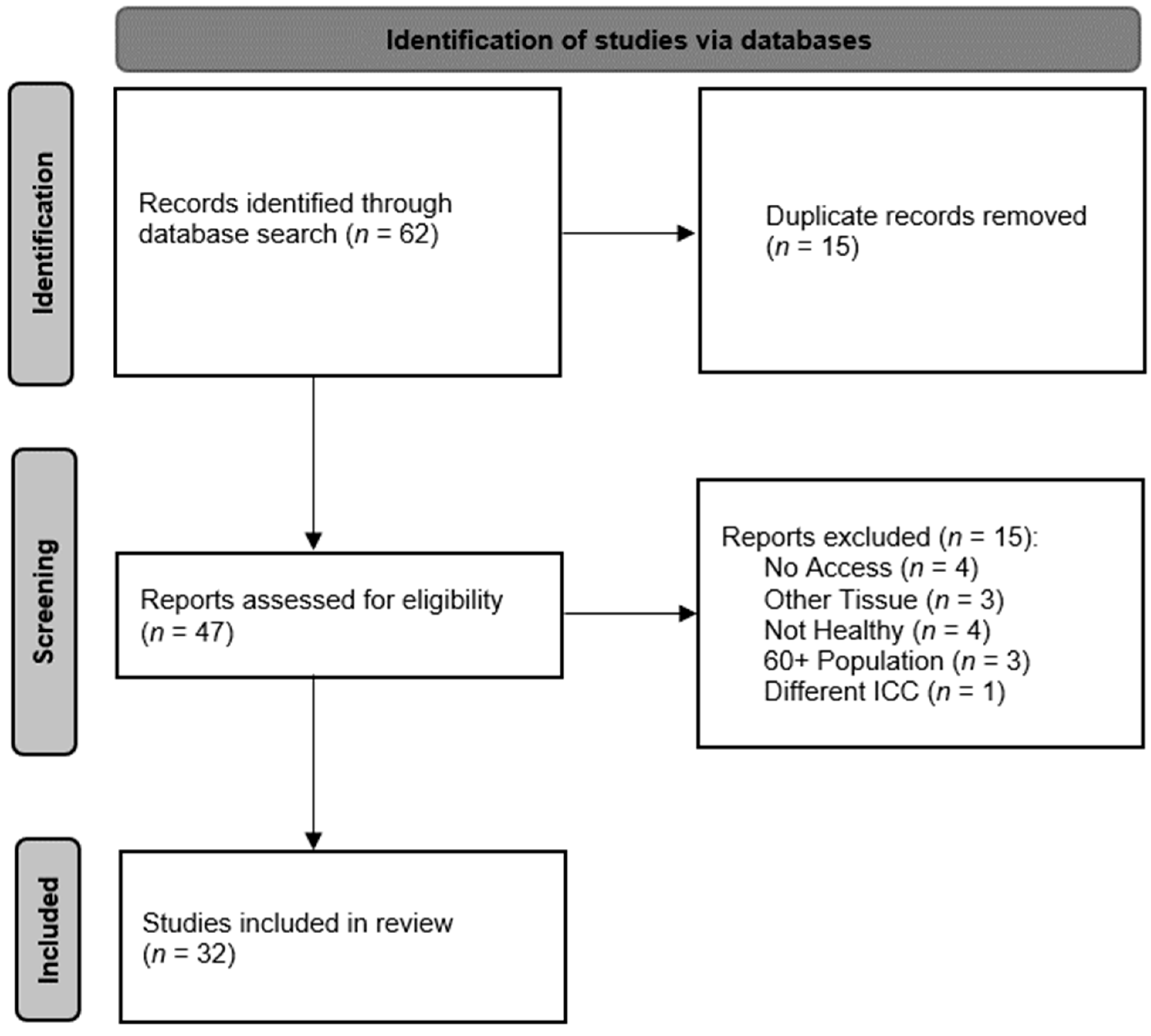
The preliminary search of information on the available objective methods for the diagnosis of muscle tone was based on the long-term specialization and research of the authors and their extensive research of the literature available. Regarding the application and reliability of the methods described, the relevant studies were identified mostly through searching electronic databases. The principal database used was PubMed. Additionally, the clinical application of Myoton was also documented using references listed on the official Myoton website. Only studies written in English were considered. The search was conducted on several occasions, the last being on 28 July 2023. A simplified PRISMA diagram [22] describing the review process of the reliability of the most frequently used methods is shown in Figure 1. The keywords used were ICC/s, healthy, and muscle in combination with of the following: CMT, MRE, myotonometer, myotonometry, MyotonPRO, SSI, or SWE. Only studies that were available in full text, tested the reliability on skeletal muscle in vivo and in a healthy population aged 18–60, and provided the ICC as an indicator of the method’s reliability were included. Three of the included studies tested multiple methods.
The keywords for application included muscle tone, mechanical properties, elasticity/stiffness, myotonometry, elastography, and similar. In order to be included in the review, the objective method described/used had to be “in vivo, in situ” and performed on skeletal muscles. The variability of clinical application of the described methods proved to be wide and extensive, so the list is not exhaustive.
4. Objective Methods of Muscle Tone Diagnosis
Latash and Zatsiorski [14] defined three possible approaches to the concept of muscle tone, from which the respective methods of its objectification proceed. The first one is based on the definition of tone as the resistance of a muscle to movement at a given joint. This approach was used by McPherson [8] and Brennan [23] in their works and proposed devices. They used the resistive force of the spring that kept the segment of the respective musculoskeletal system out of the physiological position in a given joint to determine the degree of spasticity. The very same principle is the basis of the widely used subjective assessment of spasticity based on the Ashworth scale or its modified version [3,24,25]. However, these methods assess the mechanical properties of the entire musculoskeletal chain, including the joint. Thus, they are not focused on a single muscle and small changes in muscle tone and the mechanical properties of a particular muscle (elasticity, viscosity) are difficult to determine from them, see [10,26].
The second approach is to use an EMG, where muscle tone is taken as the initial resting signal level without muscle activation. This is the approach taken by Jacobson [27], but as views on muscle tone have changed, this method has proven inadequate: Adrian and Bronk [28] found that completely relaxed normal muscle shows no spontaneous electrical activity, but under such conditions, quantifiable muscle tone (in terms of hypo-, eu-, or hypertonia) can still be detected. Latash and Zatsiorski [14] also point out that complete relaxation might not be possible in certain patients or for specific measurement scenarios.
The third group of methods are indentation stress tests. Their principle is to press a tip (indenter) of known geometry into a body surface and monitor the mechanical response, or the load characteristics, of the underlying tissue. Two types of such devices, often called myotonometers, can be distinguished. In the first case, the oscillatory response of the tissue to a single, short, rectangular pulse of the indenter is evaluated. Probably the most well-known representative of these devices is the Myoton, or the latest model MyotonPRO [29,30]. The reliability of the method varies between 0.74–0.93 (95% CIAvg, see Table 1), in different measurements [31,32,33,34,35,36,37,38,39,40,41,42,43,44,45]. Its application is so far limited to superficial muscles and other soft tissues. It cannot examine deep, hard-to-palpate muscles and cannot measure thin or small muscles. A major shortcoming of the device is that it uses tapping, an impulse different from normal palpation, which can elicit an unwanted reflex response in the tissue that can distort the result.
The second subgroup consists of myotonometers whose indenter pushes against the tissue to a predefined depth or to a predefined resistance force at a defined but relatively low speed, and then it returns. Such devices essentially simulate the palpation technique described above. The output is the dependence of the resistive force of the tissue on the indentation depth of the indenter and takes the form of a hysteresis curve. The first devices of this type were originally developed to determine the pressure required to induce pain in soft tissue [46]; only later was a measurement of pliability added [47], but this was not very reliable [48]. Gradually, different authors [49,50,51,52,53,54,55,56] worked on developing more accurate and user-friendly solutions. For some of these first devices, it was necessary to perform the indentation manually, which did not allow a constant force or speed of tip indentation to be ensured [49]. Consequently, some authors developed manual instruments that perform the indentation and calculation of rheological properties automatically, but the necessity of manual stabilization causes inaccuracies in the obtained results. For example, Leonard et al.’s Myotonometer [52], which shows variable reliability between 0.42 and 1.00 when used on selected muscles in different experimental settings [57,58,59], is worth mentioning. Other instruments [55,56,60] have a fixed design that minimizes the unreliability associated with examiner participation at the cost of less user friendliness. For example, the CMT (computerized muscle tonometer) has been found to have a reliability between 0.82 and 0.97 (95% CIAvg, see Table 1) [55,61]. Another option is to attach the device directly to the segment to be measured. In combination with constant tissue deformation by the indenter, this option is used by the MC Sensor [62].
A general criticism of Latash and Zatsiorski [14] against indentation methods is that they treat tone as a passive property only. Thus, they do not consider its participation in active movement and posturing since the examinee is always instructed to relax. However, it can be argued that, in manual palpation, complete relaxation is a requirement for correct examination as well according to some authors [2], and even Latash and Zatsiorski [14] admit that muscle tone can be defined based on the state when the muscle is “relaxed to the maximum of the examinee’s abilities”.
From a physical point of view, it is important to note that the measurement of the load characteristic alone only allows a limited assessment of the mechanical properties of the tissues under examination. It is usually based on a descriptive characterization of the tissue response curve as a function of its load. Only by using a suitable mathematical model can the viscoelastic properties of the indented tissue layers be identified. However, the validation of these models is quite complicated and often tied to computational simulations.
In addition to indentation stress tests, there are efforts to determine the mechanical properties of musculoskeletal soft tissues using free vibration techniques (e.g., [63]). Again, information on the rheological properties of the affected muscle tissue can be extracted from these using a suitable mathematical model. However, there are once more limitations associated with the validation of mathematical models, see above.
An alternative to strain stress testing, standing outside of Latash and Zatsiorski’s [14] categories and currently coming to the fore, is non-invasive imaging. Ultrasonic elastography [64,65,66], where the propagation of acoustic waves is sensed by ultrasound, has a relatively long history and widespread use. There are already many approaches for the use of ultrasound in the assessment of tissue rheological properties [67], the most widely used being the supersonic shear wave imaging (SSI) [68]. The method has so far been attributed with a variable reliability ranging between 0.67 and 0.92 (95% CIAvg, see Table 1) by various studies [33,40,41,69,70,71,72,73,74,75,76,77,78,79,80,81].
Magnetic resonance elastography (MRE) is a more costly and time consuming but also more accurate option [82,83,84,85]. Magnetic resonance imaging captures the propagation of mechanical waves in the tissue, and stiffness can then be inferred from both the wavelength and the speed of propagation. Low [86] aptly called this technique “virtual palpation.” MRE can even be used for measurement under dynamic conditions in real time [87]. The most advanced technique uses the 3T magnetic field, with a reliability of 0.65–0.98 (95% CIAvg, see Table 1) on selected muscles [88,89].
Given the high variability of the reliability indices, a variance decomposition was used for selected methods (MyotonPRO, CMT, SSI). Results (see Table 1) show that the intra-class variability of the reliability indices is significantly higher than the inter-class variability in all the analyzed methods. This virtually makes it impossible to compare the results from different measurements, even if the objective method used was the same.
In order to supplement this, infrared thermography can be mentioned as an indirect method of muscle tone assessment offered by Maršáková and Nováková [90]. In this technique, the body surface temperature is compared with the palpation findings, and the method can only be described as indicative.

Table 1.
Comparison of the reliability of the objective methods for muscle tone diagnosis.
| MyotonPRO | CMT | SWE (SSI) | MRE | |
|---|---|---|---|---|
| Number of studies included | 15 | 2 | 16 | 2 |
| Number of ICC indexes included | 240 | 10 | 123 | 14 |
| Diagnosed muscles: | ||||
| Abductor Digiti Minimi | X | |||
| Along spine (not specified) | X | |||
| Biceps Brachii | X | X | ||
| Biceps Femoris | X | X | ||
| Deltoideus Anterior | X | |||
| Diaphragma | X | |||
| Erector Spinae | X | X | ||
| Extensor Carpi Radialis Brevis | X | |||
| Flexor Carpi Ulnaris | X | |||
| Gastrocnemius Medialis | X | X | ||
| Gastrocnemius Lateralis | X | X | ||
| Gluteus Maximus | X | |||
| Infraspinatus | X | X | X | |
| Longissimus Thoracis | X | |||
| Masseter | X | X | ||
| Multifidus | X | X | ||
| Rectus Femoris | X | X | X | |
| Soleus | X | |||
| Splenius Capitis | X | |||
| Supraspinatus | X | |||
| Tibialis Anterior | X | |||
| Trapezius | X | |||
| Vastus Medialis | X | X | ||
| Vastus Lateralis | X | X | X | |
| ICCAvg (95% CI) | 0.86 (0.84–0.88) | 0.90 (0.87–0.93) | 0.83 (0.80–0.85) | 0.90 (0.85–0.95) |
| ICCmin–ICCmax | 0.06–1.00 | 0.75–0.99 | 0.08–1.00 | 0.16–1.00 |
| 95% CIAvg | 0.74–0.93 | 0.82–0.97 | 0.67–0.92 | 0.65–0.98 |
| Intergroup variability (%) | 15 | 29 | 17 | 22 |
| Intragroup variability (%) | 85 | 71 | 83 | 78 |
| Studies included | [31,32,33,34,35,36,37,38,39,40,41,42,43,44,45,55,61,69,70,71,72,73,74,75,76,77,78,79,80,81,88,89] | |||
| ICC | Intraclass Correlation Coefficient | |||
| ICCAvg | Average ICC of all included studies | |||
| CI | Confidence Interval | |||
| 95% CIAvg | Average 95% confidence interval for ICC of all included studies | |||
| CMT | Computerized Muscle Tonometer | |||
| SWE | Shear–Wave Elastography | |||
| SSI | Supersonic Shear Imaging | |||
| MRE | Magnetic Resonance Elastography | |||
5. Application of Objective Methods in Muscle Tone Diagnosis
Despite the shortcomings described above, objective methods offer an important advantage over subjective methods. While palpation can only assess muscle tone in terms of several levels (hypotonic/eutonic/hypertonic muscle), objective methods provide a numeric value. Therefore, provided that an appropriate methodology is followed, they allow quantification of intra- and possibly inter-individual differences in the measured components of muscle tone. This allows implementation of quantitative methods into a wide range of research areas. In the following subsections, we present a brief selection of them.
5.1. Physiotherapy
In physiotherapy and rehabilitation medicine, or even neurology and other related fields, objective methods can play an important role in assessing muscle tone. First and foremost, objective muscle tone data can help the health care professional determine the lesion or other deviation from the physiological norm, its extent and degree, and possibly its nature or cause. For example, using both USE (ultrasound elastography) [91] and MRE, it has been possible to detect changes in various myopathies [92], conditions preceding pressure ulcers [93], and other muscle pathologies. Furthermore, these methods or Myoton can be used to detect stiff fascicles characteristic of myofascial trigger points [66,94,95], to assess rigidity [96,97,98,99,100] or spasticity [101]. Furthermore, Myoton, MRE, or USE have been used to investigate how changes in the rheological properties of the muscle correlate with the occurrence of various pain [102,103], changes in mobility and position of body segments [104,105,106] or previous injuries [107,108], how masticatory muscle stiffness affects masticatory abilities [109], etc.
Secondly, these methods can supplement the missing link in assessing the effectiveness of various physiotherapy interventions, such as techniques targeted at influencing muscle tone (ultrasound, soft tissue techniques, Kinesio taping, post-isometric relaxation, etc.), but also techniques targeted at another problem (symptom) or holistic techniques in which the change in muscle tone is a secondary manifestation (mobilization techniques, techniques based on neurophysiology, etc.). Similarly, of course, they can be used outside physiotherapy itself, e.g., to assess the effectiveness of medication, surgical interventions, etc. This use can contribute not only to the general validation of methods according to the rules of evidence-based medicine, but also in clinical practice for the assessment of individually implemented interventions on specific patients. For example, MRE has already been used to assess the effect of eccentric exercise [110] or positive thermotherapy [111], USE in instrumental massage [112] and botulinum toxin application [113], Myoton to assess the effectiveness of petrissage (deep muscle massage) and lymphatic drainage [114] and ischemic compression of myofascial trigger points [115], neurodynamics and instrumental soft tissue mobilization [116], strengthening and stretching exercises [117], aquatic exercise and electrical neuromodulation [118], dry needling [119], botulinum toxin and shock wave application [120], mobilization [121], and many other therapeutic modalities.
Thirdly, these methods make it possible to assess and recognize some negative influences on muscle tone objectively. For example, Myoton has been used to determine the effect of army boots on the stiffness of the lower limb muscles [122] and the effect of dental protectors on the stiffness of masticatory muscles [123]. This use in ergonomics is a separate chapter (see below).
5.2. Ergonomics
Since occupational therapy is closely related to physiotherapy, the use of these methods is, to some extent, identical in these areas. Objective methods of muscle tone assessment can be used to determine the extent of impairment [66,94,95,96,97,98,99,100,101,124], which can then help in designing appropriate therapy and possible compensatory aids.
They can also be used to assess the effect of a specific workload or work environment on the musculoskeletal system, either by comparing the results of tone measurements on specific muscles before and after working hours within a single day [125] or periodically over a longer time cycle [126], or by comparing measurements under normal circumstances and under specific working conditions [127]. At the same time, it is possible to investigate how the rheological properties found in response to work/stress correlate with factors such as age, duration of employment [126], as well as perceived pain [125].
5.3. Sport
In the field of sports, objective methods of muscle tone assessment make it possible to investigate the influence of individual types of loads or even specific sports on the muscular apparatus or the rheological properties of the muscle. Myoton has been used for this purpose in many cases, either to measure the immediate effect (i.e., before and after exercise) [131,132,133,134] or to determine the long-term effect. The latter has been determined either by measuring it in a single individual before, during, and after a training program [135], or by simply comparing values in a specific group of athletes with the general population [136,137]. These measurements can give us information on how to enhance performance in some cases and in which cases muscle overload occurs. Thus, this information can be used to prevent injuries and chronic overuse, modify training and recovery methods, and so on. Similarly, the influence of various sports aids, such as the aforementioned dental protectors, can also be investigated [123]. Secondarily, how tone is influenced by other factors such as posture and positioning in different segments can be investigated, and how these may relate to pain [104].
Furthermore, these methods can be used to investigate how the rheological properties of skeletal muscles are related to specific sports performance. For example, measurements of pre-exercise muscle tone using Myoton have shown that, for some muscles, higher agonist muscle tone (or lower antagonist muscle tone [138]) predicts better performance both between different individuals [139,140,141] and within the same individual in the course of a day [142].
Finally, as in physiotherapy, these methods can be applied in sports to test the effectiveness of recovery procedures intended to affect muscle tone or to speed up the treatment of sports injuries. In addition to the cases in the Physiotherapy section and many others, we can mention, for example, the use of negative ion patches in the field of alternative medicine [143].
Apart from one exception [137], we could not find any cases in which a device other than Myoton was used in sports. It can be assumed that this is mainly due to practicality, time and money savings, and availability of the method. Imaging elastography methods, especially MRE, are more likely to be available at healthcare workplaces, whereas sports research is most often performed in sports institutions and laboratories or directly at sports venues.
5.4. Basic Research
Methods of muscle tone objectification can also contribute to research on the nature of muscle tone and its behavior in specific physiological and pathological circumstances. Some authors have already used USE [144], MRE [145,146,147], or Myoton [106,109,142,148,149] to establish normative values and to investigate changes in muscle rheological properties during the day or as a function of age, gender, menstrual cycle phase, BMI, race, individual physical activity, or stride length. As mentioned above, Myoton has also served to investigate the effect of specific physical activities on the rheological properties of muscle [131,135,136,137] or how these characteristics further relate to endurance and contractile ability and muscle strength [138,140].
Others have used these methods to investigate the rheological properties of muscles and the nature of their changes in pathologies such as hyperthyroidism [150], myopathy [91,151], other neuromuscular disorders [152,153], changes due to irradiation of tumors [154], etc., but also under specific extreme conditions [127].
6. Discussion
There seem to be several obstacles to attempts to objectify assessment of muscle tone. Firstly, it is the ambiguity of the term muscle tone (see Section 3). In this context it is worth mentioning that some authors, e.g., [3,155], believe palpation cannot be replaced by any instrument. Their argument is that the examiner not only applies static pressure but exerts subtle and purposeful movements and thus registers a combination of several pieces of information using receptors for touch, pressure, movement, and position. It must also be acknowledged that the devices mentioned above are not capable of recording the feedback, the patient’s response, that a particular purposeful movement elicits. On the other hand, the disadvantage of proprioception and palpation is that it is not reproducible, and its interpretation is purely subjective. In addition, the experience of the therapist is essential for the quality of the palpation examination. However, it must be acknowledged that the reliability and validity of objective methods depends on examiner’s experience as well, among other factors.
Furthermore, it must be considered that instrumental methods for diagnosing the mechanical properties of skeletal muscle are not intended to replace palpation. Instead, their development is driven by the desire to objectify one of the modalities that can be detected from palpation, since the therapist also inserts a finger against soft tissues, including muscle, and registers their resistance. This palpation technique is described by Kolář [3] as “applying mere pressure.” The instrumental methods can therefore support the palpation examination by objectively quantifying the resistance of the tissue.
Another obstacle that limits the broader deployment of these techniques in clinical practice is the high variability of their reliability. A separate problem, particularly relevant to indentation methods, is the frequent absence of a valid mathematical model. Low reliability and the absence of mathematical models directly affect validity of these objective methods. The measured characteristics of muscle tissue are usually interpreted only based on indirect indicators of their mechanical properties (stiffness, dynamic stiffness, energy dissipation, or curve slope or decay, etc.). Thus, results from different instruments cannot be compared. It is not even possible to compare results measured with one instrument on different muscle groups.
7. Conclusions
In this critical review, inconsistencies in the definition of muscle tone were presented, and muscle tone modalities that can be objectively measured were described. On the basis of a comparative analysis, the reliability of objective diagnostic methods for determining mechanical properties of skeletal muscles in vivo, in situ was assessed. Furthermore, clinical practice and research areas in which these methods can be used were outlined.
Based on the findings collected, it can be concluded that the objective assessment of muscle tone can be a valuable, informative tool in diagnosis and choice of therapy in the future. However, the available technologies must be understood as support service tools that cannot and must not replace human judgement. We believe that in further research, two issues need to be addressed. The first of them is the high variance in the reliability of these methods. In the second case, it would be advisable to supplement objective methods of muscle tone diagnosis with mathematical models that would give a clear physical meaning to the observed mechanical characteristics and thus allow for the intercomparison of results.
Author Contributions
Conceptualization, B.K. and V.B.; methodology, B.K. and D.R.; formal analysis, V.B.; investigation, B.K.; data curation, V.B.; writing—original draft preparation, B.K., D.R. and V.B.; writing—review and editing, B.K., D.R. and V.B.; visualization, V.B.; supervision, D.R. and K.J.; project administration, K.J.; funding acquisition, B.K. All authors have read and agreed to the published version of the manuscript.
Funding
This review is a part of a project funded by GRANTOVÁ AGENTURA, UNIVERZITA KARLOVA, grant number 397121. The APC was funded within the same grant.
Institutional Review Board Statement
Not Applicable.
Informed Consent Statement
Not Applicable.
Data Availability Statement
The data presented in this study are available in the relevant referenced studies.
Conflicts of Interest
The authors declare no conflict of interest. The funders had no role in the design of the study; in the collection, analyses, or interpretation of data; in the writing of the manuscript; or in the decision to publish the results.
References
- Véle, F. Vyšetření Hybných Funkcí z Pohledu Neurofyziologie; Triton: Praha, Czech Republic, 2012. [Google Scholar]
- Haladová, E.; Nechvátalová, L. Vyšetřovací Metody Hybného Systému; Národní Centrum Ošetřovatelství a Nelékařských Zdravotnických Oborů: Brno, Czech Republic, 2003. [Google Scholar]
- Kolar, P. Clinical Rehabilitation; Alena Kobesová: Praha, Czech Republic, 2014. [Google Scholar]
- Véle, F. Kineziologie, 2nd ed.; Triton: Praha, Czech Republic, 2006. [Google Scholar]
- Donnelly, J. Travell, Simons & Simons’ Myofascial Pain and Dysfunction: The Trigger Point Manual; Wolters Kluwer Health: Philadelphia, PA, USA, 2018. [Google Scholar]
- Foster, M. A Textbook of Physiology, 6th ed.; Macmillan and Co.: London, UK, 1892; Volume Part III. [Google Scholar]
- Clemmesen, S. Some studies on muscle tone. Proc. R. Soc. Med. 1951, 44, 637–646. [Google Scholar] [CrossRef] [PubMed] [Green Version]
- McPherson, J.J.; Kreimeyer, D.; Aalderks, M.; Gallagher, T. A comparison of dorsal and volar resting hand splints in the reduction of hypertonus. Am. J. Occup. Ther. 1982, 36, 664–670. [Google Scholar] [CrossRef] [PubMed] [Green Version]
- Masi, A.T.; Hannon, J.C. Human resting muscle tone (HRMT): Narrative introduction and modern concepts. J. Bodyw. Mov. Ther. 2008, 12, 320–332. [Google Scholar] [CrossRef] [PubMed]
- Fenn, W.O.; Garvey, P.H. The measurement of the elasticity and viscosity of skeletal muscle in normal and pathological cases; a study of socalled “muscle tonus”. J. Clin. Investig. 1934, 13, 383. [Google Scholar] [CrossRef] [Green Version]
- Simons, G.D.; Mense, S. Understanding and measurement of muscle tone as related to clinical muscle pain. Pain 1998, 75, 1–17. [Google Scholar] [CrossRef]
- Schleip, R. Fascial plasticity—A new neurobiological explanation Part 2. J. Bodyw. Mov. Ther. 2003, 7, 104–116. [Google Scholar] [CrossRef]
- Simons, D.G.; Travell, J.G.; Simons, L.S. Travell & Simons’ Myofascial Pain and Dysfunction: Upper Half of Body; Lippincott Williams & Wilkins: Philadelphia, PA, USA, 1999; Volume 1. [Google Scholar]
- Latash, M.L.; Zatsiorsky, V. Biomechanics and Motor Control: Defining Central Concepts; Academic Press: Cambridge, UK, 2016; pp. 85–98. [Google Scholar]
- Laurent, R. Disorders of Skeletal Muscle. In The Musculoskeletal System, 2nd ed.; Sambrook, P., Taylor, T., Ellis, A., Eds.; Systems of the Body; Elsevier Health Sciences: Amsterdam, The Netherlands, 2010; pp. 109–122. [Google Scholar]
- Darby, S.A.; Frysztak, R.J. Neuroanatomy of the Spinal Cord. In Clinical Anatomy of the Spine, Spinal Cord, and ANS, 3rd ed.; Cramer, G.D., Darby, S.A., Eds.; Elsevier Health Sciences: Amsterdam, The Netherlands, 2014; pp. 341–412. [Google Scholar]
- Katner, T.L.; Kasarskis, E.J. Muscle Tone. In Encyclopedia of the Neurological Sciences; Daroff, R.B., Aminoff, M.J., Eds.; Elsevier Science: Amsterdam, The Netherlands, 2014; pp. 194–196. [Google Scholar]
- Shortland, A.P. Muscle tone is not a well-defined term. Dev. Med. Child Neurol. 2018, 60, 637. [Google Scholar] [CrossRef] [PubMed]
- Ganguly, J.; Kulshreshtha, D.; Almotiri, M.; Jog, M. Muscle Tone Physiology and Abnormalities. Toxins 2021, 13, 282. [Google Scholar] [CrossRef]
- Bernstein, N.A.; Latash, M.L.; Turvey, M.T. Dexterity and Its Development; Taylor & Francis: Abingdon, UK, 2014. [Google Scholar]
- Rychlíková, E. Manuální Medicína: Průvodce Diagnostikou a Léčbou Vertebrogenních Poruch, 2nd ed.; Maxdorf: Praha, Czech Republic, 1997. [Google Scholar]
- Page, M.J.; Moher, D.; Bossuyt, P.M.; Boutron, I.; Hoffmann, T.C.; Mulrow, C.D.; Shamseer, L.; Tetzlaff, J.M.; Akl, E.A.; Brennan, S.E.; et al. PRISMA 2020 explanation and elaboration: Updated guidance and exemplars for reporting systematic reviews. BMJ (Clin. Res. Ed.) 2021, 372, n160. [Google Scholar] [CrossRef]
- Brennan, J.B. Response to stretch of hypertonic muscle groups in hemiplegia. Br. Med. J. 1959, 1, 1504–1507. [Google Scholar] [CrossRef] [Green Version]
- Bohannon, R.W.; Smith, M.B. Interrater reliability of a modified Ashworth scale of muscle spasticity. Phys. Ther. 1987, 67, 206–207. [Google Scholar] [CrossRef] [PubMed]
- Ehler, E.J.N. Spasticita-klinické škály. Neurol. Praxi 2015, 16, 20–23. [Google Scholar]
- Tognella, F.; Mainar, A.; Vanhoutte, C.; Goubel, F. A mechanical device for studying mechanical properties of human muscles in vivo. J. Biomech. 1997, 30, 1077–1080. [Google Scholar] [CrossRef]
- Jacobson, E. Innervation and “tonus” of striated muscle in man. J. Nerv. Ment. Dis. 1943, 97, 197–203. [Google Scholar] [CrossRef]
- Adrian, E.D.; Bronk, D.W. The discharge of impulses in motor nerve fibres: Part II. The frequency of discharge in reflex and voluntary contractions. J. Physiol. 1929, 67, i3–i151. [Google Scholar]
- Mullix, J.; Warner, M.; Stokes, M. Testing muscle tone and mechanical properties of rectus femoris and biceps femoris using a novel hand held MyotonPRO device: Relative ratios and reliability. Work. Pap. Health Sci. 2012, 1, 1–8. [Google Scholar]
- Peipsi, A.; Kerpe, R.; Jäger, H.; Soeder, S.; Gordon, C.; Schleip, R. Myoton pro: A novel tool for the assessment of mechanical properties of fascial tissues. J. Bodyw. Mov. Ther. 2012, 16, 527. [Google Scholar] [CrossRef]
- Aird, L.; Samuel, D.; Stokes, M. Quadriceps muscle tone, elasticity and stiffness in older males: Reliability and symmetry using the MyotonPRO. Arch. Gerontol. Geriatr. 2012, 55, e31–e39. [Google Scholar] [CrossRef]
- Feng, Y.N.; Li, Y.P.; Liu, C.L.; Zhang, Z.J. Assessing the elastic properties of skeletal muscle and tendon using shearwave ultrasound elastography and MyotonPRO. Sci. Rep. 2018, 8, 17064. [Google Scholar] [CrossRef]
- Kelly, J.P.; Koppenhaver, S.L.; Michener, L.A.; Proulx, L.; Bisagni, F.; Cleland, J.A. Characterization of tissue stiffness of the infraspinatus, erector spinae, and gastrocnemius muscle using ultrasound shear wave elastography and superficial mechanical deformation. J. Electromyogr. Kinesiol. 2018, 38, 73–80. [Google Scholar] [CrossRef]
- Lohr, C.; Braumann, K.M.; Reer, R.; Schroeder, J.; Schmidt, T. Reliability of tensiomyography and myotonometry in detecting mechanical and contractile characteristics of the lumbar erector spinae in healthy volunteers. Eur. J. Appl. Physiol. 2018, 118, 1349–1359. [Google Scholar] [CrossRef] [PubMed]
- Albin, S.R.; Koppenhaver, S.L.; Bailey, B.; Blommel, H.; Fenter, B.; Lowrimore, C.; Smith, A.C.; McPoil, T.G. The effect of manual therapy on gastrocnemius muscle stiffness in healthy individuals. Foot 2019, 38, 70–75. [Google Scholar] [CrossRef]
- Chen, G.; Wu, J.; Chen, G.; Lu, Y.; Ren, W.; Xu, W.; Xu, X.; Wu, Z.; Guan, Y.; Zheng, Y.; et al. Reliability of a portable device for quantifying tone and stiffness of quadriceps femoris and patellar tendon at different knee flexion angles. PLoS ONE 2019, 14, e0220521. [Google Scholar] [CrossRef] [Green Version]
- Tas, S.; Salkin, Y. An investigation of the sex-related differences in the stiffness of the Achilles tendon and gastrocnemius muscle: Inter-observer reliability and inter-day repeatability and the effect of ankle joint motion. Foot 2019, 41, 44–50. [Google Scholar] [CrossRef] [PubMed]
- Li, Y.P.; Feng, Y.N.; Liu, C.L.; Zhang, Z.J. Paraffin therapy induces a decrease in the passive stiffness of gastrocnemius muscle belly and Achilles tendon: A randomized controlled trial. Medicine 2020, 99, e19519. [Google Scholar] [CrossRef]
- Yu, J.F.; Chang, T.T.; Zhang, Z.J. The Reliability of MyotonPRO in Assessing Masseter Muscle Stiffness and the Effect of Muscle Contraction. Med. Sci. Monit. 2020, 26, e926578. [Google Scholar] [CrossRef] [PubMed]
- Bravo-Sánchez, A.; Abián, P.; Sánchez-Infante, J.; Esteban-Gacía, P.; Jiménez, F.; Abián-Vicén, J. Objective Assessment of Regional Stiffness in Vastus Lateralis with Different Measurement Methods: A Reliability Study. Sensors 2021, 21, 3213. [Google Scholar] [CrossRef]
- Pimentel-Santos, F.; Rodrigues Manica, S.; Masi, A.T.; Lagoas-Gomes, J.; Santos, M.B.; Ramiro, S.; Sepriano, A.; Nair, K.; Gomes-Alves, P.; Costa, J.; et al. Lumbar myofascial physical properties in healthy adults: Myotonometry vs. shear wave elastography measurements. Acta Reumatol. Port. 2021, 46, 110–119. [Google Scholar]
- Çevik Saldıran, T.; Kara, İ.; Kutlutürk Yıkılmaz, S. Quantification of the forearm muscles mechanical properties using Myotonometer: Intra- and Inter-Examiner reliability and its relation with hand grip strength. J. Electromyogr. Kinesiol. 2022, 67, 102718. [Google Scholar] [CrossRef]
- Li, Y.P.; Liu, C.L.; Zhang, Z.J. Feasibility of Using a Portable MyotonPRO Device to Quantify the Elastic Properties of Skeletal Muscle. Med. Sci. Monit. 2022, 28, e934121. [Google Scholar] [CrossRef]
- Muckelt, P.E.; Warner, M.B.; Cheliotis-James, T.; Muckelt, R.; Hastermann, M.; Schoenrock, B.; Martin, D.; MacGregor, R.; Blottner, D.; Stokes, M. Protocol and reference values for minimal detectable change of MyotonPRO and ultrasound imaging measurements of muscle and subcutaneous tissue. Sci. Rep. 2022, 12, 13654. [Google Scholar] [CrossRef] [PubMed]
- McGowen, J.M.; Hoppes, C.W.; Forsse, J.S.; Albin, S.R.; Abt, J.; Koppenhaver, S.L. Myotonometry is Capable of Reliably Obtaining Trunk and Thigh Muscle Stiffness Measures in Military Cadets during Standing and Squatting Postures. Mil. Med. 2023, usad179. [Google Scholar] [CrossRef] [PubMed]
- Fischer, A.A. Pressure threshold meter: Its use for quantification of tender spots. Arch. Phys. Med. Rehabil. 1986, 67, 836–838. [Google Scholar]
- Fischer, A.A. Tissue compliance meter for objective, quantitative documentation of soft tissue consistency and pathology. Arch. Phys. Med. Rehabil. 1987, 68, 122–125. [Google Scholar] [PubMed]
- Kawchuk, G.; Herzog, W. The reliability and accuracy of a standard method of tissue compliance assessment. J. Manip. Physiol. Ther. 1995, 18, 298–301. [Google Scholar]
- Horikawa, M.; Ebihara, S.; Sakai, F.; Akiyama, M. Non-invasive measurement method for hardness in muscular tissues. Med. Biol. Eng. Comput. 1993, 31, 623–627. [Google Scholar] [CrossRef]
- Zheng, Y.; Mak, A.F. Effective elastic properties for lower limb soft tissues from manual indentation experiment. IEEE Trans. Rehabil. Eng. 1999, 7, 257–267. [Google Scholar] [CrossRef] [Green Version]
- Murayama, M.; Nosaka, K.; Yoneda, T.; Minamitani, K. Changes in hardness of the human elbow flexor muscles after eccentric exercise. Eur. J. Appl. Physiol. 2000, 82, 361–367. [Google Scholar] [CrossRef]
- Leonard, C.T.; Stephens, J.U.; Stroppel, S.L. Assessing the spastic condition of individuals with upper motoneuron involvement: Validity of the myotonometer. Arch. Phys. Med. Rehabil. 2001, 82, 1416–1420. [Google Scholar] [CrossRef]
- Arokoski, J.P.; Surakka, J.; Ojala, T.; Kolari, P.; Jurvelin, J.S. Feasibility of the use of a novel soft tissue stiffness meter. Physiol. Meas. 2005, 26, 215–228. [Google Scholar] [CrossRef]
- Šifta, P.; Otáhal, S.; Süssová, J.; Jaeger, M. Measurement of viscoelastic properties of soft tissue in spastic syndrome. In Proceedings of the 4th Congress for Neurorehabilitation, Hong Kong, China, 12–16 February 2006; Neurorehabilitation and Neural Repair. p. 20. [Google Scholar]
- Ylinen, J.; Teittinen, I.; Kainulainen, V.; Kautiainen, H.; Vehmaskoski, K.; Hakkinen, A. Repeatability of a computerized muscle tonometer and the effect of tissue thickness on the estimation of muscle tone. Physiol. Meas. 2006, 27, 787–796. [Google Scholar] [CrossRef] [PubMed]
- Kysela, M.; Kolář, M. Myotonometer—Device for measurements of viscoelastic characteristics of soft tissues. In Proceedings of the 2016 ELEKTRO, Strbske Pleso, Slovakia, 16–18 May 2016; pp. 556–560. [Google Scholar]
- Leonard, C.T.; Deshner, W.P.; Romo, J.W.; Suoja, E.S.; Fehrer, S.C.; Mikhailenok, E.L. Myotonometer intra- and interrater reliabilities. Arch. Phys. Med. Rehabil. 2003, 84, 928–932. [Google Scholar] [CrossRef] [PubMed]
- Kerins, C.M.; Moore, S.D.; Butterfield, T.A.; McKeon, P.O.; Uhl, T.L. Reliability of the myotonometer for assessment of posterior shoulder tightness. Int. J. Sports Phys. Ther. 2013, 8, 248–255. [Google Scholar] [PubMed]
- Pamukoff, D.N.; Bell, S.E.; Ryan, E.D.; Blackburn, J.T. The Myotonometer: Not a Valid Measurement Tool for Active Hamstring Musculotendinous Stiffness. J. Sport Rehabil. 2016, 25, 111–116. [Google Scholar] [CrossRef] [PubMed]
- Williams, R.L.; Ji, W.; Howell, J.N.; Conatser, R.R., Jr. In Vivo Measurement of Human Tissue Compliance. SAE Trans. 2007, 116, 824–834. [Google Scholar]
- Alamaki, A.; Hakkinen, A.; Malkia, E.; Ylinen, J. Muscle tone in different joint positions and at submaximal isometric torque levels. Physiol. Meas. 2007, 28, 793–802. [Google Scholar] [CrossRef]
- Dordevic, S.; Stancin, S.; Meglic, A.; Milutinovic, V.; Tomazic, S. MC sensor--a novel method for measurement of muscle tension. Sensors 2011, 11, 9411–9425. [Google Scholar] [CrossRef] [Green Version]
- Fukashiro, S.; Noda, M.; Shibayama, A. In Vivo determination of muscle viscoelasticity in the human leg. Acta Physiol. Scand. 2001, 172, 241–248. [Google Scholar] [CrossRef]
- Levinson, S.F.; Shinagawa, M.; Sato, T. Sonoelastic determination of human skeletal muscle elasticity. J. Biomech. 1995, 28, 1145–1154. [Google Scholar] [CrossRef]
- Hoyt, K.; Kneezel, T.; Castaneda, B.; Parker, K.J. Quantitative sonoelastography for the in vivo assessment of skeletal muscle viscoelasticity. Phys. Med. Biol. 2008, 53, 4063–4080. [Google Scholar] [CrossRef] [Green Version]
- Sikdar, S.; Shah, J.P.; Gilliams, E.; Gebreab, T.; Gerber, L.H. Assessment of myofascial trigger points (MTrPs): A new application of ultrasound imaging and vibration sonoelastography. Annu. Int. Conf. IEEE Eng. Med. Biol. Soc. 2008, 2008, 5585–5588. [Google Scholar] [CrossRef] [PubMed]
- Parker, K.J.; Doyley, M.M.; Rubens, D.J. Imaging the elastic properties of tissue: The 20 year perspective. Phys. Med. Biol. 2011, 56, R1–R29. [Google Scholar] [CrossRef] [PubMed]
- Bercoff, J.; Tanter, M.; Fink, M. Supersonic shear imaging: A new technique for soft tissue elasticity mapping. IEEE Trans. Ultrason. Ferroelectr. Freq. Control 2004, 51, 396–409. [Google Scholar] [CrossRef] [PubMed]
- Leong, H.T.; Ng, G.Y.; Leung, V.Y.; Fu, S.N. Quantitative estimation of muscle shear elastic modulus of the upper trapezius with supersonic shear imaging during arm positioning. PLoS ONE 2013, 8, e67199. [Google Scholar] [CrossRef] [PubMed] [Green Version]
- Lima, K.; Martins, N.; Pereira, W.; Oliveira, L. Triceps surae elasticity modulus measured by shear wave elastography is not correlated to the plantar flexion torque. Muscles Ligaments Tendons J. 2017, 7, 347–352. [Google Scholar] [CrossRef]
- Taş, S.; Onur, M.R.; Yılmaz, S.; Soylu, A.R.; Korkusuz, F. Shear Wave Elastography Is a Reliable and Repeatable Method for Measuring the Elastic Modulus of the Rectus Femoris Muscle and Patellar Tendon. J. Ultrasound Med. 2017, 36, 565–570. [Google Scholar] [CrossRef] [Green Version]
- Alfuraih, A.M.; O’Connor, P.; Hensor, E.; Tan, A.L.; Emery, P.; Wakefield, R.J. The effect of unit, depth, and probe load on the reliability of muscle shear wave elastography: Variables affecting reliability of SWE. J. Clin. Ultrasound 2018, 46, 108–115. [Google Scholar] [CrossRef]
- Zhang, J.; Yu, J.; Liu, C.; Tang, C.; Zhang, Z. Modulation in Elastic Properties of Upper Trapezius with Varying Neck Angle. Appl. Bionics Biomech. 2019, 2019, 6048562. [Google Scholar] [CrossRef] [Green Version]
- Zhou, J.; Yu, J.; Liu, C.; Tang, C.; Zhang, Z. Regional Elastic Properties of the Achilles Tendon Is Heterogeneously Influenced by Individual Muscle of the Gastrocnemius. Appl. Bionics Biomech. 2019, 2019, 8452717. [Google Scholar] [CrossRef]
- Flatres, A.; Aarab, Y.; Nougaret, S.; Garnier, F.; Larcher, R.; Amalric, M.; Klouche, K.; Etienne, P.; Subra, G.; Jaber, S.; et al. Real-time shear wave ultrasound elastography: A new tool for the evaluation of diaphragm and limb muscle stiffness in critically ill patients. Crit. Care 2020, 24, 34. [Google Scholar] [CrossRef] [Green Version]
- Ma, C.Z.; Ren, L.J.; Cheng, C.L.; Zheng, Y.P. Mapping of Back Muscle Stiffness along Spine during Standing and Lying in Young Adults: A Pilot Study on Spinal Stiffness Quantification with Ultrasound Imaging. Sensors 2020, 20, 7317. [Google Scholar] [CrossRef] [PubMed]
- Liu, X.; Yu, H.K.; Sheng, S.Y.; Liang, S.M.; Lu, H.; Gu, L.X.; Fu, P.; Pan, M. Measurement consistency of dynamic stretching muscle stiffness evaluated using shear wave elastography: Comparison among different stretched levels and ROI sizes. Med. Ultrason. 2021, 23, 55–61. [Google Scholar] [CrossRef] [PubMed]
- Olchowy, C.; Olchowy, A.; Hadzik, J.; Dąbrowski, P.; Mierzwa, D. Dentists can provide reliable shear wave elastography measurements of the stiffness of masseter muscles: A possible scenario for a faster diagnostic process. Adv. Clin. Exp. Med. Off. Organ Wroc. Med. Univ. 2021, 30, 575–580. [Google Scholar] [CrossRef] [PubMed]
- Abou Karam, M.; Mukhina, E.; Daras, N.; Rivals, I.; Pillet, H.; Skalli, W.; Connesson, N.; Payan, Y.; Rohan, P.Y. Reliability of B-mode ultrasound and shear wave elastography in evaluating sacral bone and soft tissue characteristics in young adults with clinical feasibility in elderly. J. Tissue Viability 2022, 31, 245–254. [Google Scholar] [CrossRef] [PubMed]
- Niu, Y.; Yue, Y.; Zheng, Y.; Long, C.; Li, Q.; Chen, Y.; Chen, Z.; Ma, X. SWEmean of Quadriceps, a Potential Index of Complication Evaluation to Patients with Chronic Obstructive Pulmonary Disease. Int. J. Chronic Obstr. Pulm. Dis. 2022, 17, 1921–1928. [Google Scholar] [CrossRef]
- Roots, J.; Trajano, G.S.; Drovandi, C.; Fontanarosa, D. Variability of Biceps Muscle Stiffness Measured Using Shear Wave Elastography at Different Anatomical Locations with Different Ultrasound Machines. Ultrasound. Med. Biol. 2023, 49, 398–409. [Google Scholar] [CrossRef]
- Dresner, M.A.; Rose, G.H.; Rossman, P.J.; Muthupillai, R.; Manduca, A.; Ehman, R.L. Magnetic resonance elastography of skeletal muscle. J. Magn. Reson. Imaging 2001, 13, 269–276. [Google Scholar] [CrossRef]
- Papazoglou, S.; Braun, J.; Hamhaber, U.; Sack, I. Two-dimensional waveform analysis in MR elastography of skeletal muscles. Phys. Med. Biol. 2005, 50, 1313–1325. [Google Scholar] [CrossRef]
- Papazoglou, S.; Rump, J.; Braun, J.; Sack, I. Shear wave group velocity inversion in MR elastography of human skeletal muscle. Magn. Reson. Med. 2006, 56, 489–497. [Google Scholar] [CrossRef]
- Klatt, D.; Papazoglou, S.; Braun, J.; Sack, I. Viscoelasticity-based MR elastography of skeletal muscle. Phys. Med. Biol. 2010, 55, 6445–6459. [Google Scholar] [CrossRef]
- Low, G.; Kruse, S.A.; Lomas, D.J. General review of magnetic resonance elastography. World J. Radiol. 2016, 8, 59–72. [Google Scholar] [CrossRef] [PubMed]
- Schrank, F.; Warmuth, C.; Gorner, S.; Meyer, T.; Tzschatzsch, H.; Guo, J.; Uca, Y.O.; Elgeti, T.; Braun, J.; Sack, I. Real-time MR elastography for viscoelasticity quantification in skeletal muscle during dynamic exercises. Magn. Reson. Med. 2020, 84, 103–114. [Google Scholar] [CrossRef] [Green Version]
- Hong, S.H.; Hong, S.J.; Yoon, J.S.; Oh, C.H.; Cha, J.G.; Kim, H.K.; Bolster, B., Jr. Magnetic resonance elastography (MRE) for measurement of muscle stiffness of the shoulder: Feasibility with a 3 T MRI system. Acta Radiol. 2016, 57, 1099–1106. [Google Scholar] [CrossRef] [PubMed]
- Ito, D.; Numano, T.; Ueki, T.; Habe, T.; Maeno, T.; Takamoto, K.; Igarashi, K.; Maharjan, S.; Mizuhara, K.; Nishijo, H. Magnetic resonance elastography of the supraspinatus muscle: A preliminary study on test-retest repeatability and wave quality with different frequencies and image filtering. Magn. Reson. Imaging 2020, 71, 27–36. [Google Scholar] [CrossRef] [PubMed]
- Maršáková, K.; Nováková, T. Objektivizace výskytu svalového hypertonu metodou termografie u dětí a dospívajících s bolestmi hlavy cervikogenního původu. In Proceedings of the Sborník Příspěvků. Pohybové Aktivity Jako Prostředek Ovlivňování Člověka, Vědecká Konference FTVS UK, Praha, Czech Republic, 24 January 2003; pp. 179–182. [Google Scholar]
- Botar-Jid, C.; Damian, L.; Dudea, S.M.; Vasilescu, D.; Rednic, S.; Badea, R. The contribution of ultrasonography and sonoelastography in assessment of myositis. Med. Ultrason. 2010, 12, 120–126. [Google Scholar]
- Domire, Z.J.; McCullough, M.B.; Chen, Q.; An, K.N. Wave attenuation as a measure of muscle quality as measured by magnetic resonance elastography: Initial results. J. Biomech. 2009, 42, 537–540. [Google Scholar] [CrossRef] [Green Version]
- Nelissen, J.L.; Sinkus, R.; Nicolay, K.; Nederveen, A.J.; Oomens, C.W.J.; Strijkers, G.J. Magnetic resonance elastography of skeletal muscle deep tissue injury. NMR Biomed. 2019, 32, e4087. [Google Scholar] [CrossRef] [Green Version]
- Chen, Q.; Wang, H.J.; Gay, R.E.; Thompson, J.M.; Manduca, A.; An, K.N.; Ehman, R.E.; Basford, J.R. Quantification of Myofascial Taut Bands. Arch. Phys. Med. Rehabil. 2016, 97, 67–73. [Google Scholar] [CrossRef] [Green Version]
- Jiménez-Sánchez, C.; Ortiz-Lucas, M.; Bravo-Esteban, E.; Mayoral-del Moral, O.; Herrero-Gállego, P.; Gómez-Soriano, J. Myotonometry as a measure to detect myofascial trigger points: An inter-rater reliability study. Physiol. Meas. 2018, 39, 115004. [Google Scholar] [CrossRef]
- Marusiak, J.; Kisiel-Sajewicz, K.; Jaskolska, A.; Jaskolski, A. Higher muscle passive stiffness in Parkinson’s disease patients than in controls measured by myotonometry. Arch. Phys. Med. Rehabil. 2010, 91, 800–802. [Google Scholar] [CrossRef]
- Marusiak, J.; Jaskolska, A.; Budrewicz, S.; Koszewicz, M.; Jaskolski, A. Increased muscle belly and tendon stiffness in patients with Parkinson’s disease, as measured by myotonometry. Mov. Disord. 2011, 26, 2119–2122. [Google Scholar] [CrossRef] [PubMed]
- Du, L.J.; He, W.; Cheng, L.G.; Li, S.; Pan, Y.S.; Gao, J. Ultrasound shear wave elastography in assessment of muscle stiffness in patients with Parkinson’s disease: A primary observation. Clin. Imaging 2016, 40, 1075–1080. [Google Scholar] [CrossRef]
- Gao, J.; Du, L.J.; He, W.; Li, S.; Cheng, L.G. Ultrasound Strain Elastography in Assessment of Muscle Stiffness in Acute Levodopa Challenge Test: A Feasibility Study. Ultrasound. Med. Biol. 2016, 42, 1084–1089. [Google Scholar] [CrossRef] [PubMed]
- Gao, J.; He, W.; Du, L.J.; Li, S.; Cheng, L.G.; Shih, G.; Rubin, J. Ultrasound strain elastography in assessment of resting biceps brachii muscle stiffness in patients with Parkinson’s disease: A primary observation. Clin. Imaging 2016, 40, 440–444. [Google Scholar] [CrossRef] [PubMed]
- Vasilescu, D.; Vasilescu, D.; Dudea, S.; Botar-Jid, C.; Sfrangeu, S.; Cosma, D. Sonoelastography contribution in cerebral palsy spasticity treatment assessment, preliminary report: A systematic review of the literature apropos of seven patients. Med. Ultrason. 2010, 12, 306–310. [Google Scholar]
- Numano, T.; Habe, T.; Ito, D.; Onishi, T.; Takamoto, K.; Mizuhara, K.; Nishijo, H.; Igarashi, K.; Ueki, T. A new technique for motion encoding gradient-less MR elastography of the psoas major muscle: A gradient-echo type multi-echo sequence. Magn. Reson. Imaging 2019, 63, 85–92. [Google Scholar] [CrossRef]
- Alcaraz-Clariana, S.; Garcia-Luque, L.; Garrido-Castro, J.L.; Fernandez-de-Las-Penas, C.; Carmona-Perez, C.; Rodrigues-de-Souza, D.P.; Alburquerque-Sendin, F. Paravertebral Muscle Mechanical Properties and Spinal Range of Motion in Patients with Acute Neck or Low Back Pain: A Case-Control Study. Diagnostics 2021, 11, 352. [Google Scholar] [CrossRef]
- Lee, J.-H.; Kim, H.; Shin, W.-S. Characteristics of shoulder pain, muscle tone and isokinetic muscle function according to the scapular position of elite boxers. Phys. Ther. Rehabil. Sci. 2020, 9, 98–104. [Google Scholar] [CrossRef]
- Llurda-Almuzara, L.; Perez-Bellmunt, A.; Lopez-de-Celis, C.; Aiguade, R.; Seijas, R.; Casasayas-Cos, O.; Labata-Lezaun, N.; Alvarez, P. Normative data and correlation between dynamic knee valgus and neuromuscular response among healthy active males: A cross-sectional study. Sci. Rep. 2020, 10, 17206. [Google Scholar] [CrossRef]
- Tennant, L.M.; Nelson-Wong, E.; Kuest, J.; Lawrence, G.; Levesque, K.; Owens, D.; Prisby, J.; Spivey, S.; Albin, S.R.; Jagger, K.; et al. A Comparison of Clinical Spinal Mobility Measures to Experimentally Derived Lumbar Spine Passive Stiffness. J. Appl. Biomech. 2020, 36, 397–407. [Google Scholar] [CrossRef]
- Tan, S.; Kudas, S.; Ozcan, A.S.; Ipek, A.; Karaoglanoglu, M.; Arslan, H.; Bozkurt, M. Real-time sonoelastography of the Achilles tendon: Pattern description in healthy subjects and patients with surgically repaired complete ruptures. Skeletal. Radiol. 2012, 41, 1067–1072. [Google Scholar] [CrossRef] [PubMed]
- Serra-Ano, P.; Ingles, M.; Espi-Lopez, G.V.; Sempere-Rubio, N.; Aguilar-Rodriguez, M. Biomechanical and viscoelastic properties of the ankle muscles in men with previous history of ankle sprain. J. Biomech. 2021, 115, 110191. [Google Scholar] [CrossRef] [PubMed]
- Song, C.; Yu, Y.F.; Ding, W.L.; Yu, J.Y.; Song, L.; Feng, Y.N.; Zhang, Z.J. Quantification of the Masseter Muscle Hardness of Stroke Patients Using the MyotonPRO Apparatus: Intra- and Inter-Rater Reliability and Its Correlation with Masticatory Performance. Med. Sci. Monit. 2021, 27, e928109. [Google Scholar] [CrossRef]
- Green, M.A.; Sinkus, R.; Gandevia, S.C.; Herbert, R.D.; Bilston, L.E. Measuring changes in muscle stiffness after eccentric exercise using elastography. NMR Biomed. 2012, 25, 852–858. [Google Scholar] [CrossRef] [PubMed] [Green Version]
- Kennedy, P.; Macgregor, L.J.; Barnhill, E.; Johnson, C.L.; Perrins, M.; Hunter, A.; Brown, C.; van Beek, E.J.R.; Roberts, N. MR elastography measurement of the effect of passive warmup prior to eccentric exercise on thigh muscle mechanical properties. J. Magn. Reson. Imaging 2017, 46, 1115–1127. [Google Scholar] [CrossRef] [Green Version]
- Ariji, Y.; Katsumata, A.; Hiraiwa, Y.; Izumi, M.; Iida, Y.; Goto, M.; Sakuma, S.; Ogi, N.; Kurita, K.; Ariji, E. Use of sonographic elastography of the masseter muscles for optimizing massage pressure: A preliminary study. J. Oral Rehabil. 2009, 36, 627–635. [Google Scholar] [CrossRef]
- Kwon, D.R.; Park, G.Y.; Kwon, J.G. The change of intrinsic stiffness in gastrocnemius after intensive rehabilitation with botulinum toxin a injection in spastic diplegic cerebral palsy. Ann. Rehabil. Med. 2012, 36, 400–403. [Google Scholar] [CrossRef] [Green Version]
- Kablan, N.; Alaca, N.; Tatar, Y. Comparison of the Immediate Effect of Petrissage Massage and Manual Lymph Drainage Following Exercise on Biomechanical and Viscoelastic Properties of the Rectus Femoris Muscle in Women. J. Sport Rehabil. 2021, 30, 725–730. [Google Scholar] [CrossRef]
- Perez-Bellmunt, A.; Simon, M.; Lopez-de-Celis, C.; Ortiz-Miguel, S.; Gonzalez-Rueda, V.; Fernandez-de-Las-Penas, C. Effects on Neuromuscular Function after Ischemic Compression in Latent Trigger Points in the Gastrocnemius Muscles: A Randomized Within-Participant Clinical Trial. J. Manipulative Physiol. Ther. 2022, 45, 490–496. [Google Scholar] [CrossRef]
- Kim, M.-J.; Kim, T.-H. Effect of neuro dynamic technique and instrument assisted soft tissue mobilization on lower extremity muscle tone, stiffness, static balance in stroke patients. J. Korean Phys. Ther. 2020, 32, 359–364. [Google Scholar] [CrossRef]
- Milerská, I.; Lhotská, L. Investigation of Muscle Imbalance. In Proceedings of the 8th European Medical and Biological Engineering Conference: Proceedings of the EMBEC 2020, Portorož, Slovenia, 29 November–3 December 2020; pp. 733–739. [Google Scholar]
- Barassi, G.; Giannuzzo, G.; De Santis, R.; Dragonetti, A. Adaptive neuromodulation in the treatment of spasticity. J. Adv. Health Care 2020, 2. [Google Scholar] [CrossRef]
- Albin, S.R.; Koppenhaver, S.L.; MacDonald, C.W.; Capoccia, S.; Ngo, D.; Phippen, S.; Pineda, R.; Wendlandt, A.; Hoffman, L.R. The effect of dry needling on gastrocnemius muscle stiffness and strength in participants with latent trigger points. J. Electromyogr. Kinesiol. 2020, 55, 102479. [Google Scholar] [CrossRef] [PubMed]
- Megna, M.; Marvulli, R.; Fari, G.; Gallo, G.; Dicuonzo, F.; Fiore, P.; Ianieri, G. Pain and Muscles Properties Modifications after Botulinum Toxin Type A (BTX-A) and Radial Extracorporeal Shock Wave (rESWT) Combined Treatment. Endocr. Metab. Immune Disord. Drug Targets 2019, 19, 1127–1133. [Google Scholar] [CrossRef] [PubMed]
- Park, S.J.; Kim, S.H.; Kim, S.H. Effects of Thoracic Mobilization and Extension Exercise on Thoracic Alignment and Shoulder Function in Patients with Subacromial Impingement Syndrome: A Randomized Controlled Pilot Study. Healthcare 2020, 8, 316. [Google Scholar] [CrossRef]
- Wang, J.; Park, S.; Kim, J. Effect of Walking with Combat Boots on the Muscle Tone and Stiffness of Lower Extremity. J. Int. Acad. Phys. Ther. Res. 2020, 11, 2221–2228. [Google Scholar] [CrossRef]
- Wang, J.S.; Seo, D.W.; Cha, J.Y. Mouthguard-effect of high-intensity weight training on masticatory muscle tone and stiffness in taekwondo athletes. J. Exerc. Rehabil. 2020, 16, 510–515. [Google Scholar] [CrossRef] [PubMed]
- Garcia-Bernal, M.I.; Heredia-Rizo, A.M.; Gonzalez-Garcia, P.; Cortes-Vega, M.D.; Casuso-Holgado, M.J. Validity and reliability of myotonometry for assessing muscle viscoelastic properties in patients with stroke: A systematic review and meta-analysis. Sci. Rep. 2021, 11, 5062. [Google Scholar] [CrossRef]
- Oha, K.; Viljasoo, V.; Merisalu, E. Prevalence of musculoskeletal disorders, assessment of parameters of muscle tone and health status among office workers. J. Agron. Res. 2010, 8, 192–200. [Google Scholar]
- Roja, Z.; Kalkis, V.; Vain, A.; Kalkis, H.; Eglite, M. Assessment of skeletal muscle fatigue of road maintenance workers based on heart rate monitoring and myotonometry. J. Occup. Med. Toxicol. 2006, 1, 20. [Google Scholar] [CrossRef] [Green Version]
- Schneider, S.; Peipsi, A.; Stokes, M.; Knicker, A.; Abeln, V. Feasibility of monitoring muscle health in microgravity environments using Myoton technology. Med. Biol. Eng. Comput. 2015, 53, 57–66. [Google Scholar] [CrossRef] [Green Version]
- Chan, V.; Duffield, R.; Watsford, M. The effects of compression garments on performance of prolonged manual-labour exercise and recovery. Appl. Physiol. Nutr. Metab. 2016, 41, 125–132. [Google Scholar] [CrossRef] [Green Version]
- Geregei, A.; Shitova, E.; Malakhova, I.; Shuporin, E.; Bondaruk, E.; Efimov, A.; Takh, V.K. Up-to-date techniques for examining safety and physiological efficiency of industrial exoskeletons. Health Risk Anal. 2020, 147–158. [Google Scholar] [CrossRef]
- Villanueva, A.; Rabal-Pelay, J.; Berzosa, C.; Gutierrez, H.; Cimarras-Otal, C.; Lacarcel-Tejero, B.; Bataller-Cervero, A.V. Effect of a Long Exercise Program in the Reduction of Musculoskeletal Discomfort in Office Workers. Int. J. Environ. Res. Public Health 2020, 17, 9042. [Google Scholar] [CrossRef] [PubMed]
- Klich, S.; Ficek, K.; Krymski, I.; Klimek, A.; Kawczynski, A.; Madeleine, P.; Fernandez-de-Las-Penas, C. Quadriceps and Patellar Tendon Thickness and Stiffness in Elite Track Cyclists: An Ultrasonographic and Myotonometric Evaluation. Front. Physiol. 2020, 11, 607208. [Google Scholar] [CrossRef] [PubMed]
- Klich, S.; Krymski, I.; Kawczyński, A. Viscoelastic properties of lower extremity muscles after elite track cycling sprint events: A case report. Cent. Eur. J. Sport Sci. Med. 2020, 29, 5–10. [Google Scholar] [CrossRef]
- Lin, W.C.; Lee, C.L.; Chang, N.J. Acute Effects of Dynamic Stretching Followed by Vibration Foam Rolling on Sports Performance of Badminton Athletes. J. Sports Sci. Med. 2020, 19, 420–428. [Google Scholar] [PubMed]
- Saldiran, T.C.; Atici, E.; Rezaei, D.A.; Ozturk, O.; Uslu, B.; Ozcan, B.A.; Okudan, B. The Acute Effects of Different Intensity Whole-Body Vibration Exposure on Muscle Tone and Strength of the Lower Legs, and Hamstring Flexibility: A Pilot Study. J. Sport Rehabil. 2020, 30, 235–241. [Google Scholar] [CrossRef]
- Uysal, O.; Delioglu, K.; Firat, T. The effects of hamstring training methods on muscle viscoelastic properties in healthy young individuals. Scand. J. Med. Sci. Sports 2021, 31, 371–379. [Google Scholar] [CrossRef]
- Chang, T.-T.; Li, Z.; Wang, X.-Q.; Zhang, Z.-J.J. Stiffness of the gastrocnemius–Achilles tendon complex between amateur basketball players and the non-athletic general population. Front Physiol. 2020, 11, 606706. [Google Scholar] [CrossRef]
- Bravo-Sanchez, A.; Abian, P.; Sousa, F.; Jimenez, F.; Abian-Vicen, J. Influence of Badminton Practice on Age-Related Changes in Patellar and Achilles Tendons. J. Aging Phys. Act. 2021, 29, 382–390. [Google Scholar] [CrossRef]
- Hong, J.-H.; Lee, D.-H.; Kim, S.-E.; Seo, D.-K. Correlation between contraction ratio, endurance, and muscle tone of cervical muscles. Phys. Ther. Rehabil. Sci. 2020, 9, 302–308. [Google Scholar] [CrossRef]
- Colomar, J.; Baiget, E.; Corbi, F. Influence of Strength, Power, and Muscular Stiffness on Stroke Velocity in Junior Tennis Players. Front. Physiol. 2020, 11, 196. [Google Scholar] [CrossRef] [PubMed]
- Hara, K.; Namiki, C.; Yamaguchi, K.; Kobayashi, K.; Saito, T.; Nakagawa, K.; Ishii, M.; Okumura, T.; Tohara, H. Association between myotonometric measurement of masseter muscle stiffness and maximum bite force in healthy elders. J. Oral Rehabil. 2020, 47, 750–756. [Google Scholar] [CrossRef] [PubMed]
- Berzosa, C.; Gutierrez, H.; Bascuas, P.J.; Arbones, I.; Bataller-Cervero, A.V. Muscle Tone and Body Weight Predict Uphill Race Time in Amateur Trail Runners. Int. J. Environ. Res. Public Health 2021, 18, 2040. [Google Scholar] [CrossRef] [PubMed]
- Basti, A.; Yalcin, M.; Herms, D.; Hesse, J.; Aboumanify, O.; Li, Y.; Aretz, Z.; Garmshausen, J.; El-Athman, R.; Hastermann, M.; et al. Diurnal variations in the expression of core-clock genes correlate with resting muscle properties and predict fluctuations in exercise performance across the day. BMJ Open Sport Exerc. Med. 2021, 7, e000876. [Google Scholar] [CrossRef]
- Ho, C.S.; Lee, M.C.; Chang, C.Y.; Chen, W.C.; Huang, W.C. Beneficial effects of a negative ion patch on eccentric exercise-induced muscle damage, inflammation, and exercise performance in badminton athletes. Chin. J. Physiol. 2020, 63, 35–42. [Google Scholar] [CrossRef]
- Arda, K.; Ciledag, N.; Aktas, E.; Aribas, B.K.; Kose, K. Quantitative assessment of normal soft-tissue elasticity using shear-wave ultrasound elastography. AJR Am. J. Roentgenol. 2011, 197, 532–536. [Google Scholar] [CrossRef]
- Domire, Z.J.; McCullough, M.B.; Chen, Q.; An, K.N. Feasibility of using magnetic resonance elastography to study the effect of aging on shear modulus of skeletal muscle. J. Appl. Biomech. 2009, 25, 93–97. [Google Scholar] [CrossRef] [Green Version]
- Debernard, L.; Robert, L.; Charleux, F.; Bensamoun, S.F. Analysis of thigh muscle stiffness from childhood to adulthood using magnetic resonance elastography (MRE) technique. Clin. Biomech. 2011, 26, 836–840. [Google Scholar] [CrossRef]
- Kennedy, P.; Barnhill, E.; Gray, C.; Brown, C.; van Beek, E.J.R.; Roberts, N.; Greig, C.A. Magnetic resonance elastography (MRE) shows significant reduction of thigh muscle stiffness in healthy older adults. Geroscience 2020, 42, 311–321. [Google Scholar] [CrossRef] [Green Version]
- Taş, S.; Aktaş, D. Menstrual Cycle does not Affect the Mechanical Properties of Muscle and Tendon. Muscles Ligaments Tendons J. 2020, 10, 11–16. [Google Scholar] [CrossRef]
- Khowailed, I.A.; Lee, H. Neuromuscular Control of Ankle-stabilizing Muscles-specific Effects of Sex and Menstrual Cycle. Int. J. Sports Med. 2021, 42, 270–276. [Google Scholar] [CrossRef] [PubMed]
- Bensamoun, S.F.; Ringleb, S.I.; Chen, Q.; Ehman, R.L.; An, K.N.; Brennan, M. Thigh muscle stiffness assessed with magnetic resonance elastography in hyperthyroid patients before and after medical treatment. J. Magn. Reson. Imaging 2007, 26, 708–713. [Google Scholar] [CrossRef] [PubMed]
- McCullough, M.B.; Domire, Z.J.; Reed, A.M.; Amin, S.; Ytterberg, S.R.; Chen, Q.; An, K.N. Evaluation of muscles affected by myositis using magnetic resonance elastography. Muscle Nerve 2011, 43, 585–590. [Google Scholar] [CrossRef] [Green Version]
- Basford, J.R.; Jenkyn, T.R.; An, K.N.; Ehman, R.L.; Heers, G.; Kaufman, K.R. Evaluation of healthy and diseased muscle with magnetic resonance elastography. Arch. Phys. Med. Rehabil. 2002, 83, 1530–1536. [Google Scholar] [CrossRef]
- Drakonaki, E.E.; Allen, G.M. Magnetic resonance imaging, ultrasound and real-time ultrasound elastography of the thigh muscles in congenital muscle dystrophy. Skeletal. Radiol. 2010, 39, 391–396. [Google Scholar] [CrossRef]
- Kim, Y.; An, S.Y.; Park, W.; Hwang, J.H. Detection of early changes in the muscle properties of the pectoralis major in breast cancer patients treated with radiotherapy using a handheld myotonometer. Support Care Cancer 2021, 29, 2581–2590. [Google Scholar] [CrossRef]
- Lewit, K. Manipulative Therapy: Musculoskeletal Medicine; Elsevier Health Sciences: Amsterdam, The Netherlands, 2009. [Google Scholar]
Figure 1.
Simplified PRISMA diagram [22] of reliability review process.
Figure 1.
Simplified PRISMA diagram [22] of reliability review process.

Disclaimer/Publisher’s Note: The statements, opinions and data contained in all publications are solely those of the individual author(s) and contributor(s) and not of MDPI and/or the editor(s). MDPI and/or the editor(s) disclaim responsibility for any injury to people or property resulting from any ideas, methods, instructions or products referred to in the content. |
© 2023 by the authors. Licensee MDPI, Basel, Switzerland. This article is an open access article distributed under the terms and conditions of the Creative Commons Attribution (CC BY) license (https://creativecommons.org/licenses/by/4.0/).
Share and Cite
MDPI and ACS Style
Kopecká, B.; Ravnik, D.; Jelen, K.; Bittner, V. Objective Methods of Muscle Tone Diagnosis and Their Application—A Critical Review. Sensors 2023, 23, 7189. https://doi.org/10.3390/s23167189
AMA Style
Kopecká B, Ravnik D, Jelen K, Bittner V. Objective Methods of Muscle Tone Diagnosis and Their Application—A Critical Review. Sensors. 2023; 23(16):7189. https://doi.org/10.3390/s23167189
Chicago/Turabian StyleKopecká, Barbora, David Ravnik, Karel Jelen, and Václav Bittner. 2023. "Objective Methods of Muscle Tone Diagnosis and Their Application—A Critical Review" Sensors 23, no. 16: 7189. https://doi.org/10.3390/s23167189
Note that from the first issue of 2016, this journal uses article numbers instead of page numbers. See further details here.