
Application of Silicon Nanowire Field Effect Transistor (SiNW-FET) Biosensor with High Sensitivity
by

Huiping Li
1, 
Dujuan Li
1,*,
Huiyi Chen
1,
Xiaojie Yue
2,
Kai Fan
3,
Linxi Dong
1 and
Gaofeng Wang
1,* 1
Ministry of Education Engineering Research Center of Smart Microsensors and Microsystems, School of Electronic Information, Hangzhou Dianzi University, Hangzhou 310018, China
2
The Children’s Hospital of Zhejiang University School of Medicine, Hangzhou 310052, China
3
School of Automation, Hangzhou Dianzi University, Hangzhou 310018, China
*
Authors to whom correspondence should be addressed.
Sensors 2023, 23(15), 6808; https://doi.org/10.3390/s23156808
Submission received: 30 May 2023
/
Revised: 12 July 2023
/
Accepted: 25 July 2023
/
Published: 30 July 2023
(This article belongs to the Special Issue Broadband and Higher Stability Triboelectric Nanogenerators and Smart Sensors System)
Abstract
:As a new type of one-dimensional semiconductor nanometer material, silicon nanowires (SiNWs) possess good application prospects in the field of biomedical sensing. SiNWs have excellent electronic properties for improving the detection sensitivity of biosensors. The combination of SiNWs and field effect transistors (FETs) formed one special biosensor with high sensitivity and target selectivity in real-time and label-free. Recently, SiNW-FETs have received more attention in fields of biomedical detection. Here, we give a critical review of the progress of SiNW-FETs, in particular, about the reversible surface modification methods. Moreover, we summarized the applications of SiNW-FETs in DNA, protein, and microbial detection. We also discuss the related working principle and technical approaches. Our review provides an extensive discussion for studying the challenges in the future development of SiNW-FETs.
1. Introduction
A biosensor is an integrated receptor–transducer device capable of transforming a biological reaction into a measurable signal. As an analytical device, biosensors are widely used for biological sample analysis. Traditional biosensors have two components: the receptor and the sensor detection device. In general, receptors are small molecular compounds such as antibodies, enzymes, or DNA molecules, which can recognize antigen, protein, microbial and DNA, etc. Meanwhile, trace amounts of current, voltage, optical, or impedance signals [1] are read out by sensor detection devices. Up to now, the detection performance of chemical sensors or biosensors has been greatly improved due to the appliance of nanotechnology. Nanotechnology is extensively used to study the properties and applications of materials ranging from 1 to 100 nanometers [2]. In the nanometer scale, the physical and chemical properties of materials are mainly affected by a high surface-to-volume ratio, which may shorten the time of detection [3].
In recent years, the nanomaterials, including nanoparticles, nanowires (NWs) [4,5], and carbon nanotubes (CNTs) [6], have provided a new approach for biosensing due to their size in comparison to analytical targets. They have been widely used in chemical analysis [7], medical diagnosis [8], and food safety testing [9]. For example, multiwalled carbon nanotubes (MWCNTs), SnO2 nanoparticles, and chitosan can be used for the detection of pesticide residue [10]. In addition, nanoparticles can serve as an alternative to fluorescent dyes. However, it is time-consuming and probably causes conformational changes or steric hindrance. Because nanoparticles need to go through a series of labeling processes when used in fluorescent dyes, the spatial conformational change occurs when the surface is fixed to adapt, resulting in labeled adaptations that are close to the nanoparticles and induce fluorescence resonance energy transfer (FRET) [11]. This increases the operational complexity and time consumption of the process of building biosensors. Meanwhile, it is very difficult for the precise preparation of CNTs due to the unskilled interface protocol for extensive analyte binding [12].
In addition to performance factors such as sensitivity and specificity, economic cost is another key factor that must be considered in the development and fabrication of biosensors. Nanowires are becoming an excellent choice for biomedical sensor fabrication due to their sensitivity to biological targets and manufacturability in large-scale production. Therefore, the SiNW-FET biosensors, which combined nanotechnology with semiconductor silicon processing methods, have caused wide concern since they were first reported in 2001 [5].
Classically, for the fabrication of SiNW-FETs, there are two main techniques known as “bottom-up” and “top-down”. Also, the SiNWs (light doping) and source/drain electrodes (heavy doping) were defined. Like most electrochemical biosensors [13], SiNW-FETs are composed of three electrode structures: the source electrode, drain electrode, and gate electrode (back gate or solution gate). As the sensing element, SiNW is modified with small biological molecules by a series of modification methods to match the target object. The conductance modulation of SiNW-FETs can be understood by the change in charge density. Furthermore, capturing targets on the sensor surface could induce a change in the internal charge density of the devices.
At present, SiNW-FETs have emerged as an excellent technology in the field of biological research, with their superior physical properties such as high carrier mobility [14], high current switch ratio [15], and close to ideal subthreshold slope [16]. Furthermore, the materials are available at a low cost. They have been successfully applied in the detection and recognition of special proteins [17], DNA [18,19], or RNA as cancer or cell markers [2,20]. Numerous studies have reported the great achievement in the manufacturing [21] and surface modification [22] of SiNW-FETs, and the sensitivity of which has been confirmed to reach the attomole level [23]. Thus, SiNW-FETs possess wide application prospects in biosensing and biomedical fields.
Here, we summarize the recent progress of the applications of SiNW-FETs on the detection of DNA sequences, proteins, and microorganisms, including bacteria and viruses. The repetitive surface modification and stability of SiNW-FETs are also discussed. We introduce the working principle and surface modification technique of SiNW-FETs, followed by the challenges of surface modification technology and high-throughput multiplex detection.
2. Working Principles of SiNW-FETs
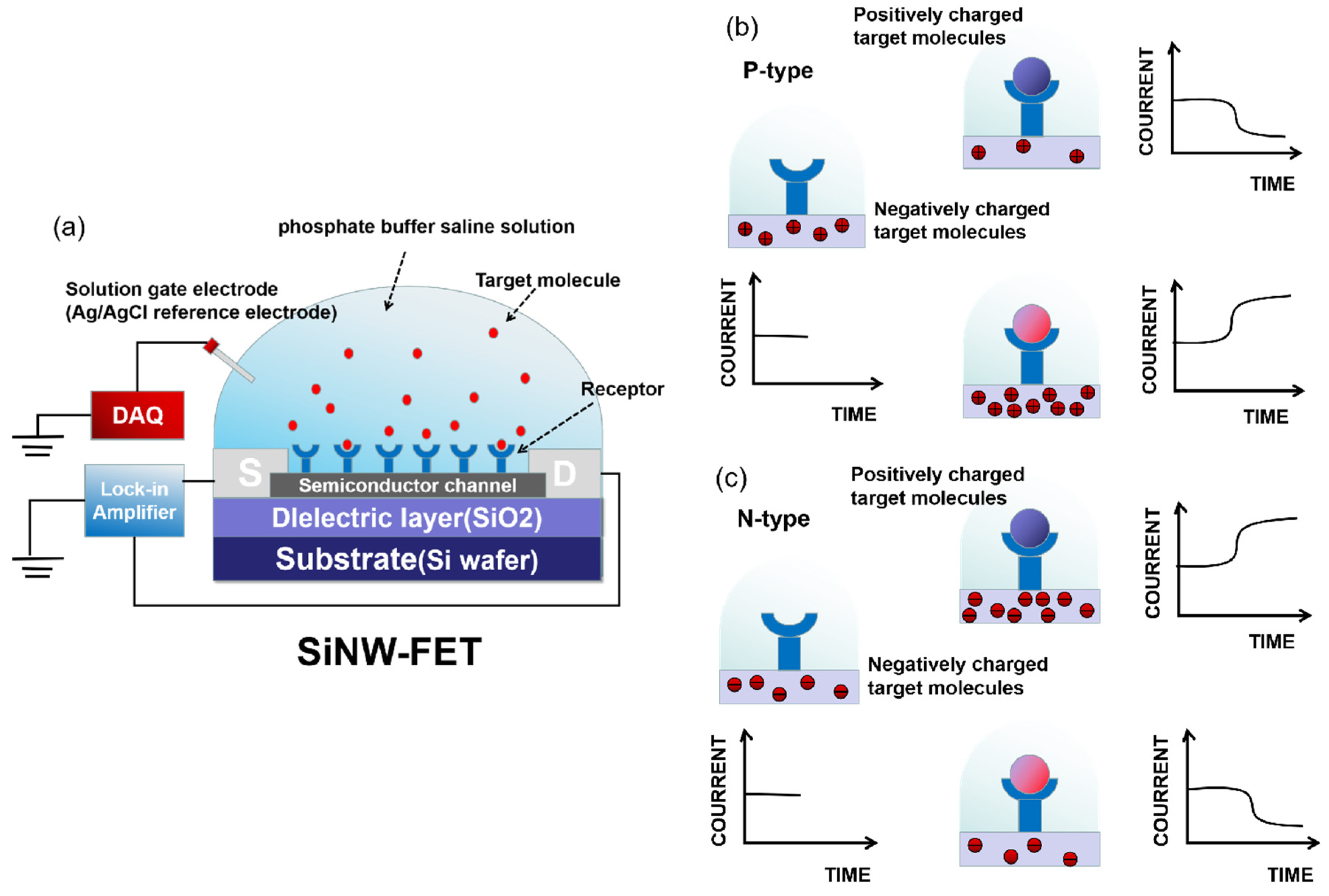
As typical FET-based devices, SiNW-FET biosensors are composed of three electrode structures: the source electrode, drain electrode, and gate electrode. The source and drain electrodes are both linked with semiconductor channels, which are made of SiNW. At the same time, the source–drain current is adjusted over the channel conductance via a varying bias voltage on the gate electrode. As an effective sensing element, SiNW can sensitively sense the change in the external electric field owing to its properties of field emission and electron transport.
The typical structure and sensing principle of SiNW-FET biosensors for identifying biomolecules are shown in Figure 1. Firstly, the target is captured by the receptor and anchored on the SiNW surface, and the data acquisition (DAQ) system supplies gate voltage (Vg) to ensure a stable output signal. Due to the charged nature of biomolecules such as DNA, the binding of the charged target to a receptor can alter the gate potential (or conductivity). The resulting signals [24] are electrically measured by a lock-in amplifier, while the sensing part of the device is usually immersed in a phosphate-buffered saline solution (PBS) to maintain the biological activity of detecting targets. For example, Cao et al. [25] used a 1 μM ssDNA-PBS solution during the SiNW-FET surface modification to maintain the biological activity of ssDNA, thereby ensuring a successful detection.
To efficiently capture the targets, bioreceptors that are capable of specifically recognizing targets are modified onto the SiNW surface. The target–receptor interaction induces the internal charge (electron or hole) distribution (accumulation or depletion) of SiNW-FET devices, thereby affecting the source–drain current. In fact, as shown in Figure 1, the direction of conductance during this change reflects the polarity (positive or negative) of the charge carried by the target, and the change in conductance reflects the target–receptor interaction.
The SiNW-FET biosensor is often used for immunoassays and is classified into an n-type or p-type according to its structural properties. For the n-type SiNW-FET device, when positively charged target molecules combine with the receptors modified on the SiNW surface, the positive charges in target molecules induce negative charges in SiNW. The electrons will accumulate in the channel, further enhancing the conductance or the source–drain current. Conversely, when the target molecule is negative, the reduction in electrons leads to a decrease in the conductivity and current. This has been demonstrated by Gao et al. [26]. They found that in the subthreshold state of the n-type SiNW-FET biosensor, the higher the negative charge concentration, the more obvious the change in the relative current. A significant increase in resistance was observed immediately upon the introduction of the DNA probe onto SiNW surface.
For p-type SiNW-FET devices, negatively charged target molecules will increase the conductivity or current since the charge carriers are mainly holes. Conversely, if the target molecules are positive, the holes’ depletion will lead to the depression of the conductance and current. Yang et al. [20] confirmed that the accumulation of the holes in the p-type channel changes the source–drain current, and the amount of negative charges ramp up at a higher pH due to deprotonation. In addition, in order to achieve a lower signal-to-noise ratio, SiNWs are integrated to form a new biosensing device that links to a semiconductor parameter analyzer or another measurement device for real-time monitoring [27,28].
3. Surface Modification Methods of SiNW
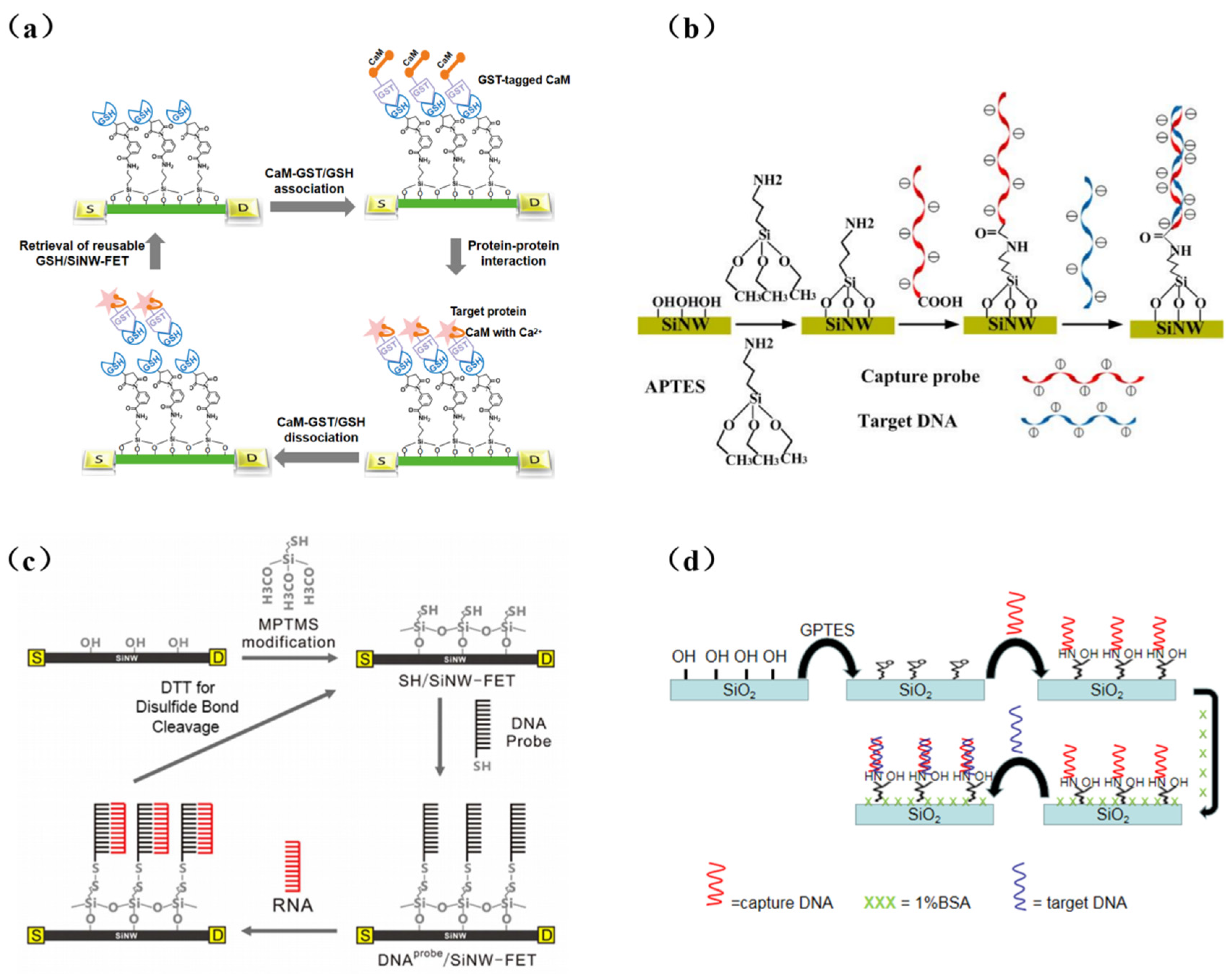
Surface modification is a critical step in the integration of biorecognition elements and transducers in biosensor fabrication. In SiNW-FET biosensors, SiNWs must be chemically modified to accomplish the integration of bioreceptors and FET sensors. The modification of biorecognition elements (i.e., receptors) onto SiNWs can provide specific binding sites for target molecules. In bioelectrical measurement, target recognition is achieved by SiNWs modified with specific receptors (antibodies, DNA, etc.). Usually, the Si/SiO2 NW surface is silanized before receptor modification. Alkoxysilane derivatives can react with hydroxyl groups (OH) on the surface of SiNWs and act as linkers to immobilize receptors onto SiNWs. 3-(trimethoxysilyl) propyl aldehyde (APTMS) is coupled with SiNW surfaces to present a terminal aldehyde group that can directly immobilize DNA [19] and antibodies [28]. In additional, 3-aminopropyltrimethoxysilane (APTES), the most popular linker, consists of a silane group and an amino group. Its amino groups can anchor biotin [29], calmodulin [30,31], and oligonucleotide [32,33,34,35]. Chu et al. [36] soaked the chip in an APTES solution (2% in acetone) at room temperature for one hour to form a single layer, and then modified the DNA probe. In other works in the literature, 3-mercapto propyl trimethoxysilane (MPTMS) has been proposed to form a thiol (SH)-termination to modify the surface. Chen et al. [37] were able to modify DNA probes using a disulfide bond formed on the SiNW surface by MPTMS. 3-glycidyloxy propyltrimethoxysilane (GPTES) is a kind of epoxy silane that can form a homogeneous silane monolayer in buffer solutions (pH 8–9). Schwartz et al. [38] silanized the SiNW device with GPTES directly. Amino-modified DNA sequences can then be directly and covalently attached to the open epoxy ring without any other cross-linking molecules. Figure 2 shows the above silicon nanowire modification method.
The natural oxide layer on the SiNW surface can prevent silanization or even shield the electric fields generated by the analyte, thereby limiting the performance of the sensor. Bunimovich et al. [39] achieved the quantitative and real-time detection of single-stranded oligonucleotides with SiNW in a physiologically relevant electrolyte solution. Compared with SiNW-FETs with an oxide layer, they found that SiNWs without a natural oxide layer exhibited better solution-gated FET characteristics and improved the sensitivity of single-strand DNA detection significantly. Therefore, prior to the silanization of SiNWs, it is necessary to remove the oxide coating by immersing them in a 2% dilute hydrogen fluoride (HF) solution for 3 s.
Furthermore, treating the SiNW surface with ultraviolet radiation (UV)/O2 plasma prior to silanization will remove the surficial inherent impurities and produce rich terminal hydroxyl groups (-OH) on the SiNW surface, thereby improving the hydrophilicity and then improving the efficiency of silanization and the sensing performance. The experimental results of Rahmnan et al. [40] showed that the detection limit of the SiNW-FET sensor without O2 plasma treatment was 4.131 × 10⁻¹³ M, while it could be as low as 1.985 × 10⁻¹⁴ M after 60 s of O2 plasma treatment. It has been demonstrated that O2 plasma treatment of SiNWs prior to silylation can improve the sensitivity of the sensor.
4. Application of SiNW-FET Biosensors
4.1. DNA Determination
Deoxyribonucleic acid (DNA), one of the most important genetic materials, functions not only as carriers for transferring genetic information, but also as ideal biomarkers for clinical diagnosis. As a good candidate for monitoring DNA/RNA hybridizations with ultra-high sensitivity (femtomolar level), SiNW-FET has higher integration and label-free detection abilities than surface plasmon resonance (SPR) [41] and quartz crystal microbalance [42].
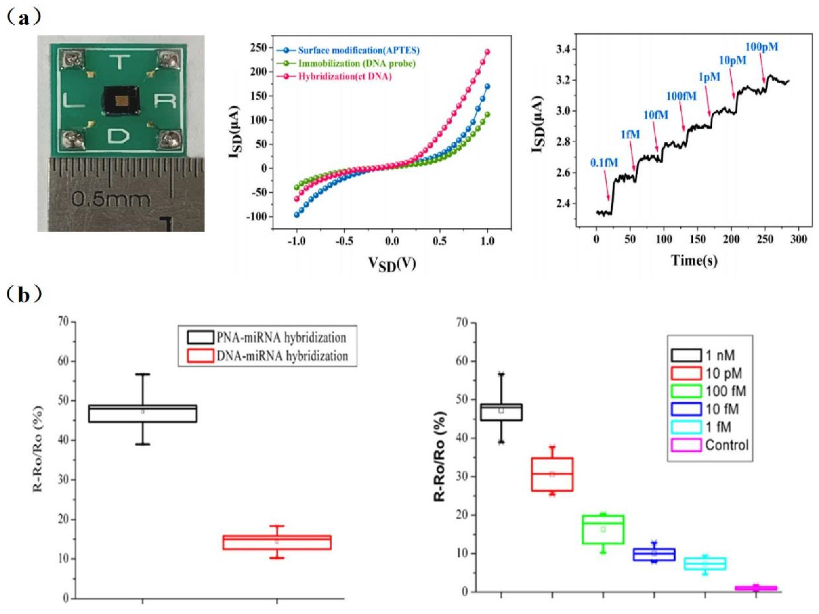
Many disease biomarkers are DNA molecules like hepatitis B virus, avian influenza virus, cancer, and others. The detection of specific DNA is extremely important from the perspective of medical diagnoses. For example, the early diagnosis of cancer is crucial to a patient’s life. Li et al. [43] achieved the detection of tumor marker circulating tumor DNA (ctDNA) with a sensitivity of 10 aM using an inverted triangle SiNW array FET (Figure 3a). The cross-sectional dimensions of SiNWs were approximately 90 nm with a deviation less than ±20 nm. Compared with a single SiNW, 120 SiNWs with a high consistency of dimensions enabled the superposition of signals, resulting in the much stronger response signal and a higher signal-to-noise ratio.
Gao et al. [25] detected the DNA of two pathogenic strains of avian influenza (H1N1 and H5N1) by immobilizing two DNA probes on the surface of SiNW, respectively. Their experimental results showed that the current was related to the logarithm of the DNA concentration. Lin et al. [44] used a Poly-SiNW FET to achieve the specific and ultra-sensitive (fM level) detection of pathogenic avian influenza virus (H5 and H7) DNA.
Due to the large amount of negative charges in the phosphate backbone of DNA/RNA, the charge carrier density of SiNW-FET biosensors varies significantly during DNA or RNA hybridization [19,45]. Although peptide nucleic acid (PNA) has no phosphate groups in its backbone, it was proven to be capable of determining the distance between target–receptor binding sites and the SiNW surface. As shown in Figure 3b, the binding of PNA/DNA or PNA/RNA strands is stronger than DNA/DNA or DNA/RNA. Hahm et al. [19] demonstrated that PNA-DNA duplex formation has successfully contributed to the detection of DNA in real time. They observed a considerable increase in conductance in the p-type PNA-modified SiNW-FET, with the detection limit down to 10 fM. PNA-based SiNW-FETs are a promising detection platform for various physiological applications [34]. Zhang et al. [32] achieved a detection limit down to 1 fM for miRNA sensing in PNA/SiNW-FET (Figure 3b). It is able to identify miRNAs from the whole RNA extract of HeLa cells, which will contribute to the early diagnosis of tumors. DNA extraction is not necessary for testing ctDNA in blood, while it is required before some viral or cellular nucleic acid assays.
Wenga, G. et al. [46] proposed a novel stepped polycrystalline silicon nanowire field effect transistor for the ultrasensitive detection of DNA hybridization. DNA probes were immobilized on the surface of polycrystalline silicon nanowires that were synthesized by sidewall spacer technology to detect DNA targets. The experimental results showed that the detection limit of complementary DNA targets could be 1 fM.
Due to the unique advantages of DNA analogues as probes for detecting target DNA, more and more scholars have begun to pay attention to DNA analogs. Hu, W.P., and colleagues [47] found that the configured neutralizing of DNA analogues as probes for target DNA can increase the Debye length, which is beneficial for improving the sensitivity and signal-to-noise ratio of the biosensor. They synthesized two partially neutralized chimeric DNA products and a fully neutralized DNA sequence using phosphomethylated nucleotides. The design of the neutralizing DNA probe reduced the ion solubility required for hybridization with the target DNA and increased the Debye length. In addition, the uncharged DNA probes avoided charge repulsion. A BTP buffer was also used to reduce the Debye shielding effect. Therefore, the detection limit of the SiNW-FET was as low as 0.1 fM.
In order to increase the sensitivity and repeatability of the SiNW-FET sensor, Mikael et al. [48] demonstrated repeatable and efficient DNA hybridization detection by coating the SiNW’s surface with a thin layer of HfO2. In a dry environment, the SiNWs were passivated by HfO2, and the DNA probes were transplanted onto the surface. Because HfO2 had a higher density of OH groups and a high dielectric constant, the density of DNA hybridization detected by this sensor was 1010 cm−2.
Now, SiNW-FET biosensors have been regarded as promising tools in DNA detection due to their label-free, ultra-highly sensitive, and rapid detection capabilities. As discussed above, SiNW-FET has been applied to detect DNA hybridization, infectious viruses, and miRNAs. However, further optimization of the device is still needed, such as reducing the width and length of the sensor or reducing unnecessary procedures to improve the sensitivity and accuracy of nanoscale devices.
Xie et al. [49] constructed an independent and integrated microfluidic nanodetection system that incorporates SiNW-FET biosensors for biodetection and analysis. All analytical processes, such as liquid sample delivery, thermostatic control, signal amplification, data acquisition, and result display, are performed automatically. This portable nanosensor detection system is mainly composed of five parts: the liquid circuit module, the light modulation module, the constant temperature control module, the signal acquisition and amplification module, and the status and result display module. The system can effectively solve the problem that SiNW-FET biosensors usually require discrete detectors and have not yet been implemented in the integrated and mobile fields. Because the surfaces of SiNW-FET biosensors are inevitably exposed to the external environment, they are susceptible to interference. They are extremely sensitive to external environmental factors such as temperature, light, and pH, which can cause detection errors. Therefore, SiNW-FET is expected to detect biomarkers in field applications in the near future.
4.2. Protein Detection
Protein is an important component of human cells and tissues, which plays a significant role in human life activities. Proteins realize the expression of genes under the control of DNA, which make organisms exhibit a variety of genetic characteristics. Many biomarkers used in disease diagnosis are proteins [17,50]. Therefore, achieving effective and sensitive detection of proteins as biomarkers is of great significance for the disease diagnosis.
The Enzyme-Linked Immunosorbent Assay (ELISA) [51], laser, and fluorescence technology [52] have all been used for the detection of proteins. Protein detection and separation technology have been constantly evolving due to improvements in microelectronic technology. Nowadays, more and more researchers are focusing on SiNW-FETs, which are suitable for protein detection.
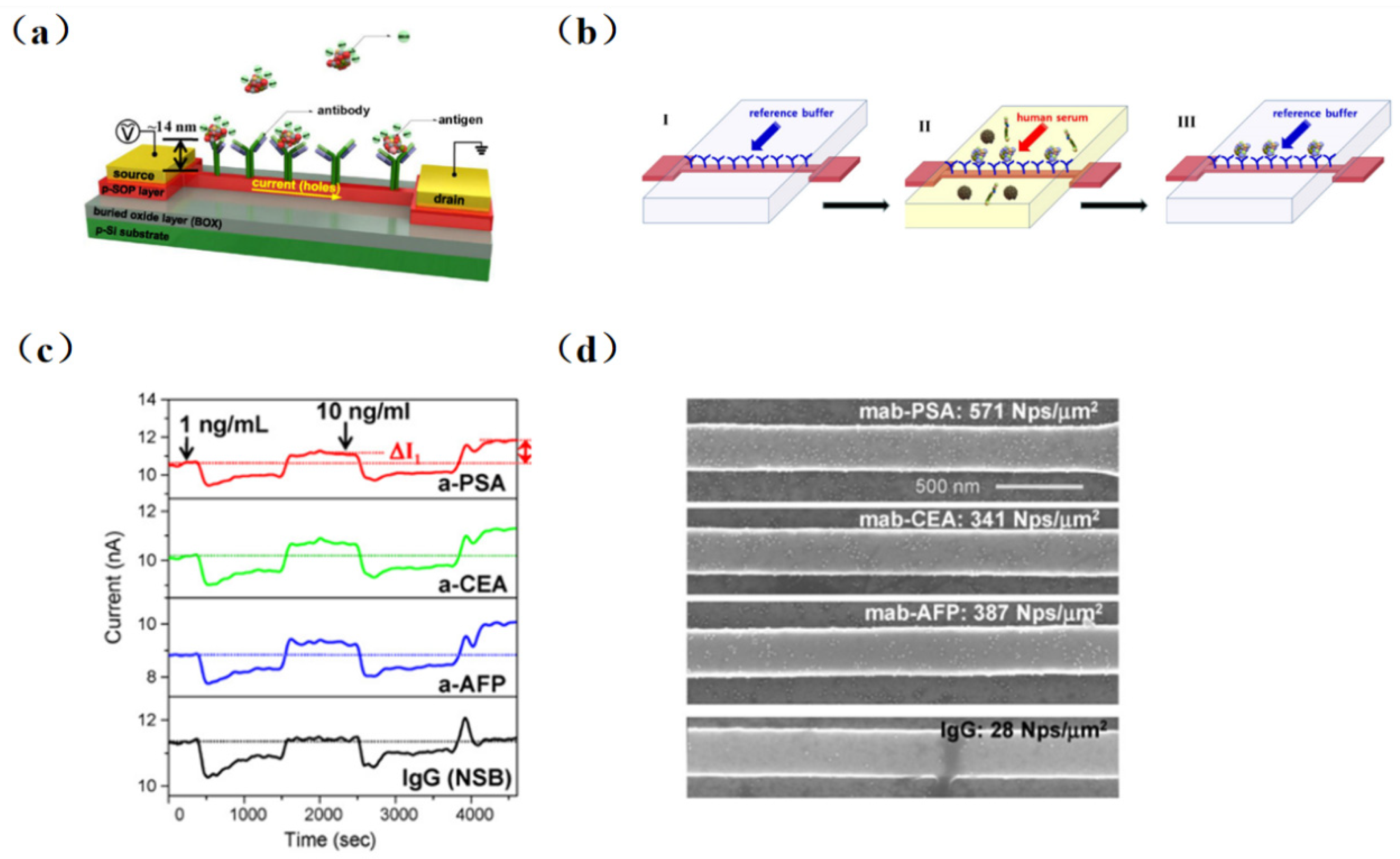
The real-time detection of biotin using an SiNW-FET biosensor was first achieved in 2001 by Cui et al. [5]. And, the detection limit reached 10 pmol/L, which was far lower than others test method. However, it was not possible to continuously detect samples with different concentrations due to the irreversible combination of biotin and streptomycin. Kim et al. [53] proposed the combination of a conventional immunoassay (Sandwich ELISA) with high-precision FET devices and accomplished the multichannel detection of three different cancer markers (carcinoembryonic antigen (CEA), prostate specific antigen (PSA), and alpha fetoprotein (AFP)) in serum without pretreatment successfully. As illustrated in Figure 4, four different antibodies were self-assembled on the chip surface (three tumor markers PSA, CEA, AFP, and monoclonal antibodies (IgG)). Then, serum antigens at concentrations of 1 and 10 ng/mL were injected into groups with tumor markers fixed, and changes before and after the injection were observed. The detection limit was as low as ng/mL level.
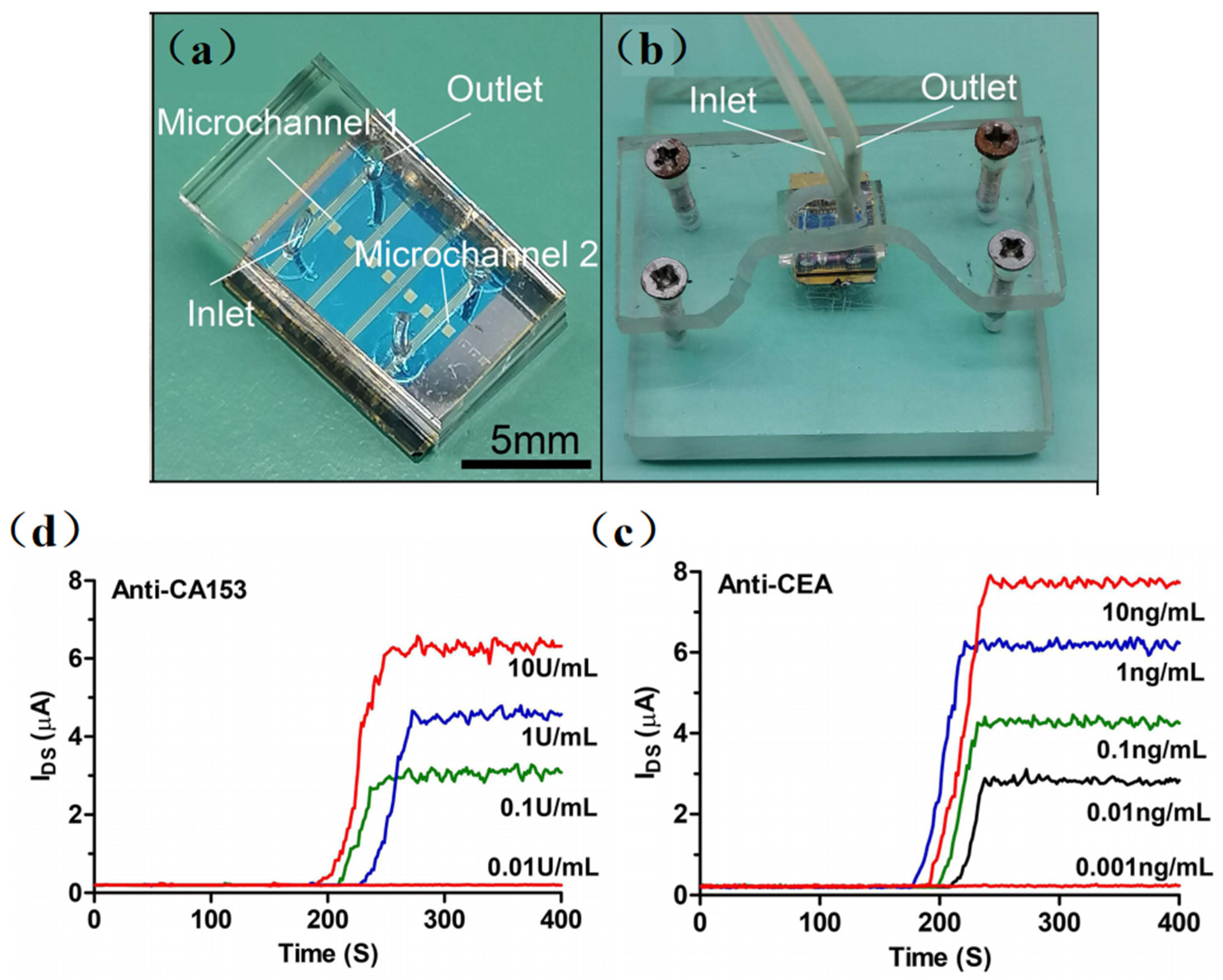
Recently, many emerging SiNW-based biosensors have been designed for in-depth studying in protein detection. Gong et al. [54] performed the ultra-sensitive detection of IgG using the SiNW-FET biosensor. They fabricated a single SiNW-FET, a double SiNW-FET, and a quad SiNW-FET into a single chip and detected the Norovirus DNA and IgG at concentrations of 1 fM and 10 fM, respectively. Then, as shown in Figure 5, Li et al. [55] developed a multichannel dual-gate SiNW-FET chip for dual-channel detection of BC tumor markers. By modifying monoclonal CA15-3 and CEA antibodies on different SiNW surfaces, the SiNW-FET could be used for the dual-channel specific detection of breast cancer tumor markers, CA15-3 and CEA, respectively. The experimental results showed that the double-gated SiNW-FET can amplify the detection signal exponentially through the capacitive coupling effect, thus improving the signal-to-noise ratio of the SiNW-FET. The sensor can accurately detect CA15-3 down to 0.1 U/mL and CEA at 0.01 ng/mL. Hence, compared with the traditional SiNW-FET, the multichannel design method increased the source–drain current signals, effectively restrained the fluctuation noise inside the transistor, and made the detection system more stable.
However, there are performance differences between the SiNW-FET devices manufactured by the same process. Lu et al. developed a novel SiNW biosensor for the detection of human tear MMP-9 based on an optimized fabrication of an optical calibration and low salt concentration scheme, achieving high sensitivity (86.96%) and specificity (90%) in the diagnosis of DED. Their device was manufactured to demonstrate minimal sensor-to-sensor variation, with optical calibration and a low salt concentration protocol allowing consistent response between sensors and sensitive and accurate detection of MMP-9 in human tears with high agreement with ELISA results [56].
SiNW-FET, as a novel kind of biosensor, could be affected by the Debye–Hückel screening effect induced by ions during protein detection [29]. The higher concentration of ions in a protein solution, the shorter the Debye length and the higher the screening efficiency. In other word, the Debye length is inversely proportional to the square root of the ionic strength. In general, because of the electrolytic medium, the interacting electric field exerted from the captured target charges is shielded. Therefore, it is necessary to reduce the Debye–Hückel screening effect when using an SiNW-FET biosensor for protein detection.
The common way to reduce the concentration of ions is by diluting the buffer solution to a low concentration. However, it is still far from effective protein detection. Kim et al. [50] explored the effect of the Debye length on the highly sensitive and label-free detection of cTnI, a biomarker for the diagnosis of acute myocardial infarction. The Debye lengths were calculated for PBS with different ion concentrations (1 × PBS, 0.1 × PBS, 0.01 × PBS3; ionic strength 180, 18, and 1.8 mM, respectively). It was found that the sensitivity increased with the decrease in buffer ion concentration, and the optimal condition was 0.01 × PBS. Therefore, the buffer concentration has a great effect on sensor determination [57,58]. But, extremely low salt concentrations may reduce the activity of proteins, aggravating the difficulty of effective detection.
Chang, S. M et al. [59] reported that the developed SiNW-FET exhibited a lower detection limit of 0.016 ng/mL than previously developed FET devices and showed great potential for future applications in point-of-care (POC) diagnostics. Puppo et al. [60] studied a terminal amnestic-modified SiNW-FET sensor that successfully detected an anti-rabbit antigen in a PBS buffer, further demonstrating the detection of anti-rabbit antigens in a tumor extract solution in the presence of 100,000 non-specific proteins [61,62]. At the same time, Meir developed a dissociative state sensing method that uses temporal differentiation between low-affinity matrix components and high-affinity target molecules as they are dissociated, enabling direct sensing when the biological samples on modifications are rinsed by low ionic strength buffer solutions [63].
It is found that the voltage power spectral density generated by current-biased SiNW-FET is related to 1/f-dependence in the frequency domain. Zheng et al. [64] performed frequency domain detection of a prostate-specific antigen, whose detection sensitivity was 10 times higher than that of time domain detection. This frequency domain measurement method showed the prospect of simple and rapid biomarker screening in medical applications, i.e., in disease diagnosis [60] and drug discovery.
So far, most of the traditional methods for detecting mycobacterium tuberculosis do not meet the requirements of actual TB detection, and the SiNW-FET sensor can solve the appeal problem. Ma [65] developed a fast and sensitive SiNW-FET biosensor for the detection of the Mycobacterium tuberculosis Ag85B protein. The detection limit of MTB Ag85B is as low as 0.01 fg/mL (0.33aM), it has good specificity and stability, and the detection response time is within 30 s. And the detection range is from 1 fg/mL to 100 fg/mL. Compared to other methods, the SiNW-FET biosensor requires only a small sample (7 μL) and can detect the Ag85B protein with a significantly wider dynamic linear range in a shorter time period.
Although the SiNW-FET biosensors were mostly used in laboratory work, they demonstrated high detection sensitivity, specificity, and relativity, which have potential for practical applications such as food safety detection and clinical diagnosis.
4.3. Microbiological Detection
A microorganism is an organism that can be seen only through a microscope, and they are pervasive in life. In fact, any environment that is devoid of microorganisms is certainly the exception rather than the rule. Microorganisms include bacteria, viruses, and fungi. A pathogenic organism is an organism that is capable of causing diseases in a host (person). So far, there have been many large-scale infection outbreaks caused by pathogenic microorganisms. Examples include swine flu in Mexico in 2009, Ebola in West Africa in 2014, and the plague in Madagascar in 2017. And especially the COVID-19 outbreak, which started in 2020, has caused global infections. The large-scale infection outbreak of these pathogenic microorganisms seriously threatens people’s health and life. So, highly sensitive detection methods are essential for the detection of bacteria and viruses to prevent the spread of infection.
Molecular detection methods are used for the detection of microorganisms. A polymerase chain reaction (PCR) [34] was used in an amplifier to amplify specific DNA fragments, and could be used for bacteria or virus detection with strong specificity, high sensitivity, and low purity requirements. Maan et al. [66] reported a real-time reverse transcription polymerase chain reactions (RT-PCR) assay to recognize Seg-2 of the eight epizootic hemorrhagic disease virus (EHDV) serotypes [67,68]. However, such detectors are difficult to implement for point-of-care testing. An immunofluorescence assay (IFA) could also be used for virus detection. Darwish et al. [69] designed a sensitive and specific optical immunosensor for detecting dengue virus markers using a fluorescent signal label with quantifiable signals [70]. Surface-enhanced Raman Scattering (SERS) [71,72] has emerged as a promising tool to guide cancer diagnosis and synergistic therapy. However, SERS requires the use of nanomaterials to enhance the fluorescence signal, and the detection equipment is relatively expensive. Yin et al. [73] reported an ultra-sensitive, highly selective, and SERS-based SARS-CoV-2 simulation sequence (N-cDNA) detection platform. The platform used a magnetic field to control the distance at which superparamagnetic iron oxide nanoparticles (DNA2/DNA3−SPIONs), DNA2-bound N-cDNA, and AuNPs coupled with DNA3 and their complementary sequence (DNA4) are bound, which effectively enhances the fluorescence signal, thereby increasing its sensitivity.
However, this method is too complicated, time-consuming, and expensive, as is the case with electrochemical biosensors [74]. Currently, the SiNW-FET-based biosensor, as an emerging biological detection technology, could be used for protein [54], DNA [75], and microorganism detection [34], providing new solutions for infectious diseases and other health aspects. To date, SiNW-FETs have successfully detected many viruses, including Dengue [4], Influenza A H3N2 [76], H1N1 [77,78], H5N1 [79], and H5N2 [80]; they have also detected two different viruses simultaneously [81].
To improve the detection sensitivity, researchers improved the SiNW-FET biosensor from different aspects. Patolsky et al. [27] functionalized the nanowire array and observed the changes in electrical conductivity before and after binding in order to detect influenza A labeled with fluorescence. The binding of the viruses resulted in a decrease in the electrical conductivity, which returned to baseline when the viruses were released. Meanwhile, their electrical measurements with antibody-functionalized nanowire field effect transistors (NWFET) illustrated that single viruses can be detected in parallel with high detection selectivity.
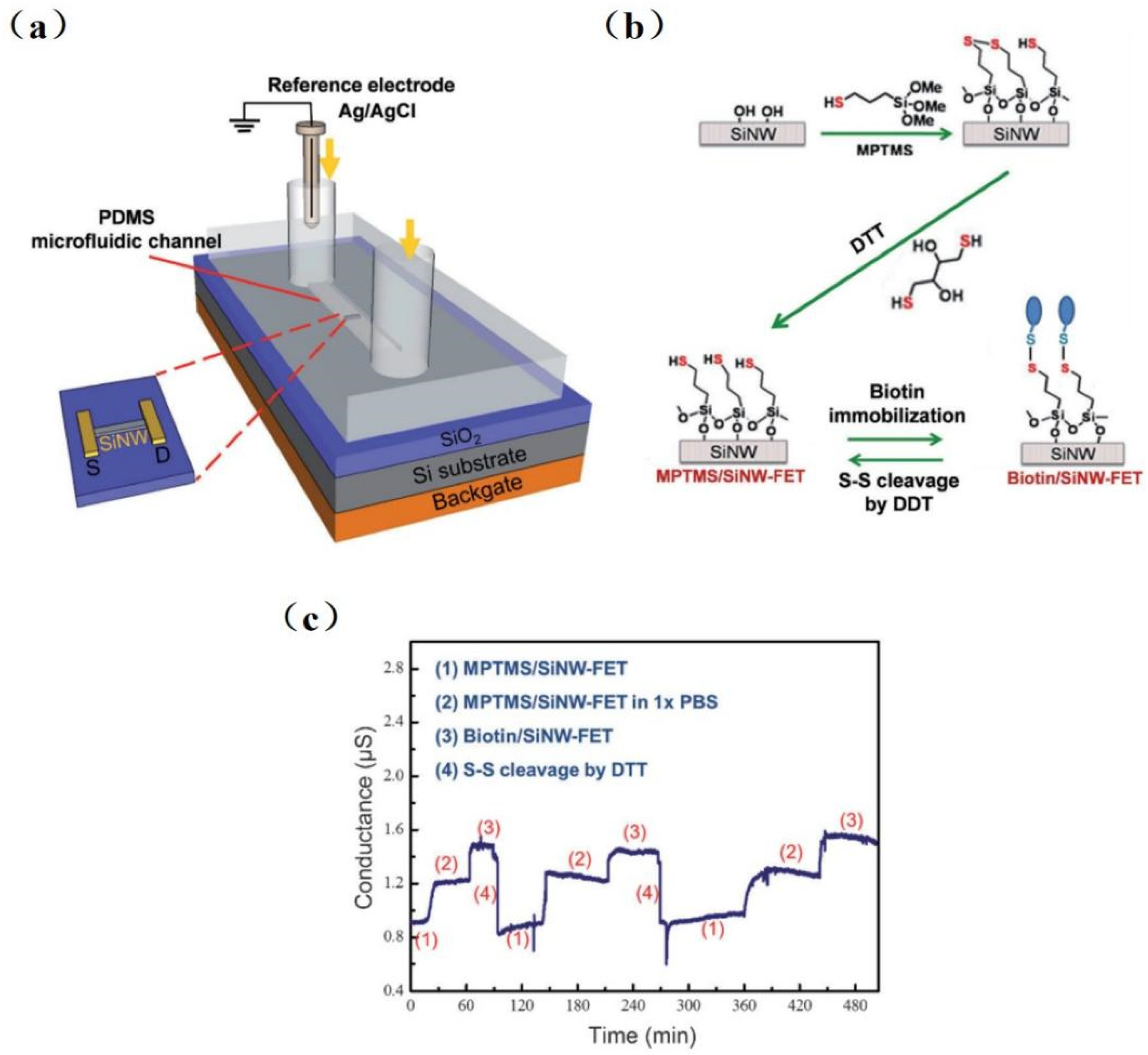
Since silanaldehydes are linked to antibodies by reductive amination, it is difficult to reverse the amine link to the original free aldehyde state after antigen detection. Therefore, removing the tightly bound antigen–antibody complex is difficult, resulting in sensors that can only be used once. Chiang et al. [80] proposed a reversible surface modification technology using a disulfide linker, where the receptor molecules were anchored in an MPTMS/SiNW-FET surface with the disulfide bond. Figure 6 shows the experimental setup of the SiNW-FET system, a schematic diagram, and experimental results. The detection limit was 10−17 M for H5N2 avian influenza virus (AIV) allergic testing. The sensing device was reusable by fixing the antibody to the SiNW-FET via disulfide bonds and then reducing the disulfide bonds with dithiothreitol (DTT) while removing the antibody–virus complex. This reversible surface functionalization by reducing disulfide bonds is comparable to another technique that modifies the surface of SiNW-FET by reversible binding–dissociation between glutathione (GSH) and glutathione S-transferase (GST)-labeled proteins [17].
The interaction between hemagglutinin (HA) on Influenza virus’ surface and glycans on the host cell was discussed by Hideshima et al. [23]. They were the first to detect H1 and H5 of influenza A viral HA molecules with glycan-immobilized FET biosensor.
In addition to enhancing surface modification, some improvements were introduced during hardware manufacturing. Borgne et al. [82] developed a simple and low-cost bacteria sensor with a self-assembled gas liquid solid method (VLS). It could be used for label-free and ultra-sensitive bacteria detection. Borgne and his colleagues also developed an SiNW-FET-based resistor fabricated by the VLS method and compatible COMS silicon technology. It was found that Escherichia coli bacteria were able to attach precisely to the SiNW networks, which resulted in a drastic decrease in the electrical resistance of the device [83]. Kim et al. [81] improved the sensitivity of biosensors through material selection. One-dimensional SiNW with a nanoscale three-dimension transistor structure was fabricated through isotropic and anisotropic patterning. This sensor could be used for the multiplex detection of AI and HIV viruses.
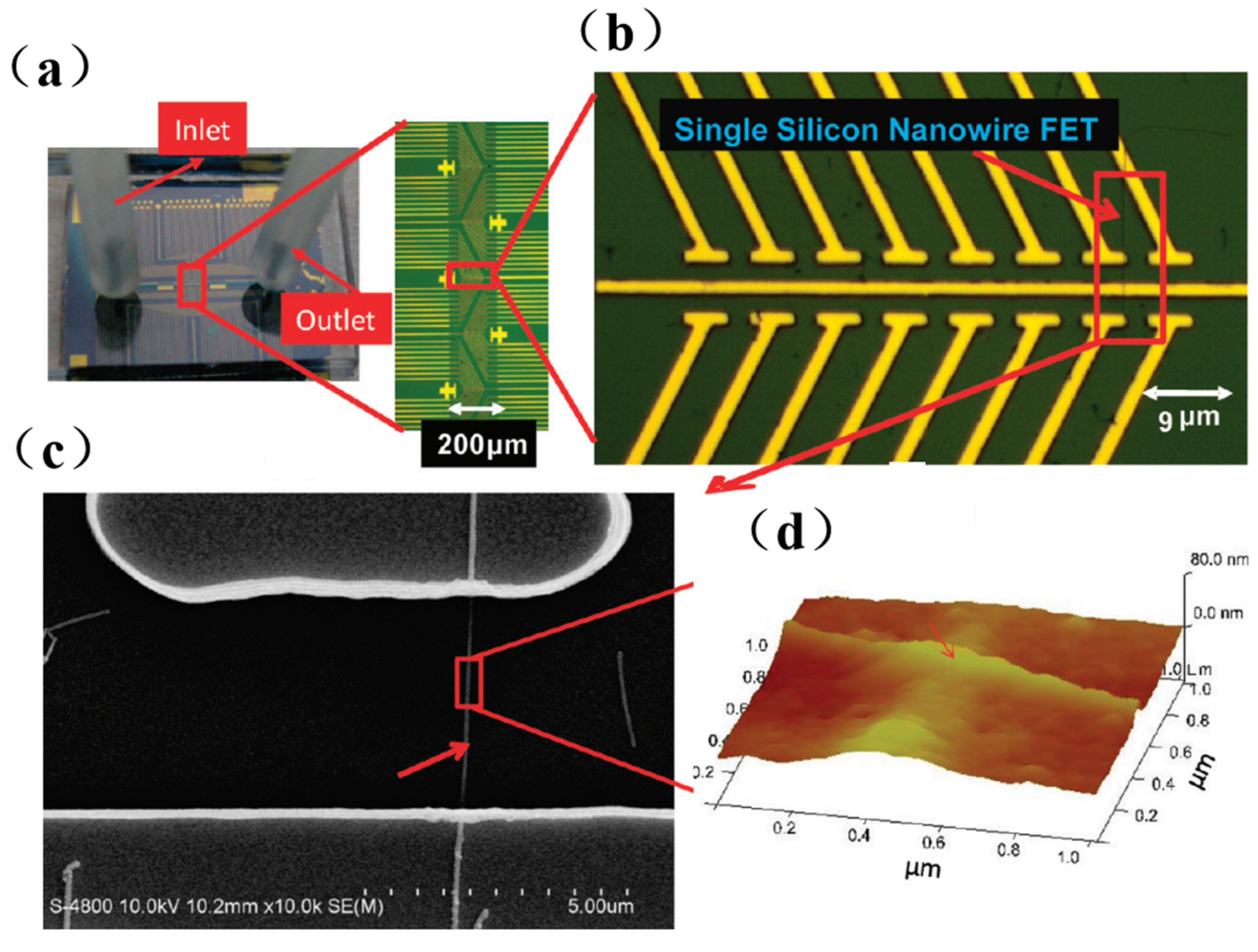
SiNW-FET, as an emerging detection method, is applied in the field of biological aerosol research. Shen et al. [76] developed a real-time monitoring system for airborne influenza H3N2, integrating electronic-addressing SiNW sensor devices, microfluidics, and bioaerosol-to-hydrosol air sampling techniques. The experimental results showed that the device can rapidly detect influenza A (H3N2) viruses by alternating clean air samples, indoor air samples, and airborne virus samples with a detection limit of 104 viruses/L and specifically differentiate between H3N2 and H1N1 viruses. The optical image chip and microfluidic channel of SiNWs are shown in Figure 7. This monitoring system was tested with an antenna and network. The remote platforms (such as cell phones and computers) observed continuous conductivity changes in seconds, demonstrating the ability to monitor the influenza virus in real time.
The outbreak of the COVID-19 pandemic started in late December 2019 and then spread quickly worldwide, leading to a global public health emergency. Hu et al. [84] proposed an SiNW-FET platform for the detection of IL-6, a disease marker of COVID-19. By using anti-IL-6 aptamers to detect IL-6, SiNW-FET can achieve effective detection concentrations as low as 2.1 pg/mL (100 fM). Moreover, the proposed aptamer-functionalized SiNW-FET detection range is sufficient to measure IL-6 expression levels in COVID-19-infected patients and distinguish between mild and severe disease.
5. Challenges and Opportunities
The SiNW-FET biosensor has ultra-sensitive and highly selective properties. It has been widely used in disease diagnosis [85], drug discovery, food monitoring [86], and environmental detection [82]. Table 1 summarizes the sensing performance of silicon nanowire-based FETs for the analysis of biomedical biomarkers. As can be seen from Table 1, the outstanding advantage of SiNW-FETs is their high sensitivity, i.e., ultra-low detection limit. These SiNW-FETs are capable of detecting nucleic acids, proteins, and microorganisms at fM levels. Despite these advantages, the commercialized CMOS-compatible SiNW-FET biosensor is still in its preliminary stage and faces some challenges. The main challenges include the following: (1) how to reduce the Debye shielding effect to improve the sensor sensitivity. The Debye shielding effect occurs when an SiNWs-FET detects charged targets in the sample. When the charge thickness generated by the charged target exceeds the charge thickness formed in the sample solution, the potential field formed by the target will be shielded, thus failing to attract carrier changes in the SiNW channel and reducing the sensitivity of the sensor. (2) Modification and reversibility of the biorecognition layer. Reversible surface modification technology can effectively improve the reusability of SiNW-FET sensors. However, the removal of covalent bonds on the SiNW surface by oxygen plasma or strong oxide can damage SiNWs and reduce the signal of the sensor (the magnitude varies from uA to nA). (3) How to achieve multitarget sensitive detection. Most SiNW-FET sensors can only be used to detect a single target, whereas diseases are often accompanied by multiple biomarkers. Microfluidic technology enables SiNW-FET sensors to detect two or more biomarkers at the same time, which is conducive to the diagnosis of diseases.
5.1. Sensor Sensitivity
SiNW-FET biosensors are mainly subject to the interacting electric field imposed by the bound molecular charges to conduct conductance modulation. The sensor sensitivity will reduce due to the Debye–Hückel screening effect. [87,88] It is difficult to trace analytes in high-salt buffer solutions. The whole blood sample [89] within a high-ionic-strength serum should be desalted before a sensor analysis [2,90].
So far, several approaches to overcome the Debye–Hückel screening effect have been proposed, such as the following: (1) replacing antibodies with aptamers [91] as biorecognition functions on silicon nanowires; (2) adding a layer of biomolecule-permeable polymers to the semiconductor material to change dielectric constant [92]; (3) purifying serum and untreated blood by an antigen–antibody binding reaction [93]. The above methods can shorten the distance between the target molecule and the surface of the silicon nanowire or increase the Debye length, which can partially overcome the Debye screening effect. However, this weakens the advantages of sensor rapidity and selectivity.
It is essential to improve the sensitivity of SiNW-FET biosensors [94] for biomarker detection. One of the challenges scientists face is how to take into account the characteristics of high sensitivity, rapidity, and good selectivity of SiNW-FET biosensors.
5.2. Surface Modification
APTES is often used in the surface modification of SiNW-FET biosensors to generate amino groups, linking the corresponding antibody or DNA of the target analytes to the sensor surface. However, low modification efficiency and antibody interactions will affect the detection result. Although cryo-electron microscopy (cryo-EM) technology [95] can distinguish structural information among organic, biological, and nanodevices, it still needs to improve the surface probe modification technology for accurate sensor detection.
In addition, one of the main challenges in the area of biosensor detection is not only the immobilization of the receptors or biological entities of target analytes, but also their complete removal from the surface to promote the reusability of the sensor platform [96]. SiNW-FET could be reusable for different biomolecules because the modified biorecognition layer can be removed by treating the SiNW surface with ultraviolet radiation (UV)/O2 plasma. Current studies have focused on the temperature-dependent reversible adhesion of cells on the surface of a stimulatory polymer PNIPAAM-modified sensor. It was shown that when detecting the signal from osteosarcoma SAOS-2 cells, the electrical signal remained constant after each reversible adhesion or dissociation of the cells. Thus, the use of stimulating polymers to modify sensor surfaces can facilitate or hinder the adhesion of biomolecules (e.g., proteins and cells), opening up new possibilities for biosensing applications.

Table 1.
Silicon nanowire-based FET biosensors for various biomarkers and their analytical performances.
Table 1.
Silicon nanowire-based FET biosensors for various biomarkers and their analytical performances.
| Channel Material | Substrate Material | Analyte | Recognition Element | LOD | Linear Range | Ref |
|---|---|---|---|---|---|---|
| N-type phosphorus-doped SiNW | Si/SiO2 | DNA | DNA probe | 0.1 fM | 0.1 fM–10 nM | [26] |
| P-type boron-doped SiNW | Si/SiO2 | α-fucosidase | Fuconojirimycin | 1.3 pM | 1.3 pM–500 pM | [97] |
| SiNW | SOI wafer | Tnl | CaM | 7 nM | 10−8 M–10−6 M | [17] |
| N-type polycrystalline SiNW | Si/SiO2 | APOA2 | Antibody | 6.7 pg/mL | 9.5 pg/mL–1.95 ug/mL | [98] |
| N-type polycrystalline SiNW | Si/SiO2 | PSA | Antibody | 5 fg/mL | 5 fg/mL–500 pg/mL | [91] |
| SiNW | SOI wafer | DNA | DNA probe | 0.83 fM | 10 fM–10 nM | [99] |
| P-type boron-doped SiNW | SOI wafer | DNA | Aptamer | 1 pM | 1 pM–10 nM | [65] |
| SiNW | SOI wafer | HA | CMP-NANA | 1 fM | 10−18 M–10−8 M | [78] |
| P-type polycrystalline SiNW | Si/SiO2 | ferritin | Antibody | 50 pg/mL | 50 pg/mL–500 ng/mL | [100] |
| SiNW | SOI wafer | cTnl | Antibody | 92 pg/mL | 92 pg/mL–46 ng/mL | [101] |
| SiNW | SOI wafer | AFP | Antibody | 100 ng/mL | 100 ng/mL–500 ng/mL | [102] |
| P-type boron-doped SiNW | SOI wafer | VEFG | Antibody | 5 fM | 5 fM–200 fM | [103] |
5.3. High-Throughput Multiplex Detection
The detection of multiple biomarkers using SiNW-FET sensors has been investigated [81]. For example, the integration of SiNW-FET sensors with Polydimethylsiloxane (PDMS) microfluidic devices can simultaneously detect microRNA and CEA [104]. PDMS-based channels are widely used in SiNW-FET biosensors, which brings the following problems: (1) particles flowing in the channels are difficult to access the nanowire surface; (2) PDMS can adsorb biomolecules, which may degrade the sensitivity of the sensor.
Scientists have also investigated improving microanalysis systems by improving microfabrication and microfluidic device technologies [85,105]. However, high-efficiency, high-sensitivity, high-throughput detection has not yet been achieved [106], which is urgently needed for precision in medical diagnosis and monitoring.
5.4. SiNW-FET Device
Silicon nanowires (SiNWs) have attracted much attention in recent years due to their excellent physical and chemical properties. Because SiNW has a high surface-to-volume ratio, Cui et al. [5] produced highly sensitive SiNW sensors to detect biological and chemical molecules. It was found that the design of SiNW-FETs, including SiNW’s size, the threshold voltage, and the manufacturing process, plays a crucial role in the detection performance [20]. The number of SiNWs is positively proportional to the sensitivity, and the size is inversely proportional to the sensitivity. He et al. [107], respectively, designed SiNW-FETs with 1–3 μm spacing between the SiNWs used for detecting the miRNA. The study results found that the SiNW-FET sensor with 1 μm spacing showed superior performance. Mohd et al. [108] proposed that tunnel field-effect transistors (TFETs) had a lower threshold voltage than FETs. Furthermore, the Schottky barrier (SB) in the source/drain was used in the design of TFETs to eliminate the doping effect of the S/D area, thus giving the TFETs better detection performance than the FETs. In addition, the introduction of high-k oxide [109] or charged plasma in TFETs [110] and the introduction of cavities on the source side for sensing biological molecules using dielectric modulation are helpful to enhance the drain current.
The above design method provides a good strategy for improving the detection performance of SiNW-FET sensors. Optimizing the size of the SiNW can enhance the sensitivity of SiNWs to the electric field, and the good detection performance of TFETs provides a solid foundation for improving the performance of SiNW-FET biosensors in the future.
6. Summary
An SiNW-FET biosensor with remarkable electronic properties is a promising biosensor for biological analysis. Changes in the internal charge distribution of the device can be sensitively detected by field-effect sensing-based SiNW-FETs. The capture of the target can cause a change in the number of carriers in the device (consumption or accumulation) and finally achieve conductance modulation.
Due to the Debye–Hückel screening effect [88], a highly sensitive strategy should be taken for biomarker detection during cancer [17] and infectious disease [68] testing. In this paper, we discussed the development process of the SiNW-FET biosensor. The detection strategies of the SiNW-FET biosensor are different for nucleic acid molecules, proteins, and microorganisms. When SiNW-FET biosensors detect DNA with PNA probes [34,111], the detection sensitivity is higher than that of DNA probes. This method could be used for virus detection [112], disease diagnosis [113], and drug therapy [114]. In addition, the SiNW-FET biosensor could be used for protein detection through the specific recognition of antigen–antibody/biotin–avidin. The detection sensitivity could be improved with sample pre-treatment (such as ion dilution [29]), chip integration, and multichannel 3D design [55]. Additionally, for microorganism detection, i.e., viruses and bacteria, the SiNW-FET biosensor can be used with a disulfide bond reversible surface modification technique [80] combined with an improved manufacturing process (such as gas liquid solid VLS [115] combined with silicon CMOS technology [116,117] and anisotropic and isotropic etching [20]). The sensitivity of an SiNW-FET biosensor is limited by factors such as serum sample desalination, small quantities of the mixed sample, a complex detection matrix, and probe sensitivity. Future research will focus on the sensitive detection of multiple biomarkers simultaneously for more accurate cancer prevention and clinical sample information.
In summary, the SiNW-FET biosensor is simple, rapid, and sensitive and can be used for real-time DNA, protein, and microorganism detection. An increasing number of portable and low-cost SiNW-FET biosensors are being developed for practical applications, demonstrating their great potential for clinical diagnosis, such as tumor testing, virus determination, and blood testing.
Author Contributions
H.L. and H.C.: manuscript draft. D.L.: project conception, designing, and revising. X.Y. and K.F.: reviewed and edited the manuscript. L.D. and G.W.: supervision and funding acquisition. All authors have contributed to the manuscript revision and approved the submitted version. All authors have read and agreed to the published version of the manuscript.
Funding
This study was supported by the NSFC Key Grant (62141409), the National Key R&D Program Grant (2018YFE0120000), the and Zhejiang Provincial Key Research and Development Project (Grant 2021C01041).
Institutional Review Board Statement
Not applicable.
Informed Consent Statement
Not applicable.
Data Availability Statement
Not applicable.
Conflicts of Interest
The authors declare no conflict of interest.
References
- Han, X.; Han, X.; Shao, Y.; Wu, Z.; Liang, Y.; Teng, J.; Bo, S.; Morthier, G.; Zhao, M. Polymer integrated waveguide optical biosensor by using spectral splitting effect. Photonic Sens. 2017, 7, 131–139. [Google Scholar] [CrossRef] [Green Version]
- Patolsky, F.; Zheng, G.; Lieber, C.M.; Patolsky, F.; Zheng, G.F.; Lieber, C.M. Nanowire sensors for medicine and the life sciences. Nanomedicine 2005, 1, 51–65. [Google Scholar] [CrossRef] [Green Version]
- Wang, Y.; Herron, N. Nanometer-Sized Semiconductor Clusters: Materials Synthesis, Quantum Size Effects, and Photophysical Properties. J. Phys. Chem. 1991, 525, 525–532. [Google Scholar] [CrossRef]
- Nuzaihan, M.; Hashim, U.; Arshad, M.M.; Kasjoo, S.R.; Rahman, S.F.A.; Ruslinda, A.R.; Fathil, M.F.M.; Adzhri, R.; Shahimin, M.M. Electrical detection of dengue virus (DENV) DNA oligomer using silicon nanowire biosensor with novel molecular gate control. Biosens. Bioelectron. 2016, 83, 106–114. [Google Scholar] [CrossRef] [PubMed]
- Cui, Y.; Wei, Q.; Park, H.; Lieber, C.M. Nanowire nanosensors for highly sensitive and selective detection of biological and chemical species. Science 2001, 293, 1289–1292. [Google Scholar] [CrossRef]
- Li, Y.; Zhao, M.; Chen, J.; Fan, S.; Liang, J.; Ding, L.; Chen, S. Multifunctional biosensor based on self-assembled multi-walled carbon nanotubes sponge. J. Mater. Sci. Mater. Electron. 2016, 27, 6911–6917. [Google Scholar] [CrossRef]
- Numnuam, A.; Thavarungkul, P.; Kanatharana, P. An amperometric uric acid biosensor based on chitosan-carbon nanotubes electrospun nanofiber on silver nanoparticles. Anal. Bioanal. Chem. 2014, 406, 3763–3772. [Google Scholar] [CrossRef]
- Sun, X.; Gong, Z.; Cao, Y.; Wang, X. Acetylcholinesterase biosensor based on poly (diallyldimethylammonium chloride)-multi-walled carbon nanotubes-graphene hybrid film. Nano-Micro Lett. 2013, 5, 47–56. [Google Scholar] [CrossRef] [Green Version]
- Jia, J.; Guan, W.; Sim, M.; Li, Y.; Li, H. Carbon Nanotubes Based Glucose Needle-type Biosensor. Sensors 2008, 8, 1712–1718. [Google Scholar] [CrossRef] [Green Version]
- Chen, D.; Sun, X.; Guo, Y.; Qiao, L.; Wang, X. Acetylcholinesterase biosensor based on multi-walled carbon nanotubes-SnO2-chitosan nanocomposite. Bioprocess Biosyst. Eng. 2014, 38, 315–321. [Google Scholar] [CrossRef]
- Taghdisi, S.M.; Danesh, N.M.; Ramezani, M.; Emrani, A.S.; Abnous, K. A simple and rapid fluorescent aptasensor for ultrasensitive detection of arsenic based on target-induced conformational change of complementary strand of aptamer and silica nanoparticles. Sens. Actuators B Chem. 2018, 256, 472–478. [Google Scholar] [CrossRef]
- Zhang, G.; Luo, Z.H.H.; Huang, M.J.; Tay, G.K.I.; Lim, E.A.; Chen, Y. Highly Sensitive and Selective Label-Free Detection of Cardiac Biomarkers in Blood Serum with Silicon Nanowire Biosensors. Sens. Actuator B Chem. 2010, 146, 138–144. [Google Scholar] [CrossRef]
- Chen, Y.; Wong, C.C.; Pui, T.S.; Nadipalli, R.; Weerasekera, R.; Chandran, J.; Yu, H.; Rahman, A.R. CMOS high density electrical impedance biosensor array for tumor cell detection. Sens. Actuators B Chem. 2012, 173, 903–907. [Google Scholar] [CrossRef]
- Song, X.; Zhang, T.; Wu, L.; Hu, R.; Qian, W.; Liu, Z.; Wang, J.; Shi, Y.; Xu, J.; Chen, K.; et al. Highly Stretchable High-Performance Silicon Nanowire Field Effect Transistors Integrated on Elastomer Substrates. Adv. Sci. 2022, 9, 2105623. [Google Scholar] [CrossRef]
- Martinez, J.; Martinez, R.V.; Garcia, R. Silicon nanowire transistors with a channel width of 4 nm fabricated by atomic force microscope nanolithography. Nano Lett. 2008, 8, 3636–3639. [Google Scholar] [CrossRef]
- Cui, Y.; Zhong, Z.; Wang, D.; Wang, W.U.; Lieber, C.M. High Performance Silicon Nanowire Field Effect Transistors. Nano Lett. 2003, 3, 149–152. [Google Scholar] [CrossRef]
- Lin, T.-W.; Hsieh, P.-J.; Lin, C.-L.; Fang, Y.-Y.; Yang, J.-X.; Tsai, C.-C.; Chiang, P.-L.; Pan, C.-Y.; Chen, Y.-T. Label-free detection of protein-protein interactions using a calmodulin-modified nanowire transistor. Proc. Natl. Acad. Sci. USA 2010, 107, 1047–1052. [Google Scholar] [CrossRef]
- Wang, C.-W.; Pan, C.-Y.; Wu, H.-C.; Shih, P.-Y.; Tsai, C.-C.; Liao, K.-T.; Lu, L.-L.; Hsieh, W.-H.; Chen, C.-D.; Chen, Y.-T. In situ detection of chromogranin a released from living neurons with a single-walled carbon-nanotube field-effect transistor. Small 2007, 3, 1350–1355. [Google Scholar] [CrossRef] [PubMed]
- Hahm, J.I.; Lieber, C.M. Direct Ultrasensitive Electrical Detection of DNA and DNA Sequence Variations Using Nanowire Nanosensors. Nano Lett. 2004, 4, 51–54. [Google Scholar] [CrossRef]
- Yang, X.; Gao, A.; Wang, Y.; Li, T. Wafer-level and highly controllable fabricated silicon nanowire transistor arrays on (111) silicon-on-insulator (SOI) wafers for highly sensitive detection in liquid and gaseous environments. Nano Res. 2018, 11, 1520–1529. [Google Scholar] [CrossRef]
- Tran, D.P.; Pham, T.T.T.; Wolfrum, B.; Offenhäusser, A.; Thierry, B. CMOS-Compatible Silicon Nanowire Field-Effect Transistor Biosensor: Technology Development toward Commercialization. Materials 2018, 11, 785. [Google Scholar] [CrossRef] [PubMed] [Green Version]
- Shen, M.-Y.; Li, B.-R.; Li, Y.-K. Silicon nanowire field-effect-transistor based biosensors: From sensitive to ultra-sensitive. Biosens. Bioelectron. 2014, 60, 101–111. [Google Scholar] [CrossRef]
- Hideshima, S.; Hinou, H.; Ebihara, D.; Sato, R.; Kuroiwa, S.; Nakanishi, T.; Nishimura, S.-I.; Osaka, T. Attomolar detection of influenza A virus hemagglutinin human H1 and avian H5 using glycan-blotted field effect transistor biosensor. Anal. Chem. 2013, 85, 5641–5644. [Google Scholar] [CrossRef]
- Li, B.-R.; Hsieh, Y.-J.; Chen, Y.-X.; Chung, Y.-T.; Pan, C.-Y.; Chen, Y.-T. An ultrasensitive nanowire-transistor biosensor for detecting dopamine release from living PC12 cells under hypoxic stimulation. J. Am. Chem. Soc. 2013, 135, 16034–16037. [Google Scholar] [CrossRef]
- Gao, A.; Lu, N.; Dai, P.; Li, T.; Pei, H.; Gao, X.; Gong, Y.; Wang, Y.; Fan, C. Silicon-nanowire-based CMOS-compatible field-effect transistor nanosensors for ultrasensitive electrical detection of nucleic acids. Nano Lett. 2011, 11, 3974–3978. [Google Scholar] [CrossRef]
- Gao, A.; Lu, N.; Wang, Y.; Dai, P.; Li, T.; Gao, X.; Wang, Y.; Fan, C. Enhanced sensing of nucleic acids with silicon nanowire field effect transistor biosensors. Nano Lett. 2012, 12, 5262–5268. [Google Scholar] [CrossRef] [PubMed]
- Patolsky, F.; Zheng, G.; Hayden, O.; Lakadamyali, M.; Zhuang, X.; Lieber, C.M. Electrical detection of single viruses. Proc. Natl. Acad. Sci. USA 2004, 101, 14017–14022. [Google Scholar] [CrossRef] [PubMed]
- Zheng, G.; Patolsky, F.; Cui, Y.; Wang, W.U.; Lieber, C.M. Multiplexed electrical detection of cancer markers with nanowire sensor arrays. Nat. Biotechnol. 2005, 23, 1294–1301. [Google Scholar] [CrossRef]
- Stern, E.; Wagner, R.; Sigworth, F.J.; Breaker, R.; Fahmy, T.M.; Reed, M.A. Importance of the Debye Screening Length on Nanowire Field Effect Transistor Sensors. Nano Lett. 2007, 7, 3405–3409. [Google Scholar] [CrossRef] [Green Version]
- Chua, J.H.; Chee, R.E.; Agarwal, A.; Wong, S.M.; Zhang, G.-J. Label-Free Electrical Detection of Cardiac Biomarker with Complementary Metal-Oxide Semiconductor-Compatible Silicon Nanowire Sensor Arrays. Anal. Chem. 2009, 81, 6266–6271. [Google Scholar] [CrossRef]
- Zhang, G.J.; Luo, Z.H.H.; Huang, M.J.; Ang, J.J.; Kang, T.G.; Ji, H. An integrated chip for rapid, sensitive, and multiplexed detection of cardiac biomarkers from fingerprick blood. Biosens. Bioelectron. 2011, 28, 459–463. [Google Scholar] [CrossRef] [PubMed]
- Zhang, G.J.; Chua, J.H.; Chee, R.-E.; Agarwal, A.; Wong, S.M. Label-free direct detection of MiRNAs with silicon nanowire biosensors. Biosens. Bioelectron. 2009, 24, 2504–2508. [Google Scholar] [CrossRef]
- Zhang, G.J.; Luo, Z.H.H.; Huang, M.J.; Tay, G.K.I.; Lim, E.-J.A. Morpholino-functionalized silicon nanowire biosensor for sequence-specific label-free detection of DNA. Biosens. Bioelectron. 2010, 25, 2447–2453. [Google Scholar] [CrossRef]
- Zhang, G.-J.; Zhang, L.; Huang, M.J.; Luo, Z.H.H.; Tay, G.K.I.; Lim, E.-J.A.; Kang, T.G.; Chen, Y. Silicon nanowire biosensor for highly sensitive and rapid detection of Dengue virus. Sens. Actuators B Chem. 2010, 146, 138–144. [Google Scholar] [CrossRef]
- Zhang, G.J.; Huang, M.J.; Luo, Z.H.H.; Tay, G.K.I.; Lim, E.-J.A.; Liu, E.T.; Thomsen, J.S. Highly sensitive and reversible silicon nanowire biosensor to study nuclear hormone receptor protein and response element DNA interactions. Biosens. Bioelectron. 2010, 26, 365–370. [Google Scholar] [CrossRef] [PubMed]
- Chu, C.-J.; Yeh, C.-S.; Liao, C.-K.; Tsai, L.-C.; Huang, C.-M.; Lin, H.-Y.; Shyue, J.-J.; Chen, Y.-T.; Chen, C.-D. Improving nanowire sensing capability by electrical field alignment of surface probing molecules. Nano Lett. 2013, 13, 2564–2569. [Google Scholar] [CrossRef] [PubMed]
- Chen, K.-I.; Pan, C.-Y.; Li, K.-H.; Huang, Y.-C.; Lu, C.-W.; Tang, C.-Y.; Su, Y.-W.; Tseng, L.-W.; Tseng, K.-C.; Lin, C.-Y.; et al. Isolation and Identification of Post-Transcriptional Gene Silencing-Related Micro-RNAs by Functionalized Silicon Nanowire Field-effect Transistor. Sci. Rep. 2015, 5, 17375. [Google Scholar] [CrossRef] [PubMed]
- Schwartz, M.; Nguyen, T.C.; Vu, X.T.; Weil, M.; Wilhelm, J.; Wagner, P.; Thoelen, R.; Ingebrandt, S. DNA detection with top-down fabricated silicon nanowire transistor arrays in linear operation regime. Phys. Status Solidi (a) 2016, 213, 1510–1519. [Google Scholar] [CrossRef]
- Bunimovich, Y.L.; Shin, Y.S.; Yeo, W.-S.; Amori, M.; Kwong, G.; Heath, J.R. Quantitative real-time measurements of DNA hybridization with alkylated nonoxidized silicon nanowires in electrolyte solution. J. Am. Chem. Soc. 2006, 128, 16323–16331. [Google Scholar] [CrossRef] [Green Version]
- Rahman, S.F.; Yusof, N.A.; Hashim, U.; Hushiarian, R.; Nuzaihan, M.M.N.; Hamidon, M.N.; Zawawi, R.M.; Fathil, M.M.F. Enhanced sensing of dengue virus DNA detection using O2 plasma treated-silicon nanowire based electrical biosensor. Anal. Chim. Acta 2016, 942, 74–85. [Google Scholar] [CrossRef]
- Tsoi, P.Y.; Yang, M. Surface plasmon resonance study of the molecular recognition between polymerase and DNA containing various mismatches and conformational changes of DNA-protein complexes. Biosens. Bioelectron. 2004, 19, 1209–1218. [Google Scholar] [CrossRef]
- Mao, X.; Yang, L.; Su, X.-L.; Li, Y. A nanoparticle amplification based quartz crystal microbalance DNA sensor for detection of Escherichia coli O157:H7. Biosens. Bioelectron. 2006, 21, 1178–1185. [Google Scholar] [CrossRef] [PubMed]
- Li, D.; Chen, H.; Fan, K.; Labunov, V.; Lazarouk, S.; Yue, X.; Liu, C.; Yang, X.; Dong, L.; Wang, G. A supersensitive silicon nanowire array biosensor for quantitating tumor marker ctDNA. Biosens. Bioelectron. 2021, 181, 113147. [Google Scholar] [CrossRef]
- Lin, C.H.; Hung, C.-H.; Hsiao, C.-Y.; Lin, H.-C.; Ko, F.-H.; Yang, Y.-S. Poly-silicon nanowire field-effect transistor for ultrasensitive and label-free detection of pathogenic avian influenza DNA. Biosens. Bioelectron. 2009, 24, 3019–3024. [Google Scholar] [CrossRef]
- Choi, A.; Kim, K.; Jung, H.-I.; Lee, S.Y. ZnO nanowire biosensors for detection of biomolecular interactions in enhancement mode. Sens. Actuators B Chem. 2010, 148, 577–582. [Google Scholar] [CrossRef]
- Wenga, G.; Jacques, E.; Salaün, A.-C.; Rogel, R.; Pichon, L.; Geneste, F. Step-gate polysilicon nanowires field effect transistor compatible with CMOS technology for label-free DNA biosensor. Biosens. Bioelectron. 2013, 40, 141–146. [Google Scholar] [CrossRef] [PubMed] [Green Version]
- Hu, W.-P.; Tsai, C.-C.; Yang, Y.-S.; Chan, H.W.-H.; Chen, W.-Y. Synergetic improvements of sensitivity and specificity of nanowire field effect transistor gene chip by designing neutralized DNA as probe. Sci. Rep. 2018, 8, 12598. [Google Scholar] [CrossRef] [Green Version]
- Jayakumar, G.; Legallais, M.; Hellström, P.-E.; Mouis, M.; Pignot-Paintrand, I.; Stambouli, V.; Ternon, C.; Östling, M. Wafer-scale HfO2 encapsulated silicon nanowire field effect transistor for efficient label-free DNA hybridization detection in dry environment. Nanotechnology 2019, 30, 184002. [Google Scholar] [CrossRef]
- Xie, X.; Ma, J.; Wang, H.; Cheng, Z.; Li, T.; Chen, S.; Du, Y.; Wu, J.; Wang, C.; Xu, X. A self-contained and integrated microfluidic nano-detection system for the biosensing and analysis of molecular interactions. Lab A Chip 2022, 22, 1702–1713. [Google Scholar] [CrossRef]
- Kim, K.; Park, C.; Kwon, D.; Kim, D.; Meyyappan, M.; Jeon, S.; Lee, J.-S. Silicon nanowire biosensors for detection of cardiac troponin I (cTnI) with high sensitivity. Biosens. Bioelectron. 2016, 77, 695–701. [Google Scholar] [CrossRef]
- Rissin, D.M.; Kan, C.W.; Campbell, T.G.; Howes, S.C.; Fournier, D.R.; Song, L.; Piech, T.; Patel, P.P.; Chang, L.; Rivnak, A.J.; et al. Single-molecule enzyme-linked immunosorbent assay detects serum proteins at subfemtomolar concentrations. Nat. Biotechnol. 2010, 28, 595–599. [Google Scholar] [CrossRef] [PubMed] [Green Version]
- Seia, M.A.; Pereira, S.V.; Fontán, C.A.; De Vito, I.E.; Messina, G.A.; Raba, J. Laser-induced fluorescence integrated in a microfluidic immunosensor for quantification of human serum IgG antibodies to Helicobacter pylori. Sens. Actuators B Chem. 2012, 168, 297–302. [Google Scholar] [CrossRef] [Green Version]
- Kim, A.; Ah, C.S.; Park, C.W.; Yang, J.-H.; Kim, T.; Ahn, C.-G.; Park, S.H.; Sung, G.Y. Direct label-free electrical immunodetection in human serum using a flow-through-apparatus approach with integrated field-effect transistors. Biosens. Bioelectron. 2010, 25, 1767–1773. [Google Scholar] [CrossRef] [PubMed]
- Gong, X.; Zhao, R.; Yu, X. High sensitive detections of norovirus DNA and IgG by using multi-SiNW-FET biosensors. In Proceedings of the Transducers 2015—2015 18th International Solid-State Sensors, Actuators and Microsystems Conference, Anchorage, AK, USA, 21–25 June 2015. [Google Scholar]
- Li, H.; Wang, S.; Li, X.; Cheng, C.; Shen, X. Dual-Channel Detection of Breast Cancer Biomarkers CA15-3 and CEA in Human Serum Using Dialysis Silicon Nanowire Field Effect Transistor. Int. J. Nanomed. 2022, 17, 6289–6299. [Google Scholar] [CrossRef] [PubMed]
- Lu, Z.; Liu, T.; Zhou, X.; Yang, Y.; Liu, Y.; Zhou, H.; Wei, S.; Zhai, Z.; Wu, Y.; Sun, F.; et al. Rapid and quantitative detection of tear MMP-9 for dry eye patients using a novel silicon nanowire-based biosensor. Biosens. Bioelectron. 2022, 214, 114498. [Google Scholar] [CrossRef]
- Joucla, G.; Le Sénéchal, C.; Bégorre, M.; Garbay, B.; Santarelli, X.; Cabanne, C. Cation exchange versus multimodal cation exchange resins for antibody capture from CHO supernatants: Identification of contaminating host cell proteins by mass spectrometry. J. Chromatogr. B Anal. Technol. Biomed. Life Sci. 2013, 942–943, 126–133. [Google Scholar] [CrossRef]
- Lawal, O.; Adebowale, K.; Ogunsanwo, B.; Sosanwo, O.; Bankole, S. On the functional properties of globulin and albumin protein fractions and flours of African locust bean. Food Chem. 2005, 92, 681–691. [Google Scholar] [CrossRef]
- Chang, S.-M.; Palanisamy, S.; Wu, T.-H.; Chen, C.-Y.; Cheng, K.-H.; Lee, C.-Y.; Yuan, S.-S.F.; Wang, Y.-M. Utilization of silicon nanowire field-effect transistors for the detection of a cardiac biomarker, cardiac troponin I and their applications involving animal models. Sci. Rep. 2020, 10, 22027. [Google Scholar] [CrossRef]
- Puppo, F.; Doucey, M.A.; Delaloye, J.F.; Moh, T.S.Y.; Pandraud, G.; Sarro, P.M.; De Micheli, G.; Carrara, S. High Sensitive Detection in Tumor Extracts with SiNW-FET in-Air Biosensors. In Proceedings of the 2014 IEEE Sensors, Valencia, Spain, 2–5 November 2014. [Google Scholar]
- Puppo, F.; Doucey, M.-A.; Moh, T.S.Y.; Pandraud, G.; Sarro, P.; De Micheli, G.; Carrara, S. Femto-Molar Sensitive Field Effect Transistor Biosensors Based on Silicon Nanowires and Antibodies. In Proceedings of the 2013 IEEE Sensors Proceedings, Baltimore, MD, USA, 3–6 November 2013. [Google Scholar]
- Puppo, F.; Di Ventra, M.; De Micheli, G.; Carrara, S. Memristive sensors for pH measure in dry conditions. Surf. Sci. 2014, 624, 76–79. [Google Scholar] [CrossRef] [Green Version]
- Meir, R.; Zverzhinetsky, M.; Harpak, N.; Borberg, E.; Burstein, L.; Zeiri, O.; Krivitsky, V.; Patolsky, F. Direct Detection of Uranyl in Urine by Dissociation from Aptamer-Modified Nanosensor Arrays. Anal. Chem. 2020, 92, 12528–12537. [Google Scholar] [CrossRef]
- Zheng, G.; Gao, X.P.A.; Lieber, C.M. Frequency domain detection of biomolecules using silicon nanowire biosensors. Nano Lett. 2010, 10, 3179–3183. [Google Scholar] [CrossRef] [PubMed] [Green Version]
- Ma, J.; Du, M.; Wang, C.; Xie, X.; Wang, H.; Li, T.; Chen, S.; Zhang, L.; Mao, S.; Zhou, X.; et al. Rapid and Sensitive Detection of Mycobacterium tuberculosis by an Enhanced Nanobiosensor. ACS Sens. 2021, 6, 3367–3376. [Google Scholar] [CrossRef] [PubMed]
- Maan, N.S.; Maan, S.; Potgieter, A.C.; Wright, I.M.; Belaganahalli, M.; Mertens, P.P.C. Development of Real-Time RT-PCR Assays for Detection and Typing of Epizootic Haemorrhagic Disease Virus. Transbound. Emerg. Dis. 2017, 64, 1120–1132. [Google Scholar] [CrossRef]
- Allison, A.B.; Goekjian, V.H.; Potgieter, A.C.; Wilson, W.C.; Johnson, D.J.; Mertens, P.P.C.; Stallknecht, D.E. Detection of a novel reassortant epizootic hemorrhagic disease virus (EHDV) in the USA containing RNA segments derived from both exotic (EHDV-6) and endemic (EHDV-2) serotypes. J. Gen. Virol. 2010, 91 Pt 2, 430–439. [Google Scholar] [CrossRef]
- Kedmi, M.; Herziger, Y.; Galon, N.; Cohen, R.M.; Perel, M.; Batten, C.; Braverman, Y.; Gottlieb, Y.; Shpigel, N.; Klement, E. The association of winds with the spread of EHDV in dairy cattle in Israel during an outbreak in 2006. Prev. Vet. Med. 2010, 96, 152–160. [Google Scholar] [CrossRef] [PubMed]
- Darwish, N.T.; Sekaran, S.D.; Alias, Y.; Khor, S.M. Immunofluorescence-based biosensor for the determination of dengue virus NS1 in clinical samples. J. Pharm. Biomed. Anal. 2018, 149, 591–602. [Google Scholar] [CrossRef] [PubMed]
- Jiang, X.; Li, D.; Xu, X.; Ying, Y.; Li, Y.; Ye, Z.; Wang, J. Immunosensors for detection of pesticide residues. Biosens. Bioelectron. 2008, 23, 1577–1587. [Google Scholar] [CrossRef]
- Zhang, Q.; Yin, B.; Huang, Y.; Gu, Y.; Yan, J.; Chen, J.; Li, C.; Zhang, Y.; Wong, S.H.D.; Yang, M. A dual “turn-on” biosensor based on AIE effect and FRET for in situ detection of miR-125b biomarker in early Alzheimer’s disease. Biosens. Bioelectron. 2023, 230, 115270. [Google Scholar] [CrossRef]
- Yin, B.; Ho, W.K.H.; Xia, X.; Chan, C.K.W.; Zhang, Q.; Ng, Y.M.; Lam, C.Y.K.; Cheung, J.C.W.; Wang, J.; Yang, M.; et al. A Multilayered Mesoporous Gold Nanoarchitecture for Ultraeffective Near-Infrared Light-Controlled Chemo/Photothermal Therapy for Cancer Guided by SERS Imaging. Small 2023, 19, 2202762–2202776. [Google Scholar] [CrossRef]
- Yin, B.; Ho, W.K.H.; Zhang, Q.; Li, C.; Huang, Y.; Yan, J.; Yang, H.; Hao, J.; Wong, S.H.D.; Yang, M. Magnetic-Responsive Surface-Enhanced Raman Scattering Platform with Tunable Hot Spot for Ultrasensitive Virus Nucleic Acid Detection. ACS Appl. Mater. Interfaces 2022, 14, 4714–4724. [Google Scholar] [CrossRef]
- Sayhi, M.; Ouerghi, O.; Belgacem, K.; Arbi, M.; Tepeli, Y.; Ghram, A.; Anik, Ü.; Österlund, L.; Laouini, D.; Diouani, M.F. Electrochemical detection of influenza virus H9N2 based on both immunomagnetic extraction and gold catalysis using an immobilization-free screen printed carbon microelectrode. Biosens. Bioelectron. 2018, 107, 170–177. [Google Scholar] [CrossRef] [PubMed]
- Abd Rashid, J.I.; Yusof, N.A.; Abdullah, J.; Hashim, U.; Hajian, R. A Novel Disposable Biosensor Based on SiNWs/AuNPs Modified-Screen Printed Electrode for Dengue Virus DNA Oligomer Detection. IEEE Sens. J. 2015, 15, 4420–4427. [Google Scholar] [CrossRef] [Green Version]
- Shen, F.; Tan, M.; Wang, Z.; Yao, M.; Xu, Z.; Wu, Y.; Wang, J.; Guo, X.; Zhu, T. Integrating silicon nanowire field effect transistor, microfluidics and air sampling techniques for real-time monitoring biological aerosols. Environ. Sci. Technol. 2011, 45, 7473–7480. [Google Scholar] [CrossRef] [PubMed]
- Kao, L.T.-H.; Shankar, L.; Kang, T.G.; Zhang, G.; Tay, G.K.I.; Rafei, S.R.M.; Lee, C.W.H. Multiplexed detection and differentiation of the DNA strains for influenza A (H1N1 2009) using a silicon-based microfluidic system. Biosens. Bioelectron. 2011, 26, 2006–2011. [Google Scholar] [CrossRef] [PubMed]
- Uhm, M.; Lee, J.-M.; Lee, J.; Lee, J.H.; Choi, S.; Park, B.-G.; Kim, D.M.; Choi, S.-J.; Mo, H.-S.; Jeong, Y.-J.; et al. Ultrasensitive Electrical Detection of Hemagglutinin for Point-of-Care Detection of Influenza Virus Based on a CMP-NANA Probe and Top-Down Processed Silicon Nanowire Field-Effect Transistors. Sensors 2019, 19, 4502. [Google Scholar] [CrossRef] [Green Version]
- Hong, H.; Park, Y.-K.; Kim, J.-Y.; Song, K.; Choi, C.-J. Label-Free and Real-Time Immunodetection of the Avian Influenza A Hemagglutinin Peptide Using a Silicon Field-Effect Transistor Fabricated by a Nickel Self-Aligned Silicide Process. Mater. Trans. 2012, 53, 1633–1637. [Google Scholar] [CrossRef] [Green Version]
- Chiang, P.-L.; Chou, T.-C.; Wu, T.-H.; Li, C.-C.; Liao, C.-D.; Lin, J.-Y.; Tsai, M.-H.; Tsai, C.-C.; Sun, C.-J.; Wang, C.-H.; et al. Nanowire transistor-based ultrasensitive virus detection with reversible surface functionalization. Chem. Asian J. 2012, 7, 2073–2079. [Google Scholar] [CrossRef]
- Kim, J.-Y.; Ahn, J.-H.; Moon, D.-I.; Park, T.J.; Lee, S.Y.; Choi, Y.-K. Multiplex electrical detection of avian influenza and human immunodeficiency virus with an underlap-embedded silicon nanowire field-effect transistor. Biosens. Bioelectron. 2014, 55, 162–167. [Google Scholar] [CrossRef]
- Borgne, B.L.; Salaun, A.C.; Pichon, L.; Jolivet-Gougeon, A.; Martin, S.; Rogel, R.; de Sagazan, O. Silicon Nanowires Based Resistors for Bacteria Detection. Proceedings 2017, 1, 496. [Google Scholar]
- Le Borgne, B.; Pichon, L.; Salaun, A.C.; Le Bihan, B.; Jolivet-Gougeon, A.; Martin, S.; Rogel, R.; de Sagazan, O. Bacteria electrical detection using 3D silicon nanowires based resistor. Sens. Actuators B Chem. 2018, 273, 1794–1799. [Google Scholar] [CrossRef] [Green Version]
- Hu, W.-P.; Wu, Y.-M.; Vu, C.-A.; Chen, W.-Y. Ultrasensitive Detection of Interleukin 6 by Using Silicon Nanowire Field-Effect Transistors. Sensors 2023, 23, 625. [Google Scholar] [CrossRef] [PubMed]
- Zhang, G.J.; Ning, Y. Silicon nanowire biosensor and its applications in disease diagnostics: A review. Anal. Chim. Acta 2012, 749, 1–15. [Google Scholar] [CrossRef] [PubMed]
- Tian, R.; Regonda, S.; Greene, S.; Zhi, G.; Ding, J.; Hu, W. Sensitive Detection of Melamine with Silicon Nanowire Field Effect Transistor Biosensor. In Proceedings of the Sensors 2013, Baltimore, MD, USA, 3–6 November 2013. [Google Scholar]
- Poghossian, A.; Weil, M.; Cherstvy, A.G.; Schöning, M.J. Electrical monitoring of polyelectrolyte multilayer formation by means of capacitive field-effect devices. Anal. Bioanal. Chem. 2013, 405, 6425–6436. [Google Scholar] [CrossRef]
- Ghatak, K.P.; Bhattacharya, S. Debye Screening Length Effects of Nanostructured Materials; Springer: New York, NY, USA, 2014. [Google Scholar]
- Stern, E.; Vacic, A.; Rajan, N.K.; Criscione, J.M.; Park, J.; Ilic, B.R.; Mooney, D.J.; Reed, M.A.; Fahmy, T.M. Label-free biomarker detection from whole blood. Nat. Nanotechnol. 2010, 5, 138–142. [Google Scholar] [CrossRef] [Green Version]
- Chen, K.-I.; Li, B.-R.; Chen, Y.-T. Silicon nanowire field-effect transistor-based biosensors for biomedical diagnosis and cellular recording investigation. Nano Today 2011, 6, 131–154. [Google Scholar] [CrossRef]
- Huang, Y.-W.; Wu, C.-S.; Chuang, C.-K.; Pang, S.-T.; Pan, T.-M.; Yang, Y.-S.; Ko, F.-H. Real-time and label-free detection of the prostate-specific antigen in human serum by a polycrystalline silicon nanowire field-effect transistor biosensor. Anal. Chem. 2013, 85, 7912–7918. [Google Scholar] [CrossRef]
- Gao, N.; Zhou, W.; Jiang, X.; Hong, G.; Fu, T.-M.; Lieber, C.M. General strategy for biodetection in high ionic strength solutions using transistor-based nanoelectronic sensors. Nano Lett. 2015, 15, 2143–2148. [Google Scholar] [CrossRef] [Green Version]
- Krivitsky, V.; Zverzhinetsky, M.; Patolsky, F. Antigen-Dissociation from Antibody-Modified Nanotransistor Sensor Arrays as a Direct Biomarker Detection Method in Unprocessed Biosamples. Nano Lett. 2016, 16, 6272–6281. [Google Scholar] [CrossRef]
- Wang, Z.; Lee, S.; Koo, K.-I.; Kim, K. Nanowire-Based Sensors for Biological and Medical Applications. IEEE Trans. Nanobiosci. 2016, 15, 186–199. [Google Scholar] [CrossRef] [PubMed]
- Glaeser, R.M.; Hall, R.J. Reaching the information limit in cryo-EM of biological macromolecules: Experimental aspects. Biophys. J. 2011, 100, 2331–2337. [Google Scholar] [CrossRef] [Green Version]
- Klinghammer, S.; Rauch, S.; Pregl, S.; Uhlmann, P.; Baraban, L.; Cuniberti, G. Surface Modification of Silicon Nanowire Based Field Effect Transistors with Stimuli Responsive Polymer Brushes for Biosensing Applications. Micromachines 2020, 11, 274. [Google Scholar] [CrossRef] [PubMed] [Green Version]
- Yang, C.-Y.; Chiang, H.-C.; Kuo, C.-J.; Hsu, C.-W.; Chan, S.-F.; Lin, Z.-Y.; Lin, C.-H.; Chen, Y.-T. Hepatocellular Carcinoma Diagnosis by Detecting alpha-Fucosidase with a Silicon Nanowire Field-Effect Transistor Biosensor. ECS J. Solid State Sci. Technol. 2018, 7, Q3153–Q3158. [Google Scholar] [CrossRef]
- Chen, H.-C.; Chen, Y.-T.; Tsai, R.-Y.; Chen, M.-C.; Chen, S.-L.; Xiao, M.-C.; Chen, C.-L.; Hua, M.-Y. A sensitive and selective magnetic graphene composite-modified polycrystalline-silicon nanowire field-effect transistor for bladder cancer diagnosis. Biosens. Bioelectron. 2015, 66, 198–207. [Google Scholar] [CrossRef] [PubMed]
- Wu, C.-C.; Manga, Y.B.; Yang, M.-H.; Chien, Z.-S.; Lee, K.-S. Label-Free Detection of BRAFV599E Gene Mutation Using Side-Gated Nanowire Field Effect Transistors. J. Electrochem. Soc. 2018, 165, B576–B581. [Google Scholar] [CrossRef]
- Yen, L.-C.; Pan, T.-M.; Lee, C.-H.; Chao, T.-S. Label-free and real-time detection of ferritin using a horn-like polycrystalline-silicon nanowire field-effect transistor biosensor. Sens. Actuators B Chem. 2016, 230, 398–404. [Google Scholar] [CrossRef]
- Kong, T.; Su, R.; Zhang, B.; Zhang, Q.; Cheng, G. CMOS-compatible, label-free silicon-nanowire biosensors to detect cardiac troponin I for acute myocardial infarction diagnosis. Biosens. Bioelectron. 2012, 34, 267–272. [Google Scholar] [CrossRef]
- Pham, V.B.; Pham, X.T.; Phan, T.N.K.; Le, T.T.T.; Dang, M.C. Facile fabrication of a silicon nanowire sensor by two size reduction steps for detection of alpha-fetoprotein biomarker of liver cancer. Adv. Nat. Sci. Nanosci. Nanotechnol. 2015, 6, 045001. [Google Scholar] [CrossRef]
- Puppo, F.; Doucey, M.-A.; Delaloye, J.-F.; Moh, T.S.Y.; Pandraud, G.; Sarro, P.M.; De Micheli, G.; Carrara, S. SiNW-FET in-Air Biosensors for High Sensitive and Specific Detection in Breast Tumor Extract. IEEE Sens. J. 2016, 16, 3374–3381. [Google Scholar] [CrossRef] [Green Version]
- Gao, A.; Yang, X.; Tong, J.; Zhou, L.; Wang, Y.; Zhao, J.; Mao, H.; Li, T. Multiplexed detection of lung cancer biomarkers in patients serum with CMOS-compatible silicon nanowire arrays. Biosens. Bioelectron. 2017, 91, 482–488. [Google Scholar] [CrossRef]
- Yang, W.; Yu, M.; Sun, X.; Woolley, A.T. Microdevices integrating affinity columns and capillary electrophoresis for multibiomarker analysis in human serum. Lab A Chip 2010, 10, 2527–2533. [Google Scholar] [CrossRef]
- Li, B.-R.; Chen, C.-C.; Kumar, U.R.; Chen, Y.-T. Advances in nanowire transistors for biological analysis and cellular investigation. Analyst 2014, 139, 1589–1608. [Google Scholar] [CrossRef] [PubMed]
- He, J.; Zhu, J.; Gong, C.; Qi, J.; Xiao, H.; Jiang, B.; Zhao, Y. Label-Free Direct Detection of miRNAs with Poly-Silicon Nanowire Biosensors. PLoS ONE 2015, 10, e0145160. [Google Scholar] [CrossRef] [PubMed]
- Iqbal, M.Y.; Alam, M.S.; Anand, S.; Amin, S.I. A FoM for Investigation of SB TFET Biosensor Considering Non-Ideality. IEEE Trans. Nanotechnol. 2022, 21, 251–258. [Google Scholar] [CrossRef]
- Ye, X.-B.; Guan, Q.; Luo, W.; Fang, L.; Lai, Z.-R.; Wang, J. Molecular substructure graph attention network for molecular property identification in drug discovery. Pattern Recognit. 2022, 128, 108659. [Google Scholar] [CrossRef]
- Anand, S.; Singh, A.; Amin, S.I.; Thool, A.S. Design and Performance Analysis of Dielectrically Modulated Doping-Less Tunnel FET-Based Label Free Biosensor. IEEE Sens. J. 2019, 19, 4369–4374. [Google Scholar] [CrossRef]
- Zhang, G.-J.; Chua, J.H.; Chee, R.-E.; Agarwal, A.; Wong, S.M.; Buddharaju, K.D.; Balasubramanian, N. Highly sensitive measurements of PNA-DNA hybridization using oxide-etched silicon nanowire biosensors. Biosens. Bioelectron. 2008, 23, 1701–1707. [Google Scholar] [CrossRef]
- Shariati, M. The field effect transistor DNA biosensor based on ITO nanowires in label-free hepatitis B virus detecting compatible with CMOS technology. Biosens. Bioelectron. 2018, 105, 58–64. [Google Scholar] [CrossRef]
- Zhang, G.-J.; Huang, M.J.; Ang, J.J.; Liu, E.T.; Desai, K.V. Self-assembled monolayer-assisted silicon nanowire biosensor for detection of protein-DNA interactions in nuclear extracts from breast cancer cell. Biosens. Bioelectron. 2011, 26, 3233–3239. [Google Scholar] [CrossRef]
- Peng, F.; Su, Y.; Ji, X.; Zhong, Y.; Wei, X.; He, Y. Doxorubicin-loaded silicon nanowires for the treatment of drug-resistant cancer cells. Biomaterials 2014, 35, 5188–5195. [Google Scholar] [CrossRef]
- Wang, H.; Zepeda-Ruiz, L.A.; Gilmer, G.H.; Upmanyu, M. Atomistics of vapour-liquid-solid nanowire growth. Nat. Commun. 2013, 4, 1956. [Google Scholar] [CrossRef] [Green Version]
- Yang, Y.; Zhu, H.; Zhu, X.; Xue, Q. A Low-Loss Bandpass Filter using Edge-Coupled Resonator with Capacitive Feeding in (Bi)-CMOS Technology. IEEE Electron Device Lett. 2018, 39, 787–790. [Google Scholar] [CrossRef]
- Selvaraja, S.K.; Bogaerts, W.; Dumon, P.; Van Thourhout, D.; Baets, R. Subnanometer Linewidth Uniformity in Silicon Nanophotonic Waveguide Devices Using CMOS Fabrication Technology. IEEE J. Sel. Top. Quantum Electron. 2010, 16, 316–324. [Google Scholar] [CrossRef]
Figure 1.
Schematic diagram and working principle of SiNW-FET biosensor. (a): The SiNW FET consists of three electrodes: the source electrode, drain electrode, and gate electrode (solution gate). The data acquisition (DAQ) system supplies a gate voltage Vg via inserting a platinum electrode (reference electrode) directly into the solution. The electrical measurements of SiNW-FET are conducted with a lock-in amplifier at VSD = 10 mV, a modulation frequency of 79 Hz, and a time constant of 100 ms. (b,c): The conductance between the source and drain varies with time for different charged target analytes and different types of semiconductor materials.

Figure 2.
(a) The strategy with APTMS to detect proteins [16]. (b) Surface modification process of SiNW-FET biosensor for DNA detection with APTES [25]. (c) A flow diagram of a reusable DNA probe/MPTMS-modified SiNW-FET device [37]. (d) Schematic of the surface modification of nanowires using GPTES [38].

Figure 3.
(a) Left to right: Photograph of packaged SiNW-array FET devices; I−V characteristics of the same SiNW-array FET sensor at different stage; the real-time response of SiNW-array FET biosensor for different concentrations of ctDNA (from 0.1 fM to 100 pM). Red arrows indicate the points of injections [43]. (b) Left to right: The response comparison of DNA-functionalized and PNA-functionalized SiNW biosensors to detect complementary miRNAs; the response of PNA-functionalized SiNW biosensors to different concentrations of complementary miRNAs. Its detection can be as low as 1 fM [32].

Figure 4.
Biosensor schematic diagram and sketch of new sensing method in physiological solution. (a) Immobilization of antibodies on the sensing channel between the source and drain electrodes to detect negatively charged antigens. (b) Schematic diagram of the novel immunoassay device. In step I, a reference buffer solution (10 μM phosphate, 20 μM NaCl, pH 7.8) is injected to fix the antibody on the surface of the sensor. Step II is for Ag–Ab interaction of target antigen (cancer marker) in the serum containing the immobilized antibody. Step III: the target antigen is bound to the antibody fixed on the FET sensor by buffer solution washing out the human serum, and the electrical measurement can be performed. (c) Multiplexed detection of cancer markers: human IgG (black line), PSA (red line), CEA (green line), and AFP (blue line). (d) SEM images of four SiNW-FET sensors incubated in immunogolds conjugated with PSA, CEA, and AFP after the multiplexed electrical measurements [53].

Figure 5.
(a) Optical image of the PDMS microfluidic device integrated with the SiNW-FET chip. (b) Optical image of the SiNW-FET biosensor integrated with acrylic fastening fixture. (c) The anti-CA15-3 SiNW-FET biosensor response to different concentrations of CA15-3 in 0.01 × PBS solution. (d) The anti-CEA SiNW-FET biosensor response to different concentrations of CEA in 0.01 × PBS solution. [55].

Figure 6.
Schematic diagram of the reversible surface functionalization. (a) An experimental setup of SiNW-FET system. (b) The surface of SiNW-FET was modified with MPTMS and then washed with DTT to form an MPTMS/ SiNW-FET. Biotin-HPDP is fixed on MPTMS/SiNW-FET by disulfide bond, and the disulfide linker is subsequently cracked by DTT, and then the device surface returns to the state of MPTMS/SiNW-FET. (c) The reusability of SiNW-FET is proven by three repeatable-period experiments. (1) DTT washes a MPTMS/SiNW-FET, then (2) it is immersed in 1 × PBS and reacts with biotin-HPDP to form (3) biotin/SiNW-FET, and last, the biotin was removed by DTT washing to return [79].

Figure 7.
Representative optical images of silicon nanowire (SiNW) chip and microfluidic channel: (a) The microfluidic channel of the SiNW sensor array with one inlet and one outlet. (b) The enlarged portion (red rectangle (A)) of the SiNW sensor device. (c) The SEM image of a single SiNW-FET indicated by arrows in (b). (d) Atomic force microscopy of antibody-modified silicon nanowires [76].

Disclaimer/Publisher’s Note: The statements, opinions and data contained in all publications are solely those of the individual author(s) and contributor(s) and not of MDPI and/or the editor(s). MDPI and/or the editor(s) disclaim responsibility for any injury to people or property resulting from any ideas, methods, instructions or products referred to in the content. |
© 2023 by the authors. Licensee MDPI, Basel, Switzerland. This article is an open access article distributed under the terms and conditions of the Creative Commons Attribution (CC BY) license (https://creativecommons.org/licenses/by/4.0/).
Share and Cite
MDPI and ACS Style
Li, H.; Li, D.; Chen, H.; Yue, X.; Fan, K.; Dong, L.; Wang, G. Application of Silicon Nanowire Field Effect Transistor (SiNW-FET) Biosensor with High Sensitivity. Sensors 2023, 23, 6808. https://doi.org/10.3390/s23156808
AMA Style
Li H, Li D, Chen H, Yue X, Fan K, Dong L, Wang G. Application of Silicon Nanowire Field Effect Transistor (SiNW-FET) Biosensor with High Sensitivity. Sensors. 2023; 23(15):6808. https://doi.org/10.3390/s23156808
Chicago/Turabian StyleLi, Huiping, Dujuan Li, Huiyi Chen, Xiaojie Yue, Kai Fan, Linxi Dong, and Gaofeng Wang. 2023. "Application of Silicon Nanowire Field Effect Transistor (SiNW-FET) Biosensor with High Sensitivity" Sensors 23, no. 15: 6808. https://doi.org/10.3390/s23156808
Note that from the first issue of 2016, this journal uses article numbers instead of page numbers. See further details here.