

Reduction in the Motion Artifacts in Noncontact ECG Measurements Using a Novel Designed Electrode Structure


School of Electronic Science and Engineering, Nanjing University, Nanjing 210046, China
*
Author to whom correspondence should be addressed.
Sensors 2023, 23(2), 956; https://doi.org/10.3390/s23020956
Submission received: 5 December 2022
/
Revised: 9 January 2023
/
Accepted: 12 January 2023
/
Published: 14 January 2023
(This article belongs to the Topic Advances in Intelligent Biosignals Processing and Analysis)
Abstract
:A noncontact ECG is applicable to wearable bioelectricity acquisition because it can provide more comfort to the patient for long-term monitoring. However, the motion artifact is a significant source of noise in an ECG recording. Adaptive noise reduction is highly effective in suppressing motion artifact, usually through the use of external sensors, thus increasing the design complexity and cost. In this paper, a novel ECG electrode structure is designed to collect ECG data and reference data simultaneously. Combined with the adaptive filter, it effectively suppresses the motion artifact in the ECG acquisition. This method adds one more signal acquisition channel based on the single-channel ECG acquisition system to acquire the reference signal without introducing other sensors. Firstly, the design of the novel ECG electrode structure is introduced based on the principle of noise reduction. Secondly, a multichannel signal acquisition circuit system and ECG electrodes are implemented. Finally, experiments under normal walking conditions are carried out, and the performance is verified by the experiment results, which shows that the proposed design effectively suppresses motion artifacts and maintains the stability of the signal quality during the noncontact ECG acquisition. The signal-to-noise ratio of the ECG signal after noise reduction is 14 dB higher than that of the original ECG signal with the motion artifact.
1. Introduction
With the development of microprocessors and MEMS sensors, wearable daily ECG monitoring is widely used to detect occasional arrhythmias or to monitor cardiac medication or postoperative recovery. Wearable ECG data acquisition suffers from multiple noise interferences, including baseline drift, industrial frequency interference, inotropic interference, and motion artifact, etc. [1]. The noise interferences seriously affect the diagnosis of an ECG, especially the identification of the ST bands and QRS waveforms. Among them, motion artifact is the destructive interference, mainly from the relative motion and friction between the body and the electrode surface. The spectral range of motion artifact is very close to the ECG signal, which leads to the difficulty in suppressing the motion artifact.
To suppress motion interference, one approach is to optimize the wearable sensing hardware, including the electrodes and front ends. The principle of electrode optimization focuses on good contact between the skin, clothes, and the electrodes. Lin et al. discovered that the adoption of flexible electrodes could provide stable contact between the electrodes and the body both horizontally and vertically, which suppressed the motion artifact by reducing the relative motion between the electrodes and the body [2]. Investigations on the composition of flexible electrodes have been conducted to achieve better stability, thus improving the quality of the acquired data and reducing the motion interference [3,4,5,6]. Xu et al. designed a stretchable strain sensor based on a CNT/MXene/PDMS composite structure and AgNWs/GO hybrid transparent conductive electrodes, and both were used for acquiring an ECG signal at the wrist end [3,4]. Dong et al. designed a flexible MXene/CNTs/TPU composite film with a bilayer conductive structure through electrospinning and vacuum filtration manufacturing. The strain sensor was used for full-scale human motion detection, including substantial joint movements and subtle movements of the facial expression, respiration, and pulse beat, indicating the feasibility of the strain sensor in practical applications [5]. Ottenbacher et al. pointed out that the presence of common mode interference would make motion interference more pronounced by simulation [7]. Motion interference can also be suppressed through software procession, such as: optimized nonlinear filters [8], wavelet transform filtering [9], adaptive filtering [10], and blind signal separation [11]. Research on adaptive noise reduction has focused on two aspects; one is the selection of a reference signal that is coherent with the motion noise component in the signal source [12,13,14,15,16,17,18,19,20,21,22], and the other is the optimization of adaptive filtering algorithms [23,24,25]. In order to improve the noise reduction effect of the adaptive noise reduction, acquiring a reference signal from external sensors that are coherent with the motion artifact is a commonly used approach. There are many kinds of reference signals acquired by external sensors. In previous studies, the reference signals selected were mainly as follows: the acceleration signal, the electrode/skin impedance signal, the skin deformation signal, etc. Thakor et al. employed the pacemaker’s pulse signal as the reference signal, which was consistent with the ECG waveform, to suppress various noise interferences, including the motion artifact and industrial frequency interference [12]. Since not everyone wears a pacemaker in daily life, the employment of pacemaker pulses as a reference signal is not universally applicable. Luo et al. reduced the EMG interference and motion artifact by taking the potential difference between the ECG electrode and the adjacent point close to it as the reference signal [13]. Hamilton et al. adopted a strain sensor mounted on the electrode surface to measure the deformation of the electrode as the reference signal [14,15,16]. Ottenbacher et al. decreased the motion artifact by introducing the variation in the electrode/skin impedance as the reference signal [17,18,19,20]. Raya et al. measured the difference in the acceleration sensor values during exercise as the reference signal to reduce the motion artifact [19,20,21,22]. The adoption of deformation sensors, acceleration sensors, and impedance sensors all increase the hardware cost and the difficulty of the device design.
The aim of this paper is to propose a new method to suppress the noncontact ECG motion artifact, which adopts a newly designed bioelectric electrode structure for acquiring the ECG signal and the reference signal. The proposed method has significant advantages, such as a simple design, easy implementation, and low cost. The article is organized as follows. Firstly, based on the principle of noise reduction, the design of the electrode structure is introduced. Secondly, the multichannel signal acquisition system and electrodes are implemented, and the performance of the system is tested. Finally, experiments under normal walking conditions are carried out, and the suppression performance of the motion artifact is analyzed.
2. Design of Electrode Structure
The ECG signal is a nonstationary signal, and noise interference due to the patient’s EMG signal and respiration is mixed in with the detection of ECG signals; thus, motion artifacts also feature a high level of uncertainty. In order to eliminate the noise while preserving the minimum loss of the signal itself, adaptive filtering calls for a reference signal that is highly correlated with the noise [26].
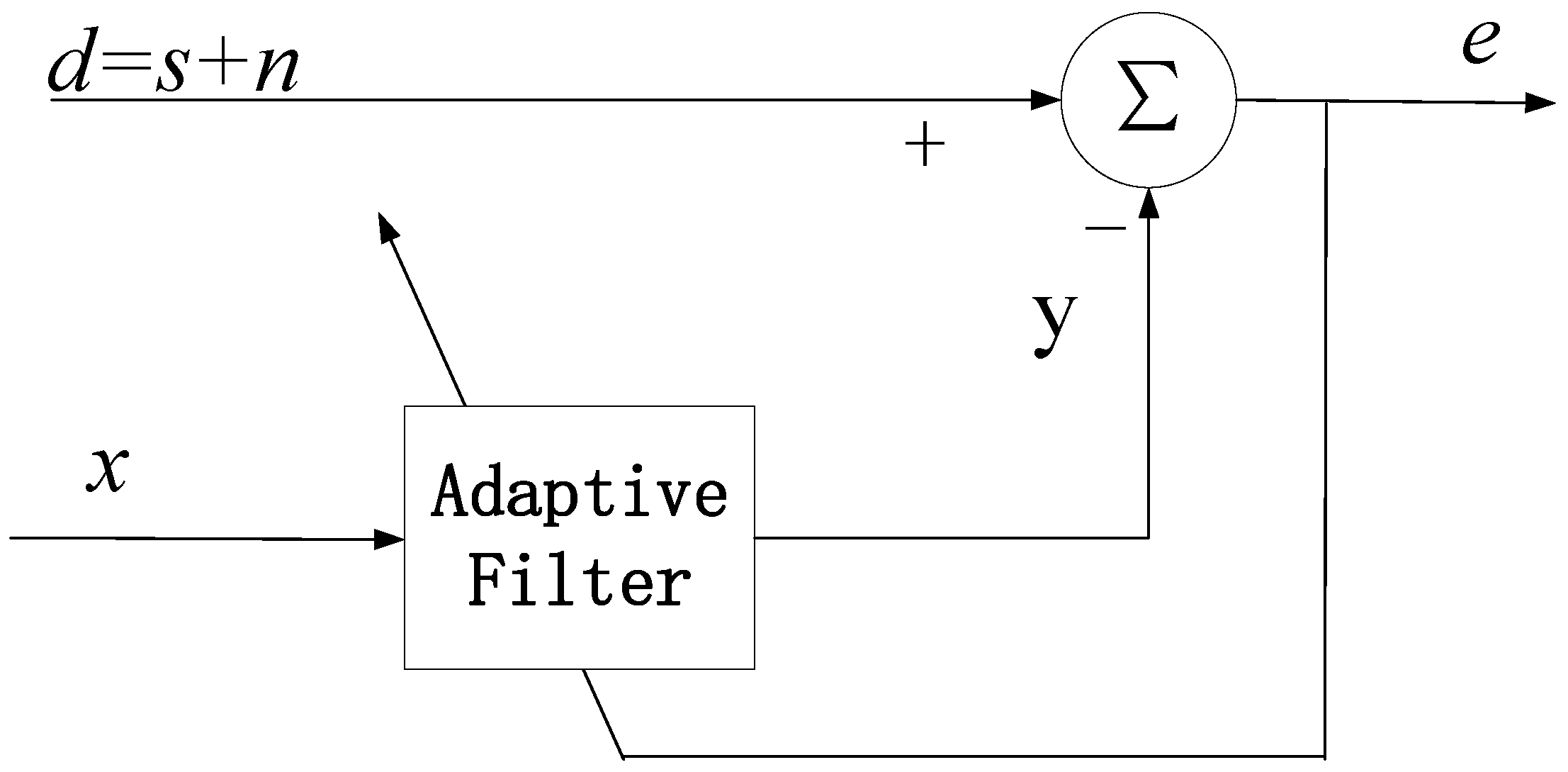
The block diagram of adaptive noise reduction is shown in Figure 1, where the input signal d contains the ECG signal s and the additive noise signal n [27,28]. The adaptive noise reduction method separates the noise signal from the main input signal d by introducing a reference signal x. It is assumed that the ECG signal s is uncorrelated with the reference signal x and the noise signal n, and the reference signal x and the noise signal n are correlated. The reference signal x is filtered to produce a filtered output y. Setting the order length of the filter as L and the weighting factor of the filter as w, the output y of the adaptive filter is expressed as
The difference between the main input signal d and the output y of the adaptive filter can be used as the error output e, which can be expressed by Equation (2).
The mean square of e is as follows:
If the ECG signal s is not correlated with the reference signal x, Equation (3) can be simplified as:
When n − y is infinitely close to 0, the error output e can be considered as the best least-squares estimate of the ECG signal s. The noise reduction performance is dependent on the level of correlation between the reference signal x and the noise signal n. Therefore, it is necessary to ensure that the reference signal x is highly correlated with the additive noise n, where the noise n will be suppressed by the adaptive filtered output y.
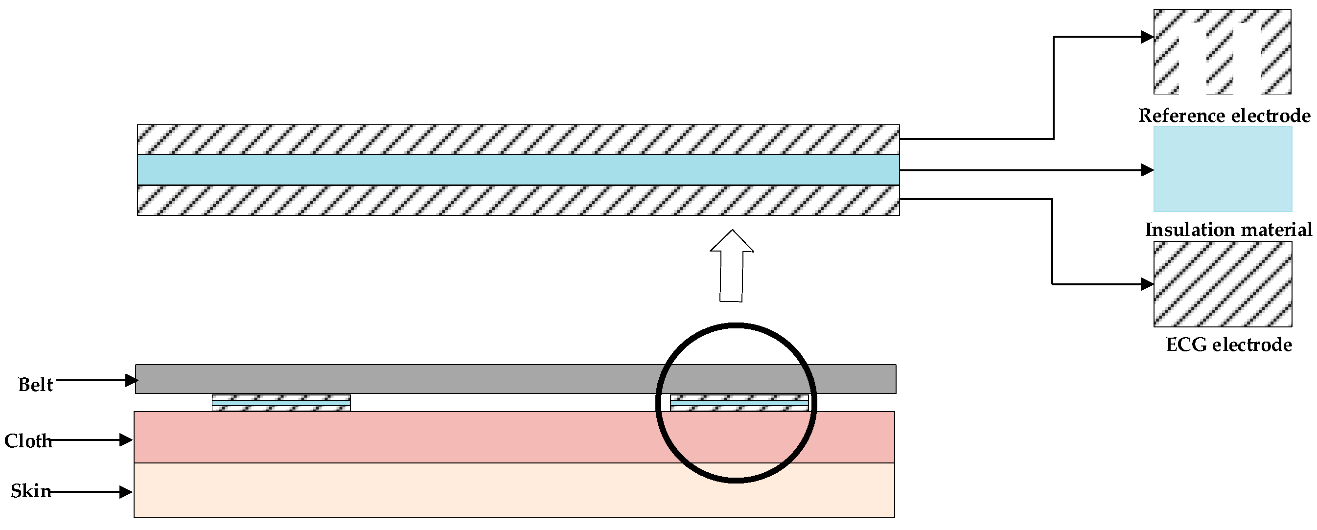
In order to obtain an appropriate reference signal x, a new electrode structure was designed. According to previous studies, the amplitude of the motion artifacts in the ECG signal is several times or even hundreds of times larger than the amplitude of the ECG signal. In noncontact ECG measurement systems, the amplitude of the ECG signal can be reduced while preserving the motion artifacts by increasing the insulation material thickness between the electrodes and the contact surface or by reducing the area of the electrodes, which provides a solution to obtain a reference signal barely containing an ECG signal.
Figure 2 shows the schematic diagram of the designed electrode structure. The carrier of the electrode is a bandage for a wearable chest ECG signal measurement. The reference signal acquisition electrode was isolated by insulating the material with the ECG signal acquisition electrode and designed with a comb-tooth shape to acquire motion artifacts, which reduced the electrode area and fit the contact surface as closely as possible. The ECG signal acquisition electrode was in the shape of a rectangle, facing the body and directly in contact with the cloth. The reference electrode, the ECG electrode, and the insulating material had the same outer contour size, thus improving the coherence of the reference signal and the motion artifacts in the ECG signal. The electrodes were all made of silver conductive fabric. The reference signal x was obtained from the difference between the two reference signal acquisition electrodes. The input signal d was obtained from the difference between the two ECG signal acquisition electrodes.
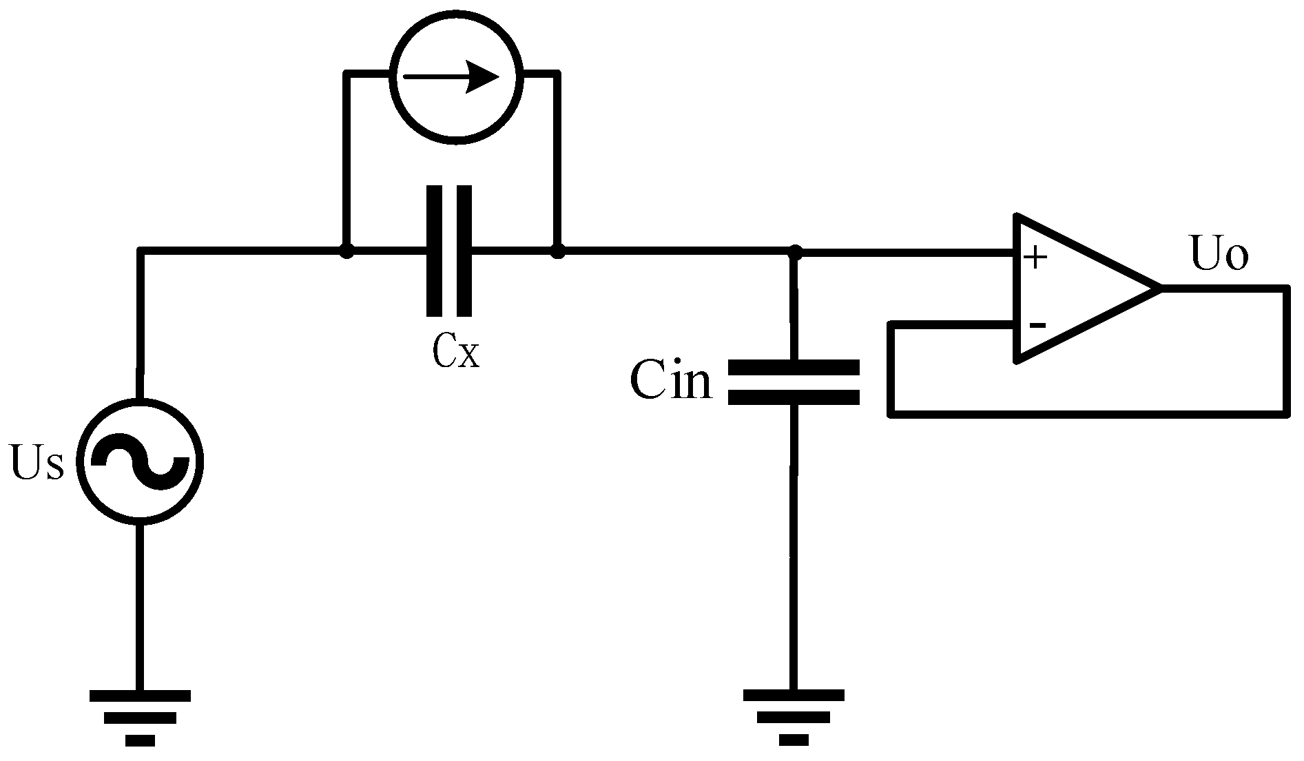
Figure 3 shows a simplified equivalent circuit diagram of the capacitive ECG measurements. The skin–electrode interface was modeled by a simple capacitor Cx, which can be considered a plate capacitor, with the electrode area A and the distance D between the electrode and the skin. Cin is the equivalent input capacitance of the acquisition front end. The voltage Us is a biosignal source, in this case the ECG. At the output of the amplifier, the voltage Uo can be measured, which is the reference input x from the reference electrodes. The motion artifacts are caused by the triboelectricity generated by friction and the variation in the skin–electrode interface. The friction between the skin, fabric, and electrode due to motion causes the charge accumulation on the textile electrode. Generally, the surface charge of the charged materials will decay with time. We adapted it here to include a current source dQ(t)/dt that represented the source of the motion artifact, where Q was the residual charge on the electrode.
For the reference electrodes, a comb-tooth shape can reduce the electrode area A, thus decreasing the value of capacitance Cx and reducing the ECG component of the reference signal x. On the other hand, the reference electrodes with a comb-tooth shape have the same outer contour size as the ECG electrodes and fit the contact surface as closely as possible. In this way, the charge variation dQ(t)/dt due to the friction between two kinds of electrodes is similar, and the reference signal x is highly correlated with the additive noise n.
3. Experimental Circuit System
3.1. Design of the Circuit System
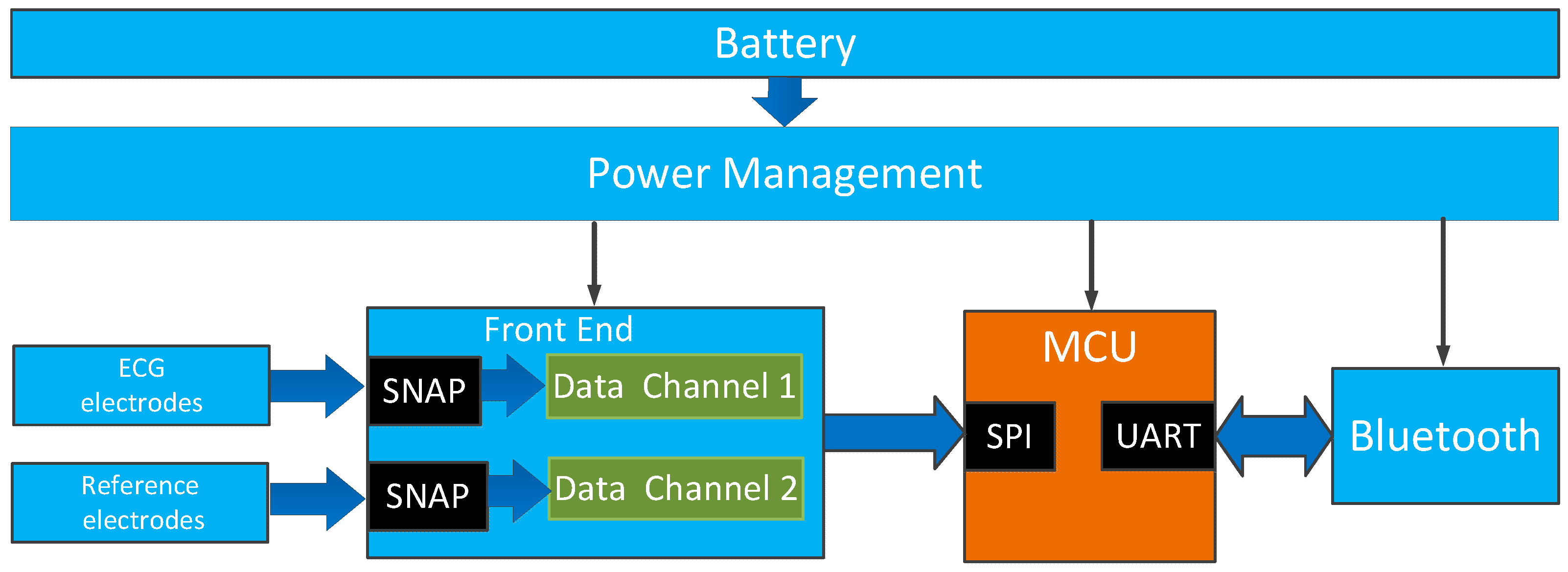
The diagram of the experimental circuit system is shown in Figure 4, including the ECG electrodes, reference electrodes, front end circuit, microcontroller, Bluetooth module, and the power management. Two pairs of stainless steel snap fasteners were used to connect the wearable electrodes with the front end. One pair of snap fasteners was used to connect the ECG electrodes, and the other was used to connect the reference electrodes. For the front end, ADS1298 was selected as the integrated ADC, where only two of the eight data channels were used. The front end was designed according to the proposed structure in reference [29]. Each channel was connected with an ultra-high input impedance sensor chip. The chip was characterized with an input capacitance of about 10 pF, and the input impedance was about 150 GΩ, which could efficiently acquire the noncontact ECG signal [29]. The microcontroller MSP430F5528 was used to sample the data from ADS1298. A 2.4 GHz Bluetooth module CC2540 was used to transfer data to the host computer, where the adaptive noise reduction processing and analysis of the acquired data were carried out. The power management circuit supplied the system using a rechargeable lithium polymer battery.
The developed experimental circuit system is shown in Figure 5, with the size of 100 × 50 × 15 mm, where the components were placed on the top layer, and two pairs of snap buttons were placed on the bottom side. The diameter of the snap was 12 mm, and the distance between the two snaps for measuring the same type signal was 45 mm; the two sets of snaps were arranged in parallel with a spacing of 18 mm. The upper two snaps were for connecting the reference electrodes, and the lower two were for connecting the ECG electrodes. Each set of electrodes or snaps was symmetrically positioned on the left and right, and the symmetrical centerline was the vertical centerline of the human body. The snaps were used as the connectors between the electrodes and the printed board.
3.2. Performance of the Designed System
In order to obtain a high quality signal for identifying the ECG characteristics, the electrical characteristics of the system needed to meet certain specification requirements. In this section, we describe the testing of the electrical characteristics of the system, including the system noise, the common mode rejection ration (CMRR), and the frequency response. According to the International Standard IEC 60601-2-47 [30] and the American National Standard ANSI/AAMI EC13 [31], the requirements are shown in Table 1.
The CMRR measurement was carried out under the condition of the common input 50 Hz sinusoidal signal with an amplitude of 400 mV and the differential input 50 Hz sinusoidal signal with an amplitude of 4 mV. As shown in Table 2, the CMRR of the system achieved 100 dB, which is about 22 dB larger than the International Standard IEC 60601-2-47 requirement of 78 dB and 5 dB larger than the American National Standard ANSI/AAMI EC13 of 95 dB.
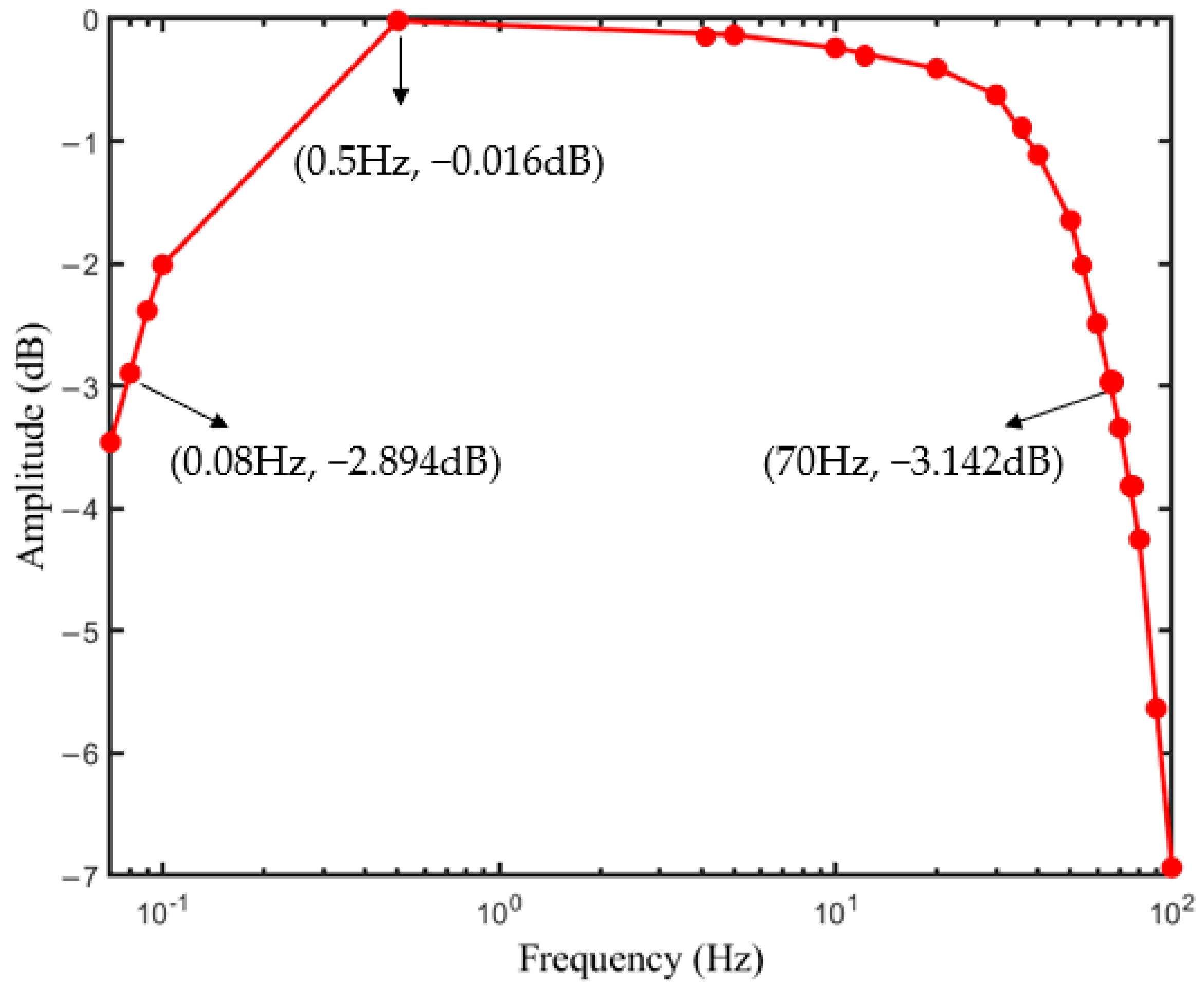
The frequency response was obtained by introducing a 4 mV sinusoidal signal with a frequency from 0.07 Hz to 100 Hz. As shown in Figure 6, the bandwidth of the ECG system was from 0.08 Hz to 70 Hz, which met the standard frequency requirement 0.67–40 Hz of the ECG signal [32].
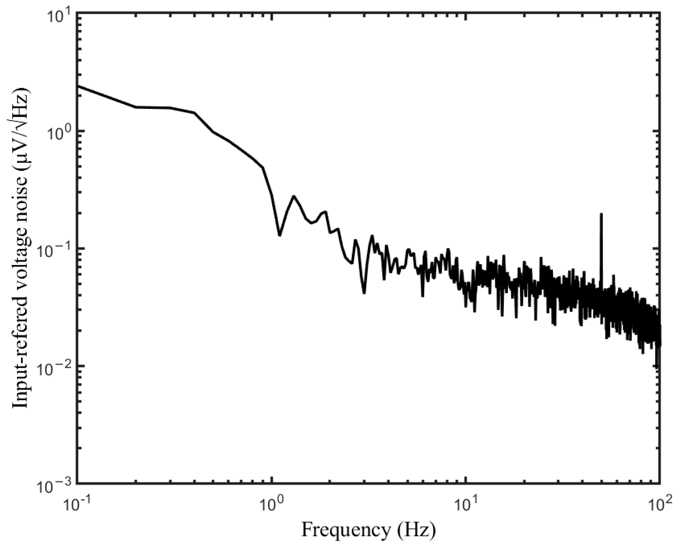
The input referred voltage noise is shown in Figure 7, which was measured with the input terminals shorted to the ground. The input-referred voltage noise for the 0.1–70 Hz bandwidth was 1.32 μVRMS.
4. Experimental Results
In this experiment, the experimental system was connected to a bandage with the snap fasteners, as shown in Figure 8a. Two sets of conductive silver fabric electrodes were stitched to the bandage. The reference signal acquisition electrodes were comb-tooth shaped, and the size of each comb tooth was 0.5 cm × 3 cm, as shown in Figure 8b. The ECG signal acquisition electrodes were rectangular electrodes with an electrode size of 6 cm × 3 cm, as shown in Figure 8c. The two types of electrodes were fixed and isolated from each other with 2-mm thick acrylic double-sided adhesive tape with the same size and shape as the ECG electrode. The reason for choosing acrylic double-sided adhesive tape was the softness of the acrylic material and its high level of adherence, which effectively reduced the relative motion between the ECG electrodes and the reference electrodes, as well as maintained the motion consistency, thus enhancing the coherence of the reference signal and motion artifacts. During the test, the bandage was tied to the volunteer’s chest, and the host computer was used to display and store the original ECG signal.
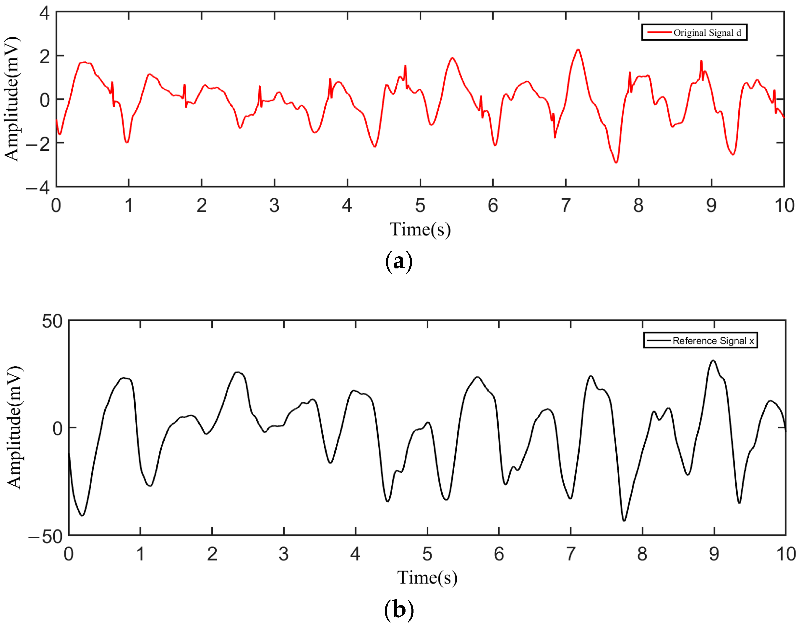
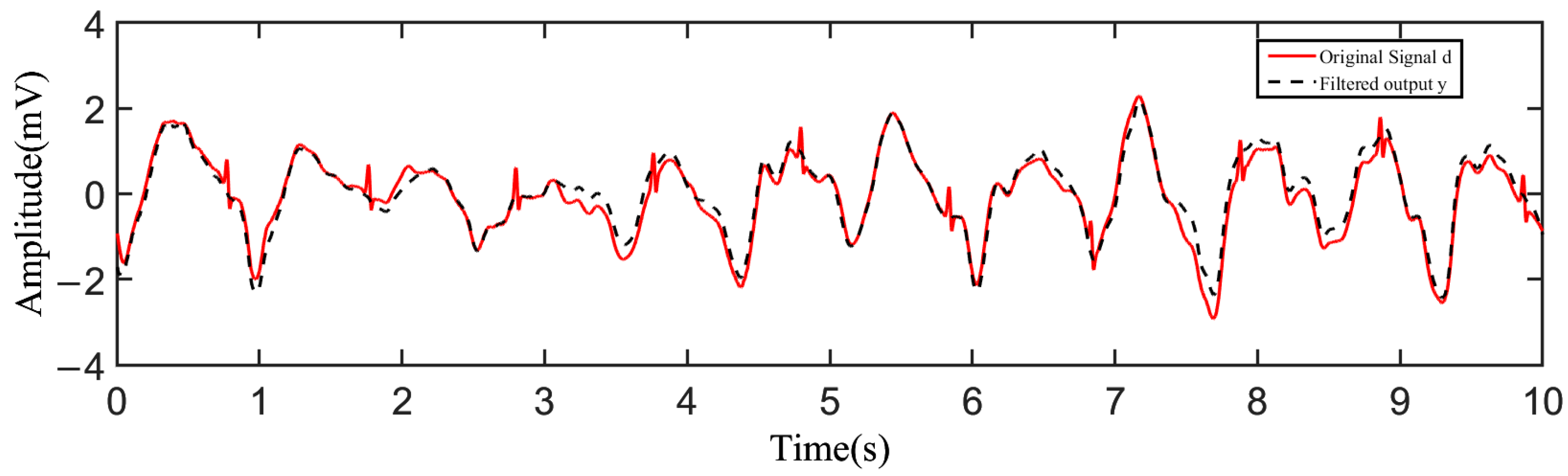
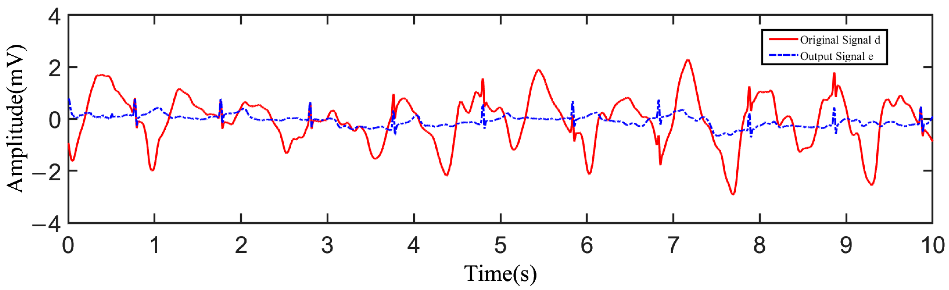
During the test, the tester wore a cotton T-shirt. After putting on the device, the tester performed the action of stepping in place with a speed of 1 m/s. The acquired raw data were transmitted to the host computer and plotted by Matlab as shown in Figure 9. Figure 9a shows the original signal d influenced by the noise obtained from one channel, and Figure 9b shows the reference signal x obtained from the other channel. As can be seen from the results, the original signal d was severely influenced by the motion artifacts, and the fluctuation in the baseline was remarkable. Through adaptive filtering, the filtered output y of the motion artifacts was derived. Figure 10 shows the comparison between the filtered output y and the original signal d, from which it can be found that the filtered output y fit the motion artifact smoothly. As shown in Figure 11, the output signal e was obtained by taking the difference between the original signal and the filtered output, where the motion artifact was significantly decreased, and the characteristics of the ECG waveform were more obvious.
For quantitative comparison, the signal to noise ratio (SNR) was calculated by Equation (5), where the (QRS) ECGp-p is the peak-to-peak amplitude of R wave in a 120 ms span centered with the R wave, and the (T-P) noisep-p is the peak-to-peak amplitude of the noise in a 40 ms T-P time interval span [33].
The SNR after adaptive noise reduction was 28.44 dB, which was about 14 dB larger than that of the original ECG signal, a value of 14.26 dB, which showed that the motion artifacts in the ECG signal were effectively suppressed by the proposed electrodes and the adaptive noise reduction processing method.
5. Conclusions and Discussion
A noncontact ECG offers good wearing comfort, which is well suited to be used for wearable long-time ECG monitoring. Compared to clinical ECG monitoring, a wearable ECG is more sensitive to motion artifacts. Adaptive noise reduction is an effective approach to suppress motion artifacts. In this paper, a novel ECG electrode structure was designed to acquire an ECG signal and reference signal simultaneously, avoiding the introduction of new sensors. The research employed a dual-channel data acquisition solution, which provided one more data acquisition channel than the conventional ECG measurements. By feeding data from both channels to an adaptive filter, the motion artifacts in the ECG acquisition were effectively suppressed.
Firstly, the design concept of the new ECG electrode structure and the principle of adaptive noise reduction were introduced. Secondly, a multichannel signal acquisition circuit system and wearable ECG electrodes were implemented, and the system performance was measured. Finally, experiments under normal walking conditions were conducted to verify the noise reduction effect. The ECG waveform characteristics of the obtained result after the noise reduction were obvious, and the motion artifacts were effectively suppressed. The signal-to-noise ratio of the noise-reduced ECG signal was 14 dB higher than that of the original ECG signal with the motion artifacts.
The proposed electrode design method was completely original and can be worn with commonly used bandages. The motion artifacts in the noncontact ECG were effectively suppressed during daily walking with the proposed design. In addition, since the solution did not involve additional sensors, the design was simple and can be applied to wearable applications, such as a chest strap, clothing, tag stickers, and other forms. By improving the SNR of the ECG signal, it could be beneficial for further waveform analysis, such as QRS identification, HRV analysis, etc. The research aimed to demonstrate the noise reduction effect of the electrode structure, so not much optimization was performed on the size of the electric board. In the future, we can optimize the PCB design to reduce the size of the electronic board and decrease the weight for better wearing comfort. Further we can also design a housing, so that the electronic board can be protected and the performance can be tested outdoors.
Author Contributions
Conceptualization, L.Z.; Data curation, L.Z.; Funding acquisition, L.Z. and F.Y.; Investigation, J.D.; Project administration, L.Z. and F.Y.; Software, Y.T., Y.L. and R.C.; Writing—original draft, J.D.; Writing—review and editing, L.Z. All authors have read and agreed to the published version of the manuscript.
Funding
This research was supported by the National Nature Science Foundation Program of China (no. 22090054 and 92059204).
Institutional Review Board Statement
Not applicable.
Informed Consent Statement
Not applicable.
Data Availability Statement
Not applicable.
Conflicts of Interest
The authors declare no conflict of interest.
References
- Clifford, G.D.; Azuaje, F. Advanced Methods and Tools for ECG Data Analysis; McSharry, P., Ed.; Artech House: Boston, MA, USA, 2006; Volume 10. [Google Scholar]
- Lin, C.-T.; Liao, L.-D.; Liu, Y.-H.; Wang, I.-J.; Lin, B.-S.; Chang, J.-Y. Novel Dry Polymer Foam Electrodes for Long-Term EEG Measurement. IEEE Trans. Biomed. Eng. 2010, 58, 1200–1207. [Google Scholar] [CrossRef] [PubMed]
- Xu, X.; Chen, Y.; He, P.; Wang, S.; Ling, K.; Liu, L.; Lei, P.; Huang, X.; Zhao, H.; Cao, J.; et al. Wearable CNT/Ti3C2Tx MXene/PDMS composite strain sensor with enhanced stability for real-time human healthcare monitoring. Nano Res. 2021, 14, 2875–2883. [Google Scholar] [CrossRef]
- Xu, X.; Liu, Z.; He, P.; Yang, J. Screen printed silver nanowire and graphene oxide hybrid transparent electrodes for long-term electrocardiography monitoring. J. Phys. D Appl. Phys. 2019, 52, 455401. [Google Scholar] [CrossRef]
- Dong, H.; Sun, J.; Liu, X.; Jiang, X.; Lu, S. Highly Sensitive and Stretchable MXene/CNTs/TPU Composite Strain Sensor with Bilayer Conductive Structure for Human Motion Detection. ACS Appl. Mater. Interfaces 2022, 14, 15504–15516. [Google Scholar] [CrossRef] [PubMed]
- Wang, Y.; Yang, B.; Hua, Z.; Zhang, J.; Guo, P.; Hao, D.; Gao, Y.; Huang, J. Recent advancements in flexible and wearable sensors for biomedical and healthcare applications. J. Phys. D Appl. Phys. 2021, 55, 134001. [Google Scholar] [CrossRef]
- Ottenbacher, J.; Heuer, S. Motion artefacts in capacitively coupled ECG electrodes. In Proceedings of the World Congress on Medical Physics and Biomedical Engineering, Munich, Germany, 7–12 September 2009; Springer: Berlin/Heidelberg, Germany, 2009. [Google Scholar]
- Łęski, J.M.; Henzel, N. ECG baseline wander and powerline interference reduction using nonlinear filter bank. Signal Process. 2005, 85, 781–793. [Google Scholar] [CrossRef]
- Tikkanen, P.E. Nonlinear wavelet and wavelet packet denoising of electrocardiogram signal. Biol. Cybern. 1999, 80, 259–267. [Google Scholar] [CrossRef] [PubMed]
- Widrow, B.; Glover, J.R.; McCool, J.M.; Kaunitz, J.; Williams, C.S.; Hearn, R.; Zeidler, J.R.; Dong, E., Jr.; Goodlin, R.C. Adaptive noise cancelling: Principles and applications. Proc. IEEE 1975, 63, 1692–1716. [Google Scholar] [CrossRef]
- Jung, H.-K.; Jeong, D.-U. Development of wearable ECG measurement system using EMD for motion artifact removal. In Proceedings of the 2012 7th International Conference on Computing and Convergence Technology (ICCCT), Seoul, Republic of Korea, 3–5 December 2012; IEEE: Piscataway, NJ, USA, 2012. [Google Scholar]
- Thakor, N.; Zhu, Y.-S. Applications of adaptive filtering to ECG analysis: Noise cancellation and arrhythmia detection. IEEE Trans. Biomed. Eng. 1991, 38, 785–794. [Google Scholar] [CrossRef] [PubMed]
- Luo, S.; Tompkins, W.J. Experimental study: Brachial motion artifact reduction in the ECG. In Proceedings of the Computers in Cardiology, Vienna, Austria, 10–13 September 1995; IEEE: Piscataway, NJ, USA, 1995. [Google Scholar]
- Hamilton, P.S.; Curley, M.G. Adaptive removal of motion artifact. In Proceedings of the 19th Annual International Conference of the IEEE Engineering in Medicine and Biology Society: Magnificent Milestones and Emerging Opportunities in Medical Engineering (Cat. No. 97CH36136), Chicago, IL, USA, 30 October–2 November 1997; IEEE: Piscataway, NJ, USA, 1997; Volume 1. [Google Scholar]
- Hamilton, P.S.; Curley, M.G.; Aimi, R.M.; Sae-Hau, C. Comparison of methods for adaptive removal of motion artifact. In Proceedings of the Computers in Cardiology 2000 (Cat. 00CH37163), Cambridge, MA, USA, 24–27 September 2000; IEEE: Piscataway, NJ, USA, 2000; Volume 27. [Google Scholar]
- Liu, Y.; Pecht, M.G. Reduction of motion artifacts in electrocardiogram monitoring using an optical sensor. Biomed. Instrument. Technol. 2011, 45, 155–163. [Google Scholar] [CrossRef] [PubMed]
- Ottenbacher, J.; Kirst, M.; Jatoba, L.; Huflejt, M.; Grossmann, U.; Stork, W. Reliable motion artifact detection for ECG monitoring systems with dry electrodes. In Proceedings of the 2008 30th Annual International Conference of the IEEE Engineering in Medicine and Biology Society, Vancouver, BC, Canada, 20–25 August 2008; IEEE: Piscataway, NJ, USA, 2008. [Google Scholar]
- An, X.; Liu, Y.; Zhao, Y.; Lu, S.; Stylios, G.K.; Liu, Q. Adaptive Motion Artifact Reduction in Wearable ECG Measurements Using Impedance Pneumography Signal. Sensors 2022, 22, 5493. [Google Scholar] [CrossRef] [PubMed]
- Raya, M.A.D.; Sison, L.G. Adaptive noise cancelling of motion artifact in stress ECG signals using accelerometer. In Proceedings of the Second Joint 24th Annual Conference and the Annual Fall Meeting of the Biomedical Engineering Society: Engineering in Medicine and Biology, Houston, TX, USA, 23–26 October 2002; IEEE: Piscataway, NJ, USA, 2002; Volume 2. [Google Scholar]
- Tong, D.A.; Bartels, K.A.; Honeyager, K.S. Adaptive reduction of motion artifact in the electrocardiogram. In Proceedings of the Second Joint 24th Annual Conference and the Annual Fall Meeting of the Biomedical Engineering Society: Engineering in Medicine and Biology, Houston, TX, USA, 23–26 October 2002; IEEE: Piscataway, NJ, USA, 2002; Volume 2. [Google Scholar]
- Romero, I.; Geng, D.; Berset, T. Adaptive filtering in ECG denoising: A comparative study. In Proceedings of the 2012 Computing in Cardiology, Krakow, Poland, 9–12 September 2012; IEEE: Piscataway, NJ, USA, 2012. [Google Scholar]
- Yu, S.; Liu, S. A novel adaptive recursive least squares filter to remove the motion artifact in seismocardiography. Sensors 2020, 20, 1596. [Google Scholar] [CrossRef] [PubMed] [Green Version]
- Olmos, S.; Sornmo, L.; Laguna, P. Block adaptive filters with deterministic reference inputs for event-related signals: BLMS and BRLS. IEEE Trans. Signal Process. 2002, 50, 1102–1112. [Google Scholar] [CrossRef]
- Ernst, F.; Schlaefer, A.; Dieterich, S.; Schweikard, A. A Fast Lane Approach to LMS prediction of respiratory motion signals. Biomed. Signal Process. Control 2008, 3, 291–299. [Google Scholar] [CrossRef]
- Rahman, M.Z.U.; Shaik, R.A.; Reddy, D.R.K. Efficient sign based normalized adaptive filtering techniques for cancelation of artifacts in ECG signals: Application to wireless biotelemetry. Signal Process. 2011, 91, 225–239. [Google Scholar] [CrossRef]
- Diniz, P.S.R. Adaptive Filtering; Springer: Berlin/Heidelberg, Germany, 1997; Volume 4. [Google Scholar]
- Choi, M.; Jeong, J.J.; Kim, S.H.; Kim, S.W. Reduction of Motion Artifacts and Improvement of R Peak Detecting Accuracy Using Adjacent Non-Intrusive ECG Sensors. Sensors 2016, 16, 715. [Google Scholar] [CrossRef] [PubMed] [Green Version]
- Alzahrani, A.; Hu, S.; Azorin-Peris, V.; Barrett, L.; Esliger, D.; Hayes, M.; Akbare, S.; Achart, J.; Kuoch, S. A multi-channel opto-electronic sensor to accurately monitor heart rate against motion artefact during exercise. Sensors 2015, 15, 25681–25702. [Google Scholar] [CrossRef] [PubMed]
- Wu, R.; Tang, Y.; Li, Z.; Zhang, L.; Yan, F. A novel high input impedance front-end for capacitive biopotential measurement. Med. Biol. Eng. Comput. 2018, 56, 1343–1355. [Google Scholar] [CrossRef] [PubMed]
- IEC 60601-2-47:2012; Medical Electrical Equipment—Part 2-47: Particular Requirements for the Basic Safety and Essential Performance of Ambulatory Electrocardiographic Systems. International Electrotechnical Commission: London, UK, 2012.
- ANSI/AAMI EC13; Cardiac Monitors, Heart Rate Meters, and Alarms. American National Standards Institute: Washington, DC, USA, 2002.
- Bragg-Remschel, D.A.; Anderson, C.M.; Winkle, R.A. Frequency response characteristics of am-bulatory ECG monitoring systems and their implications for ST segment analysis. Am. Heart J. 1982, 103, 20–31. [Google Scholar] [CrossRef] [PubMed]
- Escalona, O.J.; McFrederick, L.; Borges, M.; Linares, P.; Villegas, R.; Perpiñan, G.I.; McLaughlin, J.; McEneaney, D. Wrist and arm body surface bipolar ECG leads signal and sensor study for long-term rhythm monitoring. In Proceedings of the 2017 Computing in Cardiology (CinC), Rennes, France, 24–27 September 2017; IEEE: Piscataway, NJ, USA, 2017. [Google Scholar]
Figure 1.
The adaptive noise canceling concept.

Figure 2.
Schematic diagram of the wearable electrode structure.

Figure 3.
Simplified equivalent circuit diagram of the capacitive ECG measurements.

Figure 4.
Block diagram of the system.

Figure 5.
Printed system circuit board; (a) top layer of the circuit board; (b) bottom side of the circuit board.
Figure 5.
Printed system circuit board; (a) top layer of the circuit board; (b) bottom side of the circuit board.

Figure 6.
Frequency response measurement result.

Figure 7.
The input-referred voltage noise density.

Figure 8.
Measurement system; (a) tester wearing the experimental device; (b) reference electrodes; (c) ECG electrodes.
Figure 8.
Measurement system; (a) tester wearing the experimental device; (b) reference electrodes; (c) ECG electrodes.

Figure 9.
Original measurement results; (a) original signal; (b) reference signal.

Figure 10.
The comparison between the filtered output and the original signal.

Figure 11.
The contrast between the output signal and the original signal.


Table 1.
Performance Requirements.
| Index | Test Conditions | Requirement |
|---|---|---|
| System noise | 10 s of data | ≤30 μV (p-v RTI) |
| CMRR | f = 50 Hz | ≥78 dB (IEC 60601-2-47) ≥ 95 dB (ANSI/AAMI EC13) |
| Frequency response | −3 dB attenuation | 0.67–40 Hz |
Table 2.
CMRR measurement results.
| Mode | Input Signal | Output | Gain | CMRR (dB) | |
|---|---|---|---|---|---|
| Frequency (Hz) | Amplitude (mV) | Amplitude (mV) | |||
| Differential | 50 | 4 | 19.2 | 4.8 | 100 |
| Common | 400 | 0.0175 | 4.35 × 10⁻⁵ | ||
Disclaimer/Publisher’s Note: The statements, opinions and data contained in all publications are solely those of the individual author(s) and contributor(s) and not of MDPI and/or the editor(s). MDPI and/or the editor(s) disclaim responsibility for any injury to people or property resulting from any ideas, methods, instructions or products referred to in the content. |
© 2023 by the authors. Licensee MDPI, Basel, Switzerland. This article is an open access article distributed under the terms and conditions of the Creative Commons Attribution (CC BY) license (https://creativecommons.org/licenses/by/4.0/).
Share and Cite
MDPI and ACS Style
Ding, J.; Tang, Y.; Chang, R.; Li, Y.; Zhang, L.; Yan, F. Reduction in the Motion Artifacts in Noncontact ECG Measurements Using a Novel Designed Electrode Structure. Sensors 2023, 23, 956. https://doi.org/10.3390/s23020956
AMA Style
Ding J, Tang Y, Chang R, Li Y, Zhang L, Yan F. Reduction in the Motion Artifacts in Noncontact ECG Measurements Using a Novel Designed Electrode Structure. Sensors. 2023; 23(2):956. https://doi.org/10.3390/s23020956
Chicago/Turabian StyleDing, Jianwen, Yue Tang, Ronghui Chang, Yu Li, Limin Zhang, and Feng Yan. 2023. "Reduction in the Motion Artifacts in Noncontact ECG Measurements Using a Novel Designed Electrode Structure" Sensors 23, no. 2: 956. https://doi.org/10.3390/s23020956
Note that from the first issue of 2016, this journal uses article numbers instead of page numbers. See further details here.