
Bio-Tailored Sensing at the Nanoscale: Biochemical Aspects and Applications
by
 ,
,
Francesca Fata
†,
Federica Gabriele
†,
Francesco Angelucci
,
Rodolfo Ippoliti
,
Luana Di Leandro
,
Francesco Giansanti
and
Matteo Ardini
* Department of Life, Health and Environmental Sciences, University of L’Aquila, 67100 L’Aquila, Italy
*
Author to whom correspondence should be addressed.
†
These authors contributed equally to this work.
Sensors 2023, 23(2), 949; https://doi.org/10.3390/s23020949
Submission received: 19 December 2022
/
Revised: 4 January 2023
/
Accepted: 9 January 2023
/
Published: 13 January 2023
(This article belongs to the Special Issue New Strategies and Current Challenges for Label-Free Biosensors Improvement)
Abstract
:The demonstration of the first enzyme-based electrode to detect glucose, published in 1967 by S. J. Updike and G. P. Hicks, kicked off huge efforts in building sensors where biomolecules are exploited as native or modified to achieve new or improved sensing performances. In this growing area, bionanotechnology has become prominent in demonstrating how nanomaterials can be tailored into responsive nanostructures using biomolecules and integrated into sensors to detect different analytes, e.g., biomarkers, antibiotics, toxins and organic compounds as well as whole cells and microorganisms with very high sensitivity. Accounting for the natural affinity between biomolecules and almost every type of nanomaterials and taking advantage of well-known crosslinking strategies to stabilize the resulting hybrid nanostructures, biosensors with broad applications and with unprecedented low detection limits have been realized. This review depicts a comprehensive collection of the most recent biochemical and biophysical strategies for building hybrid devices based on bioconjugated nanomaterials and their applications in label-free detection for diagnostics, food and environmental analysis.
1. Introduction
Since 1967, when S. J. Updike and G. P. Hicks showed the first enzyme-based electrode for glucose detection [1] inspired by the previous L. C. Clark Jr.’s device [2], sensors for biological analytes or “biosensors” have passed through a three-generations development [3]. In those belonging to the third generation group the sensing molecule is physically integrated within the device rather than being freely diffusing in solution. This setup resulted in improved detection as it does not require a mediator component to transfer the signal to the transducer thereby becoming simpler and faster.
Modern biosensors are usually endowed with (i) efficient capturing of specific targets with no off-target interaction, (ii) transformation into electrochemical, electrical, optical, gravimetric or acoustic signals, (iii) sensitivity, short response time, reproducibility and low detection limit (LOD) and (iv) miniaturized components [3]. In the attempt to improve these features, great results have been achieved by taking advantage of 0D, 1D and 2D nanomaterials for their unique chemical and physical properties, which make them highly performing compared to the bulk counterparts. Biomolecules represent naturally occurring ready-to-use nanomaterials as they are nanoscopic objects with regular shape, high binding specificity and affinity, surfaces suitable for chemical functionalization and catalytic activity as in the case of enzymes, for instance [4]. Moreover, biomolecules can be modelled via genetic and protein engineering [5,6] and their surfaces enable the interaction with other nanomaterials; as a result, biomolecules have been widely exploited to functionalize nanomaterials thus enabling precise tailoring of hybrid functional bioconjugates at the nanoscale [7,8,9]. Beyond biomolecules, organic and inorganic nanomaterials with sharpen architecture have attracted interest for their high surface-to-volume ratio, conductivity, shock-bearing ability and optical tunability, e.g., nanoparticles, nanotubes, nanorods, graphene, nanofibers, molybdenum disulfide, carbides, nitrides, carbonitrides and quantum dots [3]. Their features have impelled biosensing accuracy and robustness and disclosed tunable electrochemical and physical-mechanical responses. For instance, nanomaterials integrated in a sensor can sensibly decrease the LOD and thus increase the sensitivity in minimal processing time to reach high specific detection of a target [10,11,12].
To note, as the sensors get miniaturized to the nanoscale, the so-called “label-free” detection of molecular analytes becomes affordable. One first advantage of using labeled-free detection, indeed, is the possibility of revealing molecular targets which are not easy to tag or cannot be tagged at all; secondly, many targets cannot be labeled for they are found at very low amounts thus making labeling expensive and a nonsense strategy. In such a scenario, the interplay between biomolecules and nanomaterials becomes straightforward because at the nanoscale the interactions with the target analytes turn highly specific, strong and favored; moreover, these interactions become affected by other factors which are absent in bulk macroscopic devices, for instance the molecular conformation of the biomolecules. Under these conditions, the detection inspired by structure-based high affinity biorecognition turns on and accurate targeting of the analyte can be addressed with no off-target issues even at very low concentrations.
The past two decades gave us pretty examples of biosensors based on nanometric components and highlighted the advantages of having a biological element as the sensing part [13]. Mentioning all these examples would be an endless list for such a flourishing and interdisciplinary applied science; however, a few representative examples of biosensors might be quickly recalled, e.g., DNA-coated nanoparticles for enzymatic activity detection [14], lectin-coated nanorods for detection of bacterial cells [15], antibody-coated nanowires for specific protein detection [16] and antibody-coated micropillars with nanometric roughness to detect cancer cells [17]. Beyond the ease of combining a biological moiety, be it an enzyme, nucleic acid or a protein, with a non-biological nanomaterial by means of simple adsorption or chemical crosslinking, the main advantage of obtaining such a hybrid conjugate is a reduced LOD and, therefore, increased sensitivity coming from the intrinsic properties of both the biomolecule and the nanomaterial [13,18]. To get an idea, a method based on silicon functionalization and gold nanoparticles covalently attached to antibodies enable targeting of the prostate-specific antigen within a working range of 23 fg mL−1–500 ng mL−1 with an LOD of 23 fg mL−1, which corresponds to target concentrations in the femto range (10−15 M) [19]. Thus, it is now clear that integrating an active biological moiety onto an organic or inorganic nanometric substrate is propaedeutic to realize advanced, next-generation devices for improved sensing.
In this review, several recent examples published since 2020 of label-free biosensors based on biomolecules and nanomaterials and their applications are collected and discussed. The last two years, indeed, have been propelled by many different devices where aptamers, proteins, enzymes and/or antibodies have been found as the most common biological sensing components. Note that in this review the terms “nanomaterials” and “nanostructures” exclusively refer to nanometric objects having a regular, sharpen architecture, e.g., 0D particles, dots and rods; 1D tubes and fibers; 2D and 3D organic and inorganic layered-, porous- or fiber-like matrices. Also note that, due to the huge area of label-free techniques [3,20], this review will mainly focus on biosensors exploiting electrochemical or optical features for being the most representative (Figure 1). A special emphasis is given to the biochemical and biophysical aspects concerning the strategy to assemble the hybrid bioconjugate, the chemical and/or physical signals detected and the applications ranging from detection of biomarkers and antibiotics, environment- and food-derived molecules and single-molecule detection.
2. Biosensors Based on Hybrid Biomolecules-Nanomaterials Composites
Each example, discussed in this section, is provided with a description of the chemical and/or biochemical strategies to couple the sensing biomolecular component to the nanomaterial or nanostructure and its application for practical purposes. A series of examples will also be quickly recalled that rely on the same principle of the main example just described. As mentioned before, label-free biosensors involving nanomaterials or nanostructures with regular, sharpened architecture will be exclusively considered. Furthermore, this section will mainly provide examples of aptamers-, proteins-, antibodies- and enzymes-based biosensing due to their predominant abundance in the field.
2.1. Electrochemical Biosensors
2.1.1. Aptamers-Based Electrochemical Biosensors
Aptamers are short, single-stranded nucleotide sequences or peptides that spontaneously adopt secondary conformations in solution under particular conditions, e.g., hairpin-like (stem loop) structures [21]. They are considered alternatives to antibodies as they are endowed with high binding affinity and specificity to various biological targets with little or no off-target interaction; furthermore, they show no immunogenicity, large molecular flexibility as well as thermal and chemical stability. Despite their natural occurrence, aptamers can be easily synthesized at low cost and are particularly targeted for different applications, including sensing. Indeed, though aptamers are well-known as very proficient therapeutic tools, for instance in cancer therapy [21,22,23], their application in biosensing has recently been propelled by many examples reporting on molecular, ion and cell detection in food and water as well as in diagnostics [24]. The combination of aptamers with nanomaterials is not an exception.
Most biosensors where aptamers and nanomaterials are physically integrated, so-called “aptasensors”, rely on an electrochemical setup that includes an electrode working as transducer and support for deposition of the nanomaterial, which improves the electrochemical properties of the underlying electrode and furnishes binding sites for the aptamers. In this configuration, the nanomaterial-bound aptamers act as receptors/probes that are freely accessible to capture and detect the target analytes. Namely, upon binding to the target the aptamers undergo a complementary base pairing that creates secondary structures, e.g., helical arms and single stranded loops, which in turn provide the information to form tertiary structures responsible for the high-specific and high-affinity binding to the target via weak forces. According to this structural plasticity, aptasensors show electrochemical performances that can be monitored via voltametric apparatus as, upon binding to the analytes, the molecular conformational change of aptamers alters the free surface of the electrode to accomplish an electron exchange, e.g., [Fe(CN)6]3−/4-, thus leading to an increased or decreased rate of redox reactions on the electrode and therefore to changes in the voltametric and current signals. It must be noted that the signal changes that occur during the interaction between the aptamers and the sample containing the target molecules is very dependent upon the time, which usually spans within tens of minutes before reaching a stable signal. A main drawback that must be avoided comes from the non-specific binding of the target on the surface of the electrode via adsorption that leads to an increase in the background signal therefore decreasing sensitivity of the device. In all cases, this drawback is usually fixed by allowing blocking agent to adsorb over the electrode surface, e.g., surfactants, metal-specific thionic compounds as well as proteins such as casein and bovine serum albumin (BSA). This expedient allows the target molecules to interact exclusively with the aptamer probes.
A representative example is reported by Beitollahi et al. [25] that developed an electrochemical aptasensor to detect in blood serum and urine the amino acid homocysteine, a highly reactive sulfur-containing compound that turns into an intermediate by-product during the metabolism of dietary methionine (Figure 2). In this study, gold nanoparticles (AuNPs) are synthesized via electrodeposition onto the surface of a glassy carbon electrode and exploited to stably bind thiolated aptamers (SH-aptamers) targeting homocysteine. The interaction between the AuNPs and the SH-aptamers occurs through involvement of strong thiolate-gold bonds, which allow the aptamer molecules to form a self-assembled monolayer on the metal surface, a molecular arrangement that is preferable to achieve linear detection of the analyte. The authors used voltametric measurements to reveal the binding of the homocysteine molecules to the aptamer probes with an LOD down to 0.01 μM and a linear response within the range 0.05–20.0 μM; indeed, the interaction between the aptamer probes and the homocysteine molecules triggers a fold change in the aptamer molecular structure that increases the accessibility of the underlying glassy carbon electrode leading to enhanced redox reactions and therefore increases the voltametric and current values. This aptasensor has been proposed as a potential device for healthcare purposes, especially for detecting homocysteine as a biomarker in hyperhomocysteinemia.
The aptasensor just described includes only three main components, i.e., the electrode (the transducer), a layer of NPs (the nanostructured support) and the aptamer (the biological probe). Likewise, the same simple though efficient configuration can be found in other devices having different components such as screen-printed gold electrodes coated with flower-like gold microstructures for detection of serpin in plasma [26] or coated with CeO2NPs to detect the epithelial sodium channel protein in urine [27].
As the complexity of the nanomaterial laying onto the electrode is increased or several nanomaterials are combined to produce more complex composites, the performances of the aptasensor are enhanced to reach very low LODs. For instance, the study by Xie et al. [28] is worth mentioning as it describes an aptasensor able to detect very low amounts of thrombin in blood serum with no interference from nonspecific adsorption, environmental changes or instrumental efficiency by taking advantage of a hybrid configuration (Figure 3). To realize this aptasensor, AuNPs are synthesized via heating in solution and are loaded onto a mixture reaction of aminated ionic liquid NH2-IL and 2D nanosheets of molybdenum disulfide (MoS2), the former helping the nanomaterial to stabilize via electrostatic repulsion and avoiding aggregation. The resulting AuNPs-IL-MoS2 composite is dried onto a glassy carbon electrode and furnishes a proper binding surface for the adsorption of DNA nanotetrahedrons, which are obtained by warming and cooling of four different SH-DNA sequences. The thrombin analyte molecules can bind to the aptamers and the resulting thrombin-DNA nanotetrahedron complex is sandwiched using a third nanostructured composite AuNPs-Fe-MIL-88 where AuNPs are loaded onto hydrothermally synthesized Fe-MIL-88 metal-organic frameworks (MOFs), which in turn are labeled with a fifth DNA sequence recognizing the thrombin-DNA complex. The authors recalled two import features of such high-complex devices: first, in contrast to traditional stem-loop structures or linear DNA probes, the enhanced mechanical rigidity of the DNA nanotetrahedrons increases the accessibility of the target molecules and their loading amounts; secondly, this aptasensor results bind to many interfering substances even when their concentrations are greater than the concentration of thrombin. This aptamersensor shows an LOD of 56 fM within a large linear range of 0.298–29.8 pM thus representing a potential detector for ratiometric methods in clinical applications.
This aptasensor represents a prominent example of how nanomaterials, according to their intrinsic features such as hydrophilicity, high specific surface area and good electrical conductivity, can sensibly improve the electrochemical performances of detection without affecting the biochemical features of the attached aptamers. These properties reflect the high number of similar devices which have been realized in recent years. Additional examples where the glassy carbon electrode is coated with hybrid nanostructured composites include: reduced MoS2-AuNPs for the double detection of zearalenone and fumonisin B1 mycotoxins in food [29]; (3-aminopropyl)triethoxysilane-modified graphene oxide (GO)-AgNPs for detection of chloramphenicol in food [30]; thiourea capped-ZnS quantum dots (QDs)-AuNPs for detection of β-casomorphin 7 in urine [31]; AgNPs-GQDs containing core-shell Cu-In-S/ZnSQDs for detection of ractopamine in urine and serum [32]; magnetic reduced GO-Fe3O4-Cu2O and Ag-resorcinol-formaldehyde NPs-Ag nanodots (NDs) for detection of prostate specific antigen in serum [33]; reduced GO-AgNPs and prussian blue-AuNPs for detection of acetamipridin in food [34]; reduced GO-AuNPs for detection of glycated albumin [35]; MXene-AuNPs for detection of chloramphenicol in food [36]; PtNPs-MIL-101(Fe) MOFs for detection of aflatoxin M1 in food [37]; and bimetallic CoNi-MOFs with graphene-like nanosheet structure to detect enrofloxacin in food, water and serum [38].
In this context, another prominent example is reported by Li et al. for detecting carcinoembryonic antigen in blood serum [39]. The aptasensor enables dual binding sites and dual signal amplifying electrochemical sensing by means of self-polymerized dopamine-functionalized AuNPs (Figure 4). AuNPs are chemically synthesized and decorated with dopamine moieties under oxidative and alkaline conditions to trigger dopamine polymerization over the gold surface. These polymeric hybrid constructs provide anchoring sites for Fe3+ through coordination bonds to form Fe3+-catechol MOFs with an octahedron shape and the presence of acetic acid increases the number of carboxyl groups (-COOH) regulating the nucleation and growth rate of the Fe3+-catechol MOFs on the surface of the AuNPs. The presence of several -COOH groups within the framework complexes is exploited to link carcinoembryonic antigen-specific aminated aptamers (NH2-aptamers) via classical 1-Ethyl-3-(3-dimethylaminopropyl)carbodiimide/N-hydroxysuccinimide (EDS/NHS) crosslinking reaction to achieve a colloidal aptasensor with high sensitivity and selectivity, good biocompatibility and strong stability. These functionalized AuNPs can be used to coat a glassy carbon electrode and detect carcinoembryonic antigen in serum. Note that both the polydopmine moieties and the Fe3+-catechol MOFs accelerate the electron transfer on the electrode interface, increasing the amplification of the current signals and therefore enhancing the sensitivity of the device. The authors used such a complex aptasensor to detect the carcinoembryonic antigen in the concentration range of 1 fg mL−1–1 μg mL−1 with a LOD as low as 0.33 fg mL−1 and proposed it for detection of the carcinoembryonic antigen biomarker in early clinical diagnosis.
The aforementioned examples can be further extended if considering other types of electrodes, such as: screen-printed carbon electrodes coated with carbon-black NPs-AuNPs composites to detect Cd(II) ions in water [40] or coated with exonuclease-assisted AuNPs-MOFs for detection of streptomycin in food [41]; fluorine-doped tin oxide electrodes coated with BiVO4-2D-C3N4 photoanode with II type heterojunction to detect microcystin-LR in water [42]; Au electrodes coated with highly-porous Au nanostructures for detection of acetamiprid in food [43]; indium tin oxide electrodes coated with core-shell LaFeO3-g-C3N4 heterostructures for detection of streptomycin in food [44]; Au microelectrodes coated with GQDs-AuNPs composites for detection of vitamin D in serum [45]; screen-printed graphite electrodes coated with poly-3-amino-1,2,4-triazole-5-thiol-GO-AuNPs composites for detection of lipocalin-2 in serum [46]; indium tin oxide electrodes coated with TiO2-BiOI-BiOBr visible-light photosensitive material to detect streptomycin in food and serum [47]; screen-printed carbon electrodes coated with magnetic GO-Fe3O4 to detect organophosphorus pesticides in food [48]; Pt electrodes coated with AgNPs and reacted with luminol-hydrogen peroxide to detect kanamycin in food [49]; Au electrodes coated with polyethyleneimine-functionalized reduced GO-AuNCs for detection of chloramphenicol in food [50]; Au electrodes coated with polyethyleneimine-graphite-like carbon nitride/Au nanowires (AuNWs) for detection of chloramphenicol in food [51]; Au electrodes coated with Au nanospheres (AuNSs) and functionalized with CdSQDs and PdSQDs for detection of kanamycin and tobramycin in serum [52]; and exonuclease-assisted Au electrodes coated with DNA nanostructures and horseradish peroxidase-functionalized AuNPs for detection of kanamycin in food [53].
In this context, an example of an aptasensor developed by Lv et al. [54] is significative for involving unusual electrode-supported dendrite-like nanostructures. In this example, carbon cloth-supported Au nanodendrites functionalized with AuNPs and terminal-free aptamers are realized for sensing sialic acid in blood serum (Figure 5). To realize the device, large 1 × 1.5 cm carbon cloth electrodes are used as substrate for synthesis via the electrodeposition of Au nanodendrites protruding to the solvent solution to guarantee high surface area and high conductivity along the carbon length. The resulting nanodendrites can be coated via simple adsorption with unfunctionalized aptamers targeting sialic acid. Colloidal AuNPs are then synthesized via a chemical reduction and functionalized with 4-mercaptophenylboric acid and thionine via boronic acid-diol binding. The aptamers are exploited as the sensing element from blood followed by the attachment of the modified AuNPs where the electrochemical reduction of the thionine moieties triggers the signal amplification all along the entire large surface of the gold nanodendrites. The device has been described to detect sialic acid concentrations up to LOD of 60 nM within a linear range of 0.1–440 μM and proposed to evaluate sialic acid levels in human serum.
Table 1 provides a list of the current electrochemical aptasensors reported in this review and their main features.
2.1.2. Proteins-Based Electrochemical Biosensors
Amongst biomolecules, proteins represent the larger and most intriguing group for their multiple biochemical, structural and functional features. Briefly, proteins, thanks to their self-recognition and self-assembling properties, can form macromolecular 3D globular or filamentous structures ranging from a few to millions in Daltons molecular weight [4]. Solving, predicting or creating ex novo protein structures is a challenging problem in bioinformatics but of enormous scientific interest in fundamental and applied sciences from biotechnology to medicine [55]. Unfortunately, unlike aptamers, most of the proteins are chemically and thermally unstable; furthermore, de novo synthesis with desired characteristics is challenging though many advanced techniques allow routine production from lab-scale to industrial scale. However, these drawbacks come with features which place proteins at the forefront of biotechnological and bionanotechnological applications, e.g., self-assembly behavior to form sharpened complexes, high binding affinity and specificity with other biological targets and pronounced enzymatic activity. In addition, proteins are the best biological tools to build advanced, hybrid and responsive nanostructures [56,57,58,59,60,61].
Like aptamers, proteins have been widely exploited as probes in sensing applications where traditional voltametric electrode-based sensors are the most explored to reveal the presence of specific analytes. Antibodies are found in most of the biosensors reported in literature, which are referred to as “immunosensors”. The basic principle of immunosensors recalls the one exploited for aptasensors as it is based on a high specificity and high affinity binding to the target analyte that affects the electrochemical reaction rate on the electrode surface. Accordingly, coating the surface of the electrode with a blocking agent is one main condition to accomplish and to avoid non-specific adsorption and background noise. An example is shown by Hartati et al. [62] that realized an electrochemical immunosensor based on antibodies-conjugated AuNPs to detect the epithelial sodium channel, a protein considered a biomarker in hypertension (Figure 6). The AuNPs are prepared using a wet synthesis through chemical reduction and reacted with SH-polyethylene glycol-COOH molecules through the formation of thiolate-gold bonds in order to coat the surface of the gold with –COOH monolayers. In this configuration, the surface of the AuNPs can be functionalized with antibodies specific for the epithelial sodium-channel protein acting as a molecular probe for high affinity and high-specificity recognition via the crosslinking reagents EDC/NHS that forms stable covalent bonds between the COOH– functional groups and the native NH2– groups located on the antibody surface. In these conditions, the polyethylene glycol molecules serve as spacers between the metal surface and the antibody molecules thus preventing nonspecific adsorption. As a voltametric apparatus, Au-screen-printed carbon electrodes are obtained through the electrodeposition of Au onto the surface of the carbon electrode followed by a coating with cysteamine, the latter acting as linkers between AuNPs and antibodies. The resulting modified electrode is used to detect via voltametric measurements the epithelial sodium-channel protein molecules in urine samples with an LOD of 2.8 × 10⁻¹ ng mL−1 in the range of 9.375 × 10⁻²–1.0 ng mL−1 and is proposed as a biosensor for suspected hypertensive urine sample patients and an alternative method to replace the ELISA screening.
This immunosensor involves the most common and simple configuration based on NPs to detect biomolecules and it is representative of many other similar studies such as those reporting on CeO2NPs-antibodies on screen-printed carbon, as follows: AuNPs electrodes for detection of Herceptin-2 in serum [63], divalent metal ions-functionalized AuNPs-carbon NSs-antibodies on glassy carbon electrodes for simultaneous detection of prostate specific antigen, carcinoembryonic antigen and α-fetoprotein in serum [64], magnetic Fe3O4NPs-antibodies on Au electrodes to detect Siglec 15 protein in serum [65], poly(2-hydroxyethyl methacrylate-glycidyl methacrylate)NPs-ureases on screen-printed carbon electrodes for urea detection in serum [66] and AgNPs-antibodies on graphite electrodes to detect tick-borne encephalitis virus in serum [67]. Other nanostructures can be recalled herein such as those reporting on SnS2 nanoflake-chitosan composites functionalized with biotinylated antibodies for the detection of carcinoembryonic antigen in serum [68].
When approaching the use of more advanced nanomaterials in building efficient biosensors, proteins, enzymes and antibodies are largely considered as their intrinsic biochemical and structural features make them ideal candidates to assemble hybrid composites while keeping useful properties such as enzymatic activities, self-assembly behavior and biorecognition. As a first example, Chanarsa et al. [69] reported a AgNPs-reduced GO composite as a component of an electrochemical immunosensor able to detect the immunoglobulin G as a model biomarker in blood serum (Figure 7). In their study, the authors realized an AgNPs-reduced GO composite from a heat-assisted wet chemical reduction that can be easily deposited onto a screen-printed carbon electrode and used as support for adsorption of unfunctionalized antibodies specific for immunoglobulin G (IgG). Interestingly, this very simple procedure leads to a hybrid multicomponent immunosensor with high specificity for its target being very unsensitive to 100-fold-concentrated interfering biological substances such as dopamine, glucose, interleukin-15 and myoglobin either used alone or mixed and showing a very low LOD value of 0.00086 ng mL−1 of IgG concentration within two working ranges, 0.001–0.05 and 0.05–50 ng mL−1. The authors proposed this device as a simple, low cost, high-sensitivity- and high-selectivity-method clinical diagnosis.
According to this ultrasensitive device based on IgG or its derivatives, the use of such 2D nanomaterial as a stand-alone component or combined with other nanomaterials is quickly evolving in the field of immunosensors and many other examples can be recalled, e.g.: screen-printed carbon electrodes coated with GO for detection of the epithelial sodium channels in urine [70]; GO-Fe3O4NPs-Prussian blue-AuNPs on screen-printed electrodes to detect the hepatitis B surface antibody in serum [71]; glassy carbon electrodes coated with electroreduced carboxyl G and functionalized mesoporous silica NPs-Methylene blue-AuNPs for detection of galectin-3 in serum [72]; Au colloids-TiO2NPs-chitosan composites embedded within carbon nanochips, the latter being a complex of 6–8 rolled sheets of G, on Au electrodes for detection of β-lactoglobulin in food [73]; 3D porous cryogels made using chitosan-G-ionic liquid-ferrocene composites and decorated with AuNPs-antibodies on screen-printed carbon electrodes to detect the prostate-specific antigen in serum [74]; and ZnONPs-polyaniline-chitosan functionalized with urease on indium tin oxide electrodes to detect urea in serum [75].
Interesting results have been published using alternative carbon allotropes such as carbon nanotubes (NTs), where the high aspect ratio guarantees large, functionalized surfaces compatible with biological probes. In this regard, Zhao et al. [76] described a method to realize a sandwich-like electrochemical immunosensor based on multi-walled carbon NTs to detect the carcinoembryonic antigen in blood serum (Figure 8). In this study, a glassy carbon electrode is coated with AuNPs via electrodeposition, which act as supporting surfaces to link a first antibody that specifically targets the carcinoembryonic antigen analytes. The second component of the sensor is built using commercial carboxylated multi-walled carbon NTs that are functionalized with CoS2 using the hydrothermal method and distilled aniline under reduced pressure to get a nanostructured carbon NTs-CoS2-polyalinine composite with free NH2- groups. The amine groups allow double conjugation of the composite with secondary antibodies and the horseradish peroxidase enzyme; in this configuration, the secondary antibodies recognize the carcinoembryonic antigens captured on the electrode by the first AuNPs-bound antibody probes. In the presence of hydrogen peroxide, a triple reaction occurs on the immunosensor as the hydrogen peroxide is reduced to water using electrons coming from CoS2 that in turn is reduced back via the horseradish peroxidase enzyme (Figure 8). Under this setup, the polyaniline bound to the composite act as a non-diffusing electroactive substance that effectively transfers the electrons from the active center of the enzyme to the surface of the underlying glassy carbon electrode; meanwhile, it also acts as a signal amplificatory molecule and increases the signal-to-noise ratio, thereby improving the stability of the device. The authors used this complex system to detect the analyte as a biomarker in clinical diagnostics showing an LOD of 0.33 pg mL−1 within a wide range of 0.001–40 ng mL−1.
In this context, other similar examples are worth mentioning for their use of both multi-walled and single-walled carbon NTs alone or complexed in nanostructures such as enzyme-immobilized single-walled carbon NTs for detection of glucose [77], vinyl ferrocene-, N-hydroxy succinimide acrylate-bifunctionalized carbon NTs conjugated with antibodies on glassy carbon electrodes to detect α-fetoprotein in serum [78] and carboxylated multi-walled carbon NTs-polyalinine composites functionalized with AuNPs-antibodies on glassy carbon electrodes for detection of prostate-specific antigen in serum [79]. To recall, other materials can be used as NTs in place of carbon allotropes such as porous hydrogen titanate NTs-glucose oxidases bioconjugates on titanium foils to detect glucose [80].
Taking advantage of 2D inorganic nanomaterials, many other examples can be recalled that extend the versatility of proteins in building advanced electrochemical biosensors. An interesting example is published by Ma et al. [81], where a sandwich-like electrochemical immunosensor is described for detecting the neuron-specific enolase as a tumor biomarker in blood serum, taking advantage of a hybrid nanostructured complex based on ultrathin MnO2 nanosheets (Figure 9). The authors obtained AuNPs-embedded zinc-based MOFs through heat-assisted synthesis and purified via centrifugation resulting in AuNPs coated with a 3D framework that can be applied on glassy carbon electrodes. Antibodies targeting the neuron-specific enolase analytes are activated with EDC/NHS crosslinking chemistry, allowing the COOH– groups of the biomolecules to covalently bind to the free amino groups on the AuNPs-embedded zinc-based MOFs surface. In this configuration, the embedding of AuNPs improves the conductivity of the zinc-based MOFs and accelerates the electron transfer to the underlying electrode. To further improve the electrochemical properties of the immunosensors, the authors realized colloidal trimetallic Au-Pd-Pt nanocubes under reducing conditions with the gold embedded within a shell of palladium that in turn is coated using platinum and which have been used to functionalize ultrathin MnO2 nanosheets. This composite can be treated via adsorption with a second antibody targeting the neuron-specific enolase and used to sandwich the modified electrodes upon capture of the analyte. The immunosensor has been used to catalyze the reduction of hydrogen peroxide and promote the oxidation of hydroquinone to quinone upon binding with the analytes molecules resulting in an extremely efficient sensing activity with an LOD of 4.17 fg mL−1 in a very broad working range of 10 fg mL−1–100 ng mL−1. The authors claimed the use of the sensor in ultrasensitive early clinical bioanalysis.
This example recalls similar studies about sandwich-type immunosensor with high detection efficiency including bamboo-like carbon nanostructure-toluidine blue-functionalized copper-based MOFs on glassy carbon electrodes for detecting C-reactive protein in serum [82] and antibodies-conjugated DNA dendrimers on antibodies-functionalized glassy carbon electrodes to detect the prostate-specific antigen in serum [83].
Table 2 is intended to provide a list of the current electrochemical immunosensors reported in this review and their main features.
2.2. Optical Biosensors
2.2.1. Aptamers-Based Optical Biosensors
Like aptasensors based on coated or functionalized electrodes, devices exploiting label-free colloidal or soluble nanomaterial–biomolecule nanostructures have quickly gained attention. Indeed, working in solution enables the operator to reveal the target analyte by taking advantage of very sensitive methods such as fluorescence, surface plasmon resonance (SPR) or surface-enhanced plasmonic resonance (SERS). Colorimetric, naked-eye methods able to detect analytes with good LOD values are also employed in these cases. Note that the labelling is required to detect the signal upon target engagement but exclusively regards the nanometric probes rather than the target analyte thus keeping the advantages typical of label-free methods.
As a starting example, the study reported by Wang et al. [84] describes a simple method for detecting kanamycin in milk using quantum carbon dots (CDs) and aptamers (Figure 10). In this study, AuNPs are synthesized via heat-assisted chemical reduction and used as support for coating with kanamycin-specific unfunctionalized aptamers; the aptamers-coated AuNPs remain stable in solution as single monodispersed colloidal NPs that resist to aggregation even after the addition of increasing amounts of NaCl salt, a well-known aggregating agent for metal NPs. In this status, the AuNPs can effectively quench the intrinsic fluorescence of the quantum CDs in solution as revealed using fluorescence spectroscopy. However, when kanamycin is revealed and captured it quickly starts competing for the binding with the aptamers thus breaking the low affinity adsorption interactions with the AuNPs; in these conditions, the monodispersed aptamers–AuNPs complexes are broken and the bare AuNPs become much more sensitive to the salt leading to visible aggregation. The so-formed aggregates change their absorption spectrum and no longer quench the fluorescence of the carbon QDs resulting in the recovery of the fluorescence signal that is linearly proportional to the concertation of kanamycin revealed. This aptasensor shows an LOD of 18 nM in the working range 0.04–0.24 μM and is proposed for the detection of antibiotics in food thus being adapted for several other antibiotics relying on different aptamer probes.
Colloidal probes which tend to precipitate or aggregate upon binding to the target molecules represent a common strategy for being simple and easy to monitor with common lab equipment, such as fluorescence or UV-Vis spectrophotometers. In this regard, other similar aptasensors can be recalled, highlighting the importance and efficacy of such a strategy where the aggregation of nanomaterials triggers recordable optical changes upon detection of the analyte, for example AuNPs-mediated fluorescence of Rhodamine B to detect carbendazim in water [85] or sulfamethazine in water and soil [86], AuNPs-mediated fluorescence of aptamer-complementary SYBR Green I-functionalized cDNA to detect sulphadimetoxine in water and fish [87] and fluorescent AuNPs-carbon QDs to detect adenosine triphosphate [88].
In this context, another significative example is described by Yu et al. [89] that prepared polydopamine NSs and fluorescent aptamer probes provided with DNase-I-assisted recycling amplification to detect the matrix metalloproteinase-9 and -2 in the urine and tissue homogenate of unilateral ureteral obstruction mice (Figure 11). The polydopamine NSs are obtained via a simple chemical self-aggregation under controlled wet conditions before applying the two metalloproteinase-specific aptamers marked with two different fluorophores, i.e., 5-carboxyfluorescein and Texas Red. The adsorption of the aptamer probes is likely to occur via π-π interactions and hydrogen bonding leading to quenching of the fluorophores mediated by the neighboring polydopamine NSs based on the Förster resonance energy transfer (FRET). In the presence of the metalloproteinases, the respective aptamers become detached from the polydopamine surface leading to the release as free metalloproteinase-aptamer soluble complexes and therefore to recordable and significative fluorescence emission. Interestingly, the addition of DNase-I leads to an improvement in detection as it selectively cuts the aptamers dissociated from the polydopamine nanospheres and bound to the target analytes; this reaction in turn allows the released targets to further interact with the residual fluorescent aptamers still linked to the polydopamine surface triggering signal amplification. The authors demonstrated the efficacy of this method by detecting both the metalloproteinases, whose ratio could be considered as a potential indicator for evaluating the onset of renal interstitial fibrosis. The measured LOD values are 9.6 pg mL−1 (metalloproteinase-9) and 25.6 pg mL−1 (metalloproteinase-2) within the detection ranges of 24–600 pg mL−1 (metalloproteinase-9) and 64–1600 pg mL−1 (metalloproteinase-2). This strategy can be adapted to other nanomaterials such as the aptamer-coated AuNRs for enzyme-assisted detection of adenosine triphosphate, thus justifying its versatility and usefulness [90].
Besides fluorescence-based detection to capture analytes through aptamers-functionalized nanomaterials, other easier colorimetric methods have been developed alongside allowing the biosensing without the need of instrumentations. Indeed, colorimetry detection comes with changes in the optical properties of nanomaterials when they move from non-aggregated, monodispersed nanometric objects to clusters that aggregate and/or precipitate in solution, which usually show a red shift of their absorbance spectrum in the visible region [91]. As a typical study, Soongsong et al. [92] reported on the realization of a facile colorimetric aptasensor to detect chlorpyrifos based on polyethyleneimine-induced aggregation of AuNPs (Figure 12). In their work, the authors obtained citrate-capped AuNPs using heat-assisted chemical synthesis in wet conditions and tested their aggregation upon the addition of various amounts of polyethyleneimine that is known to displace the citrate ions coating the AuNPs surfaces leading to their aggregation because of the loss of electrical repulsion in solution. The addition of chlorpyrifos-specific aptamers triggers the formation of aptamers–polyethyleneimine complexes through electrostatic bonds thus sequestering the organic polymer and avoiding the aggregation of the AuNPs that look like red colored colloids. When chlorpyrifos is added, its affinity towards the aptamer probes breaks the complexes formed by the aptamer and polyethyleneimine molecules, the latter becoming free to adsorb over the gold surface, leading to aggregation and an evident color change from red to blue through intermediate colors depending on the amount of analyte detected. It is important to recall that this simple aptasensor is very selective for chlorpyrifos, being almost blind to many other interfering compounds including divalent metal ions and biological molecules such as glucose, urea and oxalate. The authors claimed this device as capable of determining the presence of chlorpyrifos in several foods such as water, pomelo, and longan with an LOD of 7.4 ng mL−1 within a range 20–300 ng mL−1.
Such an easy, simple method based on naked-eye colorimetric detection opens up many other possibilities as reflected by several similar examples published, e.g., AuNPs coated with unfunctionalized polyadenine aptamers to detect prostate specific antigen in serum [93]; AuNPs coated with unfunctionalized truncated forms of aptamers to detect bisphenol A in food and water [94]; AuNPs coated with unfunctionalized aptamers to detect chloramphenicol and tetracycline in food [95]; AuNPs coated with unfunctionalized aptamers for detection of Bacillus carboniphilus on bacterial biofilms [96]; AuNPs coated with unfunctionalized RNA aptamers for detection of human papillomavirus type 16 L1 in clinical and vaccine samples [97]; and AuNPs coated with unfunctionalized aptamers to detect acetamiprid in food and water [98].
Other colorimetric approaches based on the aggregation of NPs have been developed to realize a ready-to-use and portable device with quick naked-eye recordable results. An example worth mentioning is the aptasensor proposed by Abedalwafa et al. [99] as the supporting material is a portable strip rather than an electrode or colloidal solution. The device has been obtained by combining an electrospinning fabrication of nanofibrous (NF) membranes and a wet colloidal synthesis of NPs for colorimetric detection of the kanamycin (Figure 13). The authors grafted glutamic acid molecules into strips made by electrospun NF membranes of cellulose acetate to obtain COOH-functionalized NFs; these have been chemically modified through a classical EDC/NHS crosslinking coupling reaction that allows NH2-aptamers targeting kanamycin to be linked to the NF membranes using amide bond. As a probe, AuNPs are synthesized chemically and coated via adsorption with a complementary single-stranded DNA (cDNA) of the kanamycin aptamer and left to hybridize with the functionalized NF membranes by taking advantage of the hydrogen-based hybridization between the two nucleic acid strands. Because the aptamer shows higher affinity to the kanamycin molecules than to its cDNA, in the presence of antibiotic the cDNA is replaced, leading to detachment of the cDNA-decorated AuNPs probes from the strips; bleaching is observed using a visual color inspection and UV-Vis spectrophotometry. The authors used this aptasensor to detect kanamycin residues in food samples with a tested LOD of 2.5 nM in the working range of 2.5–80 nM. Based on the same principle, similar devices can be found as those reporting on AuNPs functionalized with truncated aptamers run onto a nitrocellulose membrane through lateral flow assay to detect oxytetracycline in food [100] and AuNPs coated with unfunctionalized aptamers run onto nitrocellulose membrane on a paper-based microfluidic device to detect gentamicin in food [101].
The ease of making these kinds of colloidal optical sensors comes along lower LOD values, therefore showing decreased sensitivity when compared with the electrochemical counterparts. However, like the electrochemical aptamers based on voltametric measurements, the combination of colloidal probes with more efficient nanomaterials helps improve the performance of the resulting aptasensor as reported in many cases. The first example to be recalled is described by Chinnappan et al. [102] and relies on a basic improvement where GO is used as a platform for fluorescent aptamers and detection of tropomyosin, a major shrimp allergen, using fluorescence spectroscopy (Figure 14). The authors investigated the method by using several fluorescein-tagged aptamers differing for the length and provided a simple method based on GO-driven quenching to detect the analyte in solution. Namely, it is known that 2D nanomaterials interacts with both double- and single-stranded DNA sequences via non-covalent π-π stacking and hydrophobic interactions [103]; in this condition, when fluorescent aptamers interact with GO, the fluorophore is readily quenched so long as the nucleic acid is attached. In this example, a truncated fluorescent fluorescein–aptamer conjugate is used as a recognizing probe and its fluorescence is shown to increase linearly via the addition of an increasing concentration of tropomyosin, likely because the target molecules upon interaction with the truncated aptamer trigger the fluorescence signal by detaching from the GO surface to the solution state. Interestingly, the authors claimed that the truncated form of the fluorescent aptamer results are more sensitive than longer sequences, presumably due to (i) the elimination of non-essential nucleotides which may not be involved in capturing the target and (ii) their involvement in the formation of the aptamer-tertiary structure that reduces the chance of binding to tropomyosin. The authors showed that the aptasensor is provided with an LOD of 2 nM within a range of 0 to 1.3 μM to detect the tropomyosin allergen from shellfishes as well as chickens.
Accordingly, several similar studies have been described showing the advantage of having a nanometric 2D platform for fluorescent or colorimetric detection of analytes, e.g., enzyme-assisted, MnO2 nanosheets-based AuNPs aptasensor to detect the activity of the alkaline phosphate and the mycotoxin ochratoxin A in food [104], Fe3O4-GO-assisted AT-rich three-way junctions DNA-stabilized CuNPs to detect isocarbophos in food and water [105] and zirconium-porphyrin MOFs coated with fluorescein-conjugated aptamers to detect chloramphenicol in food [106].
Table 3 provides a list of the current optical aptasensors reported in this review and their main features.
2.2.2. Proteins-Based Optical Biosensors
As reported for aptamers, proteins, antibodies and enzymes are very attractive when combined to nanomaterials and build colloidal or soluble sensors with special optical features. If compared to single-stranded DNA or RNA probes, the protein surface available for functionalization is much more feasible and enables multiple chemical functionalities to coexist on the same molecule simultaneously. The surface versatility makes proteins ideal building blocks for realizing complex nanostructures soluble in hydrophilic media even when combined to hydrophobic nanomaterials.
A proper example is described by García-Rubio et al. [107] where AuNPs are conjugated to antibodies to detect the epithelial sodium-channel protein present on platelets through simple, visible spectroscopy (Figure 15). The immunosensor is obtained using commercial AuNPs reacted with 3-mercaptopropionic acid in water to ensure complete functionalization of the metal surface through thiol-gold bonds thus leaving many -COOH groups accessible for further functionalization. In this condition, antibodies specific for the epithelial sodium-channel protein can be covalently linked to the functionalized gold surface by taking advantage of the EDC/NHS crosslink method resulting in stable hydrophilic AuNPs–antibodies conjugates with optical absorption in the visible spectrum. Upon binding with the target analyte, i.e., the epithelial sodium channel, a secondary fluorescent antibody can be used to detect the complex formed. In these conditions, the samples containing the epithelial sodium channel treated with AuNPs-antibodies probes show a much larger intensity if compared to samples treated only with anti-epithelial sodium channel antibodies; this result is likely due to the plasmonic effect occurring on the surface of the metal NPs that affects the optical properties of the neighboring fluorophores [108]. To note, in this configuration the distance between the antibodies and the metal surface provided by indirect conjugation through 3-mercaptopropionic acid as a short ligand is propaedeutic for avoiding the quenching effect that is induced by the plasmonic nanostructures at very small distances from the fluorophore. The authors used their immunosensor to discriminate between hypertensive and normotensive individuals, which represents a useful diagnostic tool for arterial hypertension.
According to the same simple strategy, many other nanometric immunosensors have been proposed that exploit NPs conjugated with antibodies, as for instance magnetic Au-coated Fe3O4NPs-antibodies for detection of glyphosate in tap water [109]; AuNPs-antibodies and Fe3O4NPs-antibodies for detection of prostate-specific antigen in serum [110]; AuNPs conjugated with three fluorophore-modified proteins BSA, rhodamine B and β-lactoglobulin to detect nephrin and podocin in urine [111]; and magnetic AuNPs-antibodies coupled to SPR-active sensor disks for detection of the CD5 biomarker in serum [112].
The enhancing or quenching effect of nanomaterials based on their plasmonic behavior is found to be useful, taking advantage of 2D nanostructures as reported for GO sheets. GO, similar to metal NPs, triggers significant changes on the physical properties of molecules thus becoming a suitable tool for detecting low concentrations of analytes via optical methods. In their study, Ortiz-Riaño et al. [113] demonstrated the possibility of detecting the prostate-specific antigen in urine by taking advantage of GO-induced quenching of fluorescent QDs-functionalized antibodies (Figure 16). The authors used multi-well polystyrene amino-functionalized black plates as measuring platforms where each well is filled with cationic poly-lysine glass slides which act as support for the immobilization of GO. This is obtained by adding GO solutions within the wells and allowing the sheets to adsorb onto the glass surface via electrostatic interactions relying on the positively charged poly-lysine molecules and the negatively charged GO surface. As a probe, biotin-conjugated antibodies targeting the prostate-specific antigen are coupled to fluorescent streptavidin-conjugated QDs by taking advantage of the strong biotin–streptavidin interaction. The resulting QDs-antibodies complex allows the target analyte to be captured in solution without perturbing the fluorescence of the QDs. This immunosensor is realized in such a way to quench the fluorescence of the QDs-antibodies probes quickly and strongly in the absence of prostate-specific antigen molecules upon their adsorption onto the surface of the underlying GO substrate; however, when the probes capture the analytes, the resulting complex is weakly detached due to the low affinity between the GO-coated surface and the relatively long distance between the QDs and the GO, thus resulting in increased signal detection. To note, the authors used their immunosensor as a useful device for several clinically relevant analytes having an LOD value of 0.05 ng mL−1 with a working range of 0.15–10 ng mL−1, thus proving to be much more sensitive than commercially available immunoassays even without the use of fluorescence-enhancing nanomaterials.
The quenching effect induced via 0D and 2D nanomaterials is a very common tool exploited in immune-based sensing and is reported by other studies, as for instance antibodies-conjugated CdSe-ZnSQDs and AuNRs to detect porcine reproductive and respiratory syndrome virus in swine serum [114] and rhodamine-labelled affinity peptides adsorbed onto WS2-Pt-Fe2O3 polycaprolactone micromotors for detection of bacterial lipopolysaccharides [115].
Optical immunosensors based on papers and pads for visive colorimetric and/or fluorescence detection are also reported. An example describes how hydrophobic QDs can be coupled to protein moieties to increase both the solubility and fluorescence and detect glycosylated hemoglobin in blood samples as reported by Li et al. [116] (Figure 17). In this study, wet chemical synthesis of oleic acid-capped CdSe-ZnSQDs is realized and the resulting material is mixed in chloroform with bovine serum albumin under ultrasonication conditions at various QDs:protein ratios. The interaction between QDs and the protein moieties is likely to occur through the cysteines and the disulfide bonds contained in the amino acid sequence, which preferentially replace the hydrophobic ligands on the surface of the CdSe-ZnSQDs. Upon evaporation to remove the organic solvent, the bovine serum albumin-wrapped QDs nanobeads readily dissolve in water, accounting for the hydrophilic behavior of the abundant water-soluble groups at the protein surface that also allow a thicker silica (SiO2) shell to grow. The silica shell is obtained by mixing the nanobeads in ammonia and tetraethyl orthosilicate before the addition of (3-aminopropyl)triethoxysilane to produce the silica surface functionalized with -NH2 groups. Following the treatment with glutaric anhydride, the silica-coated nanobeads are decorated with many -COOH functional groups that allow sheep anti-human hemoglobin polyclonal antibodies to be covalently linked onto the surface through EDC/NHS crosslinking chemistry, resulting in the final fluorescent bioconjugated QDs sensors. The same procedure is applied to create fluorescent QDs conjugated to rabbit anti-deoxyribo-nucleoprotein antibodies. The authors prepared a complex device for lateral-flow immunoassay using a nitrocellulose membrane as a physical support, which is divided into a sample loading zone and a revelation zone. Both the bioconjugated QDs are sprayed onto the absorbent nitrocellulose membrane; similarly mouse anti-human hemoglobin and anti-deoxyribo-nucleoprotein-bovine serum album antibodies are adsorbed onto the revelation zone. In the presence of hemoglobin, a complex is formed with the QDs conjugated with anti-human hemoglobin polyclonal antibodies that leave the adsorption zone and migrate towards the revelation zone forming a sandwich-like structure with the mouse anti-human hemoglobin molecules and resulting in a clear luminescent band onto the nitrocellulose membrane; conversely, without hemoglobin molecules the sandwich structure is not formed and only the control luminescent band is revealed due to the formation of a complex between the QDs conjugated with the rabbit anti-deoxyribo-nucleoprotein antibodies and the anti-deoxyribo-nucleoprotein-bovine serum album antibodies adsorbed onto the revelation zone. The device has been proposed as a signal-amplification probe for a lateral-flow immunoassay biosensor for the accurate detection of biomarkers such as the glycated hemoglobin that is considered as the gold standard for long-term glycemic control in diabetes mellitus.
Further examples must be recalled that take advantage of using proteins and nanomaterials coupled to optical methods and or characteristics to reveal analytes, as for instance those reported for a multiplex lateral flow lateral flow assay device onto nitrocellulose membrane and antibodies-functionalized AuNSs for detection of metalloproteinase-9, S100 calcium-binding protein B and neuro-specific enolase in blood [117], antibodies-functionalized magnetic Fe3O4 NPs capped with a gold shell to detect the human growth hormone in plasma [118] and antibodies-conjugated Mn-ZnS QDs for analyte-induced gold amplification for detection of prostate-specific antigen in serum [119].
Table 4 provides a list of the current optical protein-based sensors reported in this review and their main features.
3. Detection of the SARS-CoV-2 Virus: A Practical and Recent Paradigm
Prevention of diseases in terms of tracing and monitoring represents the evergreen desire to take care of all pathological conditions, from cancers to infectious diseases, especially in cases of highly contagious organisms.
In this context, the recent and still current diffusion of the SARS-CoV-2 virus and its derivatives that quickly spread worldwide from January 2020 represents the prime example for at least two main reasons: first, infectious diseases usually move across the early to late stages of development inside the host organism by initially being latent and difficult to detect before becoming aggressive and causing severe health conditions; secondly, the development of pharmaceutical therapeutics takes time, usually years, to achieve final approval by regulatory institutes. Throughout 2022, local, national and global initiatives such as the World Health Organization (WHO) have unceasingly expressed main concerns regarding the SARS-CoV-2 pandemics especially related to its variants. The WHO, for instance, placed the COVID-19 disease within the list for “Prioritizing diseases for research and development in emergency contexts”, which also include pathologies caused by Ebola and Zika amongst many others [120] while updating the tracking list of all SARS-CoV-2 variants [121]. Detection of this kind of microorganism is currently based on molecular or serological assays, i.e., the polymerase chain reaction (PCR), which take time to get the result or immunological assays aimed at revealing the antibodies produced by the host organism during both the early and later stages of the infection. Finding new materials to improve the LOD and sensitivity is therefore the future in terms of advances in prevention and healthcare. As a matter of fact, the literature has been strongly prompted with hundreds of immunosensors for SARS-CoV-2 antigens rather than aptasensors.
In such a huge and broad context, it now becomes clear that methods for preventing highly diffusive diseases through detection and monitoring of the etiological agents, even at the very early stages, are the missing weapons we need to realize. The following examples are not intended to comprehensively cover the relentless growth of papers reporting on detection strategies for SARS-CoV-2 but instead aim at highlighting how very different detection strategies can be used for sensitive label-free detection based on bioconjugated nanomaterials. It must be noted that the examples described herein refer to devices able to detect whole, intact viral particles rather than single, isolated molecular components of the organism. A proper starting study is described by Shao et al. [122], a study that reported on an electrochemical immunosensor that includes a field-effect transistor (FET) based on high-purity semiconducting single-walled carbon NTs to assess the presence of SARS-CoV-2 particles in clinical nasopharyngeal samples (Figure 18). The authors prepared interdigitated Au electrodes patterned on Si/SiO2 by means of photolithography and containing micrometric channels. These electrodes are modified through dielectrophoresis with the semiconducting single-walled carbon NTs and annealed at a high temperature before the use. The carbon NTs act as supporting nanomaterials to covalently attach antibodies targeting the SARS-CoV-2 spike protein or the nucleocapsid protein to the FET via the EDC/NHS coupling chemistry. The authors tested the efficacy of both the spike- and nucleoprotein-specific immunosensors on remnant nasopharyngeal swab samples by adding a few microliters to the antibody-functionalized devices and incubating a few minutes before rinsing with water to remove unbound virus particles. To note, the electrochemical FET performances in water show that these immunosensors can detect SARS-CoV-2 viruses very quickly, within 5 min, using both the anti-spike and anti-nucleocapsid protein antibodies-functionalized carbon NTs in clinical samples without prior sample processing. To note, the FET device functionalized with the anti-spike antibodies performed better than those functionalized with anti-nucleocapsid antibodies in discriminating positive and negative nasopharyngeal samples and resulted in being less susceptible to nonspecific species. The authors reported very low LOD values of 0.55 fg mL−1 and 0.016 fg mL−1 for the anti-spike and the anti-nucleocapsid antibodies devices, respectively, therefore being suitable for clinical diagnostic applications in detecting virus particles or for realizing multiplex devices for other kinds of infections.
It must be recalled that other carbon allotropes can be exploited as supports for antibodies bioconjugation and the realization of advanced immunosensor devices. Similar FET devices, for instance, can be obtained by taking advantage of 2D graphene sheets conjugated with antibodies and used to detect viral particles without sample pre-treatment or labelling [123].
When speaking about ultrasensitive methods for detection of analytes, Raman spectroscopy stands as one of the most useful and straightforward technique. In this context, the SERS effect that occurs on nanostructured surfaces is highly desired, for it can amplify the signal coming from the analyte or the probes in contact with the nanostructure. A study relying on this approach is reported by Zhang et al. [124] that presented the use of self-assembling layers of uniform AuNPs showing plasmonic-based SERS activity to detect SARS-CoV-2 particles contained in saliva (Figure 19). In this study, SERS tags are prepared starting from silver NPs (AgNPs) synthesized in wet conditions via heat-assisted chemical reduction and functionalized on the surface with mercaptobenzoic acid. The resulting mercaptobenzoic acid-capped AgNPs are used as supports to immobilize anti-SARS-CoV-2 spike antibodies serving as probes for the viral particles. The authors realized the SERS-immune substrate using AuNPs synthesized through a seed-mediated growth method from a mixture of cetyltrimethylammonium chloride, ascorbic acid and Au seeds followed by treatment with the oil/water/oil, three-phase liquid–liquid interfaces self-assembly method that allows the AuNPs to self-assemble into a monolayer film with a metallic screen. This structure can be deposited over hydrophilic-treated silicon wafers, which can then be treated with mercaptoundecanoic acid serving as support in immobilizing the anti-SARS-CoV-2 spike antibodies. This configuration is adopted to mimic the ELISA method of detection of analytes according to a sandwich-like device. Briefly, a few microliters are applied onto the SERS-immune substrate of functionalized AuNPs before being sandwiched with the SERS nanotags of functionalized AgNPs. This device has been shown to detect the SARS-CoV-2 spike protein in both saliva and buffer with LOD of 6.07 fg mL−1 and 0.77 fg mL−1, respectively, as well as in serum and blood with LOD of 7.60 fg mL−1 and 0.10 pg mL−1, respectively. The ability of this immunosensor to detect whole viral particles from saliva samples extruded from COVID-19 patients might hold promising potential for the sensitive screening of symptomatic and asymptomatic individuals during early infection.
Accounting for the plasmonic behavior typical of metal nanostructured objects, other interesting approaches have been developed for the ultrasensitive detection of SARS-CoV-2 particles from real samples. Coupling between plasmons and optical fibers is one of the most intriguing as the light travelling across an optical fiber is not affected by electromagnetic interference and, as reported for U-shaped configurations, they are the most efficient to achieve localized plasmon response and provide the quickest response. In this context, the immunosensor described by Hadi et al. [125] and based on U-shaped optical fibers is worth mentioning; it is a simple but very effective device for the ultrasensitive detection of viral particles from real samples such as oropharyngeal or nasopharyngeal swabs (Figure 20). The authors realized a plastic optical fiber consisting of a polymethylmethacrylate core and fluorinated polymer cladding bent at the middle portion to get a U-shaped configuration fixed via heat treatment. The resulting structure is selected to have a high refractive index at the U-shaped portion of about 3.1. The resulting U-bent fibers are treated using acid hydrolysis and obtain –COOH groups on the polymethylmethacrylate core followed by treatment with hexamethylenediamine that creates -NH2 functional groups on the surface. Further treatment with silane and glutaraldehyde is required for a covalent immobilization of the while fiber structure. As plasmonic elements, AuNPs are chemically synthesized by reducing agents before being immobilized on the U-bent fiber optic probe and used as supports to covalently conjugate antibodies specific for the SARS-CoV-2 nucleoprotein. The antibodies-immobilized probes are then treated with bovine serum albumin to avoid nonspecific interactions between the viral particles and the AuNPs. In this configuration, when the AuNPs-bound antibody probes are exposed to the virus they undergo a modulation of the light transmitted and passing through the U-shaped fiber; during this process, the underlying AuNPs can amplify the interaction signal that enhanced light interaction resulting in an increase in intensity. The authors used their device to be very cost effective with minimum logistics and without the need of trained personnel. Furthermore, they argued that the device can be simply connected to a national health services system for reporting, self-analysis and limiting the COVID-19 outbreak.
4. Discussion and Conclusions
Label-free detection based on hybrid nanostructured materials where biological moieties are used as probes is rising as one of the main strategies to achieve this goal. This review summarized several examples of label-free devices containing biomolecular components, mainly nucleic acids and proteins, and nanostructured materials with 0D to 2D shape, highlighting how both the electrochemical and optical measurements can reach ultrasensitive detection, from molecules to whole organisms, even at very low LOD values down to femto moles (10−15 moles) of analyte to be detected. Many of the reported examples show very high selectivity for the target even when tested in real complex media such as serum, natural water, urine and saliva, and do not require pre-treatment of the sample therefore helping the realization of simple, ready-to-use devices; furthermore, many of these examples can realize detection within a few minutes.
Aptamers and immunoglobulins represent the two main biological components integrated in such devices for they highly ensure selectivity with no or little off-target engagement. It seems, however, that when approaching attempts of detection of viral particles, such as SARS-CoV-2, the immunosensors, i.e., devices using antibody probes, are the most exploited; conversely, aptasensors are still in a childhood stage and further development is needed even though examples are already demonstrating their efficacy for accurate and sensitive detection [126]. However, accounting for the high rate of mutation that the SARS-CoV-2 spike protein undergoes [127] and the intrinsic structural disorder shown by the nucleoprotein [128], it is shown that the development of multiplexed sensors with integrated detection systems might be needed to overcome the risk of not detecting mutant viral particles. As a matter of fact, in general the number of label-free immunosensors and aptasensors is quickly growing in terms of published scientific papers and reviews as collected, for instance, in the Scopus database (Figure 21), and the global market size of biosensors valued at 24.9 billion USD in 2021 is expected to expand at a growth rate of 8.0% from 2022 to 2030 [129], therefore proving the importance of such a spreading and flourishing area of applied science.
Now as never before, the importance of detection, tracing and monitoring activities are particularly relevant and are targeted worldwide, and the interest in obtaining advanced devices to achieve the quick and highly performing sensing of molecules and organisms grows year by year. The environmental changes, pollution and the rising of new lethal diseases caused by etiologic agents represent primary challenges for many countries. As an example, the European Commission recently adopted a set of proposals known as “Green Deal” whereby healthy and affordable foods as well as fresh air, clean water, healthy soil and biodiversity represent some of the beneficial effects it wants to achieve by taking advantage of the “NextGenerationEU Recovery Plan” and the seven-year budget Research & Innovation programme 2021–2027 “Horizon Europe” [130]. To get an idea, the latter is supporting the research community in countries belonging to the European Union with 53.5 billion €, where one-third is reserved for the “Health” and “Food, Bioeconomy, Natural Resources, Agriculture & Environment” clusters [131]. Other countries are moving the same way, and the concept of monitoring and sensing foods, environments and biological agents is undoubtedly supposed to grow and become a cultural aspect that we cannot overlook for the next few years. This cultural scene pertaining to the development of new, advanced sensors and biosensors is therefore likely to become a prominent area for the applied sciences.
Author Contributions
Conceptualization, M.A.; writing—original draft preparation, F.F. and F.G. (Federica Gabriele); writing—review and editing, F.A., R.I., F.G. (Francesco Giansanti) and L.D.L.; supervision, M.A. All authors have read and agreed to the published version of the manuscript.
Funding
This research received no external funding.
Institutional Review Board Statement
Not applicable.
Informed Consent Statement
Not applicable.
Data Availability Statement
Not applicable.
Acknowledgments
Figure 1 has been created with BioRender.com.
Conflicts of Interest
The authors declare no conflict of interest.
References
- Updike, S.J.; Hicks, G. The Enzyme Electrode. Nature 1967, 214, 986–988. [Google Scholar] [CrossRef] [PubMed]
- Clark, L.C.; Lyons, C. Electrode systems for continuous monitoring in cardiovascular surgery. Ann. N. Y. Acad. Sci. 1962, 102, 29–45. [Google Scholar] [CrossRef]
- Naresh, V.; Lee, N. A Review on Biosensors and Recent Development of Nanostructured Materials-Enabled Biosensors. Sensors 2021, 21, 1109. [Google Scholar] [CrossRef]
- Pieters, B.J.G.E.; van Eldijk, M.B.; Nolte, R.J.M.; Mecinović, J. Natural supramolecular protein assemblies. Chem. Soc. Rev. 2016, 45, 24–39. [Google Scholar] [CrossRef] [PubMed] [Green Version]
- Spicer, C.D.; Davis, B.G. Selective chemical protein modification. Nat. Commun. 2014, 5, 4740. [Google Scholar] [CrossRef] [PubMed] [Green Version]
- Shadish, J.A.; DeForest, C.A. Site-Selective Protein Modification: From Functionalized Proteins to Functional Biomaterials. Matter 2020, 2, 50–77. [Google Scholar] [CrossRef]
- Ardini, M.; Bellelli, A.; Williams, D.L.; Di Leandro, L.; Giansanti, F.; Cimini, A.; Ippoliti, R.; Angelucci, F. Taking Advantage of the Morpheein Behavior of Peroxiredoxin in Bionanotechnology. Bioconjugate Chem. 2021, 32, 43–62. [Google Scholar] [CrossRef] [PubMed]
- Passaretti, P. Graphene Oxide and Biomolecules for the Production of Functional 3D Graphene-Based Materials. Front. Mol. Biosci. 2022, 9, 774097. [Google Scholar] [CrossRef]
- Lv, Z.; Zhu, Y.; Li, F. DNA Functional Nanomaterials for Controlled Delivery of Nucleic Acid-Based Drugs. Front. Bioeng. Biotechnol. 2021, 9, 720291. [Google Scholar] [CrossRef]
- Malik, P.; Gupta, R.; Malik, V.; Ameta, R.K. Emerging nanomaterials for improved biosensing. Meas. Sens. 2021, 16, 100050. [Google Scholar] [CrossRef]
- Park, H.; Kim, G.; Seo, Y.; Yoon, Y.; Min, J.; Park, C.; Lee, T. Improving Biosensors by the Use of Different Nanomaterials: Case Study with Microcystins as Target Analytes. Biosensors 2021, 11, 525. [Google Scholar] [CrossRef] [PubMed]
- Mansuriya, B.D.; Altintas, Z. Applications of Graphene Quantum Dots in Biomedical Sensors. Sensors 2020, 20, 1072. [Google Scholar] [CrossRef] [PubMed] [Green Version]
- Welch, E.C.; Powell, J.M.; Clevinger, T.B.; Fairman, A.E.; Shukla, A. Advances in Biosensors and Diagnostic Technologies Using Nanostructures and Nanomaterials. Adv. Funct. Mater. 2021, 31, 2104126. [Google Scholar] [CrossRef]
- Cui, L.; Li, Y.; Lu, M.; Tang, B.; Zhang, C.Y. An ultrasensitive electrochemical biosensor for polynucleotide kinase assay based on gold nanoparticle-mediated lambda exonuclease cleavage-induced signal amplification. Biosens. Bioelectron. 2018, 15, 1–7. [Google Scholar] [CrossRef] [PubMed]
- Zheng, L.; Wan, Y.; Qi, P.; Sun, Y.; Zhang, D.; Yu, L. Lectin functionalized ZnO nanoarrays as a 3D nano-biointerface for bacterial detection. Talanta 2017, 15, 600–606. [Google Scholar] [CrossRef]
- Krivitsky, V.; Hsiung, L.C.; Lichtenstein, A.; Brudnik, B.; Kantaev, R.; Elnathan, R.; Pevzner, A.; Khatchtourints, A.; Patolsky, F. Si nanowires forest-based on-chip biomolecular filtering, separation and preconcentration devices: Nanowires do it all. Nano Lett. 2012, 12, 4748–4756. [Google Scholar] [CrossRef]
- Yan, S.; Zhang, X.; Dai, X.; Feng, X.; Du, W.; Liu, B.F. Rhipsalis (Cactaceae)-like Hierarchical Structure Based Microfluidic Chip for Highly Efficient Isolation of Rare Cancer Cells. ACS Appl. Mater. Interfaces 2016, 14, 33457–33463. [Google Scholar] [CrossRef]
- Nemiwal, M.; Zhang, T.C.; Kumar, D. Enzyme immobilized nanomaterials as electrochemical biosensors for detection of biomolecules. Enzym. Microb. Technol. 2022, 156, 110006. [Google Scholar] [CrossRef]
- Presnova, G.; Presnov, D.; Krupenin, V.; Grigorenko, V.; Trifonov, A.; Andreeva, I.; Ignatenko, O.; Egorov, A.; Rubtsova, M. Biosensor based on a silicon nanowire field-effect transistor functionalized by gold nanoparticles for the highly sensitive determination of prostate specific antigen. Biosens. Bioelectron. 2017, 15, 283–289. [Google Scholar] [CrossRef]
- Chadha, U.; Bhardwaj, P.; Agarwal, R.; Rawat, P.; Agarwal, R.; Gupta, I.; Panjwani, M.; Singh, S.; Ahuja, C.; Selvaraj, S.K.; et al. Recent progress and growth in biosensors technology: A critical review. J. Ind. Eng. Chem. 2022, 109, 21–51. [Google Scholar] [CrossRef]
- Ni, S.; Zhuo, Z.; Pan, Y.; Yu, Y.; Li, F.; Liu, J.; Wang, L.; Wu, X.; Li, D.; Wan, Y.; et al. Recent Progress in Aptamer Discoveries and Modifications for Therapeutic Applications. ACS Appl. Mater. Interfaces 2021, 13, 9500–9519. [Google Scholar] [CrossRef]
- Di Leandro, L.; Giansanti, F.; Mei, S.; Ponziani, S.; Colasante, M.; Ardini, M.; Angelucci, F.; Pitari, G.; d’Angelo, M.; Cimini, A.; et al. Aptamer-Driven Toxin Gene Delivery in U87 Model Glioblastoma Cells. Front. Pharmacol. 2021, 15, 588306. [Google Scholar] [CrossRef] [PubMed]
- He, S.; Gao, F.; Ma, J.; Ma, H.; Dong, G.; Sheng, C. Aptamer-PROTAC Conjugates (APCs) for Tumor-Specific Targeting in Breast Cancer. Angew. Chem. Int. Ed. 2021, 60, 23299–23305. [Google Scholar] [CrossRef]
- Zon, G. Recent advances in aptamer applications for analytical biochemistry. Anal. Biochem. 2022, 644, 113894. [Google Scholar] [CrossRef]
- Beitollahi, H.; Zaimbashi, R.; Mahani, M.T.; Tajik, S. A label-free aptasensor for highly sensitive detection of homocysteine based on gold nanoparticles. Bioelectrochemistry 2020, 134, 107497. [Google Scholar] [CrossRef] [PubMed]
- Salek Maghsoudi, A.; Hassani, S.; Rezaei Akmal, M.; Ganjali, M.R.; Mirnia, K.; Norouzi, P.; Abdollahi, M. An Electrochemical Aptasensor Platform Based on Flower-Like Gold Microstructure-Modified Screen-Printed Carbon Electrode for Detection of Serpin A12 as a Type 2 Diabetes Biomarker. Int. J. Nanomed. 2020, 31, 2219–2230. [Google Scholar] [CrossRef] [PubMed] [Green Version]
- Hartati, Y.W.; Komala, D.R.; Hendrati, D.; Gaffar, S.; Hardianto, A.; Sofiatin, Y.; Bahti, H.H. An aptasensor using ceria electrodeposited-screen-printed carbon electrode for detection of epithelial sodium channel protein as a hypertension biomarker. R. Soc. Open Sci. 2021, 8, 202040. [Google Scholar] [CrossRef]
- Xie, F.T.; Zhao, X.L.; Chi, K.N.; Yang, T.; Hu, R.; Yang, Y.H. Fe-MOFs as signal probes coupling with DNA tetrahedral nanostructures for construction of ratiometric electrochemical aptasensor. Anal. Chim. Acta 2020, 1135, 123–131. [Google Scholar] [CrossRef]
- Han, Z.; Tang, Z.; Jiang, K.; Huang, Q.; Meng, J.; Nie, D.; Zhao, Z. Dual-target electrochemical aptasensor based on co-reduced molybdenum disulfide and Au NPs (rMoS2-Au) for multiplex detection of mycotoxins. Biosens. Bioelectron. 2020, 150, 111894. [Google Scholar] [CrossRef]
- Roushani, M.; Rahmati, Z.; Farokhi, S.; Hoseinib, S.J.; Fath, R.H. The development of an electrochemical nanoaptasensor to sensing chloramphenicol using a nanocomposite consisting of graphene oxide functionalized with (3-Aminopropyl) triethoxysilane and silver nanoparticles. Mater. Sci. Eng. C 2020, 108, 110388. [Google Scholar] [CrossRef]
- Roushani, M.; Farokhi, S.; Shahdost-fard, F. Determination of BCM-7 based on an ultrasensitive aptasensor fabricated of gold nanoparticles and ZnS quantum dots. Mater. Today Commun. 2020, 23, 101066. [Google Scholar] [CrossRef]
- Roushani, M.; Ghanbarzadeh, M.; Shahdost-fard, F.; Sahraei, R.; Soheyli, E. AgNPs/QDs@GQDs nanocomposites developed as an ultrasensitive impedimetric aptasensor for ractopamine detection. Mater. Sci. Eng. C 2020, 108, 110507. [Google Scholar] [CrossRef] [PubMed]
- Zhao, Y.; Liu, H.; Shi, L.; Zheng, W.; Jing, X. Electroactive Cu2O nanoparticles and Ag nanoparticles driven ratiometric electrochemical aptasensor for prostate specific antigen detection. Sens. Actuators B Chem. 2020, 315, 128155. [Google Scholar] [CrossRef]
- Shi, X.; Sun, J.; Yao, Y.; Liu, H.; Huang, J.; Guo, Y.; Sun, X. Novel electrochemical aptasensor with dual signal amplification strategy for detection of acetamiprid. Sci. Total Environ. 2020, 705, 135905. [Google Scholar] [CrossRef] [PubMed]
- Farzadfard, A.; Shabani Shayehb, J.; Habibi-Rezaeia, M.; Omidib, M. Modification of reduced graphene/Au-aptamer to develop an electrochemical based aptasensor for measurement of glycated albumin. Talanta 2020, 211, 120722. [Google Scholar] [CrossRef]
- Yang, J.; Zhong, W.; Yu, Q.; Zou, J.; Gao, Y.; Liu, S.; Zhang, S.; Wang, X.; Lu, L. MXene-AuNP-Based Electrochemical Aptasensor for Ultra-Sensitive Detection of Chloramphenicol in Honey. Molecules 2022, 27, 1871. [Google Scholar] [CrossRef]
- Jahangiri-Dehaghani, F.; Zare, H.R.; Shekari, Z. Measurement of aflatoxin M1 in powder and pasteurized milk samples by using a label-free electrochemical aptasensor based on platinum nanoparticles loaded on Fe-based metal-organic frameworks. Food Chem. 2020, 25, 125820. [Google Scholar] [CrossRef]
- Song, Y.; Xu, M.; Liu, X.; Li, Z.; Wang, C.; Jia, Q.; Zhang, Z.; Du, M. A label-free enrofloxacin electrochemical aptasensor constructed by a semiconducting CoNi-based metal–organic framework (MOF). Electrochim. Acta 2021, 368, 137609. [Google Scholar] [CrossRef]
- Li, J.; Liu, L.; Ai, Y.; Liu, Y.; Sun, H.; Liang, Q. Self-Polymerized Dopamine-Decorated Au NPs and Coordinated with Fe-MOF as a Dual Binding Sites and Dual Signal-Amplifying Electrochemical Aptasensor for the Detection of CEA. ACS Appl. Mater. Interfaces 2020, 12, 5500–5510. [Google Scholar] [CrossRef]
- Fakude, C.T.; Arotiba, O.A.; Mabuba, N. Electrochemical aptasensing of cadmium (II) on a carbon black-gold nanoplatform. J. Electroanal. Chem. 2020, 858, 113796. [Google Scholar] [CrossRef]
- Meng, X.Z.; Gu, H.W.; Yi, H.C.; He, Y.Q.; Chen, Y.; Sun, W.Y. Sensitive detection of streptomycin in milk using a hybrid signal enhancement strategy of MOF-based bio-bar code and target recycling. Anal. Chim. Acta 2020, 1125, 1–7. [Google Scholar] [CrossRef] [PubMed]
- Li, Y.; Bu, Y.; Jiang, F.; Dai, X.; Ao, J.-P. Fabrication of ultra-sensitive photoelectrochemical aptamer biosensor: Based on semiconductor/DNA interfacial multifunctional reconciliation via 2D-C3N4. Biosens Bioelectron. 2020, 150, 111903. [Google Scholar] [CrossRef]
- Xu, Y.; Zhang, W.; Shia, J.; Lia, Z.; Huang, X.; Zou, X.; Tan, W.; Zhang, X.; Hu, X.; Wang, X.; et al. Impedimetric aptasensor based on highly porous gold for sensitive detection T of acetamiprid in fruits and vegetables. Food Chem. 2020, 322, 126762. [Google Scholar] [CrossRef]
- Xu, Y.; Ding, L.; Wen, Z.; Zhang, M.; Jiang, D.; Guo, Y.; Ding, C.; Wang, K. Core-shell LaFeO3@g-C3N4 p-n heterostructure with improved photoelectrochemical performance for fabricating streptomycin aptasensor. Appl. Surf. Sci. 2020, 511, 145571. [Google Scholar] [CrossRef]
- Wadhwa, S.; John, A.T.; Nagabooshanam, S.; Mathur, A.; Narang, J. Graphene quantum dot-gold hybrid nanoparticles integrated aptasensor for ultra-sensitive detection of vitamin D3 towards point-of-care application. Appl. Surf. Sci. 2020, 521, 146427. [Google Scholar] [CrossRef]
- Tığ, G.A.; Pekyardımcı, Ş. An electrochemical sandwich-type aptasensor for determination of lipocalin- 2 based on graphene oxide/polymer composite and gold nanoparticles. Talanta 2020, 210, 120666. [Google Scholar] [CrossRef]
- Luo, Y.; Tan, X.; Young, D.J.; Chen, Q.; Huang, Y.; Feng, D.; Ai, C.; Mi, Y. A photoelectrochemical aptasensor for the sensitive detection of streptomycin based on a TiO2/BiOI/BiOBr heterostructure. Anal. Chim. Acta 2020, 1115, 33–40. [Google Scholar] [CrossRef]
- Fu, J.; Dong, H.; Zhao, Q.; Cheng, S.; Guo, Y.; Sun, X. Fabrication of refreshable aptasensor based on hydrophobic screen-printed carbon electrode interface. Sci. Total Environ. 2020, 712, 136410. [Google Scholar] [CrossRef]
- Cheng, S.; Liu, H.; Zhang, H.; Chu, G.; Guo, Y.; Sun, X. Ultrasensitive electrochemiluminescence aptasensor for kanamycin detection based on silver nanoparticle-catalyzed chemiluminescent reaction between luminol and hydrogen peroxide. Sens. Actuators B 2020, 304, 127367. [Google Scholar] [CrossRef]
- Lu, M.; Cao, C.; Wang, F.; Liu, G. A polyethyleneimine reduced graphene oxide/gold nanocubes based electrochemical aptasensor for chloramphenicol detection using single-stranded DNA-binding protein. Mater. Des. 2021, 199, 109409. [Google Scholar] [CrossRef]
- He, B.; Wang, S. An electrochemical aptasensor based on PEI-C3N4/AuNWs for determination of chloramphenicol via exonuclease-assisted signal amplification. Microchim. Acta 2021, 188, 22. [Google Scholar] [CrossRef] [PubMed]
- Li, F.; Wu, Y.; Chen, D.; Guo, Y.; Wang, X.; Sun, X. Sensitive dual-labeled electrochemical aptasensor for simultaneous detection of multi-antibiotics in milk. Int. J. Hydrog. Energy 2021, 46, 23301–23309. [Google Scholar] [CrossRef]
- Huang, W.; Zhou, Y.; Zhan, D.; Lai, G. Homogeneous biorecognition reaction-induced assembly of DNA nanostructures for ultrasensitive electrochemical detection of kanamycin antibiotic. Anal. Chim. Acta 2021, 1154, 338317. [Google Scholar] [CrossRef]
- Lv, Y.; Zhou, Y.; Dong, H.; Liu, L.; Mao, G.; Zhang, Y.; Xu, M. Amplified Electrochemical Aptasensor for Sialic Acid Based on Carbon-Cloth-Supported Gold Nanodendrites and Functionalized Gold Nanoparticles. ChemElectroChem 2020, 7, 922–927. [Google Scholar] [CrossRef]
- Kuhlman, B.; Bradley, P. Advances in protein structure prediction and design. Nat. Rev. Mol. Cell Biol. 2019, 20, 681–697. [Google Scholar] [CrossRef]
- Ardini, M.; Giansanti, F.; Di Leandro, L.; Pitari, G.; Cimini, A.; Ottaviano, L.; Donarelli, M.; Santucci, S.; Angelucci, F.; Ippoliti, R. Metal-induced self-assembly of peroxiredoxin as a tool for sorting ultrasmall gold nanoparticles into one-dimensional clusters. Nanoscale 2014, 21, 8052–8061. [Google Scholar] [CrossRef]
- Ardini, M.; Golia, G.; Passaretti, P.; Cimini, A.; Pitari, G.; Giansanti, F.; Di Leandro, L.; Ottaviano, L.; Perrozzi, F.; Santucci, S.; et al. Supramolecular self-assembly of graphene oxide and metal nanoparticles into stacked multilayers by means of a multitasking protein ring. Nanoscale 2016, 28, 6739–6753. [Google Scholar] [CrossRef] [PubMed]
- Giovannini, G.; Ardini, M.; Maccaferri, N.; Zambrana-Puyalto, X.; Panella, G.; Angelucci, F.; Ippoliti, R.; Garoli, D.; De Angelis, F. Bio-Assisted Tailored Synthesis of Plasmonic Silver Nanorings and Site-Selective Deposition on Graphene Arrays. Adv. Opt. Mater. 2020, 8, 1901583. [Google Scholar] [CrossRef]
- Ardini, M.; Huang, J.-A.; Caprettini, V.; De Angelis, F.; Fata, F.; Silvestri, I.; Cimini, A.; Giansanti, F.; Angelucci, F.; Ippoliti, R. A ring-shaped protein clusters gold nanoparticles acting as molecular scaffold for plasmonic surfaces. Biochim. Biophys. Acta Gen. Subj. 2020, 1864, 129617. [Google Scholar] [CrossRef]
- Bittolo, B.S.; Rapi, M.; Coletta, R.; Morabito, A.; Valentini, L. Plasticised Regenerated Silk/Gold Nanorods Hybrids as Sealant and Bio-Piezoelectric Materials. Nanomaterials 2020, 10, 179. [Google Scholar] [CrossRef]
- Abbas, M.; Atiq, A.; Xing, R.; Yan, X. Silver-incorporating peptide and protein supramolecular nanomaterials for biomedical applications. J. Mater. Chem. B 2021, 9, 4444–4458. [Google Scholar] [CrossRef] [PubMed]
- Hartati, Y.W.; Gaffar, S.; Alfiani, D.; Pratomo, U.; Sofiatin, Y.; Subroto, T. A voltammetric immunosensor based on gold nanoparticle—Anti-ENaC bioconjugate for the detection of epithelial sodium channel (ENaC) protein as a biomarker of hypertension. Sens. Bio-Sens. Res. 2020, 29, 100343. [Google Scholar] [CrossRef]
- Hartati, Y.W.; Letelay, L.K.; Gaffar, S.; Wyantuti, S.; Bahti, H.H. Cerium oxide-monoclonal antibody bioconjugate for electrochemical immunosensing of HER2 as a breast cancer biomarker. Sens. Bio-Sens. Res. 2020, 27, 100316. [Google Scholar] [CrossRef]
- Li, L.; Wei, Y.; Zhang, S.; Chen, X.; Shao, T.; Feng, D. Electrochemical immunosensor based on metal ions functionalized CNSs@Au NPs nanocomposites as signal amplifier for simultaneous detection of triple tumor markers. J. Electroanal. Chem. 2021, 880, 114882. [Google Scholar] [CrossRef]
- Zhu, Z.; Zhu, J.; Zhao, J.; Zhu, K.; Xu, Q.; Chen, H. Natural receptor-based competitive immunoelectrochemical assay for ultra-sensitive detection of Siglec 15. Biosens. Bioelectron. 2020, 151, 111959. [Google Scholar] [CrossRef] [PubMed]
- Öndeş, B.; Akpınar, F.; Uygun, M.; Muti, M.; Uygun, D.A. High stability potentiometric urea biosensor based on enzyme attached nanoparticles. Microchem. J. 2021, 160, 105667. [Google Scholar] [CrossRef]
- Khristunova, E.; Barek, J.; Kratochvil, B.; Korotkova, E.; Dorozhko, E.; Vyskocil, V. Electrochemical immunoassay for the detection of antibodies to tick-borne encephalitis virus by using various types of bioconjugates based on silver nanoparticles. Bioelectrochemistry 2020, 135, 107576. [Google Scholar] [CrossRef]
- Shen, H.; Wang, C.; Ren, C.; Zhang, G.; Zhang, Y.; Li, J.; Hua, X.; Yang, Z. A streptavidin-functionalized tin disulfide nanoflake-based ultrasensitive electrochemical immunosensor for the detection of tumor markers. New J. Chem. 2020, 44, 6010–6014. [Google Scholar] [CrossRef]
- Chanarsa, S.; Jakmunee, J.; Ounnunkad, K. A Bifunctional Nanosilver-Reduced Graphene Oxide Nanocomposite for Label-Free Electrochemical Immunosensing. Front. Chem. 2021, 9, 631571. [Google Scholar] [CrossRef]
- Hartati, Y.W.; Yusup, S.F.; Fitrilawati, F.; Wyantuti, S.; Sofiatin, Y.; Gaffar, S. A voltammetric epithelial sodium channels immunosensor using screen-printed carbon electrode modified with reduced graphene oxide. Curr. Chem. Lett. 2020, 9, 151–160. [Google Scholar] [CrossRef]
- Wei, S.; Xiao, H.; Cao, L.; Chen, Z. A Label-Free Immunosensor Based on Graphene Oxide/Fe3O4/Prussian Blue Nanocomposites for the Electrochemical Determination of HBsAg. Biosensors 2020, 10, 24. [Google Scholar] [CrossRef] [PubMed] [Green Version]
- Liu, H.; Cheng, Y.; Chen, Y.; Xiao, H.; Sui, Y.; Xie, Q.; Liu, R.; Yang, X. Dual-signal sandwich-type electrochemical immunoassay of galectin-3 using methylene blue and gold nanoparticles biolabels. J. Electroanal. Chem. 2020, 861, 113952. [Google Scholar] [CrossRef]
- Hong, S.P.; Mohd-Naim, N.F.; Keasberry, N.A.; Ahmed, M.U. Electrochemical Detection of β-lactoglobulin Allergen Using Titanium Dioxide/Carbon Nanochips/Gold Nanocomposite-based Biosensor. Electroanalysis 2021, 33, 684–691. [Google Scholar] [CrossRef]
- Choosang, J.; Khumngern, S.; Thavarungkul, P.; Kanatharana, P.; Numnuam, A. An ultrasensitive label-free electrochemical immunosensor based on 3D porous chitosan–graphene–ionic liquid–ferrocene nanocomposite cryogel decorated with gold nanoparticles for prostate-specific antigen. Talanta 2021, 224, 121787. [Google Scholar] [CrossRef]
- Kushwaha, C.S.; Singh, P.; Abbas, N.S.; Shukla, S.K. Self-activating zinc oxide encapsulated polyaniline-grafted chitosan composite for potentiometric urea sensor. J. Mater. Sci. Mater. Electron. 2020, 31, 11887–11896. [Google Scholar] [CrossRef]
- Zhao, C.; Ma, C.; Wu, M.; Li, W.; Song, Y.; Hong, C.; Qiao, X. A novel electrochemical immunosensor based on CoS2 for early screening of tumor marker carcinoembryonic antigen. New J. Chem. 2020, 44, 3524–3532. [Google Scholar] [CrossRef]
- Lee, J.Y.; Cho, D.-G.; Cho, S.-P.; Choi, J.-H.; Sung, S.J.; Hong, S.; Yu, W.-R. Semiconducting carbon nanotube fibers for electrochemical biosensor platforms. Mater. Des. 2020, 192, 108740. [Google Scholar] [CrossRef]
- Ganbat, K.; Pan, D.; Chen, K.; Ning, Z.; Xing, L.; Zhang, Y.; Shen, Y. One-pot electrografting preparation of bifunctionalized carbon nanotubes for sensitive electrochemical immunosensing. J. Electroanal. Chem. 2020, 860, 113906. [Google Scholar] [CrossRef]
- Assari, P.; Rafati, A.A.; Feizollahi, A.; Joghani, R.A. Fabrication of a sensitive label free electrochemical immunosensor for detection of prostate specific antigen using functionalized multi-walled carbon nanotubes/polyaniline/AuNPs. Mater. Sci. Eng. C Mater. Biol. Appl. 2020, 115, 111066. [Google Scholar] [CrossRef]
- Ma, L.; Yue, Z.; Huo, G.; Zhang, S.; Zhu, B.; Zhang, S.; Huang, W. 3D Hydrogen Titanate Nanotubes on Ti Foil: A Carrier for Enzymatic Glucose Biosensor. Sensors 2020, 20, 1024. [Google Scholar] [CrossRef]
- Ma, E.; Wang, P.; Yang, Q.; Yu, H.; Pei, F.; Zheng, Y.; Liu, Q.; Dong, Y.; Li, Y. Electrochemical Immunosensors for Sensitive Detection of Neuron-Specific Enolase Based on Small-Size Trimetallic Au@Pd^Pt Nanocubes Functionalized on Ultrathin MnO2 Nanosheets as Signal Labels. ACS Biomater. Sci. Eng. 2020, 6, 1418–1427. [Google Scholar] [CrossRef] [PubMed]
- Li, M.; Xia, X.; Meng, S.; Ma, Y.; Yang, T.; Yang, Y.; Hu, R. An electrochemical immunosensor coupling a bamboo-like carbon nanostructure substrate with toluidine blue-functionalized Cu(II)-MOFs as signal probes for a C-reactive protein assay. RSC Adv. 2021, 11, 6699. [Google Scholar] [CrossRef] [PubMed]
- Xiong, L.; Li, Z.; Li, G.; Ju, H. A DNA dendrimer amplified electrochemical immunosensing method for highly sensitive detection of prostate specific antigen. Anal. Chim. Acta 2021, 1186, 339083. [Google Scholar] [CrossRef] [PubMed]
- Wang, J.; Lu, T.; Hu, Y.; Wang, X.; Wu, Y. A label-free and carbon dots based fluorescent aptasensor for the detection of kanamycin in milk. Spectrochim. Acta Part A Mol. Biomol. Spectrosc. 2020, 226, 117651. [Google Scholar] [CrossRef]
- Su, L.; Wang, S.; Wang, L.; Yan, Z.; Yi, H.; Zhang, D.; Shen, G.; Ma, Y. Fluorescent aptasensor for carbendazim detection in aqueous samples based on gold nanoparticles quenching Rhodamine B. Spectrochim. Acta A Mol. Biomol. Spectrosc. 2020, 15, 117511. [Google Scholar] [CrossRef]
- Wang, Y.; Yan, X.; Kou, Q.; Sun, Q.; Wang, Y.; Wu, P.; Yang, L.; Tang, J.; Le, T. An Ultrasensitive Label-Free Fluorescent Aptasensor Platform for Detection of Sulfamethazine. Int. J. Nanomed. 2021, 16, 2751–2759. [Google Scholar] [CrossRef]
- Chen, X.-X.; Lin, Z.-Z.; Hong, C.-Y.; Yao, Q.-H.; Huang, Z.-Y. A dichromatic label-free aptasensor for sulfadimethoxine detection in fish and water based on AuNPs color and fluorescent dyeing of double-stranded DNA with SYBR Green I. Food Chem. 2020, 309, 125712. [Google Scholar] [CrossRef]
- Ren, L.; Xu, P.; Zhang, P.; Qin, Z.; Zhang, Y.; Jiang, L. Label-free fluorescence aptasensor based on AuNPs and CQDs for the detection of ATP. AIP Adv. 2021, 11, 015316. [Google Scholar] [CrossRef]
- Yu, X.-A.; Hu, Y.; Zhang, Y.; Zhang, R.; Bai, X.; Gu, L.; Gao, H.; Li, R.; Tian, J.; Yu, B.-Y. Integrating the Polydopamine Nanosphere/Aptamers Nanoplatform with a DNase-I-Assisted Recycling Amplification Strategy for Simultaneous Detection of MMP-9 and MMP-2 during Renal Interstitial Fibrosis. ACS Sens. 2020, 5, 1119–1125. [Google Scholar] [CrossRef]
- Xue, N.; Wu, S.; Li, Z.; Miao, X. Ultrasensitive and label-free detection of ATP by using gold nanorods coupled with enzyme assisted target recycling amplification. Anal. Chim. Acta 2020, 1104, 117–124. [Google Scholar] [CrossRef]
- Pratap, D.; Soni, S. Review on the Optical Properties of Nanoparticle Aggregates Towards the Therapeutic Applications. Plasmonics 2021, 16, 1495–1513. [Google Scholar] [CrossRef]
- Soongsong, J.; Lerdsria, J.; Jakmunee, J. A facile colorimetric aptasensor for low-cost chlorpyrifos detection utilizing gold nanoparticle aggregation induced by polyethyleneimine. Analyst 2021, 146, 4848. [Google Scholar] [CrossRef] [PubMed]
- Shayesteh, O.H.; Ghavami, R. A novel label-free colorimetric aptasensor for sensitive determination of PSA biomarker using gold nanoparticles and a cationic polymer in human serum. Spectrochim. Acta Part A Mol. Biomol. Spectrosc. 2020, 226, 117644. [Google Scholar] [CrossRef] [PubMed]
- Jia, M.; Sha, J.; Li, Z.; Wang, W.; Zhang, H. High affinity truncated aptamers for ultra-sensitive colorimetric detection of bisphenol A with label-free aptasensor. Food Chem. 2020, 317, 126459. [Google Scholar] [CrossRef]
- Wu, Y.-Y.; Huang, P.; Wu, F.-Y. A label-free colorimetric aptasensor based on controllable aggregation of AuNPs for the detection of multiplex antibiotics. Food Chem. 2020, 30, 125377. [Google Scholar] [CrossRef]
- Kim, H.-K.; Kim, H.-R.; Yoon, S.-J.; Lee, K.-B.; Kim, J.; Kim, B.-C. Colorimetric Aptasensor for Detecting Bacillus carboniphilus Using Aptamer Isolated with a Non-SELEX-Based Method. Chemosensors 2021, 9, 121. [Google Scholar] [CrossRef]
- Zhu, L.; Zhao, Y.; Yao, S.; Xu, M.; Yin, L.; Zhai, X.; Teng, X. A colorimetric aptasensor for the simple and rapid detection of human papillomavirus type 16 L1 proteins. Analyst 2021, 146, 2712. [Google Scholar] [CrossRef]
- Xu, C.; Lin, M.; Wang, T.; Yao, Z.; Zhang, W.; Feng, X. Colorimetric aptasensor for on-site detection of acetamiprid with hybridization chain reaction-assisted amplification and smartphone readout strategy. Food Control 2022, 137, 108934. [Google Scholar] [CrossRef]
- Abedalwafa, M.A.; Tang, Z.; Qiao, Y.; Mei, Q.; Yang, G.; Li, Y.; Wang, L. An aptasensor strip-based colorimetric determination method for kanamycin using cellulose acetate nanofibers decorated DNA-gold nanoparticle bioconjugates. Microchim. Acta 2020, 187, 360. [Google Scholar] [CrossRef]
- Birader, K.; Kumar, P.; Tammineni, Y.; Barla, J.A.; Reddy, S.; Suman, P. Colorimetric aptasensor for on-site detection of oxytetracycline antibiotic in milk. Food Chem. 2021, 356, 129659. [Google Scholar] [CrossRef]
- Ramalingam, S.; Collier, C.M.; Singh, A. A Paper-Based Colorimetric Aptasensor for the Detection of Gentamicin. Biosensors 2021, 11, 29. [Google Scholar] [CrossRef] [PubMed]
- Chinnappan, R.; Rahamn, A.A.; AlZabn, R.; Kamath, S.; Lopata, A.L.; Abu-Salah, K.M.; Zourob, M. Aptameric biosensor for the sensitive detection of major shrimp allergen, tropomyosin. Food Chem. 2020, 1, 126133. [Google Scholar] [CrossRef] [PubMed]
- Zhou, J.; Chen, J.; Ge, Y.; Shao, Y. Two-dimensional nanomaterials for Förster resonance energy transfer–based sensing applications. Nanophotonics 2020, 9, 1855–1875. [Google Scholar] [CrossRef]
- He, Y.; Tian, F.; Zhou, J.; Zhao, Q.; Fu, R.; Jiao, B. Colorimetric aptasensor for ochratoxin A detection based on enzyme-induced gold nanoparticle aggregation. J. Hazard. Mater. 2020, 15, 121758. [Google Scholar] [CrossRef]
- Fan, K.; Yang, R.; Zhao, Y.; Zang, C.; Miao, X.; Qu, B.; Lu, L. A fluorescent aptasensor for sensitive detection of isocarbophos based on AT-rich three-way junctions DNA templated copper nanoparticles and Fe3O4@GO. Sens. Actuators B Chem. 2020, 321, 128515. [Google Scholar] [CrossRef]
- Liu, S.; Bai, J.; Huo, Y.; Ning, B.; Peng, Y.; Li, S.; Han, D.; Kang, W.; Gao, Z. A zirconium-porphyrin MOF-based ratiometric fluorescent biosensor for rapid and ultrasensitive detection of chloramphenicol. Biosens. Bioelectron. 2020, 149, 111801. [Google Scholar] [CrossRef]
- García-Rubio, D.L.; de la Mora, M.B.; Cerecedo, D.; Saniger Blesa, J.M.; Villagràn-Muniz, M. An optical-based biosensor of the epithelial sodium channel as a tool for diagnosing hypertension. Biosens. Bioelectron. 2020, 157, 112151. [Google Scholar] [CrossRef]
- Acunzo, A.; Scardapane, E.; De Luca, M.; Marra, D.; Velotta, R.; Minopoli, A. Plasmonic Nanomaterials for Colorimetric Biosensing: A Review. Chemosensors 2022, 10, 136. [Google Scholar] [CrossRef]
- Campanile, R.; Elia, V.C.; Minopoli, A.; Ud Din Babar, Z.; di Girolamo, R.; Morone, A.; Sakač, N.; Velotta, R.; Della Ventura, B.; Iannotti, V. Magnetic micromixing for highly sensitive detection of glyphosate in tap water by colorimetric immunosensor. Talanta 2023, 253, 123937. [Google Scholar] [CrossRef]
- Karami, P.; Khoshsafar, H.; Johari-Ahar, M.; Arduini, F.; Afkhami, A.; Bagheri, H. Colorimetric immunosensor for determination of prostate specific antigen using surface plasmon resonance band of colloidal triangular shape gold nanoparticles. Spectrochim. Acta Part A Mol. Biomol. Spectrosc. 2020, 222, 117218. [Google Scholar] [CrossRef]
- Bai, X.; Yu, X.-A.; Zhang, R.; Zhang, Y.; Hu, Y.; Zhao, L.; Zhang, M.; Tian, J.; Yu, B.-Y. Protein/Gold Nanoparticle-Based Sensors for Monitoring the Progression of Adriamycin Nephropathy. ACS Appl. Nano Mater. 2021, 4, 4919–4929. [Google Scholar] [CrossRef]
- Kausaite-Minkstimiene, A.; Popov, A.; Ramanaviciene, A. Surface Plasmon Resonance Immunosensor with Antibody-Functionalized Magnetoplasmonic Nanoparticles for Ultrasensitive Quantification of the CD5 Biomarker. ACS Appl. Mater. Interfaces 2022, 14, 20720–20728. [Google Scholar] [CrossRef] [PubMed]
- Ortiz-Riaño, E.J.; Avila-Huerta, M.D.; Mancera-Zapata, D.L.; Morales-Narváez, E. Microwell plates coated with graphene oxide enable advantageous real-time immunosensing platform. Biosens. Bioelectron. 2020, 165, 112319. [Google Scholar] [CrossRef] [PubMed]
- Li, X.; Li, J.; Yang, W.; Han, D.; Yao, N.; Zhao, H.; Chu, X.; Liang, X.; Bi, C.; Wang, C.; et al. Fluorescent immunosensor based on fluorescence resonance energy transfer between CdSe/ZnS quantum dots and Au nanorods for PRRSV detection. Prog. Nat. Sci. Mater. Int. 2022, 32, 157–162. [Google Scholar] [CrossRef]
- Pacheco, M.; de la Asunciòn-Nadal, V.; Jurado-Sànchez, B.; Escarpa, A. Engineering Janus micromotors with WS2 and affinity peptides for turn-on fluorescent sensing of bacterial lipopolysaccharides. Biosens. Bioelectron. 2020, 165, 112286. [Google Scholar] [CrossRef]
- Li, J.; Fan, J.; Wu, R.; Li, N.; Lv, Y.; Shen, H.; Li, L.S. Biomolecular Surface Functionalization and Stabilization Method to Fabricate Quantum Dots Nanobeads for Accurate Biosensing Detection. Langmuir 2022, 38, 4969–4978. [Google Scholar] [CrossRef]
- Song, Y.; Sun, J.; Zhao, S.; Gao, F.; Yuan, H.; Sun, B.; Wang, B.; Wang, Y. Based lateral flow immunosensor for ultrasensitive and selective surface-enhanced Raman spectroscopy stroke biomarkers detection. Appl. Sur. Sci. 2022, 571, 151153. [Google Scholar] [CrossRef]
- Ramanaviciene, A.; Popov, A.; Baliunaite, E.; Brasiunas, B.; Kausaite-Minkstimiene, A.; Tamer, U.; Kirdaite, G.; Bernotiene, E.; Mobasheri, A. Magneto-Immunoassay for the Detection and Quantification of Human Growth Hormone. Biosensors 2022, 12, 65. [Google Scholar] [CrossRef]
- Cid-Barrio, L.; Ruiz Encinar, J.; Costa-Fernández, J.M. Catalytic Gold Deposition for Ultrasensitive Optical Immunosensing of Prostate Specific Antigen. Sensors 2020, 20, 5287. [Google Scholar] [CrossRef]
- Available online: https://www.who.int/activities/prioritizing-diseases-for-research-and-development-in-emergency-contexts (accessed on 20 October 2022).
- Available online: https://www.who.int/activities/tracking-SARS-CoV-2-variants (accessed on 20 October 2022).
- Shao, W.; Shurin, M.R.; Wheeler, S.E.; He, X.; Star, A. Rapid Detection of SARS-CoV-2 Antigens Using High-Purity Semiconducting Single-Walled Carbon Nanotube-Based Field-Effect Transistors. ACS Appl. Mater. Interfaces 2021, 13, 10321–10327. [Google Scholar] [CrossRef]
- Seo, G.; Lee, G.; Kim, M.J.; Baek, S.-H.; Choi, M.; Ku, K.B.; Lee, C.-S.; Jun, S.; Park, D.; Kim, H.G.; et al. Rapid Detection of COVID-19 Causative Virus (SARS-CoV-2) in Human Nasopharyngeal Swab Specimens Using Field-Effect Transistor-Based Biosensor. ACS Nano 2020, 14, 5135–5142. [Google Scholar] [CrossRef] [PubMed] [Green Version]
- Zhang, M.; Li, X.; Pan, J.; Zhang, Y.; Zhang, L.; Wang, C.; Yan, X.; Liu, X.; Lu, G. Ultrasensitive detection of SARS-CoV-2 spike protein in untreated saliva using SERS-based biosensor. Biosens. Bioelectron. 2021, 15, 113421. [Google Scholar] [CrossRef] [PubMed]
- Hadi, M.U.; Khurshid, M. SARS-CoV-2 Detection Using Optical Fiber Based Sensor Method. Sensors 2022, 22, 751. [Google Scholar] [CrossRef] [PubMed]
- Mandal, M.; Dutta, N.; Dutta, G. Aptamer-based biosensors and their implications in COVID-19 diagnosis. Anal. Methods 2021, 13, 5400. [Google Scholar] [CrossRef] [PubMed]
- Mengist, H.M.; Kombe Kombe, A.J.; Mekonnen, D.; Abebaw, A.; Getachew, M.; Jin, T. Mutations of SARS-CoV-2 spike protein: Implications on immune evasion and vaccine-induced immunity. Semin. Immunol. 2021, 55, 101533. [Google Scholar] [CrossRef]
- Chang, C.-K.; Hou, M.-H.; Chang, C.-F.; Hsiao, C.-D.; Huang, T.-H. The SARS coronavirus nucleocapsid protein—Forms and functions. Antivir. Res. 2014, 103, 39–50. [Google Scholar] [CrossRef]
- Available online: https://www.grandviewresearch.com/industry-analysis/biosensors-market (accessed on 22 October 2022).
- Available online: https://ec.europa.eu/info/strategy/priorities-2019-2024/european-green-deal_en#thebenefitsoftheeuropeangreendeal (accessed on 22 October 2022).
- Available online: https://research-and-innovation.ec.europa.eu/funding/funding-opportunities/funding-programmes-and-open-calls/horizon-europe_en (accessed on 22 October 2022).
Figure 1.
Schematic overview of the main biological, organic and inorganic components and detection strategies reported in this review. The image has been created with BioRender.com.
Figure 1.
Schematic overview of the main biological, organic and inorganic components and detection strategies reported in this review. The image has been created with BioRender.com.

Figure 2.
Electrochemical aptasensor for detection of homocysteine in blood serum and urine. A glassy carbon electrode is coated with AuNPs, which in turn are functionalized with SH-aptamers. The aptamers selectively bind to homocysteine leading to increased voltametric and current values. Image adapted from [25] with permission of Elsevier, 2020.

Figure 3.
Electrochemical aptasensor for thrombin detection in blood serum. A glassy carbon electrode is coated with a AuNPs-IL-MoS2 composite and functionalized with SH-DNA nonotetrahedrons that bind thrombin molecules and serve as supports for successive binding of AuNPs-Fe-MIL-88 MOFs. Image adapted from [28] with permission of Elsevier, 2020.

Figure 4.
Electrochemical aptasensor for carcinoembryonic antigen in serum. A glassy carbon electrode is coated with AuNPs where Fe-MOFs and polydopamine are formed. NH2-aptamers for the carcinoembryonic antigen can be attached and the resulting coated AuNPs are used to detect carcinoembryonic antigen molecules. Image adapted from [39] with permission of American Chemical Society, 2020.

Figure 5.
Electrochemical aptasensor for sialic acid in serum. Carbon electrode coated with Au nanodendrites and aptamers are used to sense sialic acid molecules whose presence is amplified by taking advantage of 4-mercaptophenylboric acid-thionine-functionalized AuNPs. Image adapted from [54] with permission of John Wiley and Sons, 2020.

Figure 6.
Electrochemical immunosensor for detection of the epithelial sodium channel protein in urine. Au-screen-printed carbon electrodes are used to immobilize AuNPs–antibodies bioconjugates targeting the epithelial sodium channel and used in voltametric measurements. Image adapted from [62] with permission of Elsevier, 2020.

Figure 7.
Electrochemical immunosensor for immunoglobulin G in blood serum. Screen-printed carbon electrodes are coated with AgNPs-reduced GO composites linked to antibodies specific for immunoglobulin G. Image adapted from [69] with permission of Frontiers, 2021.
Figure 7.
Electrochemical immunosensor for immunoglobulin G in blood serum. Screen-printed carbon electrodes are coated with AgNPs-reduced GO composites linked to antibodies specific for immunoglobulin G. Image adapted from [69] with permission of Frontiers, 2021.

Figure 8.
Electrochemical enzyme-assisted immuneosensor for carcinoembryonic antigen in blood serum. Glassy carbon electrodes are coated with AuNPs–antibodies conjugates which capture the target; the target in turn is sandwiched between a complex carbon NTs-CoS2-polyaniline-antibodies-horseradish peroxidase material that increases the electrochemical detection. Image adapted from [76] with permission of Royal Society of Chemistry, 2020.

Figure 9.
Electrochemical immuneosensor for neuron-specific enolase in blood serum. (A) A hybrid structure is formed using MnO2 nanosheets decorated with trimetallic Au-Pt-Pd nanocubes and conjugated to a secondary antibody. (B) Glassy carbon electrodes are coated with antibodies-functionalized AuNPs-embedded MOFs and used as support for the targeting antibodies probes. This sandwich-like architecture increases the electrochemical efficacy upon binding to the target molecules. Image adapted from [81] with permission of American Chemical Society, 2020.

Figure 10.
Optical aptasensor for kanamycin detection in milk. Monodispersed AuNPs coated with kanamcin-specific aptamers undergo aggregation upon displacing of the aptamers by the kanamycin molecules leading to changes in the fluorescence yield of quantum CDs. Image adapted from [84] with permission of Elsevier, 2020.

Figure 11.
Optical aptasensors for matrix metalloproteinase-9 and -2 in urine and tissue homogenate. Colloidal polydopamine NSs coated with fluorescent aptamers are reacted with the metalloproteinases whose fluorescence is quenched. Following the binding with the targets, the aptamers detach and recover the fluorescence; in these conditions, DNase-I cuts the detached aptamers and leads to fluorescent amplification. Image adapted from [89] with permission of American Chemical Society, 2020.

Figure 12.
Colorimetric aptasensor for chlorpyrifos detection in water, pomelo and longan. Chlorpyrifos-specific aptamers prevent the aggregation of colloidal AuNPs by sequestering the aggregating agent polyethyleneimine. Upon detection of chlorpyrifos, the aptamer-polyethyleneimine are broken and the free polyethyleneimine molecules adsorb onto the metal surface leading to aggregation and a color change from red to blue. Image adapted from [92] with permission of Royal Society of Chemistry, 2021.

Figure 13.
Colorimetric aptasensor for kanamycin detection in water and milk. Portable glutamic acid-grafted cellulose acetate NF membranes functionalized with kanamycin aptamer and hybridized with cDNA-coated AuNPs, the probes, react to detect the analyte, i.e., kanamycin, leading to disassembly of the aptamer–cDNA complex and release of the probes, which can be visualized as bleaching of the strips. Image adapted from [99] with permission of Springer Nature, 2020.

Figure 14.
Optical aptasensor for detection of tropomyosin in food. Fluorescein-tagged aptamers are adsorbed on GO and used to detect tropomyosin according to a GO-induced quenching. When tropomyosin binds to the aptamers, they detach from the carbon surface to the solution and recover the fluorescence signal. Image adapted from [102] with permission of Elsevier, 2020.

Figure 15.
Optical immunosensor for epithelial sodium channel in blood. Fluorescein-tagged secondary antibodies are directed to epithelial sodium channel which is captured using colloidal AuNPs-antibodies conjugates. When the analyte is detected, the fluorescent antibodies are revealed and the signal is enhanced by taking advantage of the AuNPs-induced plasmonic effect. Image adapted from [107] with permission of Elsevier, 2020.

Figure 16.
Optical immunosensor for prostate-specific antigen in urine. Fluorescent QDs-antibodies conjugates are quenched upon adsorption onto GO and immobilized onto a multi-well plate. Upon detection of the prostate-specific antigen, the resulting complex is formed and its affinity for GO decreases thereby increasing the fluorescence signal. Image adapted from [113] with permission of Elsevier, 2020.

Figure 17.
Optical immunosensor for glycosylated hemoglobin in blood samples. (a) Fluorescent BSA-wrapped CdSe-ZnSQDs conjugated to antibodies targeting the glycosylated hemoglobin are adsorbed onto a nitrocellulose membrane. (b,c) When the analyte is loaded, it binds to the fluorescent QDs that migrate towards a revelation zone where anti-human hemoglobin antibodies are present. A double fluorescent signal is detected; otherwise, in the absence of the analyte only the control fluorescent band is visible. Image adapted from [116] with permission of American Chemical Society, 2022.

Figure 18.
FET immunosensor for SARS-CoV-2 detection in nasopharyngeal samples. (a) Schematic structure of the SARS-CoV-2 viral particle. (b) Single-walled carbon NTs are functionalized with spike- or nucleoprotein-specific antibodies and placed on Au microchannels. Clinical samples can be applied onto the device to reveal both the spike and nucleoprotein antigens within 5 min through current and voltametric measurements. Image adapted from [122] with permission of American Chemical Society, 2021.

Figure 19.
SERS-based immunosensor for SARS-CoV-2 detection in saliva. A two-layer AuNPs functionalized with antibodies on silicon wafers are sandwiched using AgNPs conjugated with secondary antibodies upon interaction with the viral particles. Upon detection of the analyte, the coupling between AgNPs and AuNPs increases the light scattered through plasmonic behavior. Image adapted from [124] with permission of Elsevier, 2021.

Figure 20.
Immunosensor based on optical fiber for SARS-CoV-2 detection in oropharyngeal or nasopharyngeal swabs. Two-layer AuNPs structures can be immobilized onto the surface of a U-shaped plastic optical fiber and used to immobilize anti-nucleoprotein antibodies. The detection of viral particles triggers changes in the refracted light within the fiber that are enhanced by the plasmonic behavior of AuNPs. Image adapted from [125] with permission of Multidisciplinary Digital Publishing Institute, 2022.

Figure 21.
Trend of scientific papers concerning label-free immunosensors and aptasensors. (a) Survey of immunosensors according to the keywords “label free immunosensor” in Scopus database. (b) Survey of aptasensors according to the keywords “label free aptasensors” in Scopus database. Both surveys are obtained by searching within article title, abstract and keywords.


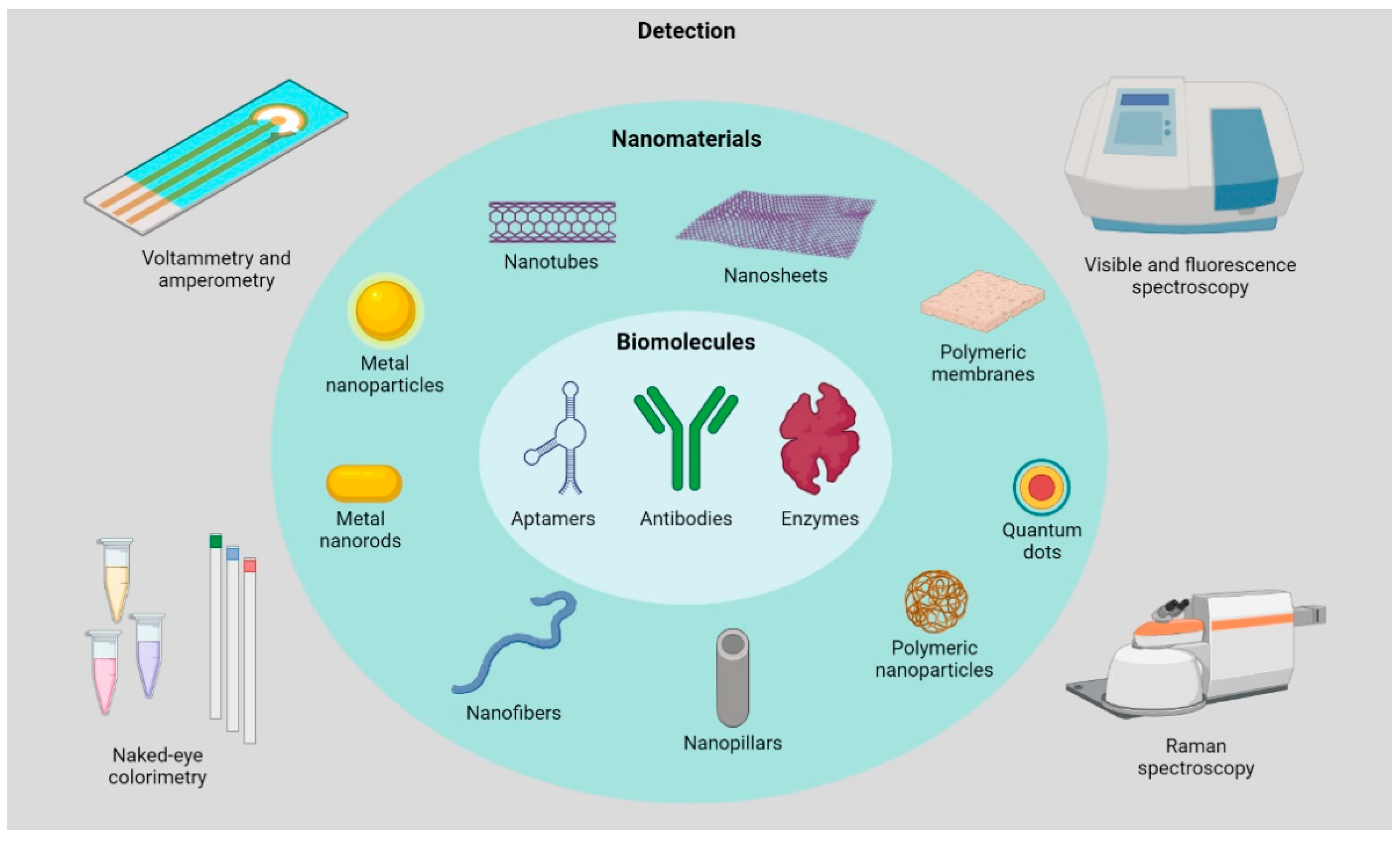
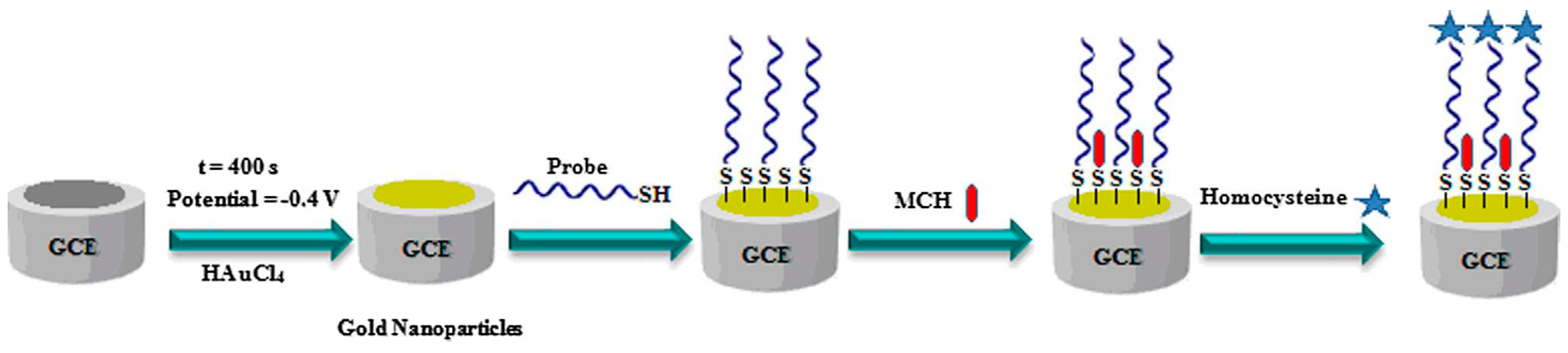
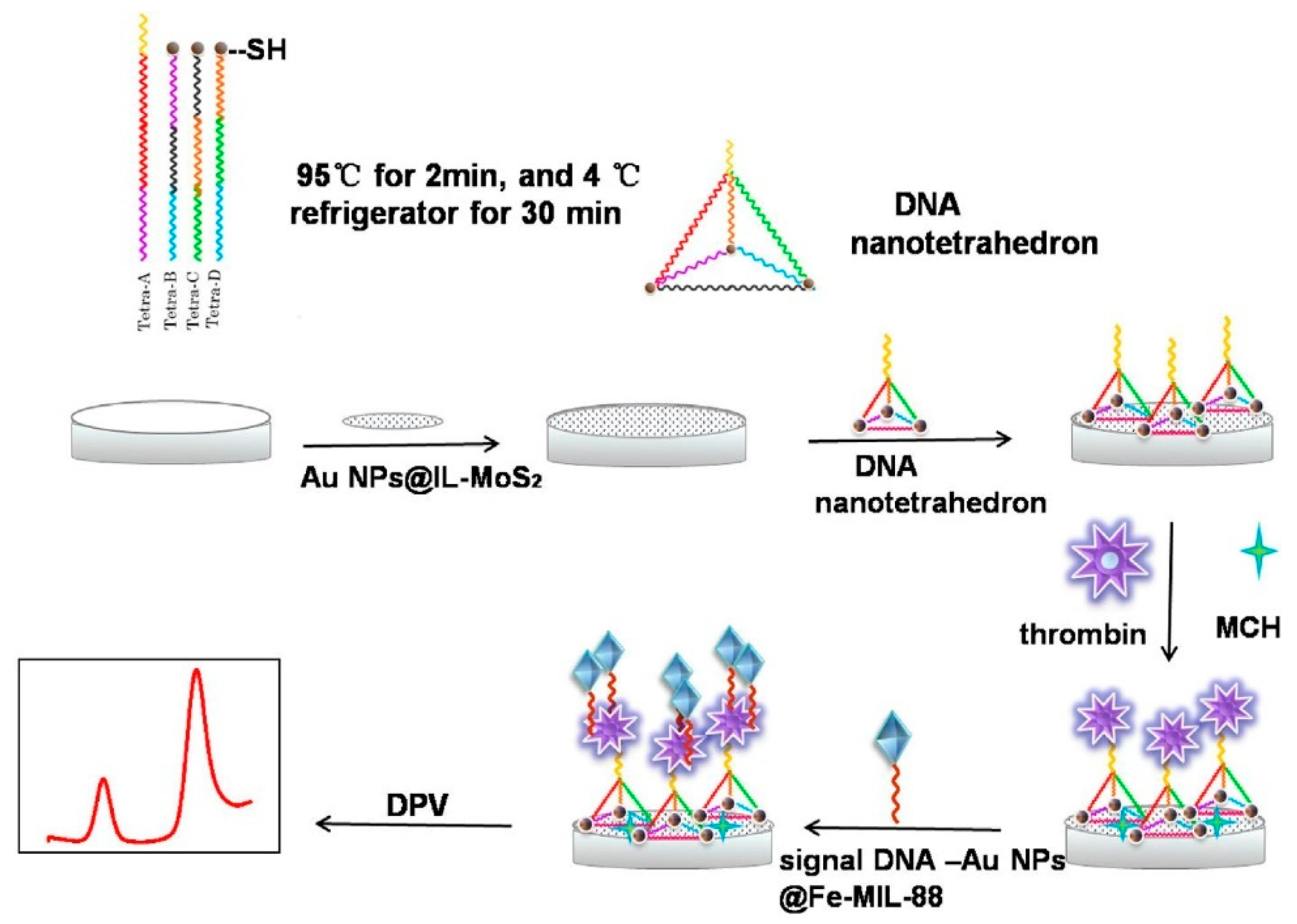
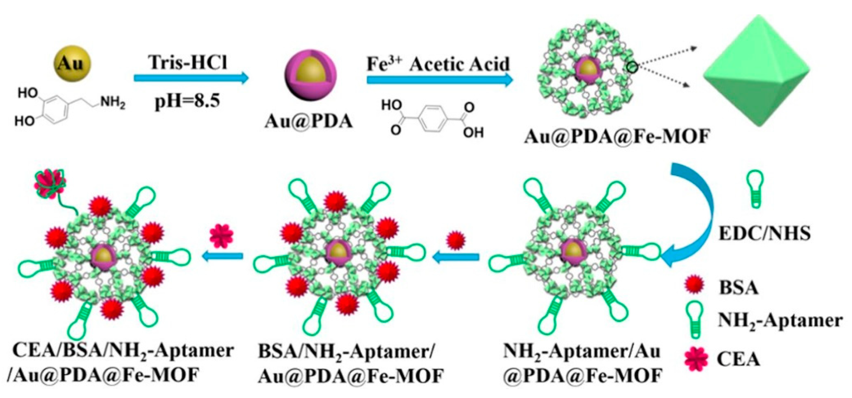
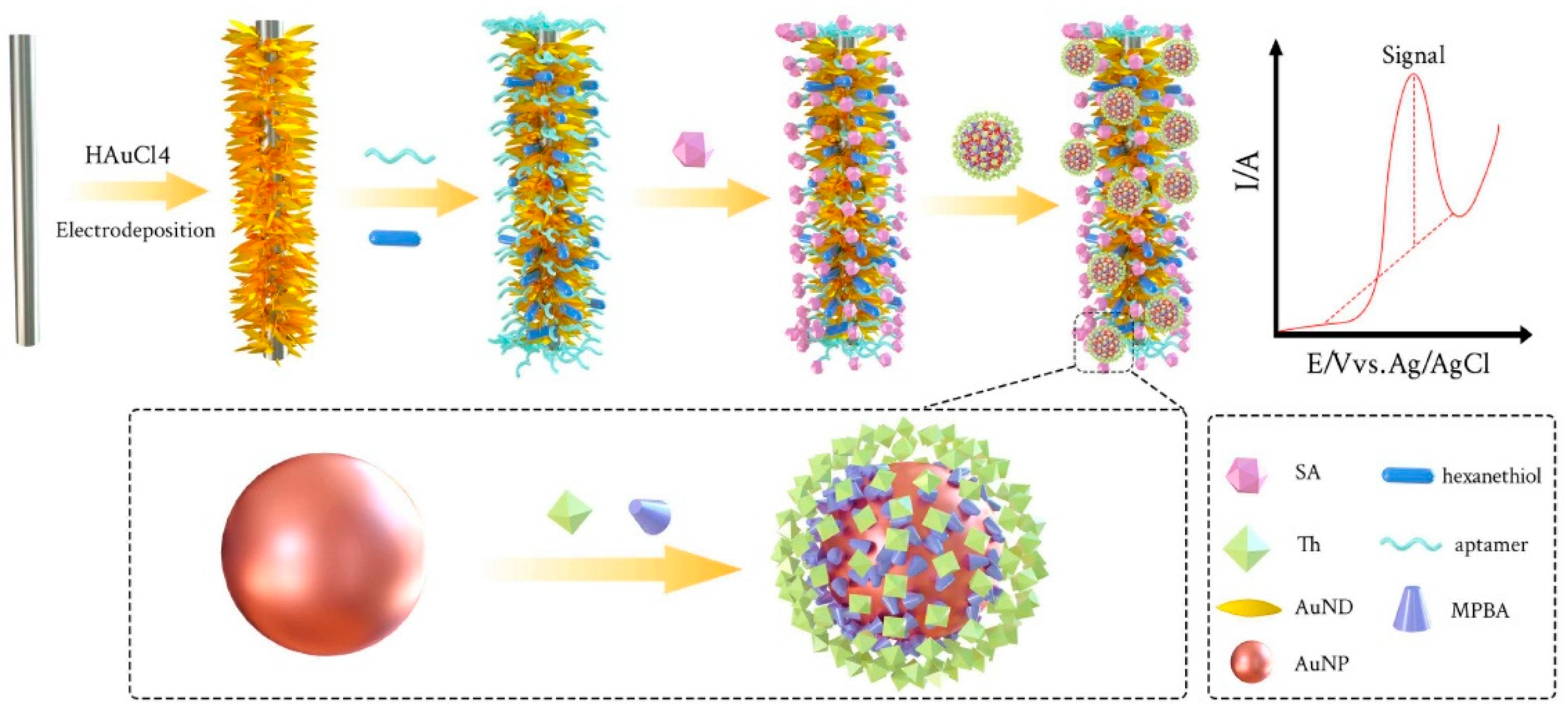
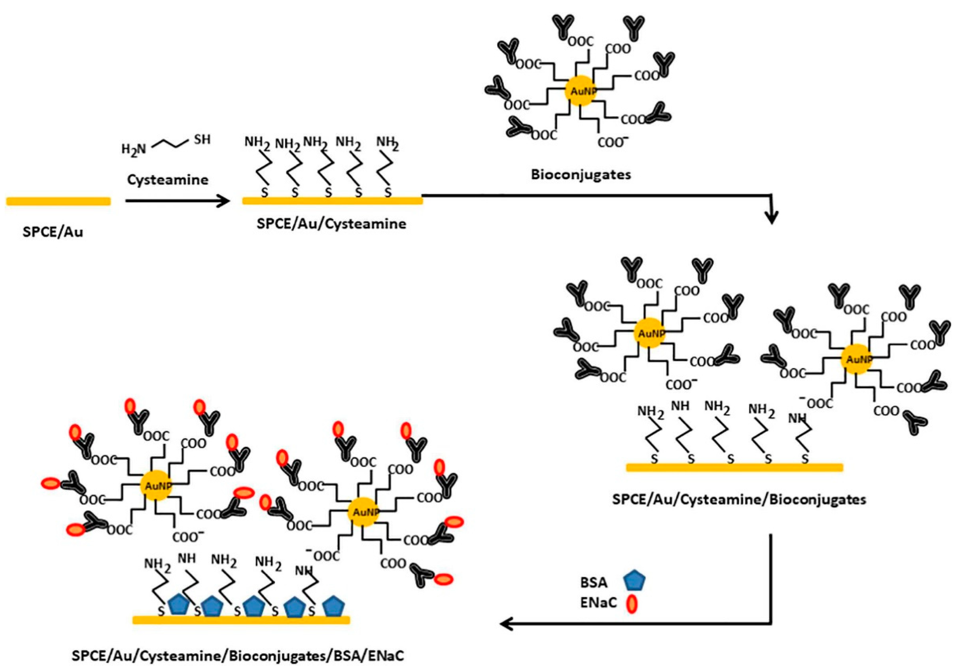
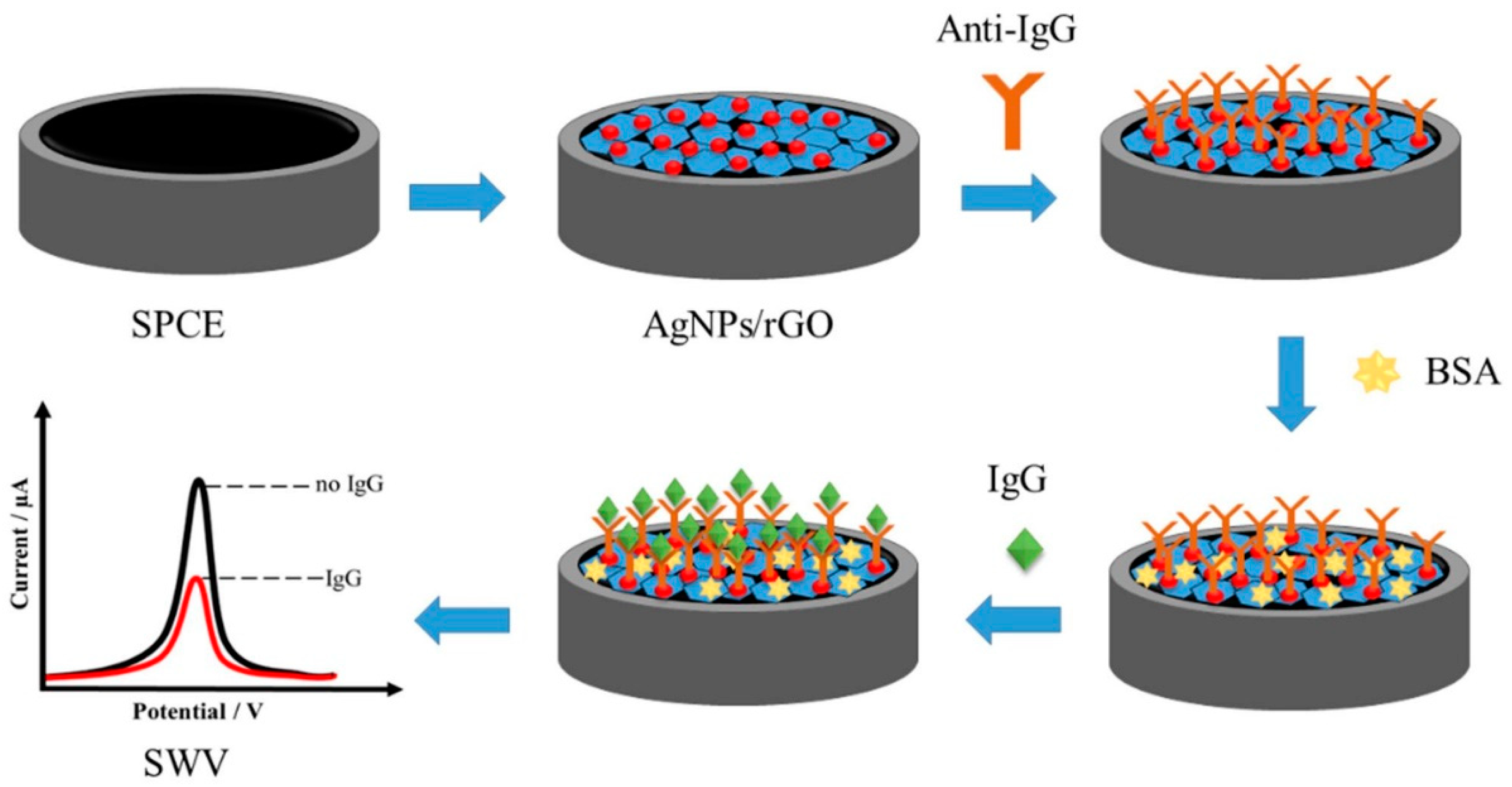
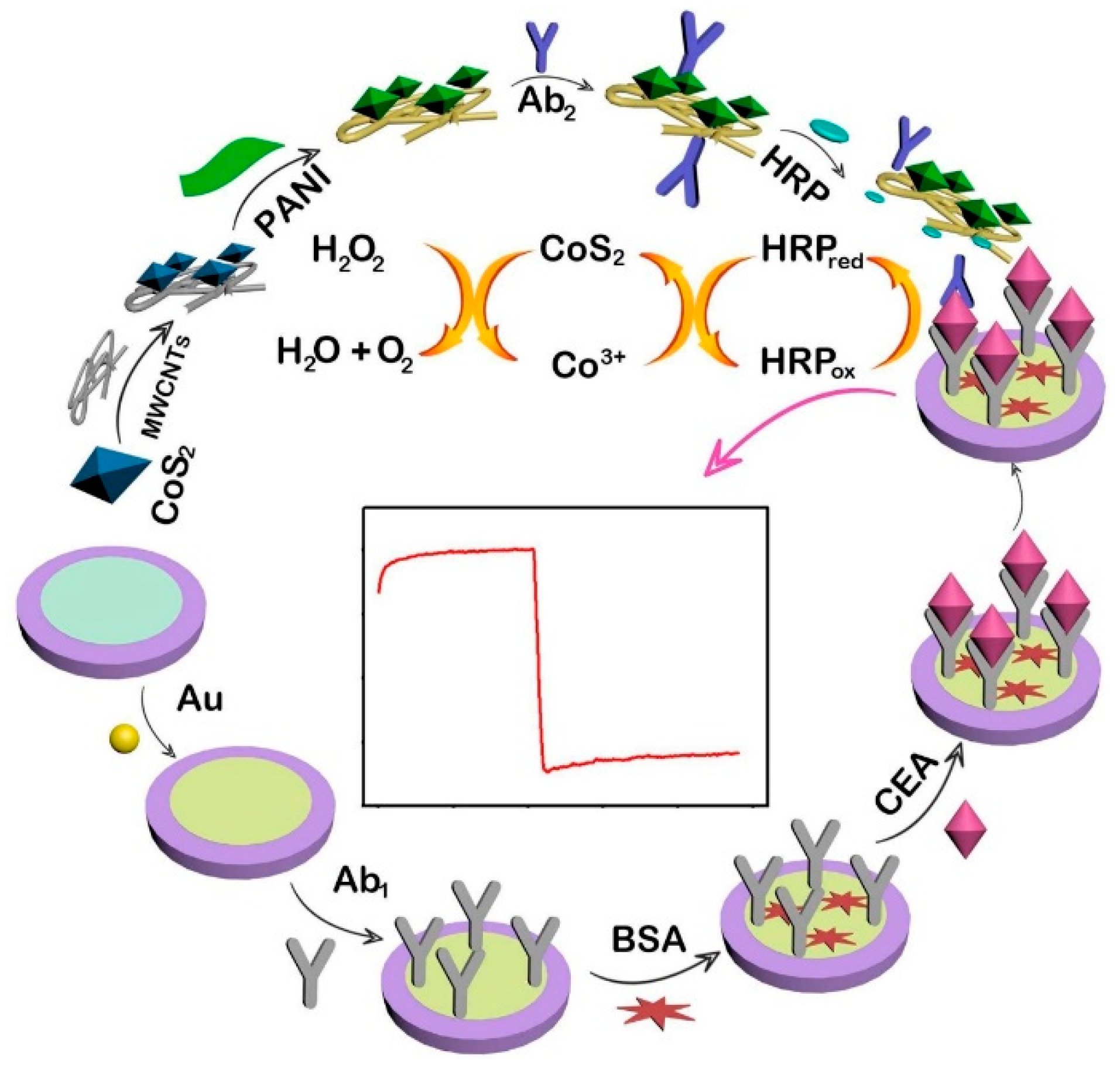
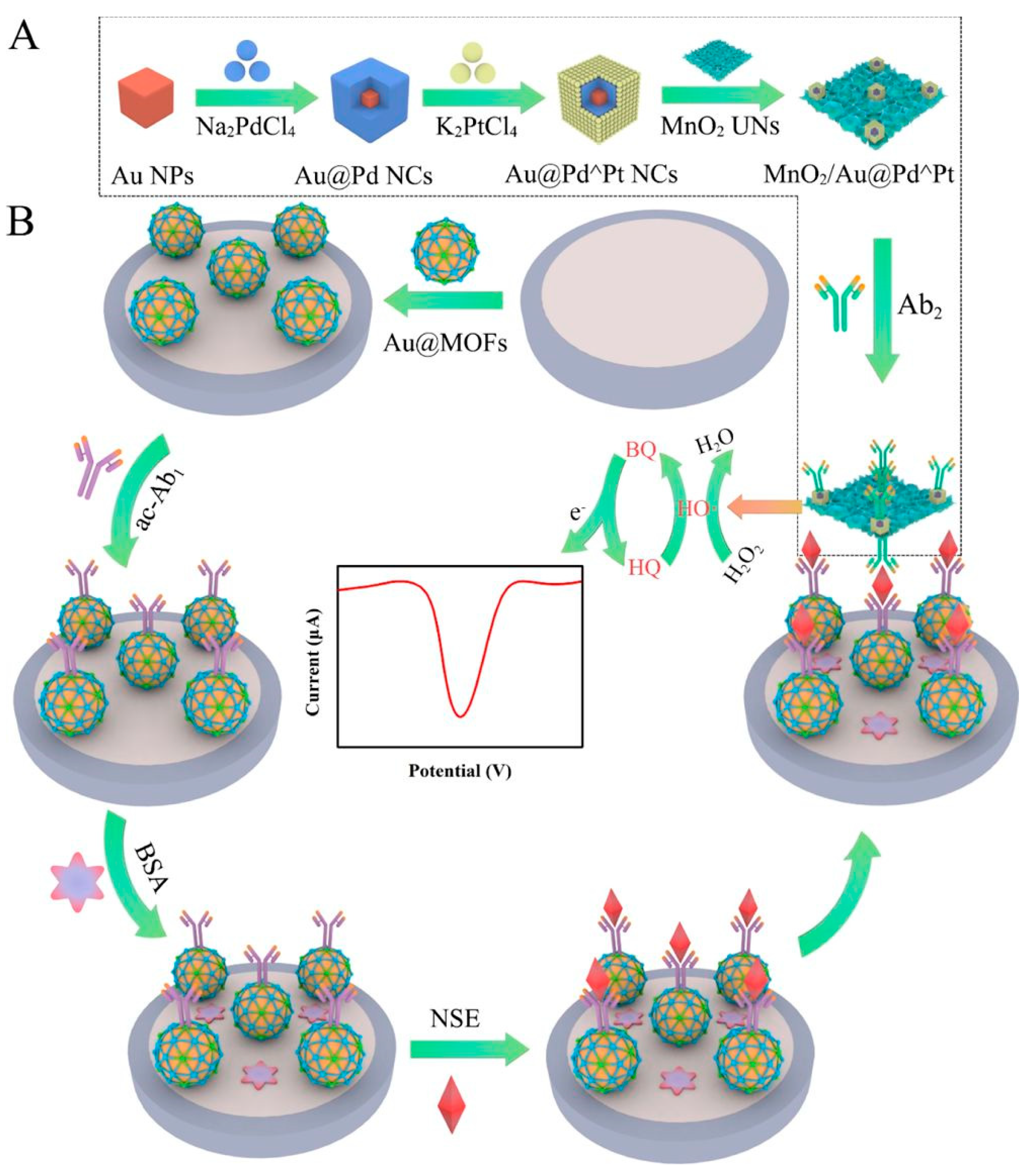
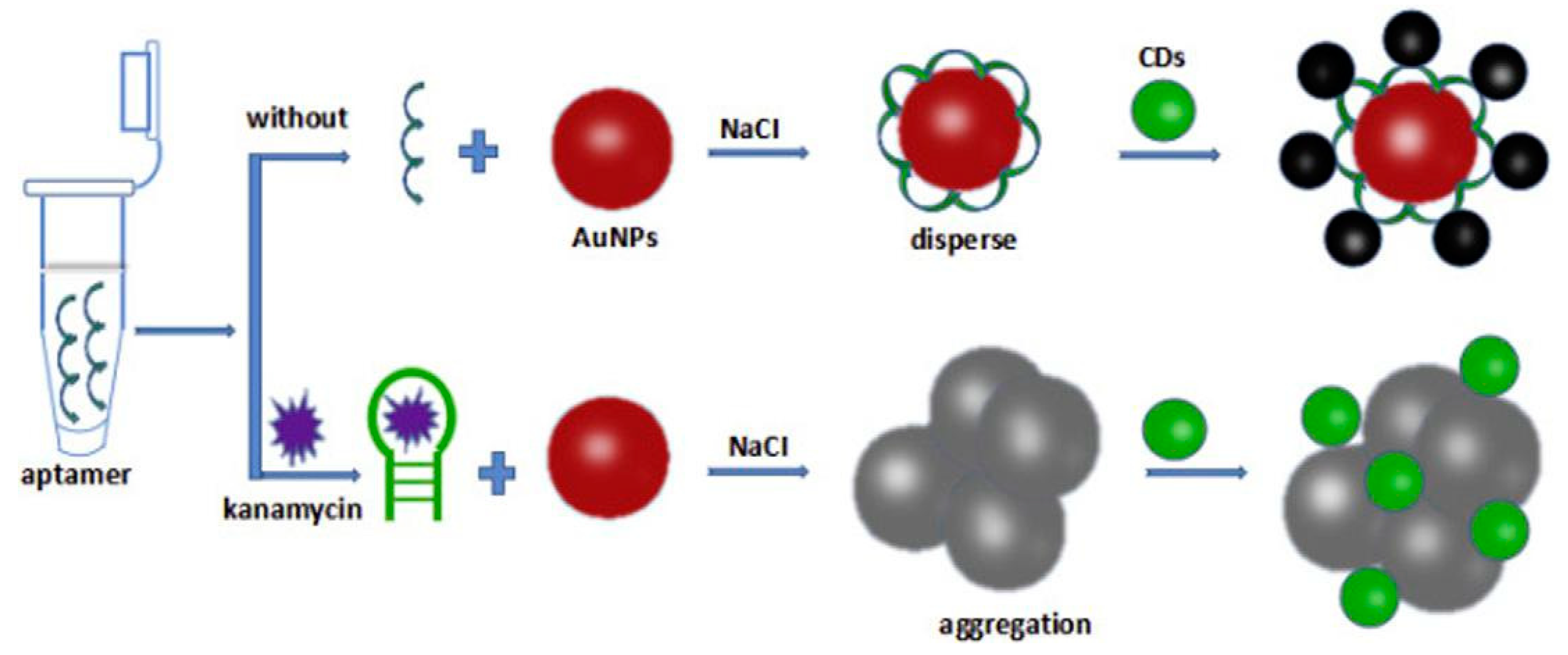
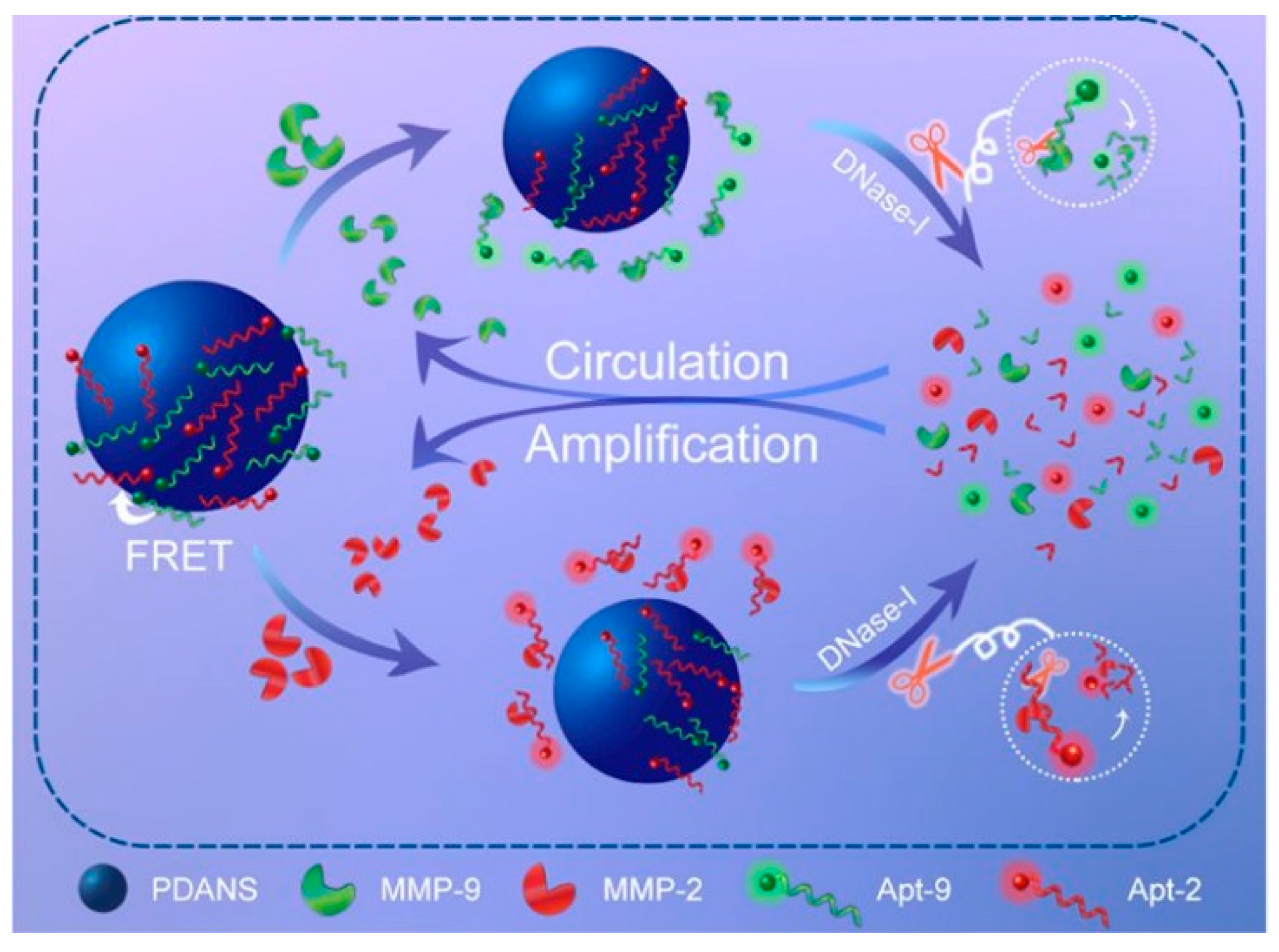
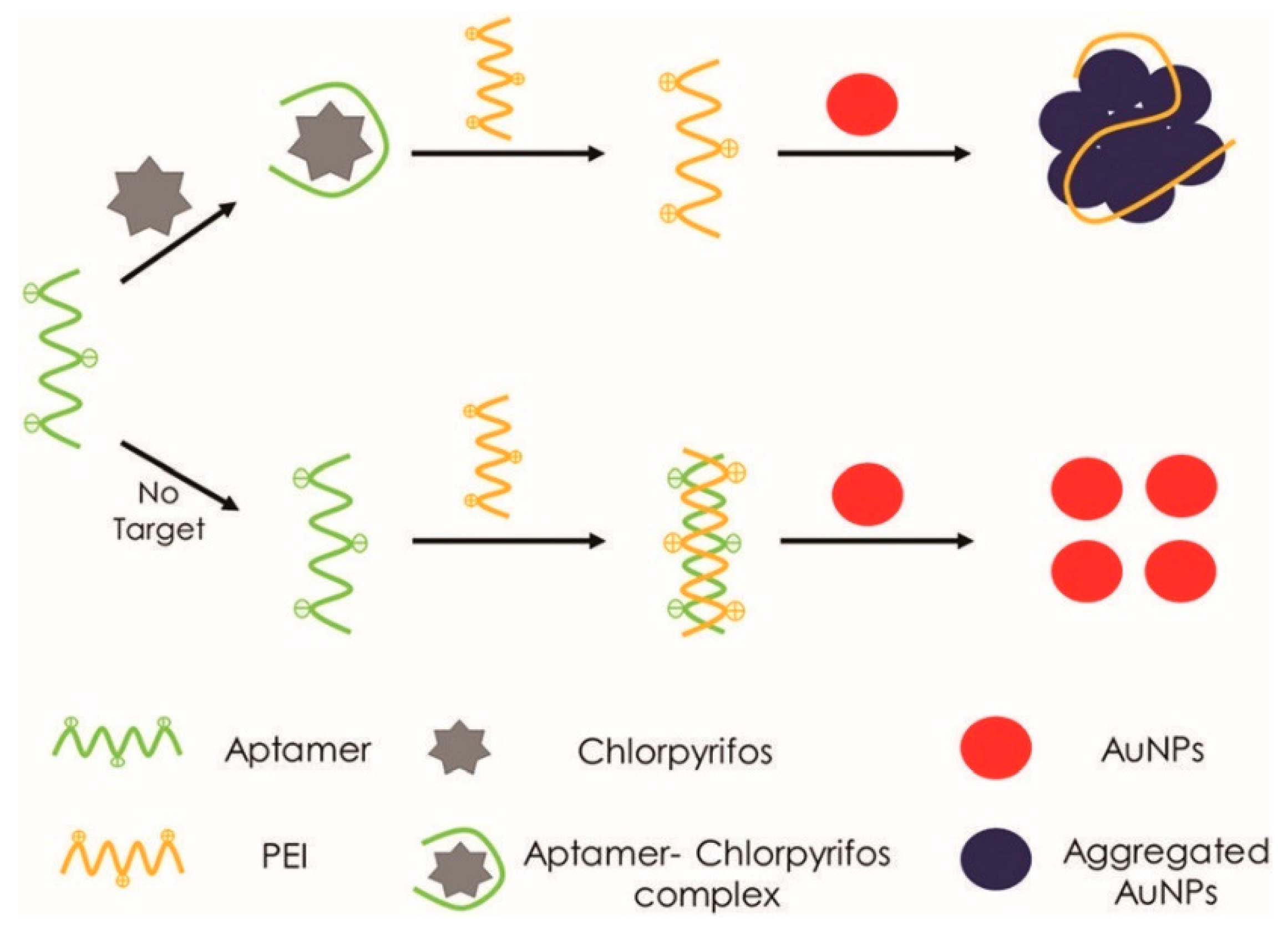
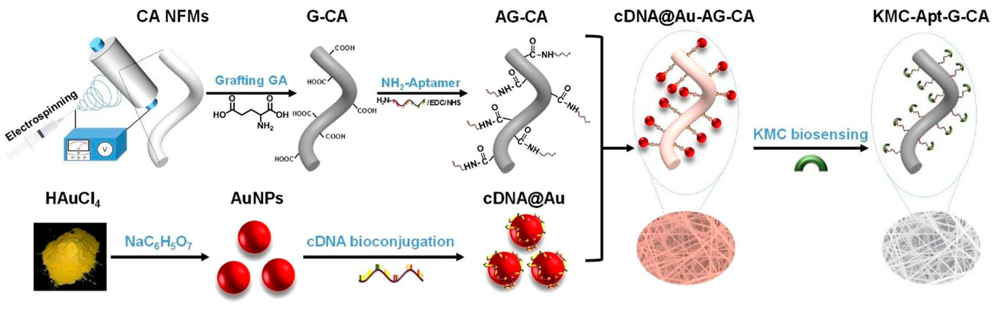
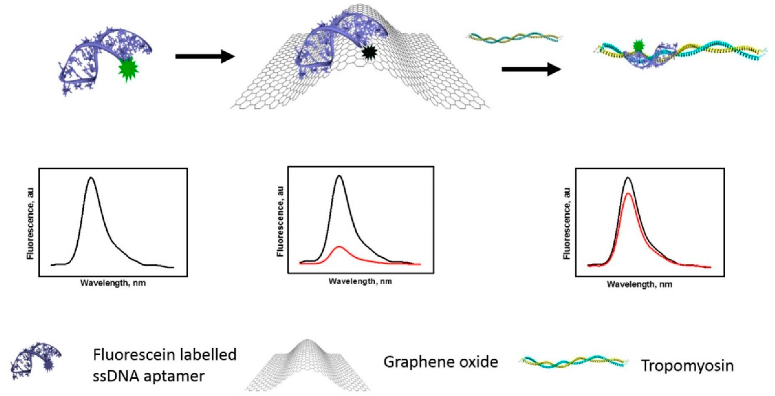
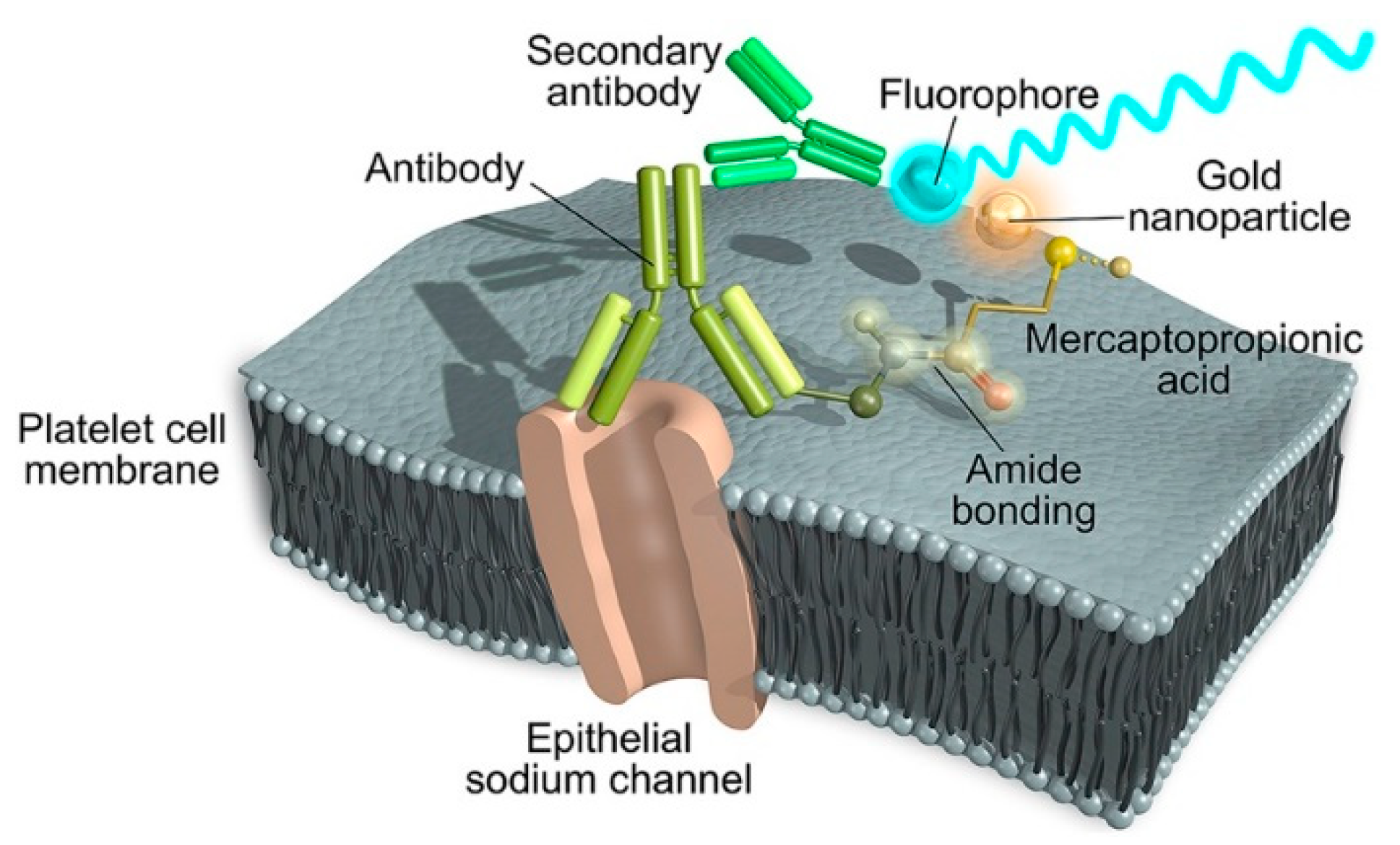
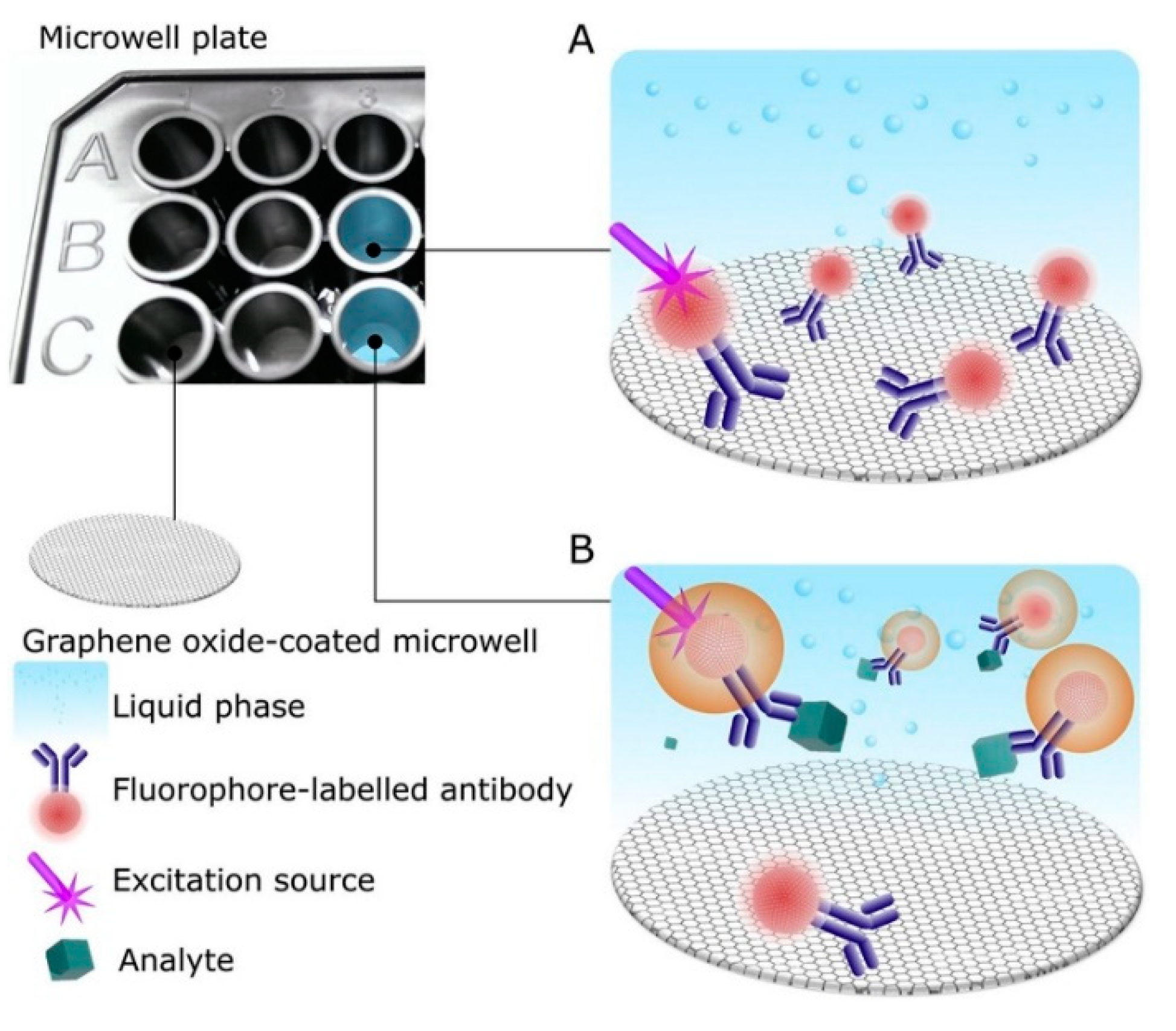
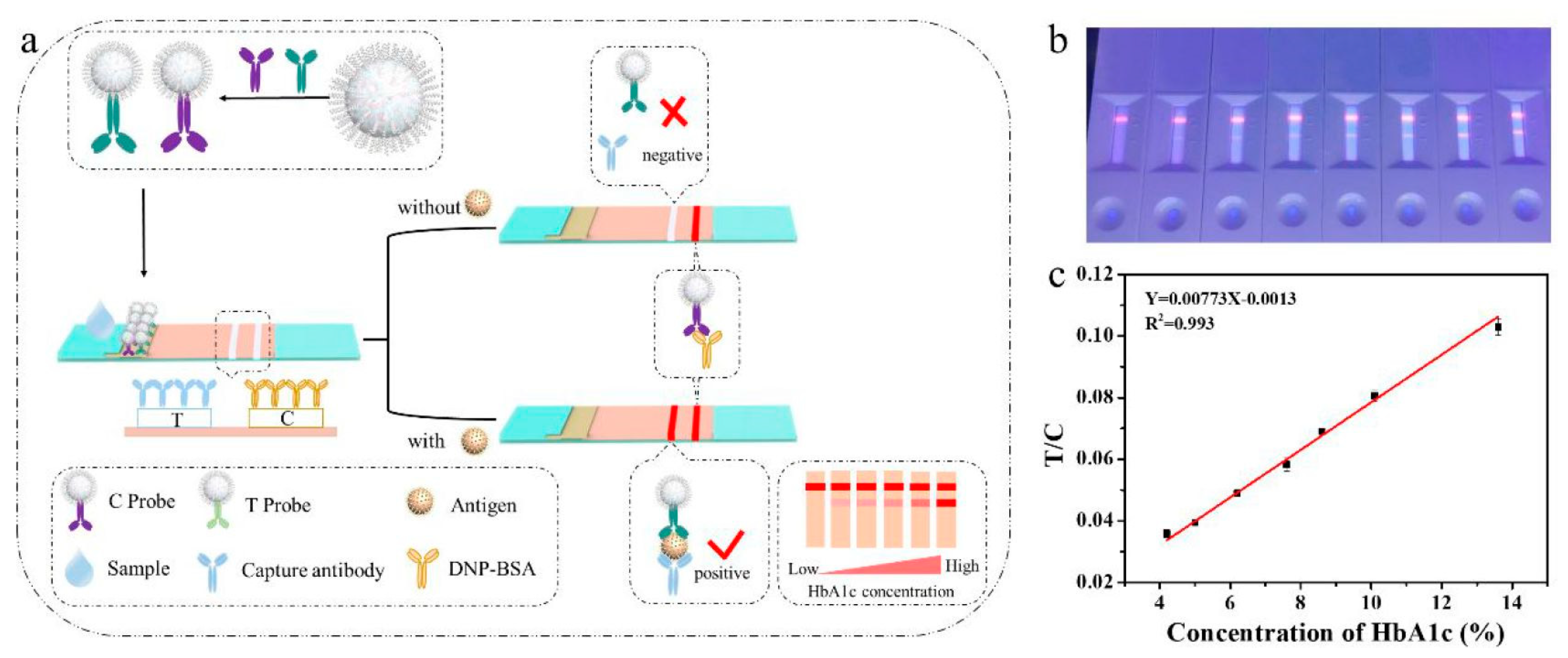
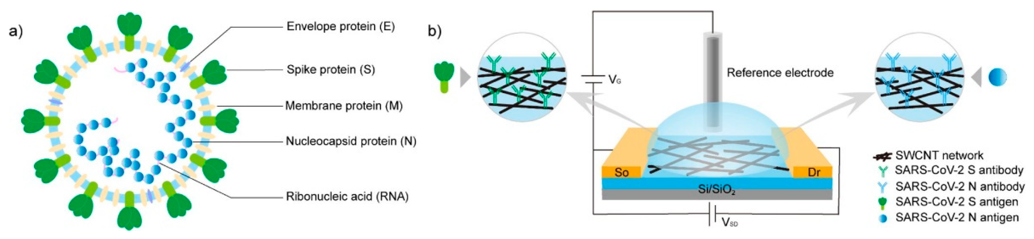
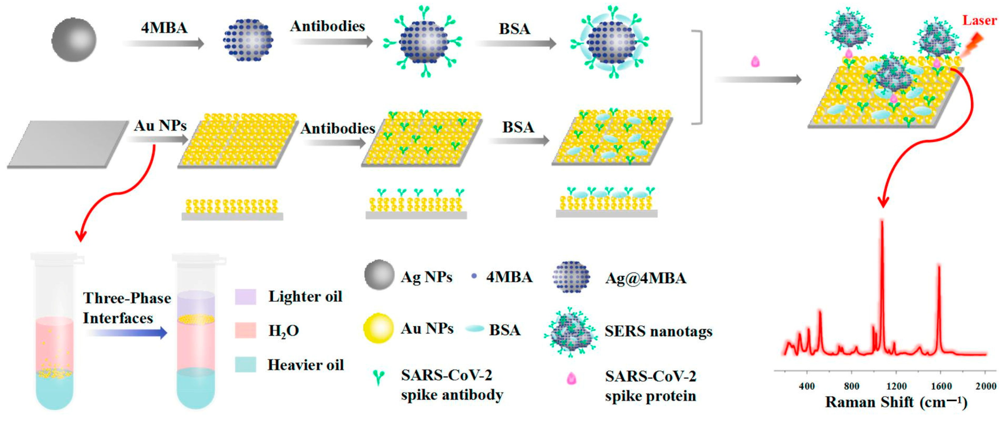
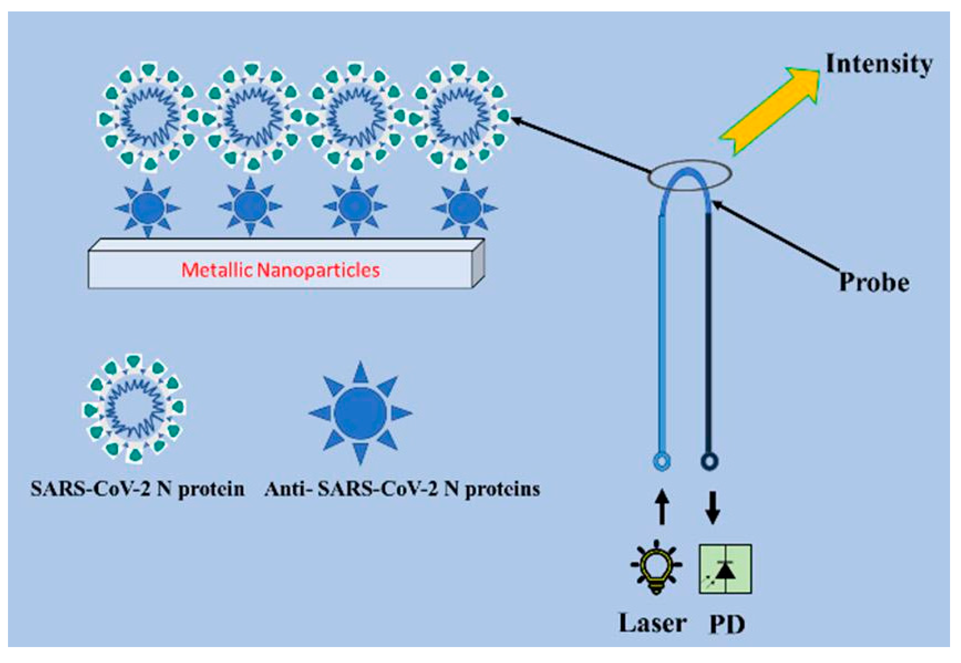
Table 1.
Electrochemical aptasensors cited in this review.
| Electrode | Main Components | Target | LOD; Range | Reference |
|---|---|---|---|---|
| Glassy carbon | Aptamers-functionalized AuNPs | Homocysteine in serum and urine | 0.01 μM; 0.05–20.0 μM | [25] |
| Screen-printed carbon | Aptamers-functionalized flower-like Au microstructures | Serpin in plasma | 0.031 ng mL−1; 0.039–10 ng mL−1 | [26] |
| Screen-printed Au | Aptamers-functionalized CeO2NPs | Epithelial sodium channel in urine | 0.012 ng mL−1; 0.05–3.0 ng mL−1 | [27] |
| Glassy carbon | DNA nonotetrahedrons-functionalized AuNPs-IL-MoS2, aptamer-functionalized AuNPs-Fe-MIL-88 MOFs | Thrombin in serum | 56 fM; 0.298–29.8 pM | [28] |
| Glassy carbon | Aptamers-functionalized reduced MoS2-AuNPs | Zearalenone and fumonisin B1 in food | 5 × 10⁻⁴ ng mL−1; 1 × 10⁻³–10 ng mL−1 (zearalenone) and 1 × 10⁻³–1 × 10² ng mL−1 (fumonisin B1) | [29] |
| Glassy carbon | Aptamers-functionalized (3-aminopropyl)triethoxysilane-modified GO-AgNPs | Chloramphenicol in food | 3.3 pM; 10 pM–0.2 μM | [30] |
| Glassy carbon | Aptamers-functionalized thiourea capped-ZnSQDs-AuNPs | β-casomorphin 7 in urine | 350 aM; 1 fM–0.6 μM | [31] |
| Glassy carbon | Aptamers-functionalized core-shell Cu-In-S-ZnSQDs-AgNPs-GQDs | Ractopamine in urine and serum | 330 aM; 1 fM–901.4 nM | [32] |
| Magnetic glassy carbon | Aptamers-functionalized magnetic reduced GO-Fe3O4-Cu2O nanosheets and Ag-resorcinol-formaldehyde NPs-AgNDs | Prostate specific antigen in serum | 6.2 pg mL−1; 0.01–100 ng mL−1 | [33] |
| Glassy carbon | Aptamers-functionalized reduced GO-AgNPs and prussian blue-AuNPs | Acetamiprid in food | 0.30 pM; 1 pM–1 μM | [34] |
| Glassy carbon | Aptamers-functionalized reduced GO-AuNPs | Glycated albumin | 0.07 μg mL−1; 2–10 μg mL−1 | [35] |
| Glassy carbon | Aptamers-functionalized MXene-AuNPs | Chloramphenicol in food | 0.03 pM; 0.0001–10 nM | [36] |
| Glassy carbon | Aptamers-functionalized PtNPs-MIL-101(Fe) MOFs | Aflatoxin M1 in food | 2 × 10⁻³ ng mL−1; 1 × 10⁻²–80 ng mL−1 | [37] |
| Au | Aptamers-functionalized CoNi-MOFs | Enrofloxacin in food, water and serum | 0.2 fg mL−1; 0.001–1 pg mL−1 | [38] |
| Glassy carbon | Aptamers-functionalized AuNPs-Fe3+-catechol MOFs | Carcinoembryonic antigen in serum | 0.33 fg mL−1; 1 fg mL−1–1 μg mL−1 | [39] |
| Screen-printed carbon | Aptamers-functionalized carbon-black NPs-AuNPs | Cd(II) ions in water | 0.14 ppb; 1–50 ppb | [40] |
| Screen-printed carbon | Aptamers-functionalized AuNPs-UiO-66-NH2 MOFs assisted by exonuclease | Streptomycin in food | 2.6 pg mL−1; 0.005–150 ng mL−1 | [41] |
| Fluorine-doped tin oxide | Aptamers-functionalized BiVO4-2D-C3N4 photoanode with II type heterojunction | Microcystin-LR in water | 4.191 × 10⁻⁸ μg L−1; 5 × 10⁻⁷ μg L−1–10 μg L−1 | [42] |
| Au | Aptamers-functionalized highly porous Au nanostructures | Acetamiprid in food | 0.34 nmol L−1; 0.5–300 nmol L−1 | [43] |
| Indium tin oxide | Aptamers-functionalized core-shell LaFeO3-g-C3N4 heterostructures | Streptomycin in food | 0.0033 nM; 0.01–10000 nM | [44] |
| Au | Aptamers-functionalized GQDs-AuNPs composites | Vitamin D in serum | 0.7 nM; 1–500 nM | [45] |
| Screen printed graphite | Aptamers-functionalized poly-3-amino-1,2,4-triazole-5-thiol-GO-AuNPs and secondary aptamer probes | Lipocalin-2 in blood serum | 0.3 ng mL−1; 1–1000 ng mL−1 | [46] |
| Indium tin oxide electrodes | Aptamers-functionalized TiO2-BiOI-BiOBr visible light photosensitive composite | Streptomycin food and blood serum | 0.04 nM; 0.05–150 nM | [47] |
| Screen-printed carbon | Aptamers-functionalized magnetic GO-Fe3O4 | Organophosphorus pesticides in food | N.D. | [48] |
| Pt | Aptamers-functionalized AgNPs and luminol-hydrogen peroxide | Kanamycin in food | 0.06 ng mL−1; 0.5–100 ng mL−1 | [49] |
| Au | Aptamers-functionalized polyethyleneimine-coated reduced GO-AuNCs and single-stranded DNA-binding protein | Chloramphenicol in food | 2.08 pmol·L−1; 5 pmol·L−1–1 μmol·L−1 | [50] |
| Au | Polyethyleneimine-graphite-like carbon nitride/AuNWs functionalized with SH-aptamers | Chloramphenicol in food | 2.96 × 10⁻⁹ μM; 1 × 10⁻⁷–1 μM | [51] |
| Au | Aptamers-functionalized AuNSs conjugated to CdSQDs and PdSQDs | Kanamycin and tobramycin in serum | 0.12 nM (kanamycin) and 0.49 nM (tobramycin); 1–4 × 10² nM (kanamycin) and 1–1 × 10⁴ nM (tobramycin) | [52] |
| Au | DNA nanostructures and horseradish peroxidase-functionalized AuNPs assisted by exonuclease | Kanamycin in food | 9.1 fg mL−1; 0.05 pg mL−1–10 ng mL−1 | [53] |
| Carbon | Aptamer-coated Au nanodendrites and 4-mercaptophenylboric acid-thionine-functionalized AuNPs | Sialic acid in serum | 60 nM; 0.1–440 μM | [54] |
Table 2.
Electrochemical immunosensors cited in this review.
| Electrode | Main Components | Target | LOD; Range | Reference |
|---|---|---|---|---|
| Screen-printed carbon | AuNPs-antibodies | Epithelial sodium channel in urine | 2.8 × 10⁻¹ ng mL−1; 9.375 × 10⁻²–1.0 ng mL−1 | [62] |
| Screen-printed carbon | CeO2NPs-antibodies | Herceptin-2 in serum | 34.9 pg mL−1; 0.001–20.0 ng mL−1 | [63] |
| Glassy carbon | Divalent metal ions-functionalized AuNPs-carbon NSs-antibodies | Prostate specific antigen, carcinoembryonic antigen and α-fetoprotein in serum | 3.6 pg mL−1 (prostate specific antigen), 3.0 pg mL−1 (carcinoembryonic antigen) and 2.6 pg mL−1 (α-fetoprotein); 0.01–100 ng mL−1 (prostate specific antigen), 0.01–80 ng mL−1 (carcinoembryonic antigen and α-fetoprotein) | [64] |
| Au | Magnetic Fe3O4NPs-antibodies | Siglec 15 protein in serum | 0.82 pg mL−1; 1 pg mL−1–100 ng mL−1 | [65] |
| Screen-printed carbon | Nafion-embedded poly(2-hydroxyethyl methacrylate-glycidyl methacrylate)NPs-urease | Urea in serum | 0.77 μM; 0.01–500 mM | [66] |
| Graphite | AgNPs-antibodies | Tick-borne encephalitis virus in serum | 90 IU mL−1; 100–1600 IU mL−1 | [67] |
| Glassy carbon | Antibodies-functionalized SnS2 nanoflakes-chitosan | Carcinoembryonic antigen in serum | 5 pg mL−1; 0.006–3.0 ng mL−1 | [68] |
| Screen-printed carbon | AgNPs-reduced GO-antibodies | Immunoglobulin G in serum | 0.00086 ng mL−1; 0.001–0.05 and 0.05–50 ng mL−1 | [69] |
| Screen-printed carbon | GO-antibodies | Epithelial sodium channels in urine | 0.198 ng mL−1; 0.01–1.5 ng mL−1 | [70] |
| Screen-printed | GO-Fe3O4NPs-Prussian blue-AuNPs-antibodies | Hepatitis B surface antibody in serum | 0.166 pg mL−1; 0.5 pg mL−1–200 ng mL−1 | [71] |
| Glassy carbon | Carboxyl G functionalized with mesoporous silica NPs-Methylene blue-AuNPs-antibodies | Galectin-3 in serum | 2.0 fg mL−1; 50 fg mL−1–500 ng mL−1 | [72] |
| Au | Carbon nanochips-embedded Au colloids-TiO2NPs-chitosan composites | β-lactoglobulin in food | 0.01 pg mL−1; 0.01–500 pg mL−1 | [73] |
| Screen-printed carbon | Chitosan-G-ionic liquid-ferrocene porous cryogel decorated with AuNPs-antibodies | Prostate-specific antigen in serum | 4.8 × 10⁻⁸ ng mL−1; 1.0 × 10⁻⁷–1.0 × 10⁻¹ ng mL−1 | [74] |
| Indium tin oxide | Urease-functionalized ZnONPs-polyaniline-chitosan | Urea in serum | 29.84 ppm; 20–500 ppm | [75] |
| Glassy carbon | Sandwich-like carbon NTs-CoS2-polyalanine-antibodies-horseradish peroxidase and AuNPs-antibodies | Carcinoembryonic antigen in serum | 0.33 pg mL−1; 0.001–40 ng mL−1 | [76] |
| Silicon oxide | Microfibers of single-walled carbon NTs-glucose oxidase | Glucose | N.D. | [77] |
| Glassy carbon | Vinyl ferrocene- and N-hydroxy succinimide acrylate-bifunctionalized carbon NTs conjugated with antibodies | α-fetoprotein in serum | 1.14 ng·mL−1; 10 ng·mL−1–50 μg·mL−1 | [78] |
| Glassy carbon | Carbon NTs-polyalanine functionalized with AuNPs-antibodies | Prostate-specific antigen in serum | 0.5 pg·mL−1; 1.66 ag·mL−1–1.3 ng·mL−1 | [79] |
| Titanium foil | Porous hydrogen titanate NTs-glucose oxidases | Glucose | 59 μM; 1–10 mM | [80] |
| Glassy carbon | Antibodies-functionalzied sandwich-like AuNPs-embedded MOFs and MnO2 nanosheets-trimetallic Au-Pt-Pd nanocubes | Neuron-specific enolase in serum | 4.17 fg mL−1; 10 fg mL−1–100 ng mL−1 | [81] |
| Glassy carbon | Antibodies-functionalized bamboo-like sandwich-like carbon nanostructure-toluidine blue-functionalized copper-based MOFs | C-reactive protein in serum | 166.7 pg mL−1; 0.5–200 ng mL−1 | [82] |
| Glassy carbon | Sandwich-like antibodies-conjugated DNA dendrimers on antibodies-functionalized electrodes | Prostate-specific antigen in serum | 0.26 pg mL−1; 1 pg mL−1–10 ng mL−1 | [83] |
Table 3.
Optical aptasensors cited in this review.
| Signal Detected | Main Components | Target | LOD; Range | Reference |
|---|---|---|---|---|
| Fluorescence | Aptamers-coated AuNPs, carbon QDs | Kanamycin in food | 18 nM; 0.04–0.24 μM | [84] |
| Fluorescence | Aptamers-coated AuNPs, Rhodamine B | Carbendazim in water | 2.33 nM; 2.33–800 nM | [85] |
| Fluorescence | Aptamers-coated AuNPs, Rhodamine B | Sulfamethazine in water and food | 0.82 ng mL−1; 1.25–40 ng mL−1 | [86] |
| Fluorescence | Aptamers-coated AuNPs, SYBR Green I-functionalized cDNA | Suplhadimetoxine in water and food | 3.41 ng mL−1 (water) and 4.41 ng L−1 (food); 2 ng mL−1–300 ng mL−1 | [87] |
| Fluorescence | Aptamers-functionalized AuNPs-carbon QDs | Adenosine triphosphate | 20 μM: 20–280 μM | [88] |
| Fluorescence | Fluorescent aptamers-coated polydopamine NSs, DNase-I enzyme | Metalloproteinase-9 and -2 in urine and tissue homogenate | 9.6 pg mL−1 (metalloproteinase-9) and 25.6 pg mL−1 (metalloproteinase-2); 24–600 pg mL−1 (metalloproteinase-9) and 64–1600 pg mL−1 (metalloproteinase-2) | [89] |
| Fluorescence | AuNRs coated with Ag nanoclusters-conjugated aptamers, exonuclease enzyme | Adenosine triphosphate in serum | 26 pM; 50 pM–1.0 nM | [90] |
| Color | Polyethyleneimine-coated AuNPs, unfunctionalized aptamers | Chlorpyrifos in water and food | 7.4 ng mL−1; 20–300 ng mL−1 | [92] |
| Color | AuNPs coated with unfunctionalized polyA aptamers | Prostate specific antigen in serum | 20 pg mL−1; 0.1–100 ng mL−1 | [93] |
| Color | Unfunctionalized truncated aptamers-coated AuNPs | Bisphenol A in food and water | 7.60 pM (38-mer) and 14.41 pM (12-mer); 20–100 pM | [94] |
| Color | Unfunctionalized aptamers-coated AuNPs | Chloramphenicol and tetracycline in food | 32.9 nM (tetracycline) and 7.0 nM (chloramphenicol); 0.05–3.0 μM (tetracycline) and 0.05–1.8 μM (chloramphenicol) | [95] |
| Color | Unfunctionalized aptamers-coated AuNPs | Bacillus carboniphilus on biofilms | 5 × 10³ CFU mL−1; 104–107 CFU mL−1 | [96] |
| Color | Unfunctionalized RNA aptamers-coated AuNPs | Human papilloma-virus type 16 L1 in clinical and vaccine samples | 9.6 ng mL-1; 9.6–201.6 ng mL−1 | [97] |
| Color | Unfunctionalized aptamers-coated AuNPs | Acetamiprid in food and water | 1.74 μM; 0–140 μM | [98] |
| Color | Glutamic acid-grafted cellulose acetate NFs functionalized with NH2-aptmaers, aptamer-complementary cDNA-coated AuNPs | Kanamycin in food and water | 2.5 nM; 2.5–80 nM | [99] |
| Color | Truncated aptamers-coated AuNPs onto nitrocellulose membrane, aptamer-complementary cDNA | Oxytetracycline in food | 5 ng mL−1; N.D. | [100] |
| Color | Unfunctionalized aptamers-coated onto nitrocellulose membrane | Gentamicin in food | 300 nM; 10–1000 nM | [101] |
| Fluorescence | GO coated with fluoresceine-conjugated aptamers | Tropomyosin allergen in food | 2 nM; 0–1.3 μM | [102] |
| Color | Enzyme-assisted, MnO2 nanosheets-induced AuNPs and biotin-labeled cDNA | Alkaline phosphate and ochratoxin A in food | 0.05 U·L−1 (alkaline phosphate) and 5.0 nM (ochratoxin A); 6.25–750 nM (ochratoxin A) | [104] |
| Fluorescence | Fe3O4-GO-assisted AT-rich three-way junctions DNA-stabilized CuNPs | Isocarbophos in food and water | 3.38 nM; 10–500 nM | [105] |
| Fluorescence | Fluorescent aptamers-coated zirconium-porphyrin MOFs | Chloramphenicol in food | 0.08 pg mL−1; 0.1 pg mL−1–10 ng mL−1 | [106] |
Table 4.
Optical immunosensors cited in this review.
| Signal Detected | Main Components | Target | LOD; Range | Reference |
|---|---|---|---|---|
| Fluorescence | AuNPs–antibodies conjugates, secondary fluorescent antibodies | Epithelial sodium channel in blood | N.D. | [107] |
| Color | Magnetic Au-coated Fe3O4NPs–antibodies conjugates | Glyphosate in tap water | 20 ng∙L−1; 0.01–100 μg∙L−1 | [109] |
| Color | AuNPs–antibodies and Fe3O4NPs–antibodies conjugates | Prostate specific antigen in serum | 0.009 ng mL−1; 0.01–20 ng mL−1 | [110] |
| Fluorescence | Fluorescent AuNPs–albumin, -rhodamine B and -β-lactoglobulin conjugates | Nephrin and podocin in urine | N.D. | [111] |
| SPR | Magnetic AuNPs-antibodies, SPR active sensor disks | CD5 in serum | 8.31 fM; N.D. | [112] |
| Fluorescence | Fluorescent QDs-antibodies, GO-coated multi-well plates | Prostate specific antigen in urine | 0.05 ng mL−1; 0.15–10 ng mL−1 | [113] |
| Fluorescence | Antibodies-conjugated CdSe-ZnSQDs and AuNRs | Porcine reproductive and respiratory syndrome virus in swine serum | 0.55 TCID50 mL−1; 101–3.5 × 10⁴ TCID50 mL−1 | [114] |
| Fluorescence | Rhodamine-labelled peptides adsorbed onto WS2-Pt-Fe2O3 micromotors | Bacterial lipopolysaccharides | 0.12 nM; 4.0–1,000,000 ng mL−1 | [115] |
| Fluorescence | Fluorescent bovine serum album-wrapped CdSe-ZnSQDs conjugated to antibodies targeting the glycosylated hemoglobin, nitrocellulose membrane | Glycosylated hemoglobin in blood | N.D. | [116] |
| SERS | Antibodies-conjugated AuNSs, nitrocellulose membrane | Metalloproteinase-9, S100 calcium-binding protein B and neuro-specific enolase in blood | 0.01 pg mL−1; 0.0001–1000 ng mL−1 | [117] |
| Absorbance | Antibodies-conjugated magnetic Fe3O4 NPs coated with a gold shell | Human growth hormone in serum | 0.082 nmol L−1; 0.1–5.0 nmol L−1 | [118] |
| Reflected light | Analyte-induced gold deposition on antibodies-conjugated Mn-ZnS QDs, microscope coverslip | Prostate-specific antigen in serum | 3.5 × 10⁻³ pg mL−1; 0.01–100 pg·mL−1 | [119] |
Disclaimer/Publisher’s Note: The statements, opinions and data contained in all publications are solely those of the individual author(s) and contributor(s) and not of MDPI and/or the editor(s). MDPI and/or the editor(s) disclaim responsibility for any injury to people or property resulting from any ideas, methods, instructions or products referred to in the content. |
© 2023 by the authors. Licensee MDPI, Basel, Switzerland. This article is an open access article distributed under the terms and conditions of the Creative Commons Attribution (CC BY) license (https://creativecommons.org/licenses/by/4.0/).
Share and Cite
MDPI and ACS Style
Fata, F.; Gabriele, F.; Angelucci, F.; Ippoliti, R.; Di Leandro, L.; Giansanti, F.; Ardini, M. Bio-Tailored Sensing at the Nanoscale: Biochemical Aspects and Applications. Sensors 2023, 23, 949. https://doi.org/10.3390/s23020949
AMA Style
Fata F, Gabriele F, Angelucci F, Ippoliti R, Di Leandro L, Giansanti F, Ardini M. Bio-Tailored Sensing at the Nanoscale: Biochemical Aspects and Applications. Sensors. 2023; 23(2):949. https://doi.org/10.3390/s23020949
Chicago/Turabian StyleFata, Francesca, Federica Gabriele, Francesco Angelucci, Rodolfo Ippoliti, Luana Di Leandro, Francesco Giansanti, and Matteo Ardini. 2023. "Bio-Tailored Sensing at the Nanoscale: Biochemical Aspects and Applications" Sensors 23, no. 2: 949. https://doi.org/10.3390/s23020949
Note that from the first issue of 2016, this journal uses article numbers instead of page numbers. See further details here.