
Non-Destructive Analytical Investigation of Decorative Wallpapers Samples of the Nineteenth Century before Their Restoration

 and
and 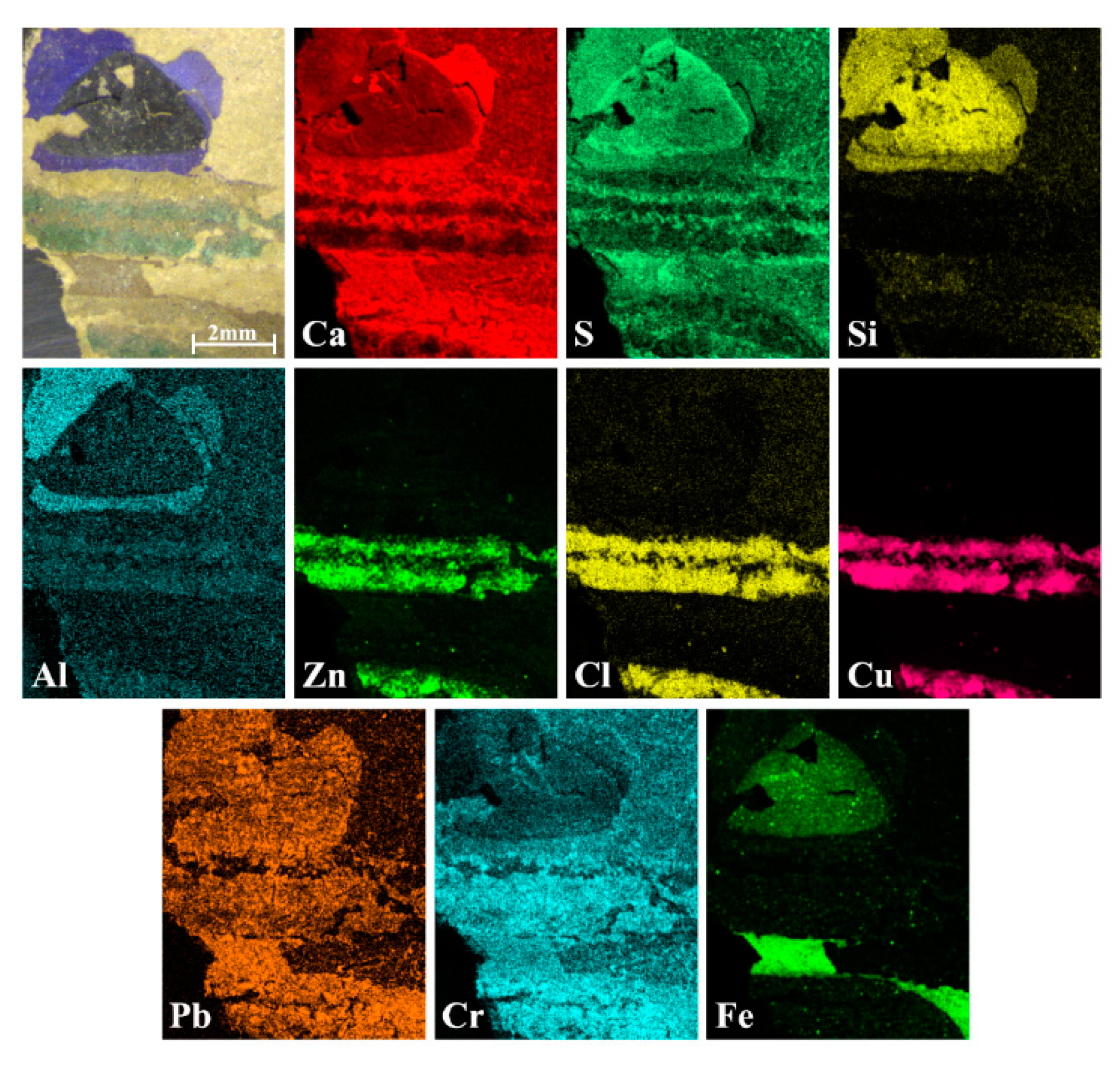
Abstract
:1. Introduction
2. Materials and Methods
2.1. Historical Information and Samples
2.2. Instrumentation
2.2.1. Micro-Energy Dispersive X-ray Fluorescence Spectroscopy (micro-EDXRF)
2.2.2. Micro-Raman Spectroscopy
2.2.3. Attenuated Total Reflection Infrared (ATR-FTIR)
2.2.4. pH-Meter
3. Results and Discussion
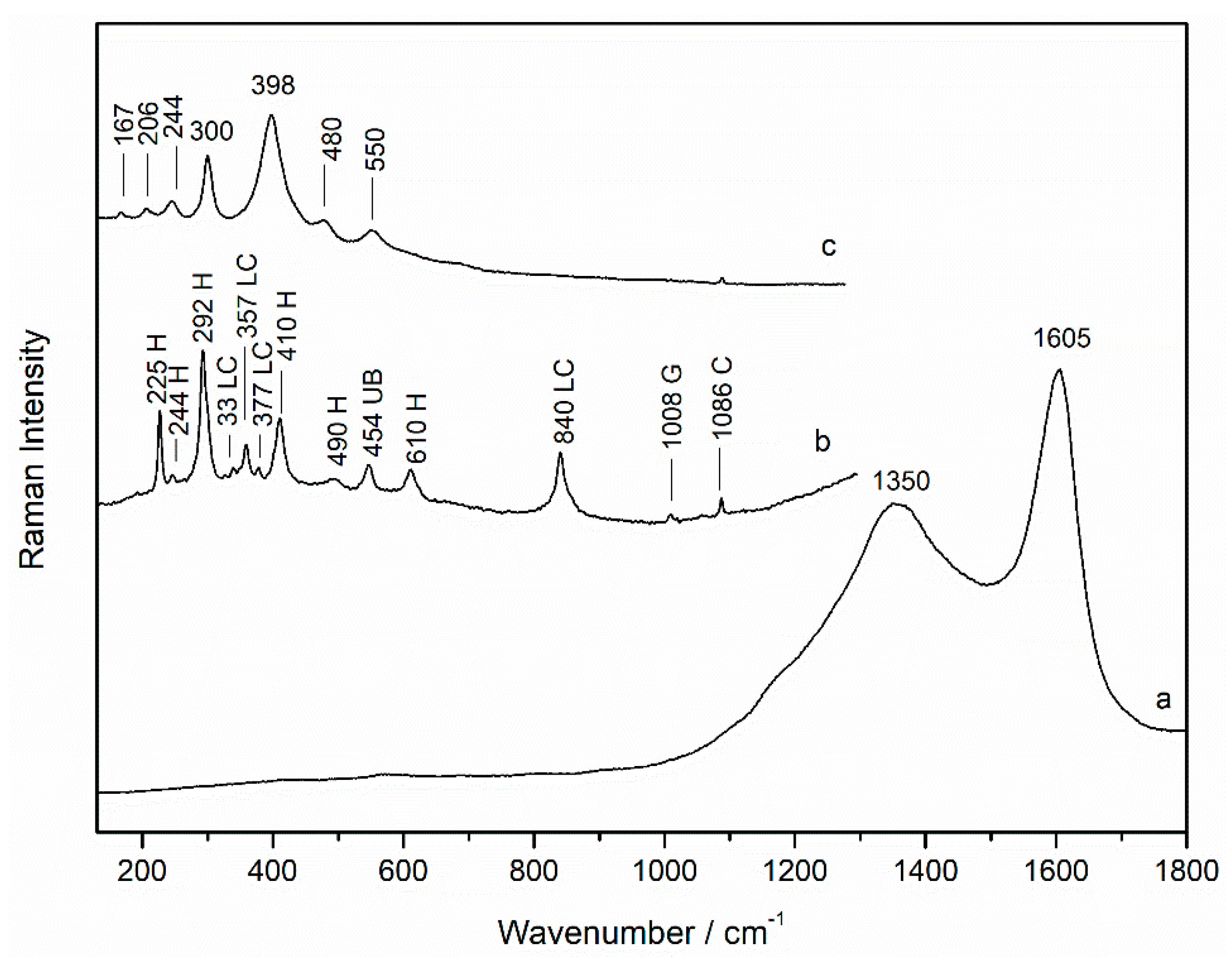
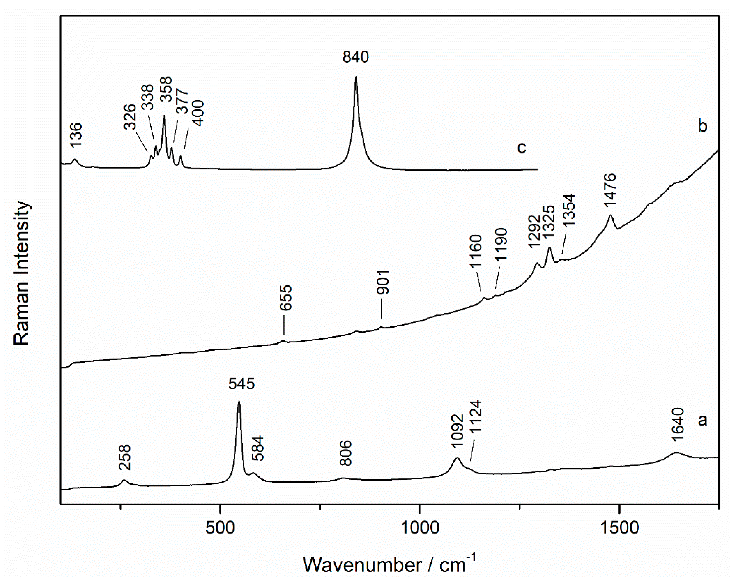
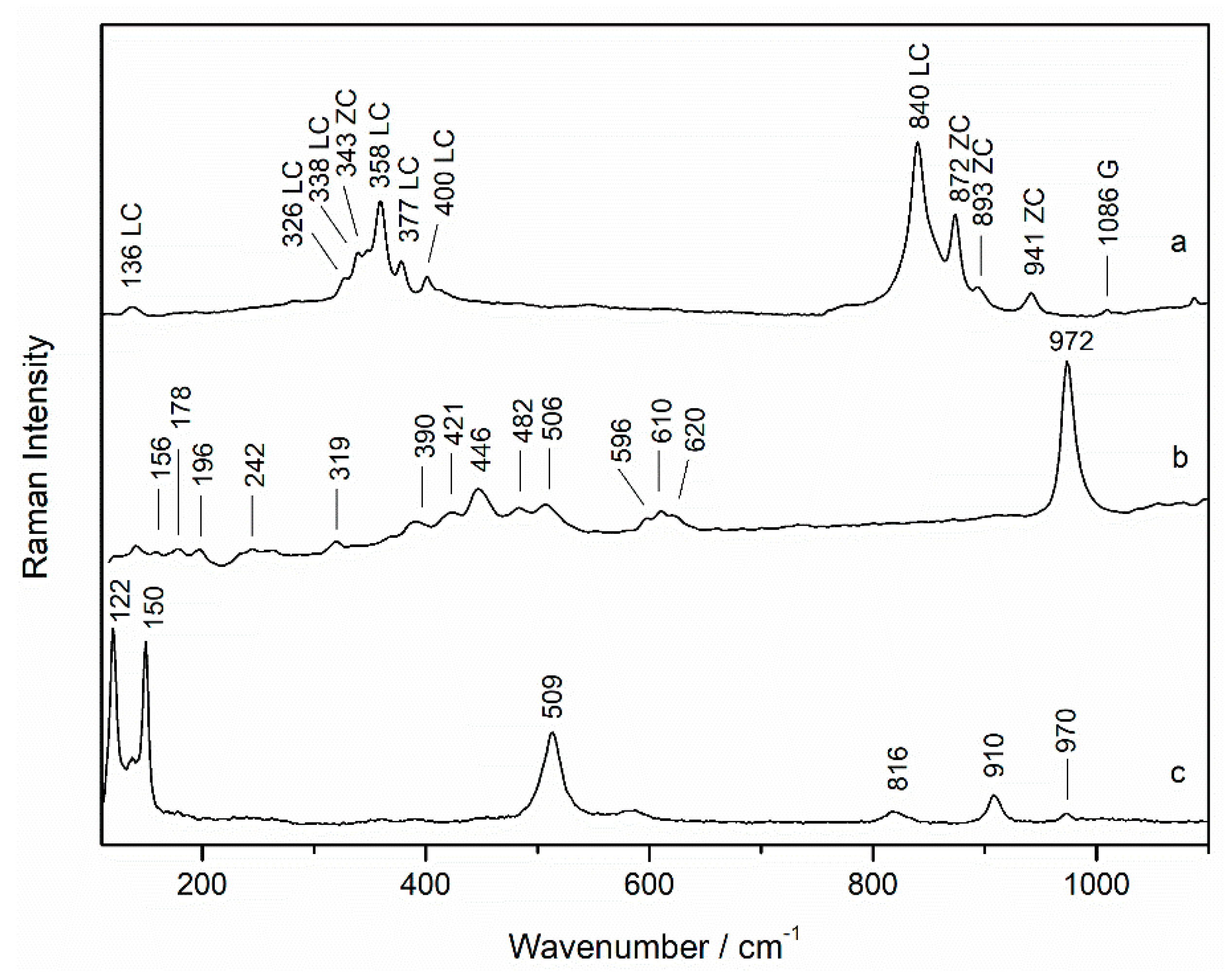
3.1. Characterization of Materials
3.2. State of Conservation of the Wallpapers
4. Conclusions
Supplementary Materials
Author Contributions
Funding
Institutional Review Board Statement
Informed Consent Statement
Data Availability Statement
Conflicts of Interest
References
- García, F.R.B. La fábrica de papeles pintados Santa Isabel de Vitoria a mediados del siglo XIX. Ars Bilduma 2019, 9, 113–136. [Google Scholar] [CrossRef]
- Lynn, C.; Frangiamore, C.L. Wallpapers in Historic Preservation; Technical Preservation Services Division, Office of Archeology and Historic Preservation, National Park Service, U.S. Department of the Interior: Washington, DC, USA, 1977.
- Hoskins, L. The Papered Wall: The History, Patterns and Techniques of Wallpaper; Thames & Hudson: London, UK, 2005; ISBN 978-0-500-28568-8. [Google Scholar]
- Pessanha, S.; Guilherme, A.; Carvalho, M.L.; Cabaço, M.I.; Bittencourt, K.; Bruneel, J.L.; Besnard, M. Study of a XVIII century hand-painted Chinese wallpaper by multianalytical non-destructive techniques. Spectrochim. Acta Part B At. Spectrosc. 2009, 64, 582–586. [Google Scholar] [CrossRef]
- Arrizabalaga, I.; Gómez-Laserna, O.; Aramendia, J.; Arana, G.; Madariaga, J.M. Determination of the pigments present in a wallpaper of the middle nineteenth century: The combination of mid-diffuse reflectance and far infrared spectroscopies. Spectrochim. Acta. A Mol. Biomol. Spectrosc. 2014, 124, 308–314. [Google Scholar] [CrossRef] [PubMed]
- Harroun, S.G.; Bergman, J.; Jablonski, E.; Brosseau, C.L. Surface-enhanced Raman spectroscopy analysis of house paint and wallpaper samples from an 18th century historic property. Analyst 2011, 136, 3453–3460. [Google Scholar] [CrossRef]
- Castro, K.; Knuutinen, U.; de Vallejuelo, S.F.-O.; Irazola, M.; Madariaga, J.M. Finnish wallpaper pigments in the 18th–19th century: Presence of KFe3(CrO4)2(OH)6 and odd pigment mixtures. Spectrochim. Acta. A Mol. Biomol. Spectrosc. 2013, 106, 104–109. [Google Scholar] [CrossRef] [PubMed]
- Haslam, J.C. Deadly Décor: A Short History of Arsenic Poisoning in the Nineteenth Century. Res. Med. 2013, 21, 76–81. [Google Scholar] [CrossRef]
- Ball, P. William Morris made poisonous wallpaper. Nature 2003. [Google Scholar] [CrossRef]
- Phillips, M. Wallpaper on Walls: Problems of Climate and Substrate. J. Am. Inst. Conserv. 1981, 20, 83–90. [Google Scholar] [CrossRef]
- Welsh, F.S. Investigation, Analysis, and Authentication of Historic Wallpaper Fragments. J. Am. Inst. Conserv. 2004, 43, 91–110. [Google Scholar] [CrossRef]
- Castro, K.; Princi, E.; Proietti, N.; Manso, M.; Capitani, D.; Vicini, S.; Madariaga, J.M.; De Carvalho, M.L. Assessment of the weathering effects on cellulose based materials through a multianalytical approach. Nucl. Instrum. Methods Phys. Res. Sect. B Beam Interact. Mater. At. 2011, 269, 1401–1410. [Google Scholar] [CrossRef]
- Castro, K.; Sarmiento, A.; Pérez-Alonso, M.; Madariaga, J.M.; Princi, E.; Vicini, S.; Pedemonte, E.; Rodríguez-Laso, M.D. Vibrational spectroscopy at the service of industrial archaeology: Nineteenth-century wallpaper. TrAC Trends Anal. Chem. 2007, 26, 347–359. [Google Scholar] [CrossRef]
- Castro, K.; Pérez-Alonso, M.; Rodríguez-Laso, M.D.; Etxebarria, N.; Madariaga, J.M. Non-invasive and non-destructive micro-XRF and micro-Raman analysis of a decorative wallpaper from the beginning of the 19th century. Anal. Bioanal. Chem. 2007, 387, 847–860. [Google Scholar] [CrossRef]
- Castro, K.; Vandenabeele, P.; Rodríguez-Laso, M.D.; Moens, L.; Madariaga, J.M. Micro-Raman analysis of coloured lithographs. Anal. Bioanal. Chem. 2004, 379, 674–683. [Google Scholar] [CrossRef] [PubMed]
- Castro, K.; Vandenabeele, P.; Rodríguez-Laso, M.D.; Moens, L.; Madariaga, J.M. Improvements in the wallpaper industry during the second half of the 19th century: Micro-Raman spectroscopy analysis of pigmented wallpapers. Spectrochim. Acta. A. Mol. Biomol. Spectrosc. 2005, 61, 2357–2363. [Google Scholar] [CrossRef]
- Janssens, K.; Grieken, R.V. Non-Destructive Micro Analysis of Cultural Heritage Materials; Elsevier: Amsterdam, The Netherlands, 2004; ISBN 978-0-08-045442-9. [Google Scholar]
- Colomban, P. Pigment identification of a rare 18th century wallpaper from Buffon library. J. Raman Spectrosc. 2011, 42, 192–194. [Google Scholar] [CrossRef]
- Lambert, I.; Laroque, C. An Eighteenth-Century Chinese Wallpaper: Historical Context and Conservation. Stud. Conserv. 2002, 47, 122–128. [Google Scholar] [CrossRef]
- Castro, K.; Rodríguez-Laso, M.D.; Fernández, L.A.; Madariaga, J.M. Fourier transform Raman spectroscopic study of pigments present in decorative wallpapers of the middle nineteenth century from the Santa Isabel factory (Vitoria, Basque Country, Spain). J. Raman Spectrosc. 2002, 33, 17–25. [Google Scholar] [CrossRef]
- Schulte, F.; Brzezinka, K.-W.; Lutzenberger, K.; Stege, H.; Panne, U. Raman spectroscopy of synthetic organic pigments used in 20th century works of art. J. Raman Spectrosc. 2008, 39, 1455–1463. [Google Scholar] [CrossRef]
- Leona, M.; Stenger, J.; Ferloni, E. Application of surface-enhanced Raman scattering techniques to the ultrasensitive identification of natural dyes in works of art. J. Raman Spectrosc. 2006, 37, 981–992. [Google Scholar] [CrossRef]
- Eremin, K.; Stenger, J.; Green, M.L. Raman spectroscopy of Japanese artists’ materials: The Tale of Genji by Tosa Mitsunobu. J. Raman Spectrosc. 2006, 37, 1119–1124. [Google Scholar] [CrossRef]
- Wang, N.; He, L.; Egel, E.; Simon, S.; Rong, B. Complementary analytical methods in identifying gilding and painting techniques of ancient clay-based polychromic sculptures. Microchem. J. 2014, 114, 125–140. [Google Scholar] [CrossRef]
- Holakooei, P.; Karimy, A.-H.; Vaccaro, C. A multi-analytical approach to the examination of nineteenth-century European wallpapers in Vasiq-Ansari House in Isfahan, Iran. Stud. Conserv. 2014, 59, 150–160. [Google Scholar] [CrossRef]
- Scott, D.A. Metallography and Microstructure of Ancient and Historic Metals; Thr Getty Conservation Institute: Singapore, 1991; p. 185. Available online: https://books.google.com.hk/books?hl=zh-CN&lr=&id=8blOAgAAQBAJ&oi=fnd&pg=PP1&dq=Metallography+and+Microstructure+of+Ancient+and+Historic+Metal&ots=5c1wqAO2oW&sig=5wbMA8wMKySu76B4BzfGKDqdHnU&redir_esc=y&hl=zh-CN&sourceid=cndr#v=onepage&q=Metallography%20and%20Microstructure%20of%20Ancient%20and%20Historic%20Metal&f=false (accessed on 31 May 2021).
- Galai, M.; Ouassir, J.; Ebn Touhami, M.; Nassali, H.; Benqlilou, H.; Belhaj, T.; Berrami, K.; Mansouri, I.; Oauki, B. α-Brass and (α + β) Brass Degradation Processes in Azrou Soil Medium Used in Plumbing Devices. J. Bio-Tribo-Corros. 2017, 3, 30. [Google Scholar] [CrossRef]
- Rodriguez-Blanco, J.D.; Shaw, S.; Benning, L.G. The kinetics and mechanisms of amorphous calcium carbonate (ACC) crystallization to calcite, viavaterite. Nanoscale 2011, 3, 265–271. [Google Scholar] [CrossRef] [PubMed]
- Anbalagan, G.; Mukundakumari, S.; Murugesan, K.S.; Gunasekaran, S. Infrared, optical absorption, and EPR spectroscopic studies on natural gypsum. Vib. Spectrosc. 2009, 50, 226–230. [Google Scholar] [CrossRef]
- Zorba, T.; Pavlidou, E.; Stanojlovic, M.; Bikiaris, D.; Paraskevopoulos, K.M.; Nikolic, V.; Nikolic, P.M. Technique and palette of XIIIth century painting in the monastery of Mileseva. Appl. Phys. A 2006, 83, 719–725. [Google Scholar] [CrossRef]
- Geweely, N.S.; Afifi, H.A.M.; Abdelrahim, S.A.; Alakilli, S.Y.M. Novel Comparative Efficiency of Ozone and Gamma Sterilization on Fungal Deterioration of Archeological Painted Coffin, Saqqara Excavation, Egypt. Geomicrobiol. J. 2014, 31, 529–539. [Google Scholar] [CrossRef]
- Monico, L.; Van der Snickt, G.; Janssens, K.; De Nolf, W.; Miliani, C.; Verbeeck, J.; Tian, H.; Tan, H.; Dik, J.; Radepont, M.; et al. Degradation Process of Lead Chromate in Paintings by Vincent van Gogh Studied by Means of Synchrotron X-ray Spectromicroscopy and Related Methods. 1. Artificially Aged Model Samples. Anal. Chem. 2011, 83, 1214–1223. [Google Scholar] [CrossRef]
- Monico, L.; Janssens, K.; Vanmeert, F.; Cotte, M.; Brunetti, B.G.; Van der Snickt, G.; Leeuwestein, M.; Salvant Plisson, J.; Menu, M.; Miliani, C. Degradation Process of Lead Chromate in Paintings by Vincent van Gogh Studied by Means of Spectromicroscopic Methods. Part 5. Effects of Nonoriginal Surface Coatings into the Nature and Distribution of Chromium and Sulfur Species in Chrome Yellow Paints. Anal. Chem. 2014, 86, 10804–10811. [Google Scholar] [CrossRef] [PubMed]
- Sun, J.X.; Xu, F.; Sun, X.F.; Xiao, B.; Sun, R.C. Physico-chemical and thermal characterization of cellulose from barley straw. Polym. Degrad. Stab. 2005, 88, 521–531. [Google Scholar] [CrossRef]
- ten Have, R.; Teunissen, P.J.M. Oxidative Mechanisms Involved in Lignin Degradation by White-Rot Fungi. Chem. Rev. 2001, 101, 3397–3414. [Google Scholar] [CrossRef]
- Chen, S.; Wu, Z.; Tanaka, H. Surface Characterization of Naturally Degraded Wood–Free Papers by Near lnfrared Fourier—Transform Raman Spectroscopy. J. Fac. Agric. Kyushu Univ. 2000, 45, 163–170. [Google Scholar] [CrossRef]
- Agarwal, U.P.; Reiner, R.S. Near-IR surface-enhanced Raman spectrum of lignin. J. Raman Spectrosc. 2009, 40, 1527–1534. [Google Scholar] [CrossRef]
- Petrou, M.; Edwards, H.G.M.; Janaway, R.C.; Thompson, G.B.; Wilson, A.S. Fourier-transform Raman spectroscopic study of a Neolithic waterlogged wood assemblage. Anal. Bioanal. Chem. 2009, 395, 2131–2138. [Google Scholar] [CrossRef] [PubMed] [Green Version]


Publisher’s Note: MDPI stays neutral with regard to jurisdictional claims in published maps and institutional affiliations. |
© 2021 by the authors. Licensee MDPI, Basel, Switzerland. This article is an open access article distributed under the terms and conditions of the Creative Commons Attribution (CC BY) license (https://creativecommons.org/licenses/by/4.0/).
Share and Cite
Costantini, I.; Castro, K.; Rodriguez-Laso, M.D.; Madariaga, J.M.; Arana, G. Non-Destructive Analytical Investigation of Decorative Wallpapers Samples of the Nineteenth Century before Their Restoration. Sensors 2021, 21, 4416. https://doi.org/10.3390/s21134416
Costantini I, Castro K, Rodriguez-Laso MD, Madariaga JM, Arana G. Non-Destructive Analytical Investigation of Decorative Wallpapers Samples of the Nineteenth Century before Their Restoration. Sensors. 2021; 21(13):4416. https://doi.org/10.3390/s21134416
Chicago/Turabian StyleCostantini, Ilaria, Kepa Castro, Maria Dolores Rodriguez-Laso, Juan Manuel Madariaga, and Gorka Arana. 2021. "Non-Destructive Analytical Investigation of Decorative Wallpapers Samples of the Nineteenth Century before Their Restoration" Sensors 21, no. 13: 4416. https://doi.org/10.3390/s21134416