Article

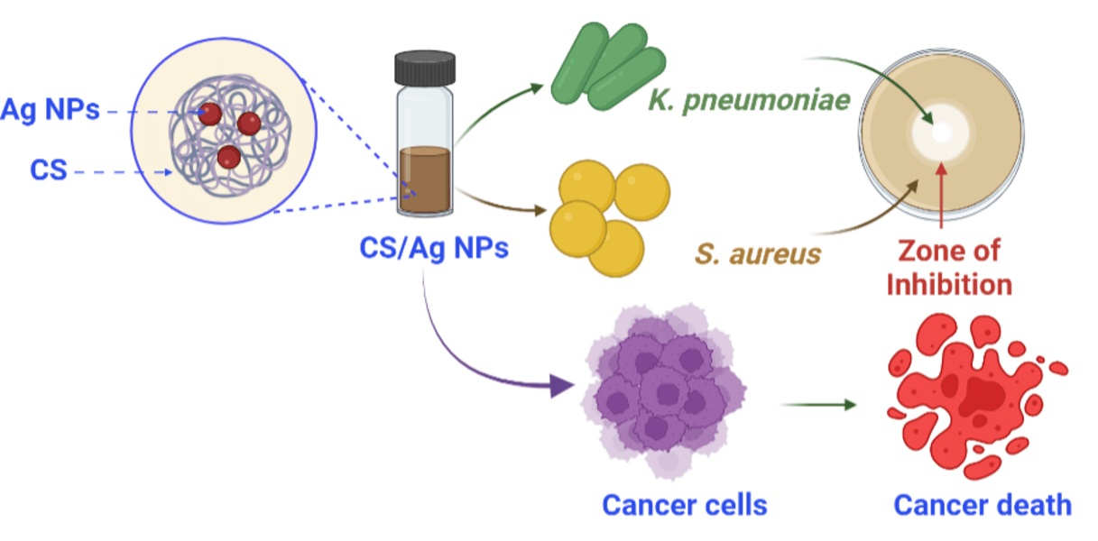
Facile Synthesis and Characterization of Chitosan Functionalized Silver Nanoparticles for Antibacterial and Anti-Lung Cancer Applications

by
Devaraj Bharathi
1,*,
Jaya Ganesh Thiruvengadam Nandagopal
2,
Jintae Lee
1 and
Rajamani Ranjithkumar
3,4,*
1
School of Chemical Engineering, Yeungnam University, Gyeongsan 38541, Gyeongbuk, Republic of Korea
2
Department of Biotechnology, Bharathiar University, Coimbatore 641046, Tamil Nadu, India
3
Viyen Biotech LLP, Coimbatore 641031, Tamil Nadu, India
4
Department of Biotechnology, Sri Ramakrishna College of Arts & Science, Nava India, Coimbatore 641006, Tamil Nadu, India
*
Authors to whom correspondence should be addressed.
Polymers 2023, 15(12), 2700; https://doi.org/10.3390/polym15122700
Submission received: 18 April 2023 / Revised: 12 June 2023 / Accepted: 14 June 2023 / Published: 16 June 2023

Abstract

:
In the treatment of bacterial contamination, the problem of multi-drug resistance is becoming an increasingly pressing concern. Nanotechnology advancements enable the preparation of metal nanoparticles that can be assembled into complex systems to control bacterial and tumor cell growth. The current work investigates the green production of chitosan functionalized silver nanoparticles (CS/Ag NPs) using Sida acuta and their inhibition efficacy against bacterial pathogens and lung cancer cells (A549). Initially, a brown color formation confirmed the synthesis, and the chemical nature of the synthesized NPs were examined by UV-vis spectroscopy, Fourier transform infrared spectroscopy (FTIR), scanning electron microscopy (SEM) coupled with energy dispersive spectroscopy (EDS), and transmission electron microscopy (TEM). FTIR demonstrated the occurrence of CS and S. acuta functional groups in the synthesized CS/Ag NPs. The electron microscopy study exhibited CS/Ag NPs with a spherical morphology and size ranges of 6–45 nm, while XRD analysis demonstrated the crystallinity of Ag NPs. Further, the bacterial inhibition property of CS/Ag NPs was examined against K. pneumoniae and S. aureus, which showed clear inhibition zones at different concentrations. In addition, the antibacterial properties were further confirmed by a fluorescent AO/EtBr staining technique. Furthermore, prepared CS/Ag NPs exhibited a potential anti-cancer character against a human lung cancer cell line (A549). In conclusion, our findings revealed that the produced CS/Ag NPs could be used as an excellent inhibitory material in industrial and clinical sectors.

Graphical Abstract

1. Introduction

Infectious diseases are the leading cause of premature death on a global scale [1,2]. At the same time, cancer is the second leading cause of early death globally [3]. Many infectious illnesses have developed resistance to commercial antibiotics and alternative remedies [4]. The growth of drug resistance has become a major concern for humans and also for pharma industries [5]. Thus, the search for new and efficient inhibitory drugs against microbial infections has intensified. Over the past few decades, nanoparticles (NPs) have demonstrated an ability to eradicate several drug-resistant infections and diseases [6]. Among the many metal and metal oxide NPs, Ag NPs have attracted interest due to their distinctive characteristics [7]. From ancient times, Ag and Ag-based compounds have been recognized for their bactericidal characteristics [8]. Ag NPs have been utilized to prevent microbiological contamination in the textile, cosmetic, and food sectors using current science and technology [9]. There are many physical, chemical, and biological methods available for synthesizing Ag NPs [10,11,12]. During the preparation of Ag NPs by physico-chemical processes, the utilization of harmful reagents, the need for high temperatures, and the emission of toxic byproducts have all had a negative impact on biological applications [13,14].
Hence, an appropriate and green alternative to the chemical and physical route of synthesis is biological methods. Living organisms, such as bacteria, plants, fungi, algae, and even animals, play a vital role in the synthesis of NPs because of some constituent biomolecules, including amino acids, vitamins, phytocompounds, and other secondary metabolites, that can be extracted from them and help in the in-situ reduction of Ag+ ions to form Ag NPs. The primary advantage of biosynthesis is the process’s safety and the purity of resulting NPs. We can be certain that the product synthesized would be free of contamination because only benign substances are utilized to aid the production of Ag NPs using this method. This has a low influence on human health, especially as these NPs are employed for a variety of medicinal applications. Aside from that, biological approaches provide a high production of NPs that are well-defined in shape and homogeneous in size: sometimes even more so than physiochemical procedures. The utilization of plants and their parts as possible factories for the production of Ag NPs is a relatively new and promising field of research [15,16,17]. Potential applications have been identified for the preparation of Ag NPs when utilizing plants such as Platycladus orientalis [18], Mangifera indica [19], Jatropha integerrima [20], Lippia citriodora [21], and Manilkara zapot [22]. Recently, Takci et al. [23] demonstrated the potential bactericidal character of Ag NPs when synthesized using Salvia officinalis against P. aeruginosa, E. coli, S. typhimurium, and S. aureus. In a recent investigation, Sharma et al. [24] revealed that phyto-synthesized Ag NPs utilizing a Callistemon lanceolatus leaf extract exhibited a dosage-dependent bactericidal effect against S. aureus, B. subtilis, M. luteus, E. coli, and K. pneumoniae. Pavan et al. [25] reported that Ag NPs developed using Dictyota ciliolate exhibited a potential anticancer activity against A549 lung cancer cells.
In this study, an aqueous S. acuta Burm.f. leaf extract was used in the production of CS/Ag NPs. S. acuta is a member of the Malvaceae family and is frequently known as wireweed. S. acuta has a long history of use as a medicinal plant, and it has been effective against a variety of illnesses. S. acuta possesses antibacterial, antioxidant, anti-inflammatory, antimalarial, insecticidal, and anti-plasmodial activities, according to pharmacological studies [26,27]. In recent years, the green synthesis of organic-inorganic NPs has been a developing subject in nanoscience and nanotechnology due to its sustainable uses in the biopharmaceutical, food, and clinical sectors [28,29]. Organic-inorganic nano-complexes, such as polymer-coated metal nanoparticles have attracted considerable attention due to their low toxicity, eco-friendliness, simple production procedures, and good surface binding characteristics [30]. Among the different combinations of organic-metal NPs, CS functionalized Ag NPs represent a novel class of nano-complexes with enhanced characteristics, properties, and uses [31]. Chitosan (CS) is a natural polymer that is generated from the deacetylation of chitin and possesses unique features, including biodegradability, biocompatibility, and antibacterial capabilities [32,33]. A study by Wongpreecha et al. [34] reported that one-pot synthesized Ag NPs-CS exhibited effective bactericidal properties against E. coli and S. aureus. Another study by Gobinath et al. [35] documented that biocompatible CS-decorated Ag NPs showed potential anticancer activity against A549 cells.
The green production of Ag NPs has been broadly investigated; however, those for the synthesis of CS-loaded Ag NPs with increased properties and potential applications are still challenging. Thus, the current study was designed to synthesize CS/Ag NPs using S. acuta by an eco-friendly route (Figure 1). Our investigation followed a facile protocol to generate CS/Ag NPs using the leaf extract of S. acuta without extra reagents, solvents, precipitating agents, and templates. Further, CS/Ag NPs were characterized by various physical and chemical techniques, and assessed for their antibacterial activity against K. pneumonia and S. aureus by well diffusion and dual staining fluorescence method. Moreover, the anticancer activity of CS/Ag NPs was investigated against a human lung cancer cell line (A549) by the MTT assay, and its apoptosis induction ability was also analyzed.

2. Experimental Sections

2.1. Chemicals and Cultures

Chitosan (deacetylation degree ≥ 75%, molecular weight; 190–375 kDa), silver nitrate, 3-[4,5-96 dimethylthiazole-2-yl]-2, 5-diphenyltetrazolium bromide (MTT), and Muller-Hinton agar were procured from HiMedia, Mumbai, India. The fresh leaves of S. acuta were obtained from Bharathiar University and authenticated by the Botanical Survey of India (Reference number: BSI/SRC/5/23/2020/651). The bacterial strains (K. pneumoniae and S. aureus) were acquired from a clinical lab in Coimbatore.

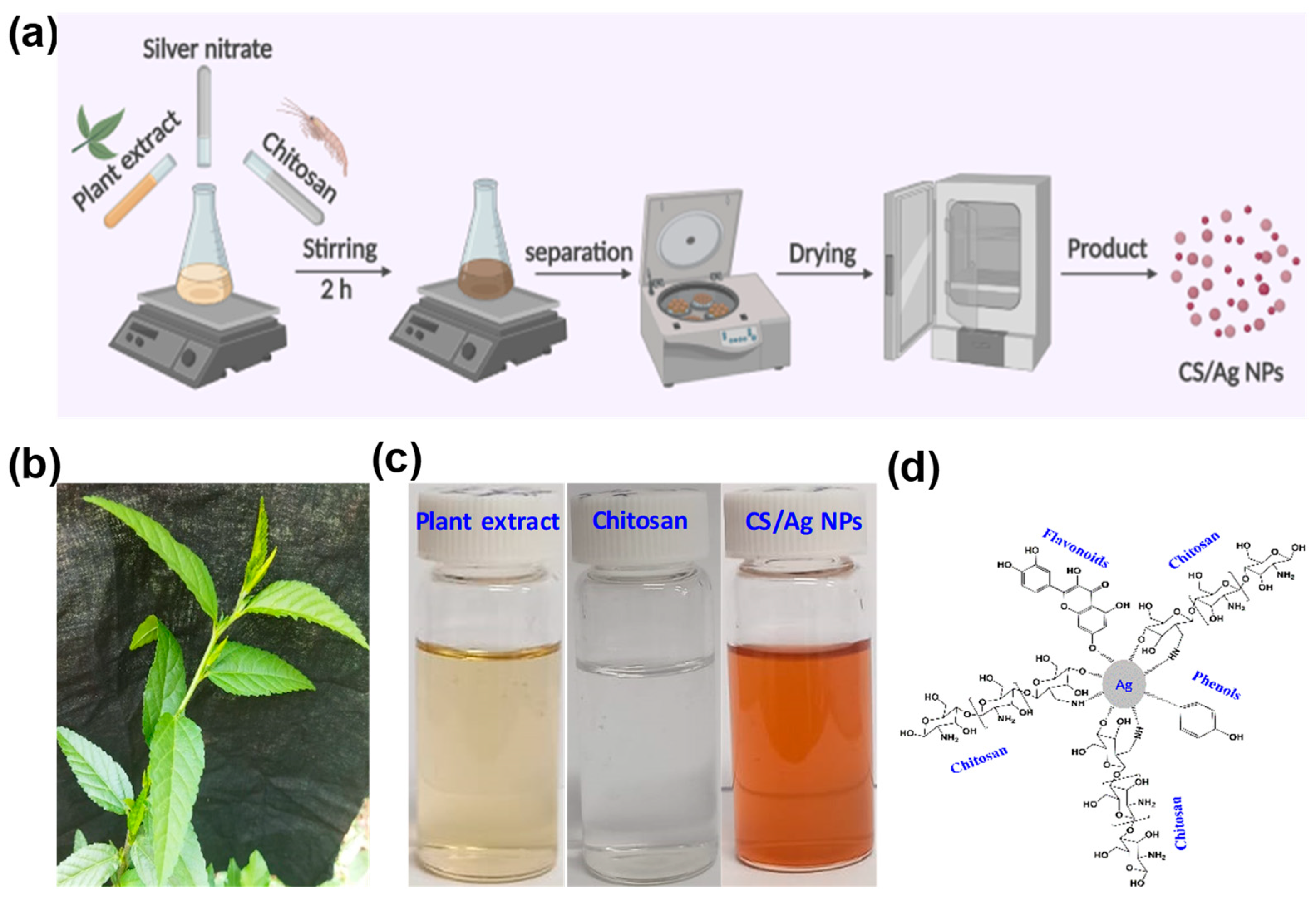
2.2. Extraction of S. acuta Leaf Samples

The harvested leaves were initially cleaned, dried for four days, and ground into a fine powder. A Soxhlet extractor was utilized to obtain an aqueous extract by combining 10 g of fine powder with 100 mL of distilled water. Then, the extracted substance was kept at 4 °C until further usage.

2.3. Synthesis of CS/Ag NPs

The synthesis of CS/Ag NPs was carried out by a green chemistry method, as previously reported by Nandana et al. [30], with some modifications. About 10 mL of the S. acuta extract and 10 mL of CS solution (0.1 g of CS in 10 mL of 1% acetic acid) were mixed with 80 mL of a 1 mM AgNO3 solution, and then the solution was agitated for 2 h at room temperature under a dark state. After this, the resulting colloidal solution was configured at 15,000 rpm for 10 min before being separated by washing with distilled water to remove unbound S. acuta compounds and CS in the synthesized CS/Ag NPs and dried at 80 °C.

2.4. Physico-Chemical Characterization

2.4.1. Optical Studies

UV-vis spectroscopy (JASCO-V-670) was subjected to observe the reaction mixture as it underwent the formation of CS/Ag NPs. The visible spectra of NPs were observed from 300 to 800 nm.

2.4.2. FTIR

The IR analysis was used to identify the molecules of chitosan and S. acuta that were accountable for the formation of NPs. Readings from the FTIR (FTIR-00585, PerkinElmer) spectrum were taken with a resolution of 4 cm−1 across the range of 400–4000 cm−1.

2.4.3. SEM-EDS

The SEM was used to characterize the NPs’ morphology (JEOL-Model JSM 6390 coupled with EDS). In order to prepare a SEM sample on a grid, a pinch of NPs was positioned on the grid then the surfaces of the sample were sputter-coated with carbon tape and then imaged. This experiment was carried out with a voltage of 10 kV applied to the accelerator.

2.4.4. TEM

The structure of synthesized NPs was observed using TEM. A very small pinch of NPs was placed on a grid, and NPs images were observed at 120 kV (JEOL-Japan).

2.5. Disc Diffusion Antibacterial Activity Assay

The bactericidal activity of CS/Ag NPs was assessed against K. pneumoniae and S. aureus using a disc diffusion assay [36]. Briefly, the chosen bacterial strains (105 CFU/mL) were spreaded on the MHA plates. Then, the empty discs were filled with various concentrations of CS/Ag NPs (10, 20 and 30 µg/mL) and allowed to dry for a few minutes. Then, coated discs were positioned on the dish and kept at 37 °C for 24 h to observe the activity.

2.6. AO/EtBr Dual Staining and Growth Curve Assay

The changes in the K. pneumonia and S. aureus cell membrane upon action with NPs were examined using an AO/EtBr staining technique, as described by Kumar et al. [37]. Briefly, 1 mL of overnight-grown pathogens were incubated with 20 µg/mL of NPs for 24 h at 37 °C. The cells without NPs were kept as a control. Then, the cells were cleaned with a buffer solution and further stained with 1 µL of AO/EtBr, and images of fluorochemical staining were attained using a Nikon Eclipse Ni-E Microscope with a Nikon DS-Ri2 digital camera. The digitized images were processed using Nikon’s proprietary software NIS-Elements BR.

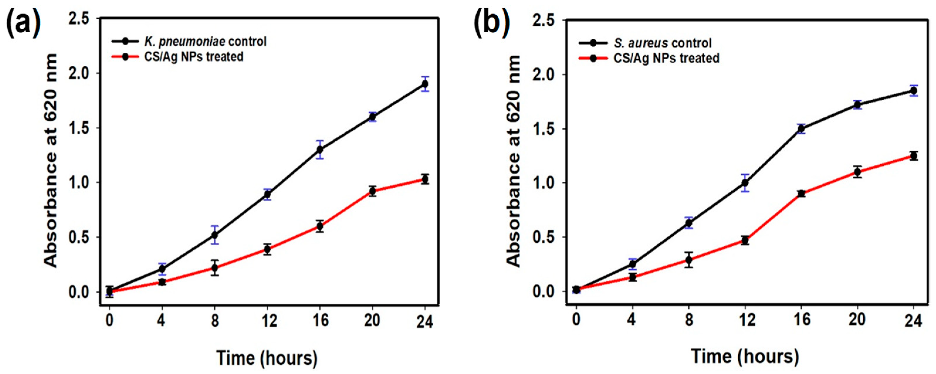
2.7. Growth Curve Analysis

The growth curve analysis of synthesized CS/Ag NPs was tested against the chosen pathogens (K. pneumoniae and S. aureus) [38]. Briefly, overnight cultures of K. pneumoniae and S. aureus were incubated with 20 µg/mL of CS/Ag NPs, and the O.D. was monitored at every 4 h interval up to 24 h.

2.8. Anticancer MTT Assay against A549 Cells

Human lung cancer cells were obtained from the National Centre for Cell Science (NCCS), Pune, India. Initially, the monolayer cell culture was trypsinized, and the cell amount was attuned to 1 × 10⁴ cells/mL with 10% FBS medium. About 0.2 mL of the diluted cell suspension was added to each well of the 96-micro titer plate. When a partial monolayer formed after 24 h, the supernatant was flicked off, and the monolayer was washed once with a buffer. Different concentrations of CS/Ag NPs (20, 40, 60, 80, and 100 μg/mL) being diluted in media were added and incubated for 24 h to determine the effect of CS/Ag NPs on cell viability. After that 10 µL of MTT (5 mg/mL) was added and further incubated for 4 h, then removed, and a further 200 µL of DMSO was added and thoroughly mixed to dissolve the dark blue crystals. Then, the absorbance was measured at 570 nm, and the percentage of viability was calculated using Formula (1).
Cell viability (%) = O.D. of test/O.D. of control × 100

2.9. Apoptosis Induction Studies

Apoptosis causing ability of synthesized CS/Ag NPs was studied using a fluorescence microscopic dual staining method as previously described by Bharathi et al. [39]. About 300 μL of lung cancer cells were treated with the IC50 concentration of CS/Ag NPs in a 16-well plate and incubated for 24 h. After that, the treated and untreated cells were washed with PBS and stained with 10 μL of an AO/EtBr mixture before the morphology changes were imaged using a fluorescence microscope (Nikon Eclipse Ni-E Microscope with a Nikon DS-Ri2 digital camera).

2.10. Statistical Analysis

The experimental investigation, including testing for antibacterial and anticancer activities, was performed in triplicates, and the findings were reported as the mean ± standard deviation. The statistical significance of the differences were determined using a p-value of 0.05.

3. Results and Discussion

3.1. Formation of CS/Ag NPs

The green production of CS/Ag NPs was carried out using S. acuta leaves extract (Figure 1b). Previously, Uysal et al. [27] reported that S. acuta contained various phytochemical properties such as hydroxybenzoic, hydroxycinnamic and acylquinic acids, hydroxycinnamoyl tartarates, flavonoids, cinnamic acid amides, alkaloids, and amino acids. The presence of these phytocompounds in S. acuta and CS might have acted as reducing and capping agents for the synthesis of CS/Ag NPs [39]. The development of NPs was established by the color change from yellow to brown (Figure 1c). The development of this brown color was well known to be emerged from SPR vibrations of nano-CS/Ag [40]. Similarly, various phytocompounds of Myristica fragrans [41], Muntingia calabura [42], and Rumex nervosus [43] mediated and synthesized Ag NPs and exhibited brown color formation. The mechanism of this chain network formation by CS and phyto-compounds with Ag is shown in Figure 1d. The active phyto-compounds presented in S. acuta and CS acted as ligation agents. The existence of long pair of e, free -NH2, and O-H groups in CS and phytocompounds of S. acuta might have ligated with Ag2+ and developed an Ag–CS–ellagate complex. Then, this network system naturally led to a nucleation process that went into reverse micellization, which further rooted the reduction of Ag ions (Ag+) to nano Ag [30,44,45].

3.2. Characterization Studies

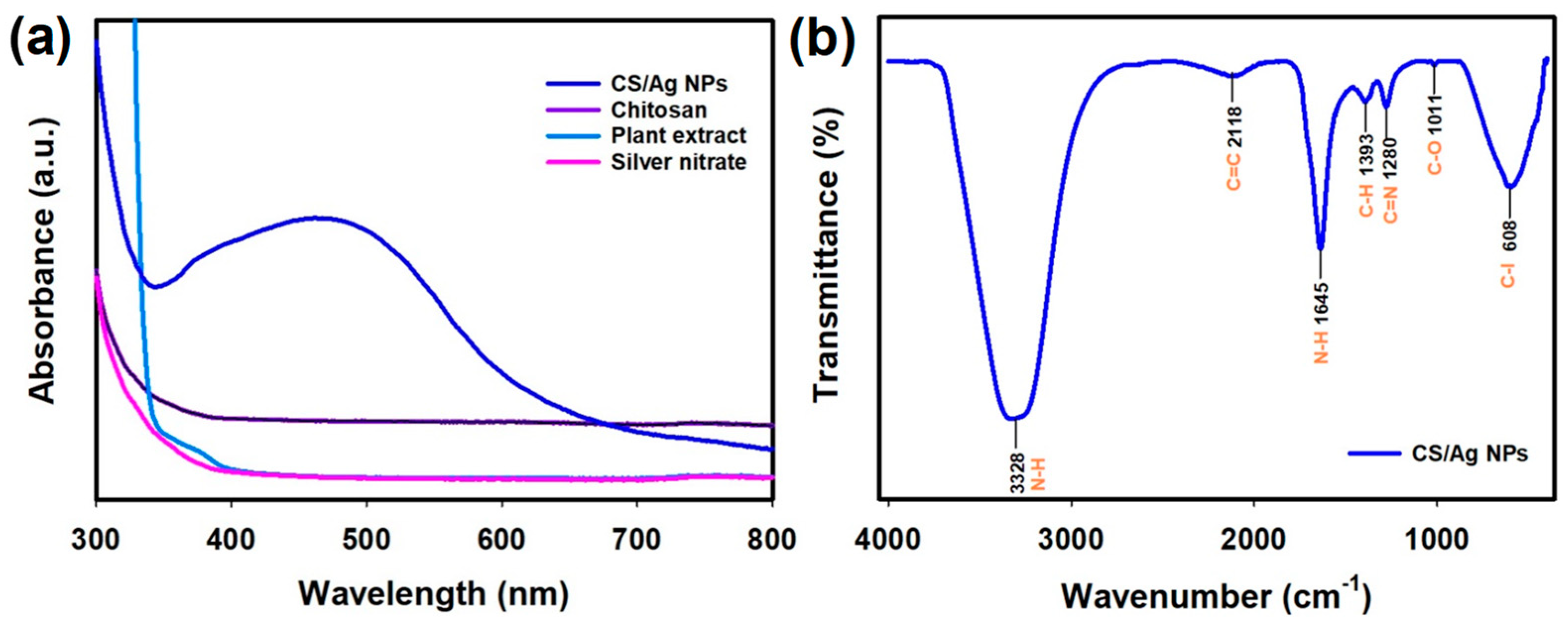
3.2.1. Optical Studies

The UV-vis spectroscopy investigation of CS/Ag NPs exhibited an absorbance peak at 464 nm and thus supported the synthesis of CS/Ag NPs (Figure 2a). According to reports, the UV absorbance peak around 400–480 nm was a distinct property of nano-Ag [46]. Similarly, CS-Aegle marmelos entrapped Ag NPs exhibited UV-vis absorbance at 420 nm [47]. The UV-visible spectrum of CS-decorated Ag NPs was prepared using Piper betle exhibited an absorption peak at 430 nm [35]. Recently, Ag NPs were synthesized using CS, and seaweed showed the UV-vis absorbance peak to be around 425 nm [48].

3.2.2. FTIR-Functional Groups Investigation

The attraction of active groups and the development of end product CS/Ag NPs were studied using FTIR. The IR analysis of CS/Ag NPs is depicted in Figure 2b. The FTIR of NPs showed a typical primary N-H band at 3328 cm−1, C=C vibration at 2118 cm−1, N-H band at 1645 cm−1, C-H bending at 1393 cm−1, C-N band at 1280 cm−1, C-O stretching at 1011 cm−1 and a C-I band at 608 cm−1. The obtained N-H and C-O functional groups could be derived from -NH2 groups and acetylated parts of CS [49]. Other groups, namely C=C, C-H, C-N, and C-I, may be derived from the phyto-extract of S. acuta. Moreover, certain band vibrations around 600–400 cm−1 could be accredited to the presence of metal (Ag) in the synthesized CS/Ag NPs. The presence of these bio-active derivatives might have acted as a reductant for the formation of final CS/Ag NPs [36].

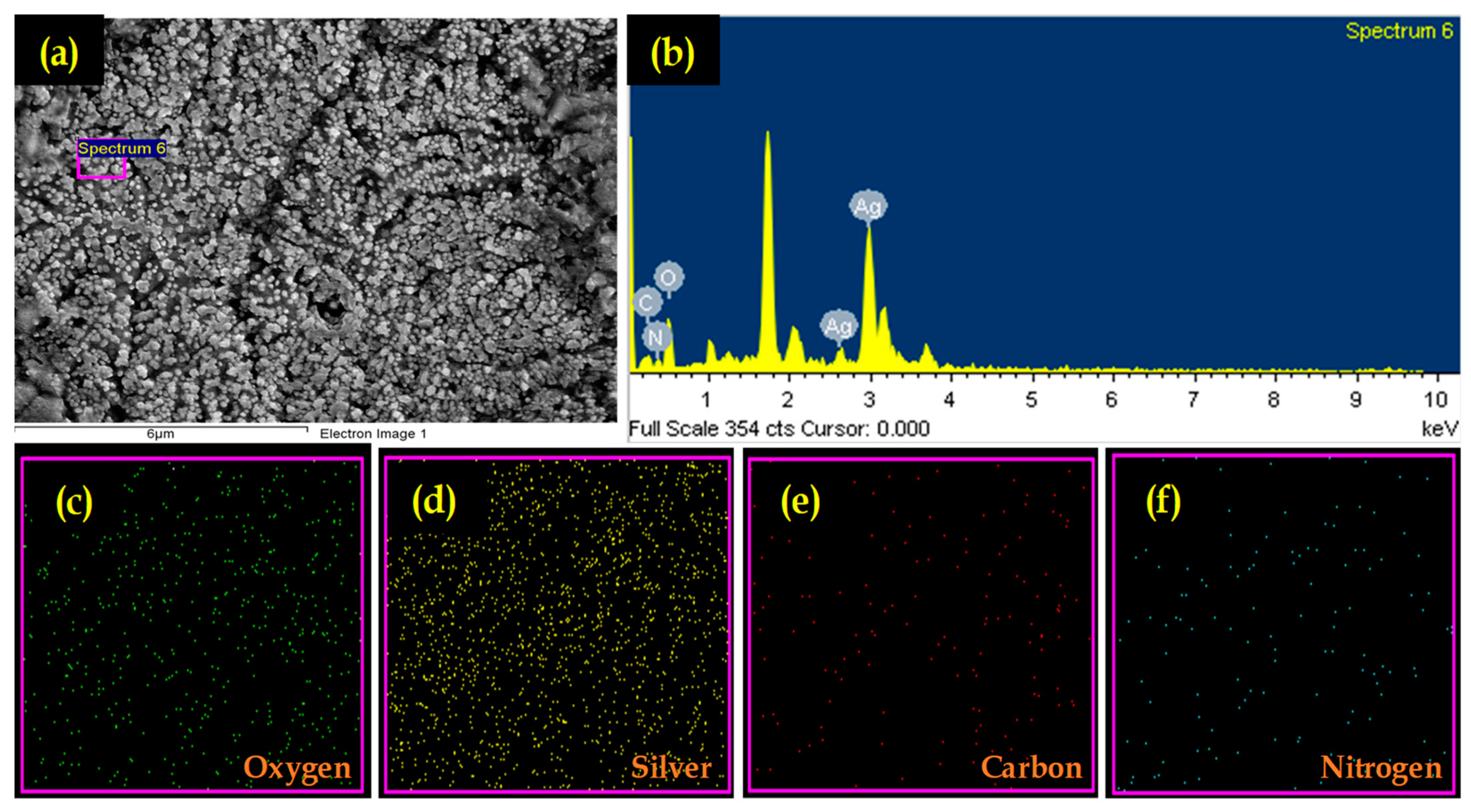
3.2.3. Microscopic and EDS Analysis
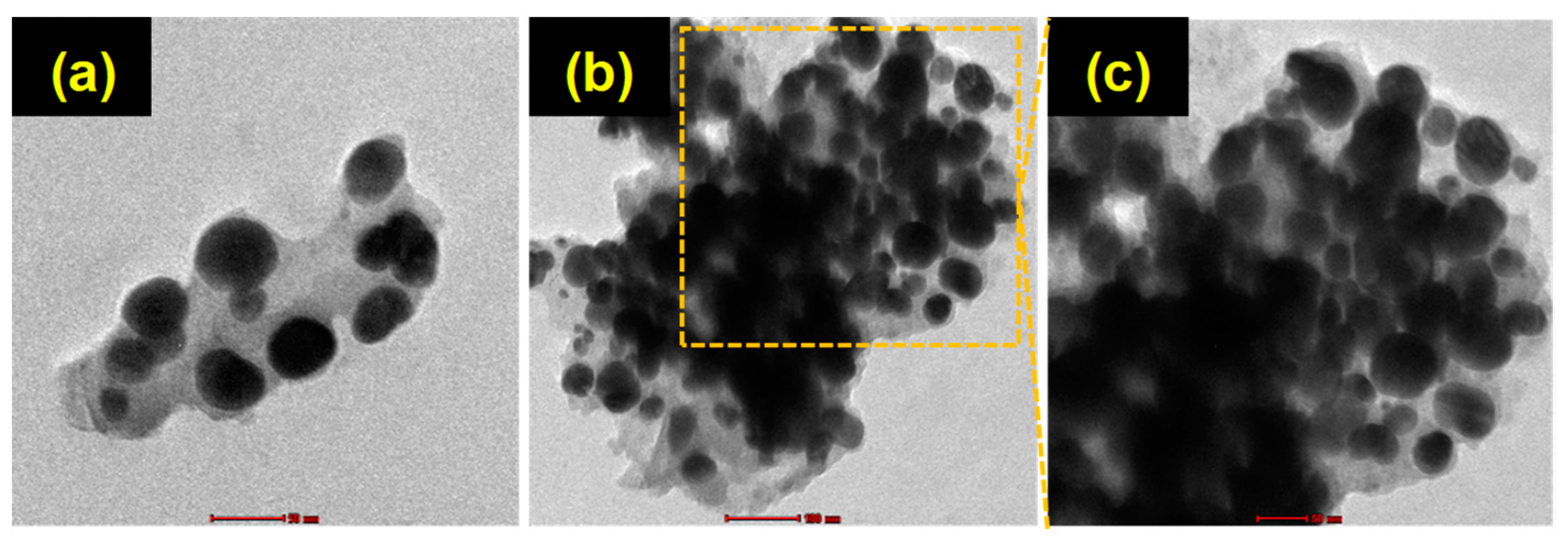
Figure 3a shows the FE-SEM of CS/Ag NPs. FE-SEM images showed that the NPs had a spherical morphology. Ag NPs were synthesized using various plant extracts, which showed a spherical-shaped morphology [50,51]. Further, EDS was performed to analyze the elemental composition of CS/Ag NPs (Figure 3b). The elemental spectra of CS/Ag NPs exhibited a peak of silver (Ag), which supported the formation of Ag NPs. The other peaks of C, N, and O were detected due to the emission of X-rays from the -NH2 and de-acetylated groups of CS. The elemental percentage in the synthesized CS/Ag NPs is given in Table 1. Similar to our study, Nandhana et al. reported on the presence of Ag, N, C, and O in the green synthesized CS/Ag nanocomposite using rutin. Further, elemental mapping analysis confirmed the random distribution of observed elements in the synthesized NPs (Figure 3e,f).

3.2.4. TEM Analysis

Further, the morphology and size of produced NPs were analyzed using TEM. The TEM images of synthesized CS/Ag NPs are shown in Figure 4a–c. TEM showed the particle sizes varied from 6 to 45 nm, and it also showed the synthesized NPs to have a spherical morphology with Ag crystallinity. Similar to our study, gallic acid-chitosan-modified Ag NPs exhibited a spherical-shaped morphology [52].

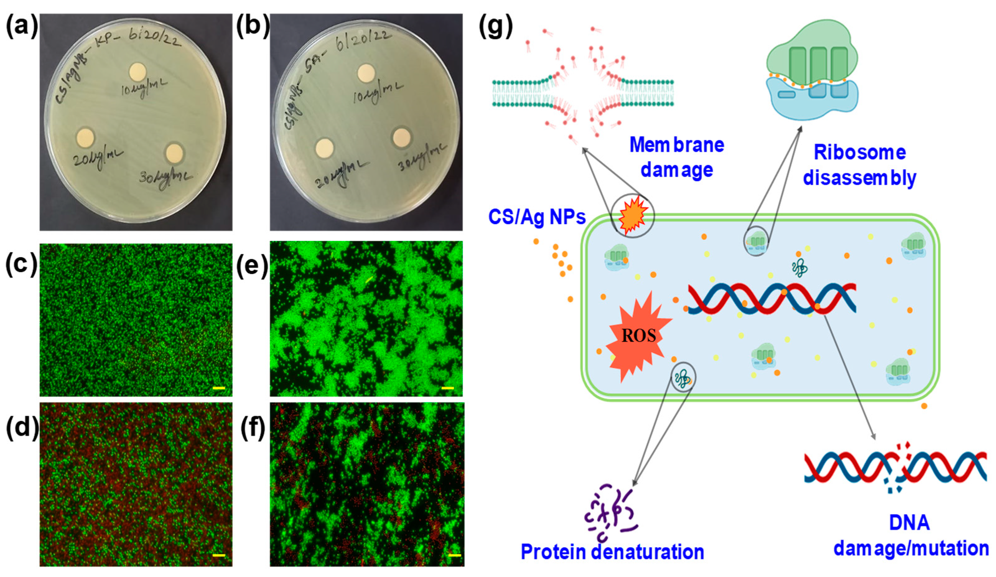
3.3. Disc Diffusion Bactericidal Assay

The antibacterial properties of CS/Ag NPs were investigated against K. pneumoniae and S. aureus. The synthesized NPs inhibited potential and concentration-dependent inhibitory activities against the tested bacterial pathogens. Figure 5a,b depicts a well diffusion plate experiment with various concentrations (10, 20, and 30 µg/mL) of CS/Ag NPs. Against all the pathogens examined, clear zones of bacterial suppression appeared around the CS/Ag NPs embedded discs. At an increasing concentration of NPs, the bactericidal ZOI increased. The ZOI for K. pneumoniae was found to be 9 ± 0.5 mm for 10 µg/mL, 11 ± 0.8 mm for 20 µg/mL and 13 ± 0.2 for 30 µg/mL of CS/Ag NPs, whereas S. aureus exhibited 10 ± 0.2 mm for 10 µg/mL, 11 ± 0.6 mm for 20 µg/mL, and 13 ± 1 mm for 30 µg/mL. Synthesized NPs exhibited equal activity for both tested pathogens. The results of NPs’ action could change depending on the bacteria’s cell wall and membrane structure [36]. Moreover, nano-scale CS possessed a significant bactericidal and broad-range antibacterial action against bacterial infections [53]. Similar to our study, CS-Ag NPs coated linen fabrics showed their potential antibacterial activity against E. coli and S. aureus [54].

3.4. Dual Fluorescent Staining

Furthermore, the antibacterial efficacy of produced CS/Ag NPs against K. pneumoniae and S. aureus was validated by utilizing a fluorescent-based live/dead cell test. The fluorescent microscopic pictures of the control and NPs treated cells are shown in Figure 5c–f. The untreated cells fluoresced green due to the existence of live cells, whereas treated cells fluoresced red, confirming their death. Fluorescent AO is a green dye that stains both live and dead bacteria, whereas EtBr is a red dye that exclusively stains dead bacteria. The EtBr penetrates dead cells via the cell membrane and reduces the green fluorescent color of AO [55]. Accordingly, the green color denotes living bacterial cells, whereas the red hue signifies dead bacterial cells. The obtained results revealed that produced NPs had a bacteriostatic impact against the tested bacterial pathogens. Further, the growth curve analysis revealed that CS/Ag NPs were able to prevent the growth of K. pneumoniae and S. aureus and also showed delayed growth (Figure 6).
A potential mechanism for the bacterial suppression of CS/Ag NPs is shown in Figure 5g. One of the two fundamental processes that may be responsible for the inhibition action of Ag-based NPs is the breakdown of cell walls and membrane destruction (1). The attraction and interaction between NPs and bacteria begin with Ag adhesion to the plasma membrane, which causes membrane structural alterations, resulting in membrane depolarization, disruption of permeability, and disruption of cell wall integrity. As a consequence of depolarization, the internal material of bacterial cells escapes into the surrounding environment, leading to the death of the cells. The second approach involves the creation of reactive oxygen species (ROS), which can include superoxide, hydroxyl radicals, and hydrogen peroxides, among other things. These limit the development of bacteria by binding to the genetic elements and proteins that are found on their cells [56,57,58].

3.5. Anticancer Studies
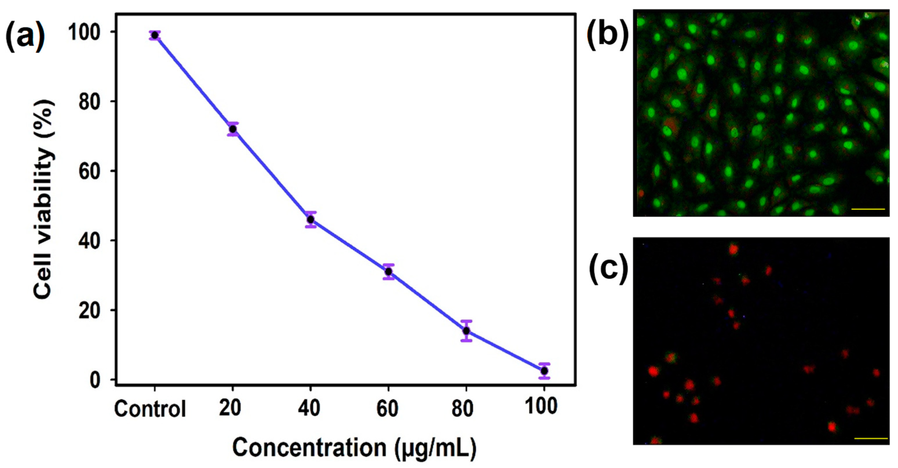
3.5.1. MTT Assay

The anticancer property of CS/Ag NPs was assessed against human lung cancer cells using a MTT assay. The obtained findings exhibited a significant decrease in the growth of A549 cells (97.2%) when the concentration of CS/Ag NPs increased. CS/Ag NPs induced a 50% growth defeat property at a concentration of 34.5 ± 0.5 µg/mL. Similarly, Murugesan et al. [47] reported that the synthesized Ag NPs using Gloriosa superba exhibited potential anticancer activity against A549 cells. Similar findings were reported by Priya et al. [59], who observed the dose-dependent anticancer activity of biogenic CS-Ag NPs against hepatocellular carcinoma cells. The biogenic synthesized Ag NPs entrapped with CS showed anticancer activity against HeLa cells [47]. It was reported that the anticancer properties of Ag NPs were dependent on the morphology, size, and reducing agents of the NPs.

3.5.2. AO/EtBr Fluorescent Assay

Nearly all unicellular creatures undergo apoptosis, which is a type of planned cell death. A helpful strategy for the therapy of cancer is the induction of apoptosis [60]. Using a fluorescence microscope, the apoptosis cells were separated from one another by their orange or red-colored bodies. Fluorescence microscope images revealed that the prepared CS/Ag NPs could induce apoptosis in treated A549 cells. The control cells displayed a green color, and the treated cells showed a red color, thus supporting the induction property of CS/Ag NPs (Figure 7a,b). The appearance of a red hue indicated the presence of apoptotic bodies and displayed cell shrinkage as well as membrane blebbing. In most cases, the morphological and biochemical alterations in such cell shrinkage, membrane blebbing, membrane unity, nuclear fragmentation, and cytoplasmic condensation contributed to cell death by initiating the apoptotic pathway. This process was responsible for the death of cells. [61,62]. The apoptosis induction property of NPs depended on the permeability of Ag ions into the cancer cells, which stimulated cell damage, and DNA fragmentation and, thus spontaneously led to apoptosis [63].

4. Conclusions

Silver is one of the most often utilized metals that is accessible and has a diverse variety of applications. In biomedical applications, the nanoform of Ag, also known as Ag NPs, has provided a novel structure for particles. The synthesis of Ag NPs using chemical methods is both expensive and harmful; thus, efforts have been made to utilize biological sources such as plants, bacteria, and algae in order to cut down on both costs and toxicity. In the present investigation, S. acuta performed outstandingly both as a reducing and capping agent. The production of CS/Ag NPs using the S. acuta aqueous leaf extract has been presented as an eco-friendly and simple green technique. To our knowledge, this is the first report on the synthesis of CS/Ag NPs using S. acuta without any additional chemicals and reagents. The size of the prepared CS/Ag NPs ranged from 6 to 45 nm and had a spherical form. The occurrence of CS in prepared CS/Ag NPs was validated by EDS and FTIR analyses. Significant antibacterial activity was exhibited by the synthesized CS-Ag NPs against K. pneumonia and S. aureus pathogens. In addition, the MTT and fluorescent-based assay confirmed its anti-cancer capabilities with the apoptosis induction property of synthesized CS/Ag NPs against the human lung cancer cell line (A549). The potential antibacterial and anticancer properties of CS/Ag NPs could be due to the synergetic properties of both CS and Ag NPs. Thus, we believe that the CS/Ag NPs synthesized using S. acuta have the potential to be employed as NPs in clinical sectors to reduce the growth of bacterial and also cancer cells.

Author Contributions

Conceptualization, R.R. and D.B.; methodology, D.B.; analysis, D.B. and J.L.; writing—original draft preparation, D.B. and J.G.T.N.; visualization, D.B. and R.R.; review and editing, D.B. and J.G.T.N.; supervision, D.B. and R.R.; project administration, D.B. and R.R. All authors have read and agreed to the published version of the manuscript.

Funding

This research received no external funding.

Institutional Review Board Statement

Not applicable.

Data Availability Statement

Not applicable.

Acknowledgments

The authors would like to thank Bharathiar University and Viyen Biotech LLP, Coimbatore, for providing the necessary instrumentation facilities.

Conflicts of Interest

The authors declare no conflict of interest.

References

  1. Zhang, Y.; Hao, M.; Li, L.; Luo, Q.; Deng, S.; Yang, Y.; Liu, Y.; Fang, W.; Song, E. Research Progress of Contrast Agents for Bacterial Infection Imaging in Vivo. TrAC-Trends Anal. Chem. 2023, 159, 116916. [Google Scholar] [CrossRef]
  2. Tsikourkitoudi, V.; Henriques-Normark, B.; Sotiriou, G.A. Inorganic Nanoparticle Engineering against Bacterial Infections. Curr. Opin. Chem. Eng. 2022, 38, 100872. [Google Scholar] [CrossRef]
  3. Bharathi, D.; Rajamani, R.; Sibuh, B.Z.; Pandit, S.; Agrawal, S.; Mishra, N.; Sahni, M.; Thakur, V.K.; Gupta, P.K. Biogenic Preparation, Characterization, and Biomedical Applications of Chitosan Functionilized Iron Oxide Nanocomposite. J. Compo. Sci. 2022, 6, 120. [Google Scholar] [CrossRef]
  4. Mishra, A.; Pradhan, D.; Halder, J.; Biswasroy, P.; Rai, V.K.; Dubey, D.; Kar, B.; Ghosh, G.; Rath, G. Metal Nanoparticles against Multi-Drug-Resistance Bacteria. J. Inorg. Biochem. 2022, 237, 111938. [Google Scholar] [CrossRef]
  5. Xuan, J.; Feng, W.; Wang, J.; Wang, R.; Zhang, B.; Bo, L.; Chen, Z.-S.; Yang, H.; Sun, L. Antimicrobial Peptides for Combating Drug-Resistant Bacterial Infections. Drug Resist. Updat. 2023, 68, 100954. [Google Scholar] [CrossRef]
  6. Xu, Y.; Li, H.; Li, X.; Liu, W. What Happens When Nanoparticles Encounter Bacterial Antibiotic Resistance? Sci. Total Environ. 2023, 876, 162856. [Google Scholar] [CrossRef]
  7. Anees Ahmad, S.; Sachi Das, S.; Khatoon, A.; Tahir Ansari, M.; Afzal, M.; Saquib Hasnain, M.; Kumar Nayak, A. Bactericidal Activity of Silver Nanoparticles: A Mechanistic Review. Mater. Sci. Energy Technol. 2020, 3, 756–769. [Google Scholar] [CrossRef]
  8. Malik, M.; Aamir Iqbal, M.; Iqbal, Y.; Malik, M.; Bakhsh, S.; Irfan, S.; Ahmad, R.; Pham, P.V. Biosynthesis of Silver Nanoparticles for Biomedical Applications: A Mini Review. Inorg. Chem. Commun. 2022, 145, 109980. [Google Scholar] [CrossRef]
  9. Tharani, S.; Bharathi, D.; Ranjithkumar, R. Extracellular Green Synthesis of Chitosan-Silver Nanoparticles Using Lactobacillus Reuteri for Antibacterial Applications. Biocatal. Agric. Biotechnol. 2020, 30, 101838. [Google Scholar] [CrossRef]
  10. Nie, P.; Zhao, Y.; Xu, H. Synthesis, Applications, Toxicity and Toxicity Mechanisms of Silver Nanoparticles: A Review. Ecotoxicol. Environ. Saf. 2023, 253, 114636. [Google Scholar] [CrossRef]
  11. Hossain, N.; Islam, M.A.; Chowdhury, M.A. Synthesis and Characterization of Plant Extracted Silver Nanoparticles and Advances in Dental Implant Applications. Heliyon 2022, 8, e12313. [Google Scholar] [CrossRef]
  12. Islam, M.A.; Jacob, M.V.; Antunes, E. A Critical Review on Silver Nanoparticles: From Synthesis and Applications to Its Mitigation through Low-Cost Adsorption by Biochar. J. Environ. Manage. 2021, 281, 111918. [Google Scholar] [CrossRef]
  13. Bharathi, D.; Josebin, M.D.; Vasantharaj, S.; Bhuvaneshwari, V. Biosynthesis of Silver Nanoparticles Using Stem Bark Extracts of Diospyros Montana and Their Antioxidant and Antibacterial Activities. J. Nanostructure Chem. 2018. [Google Scholar] [CrossRef] [Green Version]
  14. Seerangaraj, V.; Sathiyavimal, S.; Shankar, S.N.; Nandagopal, J.G.T.; Balashanmugam, P.; Al-Misned, F.A.; Shanmugavel, M.; Senthilkumar, P.; Pugazhendhi, A. Cytotoxic Effects of Silver Nanoparticles on Ruellia Tuberosa: Photocatalytic Degradation Properties against Crystal Violet and Coomassie Brilliant Blue. J. Environ. Chem. Eng. 2021, 9, 105088. [Google Scholar] [CrossRef]
  15. Ahmed, A.; Usman, M.; Ji, Z.; Rafiq, M.; Yu, B.; Shen, Y.; Cong, H. Nature-Inspired Biogenic Synthesis of Silver Nanoparticles for Antibacterial Applications. Mater. Today Chem. 2023, 27. [Google Scholar] [CrossRef]
  16. Heinemann, M.G.; Rosa, C.H.; Rosa, G.R.; Dias, D. Biogenic Synthesis of Gold and Silver Nanoparticles Used in Environmental Applications: A Review. Trends Environ. Anal. Chem. 2021, 30. [Google Scholar] [CrossRef]
  17. Ashique, S.; Upadhyay, A.; Hussain, A.; Bag, S.; Chaterjee, D.; Rihan, M.; Mishra, N.; Bhatt, S.; Puri, V.; Sharma, A.; et al. Green Biogenic Silver Nanoparticles, Therapeutic Uses, Recent Advances, Risk Assessment, Challenges, and Future Perspectives. J. Drug Deliv. Sci. Technol. 2022, 77, 103876. [Google Scholar] [CrossRef]
  18. Anshiba, J.; Poonkothai, M.; Karthika, P.; Mythili, R. Green Route to a Novel and Ecofriendly Phytosynthesis of Silver Nanoparticles Using Platycladus Orientalis L. Leaf Extract. Mater. Lett. 2022, 309, 131347. [Google Scholar] [CrossRef]
  19. Hai, N.D.; Dat, N.M.; Huong, L.M.; Tai, L.T.; Thinh, D.B.; Nam, N.T.H.; Dat, N.T.; Phong, M.T.; Hieu, N.H. Phytosynthesis of Silver Nanoparticles Using Mangifera Indica Leaves Extract at Room Temperature: Formation Mechanism, Catalytic Reduction, Colorimetric Sensing, and Antimicrobial Activity. Colloids Surfaces B Biointerfaces 2022, 220, 112974. [Google Scholar] [CrossRef]
  20. Suriyakala, G.; Sathiyaraj, S.; Devanesan, S.; AlSalhi, M.S.; Rajasekar, A.; Maruthamuthu, M.K.; Babujanarthanam, R. Phytosynthesis of Silver Nanoparticles from Jatropha Integerrima Jacq. Flower Extract and Their Possible Applications as Antibacterial and Antioxidant Agent. Saudi J. Biol. Sci. 2022, 29, 680–688. [Google Scholar] [CrossRef]
  21. Elemike, E.E.; Onwudiwe, D.C.; Ekennia, A.C.; Ehiri, R.C.; Nnaji, N.J. Phytosynthesis of Silver Nanoparticles Using Aqueous Leaf Extracts of Lippia Citriodora: Antimicrobial, Larvicidal and Photocatalytic Evaluations. Mater. Sci. Eng. C 2017, 75, 980–989. [Google Scholar] [CrossRef] [PubMed]
  22. Otari, S.V.; Patil, R.M.; Ghosh, S.J.; Pawar, S.H. Green Phytosynthesis of Silver Nanoparticles Using Aqueous Extract of Manilkara Zapota (L.) Seeds and Its Inhibitory Action against Candida Species. Mater. Lett. 2014, 116, 367–369. [Google Scholar] [CrossRef]
  23. Takcı, D.K.; Ozdenefe, M.S.; Genc, S. Green Synthesis of Silver Nanoparticles with an Antibacterial Activity Using Salvia Officinalis Aqueous Extract. J. Cryst. Growth 2023, 614, 127239. [Google Scholar] [CrossRef]
  24. Sharma, K.; Guleria, S.; Hussain, K.; Majeed, A.; Sharma, N.; Pawar, K.D.; Kumar, V.; Kumar, V. Photocatalytic and Biological Properties of Silver Nanoparticles Synthesized Using Callistemon Lanceolatus Leaf Extract. Ind. Crop. Prod. 2023, 202, 116951. [Google Scholar] [CrossRef]
  25. Pavan, S.R.; Venkatesan, J.; Prabhu, A. Anticancer Activity of Silver Nanoparticles from the Aqueous Extract of Dictyota Ciliolata on Non-Small Cell Lung Cancer Cells. J. Drug Deliv. Sci. Technol. 2022, 74, 103525. [Google Scholar] [CrossRef]
  26. Kamdoum, B.C.; Simo, I.; Wouamba, S.C.N.; Tchatat Tali, B.M.; Ngameni, B.; Fotso, G.W.; Ambassa, P.; Fabrice, F.B.; Lenta, B.N.; Sewald, N.; et al. Chemical Constituents of Two Cameroonian Medicinal Plants: Sida Rhombifolia L. and Sida Acuta Burm. f. (Malvaceae) and Their Antiplasmodial Activity. Nat. Prod. Res. 2022, 36, 5311–5318. [Google Scholar] [CrossRef] [PubMed]
  27. Uysal, S.; Gevrenova, R.; Sinan, K.I.; Bayarslan, A.U.; Altunoglu, Y.C.; Zheleva-Dimitrova, D.; Ak, G.; Baloglu, M.C.; Etienne, O.K.; Lobine, D.; et al. New Perspectives into the Chemical Characterization of Sida Acuta Burm. f. Extracts with Respect to Its Anti-Cancer, Antioxidant and Enzyme Inhibitory Effects. Process Biochem. 2021, 105, 91–101. [Google Scholar] [CrossRef]
  28. Liu, K.-G.; Bigdeli, F.; Sharifzadeh, Z.; Gholizadeh, S.; Morsali, A. Role of Metal-Organic Framework Composites in Removal of Inorganic Toxic Contaminants. J. Clean. Prod. 2023, 404, 136709. [Google Scholar] [CrossRef]
  29. Niu, B.; Chen, Y.; Zhang, L.; Tan, J. Organic-Inorganic Hybrid Nanomaterials Prepared via Polymerization-Induced Self-Assembly: Recent Developments and Future Opportunities. Polym. Chem. 2022, 2554–2569. [Google Scholar] [CrossRef]
  30. Nandana, C.N.; Christeena, M.; Bharathi, D. Synthesis and Characterization of Chitosan/Silver Nanocomposite Using Rutin for Antibacterial, Antioxidant and Photocatalytic Applications. J. Clust. Sci. 2022, 33, 269–279. [Google Scholar] [CrossRef]
  31. Ramachandran, G.; Alharbi, N.S.; Chackaravarthy, G.; Kanisha Chelliah, C.; Rajivgandhi, G.; Maruthupandy, M.; Quero, F.; Natesan, M.; Li, W.J. Chitosan/Silver Nanocomposites Enhanced the Biofilm Eradication in Biofilm Forming Gram Positive S. Aureus. J. King Saud Univ.-Sci. 2023, 35, 102597. [Google Scholar] [CrossRef]
  32. Saberi Riseh, R.; Vatankhah, M.; Hassanisaadi, M.; Kennedy, J.F. Chitosan-Based Nanocomposites as Coatings and Packaging Materials for the Postharvest Improvement of Agricultural Product: A Review. Carbohydr. Polym. 2023, 309, 120666. [Google Scholar] [CrossRef] [PubMed]
  33. Chen, Y.; Liu, Y.; Dong, Q.; Xu, C.; Deng, S.; Kang, Y.; Fan, M.; Li, L. Application of Functionalized Chitosan in Food: A Review. Int. J. Biol. Macromol. 2023, 235. [Google Scholar] [CrossRef] [PubMed]
  34. Wongpreecha, J.; Polpanich, D.; Suteewong, T.; Kaewsaneha, C.; Tangboriboonrat, P. One-Pot, Large-Scale Green Synthesis of Silver Nanoparticles-Chitosan with Enhanced Antibacterial Activity and Low Cytotoxicity. Carbohydr. Polym. 2018, 199, 641–648. [Google Scholar] [CrossRef] [PubMed]
  35. Gopinath, V.; MubarakAli, D.; Vadivelu, J.; Manjunath Kamath, S.; Syed, A.; Elgorban, A.M. Synthesis of Biocompatible Chitosan Decorated Silver Nanoparticles Biocomposites for Enhanced Antimicrobial and Anticancer Property. Process Biochem. 2020, 99, 348–356. [Google Scholar] [CrossRef]
  36. Sathiyavimal, S.; Vasantharaj, S.; Bharathi, D.; Saravanan, M.; Manikandan, E.; Kumar, S.S.; Pugazhendhi, A. Biogenesis of Copper Oxide Nanoparticles (CuONPs) Using Sida Acuta and Their Incorporation over Cotton Fabrics to Prevent the Pathogenicity of Gram Negative and Gram Positive Bacteria. J. Photochem. Photobiol. B Biol. 2018. [Google Scholar] [CrossRef]
  37. Kumar, S.S.D.; Houreld, N.N.; Kroukamp, E.M.; Abrahamse, H. Cellular Imaging and Bactericidal Mechanism of Green-Synthesized Silver Nanoparticles against Human Pathogenic Bacteria. J. Photochem. Photobiol. B Biol. 2018, 178, 259–269. [Google Scholar] [CrossRef]
  38. Sathiskumar, S.; Vanaraj, S.; Sabarinathan, D.; Preethi, K. Evaluation of Antibacterial and Antibiofilm Activity of Synthesized Zinc-Hydroxyapatite Biocomposites from Labeo Rohita Fish Scale Waste. Mater. Res. Express 2018, 5, 025407. [Google Scholar] [CrossRef]
  39. Bharathi, D.; Preethi, S.; Abarna, K.; Nithyasri, M.; Kishore, P.; Deepika, K. Bio-Inspired Synthesis of Flower Shaped Iron Oxide Nanoparticles (FeONPs) Using Phytochemicals of Solanum Lycopersicum Leaf Extract for Biomedical Applications. Biocatal. Agric. Biotechnol. 2020, 27, 101698. [Google Scholar] [CrossRef]
  40. George, I.E.; Cherian, T.; Ragavendran, C.; Mohanraju, R.; Dailah, H.G.; Hassani, R.; Alhazmi, H.A.; Khalid, A.; Mohan, S. One-Pot Green Synthesis of Silver Nanoparticles Using Brittle Star Ophiocoma Scolopendrina: Assessing Biological Potentialities of Antibacterial, Antioxidant, Anti-Diabetic and Catalytic Degradation of Organic Dyes. Heliyon 2023, 9, e14538. [Google Scholar] [CrossRef]
  41. Thomas, T.; Thalla, A.K. Synthesis of Silver Nanoparticles Using Myristica Fragrans Seed Shell: Assessment of Antibacterial, Antioxidant Properties and Photocatalytic Degradation of Dyes. J. Environ. Chem. Eng. 2023, 11, 109585. [Google Scholar] [CrossRef]
  42. Vankudoth, S.; Dharavath, S.; Veera, S.; Maduru, N.; Chada, R.; Chirumamilla, P.; Gopu, C.; Taduri, S. Green Synthesis, Characterization, Photoluminescence and Biological Studies of Silver Nanoparticles from the Leaf Extract of Muntingia Calabura. Biochem. Biophys. Res. Commun. 2022, 630, 143–150. [Google Scholar] [CrossRef] [PubMed]
  43. Alshameri, A.W.; Owais, M.; Altaf, I.; Farheen, S. Rumex Nervosus Mediated Green Synthesis of Silver Nanoparticles and Evaluation of Its in Vitro Antibacterial, and Cytotoxic Activity. OpenNano 2022, 8, 100084. [Google Scholar] [CrossRef]
  44. Soto-Robles, C.A.; Luque, P.A.; Gómez-Gutiérrez, C.M.; Nava, O.; Vilchis-Nestor, A.R.; Lugo-Medina, E.; Ranjithkumar, R.; Castro-Beltrán, A. Study on the Effect of the Concentration of Hibiscus Sabdariffa Extract on the Green Synthesis of ZnO Nanoparticles. Results Phys. 2019, 15. [Google Scholar] [CrossRef]
  45. Luque, P.A.; Soto-Robles, C.A.; Nava, O.; Gomez-Gutierrez, C.M.; Castro-Beltran, A.; Garrafa-Galvez, H.E.; Vilchis-Nestor, A.R.; Olivas, A. Green Synthesis of Zinc Oxide Nanoparticles Using Citrus Sinensis Extract. J. Mater. Sci. Mater. Electron. 2018, 29, 9764–9770. [Google Scholar] [CrossRef]
  46. Laib, I.; Djahra Ali, B.; Boudebia, O. Green Synthesis of Silver Nanoparticles Using Helianthemum Lippii Extracts (Hl-NPs): Characterization, Antioxidant and Antibacterial Activities, and Study of Interaction with DNA. J. Organomet. Chem. 2023, 986, 122619. [Google Scholar] [CrossRef]
  47. Sukumar, D.T.; Gunasangkaran, G.; Arumugam, V.A.; Muthukrishnan, S. Effects of Biogenic Synthesis of Chitosan Entrapped Silver Nanoparticle from Aegle Marmelos on Human Cervical Cancer Cells (HeLa). J. Drug Deliv. Sci. Technol. 2022, 70, 103189. [Google Scholar] [CrossRef]
  48. Rezazadeh, N.H.; Buazar, F.; Matroodi, S. Synergistic Effects of Combinatorial Chitosan and Polyphenol Biomolecules on Enhanced Antibacterial Activity of Biofunctionalaized Silver Nanoparticles. Sci. Rep. 2020, 10. [Google Scholar] [CrossRef]
  49. Liu, C.; Ling, J.; Yang, L.Y.; Ouyang, X.K.; Wang, N. Chitosan-Based Carbon Nitride-Polydopamine-silver Composite Dressing with Antibacterial Properties for Wound Healing. Carbohydr. Polym. 2023, 303, 120436. [Google Scholar] [CrossRef]
  50. Kocadag Kocazorbaz, E.; Moulahoum, H.; Tut, E.; Sarac, A.; Tok, K.; Yalcin, H.T.; Zihnioglu, F. Kermes Oak (Quercus Coccifera L.) Extract for a Biogenic and Eco-Benign Synthesis of Silver Nanoparticles with Efficient Biological Activities. Environ. Technol. Innov. 2021, 24, 102067. [Google Scholar] [CrossRef]
  51. Gecer, E.N.; Erenler, R. Biogenic Synthesis of Silver Nanoparticles Using Echium Vulgare: Characterisation, Quantitative Analysis of Bioactive Compounds, Antioxidant Activity and Catalytic Degradation. J. Indian Chem. Soc. 2023, 100, 101003. [Google Scholar] [CrossRef]
  52. Lunkov, A.; Shagdarova, B.; Konovalova, M.; Zhuikova, Y.; Drozd, N.; Il’ina, A.; Varlamov, V. Synthesis of Silver Nanoparticles Using Gallic Acid-Conjugated Chitosan Derivatives. Carbohydr. Polym. 2020, 234, 115916. [Google Scholar] [CrossRef] [PubMed]
  53. Yilmaz Atay, H. Antibacterial Activity of Chitosan-Based Systems. Fun. Chitosan. 2020, 457–489. [Google Scholar] [CrossRef]
  54. Shahid-ul-Islam; Butola, B.S.; Verma, D. Facile Synthesis of Chitosan-Silver Nanoparticles onto Linen for Antibacterial Activity and Free-Radical Scavenging Textiles. Int. J. Biol. Macromol. 2019, 133, 1134–1141. [Google Scholar] [CrossRef]
  55. Choudhary, P.; Parandhaman, T.; Ramalingam, B.; Duraipandy, N.; Kiran, M.S.; Das, S.K. Fabrication of Nontoxic Reduced Graphene Oxide Protein Nanoframework as Sustained Antimicrobial Coating for Biomedical Application. ACS Appl. Mater. Interfaces 2017, 9, 38255–38269. [Google Scholar] [CrossRef]
  56. Singh, A.; Dubey, A.K. Various Biomaterials and Techniques for Improving Antibacterial Response. ACS Appl. Bio Mater. 2018, 1, 3–20. [Google Scholar] [CrossRef]
  57. Roy, A.; Bulut, O.; Some, S.; Mandal, A.K.; Yilmaz, M.D. Green Synthesis of Silver Nanoparticles: Biomolecule-Nanoparticle Organizations Targeting Antimicrobial Activity. RSC Adv. 2019, 9, 2673–2702. [Google Scholar] [CrossRef] [PubMed] [Green Version]
  58. Fayaz, A.M.; Balaji, K.; Girilal, M.; Yadav, R.; Kalaichelvan, P.T.; Venketesan, R. Biogenic Synthesis of Silver Nanoparticles and Their Synergistic Effect with Antibiotics: A Study against Gram-Positive and Gram-Negative Bacteria. Nanomedicine Nanotechnology, Biol. Med. 2010, 6, 103–109. [Google Scholar] [CrossRef]
  59. Priya, K.; Vijayakumar, M.; Janani, B. Chitosan-Mediated Synthesis of Biogenic Silver Nanoparticles (AgNPs), Nanoparticle Characterisation and in Vitro Assessment of Anticancer Activity in Human Hepatocellular Carcinoma HepG2 Cells. Int. J. Biol. Macromol. 2020, 149, 844–852. [Google Scholar] [CrossRef]
  60. Jan, R.; Chaudhry, G. Understanding Apoptosis and Apoptotic Pathways Targeted Cancer Therapeutics. Tabriz Univ. Med. Sci. 2019, 7, 113–117. [Google Scholar] [CrossRef] [Green Version]
  61. Aswini, R.; Murugesan, S.; Kannan, K. Bio-Engineered TiO2 Nanoparticles Using Ledebouria Revoluta Extract: Larvicidal, Histopathological, Antibacterial and Anticancer Activity. Int. J. Environ. Anal. Chem. 2021, 101, 2926–2936. [Google Scholar] [CrossRef]
  62. Kannan, K.; Radhika, D.; Vijayalakshmi, S.; Sadasivuni, K.K.; Ojiaku, A.A.; Verma, U. Facile Fabrication of CuO Nanoparticles via Microwave-Assisted Method: Photocatalytic, Antimicrobial and Anticancer Enhancing Performance. Int. J. Environ. Anal. Chem. 2022, 102, 1095–1108. [Google Scholar] [CrossRef]
  63. Bharathi, D.; Ranjithkumar, R.; Chandarshekar, B.; Bhuvaneshwari, V. Bio-Inspired Synthesis of Chitosan/Copper Oxide Nanocomposite Using Rutin and Their Anti-Proliferative Activity in Human Lung Cancer Cells. Int. J. Biol. Macromol. 2019, 141, 476–483. [Google Scholar] [CrossRef] [PubMed]
Figure 1. (a) Schematic depiction of the synthesis approach to develop CS/Ag NPs using S. acuta, (b) S. acuta image, (c) Colorization images of S. acuta, CS and CS/Ag NPs and (d) Chain network formation of CS/Ag NPs from CS and S. acuta leaf extract.
Polymers 15 02700 g001
Figure 2. (a) UV−Visible spectroscopy analysis of silver nitrate, plant extract, CS and synthesized CS/Ag NPs: The CS/Ag NPs were examined in cuvettes with a route length of one centimeter across the range of 300 to 800 nm, and (b) FTIR fingerprint of synthesized CS/Ag NPs: The resolution for the IR study was set at 4 cm−1, and the analysis was performed across the range of 4000–400 cm−1.
Polymers 15 02700 g002
Figure 3. Electron microscopic analysis of synthesized CS/Ag NPs using S. acuta leaf extract: (a) SEM image, (b) EDS analysis and (cf) mapping of O, Ag, C and N, respectively. The X-ray emission peaks of Ag, O, C and N are labelled.
Polymers 15 02700 g003
Figure 4. (ac) TEM images of synthesized CS/Ag NPs at different magnifications. An experiment using a TEM was carried out with an accelerator voltage of 100 kV.
Polymers 15 02700 g004
Figure 5. Antibacterial activity assays: disc diffusion antibacterial plates of (a) K. pneumonia, (b) S. aureus treated with CS/Ag NPs at different concentrations (10, 20 and 30 µg/mL), fluorescence microscopic images of the control and CS/Ag NPs treated (c,d) K. pneumonia and (e,f) S. aureus, respectively, and (g) proposed antibacterial mechanism of CS functionalized Ag NPs. In order to evaluate the samples, sterile discs with a diameter of 5 mm were utilized, and the ZOI was determined in millimeters of diameter. The AO/EtBr dual fluorescent dye was used for fluorescent staining, and the results showed that the green, fluorescent color represented alive cells while the red fluorescent color depicted dead cells.
Polymers 15 02700 g005
Figure 6. Growth curve analysis of CS/Ag NPs: (a) K. pneumoniae and (b) S. aureus. Data denote the mean (±) standard errors of three independent assays.
Polymers 15 02700 g006
Figure 7. Anticancer activity of CS/Ag NPs against human lung cancer cell line (A549): (a) MTT assay, and fluorescent images of (b) control cells, and (c) treated cells. The data shown here are the means and standard errors obtained from three separate tests.
Polymers 15 02700 g007
Table 1. The percentage of elements present in the CS/Ag NPs.
ElementsWeight(s)
Ag53
C02
N14
O31
Disclaimer/Publisher’s Note: The statements, opinions and data contained in all publications are solely those of the individual author(s) and contributor(s) and not of MDPI and/or the editor(s). MDPI and/or the editor(s) disclaim responsibility for any injury to people or property resulting from any ideas, methods, instructions or products referred to in the content.

