New Eudesmane-Type Sesquiterpene Glycosides from the Leaves of Aster koraiensis
by
 , , and
Ji-Young Kim
1,† ,
,
Young Hye Seo
2,†,
Im-Ho Lee
1,
He Yun Choi
1,
Hak Cheol Kwon
3,
Jung-Hye Choi
1,
Jun Lee
2,* and
Dae Sik Jang
1,* 1
Department of Life and Nanopharmaceutical Sciences, Graduate School, Kyung Hee University, Seoul 02447, Korea
2
Herbal Medicine Resources Research Center, Korea Institute of Oriental Medicine (KIOM), Naju 58245, Korea
3
Natural Product Informatics Research Center, Korea Institute of Science and Technology (KIST) Gangneung Institute, Gangneung 25451, Korea
*
Authors to whom correspondence should be addressed.
†
These authors contributed equally to this work and joint first authors.
Plants 2020, 9(12), 1811; https://doi.org/10.3390/plants9121811
Submission received: 20 November 2020
/
Revised: 16 December 2020
/
Accepted: 18 December 2020
/
Published: 21 December 2020
(This article belongs to the Special Issue Structurally and/or Biologically Novel Natural Products from Medicinal Plants)
Abstract
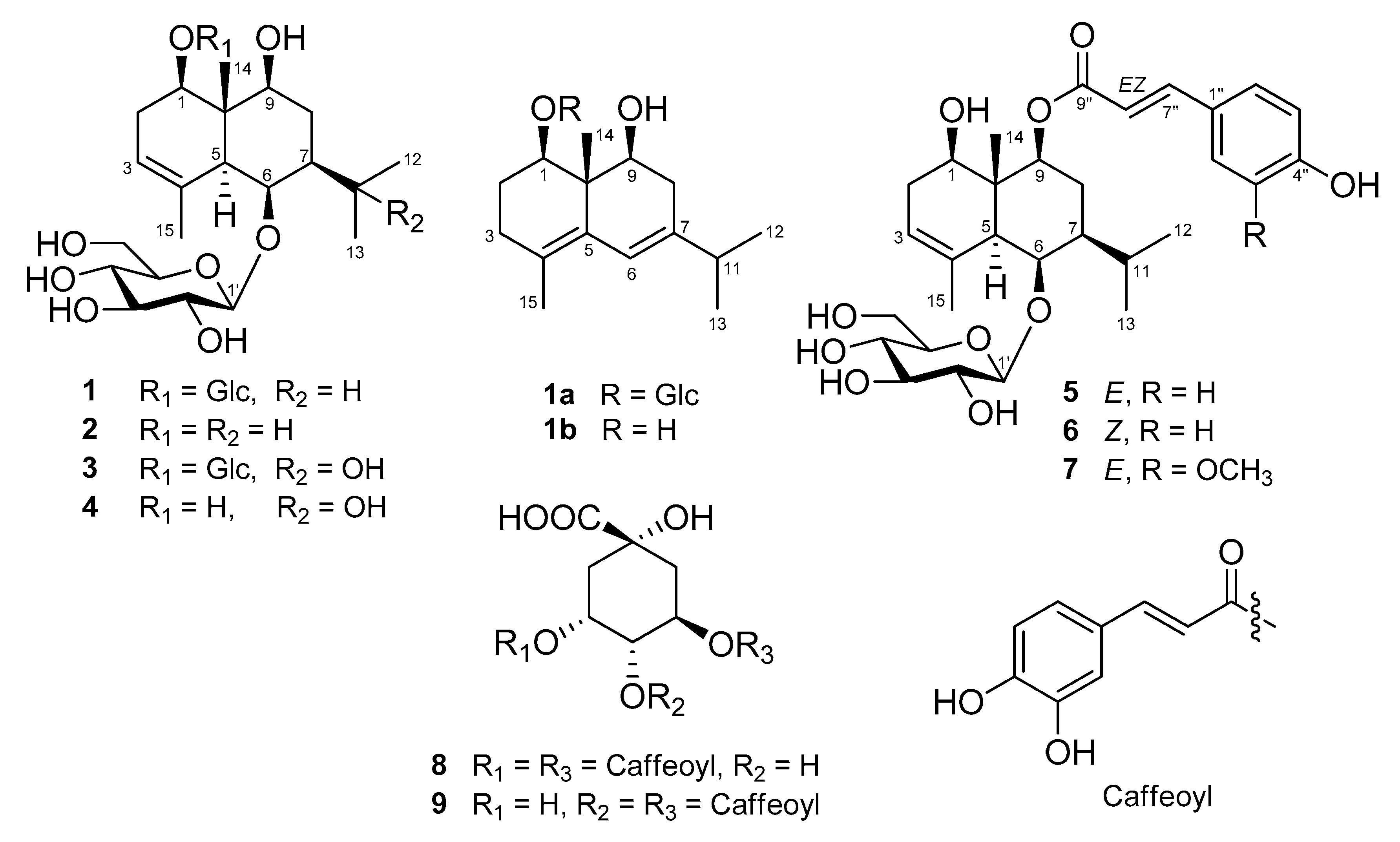
:Four new eudesmane-type sesquiterpenoids, (1R,5S,6R,7S,9S,10S)-1,6,9-trihydroxy-eudesm-3-ene-1,6-di-O-β-d-glucopyranoside (1), (1R,5S,6S,7R,9S,10S)-1,6,9,11-tetrahydroxy-eudesm-3-ene-1,6-di-O-β-d-glucopyranoside (3), (1R,5S,6R,7S,9S,10R)-9-O-(Z-p-coumaroyl)-1,6,9-trihydroxy-eudesm-3-ene-6-O-β-d-glucopyranoside (6), and (1R,5S,6R,7S,9S,10R)-9-O-(E-feruloyl)-1,6,9-trihydroxy-eudesm-3-ene-6-O-β-d-glucopyranoside (7), were isolated from a 95% EtOH extract of the leaves of Aster koraiensis by repeated chromatography. Moreover, three sesquiterpenoids (2, 4, and 5) and two caffeoylquinic acids (8 and 9) having previously known chemical structures were isolated during the isolation procedure. The four new compounds (1, 3, 6, and 7) were elucidated by spectroscopic data (1D- and 2D-NMR, MS, and ECD) interpretation and hydrolysis. Moreover, the absolute configurations of 2, 4, and 5 were determined for the first time in this study. The compounds isolated were tested for their viability on nitric oxide (NO) and prostaglandin E2 (PGE2) production on LPS-stimulated RAW 264.7 cells. Among them, only 7 presented weak inhibitory effects on both NO and PGE2 production.
1. Introduction
Aster koraiensis Nakai (syn. Gymnaster koraiensis (Nakai) Kitamura; Compositae) is an endemic species to Korea that is distributed from Jeju Island to the southern parts of Gangwon-do [1]. The young leaves and stems of A. koraiensis have been used in Korean cuisines and also used to ornamental plant for beautiful flowers [1]. It has been used for chronic bronchitis, pneumonia, antitussive, and pertussis in Korean folk medicine [2].
It has been reported that the extracts of A. koraiensis have a variety of biological activities including anti-proliferative activity on human and mouse tumor cell lines [3,4]. The extracts also exhibited antioxidant and α-glucosidase inhibitory activities [5]. Furthermore, it was reported that A. koraiensis protects retinal ganglion cells against oxidative stress in diabetic rats [6]. 3,5-O-Dicaffeoylepiquinic acid isolated from A. koraiensis showed inhibitory activity on AKR1B10 for cancer therapy and on formation of advanced glycation end products (AGEs) to treat diabetic nephropathy [7,8]. Similarly, chlorogenic acid from A. koraiensis reduced AGE formation and AGE/RAGE binding activity [9]. A polyacetylene, gymnasterkoreayne B (GKB), from this plant induced a variety of detoxification enzymes and exhibited a hepatoprotective effect and cytotoxicity against cancer cells [10]. Besides, gymnasterkoreayne G, which has a similar chain structure to GKB, has activity on the modulation of NFAT transcription factor [11]. Other polyacetylenes from A. koraiensis have acyl CoA: cholesterol acyltransferase (ACAT) activities in rat liver microsomes [12,13].
Previous chemical studies on A. koraiensis have led to the identification of several types of secondary metabolites, such as polyacetylenes [3,10,11,12,14], sesquiterpenoids [4,15,16,17], triterpenoids [18], flavonoids [4,16,17], ionones [17], caffeoylquinic acids [16], and benzofurans [16,19]. However, active compounds with anti-inflammatory activities in this plant has been poorly studied. In an ongoing project directed toward the search for bioactive compounds in plants, the leaves of A. koraiensis were chosen for phytochemical investigation, since its 95% EtOH extract was found to inhibit production of prostaglandin E2 (PGE2) and lipopolysaccharide (LPS)-induced nitric oxide (NO) in RAW 264.7 macrophages.
Various chromatographic separation led to the isolation and characterization of four new eudesmane-type sesquiterpenoid glycosides (1, 3, 6, and 7) and five known compounds (2, 4, 5, 8, and 9) from the leaves of A. koraiensis. The structures of 1–7 were elucidated by interpreting one- and two-dimensional (D) nuclear magnetic resonance (NMR) spectroscopic data analysis, enzymatic and acid hydrolysis, and electronic circular dichroism (ECD) calculation. Two caffeoylquinic acids (8 and 9) were identified by measurement of NMR spectroscopic data and by comparison with published values. The compounds obtained were evaluated for their activities on the production of the pro-inflammatory mediators, NO and PGE2, in RAW264.7 macrophages. We describe in this paper the isolation of compounds from the leaves of A. koraiensis, structure elucidation of the seven sesquiterpenes, and inhibitory activities of the isolates against production of NO and PGE2.
2. Results and Discussion
2.1. Structure Elucidation of 1–7
Four new compounds (1, 3, 6, and 7) and five known compounds (2, 4, 5, 8, and 9) were isolated from 95% EtOH extract of the leaves of A. koraiensis in the present research (Figure 1).
Compound 1 was isolated as a white powder. The molecular formula of 1 was established as C27H46O13 by HR-ESI-MS (m/z = 601.2938 [M + Na]+; calcd for C27H46O13Na, 601.2836) (Figure S1). The infrared absorption spectrum showed absorption bands at 3437, 2917, 1358, and 1010 cm−1, implicating that 1 has hydroxyl and olefinic groups. The 1H-NMR spectroscopic data of 1 exhibited two doublet methyl signals at δH 1.11 (3H, d, J = 6.0 Hz) and 0.85 (3H, d, J = 6.5 Hz), two singlet methyl signals at δH 2.10 (3H, s) and 1.64 (3H, s), two anomeric protons at δH 4.99 (1H, d, J = 8.0 Hz) and 4.92 (1H, d, J = 8.0 Hz), and an olefinic proton at δH 5.30 (1H, br s) (Table 1, Figure S2). The 13C-NMR spectrum of 1 indicated four methyl carbons (δC 22.0, 21.9 × 2, and 10.7), two methylene carbons (δC 30.3 and 29.4), three oxygenated methine carbons (δC 80.2, 78.4, and 76.6), and four methine carbons (δC 120.4, 52.3, 51.7, and 29.0), 12 carbons (δC 104.6, 102.1, 86.4, 78.4, 78.1, 77.4, 76.2, 75.4, 72.1, 71.6, 63.4, and 62.9) assignable to the glucose moieties including two anomeric carbons and two quaternary carbons (δC 136.7 and 43.0) (Table 1, Figure S3).
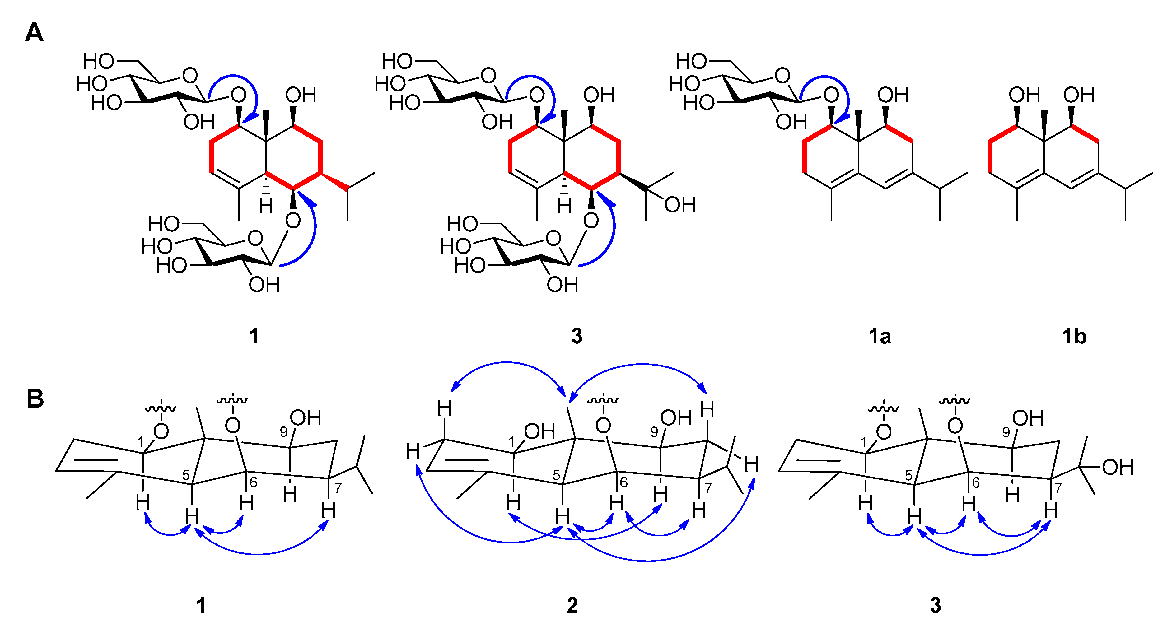
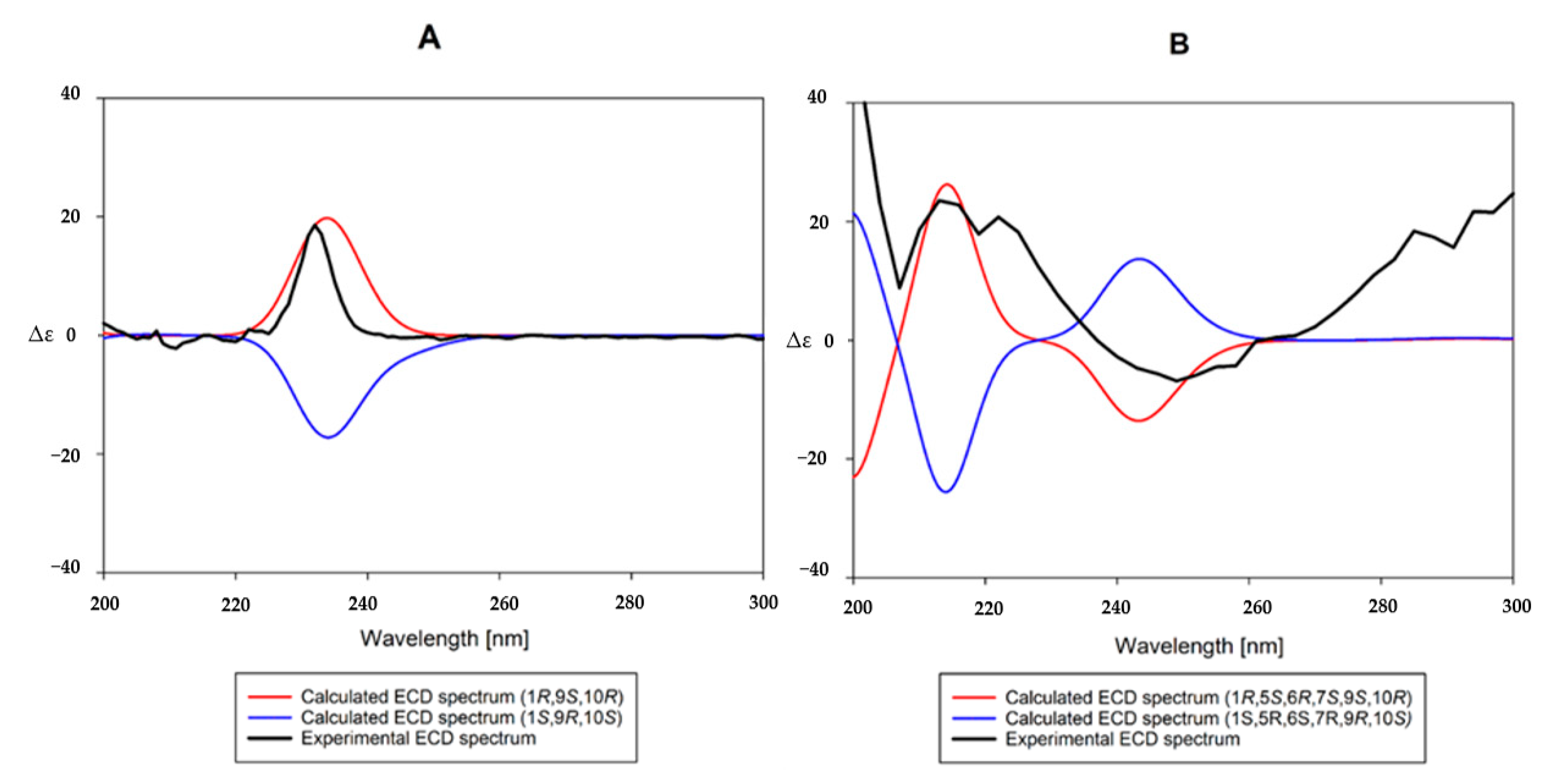
Based on the interpretation of 1H- and 13C-NMR data of 1, it was inferred that 1 is a eudesmane-type sesquiterpene containing two glucopyranosyl moieties. An enzymatic hydrolysis of 1, high performance liquid chromatography (HPLC) experiment, and analysis of coupling constants (both 8.0 Hz) of two anomeric protons led to the establishment of the sugars in 1 as β-d-glucopyranose. The heteronuclear multiple bond correlation (HMBC) experimental data showed long-range correlations between the signal of H-6 (δH 4.64) and Glc-1’ (δC 104.6) and the signal of H-1 (δH 4.18) and Glc-1” (δC 102.1) (Figure S6), indicating that the positions of the glucopyranosyl moieties are C-1 and C-2. The positions of the three hydroxyl groups were also determined by HMBC correlations to be C-1, 6, and 9. The relative stereochemistry of the hydroxyl groups at C-1, 6, and 9 were determined as all β-forms on the basis of the NOESY correlations (H-1 with H-5, H-5 with H-6/H-7) (Figure 2, Figure S7). To make the stereochemistry of 1 clearer, we generated 2 by enzymatic hydrolysis of 1 (Figure 1). The NOESY spectra of 2 revealed correlations of H-1 with H-9, H-5 with H-2α/H-6/H-8α, H-6 with H-7, and H-14 with H-2β/H-8β (Figure 2). To determine absolute configurations of 1 and 2, we obtained a derivative 1a by acidic hydrolysis of 1 (Figure 1). Additionally, an enzymatic hydrolysis of 1a was performed to remove the sugar moiety at C-1 and to produce 1b (Figure 1). In the 1H- and 13C-NMR spectra of both molecules, there are similar signals that correspond to the protons of the eudesmane-type skeleton, except for the chemical shifts of the proton in C-1 in both 1a and 1b, which are different due to the presence or absence of the sugar residue. The absolute configurations at C-1, 9, and 10 of 1b were established by comparing its experimental ECD spectrum with calculated spectra of (1R,9S,10R) and (1S,9R,10S) models using the time-dependent density functional theory (TDDFT) method. The experimental ECD spectrum of 1b displayed a positive Cotton effect (CE) at 232 nm ( + 24.8). The experimental data were in good agreement with the calculated ECD spectrum of the (1R,9S,10R) model (Figure 3), suggesting the absolute configuration of 1b as (1R,9S,10R). Therefore, the structure of the new compound 1 was elucidated as (1R,5S,6R,7S,9S,10S)-1,6,9-trihydroxy-eudesm-3-ene-1,6-di-O-β-d-glucopyranoside.
The planar structure of 2 turned out to be the same as 1β,6β,9α -trihydroxy-trans-eudesm-3-ene-6-O-β-d-glucopyranoside, which was isolated previously from the flower of A. koraiensis and reported only its relative configuration [4]. Although their 1H- and 13C-NMR spectroscopic data were identical, indicating they are the same compounds, we found that 2 has different configuration (9β-hydroxy-) from the other one (9α-hydroxy-). Thus, we propose the structure of the known compound 2 as (1R,5S,6R,7S,9S,10R)-1,6,9-trihydroxy-eudesm-3-ene-6-O-β-d-glucopyranoside.
Compound 3 was isolated as a white powder. The molecular formula of 3 was established as C27H46O14 by HR-ESI-MS (m/z = 617.2791 [M + Na]+; calcd for C27H46O14Na, 617.2785) (Figure S15). The 1H-NMR spectrum of 3 exhibited two singlet methyl signals at δH 1.58 (3H) and 1.38 (3H), two anomeric protons at δH 4.99 (1H, d, J = 8.0 Hz) and 4.87 (1H, d, J = 8.0 Hz), and an olefinic proton at δH 4.98 (1H, br s) (Table 1, Figure S16). The 13C-NMR spectrum of 3 showed 27 signals including four methyl carbons (δC 29.5, 29.2, 22.4, and 10.9), two methylene carbons (δC 28.5 and 27.3), 12 signals for glucose moieties, and three quaternary carbons (δC 136.7, 72.6, and 42.9) (Table 1, Figure S17). The NMR data for 3 were very similar to 1 except for the presence of a quaternary oxygenated carbon signal instead of a methine carbon signal. The position of the quaternary carbon at C–11 was deduced on the basis of the coupling pattern for two methyl groups changed from (δH 1.11 (3H, d, J = 6.0 Hz) and 0.85 (3H, d, J = 6.5 Hz)) to (δH 1.58 (3H, s) and 1.38 (3H, s)) in the 1H-NMR spectrum. It was supported by HMBC correlations between the signal of C-11 and H-12, H-13, and H-7 (Figure S20). The relative stereochemistry of hydroxyl groups at C-1, 6, and 9 were determined as all β-forms like 1 by analyzing NOESY correlations (H-1 with H-5, H-5 with H-6/H-7, and H-6 with H-7), indicating 3 is a 11-hydroxy derivative of 1 (Figure 2, Figure S21). Considering a biogenetic relationship with 1, the structure of the new compound 3 was proposed as (1R,5S,6S,7R,9S,10S)-1,6,9,11-tetrahydroxy-eudesm-3-ene-1,6-di-O-β-d-glucopyranoside.
The planar structure of 4 was also reported from the flowers of A. koraiensis together with 2 (Figure 2) [4]. The authors reported the chemical structure of the compound as 1β,6β,9α,11-tetrahydroxy-trans-eudesm-3-ene-6-O-β-d-glucopyranoside on the basis of the NOESY experiment. The 1H- and 13C-NMR spectroscopic data of 4 were identical with those of published values [4]. However, the NOESY correlations indicated that the relative configuration of 4 is the same as 1–3. Therefore, we propose the structure of the known compound 4 as (1R,5S,6S,7R,9S,10R)-1,6,9,11-tetrahydroxy-eudesm-3-ene-6-O-β-d-glucopyranoside.
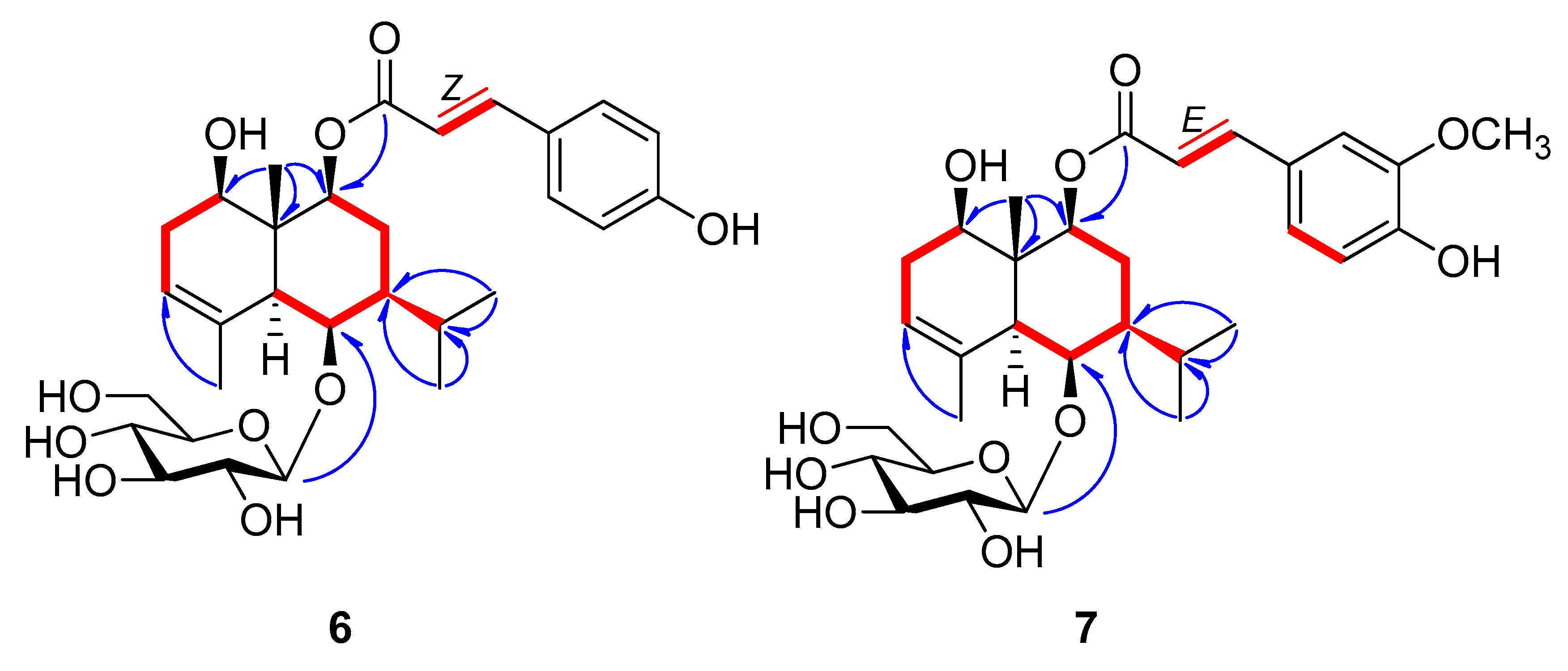
Compound 6 was obtained as a pale yellow amorphous powder and the molecular formula was established as C30H42O10 by HR–ESI–MS (m/z 561.2710 [M-H]−, calcd for C30H41O10, 561.2705) (Figure S15). The 1H-NMR spectrum of 6 showed four methyl signals at δH 1.88 (3H, s), 1.26 (3H, s), 1.02 (3H, d, J = 6.5 Hz), and 0.93 (3H, d, J = 6.5 Hz); one anomeric proton at δH 4.38 (1H, d, J = 8.0 Hz; and cis-olefinic group at δH 6.89 (1H, d, J = 12.5 Hz) and 5.80 (1H, d, J = 12.5 Hz) (Table 2, Figure S16). The 13C-NMR spectrum of 6 exhibited four methyl signals (δC 22.0, 21.9, 21.8, and 11.8), six glucosyl signals including an anomeric carbon (δC 104.5, 78.5, 77.5, 76.2, 72.1, and 63.3), three oxymethine signals (δC 80.8, 80.3, and 76.0), and cis-para-coumaroyl group (δC 167.8, 160.3, 145.7, 134.2 × 2, 127.9, 117.7, and 116.0 × 2), indicating that 6 is a p-coumaroyl derivative of 2 (Table 2, Figure S17). The positions of hydroxyl, p-coumaroyl and β-d-glucopyranosyl groups and the relative configurations were determined by analysis of the HMBC and NOESY correlations (Figure 4, Figures S20 and S21). On the basis of the NMR data and a biogenetic relationship with 1-4, the structure of the new compound 6 was proposed as (1R,5S,6R,7S,9S,10R)-9-O-(Z-p-coumaroyl)-1,6,9-trihydroxy-eudesm-3-en-6-O-β-d-glucopyranoside. A literature survey revealed that 6 is the geometric isomer of 5, 9β-O-(E-p-hydroxycinnamoyl)-1β,6β-dihydroxy-trans-eudesm-3-ene-6-O-β-d-glucopyranoside, which was isolated previously from the aerial parts of A. koraiensis and reported only its relative configuration [16]. The absolute configuration of 5 was established by comparing its experimental ECD spectrum with those calculated spectra of (1R,5S,6R,7S,9S,10R) and (1S,5R,6S,7R,9R,10S) models using the same method as 1c. The experimental data (Figure 3) were in accordance with the calculated ECD spectrum of the (1R,5S,6R,7S,9S,10R) model, offering the absolute configuration of 5 as (1R,5S,6R,7S,9S,10R)-9-O-(E-p-coumaroyl)-1,6,9-trihydroxy-eudesm-3-ene-6-O-β-d-glucopyranoside and also supporting our proposed absolute configurations for 1–6.
The molecular formula of 7 was established as C31H44O11 by HR-ESI-MS (m/z 575.2848 [M-H2O-H]−, calcd for C31H43O10, 575.2856) (Figure S22). The 1H- and 13C-NMR spectroscopic data of 7 were very similar with those of 5 and 6 except for the presence of E-p-feruloyl group in 7 instead of E- or Z-p-coumaroyl group (Table 2). An ABX system (δH 6.81 (d, J = 8.5 Hz), 7.08 (dd, J = 8.5, 2.0 Hz), and 7.22 (d, J = 2.0 Hz)), trans-olefinic group (δH 7.61 (1H, d, J = 16.0 Hz) and 6.38 (1H, d, J = 16.0 Hz)), and a methoxy signal (δH 3.91 (3H, s)) were revealed in the 1H-NMR spectrum of 7, indicating the presence of E-p-feruloyl group in 7. The positions of the functional groups in 7 and the relative configuration were confirmed by the HMBC and NOESY correlations (Figure 4, Figures S26 and S27). Thus, from the analysis of above data, the structure of the new compound 7 was elucidated as (1R,5S,6R,7S,9S,10R)-9-O-(E-p-feruloyl)-1,6,9-trihydroxy-eudesm-3-ene-6-O-β-d-glucopyranoside.
2.2. Inhibitory Activities of the Isolates on LPS-Stimulated NO and PGE2 Production
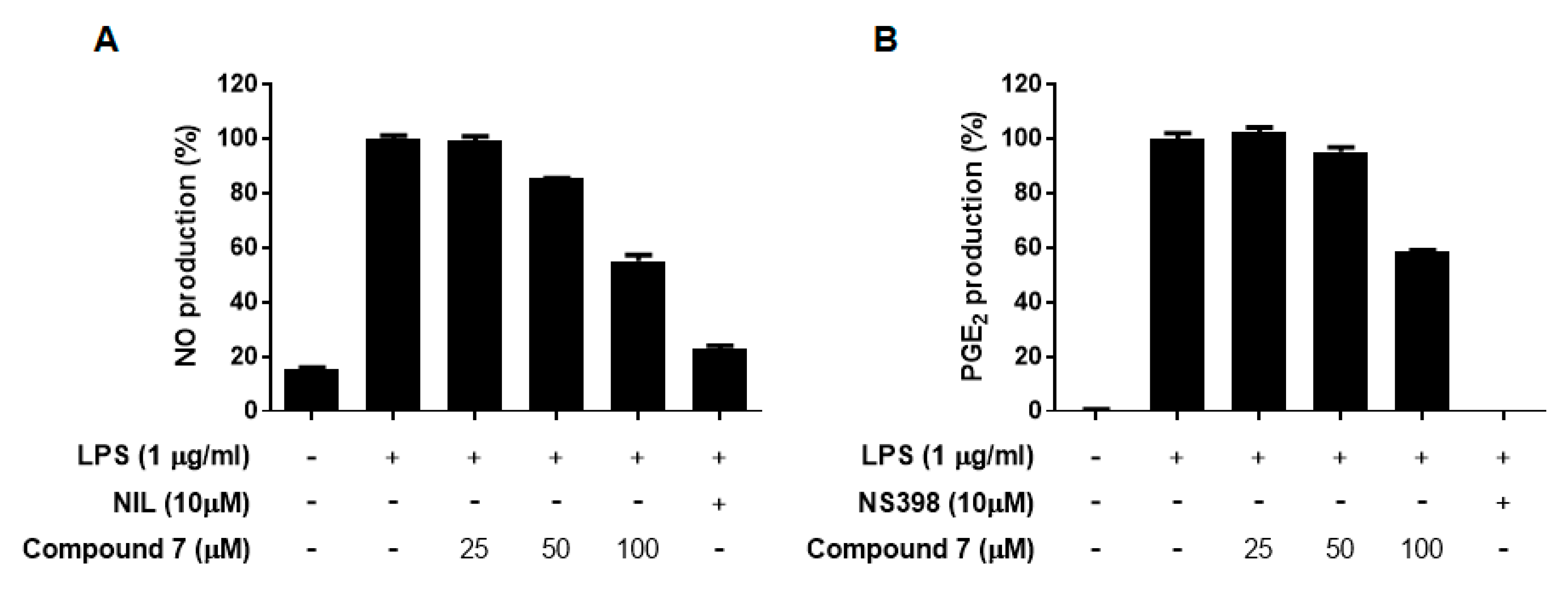
All the isolates 1–9 obtained from the leaves of A. koraiensis were evaluated for their inhibitory effects of LPS-stimulated NO and PGE2 production in RAW 264.7 macrophages at non-toxic concentrations (Table 3). Of these, only the new compound 7 presented weak inhibitory effects on both NO and PGE2 production with observed IC50 values of 95.7 and 111.6 μM, respectively, while others were inactive (Table 3, Figure 5).
3. Materials and Methods
3.1. Plant Material
The leaves of Aster koraiensis Nakai (Compositae) were collected at Pyeongchang, Gangwon-Do, Korea, in 2017. The origin of the plant was authenticated by one of the authors D.S.J. and a voucher specimen (ASKO1-2017) was deposited at the College of Pharmacy, Kyung Hee University, Korea.
3.2. General Experimental Procedures
General experimental procedures are in the Supplementary Materials.
3.3. Extraction and Isolation
The dried and ground leaves (5.0 kg) of A. koraiensis were extracted twice with 25 L of 95% EtOH at 70 °C for 3 hours and extract solutions were condensed using a steam heated evaporator. The 95% EtOH extract (500 g) was chromatographed over Diaion HP-20 (9.8 × 63.0 cm) eluting with an acetone-H2O gradient (from 0:1 to 1:0 v/v) to afford 28 fractions (C1 ~ C28).
Fraction C4 (11.82 g) was separated into five subfractions by Sephadex LH-20 column chromatography (CC) (4.8 × 63.0 cm) with 50 % acetone (C4-1 ~ C4-5). Compound 3 (18.7 mg) was purified by repetitive chromatography from subfraction C4-2 (2.99 g). Fraction C5 (8.32 g) was fractionated further by Sephadex LH-20 CC (4.8 × 43.0 cm) with MeOH-H2O (1:1 v/v), yielding 14 fractions (C5-1 ~ C5-14) and compound 1 (1.76 g). Compound 4 (61.3 mg) was purified using a flash chromatography system (Redi Sep-C18 cartridge, 43 g, MeOH-H2O gradient (from 10:90 to 40:60 v/v)) from fraction C5-3-5 (133.8 mg). Compound 1 (324.1 mg) was additionally obtained by flash chromatography with Redi Sep-C18 cartridge (120 g, MeOH-H2O, from 20:80 to 50:50 v/v) from fraction C5-3-11 (701.6 mg). Fraction C9 (3.64 g) was subjected to Sephadex LH-20 CC with a MeOH-H2O mixture (1:1 v/v) to give nine subfractions (C9-1 ~ C9-9). Subfraction C9-5 (522.9 mg) was separated further using a flash chromatography system with Redi Sep-C18 (43 g, MeOH-H2O, 40:60 to 70:30, v/v) to afford compound 2 (41.8 mg). Fraction C11 (10.0 g) was separated into seven subfractions (C11-1 ~ C11-7) by Sephadex LH-20 CC (3.6 × 65.0 cm) with 50% acetone. Subfraction C11-6 (2.27 g) was chromatographed over silica gel (230–400 mesh; 4.8 × 28.3 cm) with an EtOAc-acetone-H2O mixture (from 50:45:5 to 30:60:10 v/v/v) as mobile phase to obtain compounds 8 (948.8 mg) and 9 (10.2 mg). Fraction C17 (3.12 g) was fractionated into five subfractions (C17-1 ~ C17-5) by Sephadex LH-20 CC (4.0 × 69.0 cm) with MeOH-H2O (1:1 v/v). Compounds 5 (25.6 mg), 6 (7.6 mg), and 7 (4.8 mg) were isolated from subfraction C17-3 (300.0 mg) using a flash chromatography system with silica cartridge (48 g, CH2Cl2-MeOH-H2O, 35:65 to 50:50, v/v).
3.3.1. (1R,5S,6R,7S,9S,10S)-1,6,9-trihydroxy-eudesm-3-ene-1,6-di-O-β-d-glucopyranoside (1)
White powder; m.p.: 167.2 °C; : −0.4° (c 0.1, MeOH); UV (acetonitrile) λmax (log ε): 205 (3.83), 376 (4.08) nm; IR (ATR) νmax 3437, 2917, 1358, 1010 cm−1; HR-ESI-MS m/z = 601.2838 [M + Na]+, (calcd for C27H46O13Na, 601.2836); NMR data: Table 1.
3.3.2. 1R,9S,10S-1,9-Dihydroxy-eudesm-4,6-diene-1-O-β-d-glucopyranoside (1a)
Pale yellow amorphous powder; HR-Q-TOF-MS m/z = 397.2237 [M–H]− (calcd for C21H33O7, 397.2226); : 58.3° (c 0.03, MeOH); UV (MeOH) λmax nm (log ε): 240 (3.37), 296 (3.34); IR (ATR) νmax 1937, 1587, 1348, 1013 cm−1; 1H-NMR (500 MHz, CD3OD) δH 6.05 (1H, brs, H-6), 4.48 (1H, d, J = 8.0 Hz, Glc-1), 3.94 (1H, dd, J = 12.0, 4.0 Hz, H-1), 3.88 (1H, dd, J = 12.0, 2.0 Hz, Glc-6a), 3.83 (1H, dd, J = 11.0, 5.5 Hz, H-9), 3.65 (1H, dd, J = 12.0, 6.0 Hz, Glc-6b), 3.36 (1H, m, Glc-5), 3.29 (1H, m, Glc-4), 3.27 (1H, m, Glc-3), 3.17 (1H, dd, J = 9.5, 8.0 Hz, Glc-2), 2.31 (1H, m, H-11), 2.19 (2H, m, H-8), 2.08 (2H, m, H-3), 1.93 (2H, m, H-2), 1.69 (3H, s, H-15), 1.07 (3H, d, J = 2.0 Hz, H-12), 1.06 (3H, d, J = 2.0 Hz, H-13), 1.01 (3H, s, H-14); 13C–NMR (125 MHz, CD3OD) δC 141.9 (C-7), 133.3 (C-5), 129.1 (C-4), 118.5 (C-6), 102.4 (Glc-1), 84.9 (C-1), 78.6 (Glc-5), 78.3 (Glc-3), 78.1 (C-9), 75.1 (Glc-2), 71.7 (Glc-4), 63.0 (Glc-6), 43.1 (C-10), 36.4 (C-11), 33.0 (C-8), 32.5 (C-3), 24.6 (C-2), 22.2 (C-12), 21.7 (C-13), 19.2 (C-15), 12.5 (C-14).
3.3.3. 1R,9S,10R-1,9-Dihydroxy-eudesm-4,6-diene (1b)
Pale yellow amorphous powder; : 67.2° (c 0.03, MeOH); UV (MeOH) λmax nm (log ε): 239 (3.27), 290 (3.22); IR (ATR) νmax 1569, 1418, 1363, 1016 cm−1; 1H–NMR (500 MHz, CD3OD) δH 6.07 (1H, brs, H-6), 3.77 (1H, dd, J = 12.0, 4.0 Hz, H-1), 3.68 (1H, dd, J = 11.0, 5.5 Hz, H-9), 2.30 (1H, m, H-11), 2.22 (2H, m, H-8), 2.14 (2H, m, H-3), 1.80 (2H, m, H-2), 1.68 (3H, s, H-15), 1.07 (3H, d, J = 2.0 Hz, H-12), 1.05 (3H, d, J = 2.0 Hz, H-13), 0.93 (3H, s, H-14); 13C–NMR (125 MHz, CD3OD) δC 141.9 (C-7), 133.3 (C-5), 129.1 (C-4), 118.5 (C-6), 78.6 (C-1), 78.3 (C-9), 43.1 (C-10), 36.4 (C-11), 33.0 (C-8), 32.5 (C-3), 24.6 (C-2), 22.2 (C-12), 21.7 (C-13), 19.2 (C-15), 12.5 (C-14).
3.3.4. (1R,5S,6S,7R,9S,10S)-1,6,9,11-tetrahydroxy-eudesm-3-ene-1,6-di-O-β-d-glucopyranoside (3)
White powder; m.p.: 230 °C; : −1.5° (c 0.1, MeOH); UV (acetonitrile) λmax (log ε): 205 (3.85), 322 (4.02), 359 (4.08), 373 (4.43) nm; IR (ATR) νmax 3314, 2876, 1360, 1011 cm−1; HR-ESI-MS (positive mode) m/z = 617.2791 [M + Na]+ (calcd for C27H46O14Na, 617.2785); NMR data: Table 1.
3.3.5. (1R,5S,6R,7S,9S,10R)-O-(Z-p-coumaroyl)-1,6,9-Trihydroxy-eudesm-3-ene-6-O-β-d-glucopyranoside (6)
Pale yellow amorphous powder; HR-ESI-MS m/z = 561.2701 [M–H]− (calcd for C30H41O10, 561.2705); : 48.4° (c 0.1, MeOH); UV (MeOH) λmax nm (log ε): 313 (4.48); IR (ATR) νmax 1705, 1603 1513, 1165, 1074, 989 cm−1; NMR data: Table 2.
3.3.6. (1R,5S,6R,7S,9S,10R)-O-(E-feruloyl)-1,6,9-Trihydroxy-eudesm-3-ene-6-O-β-d-glucopyranoside (7)
Pale yellow amorphous powder; HR-ESI-MS m/z = 575.2845 [M–H2O–H]− (calcd for C31H43O10, 575.2856); : 5.8° (c 0.1, MeOH); UV (MeOH) λmax nm (log ε): 237 (3.47), 327 (3.22); IR (ATR) νmax 1705, 1596, 1517, 1268, 1159, 1074 cm−1; NMR data: Table 2.
3.4. Acidic and Enzymatic Hydrolysis of 1
Compound 1 (10.0 mg) was incubated together with β-glucosidase (25.0 mg), toluene (2 drops), and H2O (15.0 mL) in a CO2 incubator at 35 °C for 3 days. EtOH was added to the reaction mixture to stop the reaction and β-glucosidase was removed by filtration. Compound 2 (2.0 mg) was isolated from the hydrolysate by flash CC with Redi Sep-C18 cartridge (13 g, MeOH-H2O, from 50:50 to 80:20 v/v). Meanwhile, compound 1 (93.8 mg) was hydrolyzed with 2N HCl at 80 °C for one hour. The reaction was stopped by the addition of sodium bicarbonate and 1a (8.0 mg) was isolated from the hydrolysate by flash CC with a Redi Sep-C18 cartridge (13 g, MeOH-H2O, 60:40 to 80:20, v/v). An enzymatic hydrolysis of 1a (8.0 mg) was performed using the same method as 1 to give 1b (1.5 mg).
3.5. Absolute Configurations of β-Glucose in 1
The absolute configuration of β-glucose in 1 was determined by the previously reported method [21]. Pyridine (500 μL) and l-cysteine methyl ester hydrochloride (1.2 mg) were added in the hydrolysate and the mixture was heated at 60 °C for 1 h. σ-Tolyl isothiocyanate (100 μL) was added in the mixture and heated again at 60 °C for 1 hour. The reaction was analyzed directly by HPLC with a gradient system (10–50% of B, A: 0.1% (v/v) formic acid in water, B: 0.1% (v/v) formic acid in acetonitrile). The reaction mixture of 1 was detected at 27.4 min. At the same HPLC conditions, authentic l- and d-glucoses were detected at 26.8 and 27.4 min, respectively. Therefore, the absolute configuration of β-glucose in 1 was established as the d configuration.
3.6. Computational Methods
3.7. Measurement of Cell Viability and NO Production
Cell viability and nitrite levels were measured using MTT and Griess reaction assays, respectively [24].
3.8. Measurement of PGE2
PGE2 levels in cell culture mediums were determined using EIA kits (R&D Systems, MN) as reported in the previous paper [24].
4. Conclusions
Four new eudesmene-type sesquiterpenoids (1, 3, 6, and 7) were obtained from a 95% EtOH extract of the leaves of Aster koraiensis by repeated chromatography, along with five known compounds (2, 4, 5, 8, and 9). The chemical structures of the four new compounds and absolute configurations of the known compounds 2, 4, and 5 were established by their spectroscopic data (HR-MS, 1D- & 2D-NMR, and ECD) measurement and by acidic and enzymatic hydrolysis. Among the isolates, the new compound 7 exhibited weak inhibitory activities on both NO and PGE2 production. The compounds found in this study do not appear to contribute to the anti-inflammatory activity of the extract from which they were isolated. Thus, compounds with higher activity in the leaves of A. koraiensis needs to be identified through further studies.
Supplementary Materials
The following are available online at https://www.mdpi.com/2223-7747/9/12/1811/s1, General Experimental Procedure, The HR-ESI-MS, 1H-NMR (500 MHz, C5D5N), 13C-NMR (125 MHz, C5D5N), HSQC, COSY, HMBC, and NOESY spectra of 1 (Figures S1~S8), The HR-ESI-MS, 1H-NMR (500 MHz, C5D5N), 13C-NMR (125 MHz, C5D5N), HSQC, COSY, HMBC, and NOESY spectra of 3 (Figures S9~S15), The HR-ESI-MS, 1H-NMR (500 MHz, CD3OD), 13C-NMR (125 MHz, CD3OD), HSQC, COSY, HMBC, and NOESY spectra of 6 (Figures S16~S21), The HR-ESI-MS, 1H-NMR (500 MHz, CD3OD), 13C-NMR (125 MHz, CD3OD), COSY, HMBC, and NOESY spectra of 7 (Figures S22~S27).
Author Contributions
Conceptualization, D.S.J.; Funding acquisition, D.S.J.; Investigation, J.-Y.K., Y.H.S., I.-H.L. and H.Y.C.; Project administration, H.C.K.; Resources, H.C.K.; Supervision, J.-H.C., J.L. and D.S.J.; Writing—original draft, J.-Y.K. and Y.H.S.; Writing—review & editing, J.L. and D.S.J. All authors have read and agreed to the published version of the manuscript.
Funding
This research was supported by a grant (grant number: NRF-2019R1A2C1083945) from the National Research Foundation of Korea (NRF) funded by the Ministry of Science and ICT (MSIT), Korea and by the Korea Institute of Science & Technology (KIST) Institutional Program (Project No. 2E30650-20-154). This research was also supported by the project entitled “Development of Sustainable Application for Standard Herbal Resources” (grant number: KSN2012320) from the Korea Institute of Oriental Medicine.
Conflicts of Interest
The authors declare no conflict of interest.
References
- Ahn, D.K. Illustrated Book of Korean Medicinal Herbs; Kyo-hak Publishing Co.: Seoul, Korea, 1998; p. 107. [Google Scholar]
- Kwon, J.H.; Kang, S.W.; Shen, S.S.; Park, C.S. Occurrence of stem rot of wild Aster (Aster koraiensis) caused by Sclerotium rolfsii in Korea. Mycobiology 2001, 29, 58–60. [Google Scholar] [CrossRef] [Green Version]
- Jung, H.J.; Min, B.S.; Park, J.Y.; Kim, Y.H.; Lee, H.K.; Bae, K.H. Gymnasterkoreaynes A–F, Cytotoxic polyacetylenes from Gymnaster koraiensis. J. Nat. Prod. 2002, 65, 897–901. [Google Scholar] [CrossRef] [PubMed]
- Lee, I.K.; Kim, K.H.; Ryu, S.Y.; Choi, S.U.; Lee, K.R. Phytochemical constituents from the flowers of Gymnaster koraiensis and their cytotoxic activities in vitro. Bull. Korean Chem. Soc. 2010, 31, 227–229. [Google Scholar] [CrossRef] [Green Version]
- Lee, T.G.; Hyun, S.W.; Lee, I.S.; Park, B.K.; Kim, J.S.; Kim, C.S. Antioxidant and α-glucosidase inhibitory activities of the extracts of Aster koraiensis leaves. Korean J. Med. Crop Sci. 2018, 26, 382–390. [Google Scholar]
- Kim, K.A.; Kang, K.D.; Lee, E.H.; Nho, C.W.; Jung, S.H. Edible wild vegetable, Gymnaster koraiensis protects retinal ganglion cells against oxidative stress. Food Chem. Toxicol. 2011, 49, 2131–2143. [Google Scholar] [CrossRef]
- Lee, J.Y.; Song, D.G.; Lee, E.H.; Jung, S.H.; Nho, C.W.; Cha, K.H.; Pan, C.H. Inhibitory effects of 3, 5−O−dicaffeoyl−epi−quinic acid from Gymnaster koraiensis on AKR1B10. J. Korean Soc. Appl. Biol. Chem. 2009, 52, 731–734. [Google Scholar] [CrossRef]
- Sohn, E.; Kim, J.; Kim, C.S.; Kim, Y.S.; Jang, D.S.; Kim, J.S. Extract of the aerial parts of Aster koraiensis reduced development of diabetic nephropathy via anti−apoptosis of podocytes in streptozotocin−induced diabetic rats. Biochem. Biophys. Res. Commun. 2010, 391, 733–738. [Google Scholar] [CrossRef]
- Kim, J.; Jo, K.; Lee, I.S.; Kim, C.S.; Kim, J.S. The extract of Aster koraiensis prevents retinal pericyte apoptosis in diabetic rats and its active compound, Chlorogenic acid inhibits AGE formation and AGE/RAGE interaction. Nutrients 2016, 8, 585. [Google Scholar] [CrossRef] [Green Version]
- Lee, S.B.; Kang, K.; Oidovsambuu, S.; Jho, E.H.; Yun, J.H.; Yoo, J.H.; Nho, C.W. A polyacetylene from Gymnaster koraiensis exerts hepatoprotective effects in vivo and in vitro. Food. Chem. Toxicol. 2010, 48, 3035–3041. [Google Scholar] [CrossRef]
- Dat, N.T.; Cai, X.F.; Shen, Q.; Im, S.L.; Lee, E.J.; Park, Y.K.; Kim, Y.H. Gymnasterkoreayne G, a new inhibitory polyacetylene against NFAT transcription factor from Gymnaster koraiensis. Chem. Pharm. Bull. 2005, 53, 1194–1196. [Google Scholar] [CrossRef] [Green Version]
- Jung, H.J.; Hung, T.M.; Na, M.K.; Min, B.S.; Kwon, B.M.; Bae, K.H. ACAT Inhibition of polyactylenes from Gymnaster koraiensis. Nat. Prod. Sci. 2009, 15, 110–113. [Google Scholar]
- Butler, S.M.; Wallig, M.A.; Nho, C.W.; Pan, C.H.; Lee, E.H.; Jung, S.H.; Jeffery, E.H. A polyacetylene−rich extract from Gymnaster koraiensis strongly inhibits colitis−associated colon cancer in mice. Food. Chem. Toxicol. 2013, 53, 235–239. [Google Scholar] [CrossRef] [PubMed]
- Park, J.Y.; Min, B.S.; Jung, H.J.; Kim, Y.H.; Lee, H.K.; Bae, K.H. Polyacetylene glycosides from Gymnaster koraiensis. Chem. Pharm. Bull. 2002, 50, 685–687. [Google Scholar] [CrossRef] [PubMed] [Green Version]
- Lee, I.K.; Kim, K.H.; Ryu, S.Y.; Lee, K.R. Two new sesquiterpene glucosides from Gymnaster koraiensis. Heterocycles 2009, 8, 2827–2835. [Google Scholar] [CrossRef] [Green Version]
- Lee, J.; Lee, Y.M.; Lee, B.W.; Kim, J.H.; Kim, J.S. Chemical constituents from the aerial parts of Aster koraiensis with protein glycation and aldose reductase inhibitory activities. J. Nat. Prod. 2012, 75, 267–270. [Google Scholar] [CrossRef] [PubMed]
- Nhoek, P.; Ahn, J.; Chae, H.S.; Pel, P.; Kim, Y.M.; Lee, S.E.; Lee, J.H.; Kim, J.; Choi, Y.H.; Lee, K.; et al. Isolation of polyacetylenes with proprotein convertase/kexin type 9 downregulating activity and two new sesquiterpenes from the aerial parts of Aster koraiensis. Tetrahedron Lett. 2020, 151957. [Google Scholar] [CrossRef]
- Kwon, J.; Ko, K.; Zhang, L.; Zhao, D.; Yang, H.O.; Kwon, H.C. An autophagy inducing triterpene saponin derived from Aster koraiensis. Molecules 2019, 24, 4489. [Google Scholar] [CrossRef] [Green Version]
- Dat, N.T.; Van Kiem, P.; Cai, X.F.; Shen, Q.; Bae, K.; Kim, Y.H. Gymnastone, a new benzofuran derivative from Gymnaster koraiensis. Arch. Pharm. Res. 2004, 27, 1106–1108. [Google Scholar] [CrossRef]
- Wu, Q.Z.; Zhao, D.X.; Xiang, J.; Zhang, M.; Zhang, C.F.; Xu, X.H. Antitussive, expectorant, and anti-inflammatory activities of four caffeoylquinic acids isolated from Tussilago farfara. Pharm. Biol. 2016, 54, 1117–1124. [Google Scholar] [CrossRef] [Green Version]
- Tanaka, T.; Tatsuya, N.; Toshihisa, U.; Kenji, T.; Isao, K. Facile discrimination of aldose enantiomers by reversed-phase HPLC. Chem. Pharm. Bull. 2007, 55, 899–901. [Google Scholar] [CrossRef] [Green Version]
- Kwon, J.; Lee, H.; Ko, W.; Kim, D.C.; Kim, K.W.; Kwon, H.C.; Guo, Y.; Sohn, J.H.; Yim, J.H.; Kim, Y.C. Chemical constituents isolated from Antarctic marine-derived Aspergillus sp. SF-5976 and their anti-inflammatory effects in LPS-stimulated RAW 264.7 and BV2 cells. Tetrahedron 2017, 73, 3905–3912. [Google Scholar] [CrossRef]
- Lee, J.S.; Jeong, M.; Park, S.; Ryu, S.M.; Lee, J.; Song, Z.; Guo, Y.; Choi, J.H.; Lee, D.; Jang, D.S. Chemical constituents of the leaves of butterbur (Petasites japonicus) and their anti-inflammatory effects. Biomolecules 2019, 9, 806. [Google Scholar] [CrossRef] [PubMed] [Green Version]
- Shin, J.S.; Lee, K.G.; Lee, H.H.; Lee, H.J.; An, H.J.; Nam, J.H.; Jang, D.S.; Lee, K.T. α−Solanine isolated from Solanum tuberosum, L. cv Jayoung abrogates LPS−induced inflammatory responses via NF−κB inactivation in RAW 264.7 macrophages and endotoxin−induced shock model in mice. J. Cell. Biochem. 2016, 117, 2327–2339. [Google Scholar] [CrossRef] [PubMed]
Figure 1.
Chemical structures of 1–9 isolated from the leaves of A. koraiensis.

Figure 2.
Key COSY (▬) and HMBC ( ![Plants 09 01811 i001]() ) correlations of 1, 3, 1a, and 1b (A). Key NOESY (
) correlations of 1, 3, 1a, and 1b (A). Key NOESY ( ![Plants 09 01811 i002]() ) correlations of 1, 2, and 3 (B).
) correlations of 1, 2, and 3 (B).
 ) correlations of 1, 3, 1a, and 1b (A). Key NOESY (
  ) correlations of 1, 2, and 3 (B).
Figure 2.
Key COSY (▬) and HMBC ( ![Plants 09 01811 i001]() ) correlations of 1, 3, 1a, and 1b (A). Key NOESY (
) correlations of 1, 3, 1a, and 1b (A). Key NOESY ( ![Plants 09 01811 i002]() ) correlations of 1, 2, and 3 (B).
) correlations of 1, 2, and 3 (B).
) correlations of 1, 3, 1a, and 1b (A). Key NOESY ( ) correlations of 1, 2, and 3 (B).
Figure 3.
Measured and calculated electronic circular dichroism (ECD) spectra of 1b (A) and 5 (B).

Figure 4.
Key 1H-1H COSY (▬) and HMBC ( ![Plants 09 01811 i001]() ) correlations of 6 and 7.
) correlations of 6 and 7.
) correlations of 6 and 7.
Figure 5.
Inhibitory effects of compound 7 on LPS-stimulated NO (A) and PGE2 productions (B) in RAW 264.7 macrophages. Cells were pretreated with different concentrations (25, 50, or 100 μM) of compound 7 for 1 h, then with LPS (1 μg/mL), and then were incubated for 24 h. l-N6-(1-Iminoethyl)lysine (l-NIL, 10 μM) and N-[2-(cyclohexyloxy)-4-nitrophenyl]-methanesulfonamide (NS-398, 10 μM) were used as positive NO and PGE2 production inhibitors, respectively.


Table 1.
1H and 13C nuclear magnetic resonance (NMR) spectroscopic data of 1 and 3 (δ in ppm, C5D5N, 500, and 125 MHz).
Table 1.
1H and 13C nuclear magnetic resonance (NMR) spectroscopic data of 1 and 3 (δ in ppm, C5D5N, 500, and 125 MHz).
| Position a | 1 | 3 | ||
|---|---|---|---|---|
| δH Multi (J in Hz) | δC | δH Multi (J in Hz) | δC | |
| 1 | 4.18 d (9.5) | 78.4 | 4.17 m | 79.4 |
| 2 | 2.06 m/2.25 td (13.0, 11.0) | 30.3 | 2.73 q (12.0)/2.21 m | 28.5 |
| 3 | 5.30 br s | 120.4 | 4.98 br s | 120.7 |
| 4 | - | 136.7 | - | 136.7 |
| 5 | 1.83 br s | 52.3 | 1.83 s | 52.3 |
| 6 | 4.64 m | 76.6 | 4.66 br s | 80.2 |
| 7 | 0.99 m | 51.7 | 1.37 s | 51.6 |
| 8 | 2.37 m/2.53 m | 29.4 | 2.48 m | 27.3 |
| 9 | 4.08 m | 80.2 | 4.16 m | 85.0 |
| 10 | - | 43.0 | - | 42.9 |
| 11 | 2.37 m | 29.0 | - | 72.6 |
| 12 | 0.85 d (6.5) | 21.9 | 1.58 s | 29.2 |
| 13 | 1.11 d (6.0) | 21.9 | 1.38 s | 29.5 |
| 14 | 1.64 s | 10.7 | 1.68 s | 10.9 |
| 15 | 2.10 s | 22.0 | 1.93 s | 22.4 |
| Glc-1’ | 4.92 d (8.0) | 104.6 | 4.87 d (8.0) | 105.5 |
| Glc-2’ | 3.96 t (8.5) | 76.2 | 3.97 dd (8.0, 8.0) | 75.7 |
| Glc-3’ | 4.10 m | 78.4 | 4.21–4.15 | 78.8 |
| Glc-4’ | 4.15 m | 72.1 | 4.01 dd (9.5) | 72.1 |
| Glc-5’ | 3.86 m | 77.4 | 3.82 | 77.6 |
| Glc-6’ | 4.31 t (6.0)/4.47 dd (11.5, 3.0) | 63.4 | 4.38 m/4.21–4.15 | 64.0 |
| Glc-1” | 4.99 d (8.0) | 102.1 | 4.99 d (8.0) | 102.2 |
| Glc-2” | 4.64 m | 75.4 | 3.90 dd (8.0, 8.0) | 75.5 |
| Glc-3” | 4.27 t (9.0) | 78.1 | 4.21–4.15 | 78.8 |
| Glc-4” | 4.14 t (4.0) | 71.6 | 4.10 m | 71.5 |
| Glc-5” | 4.18 d (9.5) | 86.4 | 4.21–4.15 | 79.3 |
| Glc-6” | 4.41 dd (12.0, 6.0)/4.64 m | 62.9 | 4.68 m/4.42 m | 63.4 |
a All assignments were supported with 1H-1H correlation spectroscopy (COSY), 1H-13C heteronuclear single quantum coherence spectroscopy (HSQC), and 1H-13C heteronuclear multiple bond correlation (HMBC) experiments.
Table 2.
1H and 13C NMR spectroscopic data of 6 and 7 (δ in ppm, CD3OD, 500, and 125 MHz).
| Position a | 6 | 7 | ||
|---|---|---|---|---|
| δH Multi (J in Hz) | δC | δH Multi (J in Hz) | δC | |
| 1 | 4.87 overlapped | 80.8 | 4.89 overlapped | 80.5 |
| 2 | 2.22 m/2.09 m | 29.6 | 2.25 m/2.09 m | 29.7 |
| 3 | 5.29 br s | 120.4 | 5.30 br s | 120.7 |
| 4 | 136.2 | 136.0 | ||
| 5 | 2.10 br s | 52.3 | 2.11 br s | 52.4 |
| 6 | 4.46 s | 76.0 | 4.47 s | 76.0 |
| 7 | 1.18 m | 51.5 | 1.18 m | 51.5 |
| 8 | 1.98 m | 29.1 | 1.93 m | 29.1 |
| 9 | 4.92 overlapped | 80.3 | 4.98 overlapped | 80.4 |
| 10 | 42.2 | 42.3 | ||
| 11 | 1.89 m | 29.0 | 1.98 m | 29.1 |
| 12 | 1.02 d (6.5) | 21.9 | 1.02 d (6.5) | 21.9 |
| 13 | 0.93 d (6.5) | 21.8 | 0.92 d (6.5) | 21.8 |
| 14 | 1.26 s | 11.8 | 1.37 s | 11.9 |
| 15 | 1.88 s | 22.0 | 1.87 s | 22.0 |
| Glc-1’ | 4.38 d (8.0) | 104.5 | 4.39 d (8.0) | 104.5 |
| Glc-2’ | 3.14 br t (8.5) | 76.2 | 3.16 br t (8.0) | 76.2 |
| Glc-3’ | 3.32 m | 78.5 | 3.32 m | 78.5 |
| Glc-4’ | 3.31 m | 72.1 | 3.31 m | 72.1 |
| Glc-5’ | 3.21 m | 77.5 | 3.21 m | 77.6 |
| Glc-6’ | 3.83 dd (11.5, 2.5)/3.68 dd(11.5, 5.5) | 63.3 | 3.83 dd (11.5, 2.5)/3.69 dd(11.5, 5.5) | 63.3 |
| 1” | 127.9 | 124.5 | ||
| 2” | 7.70 d (8.5) | 134.2 | 7.22 d (2.0) | 111.9 |
| 3” | 6.76 d (8.5) | 116.0 | 7.08 dd (8.5, 2.0) | 120.7 |
| 4” | 160.3 | 149.6 | ||
| 5” | 6.76 d (8.5) | 116.0 | 6.81 d(8.5) | 116.7 |
| 6” | 7.70 d (8.5) | 134.2 | 150.8 | |
| 7” | 6.89 d (12.5) | 145.7 | 7.61 d (16.0) | 147.0 |
| 8” | 5.80 d (12.5) | 117.7 | 6.38 d (16.0) | 116.7 |
| 9” | 167.8 | 168.8 | ||
| OCH3 | - | - | 3.91 s | 56.7 |
a All assignments were supported with COSY, HSQC, and HMBC experiments.
Table 3.
The cytotoxicities and inhibitory activities of 1–9 obtained from the leaves of A. koraiensis on LPS-induced NO and PGE2 production in RAW 264.7 macrophages.
Table 3.
The cytotoxicities and inhibitory activities of 1–9 obtained from the leaves of A. koraiensis on LPS-induced NO and PGE2 production in RAW 264.7 macrophages.
| Compound | Cell Viabilities (%) a | Inhibition Rate (%) a [IC50 (μM)] | |
|---|---|---|---|
| NO | PGE2 | ||
| 1 | 102.46 | 1.79 [>100] | 0 [>100] |
| 2 | 105.31 | 3.20 [>100] | 0 [>100] |
| 3 | 94.03 | 4.66 [>100] | 10.48 [>100] |
| 4 | 102.18 | 1.31 [>100] | 6.15 [>100] |
| 5 | 103.83 | 8.39 [>100] | 0 [>100] |
| 6 | 107.78 | 7.01 [>100] | 2.16 [>100] |
| 7 | 89.35 | 53.12 [95.7] | 41.26 [111.6] |
| 8 | 93.38 | 6.25 [>100] | 1.27 [>100] |
| 9 | 100.41 | 4.64 [>100] | 20.42 [>100] |
a Cells were pretreated with 1–9 (100 μM) and LPS (1 μg/mL) for 1 h, and incubated for 24 h.
Publisher’s Note: MDPI stays neutral with regard to jurisdictional claims in published maps and institutional affiliations. |
© 2020 by the authors. Licensee MDPI, Basel, Switzerland. This article is an open access article distributed under the terms and conditions of the Creative Commons Attribution (CC BY) license (http://creativecommons.org/licenses/by/4.0/).
Share and Cite
MDPI and ACS Style
Kim, J.-Y.; Seo, Y.H.; Lee, I.-H.; Choi, H.Y.; Kwon, H.C.; Choi, J.-H.; Lee, J.; Jang, D.S. New Eudesmane-Type Sesquiterpene Glycosides from the Leaves of Aster koraiensis. Plants 2020, 9, 1811. https://doi.org/10.3390/plants9121811
AMA Style
Kim J-Y, Seo YH, Lee I-H, Choi HY, Kwon HC, Choi J-H, Lee J, Jang DS. New Eudesmane-Type Sesquiterpene Glycosides from the Leaves of Aster koraiensis. Plants. 2020; 9(12):1811. https://doi.org/10.3390/plants9121811
Chicago/Turabian StyleKim, Ji-Young, Young Hye Seo, Im-Ho Lee, He Yun Choi, Hak Cheol Kwon, Jung-Hye Choi, Jun Lee, and Dae Sik Jang. 2020. "New Eudesmane-Type Sesquiterpene Glycosides from the Leaves of Aster koraiensis" Plants 9, no. 12: 1811. https://doi.org/10.3390/plants9121811
Note that from the first issue of 2016, this journal uses article numbers instead of page numbers. See further details here.