Article

Mulberroside F from In Vitro Culture of Mulberry and the Potential Use of the Root Extracts in Cosmeceutical Applications

by
Jiralapat Thamrongwatwongsa
1,
Nattaya Pattarapipatkul
1,
Titiradsadakorn Jaithon
1,
Ananya Jindaruk
1,
Atchara Paemanee
2,
Nattanan Panjaworayan T-Thienprasert
1 and
Wannarat Pornsiriwong Phonphoem
1,*
1
Department of Biochemistry, Faculty of Science, Kasetsart University, Bangkok 10900, Thailand
2
Metabolomics Research Team, National Omics Center, National Science and Technology Development Agency, Pathum Thani 12120, Thailand
*
Author to whom correspondence should be addressed.
Plants 2023, 12(1), 146; https://doi.org/10.3390/plants12010146
Submission received: 1 November 2022 / Revised: 21 December 2022 / Accepted: 23 December 2022 / Published: 28 December 2022
(This article belongs to the Special Issue New Insight into Research in In Vitro Plants Propagation)

Abstract

:
Mulberry (Morus spp.) is primarily used in sericulture, and its uses also extend to the food, pharmaceutical, and cosmetic industries. Mulberry extracts are rich in many bioactive compounds that exhibit a wide range of biological properties. Mulberroside F (Moracin M-6, 3′-di-O-β-D-glucopyranoside), one of the bioactive compounds found in mulberry, has previously been reported as a whitening agent by inhibiting melanin synthesis and exhibiting antioxidant effects. However, there is still limited information on the presence of this compound in plants cultured in vitro. In this study, the mulberroside F content, biochemical, and cytotoxic properties of the extracts from mulberry cultured in vitro were determined. The results revealed that both root and callus were found to be a potential source of mulberroside F. Furthermore, the mulberroside F content was positively correlated with the inhibitory effects on tyrosinase activity. Cell viability assay also revealed that crude extract of the mulberry root has no cytotoxicity in both human keratinocyte cell line (HaCaT) and Vero cells. Taken together, mulberry tissue culture represents a possible alternative and continuous production of mulberroside F, which could be further utilized in cosmeceutical applications.

Graphical Abstract

1. Introduction

Plant-derived ingredients have gained considerable interest, due to their safeness and value, which could be used in several types of industries, such as food and pharmaceuticals. Their uses also extend to cosmeceuticals, which represent cometic products with pharmaceutical actions [1,2,3]. Many cosmeceutical agents with photoprotection activities have been developed for the prevention of skin-aging caused by UV radiation. Photoprotection has been considered an essential mechanism for reducing the effects caused by UV radiation. Previous studies demonstrated that plant-derived ingredients have several benefits, such as UV absorption capacity, high antioxidants, and fewer side effects compared to synthetic sunscreen agents [4]. Some examples are quercetin [5], apigenin [1], arbutin [6], and curcumin [7].
Mulberry (Morus spp.) is a tropical plant growing naturally and under cultivation in many countries, such as China, Japan, India, and Thailand. It is well documented in some traditional and folk Chinese medicine. This plant can be classified into two groups according to its uses: mulberries that are grown for feeding the silkworm (Bombyx mori) for silk production, and mulberries for human consumption. Mulberry fruits are rich in carbohydrates, vitamins, dietary fibers, and many bioactive compounds [8], whereas the leaves have been considered an excellent source of antioxidants [9]. Furthermore, other parts of the plant, such as stem bark and root, are reported to have many biological activities [10,11]. Several bioactive compounds have been identified in this plant. Due to their potent biological activities, these compounds have become of interest to the pharmaceutical and cosmetic industries [12].
Ultraviolet (UV) radiation may cause several types of skin damage, including sunburn, photoaging, and skin cancer, in the worst-case scenario [13]. Natural substances, plants for instance, have the potential to be used in sunscreen products as a photoprotective agent. A previous study demonstrated that mulberroside F (Moracin M-6, 3′-di-O-β-D-glucopyranoside), a secondary metabolite that belongs to the class of organic compounds known as 2-arylbenzofuran flavonoids, has been found in leaves, roots, and stem barks [14,15]. This compound can inhibit melanogenesis by acting as an anti-tyrosinase, the enzyme involved in the melanin production process in the skin and the antioxidant mechanism [14,15,16]. This data suggested that mulberroside F has the potential to be developed for cosmeceutical uses. Tyrosinase (EC 1.14.18.1), also known as phenol oxidase, is the immediate enzyme responsible for the enzymatic browning and melanogenesis represented in animals, plants, and microorganisms [17]. This enzyme catalyzes two major reactions: the hydroxylation of monophenols to o-diphenols (monophenolase or cresolase activity) and the oxidation of o-diphenols to o-quinones (diphenolase or catecholase activity).
In addition, various biological and pharmacological activities of extracts from different parts of mulberry plants have been reported. For example, acetonic extract of Morus nigra L. root barks demonstrated antinociceptive activity in mice [18], whereas 70% ethanol extract of Morus alba fruits indicated a protective effect against cisplatin-induced kidney cell damage in pig cell line [19]. However, compared to field cultivation, only a few studies have reported the biological activity of in vitro mulberry cultures.
Plant tissue culture is now a reliable and affordable method for producing bioactive compounds with industrial significance [20,21,22]. This technique has been utilized for in vitro plant cultivation under a controllable environment, allowing for the production of multiple plants in a short period [23]. Several bioactive compounds have been produced using plant tissue culture systems. These include ginsenoside from Panax ginseng [24,25], paclitaxel from Taxus baccata [26], and resveratrol from Arachis hypogaea and Vitis vinifera [27]. In mulberry, a tissue culture system has been established to produce mulberroside A [28], 1-deoxynojirimycin (DNJ) [29], and flavonoids [30]. Nevertheless, the potential of mulberry tissue for mulberroside F production has yet to be reported.
Therefore, this research aimed to determine the content of mulberroside F in different parts of the mulberry and investigate biochemical properties and cytotoxicity of root and callus extracts on human tissue culture cells.

2. Results

2.1. Investigation of Mulberroside F Content in Mulberry

Mulberroside F is a diglycosidic phenolic compound of moracin M and benzofuran derivative. The mulberroside F content was preliminary studied in the soil-grown mulberry. Leaves and roots were harvested, freeze-dried, and subsequently used for extraction, before being subjected to HPLC analysis. The mulberroside F structure is shown in Figure 1a. The working calibration curve was generated from 0–100 µg mL−1 (R2 = 0.998). The sample mulberroside F was identified by comparing with the retention time (7.3 min) in a standard sample (Figure 1b). It was found that the mulberroside F contents in the leaf and root of soil-grown plants were 30.85 ± 5.98 µg g−1 and 121.52 ± 6.59 µg g−1, respectively.

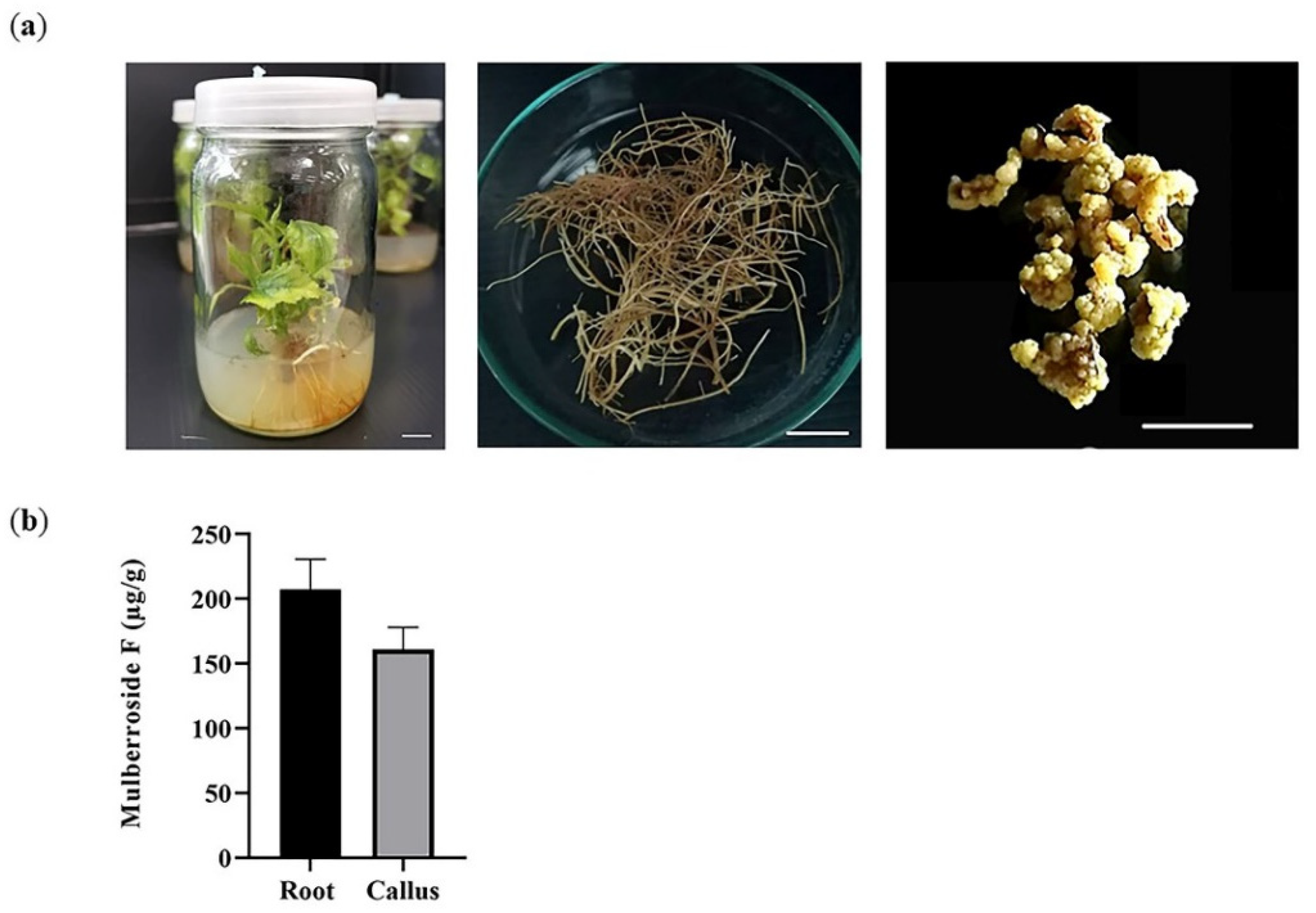
2.2. Mulberroside F Content in Different Parts of Mulberry Cultured In Vitro

Plant tissue culture has become a potential source for valuable bioactive compound production [21]. Thus, the amount of mulberroside F was investigated in mulberry plants cultured in vitro. Based on the data of soil-grown plants, root tissue was selected and compared with the callus (Figure 2a). The result showed that mulberroside F was found in both root (207.35 ± 23.42 µg g−1) and callus (160.97 ± 17.08 µg g−1) (Figure 2b). The root extracts contained a slightly higher mulberroside F than the callus; however, this was not statistically significant.

2.3. Evaluation of Anti-Tyrosinase Activity

In this study, the tyrosinase inhibitory activity of root and callus extracts was investigated using L-3,4-dihydroxyphenylalanine (L-DOPA) as a substrate. At a concentration of 5 mg mL−1, the tyrosinase inhibition activities by root and callus were 66.69% and 60.12%, respectively (Figure 3a). Moreover, the IC50 values of root and callus extracts were determined, and the values were 10.57 and 92.06 μg mL−1, respectively (Figure 3b,c). This result revealed that root and callus extracts possess anti-tyrosinase activity and mulberroside F is one of the bioactive constituents that could contribute to this inhibition.

2.4. UV Absorption Capacity

To evaluate the photoprotective activity, the UV absorption of mulberry extracts was determined between the range of 290–400 nm, which cover UVA (320–400 nm) and UVB (290–320 nm) regions, with intervals of 5 nm. As shown in Figure 4, the result suggested that root and callus extracts contain compounds responsible for UV absorption.

2.5. Assessment of Cytotoxicity Effects

Next, the cytotoxicity effects of root crude extract of Morus spp. were investigated using MTT assay in Vero and HaCaT cells. Vero is an African green monkey kidney cell that is widely used to validate toxicity of toxin [31,32] or cytotoxic effects of drugs [33] and natural products in mammalian cells [34,35]. HaCaT, on the other hand, is a type of immortalized human keratinocyte that has been frequently applied to examine the homeostasis of the epidermis [36,37]. As a result, crude extract of mulberry root showed no cytotoxicity in both HaCaT (Figure 5a) and Vero cells (Figure 5b).

3. Discussion

Plant tissue culture systems provide rapid and intensive scale production of phytochemicals for pharmaceutical and cosmeceutical applications [21,38]. In contrast to the conventionally cultivated mulberry, this system can produce uniform and disease-free plants with less variation in the content of bioactive compounds [39].
Using alternative sources such as callus and root cultures has become a new approach for secondary metabolite production. Root has been considered a good source of bioactive compounds, capable of scaling up the production through adventitious root or hairy root culture systems [40,41]. Callus culture is another choice for the enhanced production of secondary metabolites in the pharmaceuticals, cosmetic, and food industries. Several bioactive phytochemicals produced from callus cultures of medicinal plants can be used to treat various diseases, such as cancer, infectious diseases, and cardiovascular diseases [42]. Its advantage also includes the ability to be converted to single-cell suspension culture growing in shake flasks or bioreactors to enhance yield production [43].
Mulberry has long been considered a valuable resource for various food, beverage, cosmetic, and pharmaceutical industries, due to the presence of many phytochemicals with biological functions [11]. Previous studies demonstrated that root barks of mulberry are rich in mulberroside A, kuwanon G, and morusin [44]. Here, the mulberroside F was investigated in different parts of mulberry plants. Previously, mulberroside F was isolated from mulberry leaves. The researchers also reported that this compound could inhibit the activity of tyrosinase, which converts dopa to dopachrome in the biosynthetic activity of melanin [13]. However, there is still a lack of studies regarding the mulberroside F. In this study, the presence of mulberroside F in the roots of mulberry cultured in vitro was reported for the first time. Further, enhancement strategies such as cell suspension culture, hairy root culture, precursor feeding culture, or elicitors [25,45] could be utilized for the production of mulberroside F towards industrial needs.
The primary pigment responsible for the different pigmentations found in animal and human skin, hair, and eyes is melanin, produced by melanocytes via the melanogenesis pathway [46]. Tyrosinase is a rate-limiting, copper-containing enzyme that controls the production of melanin in the human body, which can lead to a variety of skin disorders when overproduced [6]. Thus, the compounds that inhibit melanin synthesis are expected to have cosmetic applications such as whitening agents [47]. Due to skin-whitening properties, the use of tyrosinase inhibitors is becoming increasingly important. Apart from kojic acid, which is commonly known as an inhibitor of tyrosinase, several bioactive compounds from different plant species, including quercetin, chalcones, arbutin, and stilbenes, have also been reported [6,48,49,50]. A previous study showed that the oxyresveratrol, oxyresveratrol-3-O-glucoside, and mulberroside A found in mulberry roots could suppress the expression of tyrosinase-related protein-1, and microphthalmia-associated transcription factor (MITF), which involves melanocyte survival and differentiation [51]. The present study further demonstrated that the extracts of root and callus derived from the in vitro culture of mulberry possess anti-tyrosinase activity, and that mulberroside F is one of the bioactive constituents that could contribute to this effect [14]. Additionally, the UV absorption property was also observed, suggesting that the root and callus extracts have a high potential to be used as natural skin care agents. Nevertheless, further study is required to gain insight into the role of mulberroside F on melanogenesis inhibitory mechanism.
Various biological properties of different mulberry extracts have been investigated and reported including medicinal potentials such as anti-cancer activity [52], anti-bacterial activity, and antioxidant activity [53]. Importantly, their toxicity needs to be assessed concurrently for a safe application. According to Li and coworkers (2018), M. alba leaf extract had no effect on mortality, hematology, and biochemical parameters in the exposed mice [54]. Moreover, mulberry plant extracts from the leaves and fruits had a protective effect against lead-induced damage to the tissues in rat testes [55]. Additionally, this study showed that culture mulberry root extract was not cytotoxic to Vero and HaCaT cells, but it significantly inhibited tyrosinase activity. Taken together, mulberry extracts include active compounds that may be used in cosmeceutical products.

4. Materials and Methods

4.1. Chemicals and Reagents

Mulberroside F standard was purchased from Chengdu Biopurify Phytochemicals, China. For anti-tyrosinase assays, mushroom tyrosinase (Sigma-Aldrich, St. Louis, MO, USA), kojic acid (Thermo Fisher Scientific, Waltham, MA, USA), and L-DOPA (Acros organics, Fair Lawn, NJ, USA) were used.

4.2. Plant Materials

Mulberry cv. Kamphaeng Saen-MB-42-1 was obtained from the Department of Biochemistry, Faculty of Science, Kasetsart University, Thailand (N 14.015619 E 99.958970), in April 2020. Shoot tips of soil-grown mulberry were excised and used to initiate in vitro culture.

4.3. Establishment of Mulberry Tissue Culture

Shoot tips were soaked in 70% ethanol for 1 min and sterilized with 0.1% mercury (II) chloride (HgCl2) for 10 min. Then, the explants were washed with distilled water and transferred into Murashige and Skoog (MS) medium (PhytoTechnology, Lenexa, KS, USA), containing 30 g/L of sucrose and 8 g/L of agar powder. The pH of the MS media was adjusted to 5.6–5.8 using 1 M sodium hydroxide (NaOH) (Fisher Chemical, Geel, Belgium) before autoclaving. All plant tissue samples were initially grown in a tissue culture room at 25 ± 1 °C under white fluorescent light (light/dark period of 16/8 h and ~75% RH) for 4–6 weeks.
For callus initiation, the 4-week-old roots from in vitro mulberry plants were excised and placed on solid MS media, supplemented with 0.5 mg/L 2,4-D and 0.5 mg/L BAP. Samples were kept in the dark and sub-cultured every 4 weeks.

4.4. Extraction and Quantification of Mulberroside F

Samples of leaves, roots, and callus of mulberry were collected and freeze-dried for 2 days. After that, the samples were powdered by tissue lyser (Retsch GmbH, Haan, Germany). The powdered samples (100 mg) were mixed well in 1 mL of 60 % ethyl alcohol by vortex for 5 min, sonicated in cold water for 20 min, and then incubated for an hour at room temperature. After centrifugation at 13,000 rpm, 4 °C for 10 min, the upper phase was collected to a new tube and stored at −20 °C for further experiments. For anti-tyrosinase activity, UV absorption capacity, and cytotoxicity assays, the extracts were evaporated into dryness with the use of a vacuum centrifuge (Eppendorf, Hamburg, Germany) to obtain the crude extract at a yield of 26%. The extract was resuspended in water prior to use.
HPLC analysis of mulberroside F in leaf, root, and callus was performed using a Waters Alliance Separations Module (Waters, Milford, MA, USA). Mulberroside F were separated on a 4.6 mm x 250 mm, 5 μm particle size, C18 column (Thermo Scientific, Waltham, MA, USA). Solvent A was ultrapure water and solvent B was 100% acetonitrile. The column temperature was maintained at 35 °C with 20 μL of sample. Flow rate was set as 1.0 mL min−1 and detected at 320 nm. Samples were filtered before HPLC injection by using 0.2 μm syringe filter. The concentrations of mulberroside F in each part of the mulberry plants were quantified using Empower® 3 software and the results were expressed as μg g−1 DW.

4.5. Evaluation of Anti-Tyrosinase Activity

Tyrosinase inhibition assays were performed with L-DOPA as substrate. First, the reaction combination of 160 μL of assay mixer containing 120 μL of phosphate buffer (20 mM, pH 6.8), 20 μL of the sample, and 20 μL of mushroom tyrosinase (100 U mL−1), was added to each well of the 96-well plate. Then, the microplate was preincubated at 25 °C for 10 min. After that, 20 μL of 2 mM L-DOPA (pH 6.8) was added to each well and further incubated for 20 min. The reaction was observed at 490 nm for dopachrome formation in the reaction mixture. In this experiment, kojic acid was used as a positive control. Each measurement was performed in triplicate (n = 3). The percentage of inhibition was calculated as previously described [56].

4.6. Investigation of UV Protective Activity

For the determination of UV-filtering potential, the UV absorption of each extract (1 mg mL−1) was measured between 290 and 400 nm, using a SparkTM 10M multimode microplate reader (Tecan, Männedorf, Switzerland).

4.7. Cytotoxicity Effects of Normal Cell Cultures

For cell culture and maintenance, normal African green monkey kidney fibroblast cells (Vero; ATCC® CCL-81™) and immortalized human keratinocyte cell line (HaCaT; CLS 300493) were cultured in Dulbecco’s Modified Eagle’s Medium (DMEM; Gibco, Invitrogen, Carlsbad, CA, USA), supplemented with 10 % fetal bovine serum (v/v), and 1% Antibiotic-antimycotic (v/v) (Gibco, Invitrogen, CA, USA) at 37 °C in the present of 5% CO2.
To test the cytotoxicity effects of normal cell cultures, Vero and HaCaT cells were seeded approximately 1.5 × 10⁴ cells per well into 96 well-plates (Falcon® a Corning brand, USA) and incubated for 24 h. All cells were then treated with 0 (distilled water only), 500, 1000, and 1500 μg /mL of crude extract. After 96 h of incubation, the mixing solutions were discarded; then, 4 μg of MTT solution was added and incubated for 3 h. Lastly, MTT solution (Invitrogen, USA) was replaced with 50 μL of dimethyl sulfoxide (Thermo Fisher Scientific, CA, USA) prior to the absorbance measurement at 570 nm by using a Infinite® M Nano microplate reader (Tecan, Männedorf, Switzerland).

4.8. Statistical Analysis

Data are presented as the mean ± standard error. The statistical analyses were performed using GraphPad Prism 8.0.1 software. Differences were considered significant at the p < 0.05 levels using Tukey’s multiple comparison test.

5. Conclusions

Plants have been considered rich sources of biologically active compounds with industry potential. In this study, mulberroside F was identified in the root and callus of mulberry cultured in vitro. The root and callus extracts also have the potential for cosmeceutical applications, due to the anti-tyrosinase activity and the high UV absorption property with no cytotoxic effects. Further studies are necessary for increasing the amount of mulberroside F, for instance, cell suspension culture, precursor feeding, or elicitation strategies.

Author Contributions

Conceptualization, N.P.T.-T. and W.P.P.; investigation, J.T., N.P., T.J., A.J., A.P., N.P.T.-T. and W.P.P.; data curation, J.T., T.J., A.J., N.P.T.-T. and W.P.P.; writing—original draft preparation, J.T., N.P.T.-T. and W.P.P.; writing—review and editing, N.P.T.-T. and W.P.P. All authors have read and agreed to the published version of the manuscript.

Funding

This research is supported by the International SciKU Branding (ISB), Faculty of Science, Kasetsart University.

Data Availability Statement

Not applicable.

Acknowledgments

This research is supported by the International SciKU Branding (ISB), Faculty of Science, Kasetsart University. Authors are also grateful to Associate Professor Amornrat Promboon (Biochemistry, KU) and Associate Professor Pakorn Wattana-Amorn (Chemistry, KU) for helpful discussions.

Conflicts of Interest

The authors declare no conflict of interest.

References

  1. Svobodová, A.; Psotová, J.; Walterová, D. Natural phenolics in the prevention of UV-induced skin damage. A review. Biomed. Pap. 2003, 147, 137–145. [Google Scholar] [CrossRef] [Green Version]
  2. Martin, K.I.; Glaser, D.A. Cosmeceuticals: The new medicine of beauty. Mo. Med. 2011, 108, 60–63. [Google Scholar]
  3. Seok, J.K.; Kwak, J.Y.; Choi, G.W.; An, S.M.; Kwak, J.-H.; Seo, H.-H.; Suh, H.-J.; Boo, Y.C. Scutellaria radix Extract as a Natural UV Protectant for Human Skin. Phytotherapy Res. 2016, 30, 374–379. [Google Scholar] [CrossRef] [PubMed]
  4. Korać, R.R.; Khambholja, K.M. Potential of herbs in skin protection from ultraviolet radiation. Pharmacogn. Rev. 2011, 5, 164–173. [Google Scholar] [CrossRef] [PubMed] [Green Version]
  5. Choquenet, B.; Couteau, C.; Paparis, E.; Coiffard, L.J.M. Quercetin and Rutin as Potential Sunscreen Agents: Determination of Efficacy by an in Vitro Method. J. Nat. Prod. 2008, 71, 1117–1118. [Google Scholar] [CrossRef]
  6. Pillaiyar, T.; Manickam, M.; Namasivayam, V. Skin whitening agents: Medicinal chemistry perspective of tyrosinase inhib-itors. J. Enzyme Inhib. Med. Chem. 2017, 32, 403–425. [Google Scholar] [CrossRef] [Green Version]
  7. García-Bores, A.; Avila, J. Natural products: Molecular mechanisms in the photochemoprevention of skin cancer. Rev. Lati-noamer. Quím. 2007, 36, 83–102. [Google Scholar]
  8. Sadia, H.; Ahmad, M.; Sultana, S.; Abdullah, A.Z.; Teong, L.; Zafar, M.; Bano, A. Nutrient and mineral assessment of edible wild fig and mulberry fruits. Fruits 2014, 69, 159–166. [Google Scholar] [CrossRef] [Green Version]
  9. Zhang, D.-Y.; Wan, Y.; Xu, J.-Y.; Wu, G.-H.; Li, L.; Yao, X.-H. Ultrasound extraction of polysaccharides from mulberry leaves and their effect on enhancing antioxidant activity. Carbohydr. Polym. 2016, 137, 473–479. [Google Scholar] [CrossRef]
  10. Nattapong, S.; Omboon, L. A new source of whitening agent from a Thai Mulberry plant and its betulinic acid quantitation. Nat. Prod. Res. 2008, 22, 727–734. [Google Scholar] [CrossRef]
  11. Memete, A.R.; Timar, A.V.; Vuscan, A.N.; Miere, F.; Venter, A.C.; Vicas, S.I. Phytochemical Composition of Different Botanical Parts of Morus Species, Health Benefits and Application in Food Industry. Plants 2022, 11, 152. [Google Scholar] [CrossRef]
  12. Chan, E.W.C.; Lye, P.-Y.; Wong, S.-K. Phytochemistry, pharmacology, and clinical trials of Morus alba. Chin. J. Nat. Med. 2016, 14, 17–30. [Google Scholar] [CrossRef] [PubMed]
  13. Ichihashi, M.; Ueda, M.; Budiyanto, A.; Bito, T.; Oka, M.; Fukunaga, M.; Tsuru, K.; Horikawa, T. UV-induced skin damage. Toxicology 2003, 189, 21–39. [Google Scholar] [CrossRef]
  14. Lee, S.H.; Choi, S.Y.; Kim, H.; Hwang, J.S.; Lee, B.G.; Gao, J.J.; Kim, S.Y. Mulberroside F Isolated from the Leaves of Morus alba Inhibits Melanin Biosynthesis. Biol. Pharm. Bull. 2002, 25, 1045–1048. [Google Scholar] [CrossRef] [Green Version]
  15. Chang, L.-W.; Juang, L.-J.; Wang, B.-S.; Wang, M.-Y.; Tai, H.-M.; Hung, W.-J.; Chen, Y.-J.; Huang, M.-H. Antioxidant and antityrosinase activity of mulberry (Morus alba L.) twigs and root bark. Food Chem. Toxicol. 2011, 49, 785–790. [Google Scholar] [CrossRef] [PubMed]
  16. Kim, J.-M.; Chang, S.-M.; Kim, I.-H.; Kim, Y.-E.; Hwang, J.-H.; Kim, K.-S.; Kim, W.-S. Design of optimal solvent for extraction of bio-active ingredients from mulberry leaves. Biochem. Eng. J. 2007, 37, 271–278. [Google Scholar] [CrossRef]
  17. Chang, T.-S. An Updated Review of Tyrosinase Inhibitors. Int. J. Mol. Sci. 2009, 10, 2440–2475. [Google Scholar] [CrossRef] [Green Version]
  18. de Souza, M.M.; Bittar, M.; Cechinel-Filho, V.; Yunes, R.A.; Messana, I.; Monache, F.D.; Ferrari, F. Antinociceptive Properties of Morusin, a Prenylflavonoid Isolated from Morus nigra Root Bark. Zeitschrift für Naturforschung C 2000, 55, 256–260. [Google Scholar] [CrossRef] [PubMed] [Green Version]
  19. Lee, D.; Yu, J.S.; Lee, S.R.; Hwang, G.S.; Kang, K.S.; Park, J.G.; Kim, H.Y.; Kim, K.H.; Yamabe, N. Beneficial Effects of Bioactive Compounds in Mulberry Fruits against Cisplatin-Induced Nephrotoxicity. Int. J. Mol. Sci. 2018, 19, 1117. [Google Scholar] [CrossRef] [PubMed] [Green Version]
  20. Hasnain, A.; Naqvi, S.A.H.; Ayesha, S.I.; Khalid, F.; Ellahi, M.; Iqbal, S.; Hassan, M.Z.; Abbas, A.; Adamski, R.; Markowska, D.; et al. Plants in vitro propagation with its applications in food, pharmaceuticals and cosmetic industries; current scenario and future approaches. Front. Plant Sci. 2022, 13, 3894. [Google Scholar] [CrossRef]
  21. Chandran, H.; Meena, M.; Barupal, T.; Sharma, K. Plant tissue culture as a perpetual source for production of industrially important bioactive compounds. Biotechnol. Rep. 2020, 26, e00450. [Google Scholar] [CrossRef]
  22. Fazili, M.A.; Bashir, I.; Ahmad, M.; Yaqoob, U.; Geelani, S.N. In vitro strategies for the enhancement of secondary metabolite production in plants: A review. Bull. Natl. Res. Cent. 2022, 46, 35. [Google Scholar] [CrossRef] [PubMed]
  23. Oseni, O.M.; Pande, V.; Nailwal, T.K. A Review on Plant Tissue Culture, A Technique for Propagation and Conservation of Endangered Plant Species. Int. J. Curr. Microbiol. Appl. Sci. 2018, 7, 3778–3786. [Google Scholar] [CrossRef]
  24. Palazón, J.; Cusidó, R.M.; Bonfill, M.; Mallol, A.; Moyano, E.; Morales, C.; Piñol, M. Elicitation of different Panax ginseng transformed root phenotypes for an improved ginsenoside production. Plant Physiol. Biochem. 2003, 41, 1019–1025. [Google Scholar] [CrossRef]
  25. Hu, X.; Neill, S.J.; Cai, W.; Tang, Z. Nitric oxide mediates elicitor-induced saponin synthesis in cell cultures of Panax ginseng. Funct. Plant Biol. 2003, 30, 901–907. [Google Scholar] [CrossRef] [PubMed]
  26. Tabata, H. Paclitaxel Production by Plant-Cell-Culture Technology. In Biomanufacturing; Springer: Berlin/Heidelberg, Germany, 2004. [Google Scholar] [CrossRef]
  27. Nandagopal, K.; Halder, M.; Dash, B.; Nayak, S.; Jha, S. Biotechnological Approaches for Production of Anti-Cancerous Compounds Resveratrol, Podophyllotoxin and Zerumbone. Curr. Med. Chem. 2018, 25, 4693–4717. [Google Scholar] [CrossRef] [PubMed]
  28. Komaikul, J.; Kitisripanya, T.; Tanaka, H.; Sritularak, B.; Putalun, W. Enhanced mulberroside A production from cell sus-pension and root cultures of Morus alba using elicitation. Nat. Prod. Commun. 2015, 10, 1253–1256. [Google Scholar] [PubMed] [Green Version]
  29. Sonthisut, M.; Wongpanya, R.; Phonphoem, A.; Phonphoem, W.P. Enhancement of 1-deoxynojirimycin production in mulberry (Morus spp.) using LED irradiation. Plant Cell Tissue Organ Cult. 2021, 148, 167–176. [Google Scholar] [CrossRef]
  30. El-Mawla, A.M.A.A.; Mohamed, K.M.; Mostafa, A.M. Induction of Biologically Active Flavonoids in Cell Cultures of Morus nigra and Testing their Hypoglycemic Efficacy. Sci. Pharm. 2011, 79, 951–961. [Google Scholar] [CrossRef] [Green Version]
  31. Godoy, M.G.; Fernandes, K.V.; Gutarra, M.L.; Melo, E.J.; Castro, A.M.; Machado, O.L.; Freire, D.M. Use of Vero cell line to verify the biodetoxification efficiency of castor bean waste. Process. Biochem. 2012, 47, 578–584. [Google Scholar] [CrossRef] [Green Version]
  32. Menezes, C.; Valério, E.; Dias, E. The Kidney Vero-E6 Cell Line: A Suitable Model to Study the Toxicity of Microcystins. In New Insights into Toxicity and Drug Testing; Sivakumar, G., Ed.; IntechOpen: Rijeka, Croatia, 2013. [Google Scholar] [CrossRef]
  33. Freire, P.F.; Labrador, V.; Martín, J.P.; Hazen, M. Cytotoxic effects in mammalian Vero cells exposed to pentachlorophenol. Toxicology 2005, 210, 37–44. [Google Scholar] [CrossRef] [PubMed]
  34. Khamwut, A.; Jevapatarakul, D.; Reamtong, O.; T-Thienprasert, N.P. In vitro evaluation of anti-epidermoid cancer activity of Acanthus ebracteatus protein hydrolysate and their effects on apoptosis and cellular proteins. Oncol. Lett. 2019, 18, 3128–3136. [Google Scholar] [CrossRef] [Green Version]
  35. Budchart, P.; Khamwut, A.; Sinthuvanich, C.; Ratanapo, S.; Poovorawan, Y.; T-Thienprasert, N.P. Partially Purified Gloriosa superba Peptides Inhibit Colon Cancer Cell Viability by Inducing Apoptosis Through p53 Upregulation. Am. J. Med. Sci. 2017, 354, 423–429. [Google Scholar] [CrossRef]
  36. Boukamp, P.; Petrussevska, R.T.; Breitkreutz, D.; Hornung, J.; Markham, A.; Fusenig, N.E. Normal Keratinization in a Spontaneously Immortalized Aneuploid Human Keratinocyte Cell Line. J. Cell Biol. 1988, 106, 761–771. [Google Scholar] [CrossRef] [Green Version]
  37. Colombo, I.; SanGiovanni, E.; Maggio, R.; Mattozzi, C.; Zava, S.; Corbett, Y.; Fumagalli, M.; Carlino, C.; Corsetto, P.A.; Scaccabarozzi, D.; et al. HaCaT Cells as a Reliable In Vitro Differentiation Model to Dissect the Inflammatory/Repair Response of Human Keratinocytes. Mediat. Inflamm. 2017, 2017, 7435621. [Google Scholar] [CrossRef] [Green Version]
  38. Karuppusamy, S. A review on trends in production of secondary metabolites from higher plants by in vitro tissue, organ and cell cultures. J. Med. Plants Res. 2009, 3, 1222–1239. [Google Scholar]
  39. Espinosa-Leal, C.A.; Puente-Garza, C.A.; García-Lara, S. In vitro plant tissue culture: Means for production of biological active compounds. Planta 2018, 248, 1–18. [Google Scholar] [CrossRef]
  40. Hussain, M.J.; Abbas, Y.; Nazli, N.; Fatima, S.; Drouet, S.; Hano, C.; Abbasi, B.H. Root Cultures, a Boon for the Production of Valuable Compounds: A Comparative Review. Plants 2022, 11, 439. [Google Scholar] [CrossRef]
  41. Jiang, Y.J.; Piao, X.C.; Liu, J.S.; Jiang, J.; Lian, Z.X.; Kim, M.J.; Lian, M.L. Bioactive compound production by adventitious root culture of Oplopanax elatus in balloon-type airlift bioreactor systems and bioactivity property. Plant Cell Tissue Organ Cult. 2015, 123, 413–425. [Google Scholar] [CrossRef]
  42. Efferth, T. Biotechnology Applications of Plant Callus Cultures. Engineering 2018, 5, 50–59. [Google Scholar] [CrossRef]
  43. Fischer, R.; Emans, N.; Schuster, F.; Hellwig, S.; Drossard, J. Towards molecular farming in the future: Using plant-cell-suspension cultures as bioreactors. Biotechnol. Appl. Biochem. 1999, 30, 109–112. [Google Scholar]
  44. Kim, J.-H.; Doh, E.-J.; Lee, G. Quantitative Comparison of the Marker Compounds in Different Medicinal Parts of Morus alba L. Using High-Performance Liquid Chromatography-Diode Array Detector with Chemometric Analysis. Molecules 2020, 25, 5592. [Google Scholar] [CrossRef]
  45. Shakya, P.; Marslin, G.; Siram, K.; Beerhues, L.; Franklin, G. Elicitation as a tool to improve the profiles of high-value secondary metabolites and pharmacological properties of Hypericum perforatum. J. Pharm. Pharmacol. 2019, 71, 70–82. [Google Scholar] [CrossRef] [Green Version]
  46. D’Ischia, M.; Wakamatsu, K.; Cicoira, F.; Di Mauro, E.; Garcia-Borron, J.C.; Commo, S.; Galván, I.; Ghanem, G.; Kenzo, K.; Meredith, P.; et al. Melanins and melanogenesis: From pigment cells to human health and technological applications. Pigment. Cell Melanoma Res. 2015, 28, 520–544. [Google Scholar] [CrossRef] [Green Version]
  47. Yamauchi, K.; Mitsunaga, T.; Batubara, I. Isolation, Identification and Tyrosinase Inhibitory Activities of the Extractives from Allamanda cathartica. Nat. Resour. 2011, 02, 167–172. [Google Scholar] [CrossRef] [Green Version]
  48. Lee, S.Y.; Baek, N.; Nam, T.-G. Natural, semisynthetic and synthetic tyrosinase inhibitors. J. Enzym. Inhib. Med. Chem. 2015, 31, 1–13. [Google Scholar] [CrossRef]
  49. Gug, K. Physiological and Whitening Effects of Morus alba Extracts. J. Chosun Nat. Sci. 2012, 5, 46–52. [Google Scholar] [CrossRef] [Green Version]
  50. Cui, T.; Nakamura, K.; Ma, L.; Li, J.-Z.; Kayahara, H. Analyses of Arbutin and Chlorogenic Acid, the Major Phenolic Constituents in Oriental Pear. J. Agric. Food Chem. 2005, 53, 3882–3887. [Google Scholar] [CrossRef]
  51. Park, K.-T.; Kim, J.-K.; Hwang, D.; Yoo, Y.; Lim, Y.-H. Inhibitory effect of mulberroside A and its derivatives on melanogenesis induced by ultraviolet B irradiation. Food Chem. Toxicol. 2011, 49, 3038–3045. [Google Scholar] [CrossRef]
  52. Yu, M.-H.; Tsai, M.-C.; Wang, C.-C.; Wu, S.-W.; Chang, Y.-J.; Wu, C.-H.; Wang, C.-J. Mulberry Leaf Polyphenol Extract and Rutin Induces Autophagy Regulated by p53 in Human Hepatoma HepG2 Cells. Pharmaceuticals 2021, 14, 1310. [Google Scholar] [CrossRef]
  53. Suriyaprom, S.; Kaewkod, T.; Promputtha, I.; Desvaux, M.; Tragoolpua, Y. Evaluation of antioxidant and antibacterial activ-ities of white mulberry. Plants 2021, 10, 2736. [Google Scholar] [CrossRef]
  54. Li, Y.; Zhang, X.; Liang, C.; Hu, J.; Yu, Z. Safety evaluation of mulberry leaf extract: Acute, subacute toxicity and genotoxicity studies. Regul. Toxicol. Pharmacol. 2018, 95, 220–226. [Google Scholar] [CrossRef]
  55. Ramis, E.S.; Hammoud, G.M.; ElSawy, K.M. In vitro and In vivo Studies on Mulberry Extracts: Evaluation of Chemical and Anticancer Activities and Attenuation of Lead Toxicity. Cerebellum 2018, 4, 5. [Google Scholar] [CrossRef]
  56. Tu, P.T.B.; Tawata, S. Anti-Oxidant, Anti-Aging, and Anti-Melanogenic Properties of the Essential Oils from Two Varieties of Alpinia zerumbet. Molecules 2015, 20, 16723–16740. [Google Scholar] [CrossRef]
Figure 1. Mulberroside F (moracin M-6, 3′-di-O-beta-D-glucopyranoside) from Morus species. (a) Chemical structure; (b) HPLC chromatogram of mulberroside F standard.
Plants 12 00146 g001
Figure 2. Quantification of mulberroside F in mulberry cultured in vitro. (a) Plant material used in this study. 6-week-old mulberry (left), root tissue (middle), and callus (right); (b) HPLC analysis of mulberroside F. Error bars were from 3–5 replicates. Scale bar: 1 cm.
Plants 12 00146 g002
Figure 3. The tyrosinase inhibitory effects of the extracts from root and callus of the mulberry. (a) Tyrosinase inhibitory activity of root and callus extracts for the catalysis of L-DOPA. Kojic acid was included as a positive control. Different lowercase letters indicate significant differences between each part analyzed (p < 0.05). Error bars represent the standard error of the mean of 3 replicates; (b) IC50 curve of mulberry root extract; (c) IC50 curve of callus extract. The values were calculated by nonlinear regression using GraphPad Prism 8.0.1.
Plants 12 00146 g003
Figure 4. Ultraviolet (UV) absorption spectra of root and callus extracts.
Plants 12 00146 g004
Figure 5. The cytotoxic effects of mulberry root extract on tissue culture cells. (a) HaCaT (b) Vero. The results are the means of two independent experiments, and each data point was obtained in triplicate. Error bars represent the standard error of the mean.
Plants 12 00146 g005
Disclaimer/Publisher’s Note: The statements, opinions and data contained in all publications are solely those of the individual author(s) and contributor(s) and not of MDPI and/or the editor(s). MDPI and/or the editor(s) disclaim responsibility for any injury to people or property resulting from any ideas, methods, instructions or products referred to in the content.

