

Effects of COVID-19 on the Liver and Mortality in Patients with SARS-CoV-2 Pneumonia Caused by Delta and Non-Delta Variants: An Analysis in a Single Centre
 ,
,  , ,
, ,
Abstract
:1. Introduction
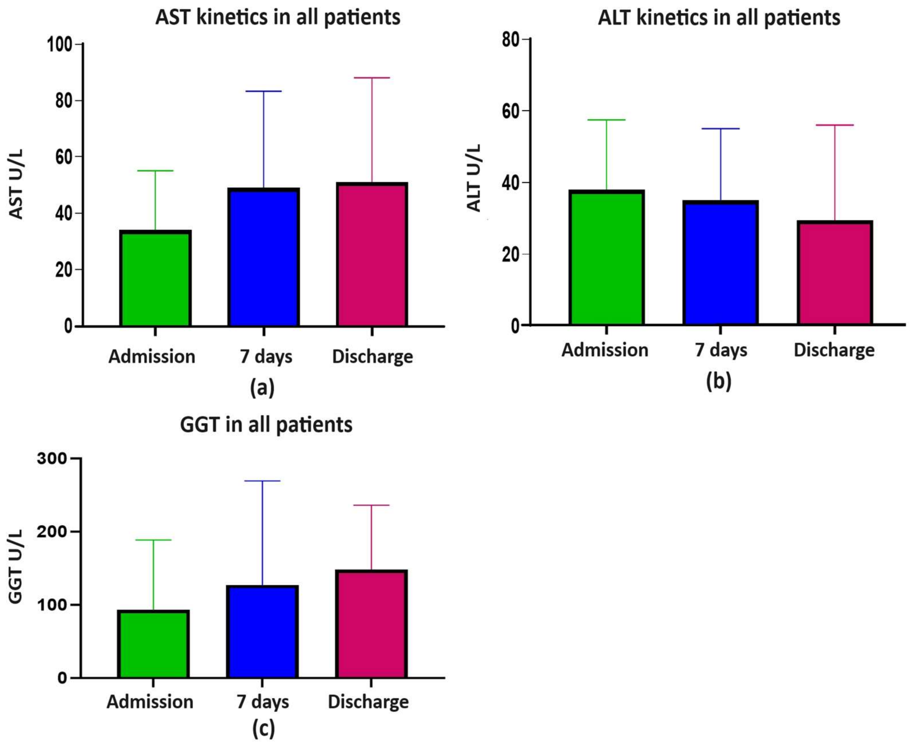
2. Results
2.1. Patient Comparison in the Delta and Non-Delta Waves
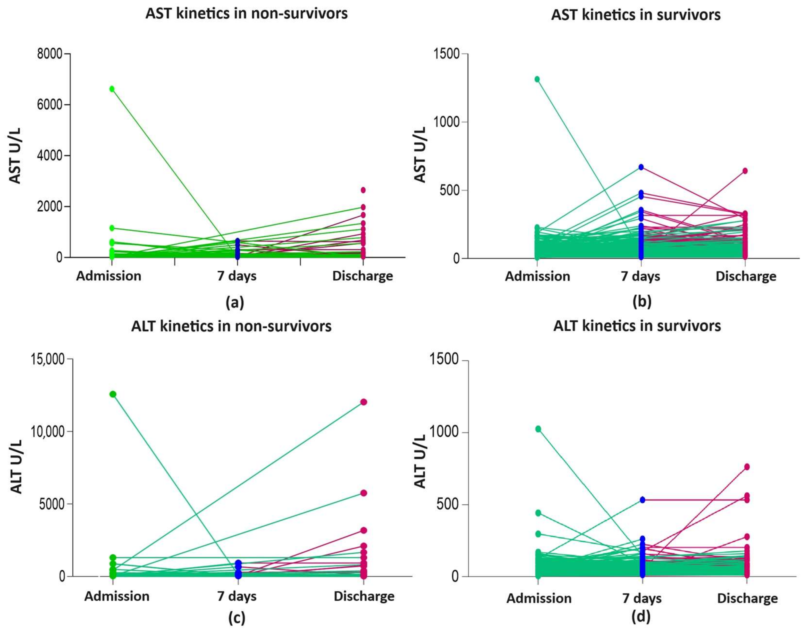
2.2. Patient Comparison between Non-Survivors and Survivors
3. Discussion
3.1. Patient Comparison in the Delta and Non-Delta Waves
3.2. Prognosis and Liver Injury
3.3. DILI
3.4. Study Limitations and Strengths
4. Materials and Methods
4.1. Study Design and Setting
4.2. Participants and Variables
4.3. Statistical Analysis
5. Conclusions
Supplementary Materials
Author Contributions
Funding
Institutional Review Board Statement
Informed Consent Statement
Data Availability Statement
Acknowledgments
Conflicts of Interest
References
- Idalsoaga, F.; Ayares, G.; Arab, J.P.; Díaz, L.A. COVID-19 and indirect liver injury: A narrative synthesis of the evidence. J. Clin. Transl. Hepatol. 2021, 9, 760–768. [Google Scholar] [CrossRef]
- Vitiello, A.; La Porta, R.; D’Aiuto, V.; Ferrara, F. The risks of liver injury in COVID-19 patients and pharmacological management to reduce or prevent the damage induced. Egypt. Liver J. 2021, 11, 11. [Google Scholar] [CrossRef] [PubMed]
- Hamming, I.; Timens, W.; Bulthuis, M.L.; Lely, A.T.; Navis, G.; van Goor, H. Tissue distribution of ACE2 protein, the functional receptor for SARS coronavirus. A first step in understanding SARS pathogenesis. J. Pathol. 2004, 203, 631–637. [Google Scholar] [CrossRef] [PubMed]
- Mihai, N.; Tiliscan, C.; Visan, C.A.; Stratan, L.; Ganea, O.; Arama, S.S.; Lazar, M.; Arama, V. Evaluation of Drug-Induced Liver Injury in Hospitalized Patients with SARS-CoV-2 Infection. Microorganisms 2022, 10, 2045. [Google Scholar] [CrossRef] [PubMed]
- Zghal, M.; Bouhamed, M.; Mellouli, M.; Triki, M.; Kallel, R.; Ayedi, L.; Boudawara, T.S.; Makni, S. Liver injury in COVID-19: Pathological findings. Pan Afr. Med. J. 2022, 41, 56. [Google Scholar] [CrossRef] [PubMed]
- Xu, W.; Huang, C.; Fei, L.; Li, Q.; Chen, L. Dynamic Changes in Liver Function Tests and Their Correlation with Illness Severity and Mortality in Patients with COVID-19: A Retrospective Cohort Study. Clin. Interv. Aging 2021, 16, 675–685. [Google Scholar] [CrossRef] [PubMed]
- Wang, X.; Lei, J.; Li, Z.; Yan, L. Potential effects of coronaviruses on the liver: An update. Front. Med. 2021, 8, 651658. [Google Scholar] [CrossRef] [PubMed]
- Deng, H.; Lin, H.; Mai, Y.; Liu, H.; Chen, W. Clinical features and predictive factors related to liver injury in SARS-CoV-2 Delta and Omicron variant-infected patients. Eur. J. Gastroenterol. Hepatol. 2022, 34, 933–939. [Google Scholar] [CrossRef]
- Sun, J.; Aghemo, A.; Forner, A.; Valenti, L. COVID-19 and liver disease. Liver Int. 2020, 40, 1278–1281. [Google Scholar] [CrossRef]
- Zhang, C.; Shi, L.; Wang, F.S. Liver injury in COVID-19: Management and challenges. Lancet Gastroenterol. Hepatol. 2020, 5, 428–430. [Google Scholar] [CrossRef]
- Liatsos, G.D. SARS-CoV-2 induced liver injury: Incidence, risk factors, impact on COVID-19 severity and prognosis in different population groups. World J. Gastroenterol. 2023, 29, 2397–2432. [Google Scholar] [CrossRef] [PubMed]
- Ekpanyapong, S.; Bunchorntavakul, C.; Reddy, K.R. COVID-19 and the liver: Lessons learnt from the EAST and the WEST, a year later. J. Viral Hepat. 2022, 29, 4–20. [Google Scholar] [CrossRef] [PubMed]
- Salton, F.; Confalonieri, P.; Campisciano, G.; Cifaldi, R.; Rizzardi, C.; Generali, D.; Pozzan, R.; Tavano, S.; Bozzi, C.; Lapadula, G.; et al. Cytokine Profiles as Potential Prognostic and Therapeutic Markers in SARS-CoV-2-Induced ARDS. J. Clin. Med. 2022, 11, 2951. [Google Scholar] [CrossRef] [PubMed]
- Amri, N.; Bégin, R.; Tessier, N.; Vachon, L.; Villeneuve, L.; Bégin, P.; Bazin, R.; Loubaki, L.; Martel, C. Use of Early Donated COVID-19 Convalescent Plasma Is Optimal to Preserve the Integrity of Lymphatic Endothelial Cells. Pharmaceuticals 2022, 15, 365. [Google Scholar] [CrossRef] [PubMed]
- Kotfis, K.; Karolak, I.; Lechowicz, K.; Zegan-Barańska, M.; Pikulska, A.; Niedźwiedzka-Rystwej, P.; Kawa, M.; Sieńko, J.; Szylińska, A.; Wiśniewska, M. Mineralocorticoid Receptor Antagonist (Potassium Canrenoate) Does Not Influence Outcome in the Treatment of COVID-19-Associated Pneumonia and Fibrosis-A Randomized Placebo Controlled Clinical Trial. Pharmaceuticals 2022, 15, 200. [Google Scholar] [CrossRef] [PubMed]
- Chew, M.; Tang, Z.; Radcliffe, C.; Caruana, D.; Doilicho, N.; Ciarleglio, M.M.; Deng, Y.; Garcia-Tsao, G. Significant Liver Injury During Hospitalization for COVID-19 Is Not Associated with Liver Insufficiency or Death. Clin. Gastroenterol. Hepatol. 2021, 19, 2182–2191.e7. [Google Scholar] [CrossRef] [PubMed]
- Marjot, T.; Webb, G.J.; Barritt, A.S., 4th; Moon, A.M.; Stamataki, Z.; Wong, V.W.; Barnes, E. COVID-19 and liver disease: Mechanistic and clinical perspectives. Nat. Rev. Gastroenterol. Hepatol. 2021, 18, 348–364. [Google Scholar] [CrossRef]
- Baldelli, L.; Marjot, T.; Barnes, E.; Barritt, A.S.; Webb, G.J.; Moon, A.M. SARS-CoV-2 Infection and Liver Disease: A Review of Pathogenesis and Outcomes. Gut Liver 2023, 17, 12–23. [Google Scholar] [CrossRef]
- Wanner, N.; Andrieux, G.; Badia-I-Mompel, P.; Edler, C.; Pfefferle, S.; Lindenmeyer, M.T.; Schmidt-Lauber, C.; Czogalla, J.; Wong, M.N.; Okabayashi, Y.; et al. Molecular consequences of SARS-CoV-2 liver tropism. Nat. Metab. 2022, 4, 310–319. [Google Scholar] [CrossRef]
- Luxenburger, H.; Thimme, R. SARS-CoV-2 and the liver: Clinical and immunological features in chronic liver disease. Gut 2023, 72, 1783–1794. [Google Scholar] [CrossRef]
- Bergamaschi, L.; Mescia, F.; Turner, L.; Hanson, A.L.; Kotagiri, P.; Dunmore, B.J.; Cambridge Institute of Therapeutic Immunology and Infectious Disease-National Institute of Health Research (CITIID-NIHR) COVID BioResource Collaboration; Göttgens, B.; Toshner, M.; Hess, C.; et al. Longitudinal analysis reveals that delayed bystander CD8+ T cell activation and early immune pathology distinguish severe COVID-19 from mild disease. Immunity 2021, 54, 1257–1275.e8. [Google Scholar] [CrossRef] [PubMed]
- Laffont-Lozes, P.; Laureillard, D.; Loubet, P.; Stephan, R.; Chiaruzzi, M.; Clemmer, E.; Martin, A.; Roger, C.; Muller, L.; Claret, P.G.; et al. Effect of Tocilizumab on Mortality in Patients with SARS-CoV-2 Pneumonia Caused by Delta or Omicron Variants: A Propensity-Matched Analysis in Nimes University Hospital, France. Antibiotics 2023, 12, 88. [Google Scholar] [CrossRef] [PubMed]
- Cai, Q.; Huang, D.; Yu, H.; Zhu, Z.; Xia, Z.; Su, Y.; Li, Z.; Zhou, G.; Gou, J.; Qu, J.; et al. COVID-19: Abnormal liver function tests. J. Hepatol. 2020, 73, 566–574. [Google Scholar] [CrossRef] [PubMed]
- Weber, S.; Hellmuth, J.; Scherer, C.; Muenchhoff, M.; Mayerle, J.; Gerbes, A. Liver function test abnormalities at hospital admission are associated with severe course of SARS-CoV-2 infection: A prospective cohort study. Gut 2021, 70, 1925–1932. [Google Scholar] [CrossRef] [PubMed]
- Vijayaraghavan, B.K.T.; Bishnu, S.; Baruch, J.; Citarella, B.W.; Kartsonaki, C.; Meeyai, A.; Zubair, M.; Ohshimo, S.; Al-Fares, A.; Calvache, J.A.; et al. ISARIC Clinical Characterisation Group. Liver injury in hospitalized patients with COVID-19: An International observational cohort study. PLoS ONE 2023, 18, e0277859. [Google Scholar]
- Ponziani, F.; Del Zompo, F.; Nesci, A.; Santopaolo, F.; Ianiro, G.; Pompili, M.; Gasbarrini, A.; “Gemelli against COVID-19” group. Liver involvement is not associated with mortality: Results from a large cohort of SARS-CoV-2-positive patients. Aliment. Pharmacol. Ther. 2020, 52, 1060–1068. [Google Scholar] [CrossRef]
- Fu, Y.; Zhu, R.; Bai, T.; Han, P.; He, Q.; Jing, M.; Xiong, X.; Zhao, X.; Quan, R.; Chen, C.; et al. Clinical features of patients infected with coronavirus disease 2019 with elevated liver biochemistries: A multicenter, retrospective study. Hepatology 2021, 73, 1509–1520. [Google Scholar] [CrossRef]
- Wijarnpreecha, K.; Ungprasert, P.; Panjawatanan, P.; Harnois, D.; Zaver, H.; Ahmed, A.; Kim, D. COVID-19 and liver injury: A meta-analysis. Eur. J. Gastroenterol. Hepatol. 2021, 33, 990–995. [Google Scholar] [CrossRef]
- Imam, M.; Almalki, Z.; Alzahrani, A.; Al-Ghamdi, S.; Falemban, A.; Alanazi, I.; Shahzad, N.; Muhammad Alrooqi, M.; Jabeen, Q.; Shahid, I. COVID-19 and severity of liver diseases: Possible crosstalk and clinical implications. Int. Immunopharmacol. 2023, 121, 110439. [Google Scholar] [CrossRef]
- Kulkarni, A.V.; Kumar, P.; Tevethia, H.V.; Premkumar, M.; Arab, J.P.; Candia, R.; Talukdar, R.; Sharma, M.; Qi, X.; Rao, P.N.; et al. Systematic review with meta-analysis: Liver manifestations and outcomes in COVID-19. Aliment. Pharmacol. Ther. 2020, 52, 584–599. [Google Scholar] [CrossRef]
- Briciu, V.; Topan, A.; Calin, M.; Dobrota, R.; Leucuta, D.C.; Lupse, M. Comparison of COVID-19 Severity in Vaccinated and Unvaccinated Patients during the Delta and Omicron Wave of the Pandemic in a Romanian Tertiary Infectious Diseases Hospital. Healthcare 2023, 11, 373. [Google Scholar] [CrossRef] [PubMed]
- Roca-Fernández, A.; Dennis, A.; Nicholls, R.; McGonigle, J.; Kelly, M.; Banerjee, R.; Sanyal, A.J. Hepatic Steatosis, Rather Than Underlying Obesity, Increases the Risk of Infection and Hospitalization for COVID-19. Front. Med. 2021, 29, 636637. [Google Scholar] [CrossRef] [PubMed]
- Ebrahimi, F.; Simon, T.G.; Hagström, H.; Söderling, J.; Wester, A.; Roelstraete, B.; Ludvigsson, J.F. Risk of Severe Infection in Patients With Biopsy-proven Nonalcoholic Fatty Liver Disease—A Population-based Cohort Study. Clin. Gastroenterol. Hepatol. 2023, 21, 3346–3355.e19. [Google Scholar] [CrossRef] [PubMed]
- Lamers, M.M.; Haagmans, B.L. SARS-CoV-2 pathogenesis. Nat. Rev. Microbiol. 2022, 20, 270–284. [Google Scholar] [CrossRef] [PubMed]
- Dufour, J.F.; Marjot, T.; Becchetti, C.; Tilg, H. COVID-19 and liver disease. Gut 2022, 71, 2350–2362. [Google Scholar] [CrossRef] [PubMed]
- Moga, T.V.; Foncea, C.; Bende, R.; Popescu, A.; Burdan, A.; Heredea, D.; Danilă, M.; Miutescu, B.; Ratiu, I.; Bizerea-Moga, T.O.; et al. Impact of COVID-19 on Patients with Decompensated Liver Cirrhosis. Diagnostics 2023, 13, 600. [Google Scholar] [CrossRef] [PubMed]
- Zhao, S.W.; Li, Y.M.; Li, Y.L.; Su, C. Liver injury in COVID-19: Clinical features, potential mechanisms, risk factors and clinical treatments. World J. Gastroenterol. 2023, 29, 241–256. [Google Scholar] [CrossRef] [PubMed]
- Bhaskaran, K.; Bacon, S.; Evans, S.; Bates, C.; Rentsch, C.; MacKenna, B.; Tomlinson, L.; Walker, A.J.; Schultze, A.; Morton, C.E.; et al. Factors associated with deaths due to COVID-19 versus other causes: Population-based cohort analysis of UK primary care data and linked national death registrations within the OpenSAFELY platform. Lancet Reg. Health Eur. 2021, 6, 100109. [Google Scholar] [CrossRef]
- Bennett, T.D.; Moffitt, R.A.; Hajagos, J.G.; Amor, B.; Anand, A.; Bissell, M.M.; Bradwell, K.R.; Bremer, C.; Byrd, J.B.; Denham, A.; et al. National COVID Cohort Collaborative (N3C) Consortium. Clinical Characterization and Prediction of Clinical Severity of SARS-CoV-2 Infection among US Adults Using Data From the US National COVID Cohort Collaborative. JAMA Netw. Open 2021, 4, e2116901. [Google Scholar] [CrossRef]
- Radulescu, A.; Lupse, M.; Istrate, A.; Calin, M.; Topan, A.; Kormos, N.F.; Macicasan, R.V.; Briciu, V. Validation of Mortality Scores among High-Risk COVID-19 Patients: A Romanian Retrospective Study in the First Pandemic Year. J. Clin. Med. 2022, 11, 5630. [Google Scholar] [CrossRef]
- Nab, L.; Parker, E.P.K.; Andrews, C.D.; Hulme, W.J.; Fisher, L.; Morley, J.; Mehrkar, A.; MacKenna, B.; Inglesby, P.; Morton, C.E.; et al. Changes in COVID-19-related mortality across key demographic and clinical subgroups in England from 2020 to 2022: A retrospective cohort study using the OpenSAFELY platform. Lancet Public Health 2023, 8, e364–e377. [Google Scholar] [CrossRef] [PubMed]
- Gottlieb, R.L.; Vaca, C.E.; Paredes, R.; Mera, J.; Webb, B.J.; Perez, G.; Oguchi, G.; Ryan, P.; Nielsen, B.U.; Brown, M.; et al. GS-US-540-9012 (PINETREE) Investigators. Early Remdesivir to Prevent Progression to Severe Covid-19 in Outpatients. N. Engl. J. Med. 2022, 386, 305–315. [Google Scholar] [CrossRef] [PubMed]
- Turtle, L.; Thorpe, M.; Drake, T.; Swets, M.; Palmieri, C.; Russell, C.; Ho, A.; Aston, S.; Wootton, D.G.; Richter, A.; et al. Outcome of COVID-19 in hospitalised immunocompromised patients: An analysis of the WHO ISARIC CCP-UK prospective cohort study. PLoS Med. 2023, 20, e1004086. [Google Scholar] [CrossRef] [PubMed]
- Institutul National de Sanatate Publica (INSP) (The Romanian Nationale Institute of Public Health). Informare Privind Cazurile de COVID-19 Confirmate cu Variante ale SARS-CoV-2 Care Determina Îngrijorare (VOC). Available online: https://www.cnscbt.ro/index.php/analiza-cazuri-confirmate-covid19/3437-s-13-2023-informare-cazuri-cu-variante-de-ingrijorare-voc-si-variante-de-interes-voi/file (accessed on 5 May 2023).
- Liakina, V.; Stundiene, I.; Milaknyte, G.; Bytautiene, R.; Reivytyte, R.; Puronaite, R.; Urbanoviciute, G.; Kazenaite, E. Effects of COVID-19 on the liver: The experience of a single center. World J. Gastroenterol. 2022, 28, 5735–5749. [Google Scholar] [CrossRef] [PubMed]
- Lei, F.; Liu, Y.M.; Zhou, F.; Qin, J.J.; Zhang, P.; Zhu, L.; Zhang, X.J.; Cai, J.; Lin, L.; Ouyang, S.; et al. Longitudinal Association Between Markers of Liver Injury and Mortality in COVID-19 in China. Hepatology 2020, 72, 389–398. [Google Scholar] [CrossRef] [PubMed]
- Vadiraj, P.K.; Thareja, S.; Raman, N.; Karantha, S.C.; Jayaraman, M.; Vardhan, V. Does Raised Transaminases Predict Severity and Mortality in Patients with COVID-19? J. Clin. Exp. Hepatol. 2022, 12, 1114–1123. [Google Scholar] [CrossRef] [PubMed]
- Yanez, N.D.; Weiss, N.S.; Romand, J.A.; Treggiari, M.M. COVID-19 mortality risk for older men and women. BMC Public Health 2020, 20, 1742. [Google Scholar] [CrossRef]
- Shehab, M.; Alrashed, F.; Shuaibi, S.; Alajmi, D.; Barkun, A. Gastroenterological and hepatic manifestations of patients with COVID-19, prevalence, mortality by country, and intensive care admission rate: Systematic review and meta-analysis. BMJ Open Gastroenterol. 2021, 8, e000571. [Google Scholar] [CrossRef]
- Briciu, V.; Leucuta, D.-C.; Muntean, M.; Radulescu, A.; Cismaru, C.; Topan, A.; Herbel, L.; Horvat, M.; Calin, M.; Dobrota, R.; et al. Evolving Clinical Manifestations and Outcomes in COVID-19 Patients: A Comparative Analysis of SARS-CoV-2 Variant Waves in a Romanian Hospital Setting. Pathogens 2023, 12, 1453. [Google Scholar] [CrossRef]
- Malik, P.; Patel, U.; Mehta, D.; Patel, N.; Kelkar, R.; Akrmah, M.; Gabrilove, J.L.; Sacks, H. Biomarkers and outcomes of COVID-19 hospitalisations: Systematic review and meta-analysis. BMJ Evid.-Based Med. 2021, 26, 107–108. [Google Scholar] [CrossRef]
- Paranga, T.G.; Pavel-Tanasa, M.; Constantinescu, D.; Plesca, C.E.; Petrovici, C.; Miftode, I.L.; Moscalu, M.; Cianga, P.; Miftode, E.G. Comparison of C-reactive protein with distinct hyperinflammatory biomarkers in association with COVID-19 severity, mortality and SARS-CoV-2 variants. Front. Immunol. 2023, 14, 1213246. [Google Scholar] [CrossRef] [PubMed]
- Loomba, R.S.; Villarreal, E.G.; Farias, J.S.; Aggarwal, G.; Aggarwal, S.; Flores, S. Serum biomarkers for prediction of mortality in patients with COVID-19. Ann. Clin. Biochem. 2022, 59, 15–22. [Google Scholar] [CrossRef] [PubMed]
- Martínez-Sanz, J.; Muriel, A.; Ron, R.; Herrera, S.; Perez-Molina, J.A.; Moreno, S.; Serrano-Villar, S. Effects of tocilizumab on mortality in hospitalized patients with COVID-19: A multicentre cohort study. Clin. Microbiol. Infect. 2021, 27, 238–243. [Google Scholar] [CrossRef]
- Moreno Diaz, R.; Amor García, M.A.; Teigell Muñoz, F.J.; Saldana Perez, L.E.; Mateos Gonzalez, M.; Melero Bermejo, J.A.; López Hernández, A.; Reyes Marquez, L.; De Guzman García-Monge, M.T.; Perez Quero, J.L.; et al. Does timing matter on tocilizumab administration? Clinical, analytical and radiological outcomes in COVID-19. Eur. J. Hosp. Pharm. 2021, 29, 340–345. [Google Scholar] [CrossRef] [PubMed]
- Conrozier, T.; Lohse, A.; Balblanc, J.C.; Dussert, P.; Royer, P.Y.; Bossert, M.; Bozgan, A.M.; Gendrin, V.; Charpentier, A.; Toko, L.; et al. Biomarker variation in patients successfully treated with tocilizumab for severe coronavirus disease 2019 (COVID-19): Results of a multidisciplinary collaboration. Clin. Exp. Rheumatol. 2019, 38, 742–747. [Google Scholar]
- Radulescu, A.; Istrate, A.; Muntean, M. Treatment with Tocilizumab in Adult Patients with Moderate to Critical COVID-19 Pneumonia: A Single-Center Retrospective Study. Int. J. Infect. Dis. 2022, 117, 1–7. [Google Scholar] [CrossRef] [PubMed]
- Phipps, M.M.; Barraza, L.H.; LaSota, E.D.; Sobieszczyk, M.E.; Pereira, M.R.; Zheng, E.X.; Fox, A.N.; Zucker, J.; Verna, E.C. Acute Liver Injury in COVID-19: Prevalence and Association with Clinical Outcomes in a Large U.S. Cohort. Hepatology 2020, 72, 807–817. [Google Scholar] [CrossRef] [PubMed]
- Papagiouvanni, I.; Kotoulas, S.C.; Pataka, A.; Spyratos, D.G.; Porpodis, K.; Boutou, A.K.; Papagiouvannis, G.; Grigoriou, I.; Vettas, C.; Goulis, I. COVID-19 and liver injury: An ongoing challenge. World J. Gastroenterol. 2023, 29, 257–271. [Google Scholar] [CrossRef]
- Gabrielli, M.; Franza, L.; Esperide, A.; Gasparrini, I.; Gasbarrini, A.; Franceschi, F. On Behalf of Gemelli Against Covid. Liver Injury in Patients Hospitalized for COVID-19: Possible Role of Therapy. Vaccines 2022, 10, 192. [Google Scholar] [CrossRef]
- Crisan, D.; Avram, L.; Grapa, C.; Dragan, A.; Radulescu, D.; Crisan, S.; Grosu, A.; Militaru, V.; Buzdugan, E.; Stoicescu, L.; et al. Liver Injury and Elevated FIB-4 Define a High-Risk Group in Patients with COVID-19. J. Clin. Med. 2021, 11, 153. [Google Scholar] [CrossRef]
- Chaibi, S.; Boussier, J.; Hajj, W.E.; Abitbol, Y.; Taieb, S.; Horaist, C.; Jouannaud, V.; Wang, P.; Piquet, J.; Maurer, C.; et al. Liver function test abnormalities are associated with a poorer prognosis in COVID-19 patients: Results of a French cohort. Clin. Res. Hepatol. Gastroenterol. 2021, 45, 101556. [Google Scholar] [CrossRef] [PubMed]
- NIH Coronavirus Disease 2019 (COVID-19) Treatment Guidelines 2023. Available online: https://files.covid19treatmentguidelines.nih.gov/guidelines/covid19treatmentguidelines.pdf (accessed on 10 May 2023).
- RECOVERY Collaborative Group. Tocilizumab in patients admitted to hospital with COVID-19 (RECOVERY): A randomised, controlled, open-label, platform trial. Lancet 2021, 397, 1637–1645. [Google Scholar] [CrossRef] [PubMed]
- Veklury (Remdesivir). Package Insert. 2020. Available online: https://www.accessdata.fda.gov/drugsatfda_docs/label/2020/214787Orig1s000lbl.pdf (accessed on 28 January 2023).
- Bhimraj, A.; Morgan, R.L.; Shumaker, A.H.; Baden, L.; Cheng, V.C.; Edwards, K.M.; Gandhi, R.; Muller, W.J.; O’Horo, J.C.; Shoham, S.; et al. Infectious Diseases Society of America Guidelines on the Treatment and Management of Patients with COVID-19. 2023. Available online: https://www.idsociety.org/globalassets/idsa/practice-guidelines/covid-19/treatment/idsa-covid-19-gl-tx-and-mgmt-v11.0.0.pdf (accessed on 10 June 2023).
- Tuteja, S.; Yu, Z.; Wilson, O.; Chen, H.C.; Wendt, F.; Chung, C.P.; Shah, S.C.; Hunt, C.M.; Suzuki, A.; Chanfreau, C.; et al. Pharmacogenetic variants and risk of remdesivir-associated liver enzyme elevations in Million Veteran Program participants hospitalized with COVID-19. Clin. Transl. Sci. 2022, 15, 1880–1886. [Google Scholar] [CrossRef] [PubMed]
- WHO. Clinical Management of COVID-19: Living Guideline. 13 January 2023. Available online: https://iris.who.int/bitstream/handle/10665/365580/WHO-2019-nCoV-clinical-2023.1-eng.pdf (accessed on 30 January 2023).
- Hermine, O.; Mariette, X.; Tharaux, P.L.; Resche-Rigon, M.; Porcher, R.; Ravaud, P. CORIMUNO-19 Collaborative Group. Effect of Tocilizumab vs Usual Care in Adults Hospitalized with COVID-19 and Moderate or Severe Pneumonia: A Randomized Clinical Trial. JAMA Intern. Med. 2021, 181, 32–40. [Google Scholar] [CrossRef] [PubMed]
- Tleyjeh, I.M.; Kashour, Z.; Damlaj, M.; Riaz, M.; Tlayjeh, H.; Altannir, M.; Al-Tannir, M.; Tleyjeh, R.; Hassett, L.; Kashour, T. Efficacy and safety of tocilizumab in COVID-19 patients: A living systematic review and meta-analysis. Clin. Microbiol. Infect. 2021, 27, 215–227. [Google Scholar] [CrossRef] [PubMed]
- Batool, S.; Vuthaluru, K.; Hassan, A.; Bseiso, O.; Tehseen, Z.; Pizzorno, G.; Rodriguez Reyes, Y.; Saleem, F. Efficacy and Safety of Favipiravir in Treating COVID-19 Patients: A Meta-Analysis of Randomized Control Trials. Cureus 2023, 15, e33676. [Google Scholar] [CrossRef] [PubMed]
- Shah, P.L.; Orton, M.C.; Grinsztejn, B.; Donaldson, G.C.; Ramírez, B.C.; Tonkin, J.; Santos, B.R.; Cardoso, S.W.; Ritchie, A.I.; Conway, F.; et al. Favipiravir in patients hospitalised with COVID-19 (PIONEERtrial): A multicentre, open-label, phase 3, randomised controlled trial of early intervention versus standard care. Lancet Respir. Med. 2023, 11, 415–424. [Google Scholar] [CrossRef] [PubMed]
- Portal Legislativ. Protocol of Treatment of SARS-CoV-2 Infection. 2020. Available online: https://legislatie.just.ro/Public/DetaliiDocumentAfis/249520 (accessed on 30 January 2023).
- WHO. Working Group on the Clinical Characterisation and Management of COVID-19 infection, a minimal common outcome measure set for COVID-19 clinical research. Lancet Infect. Dis. 2020, 20, e192–e197. [Google Scholar] [CrossRef]
- National Institutes of Health. COVID-19 Treatment Guidelines Panel. Coronavirus Disease 2019 (COVID-19) Treatment Guidelines. Available online: https://www.covid19treatmentguidelines.nih.gov/ (accessed on 30 May 2023).
- WHO Solidarity Trial Consortium; Pan, H.; Peto, R.; Henao-Restrepo, A.M.; Preziosi, M.P.; Sathiyamoorthy, V.; Abdool Karim, Q.; Alejandria, M.M.; Hernández García, C.; Kieny, M.P.; et al. Repurposed antiviral drugs for COVID-19—Interim WHO Solidarity Trial Results. N. Engl. J. Med. 2021, 384, 497–511. [Google Scholar] [CrossRef]
- Danan, G.; Teschke, R. RUCAM in Drug and Herb Induced Liver Injury: The Update. Int. J. Mol. Sci. 2015, 17, 14. [Google Scholar] [CrossRef]
- Karlafti, E.; Paramythiotis, D.; Pantazi, K.; Georgakopoulou, V.E.; Kaiafa, G.; Papalexis, P.; Protopapas, A.A.; Ztriva, E.; Fyntanidou, V.; Savopoulos, C. Drug-Induced Liver Injury in Hospitalized Patients during SARS-CoV-2 Infection. Medicina 2022, 58, 1848. [Google Scholar] [CrossRef]
- Ahlstrand, E.; Cajander, S.; Cajander, P.; Ingberg, E.; Löf, E.; Wegener, M.; Lidén, M. Visual scoring of chest CT at hospital admission predicts hospitalization time and intensive care admission in COVID-19. Infect. Dis. 2021, 53, 622–632. [Google Scholar] [CrossRef]
- R Core Team. R. A Language and Environment for Statistical Computing; R Foundation for Statistical Computing: Vienna, Austria, 2020; Available online: https://www.R-project.org/ (accessed on 15 January 2023).


| Variable | Details | Delta N = 172 (31.2%) | Non-Delta N = 379 (68.8%) | Total N = 551 | Statistics |
|---|---|---|---|---|---|
| Age | M (IQR) | 68 (51–76) | 67 (57–76) | 67 (56–76) | p = 0.07 |
| Male gender | 85 (49.4) | 195 (51.4%) | 280 (50.8%) | NS | |
| Length of stay (days) LOS | M (IQR) | 13 (9–19) | 11 (7–16) | 12 (8–17) | MW: p = 0.0001 |
| Non-survivors | N (%) | 47 (27.3%) | 69 (18.2%) | 116 (21%) | OR = 1.69 [1.09, 2.56] p = 0.0018 |
| ICU admission | N (%) | 35 (20.3%) | 116 (30.6%) | 151 (27.4%) | OR = 0.56 [0.37, 0.89] p = 0.013 |
| Lung damage (%) | M (IQR) | 50 (40–80) | 50 (40–70) | 50 (40–70) | MW: NS |
| Cardiovascular comorbidities (>2) | N (%) | 29 (16.9%) | 66 (17.4%) | 95 (17.2%) | OR = 0.96 [0.59, 1.55] NS |
| Hypertension | N (%) | 107 (62.2%) | 248 (65.4%) | 355 (64.4%) | OR = 0.86 [0.59, 1.26] NS |
| Diabetes type II | N (%) | 62 (36%) | 122 (32.2%) | 184 (33.4%) | OR = 1.18 [0.81, 1.74] NS |
| Obesity/overweight | N (%) | 84 (49%) | 202 (53.3%) | 286 (52%) | OR = 0.83 [0.58, 1.19] NS |
| Chronic kidney disease/Hemodialysis | N (%) | 14 (8%) | 33 (8.7%) | 47 (8.5%) | OR = 0.92 [0.49, 1.79] NS |
| COPD/asthma | N (%) | 26 (15%) | 42 (11%) | 68 (12.3%) | OR = 1.42 [0.83, 2.41] NS |
| Chronic liver disease/cirrhosis | N (%) | 10 (5.8%) | 16 (4.2%) | 26 (4.7%) | OR = 1.4 [0.64, 3.2] NS |
| Cancers | N (%) | 9 (5.2%) | 46 (12.1%) | 55 (10%) | OR = 0.39 [0.19, 0.80] p = 0.013 |
| Remdesivir use | N (%) | 95 (55.2%) | 192 (51%) | 287 (52%) | OR = 0.83 [0.57, 1.18] NS |
| Tocilizumab use | N (%) | 16 (9.3%) | 57 (15%) | 73 (13.2%) | OR = 0.57 [0.32, 1.05] NS |
| Non-survivors and remdesivir treatment | N (%) | 28/95 (29.5%) | 43/192 (22.4%) | 71/287 (24.7%) | OR= 0.69 [0.39, 1.18] NS |
| Non-survivors and antivirals + immunomodulators | N (%) | 17/57 (29.8%) | 32/109(29.4%) | 49/166 (29.5%) | OR= 0.98 [0.49, 1.92] NS |
| Variable | Delta N = 172 (31.2%) | Non-Delta N = 379 (68.8%) | Total N = 551 | Statistics | |
|---|---|---|---|---|---|
| CRP > 75 mg/L | N (%) | 103/172 (60%) | 215/379 (56.7%) | 318/551 (58%) | OR = 1.13 [0.79, 1.65] NS |
| D-dimer > 1 mg/L | N (%) | 69/172 (40%) | 148 (39%) | 217 (39.4%) | OR = 1.06 [0.74, 1.54] NS |
| Lymphocyte count | M (IQR) | 0.66 (0.52–0.99) | 0.75 (0.49–1) | 0.73 (0.51–1) | MW—NS |
| Ferritin | M (IQR) | 597 (248–1183) | 580 (311–1025 | 585 (290–1025) | MW—NS |
| LDH | M (IQR) | 384 (302–519) | 373 (279–459) | 376 (283–479) | MW—NS |
| Abnormal AST < 2 ULN, admission | N (%) | 60/172 (34.9%) | 70/379 (18.5%) | 130/551 (23.6%) | OR = 2.36 [1.56, 3.56] p < 0.0001 |
| AST ≥ 2 ULN, admission | N (%) | 15/172 (8.7%) | 35/379 (9.2%) | 50/551 (9%) | OR = 0.93 [0.49, 1.75] NS |
| AST ≥ 5 ULN, admission | N (%) | 2/172 (0.6%) | 7/379 (1.8%) | 9/551 (1.6%) | OR = 0.6 [0.13, 2.83] NS |
| Abnormal ALT < 2 ULN, admission | N %) | 59/172 (34.3%) | 95/379 (25%) | 154/551 (28%) | OR = 1.52 [1.03, 2.26] p = 0.040 |
| ALT ≥ 2 ULN, admission | N (%) | 15/172 (8.7%) | 24/379 (6.3%) | 39/551 (7%) | OR = 1.41 [0.74, 2.78] NS |
| ALT ≥ 5 ULN, admission | N (%) | 1/172 (0.58%) | 8/379 (2.1%) | 9/551 (1.6%) | OR = 0.54 [0.11, 2.25] NS |
| Abnormal GGT < 2 ULN, admission | N (%) | 13/70 (18.6%) | 48/155 (31%) | 61/225 (27.1%) | OR = 0.32 [0.16, 0.44] p = 0.0012 |
| GGT ≥ 2 ULN, admission | N (%) | 40/70 (57.1%) | 64/155 (41.3%) | 104/225 (46.2%) | OR = 1.89 [1.05, 3.34] p = 0.0308 |
| AST increase ≥ 2 ULN at 7 days | N (%) | 20/119 (17%) | 41/204 (20%) | 61/323 (19%) | OR = 0.77 [0.42, 1.41] NS |
| ALT increase ≥ 2 ULN at 7 days | N (%) | 9/119 (7.5%) | 19/204 (9.3%) | 28/323 (8.7%) | OR = 0.79 [0.33, 1.80] NS |
| Abnormal AST < 2 ULN at discharge | N %) | 35/143 (24.5%) | 86/278 (31%) | 121/421 (28.7%) | OR = 0.73 [0.46, 1.41] NS |
| AST increase ≥ 2 ULN at discharge | N (%) | 29/143 (20.2%) | 56/278 (20%) | 85/421 (20%) | OR = 1.0 [0.61, 1.66] NS |
| Abnormal ALT < 2 ULN at discharge | N %) | 25/143 (17.5%) | 52/278 (18.7%) | 77/421 (18.3%) | OR = 0.96 [0.57, 1.60] NS |
| ALT increase ≥ 2 ULN at discharge | N (%) | 25/143 (17.5%) | 27/278 (10%) | 52/421 (12.3%) | OR = 1.97 [1.08, 3.54] p = 0.028 |
| GGT increase ≥ 2 ULN at discharge | N (%) | 16/23 (69.6%) | 37/52 (71.2%) | 53/75 (70.7%) | OR= 0.92 [0.34, 2.5] NS |
| AST ≥ 2 ULN at discharge and remdesivir use | N (%) | 17/95 (18%) | 24/192 (12.5%) | 41/287 (14.3%) | OR = 1.52 [0.75, 2.9] NS |
| ALT ≥ 2 ULN at discharge and remdesivir use | N (%) | 16/95 (17%) | 14/192 (7.3%) | 30/287 (10.5%) | OR = 2.57 [1.18, 5.28] p = 0.022 |
| Variable | Details | Non-Survivors N = 116 (21%) | Survivors N = 435 (79%) | Total N = 551 | Statistics |
|---|---|---|---|---|---|
| Age | M (IQR) | 77 (70–83) | 64 (53–73) | 67 (56–76) | MW p < 0.0001 |
| Age ≥ 65 years | N (%) | 102 (88%) | 212 (49%) | 314 (57%) | OR = 7.66 [4.32, 14.23] p < 0.0001 |
| Male gender | N (%) | 51 (44%) | 229 (52.6%) | 280 (51%) | NS |
| Length of stay (days) | M (IQR) | 12 (6–19) | 12 (8–17) | 12 (8–17) | MW: NS |
| Lung damage | M (IQR) | 70 (50–80) | 50 (40–70) | 50 (40–70) | MW: NS |
| Lung damage > 50% | N (%) | 90/116 (77.6%) | 288/435 (66.2%) | 378 (68.6%) | OR = 1.76 [1.10, 2.81] p = 0.018 |
| ICU admission | N (%) | 68/116 (58.7%) | 83/435 (19%) | 151 (27.4%) | OR = 6 [3.84, 9.33] p = 0.0001 |
| Comorbidities (N) | M (IQR) | 3.5 (2–5) | 2 (1–3) | 2 (1- 4) | MW p < 0.0001 |
| Comorbidities > 2 | N (%) | 83 (71.5%) | 170 (39%) | 253 (46%) | OR = 3.92 [2.51, 6.13] p < 0.0001 |
| Cardiovascular comorbidities (>2) | N (%) | 40 (34.5%) | 55 (12.6%) | 95 (17.2%) | OR = 3.63 [2.24, 5.80] p < 0.0001 |
| Diabetes mellitus type II | N (%) | 49 (42.2%) | 135 (31%) | 184 (33.3%) | OR = 1.62 [1.06, 2.44] p = 0.026 |
| Obesity/overweight | N (%) | 55 (47.4%) | 231 (53.1%) | 286 (52%) | OR = 0.7 [0.54, 1.2] NS |
| Hypertension | N (%) | 99 (85.3%) | 256 (59%) | 355 (64.4%) | OR = 4.07 [2.36, 7.13] p < 0.0001 |
| Chronic kidney disease/HD | N (%) | 23 (20%) | 24 (5.5%) | 47 (8.5%) | OR = 4.23 [2.3, 7.69] p < 0.0001 |
| COPD/asthma | N (%) | 20 (17.2%) | 48 (11%) | 68 (12.3%) | OR = 1.68 [0.96, 2.9] NS |
| Chronic liver disease/cirrhosis | N (%) | 7 (6%) | 19 (4.4%) | 26 (4.7%) | OR = 1.4 [0.53, 3.45] NS |
| Cancers | N (%) | 15 (13%) | 40 (9.2%) | 55 (10%) | OR = 1.46 [0.76, 2.7] NS |
| Remdesivir use | N (%) | 71 (61.2%) | 216 (49.6%) | 287 (52%) | OR = 0.62 [0.4, 0.95] p = 0.028 |
| Favipiravir use | N (%) | 23/116 (19.8%) | 126/435 (29%) | 149 (27%) | OR = 0.60 [0.36, 1.001] p = 0.0506 |
| Tocilizumab use | N (%) | 18 (15.5%) | 55 (12.6%) | 73 (13.2%) | OR = 0.78 [0.45, 1.42] NS |
| Antivirals+ immunomodulators | N (%) | 49 (42.2%) | 117 (26.9%) | 166 (30%) | OR =0.50 [0.33, 0.77] p = 0.002 |
| Variable | Details | Non-Survivors N = 116 (21%) | Survivors N = 435 (79%) | Total N = 551 | Statistics |
|---|---|---|---|---|---|
| D-dimer (mg/L) | M (IQR) | 1.2 (0.7–2.3) | 0.72 (0.47–1.4) | 0.81 (0.49–1.5) | MW: p < 0.0001 |
| D-dimer >1 mg/L | N (%) | 67/110 (61%) | 146/417 (35%) | 213/527 (40.4%) | OR = 2.89 [1.88, 4.43] p < 0.0001 |
| CRP (mg/L) | M (IQR) | 130 (68–230) | 83 (47–140) | 91 (50–160) | MW: p < 0.0001 |
| CRP > 75 mg/L | N (%) | 84/116 (72.4%) | 234/435 (53.9%) | 318/551 (57.7%) | OR = 2.25 [1.44, 3.52] p < 0.0003 |
| Lymphocyte N × 103/µL | M (IQR) | 0.64 (0.41–0.85) | 0.75 (0.54–1.1) | 0.73 (0.51–1) | MW: p = 0.0001 |
| Lymphocyte <1 × 10³/µL | N (%) | 97/116 (83.6%) | 310/435 (71.3%) | 407/551 (74%) | OR =2.06 [1.21, 3.45] p = 0.0063 |
| Ferritin (ng/mL) | M (IQR) | 756 (299–145) | 548 (285–1019) | 585 (290–1068) | MW: p = 0.017 |
| Il-6 (pg/mL) | M (IQR) | 67 (33–135) | 25 (11–64) | 33 (13–77) | MW: p < 0.0001 |
| LDH (U/L) | M (IQR) | 441 (313–613) | 367 (278–456) | 376 (283–479) | MW: p < 0.0002 |
| LDH > 400 U/L | M (IQR) | 71/116 (61.2%) | 155/435 (35.6%) | 226/551 (41%) | OR = 2.69 [1.75, 4.06] p < 0.0001 |
| AST (U/L) | M (IQR) | 35 (24–59) | 37 (25–59) | 37 (25–59) | MW: NS |
| AST ≥ 2 ULN | N (%) | 16/116 (13.8%) | 34/435 (7.8%) | 49/551 (8.9%) | OR = 1.90 [0.99, 3.59] NS |
| AST ≥ 5 ULN | N (%) | 7/116 (6%) | 1/435 (0.23%) | 8/551 (1.45%) | OR = 27.9 [3.83, 314.6] p < 0.0001 |
| ALT (U/L) | M (IQR) | 43 (24–68) | 35 (24–52) | 36 (24–54) | MW: NS |
| ALT ≥ 2 ULN | N (%) | 11/116 (9.5%) | 28/435 (6.4%) | 39/551 (7.1%) | OR = 1.51 [0.72, 3.07] NS |
| ALT ≥ 5 ULN | N (%) | 6/116 (5.5%) | 3/435 (0.69%) | 9/551 (1.6%) | OR = 7.9 [2, 28.9] p = 0.0037 |
| GGT (U/L) | M (IQR) | 82 (49–164) | 94 (46–196) | 93 (46–189) | MW: NS |
| GGT ≥ 2 ULN | N (%) | 26/62 (41.9%) | 78/163 (47.9%) | 104/225 (46.2%) | OR = 1.32 [0.8, 2.15] NS |
| GGT ≥ 5 ULN | N (%) | 9/62 (14.5%) | 33/163 (20.2%) | 42/225 (18.7%) | OR = 0.66 [0.29, 1.49] NS |
| ALP (U/L) | M (IQR) | 84 (68–123) | 76 (59–96) | 78 (60–101) | MW: p = 0.038 |
| ALP (≥2 ULN) | N (%) | 14/60 (23.3%) | 28/152 (18.4%) | 42/ 212 (19.8%) | OR = 1.35 [0.63, 2.76] NS |
| Variable | Non-Survivors N = 116 (21%) | Survivors N = 435 (79%) | Total N = 551 | Statistics | |
|---|---|---|---|---|---|
| D-dimer (mg/L) | M (IQR) | 3.1 (1.5–5.2) | 0.67 (0.38–1.3) | 0.82 (0.4–2.1) | MW: p < 0.0001 |
| D-dimer > 1 mg/L | N % | 63/75 (84%) | 118/350 (33.7%) | 181/425 (42.6%) | OR = 10.32 [5.31, 19.76] p < 0.0001 |
| CRP (mg/L) | M (IQR) | 93 (43–200) | 6.3 (2.1–17) | 10 (2.8–43) | MW: p < 0.0001 |
| CRP > 75 mg/L | N % | 66/108 (61%) | 19/375 (5%) | 88/483 (17.6%) | OR = 29.44 [16, 51.8] p < 0.0001 |
| Lymphocyte N × 103/µL | M (IQR) | 0.68 (0.36–1.2) | 1.3 (0.83–1.9) | 1.2 (0.7–1.8) | MW: p < 0.0001 |
| Lymphocyte < 1 × 10³/µL | N % | 72/109 (66%) | 126/373 (33.8%) | 198/482 (41%) | OR= 3.8 [2.41, 5.95] p < 0.0001 |
| Ferritin (ng/mL) | M (IQR) | 1218 (619–2163) | 460 (269–746) | 515 (294–746) | MW: p < 0.0001 |
| Il-6 | M (IQR) | 128 (29–726) | 5.5 (2–19) | 7 (2.5–38) | MW: p < 0.0001 |
| LDH (U/L) | M (IQR) | 588 (413–846) | 250 (206–311) | 270 (213–270) | MW: p < 0.0001 |
| LDH >400 U/L | N (%) | 52/69 (75.4%) | 30/265 (11.3%) | 82/334 (24.5%) | OR = 23.96 [12.032, 44.83] p < 0.0001 |
| AST (U/L) | M (IQR) | 47 (29–121) | 50 (27–86) | 48 (28–89) | MW: NS |
| AST ≥ 2 ULN | N % | 28/84 (33.3%) | 60/336 (17.8%) | 88/420 (21%) | OR = 2.3 [1.33, 3.93] p = 0.0022 |
| AST ≥ 5 ULN | N % | 13/84 (15.5%) | 9/336 (2.7%) | 22/420 (5.2%) | OR = 6.65 [2.66, 15.86] p < 0.0001 |
| AST≥ 2 ULN and remdesivir use | N % | 15/71 (21%) | 26/216 (12%) | 41/287 (14.3%) | OR= 1.97 [0.97, 3.85] NS |
| ALT (U/L) | M (IQR) | 49 (29–112) | 29 (20–51) | 31 (21–59) | MW: p < 0.0001 |
| ALT ≥ 2 UNV | N % | 23/84 (27.4%) | 28/336 (8.3%) | 51/420 (12%) | OR = 4.14 [2.42, 7.48] p < 0.0001 |
| ALT ≥ 5 UNV | N % | 13/84 (15.5%) | 4/336 (1.2%) | 17/420 (4%) | OR = 15.2 [4.9, 43.4] p < 0.0001 |
| ALT ≥ 2 ULN and remdesivir | N % | 13/71 (18.3%) | 17/216 (7.8%) | 30/287 (10.5%) | OR= 2.62 [1.22, 5.75] p = 0.023 |
| GGT (UI/L) | M (IQR) | 177 (95–240) | 133(87–237) | 148 (87–236) | MW: NS |
| GGT ≥ 2 ULN | N (%) | 13/17 (76.5%) | 40/58 (68.9%) | 53/75 (70.7%) | OR = 1.46 [0.43, 4.53] NS |
| GGT ≥ 5 ULN | N (%) | 4/17 (23.5%) | 40/58 (20.7%) | 44/75 (58.7%) | OR = 1.03 [0.32, 3.48] NS |
| ALP UI/L | M (IQR) | 127 (95–164) | 84 (58–119) | 103 (65–128) | MW: p = 0.0385 |
| ALP ≥ 2 ULN | N (%) | 1/9 (11%) | 2/27 (7.4%) | 3/36 (8.3%) | OR = 1.56 [0.09, 14.64] NS |
Disclaimer/Publisher’s Note: The statements, opinions and data contained in all publications are solely those of the individual author(s) and contributor(s) and not of MDPI and/or the editor(s). MDPI and/or the editor(s) disclaim responsibility for any injury to people or property resulting from any ideas, methods, instructions or products referred to in the content. |
© 2023 by the authors. Licensee MDPI, Basel, Switzerland. This article is an open access article distributed under the terms and conditions of the Creative Commons Attribution (CC BY) license (https://creativecommons.org/licenses/by/4.0/).
Share and Cite
Muntean, M.; Briciu, V.; Lupse, M.; Colcear, D.; Macicasan, R.V.; Csiszer, A.; Manole, A.; Radulescu, A. Effects of COVID-19 on the Liver and Mortality in Patients with SARS-CoV-2 Pneumonia Caused by Delta and Non-Delta Variants: An Analysis in a Single Centre. Pharmaceuticals 2024, 17, 3. https://doi.org/10.3390/ph17010003
Muntean M, Briciu V, Lupse M, Colcear D, Macicasan RV, Csiszer A, Manole A, Radulescu A. Effects of COVID-19 on the Liver and Mortality in Patients with SARS-CoV-2 Pneumonia Caused by Delta and Non-Delta Variants: An Analysis in a Single Centre. Pharmaceuticals. 2024; 17(1):3. https://doi.org/10.3390/ph17010003
Chicago/Turabian StyleMuntean, Monica, Violeta Briciu, Mihaela Lupse, Doina Colcear, Raul Vlad Macicasan, Agnes Csiszer, Alexandra Manole, and Amanda Radulescu. 2024. "Effects of COVID-19 on the Liver and Mortality in Patients with SARS-CoV-2 Pneumonia Caused by Delta and Non-Delta Variants: An Analysis in a Single Centre" Pharmaceuticals 17, no. 1: 3. https://doi.org/10.3390/ph17010003