
Dermal Drug Delivery of Phytochemicals with Phenolic Structure via Lipid-Based Nanotechnologies


Department of Pharmaceutical Technologies, Faculty of Pharmacy, Medical University of Varna, 55 Marin Drinov Str., 9000 Varna, Bulgaria
*
Author to whom correspondence should be addressed.
Pharmaceuticals 2021, 14(9), 837; https://doi.org/10.3390/ph14090837
Submission received: 30 July 2021
/
Revised: 17 August 2021
/
Accepted: 20 August 2021
/
Published: 24 August 2021
(This article belongs to the Special Issue Current Insights on Lipid-Based Nanosystems)
Abstract
:Phenolic compounds are a large, heterogeneous group of secondary metabolites found in various plants and herbal substances. From the perspective of dermatology, the most important benefits for human health are their pharmacological effects on oxidation processes, inflammation, vascular pathology, immune response, precancerous and oncological lesions or formations, and microbial growth. Because the nature of phenolic compounds is designed to fit the phytochemical needs of plants and not the biopharmaceutical requirements for a specific route of delivery (dermal or other), their utilization in cutaneous formulations sets challenges to drug development. These are encountered often due to insufficient water solubility, high molecular weight and low permeation and/or high reactivity (inherent for the set of representatives) and subsequent chemical/photochemical instability and ionizability. The inclusion of phenolic phytochemicals in lipid-based nanocarriers (such as nanoemulsions, liposomes and solid lipid nanoparticles) is so far recognized as a strategic physico-chemical approach to improve their in situ stability and introduction to the skin barriers, with a view to enhance bioavailability and therapeutic potency. This current review is focused on recent advances and achievements in this area.
1. Introduction
Phenolics are a large group of secondary metabolites comprising one or more phenolic rings in their chemical composition [1]. The myriad structural variations determine an inherent diversity and heterogeneity in the group. The over 8000 identified representatives of herbal/vegetable origin differ in the number of phenolic rings and phenolic groups, the presence of other substitutes of the H-atom/s in the aromatic core, and the level of saturation/dehydration [2,3]. Subgroups are the simple phenols (phenolic acids, alcohols, and others), the flavonoids, anthraquinones, naphtoquinones, acetophenones, xanthones, stilbenes, tannins, phloroglucinols, and lignans [2,3]. Despite the structural variety, the majority of phenolics exhibit antioxidant, anti-inflammatory, and antimicrobial activity in vivo [4,5,6], to which they principally owe their therapeutical potential in the treatment of series of health disorders [7]. Furthermore, such a pharmacological profile justifies the increasing interest in the utilization of phenolic compounds in cosmetics for esthetic purposes (antiaging, antihyperpigmentation products, and others) [8,9,10,11]. From the clinical perspective of dermatology, the local or systemic application of phenolic compounds may contribute to the cure or prevention of many skin diseases. Among them are cancerous or precancerous conditions, acne vulgaris, allergies, rosacea, atopic dermatitis, psoriasis, vitiligo, wounds, and many more [12]. Widely explored members of the phytophenolics group in the therapy of dermatological problems include caffeic, ferulic, chlorogenic, coumaric and gallic acids, resveratrol, catechins, quercetin, rutin, kaempferol, curcumin, luteolin, hypericin, hyperforin [2,13,14,15,16]. Many other potent representatives, as well as herbal extracts rich in phenolic content, have fallen under the therapeutic focus of skin diseases/disorders. However, the main setbacks to the dermal delivery of phenolic compounds appear to be their chemical instability and potential discrepancy with the biopharmaceutical requirements for this route of application [17,18]. Dermal drug transport is dictated, to a large extent, by the physico-chemical particularities of the active ingredients. Extreme polarity or strict hydrophobicity, high molecular mass, the presence of ionizable functional groups and their dissociation at the physiological/pathophysiological pH of the skin layers are all prerequisites for limited cutaneous permeation of the drug [19,20]. Since one or more of them are intrinsic for the majority of phenolic compounds, they do not always represent the best candidates for dermal transport [21,22]. Another limitation is often set by insufficient chemical stability of particular representatives [23,24] (e.g., resveratrol [25,26,27], hypericin [28,29], hyperforin [30,31,32], quercetin [33,34,35], cathehin [36,37,38]), for which precise control over the selection of dermal vehicles and technological operations for drug introduction in preformulation stage is required. It is worth mentioning that physico-chemical properties, skin permeation, and chemical stability of phenolics are strongly affected by the presence of the glycoside part attached to the aglycone, and its type [39,40]. Most often, but not always, the phenolic aglycons are preferred for dermal delivery as a result of their higher permeability coefficient and skin deposition, unless pharmacological/toxicological reasons or stability considerations direct the choice of researchers in favor of a glycoside form [21,41,42,43].
The inclusion of active pharmaceutical ingredients in drug delivery systems is the contemporary approach to overcome problems such as poor solubility, stability, and permeation [44,45,46]. Indisputably, lipid-based nanoparticles are among the most attractive drug carriers in the field of dermal and transdermal drug delivery [47,48]. This is in compliance with their structural similarity to skin barriers and compatibility with the majority of dermal bases. The nanotechnologies in question are based on the physico-chemical interaction between liquid, soft or hard lipids (phospholipids, mono, di or triglycerides, fatty acids or alcohols, waxes, cholesterol) and surfactants, in the presence or not of other excipients under different type of processing [49]. Depending on the nature of the lipids, the experimental conditions, and the ingredients ratio, nanosized aggregates may occur with different morphology, from liquid core-elastic wall vesicles (liposomes, niosomes, ethosomes) to thermodynamically stable liquid-in-liquid systems (nanoemulsions) or variously structured solid or soft particles (solid lipid nanoparticles or other types of nanostructured lipid carriers). However, all of the above-mentioned lipid-based nanocarriers possess some universal features, such as the ability to modify drug release, encapsulate efficiently hydrophobic molecules (and some hydrophilic ones, as well), improve drug solubility and permeation, and increase drug stability by providing a protective microenvironment [47,48,49]. As the lipid-based nanotechnologies often involve steps in preparation at higher temperatures and/or sonication [50], the chemical stability of the active compounds under such conditions should be investigated and considered. This is highly relevant, although not widely discussed for the phenolic compounds and their introduction to lipid-based nanostructures.
2. Phenolic Compounds
The term ‘phenolics’ relates to all biologically active compounds having at least one phenolic ring in their structure. Being a major class of secondary metabolites with a vital role in growth regulation, defense, and signaling, they are widely distributed among the plant kingdom [51,52]. Phenolic compounds originate from the shikimic and acetic acid biosynthetic pathways. Besides being united by a common genesis, and, therefore, elements in the structure [53], the representatives of this group share similarities in their pharmacological activity, mechanism of action, and therapeutic effects. An essential quality of phenolics is the reduction of oxidative stress in vivo [4,5] by scavenging reactive oxygen and nitrogen species (ROS, RNS) and free radicals, inhibition of key enzymes (xanthine oxidase, lipoxygenases, cyclooxygenases, monoamine oxidase, nicotinamide adenine dinucleotide phosphate oxidase and other), suppressing ROS/RNS generation, activating natural antioxidant systems (as superoxide dismutase, catalase, glutathione peroxidase [13,54]) and chelating metal ions (well-known to act as catalysts of oxidation processes [55]) [13,56,57]. The antioxidant ability of phenolics is determined, in the utmost, by the presence of electron-donating phenolic group/s, whereas the electron-donation process is highly dependent on the electron-density distribution in the aromatic core and thus the nature of the other substitutes in the structure [58,59]. Phenolic compounds are known to build stable radicals after neutralizing reactive species and free radicals, and terminate the oxidative chain reactions by interaction with one another [60]. Since oxidative stress is a fundamental element in the genesis of inflammatory, allergic, oncogenic, and atherogenic pathologies [61,62,63,64], the emphatic antioxidant properties of phenolics are the underlying prerequisite for their numerous health benefits in humans. It is known that many molecular mechanisms other than antioxidant activity are involved and contribute to the anticancer, anti-inflammatory, antiallergic, immunomodulatory, antimicrobial, antiaging/regenerative, antiatherogenic, and vasoprotective potency of phenolics. However, they are particularly related to the individual structures, the presence, and the type of glycoside parts, which we discuss below by groups and members. More importantly, the mechanisms of action of phenolics suit the functional and structural deficiencies related to many skin diseases and conditions, wherefore they are widely investigated and applied in the field of dermatology.
3. Fields of Application of Phenolic Compounds in Dermatology
The prevalence of skin diseases has substantially increased in recent years (by almost 50% for the past three decades) [65,66]. Today, they represent the fourth most common cause of all human diseases and affect approximately one-third of the world population [67]. The need exists for new therapeutic alternatives to be sought, as the treatment of the most frequently encountered skin disorders often includes the local or systemic use of steroids and antibiotics, both known to exhibit explicit side effects and long-term health risks [68,69,70,71].
The majority of most common dermatological diseases are associated with oxidative stress (ROS/RNS generation) and activation of the immune-inflammatory cascade [61,62,72]; such diseases are referred as inflammatory skin diseases [73,74]. These include atopic dermatitis (eczema), acne vulgaris, psoriasis, allergic contact dermatitis, urticaria (hives), seborrheic dermatitis, lupus erythematosus [75,76], alopecia areata [77], rosacea [78], vitiligo [79], skin malignancies (for whose pathogenesis inflammation is a key mechanism) [80,81] and others. In this regard, the phenolics’ antioxidant activity makes them suitable therapeutic agents for the local treatment of these pathologies. Furthermore, reduction of oxidative stress in the skin tissues is also important in the name of prevention against UV-radiation-mediated aging, loss of natural antioxidant capacity, DNA damage, and initialization of carcinogenesis. The most promising protective agencies, in this regard, are representatives of the anthocyanins and catechins (flavan-3-ols) [82,83], which, indeed, are among the strongest antioxidants in the flavonoid class [58]. Other molecular mechanisms of action, unrelated or indirectly related to antioxidant activity, are also established for the set of representatives, and they extend further the phytophenolics’ therapeutic field. The most important of such effects, with relevance to skin diseases and dermal drug delivery, are described below.
3.1. Interaction with Bacterial Cell Walls, Cell Membranes, and Synergism with Antibiotics
The ability of some phenolic compounds to interact with bacterial cell walls and cell membranes is fundamental to their antibacterial activity [84]. In several studies, phenolic compounds (epigallocatechin gallate, epicatechin gallate, gallic and caffeic acids) have been demonstrated to interfere with bacterial cell wall integrity, causing damage in its structure and leakage of cellular constituents [85,86,87]. The interaction is attributed to a bonding of the active phenolic molecules with the peptidoglycan layer through hydrogen and/or covalent bonds (for Gram-positive bacteria) and/or the lipopolysaccharides (for Gram-negative bacteria) [84,85]. Furthermore, inhibitory actions on the penicillinase enzyme and the efflux pump are found to contribute to a decrease in antibiotic resistance and synergistic antibacterial activity of phenolics with antibiotics [88,89,90,91]. Much more of the phenolic group representatives are proven to owe their antibacterial potency to alteration of bacterial cell membrane permeability, fluidity, ion transport, and respiration [82]. Rigidification or fluidization may be observed depending on the chemical structure of the phenolic molecule (polarity, molecular mass, and conformation) and its positioning among the lipid bilayer [92,93]. For example, the flavonoids kaempferol, chrysin, quercetin, baicalein, luteolin, epigallocatechin gallate, gallocatechin, theaflavin, and theaflavin gallate, when in contact with the bacterial cell membrane, decrease its fluidity, while the isoflavonoids puerarin, ononin, daidzein, genistein, and the stilbene resveratrol have shown the opposite effect [90,91]. Destabilitization of the bacterial cell membranes could also result from phenolics stepping into reaction with enzymes responsible for cell membrane stability and integrity [68,94]. In addition, some phenolic acids (caffeic and gallic acids) acidify the bacterial membrane, leading to its disruption and changes in permeability and ion transport [95]. Membrane damage and subsequent potassium loss from the bacterial intercellular space are also reported for galangin [96], a flavonoid (flavanol) found in propolis, to which the antibacterial properties of the latter could be partially attributed [97].
3.2. Interaction with Microbial DNA/RNA Polymerases and Topoisomerases, Proteases, Transcriptases, Surface Proteins (Adhesins), and Other Virulence Factors
In general, the biologically active aglycons of phenolic compounds possess a structure rich in reactive functional groups, multiple phenolic groups, carbonyl groups (e.g., xanthones, anthraquinones, most flavonoids), free or esterified carboxylic groups (phenolic acids), among others. They easily step into hydrogen bonding with other biomolecules (nucleotides, proteins, including adhesins and receptors, enzymes as DNA/RNA polymerases and topoisomerases, transcriptases, proteases, and many others) [94,98,99] or complexation with metal ions (iron ions) [100] that are essential for the infectious cycle of pathogenic bacteria and viruses (adhesion, entry, replication and spread) [84,101]. This is a wide-ranging and nonspecific complex of potential interactions of phenolics that has led many researchers to understand their antiviral and antibacterial properties. Examples relevant to skin infections to support this theory include curcumin (diferuloylmethane), which exerts its antiviral activity against human herpesvirus -1 (and other DNA viruses) by blocking the histone-acetyltransferase activity of specific transcriptional coactivator proteins (p300 and the CREB-binding proteins) [102,103]. Curcumin, again, is also found to inhibit the adhesins-mediated adsorption and replication of human herpesvirus 1 and 2 [104,105]. Epigallocatechin gallate, which was previously mentioned to possess a destructive effect on bacterial cell walls, exhibits its antibacterial action against methicillin-resistant Staphylococcus aureus also by inhibiting multiple staphylococcal virulence factors [6,90]. Quercetin, kaempferol and other flavonoids inhibit staphylococcal topoisomerases [106,107,108].
Today, the antibacterial, antiviral, and antifungal activity of phenolic compounds is considered a fact after being a subject of study for decades [109,110]. They have shown activity against the most frequent causative agents of skin infections, such as bacteria of the genera Staphylococcus, Pseudomonas, Enterococcus, the Herpes virus 1 and 2, the dermatophytes genera Trichophyton, Epidermophyton, and Microsporum [2,95,111]. Therefore, their topical use is highly beneficial for the purposes of infectious skin diseases’ healing (dermatophytosis, impetigo, herpes infections, infected wounds, and others). Special attention is dedicated to dermal products containing phenolic compounds in cases of antibiotic-resistant infections, which have become more and more commonly encountered problem [2,112]. However, the exact mechanism of antimicrobial activity of a given phenolic compound is not always thoroughly investigated and fully understood. Even so, the significance of many other phenolic representatives as antimicrobial agents, beyond the list of examples given above, needs to be acknowledged. Among them are the main active compounds in Hypericum perfuratum preparations: hypericin, pseudohypericin and hyperforin [113,114,115,116], resveratrol [117,118], vitexin and isovitexin [119], hesperidin [120,121], and eugenol [122].
3.3. Effects on Skin Renewal, Proliferation, Collagen, and Elastin Synthesis
Indisputably, the regenerative properties of the phenolic compounds are among their strengths and justify the role of this phytochemical group in the therapy of wounds, incised or chronic, burns, infected wounds, etc. (for which, of course, the antimicrobial properties also contribute) [123,124,125,126]. Skin regeneration is a complex process that involves a vascular response (hemostasis and coagulation), cellular response (inflammation), proliferation phase (re-epithelialization), neovascularization (angiogenesis), granulation tissue formation, and remodeling (strengthening by conversion of collagen type III to type I) [127]. The reduction of oxidative stress in the early stages of injury may facilitate physiological responses (swelling, redness, pain) because of the direct relationship of reactive species and free radicals with the inflammatory mediators’ secretion [128] (vasoactive amines and proteins, cytokines, prostaglandins [128,129]). The late phases of wound healing are based primarily on the proliferation and migration of fibroblasts, keratinocytes, and endothelial cells, and the activation of collagen and fibronectin synthesis [130]. The signaling pathways responsible for these processes also include cytokines and growth factors release from epithelial and nonepithelial cells, and are dependent on oxidative balance and supported by antioxidant-acting molecules. It is clear now that many phenolic antioxidants favor skin regeneration and renewal by reducing inflammation, inhibiting matrix metalloproteases, collagenases, elastases, increasing the expression of endothelial growth factor and the transforming growth factor, and thereby promote re-epithelization, angiogenesis, maturation, and thus tissue regeneration. Such activity is also highly desirable in the fight against age-related changes of the skin [131] (wrinkles appearance, loss of elasticity, thinning). Examples of phenolic compounds or herbal preparations that have been demonstrated to exert these mechanisms in vitro and/or in vivo are luteolin [132], epigallocatechin gallate and extracts rich in it, and other tannins [133,134], crude grape pomace and its main constituent gallic acid [135], lignans in seedcake extract [126], other phenolic-rich content extracts from the cacao pod [136], Phyllanthus emblica, Manilkara zapota [137], Clausena excavate [138], Sphaeranthus amaranthoides [139], Meum athamanticum, Centella asiatica, Aegopodium podagraria [140] and many more. Despite the undeniable role of the antioxidant properties of phenolics for skin regeneration, other supplementary mechanisms are found to be involved in the healing/protective processes. For instance, several genes involved in skin renewal (Kruppel-like factor 10, E2F-4 transcription factor, and epidermal growth response factor) have been up-regulated in human dermal fibroblast cell cultures when treated with Populous nigra preparations (rich in caffeic, p-coumaric, cinnamic, isoferulic acids, pinocembrin, salicin, and other phenolic compounds) [141]. Similar modulatory effects on gene transcription have been established for ellagitannins from oak wood, caffeoyl- derivatives from mate leaf, and phenolic acids from benzoin resin [142].
3.4. Effects on Melanin Synthesis
Melanin is a term referring to a complex of natural pigments with a crucial role in skin coloring and photoprotection. It is deposed in the keratinocytes after migration from the melanocytes cells, where it is produced from tyrosine through multiple oxidation reactions catalyzed by the enzyme tyrosinase [143,144,145]. Many phenolic compounds have shown competitive inhibitory activity on tyrosinase due to a structural resemblance with its initial substrate tyrosine, and chelation of the copper ions present at the binding sites of the enzyme [10,146,147]. In this regard, phenolics have found their application as tyrosinase inhibitors in the treatment of hyperpigmentation skin disorders [10]. Furthermore, melanogenesis suppression is considered to be one of several mechanisms of the phenolics’ anticancer activity in the therapy of melanoma-type skin tumors [2,148,149]. Among the strongest tyrosinase inhibitors from the phytophenolic group are isoliquiritigenin [150] (chalcone structure), galangin [151], kaempferol [152], luteolin [153], apigenin [153], resveratrol [154], isoeugenol [155], p-coumaric, caffeic and rosmarinic acid [156,157]. With respect to antimelanogenic activity, glycoside forms of some phenolic compounds have shown higher efficacy due to increased tyrosinase inhibitory capacity [158,159], and/or lower toxicity [160]. It should be noted that significant cytotoxicity on melanocytes and risk of leucodermia (induced vitiligo), ochronosis (diffuse skin bluish-black discoloration), and carcinogenesis are inherent for many skin-lightening substances, including those of the natural phenolics class [145,161,162]. The very potent whitening agent hydroquinone, for instance, has fallen into the list of forbidden substances in cosmetics as a result of confirmed relation between its topical use and the above-mentioned adverse reactions [160,163]. In general, the contemporary research in this area is focused on seeking synthetic or semisynthetic phenolic molecules that will inherit the natural compounds’ high depigmentation activity with less toxicity and higher stability [10,164].
Paradoxically, in some cases, the application of phenolic compounds has shown beneficial effects in the therapy of vitiligo [165], an autoimmune-determined disturbance in melanogenesis manifesting itself as white patches on the skin [166]. Such cases occur when (1) phenolics are used as whitening agents in order simulate merging of the white vitiligo spots and lead to total whitening (depigmentation therapy; synthetic or semisynthetic phenolics), or (2) antioxidants protect the melanocytes and keratinocytes [167] (mostly natural or semisynthetic phenolics). Oxidative stress is considered one of the main inducers of auto-reactive T-cells against the epidermal melanocytes and the destruction of the latter [166]. Therefore, in terms of an ongoing oxidative stress-related autoimmune response against the melanocytes, phenolic compounds may exhibit a protective action toward melanogenesis [166,168] (e.g., curcumin and its metabolite tetrahydrocurcumin [169], quercetin [170,171], the green tea polyphenols, epicatechin, epicatechin-3-gallate, and epigallocatechin [170]).
3.5. Photosensitization
Photosensitization may occur due to a phototoxic reaction (an acute light-induced tissue response to a photoreactive chemical) or photoallergy (an immunologically mediated response to a chemical, initiated by the formation of photoproducts following a photochemical reaction). It is a concern for compounds that possess high molar absorptivity (>1000 L mol−1·cm−1) at each wavelength within the range of natural sunlight (from 290 to 700 nm), generate reactive species after absorption of light, and distribute/accumulate in the skin [172]. Such properties among the phytophenolic group are characteristic for anthracene derivatives [173] (anthraquinones—aloin A, aloe-emodin, hypericin), lignans (in the composition of silymarin), and curcumin, and some of its derivatives [174]. They may cause photosensitization (sunburn-like symptomatic such as skin irritation, erythema, pruritis, edema [175]) after systemic, as well as dermal administration [172].
3.6. Antitumor Activity of Phenolics
Skin cancers, being the most serious group of skin diseases (incl. basal cell carcinoma, squamous cell carcinoma, malignant melanoma) [2], are among the most tempting research areas for scientists to explore the potency of alternative treatment options, and the therapeutic application of a promising group such as phenolic compounds makes no exception. The anticancer activity of phenolics is primarily due to their antioxidant properties and high reactivity (hydrogen and/or covalent bonding with essential biomolecules), whereas one or both of which lead to additional mechanisms determining their complex action and high efficiency. In particular, phenolic compounds are proven to interfere with the cancer cell life cycle by inducing caspases activity and apoptosis of cancer cells (curcumin [176,177,178], luteolin [179], vitexin [180], epicatechin gallate [177,181], gallic acid [182], eugenol [183]) and regulation of gene expression in cancer cells (for example, eugenol is found to induce down-regulation of c-Myc, H-ras and Bcl2 expression and up-regulation of p53); to inhibit epidermal growth factor-induced neoplastic transformations in cell lines (caffeic acid [184]); to inhibit tyrosinase and melanogenesis (a mechanism relevant to melanoma type of skin cancer; examples are given in a previous section); to inhibit the proteasome; an enzyme complex responsible for the degradation of essential proteins involved in cell development, and lead to subsequent suppression of cancer cell growth and spread (catechin-3-gallate and epigallocatechin gallate [185], gallic acid [186], apigenin [187], quercetin [187], curcumin [188]), and to destabilize lysosomal membrane through permeabilization and cause cancer cell death (pterostilbene, a dimethoxylated analog of resveratrol [189]). These examples are only a few concrete representatives chosen for subjects of specific investigation, whereas the whole complex of proposed mechanisms of action is potentially valid for a much larger sample of natural and modified phenolic compounds.
3.7. Phenolics as Pro-Oxidants
Amongst the abundance of scientific reports regarding the mechanisms of action of phenolic compounds (including those mentioned in this review), it is not hard to follow an apparent controversy. For example, some phenolic compounds are found to promote injured skin regeneration by inducing the epidermal growth factor and transforming growth factor, whereas the same or similar compounds are shown in different studies to suppress epithelial cancer cell development and spread due to opposing effects on growth regulation factors [190,191]. There is a theory based on the concept of a switch between anti-and pro-oxidant properties of phenolics as a function of microenvironmental factors. A decrease in antioxidant properties and switch to pro-oxidant activity of phenolic compounds is observed under conditions of decreased pH (intrinsic for cancer cell lines) and upon complexation in the presence of transition metals (Cu, Fe: Cu2+→Cu+, Fe3+→Fe2+; extracted from the herbal drugs, for example), which indeed stabilizes the phenoxyl radicals and enhances the production of reactive species [192]. Formation of metal-phenolic networks is more likely for 3-hydroxy-, 4-carbonyl flavonoids (flavonols—e.g., quercetin, kaempferol, galangin, morin, myricetin) [193]. The environment-determined switch to pro-oxidant properties is a matter of potential toxicity and is among the possible explanations for anticancer activity [194].
4. Dermal Drug Delivery of Phenolic Compounds
Despite the countless proofs for the multidirectional therapeutic potential of phenolic compounds in dermatology, a few simple facts must be acknowledged. (1) In order for them to exert their molecular mechanisms on targeted structures, they must reach the latter and accumulate in sufficient concentrations. (2) They must possess sufficient stability during storage and until deposition in the relevant skin layer, and hence be included in proper dosage forms by suitable technological operations with the aid or not of drug-delivery vehicles. (3) Additional factors, such as potential toxicity under certain conditions, should be considered.
4.1. Biopharmaceutical Considerations of the Dermal Drug Delivery
Absorption is not a primary physiological function of the skin; on the contrary, the epidermal layer, in particular, is an effective barrier for the intrusion of foreign matter (including potentially hazardous matter). Therefore, dermal drug delivery is challenging and sets numerous requirements for the chosen therapeutic agents and dermal bases/vehicles. The skin possesses a complex structure of multiple layers with different morphology and function, starting with the corneum, the outermost nonviable, keratinized epidermal stratum responsible for the limited permeability of the epidermis. Molecules can pervade in it either by paracellular transport (through the lipid matrix; preferable route for mostly lipophilic compounds, log P ≥ 2) or via the transcellular route (through the corneocytes, the constructive type of cells in stratum corneum, often compared with bricks walled up in the “mortar” of lipid milieu; alternative transportation for more hydrophilic molecules). At this stage of entry (referred as penetration), it is evident that lipophilic properties of the applied therapeutic agent are preferable. However, further transportation of the substrates to the viable epidermis and the derma (permeation), and/or their percutaneous absorption, requires sufficient water solubility (~0.5–1.0 mg/mL) otherwise, they are retained in the congenial surrounding of stratum corneum and not be able to overcome the amphiphilic nature of the underlying cutaneous stratums. Other possible, but rather supplementary mechanisms of drug permeation through the skin, are the transfollicular transport or passage across the sweat glands [195,196]. The pathophysiology of the most common skin diseases (impartially reviewed in the previous sections) suggests that the therapeutic targets for phenolic compounds are settled either in the viable epidermis or the derma, rarely in the hypodermis (e.g., keratinocytes, melanocytes, immune cells, mast cells, endothelium, hair follicles, etc.). Therefore, permeation is essential for a practical manifestation of their activity. Besides, a balanced hydrophilic-lipophilic profile (and a respective suitable partition coefficient, ideally in the range of log P 2–3 [197,198]) is only one of the desired qualities for successful skin permeation. Further limitations are set by the molecular weight (<500 Da, but often a lower limit is set with respect to the other molecular particularities of the active compound) and the potential for ionization [199].
4.2. Physico-Chemical Properties of Some Common Phenolic Compounds and Their Glycosides
The separate classes of phenolic compounds differ substantially in their physico-chemical properties and skin permeation. The simple phenols, including phenolic acids, for example, are characterized by lower molecular weight and higher water solubility compared to the majority of other phenolics [200] (Table 1). A limitation for their cutaneous permeation is the presence of multiple ionizable groups (alcohol and carboxyl groups). The flavonoid aglycons (e.g., quercetin, kaemferol, luteolin, apigenin) are distinguished with extreme hydrophobicity, with the exception of the class of catechins that cross the water solubility barrier of >1 mg/mL (needed for effective skin permeation). The same undesirable practical insolubility in water is also inherent for the majority of other polyphenols (xanthones, anthraquinones, stilbenes, lignans, tannins, phloroglucinols). Glycosylation, as a biosynthetically occurring metabolic process, leads to the formation of more hydrophilic derivatives. However, the water solubility improvement of the phenolics’ glycoside forms is sometimes insufficient (e.g., apigenin→vitexin, hesperetin→hesperidin, Table 1). In general, many approaches involving chemical modification of the phenolic compounds (sulfonation, phosphorylation, complexation, incl. with metal ions, biomacromolecules or cyclodextrins [99,201,202,203]) are studied for their potential to obtain analogs or prodrugs with increased water solubility and bioavailability. On the other hand, the “blocking” of reactive groups by etherification, esterification, and other processes, is a well-known approach to improve skin permeation due to reduced ionizability [204], whereas such operations, depending on the substrates’ nature, may lead to an increase or a decrease in water solubility [205]. Examples could be given for caffeic acid and chlorogenic acid, where the latter, being an ester of the former with quinic acid, despite its higher molecular mass has better skin permeation [206]. A few methoxylated quercetin derivatives were shown to possess increased skin permeation compared to the native quercetin by Lin et al. [40]. The same authors reported even better dermal penetration of rutin (quercetin-3-O-rutinoside; Mw 610.52) compared to quercetin (Mw 302.24), due to higher hydrophilicity, although these results contradict other research findings [207].
4.3. Stability of Phenolics
The chemical stability of phytophenolics is a priority concern since these highly reactive molecules take part in all types of degradation processes (incl. oxidation/autooxidation, hydrolysis, isomerization) and lose their therapeutical efficacy over time [34,259,260,261]. Furthermore, for the majority of chemically sensitive phenolics, light irradiation has been identified as a determining factor for decomposition process mechanisms and rates [259,262]. Therefore, many phenolic compounds are known to be susceptible to photodegradation (resveratrol [263], curcumin [264], hypericin [255], hyperforin [31], eugenol [265], quercetin [266], and many others) [267]. Furthermore, polymerization is another undesirable event for some simple phenols and polyphenols (catalyzed or not by oxidation processes) [259,268], which leads to substantial changes in their pharmacological activity, molecular mass, and skin permeation potential.
Considering this information, the choice of a dermal drug delivery vehicle (viz. the inclusion of permeation enhancers in the composition or the utilization of nanoparticulate delivery systems, the type of solvents, etc.) determines the penetration potential and stability of the chosen phenolic compound(s). Among the numerous investigated approaches with respect to improved skin permeation and stability (including chemical modification, prodrug development, complexation, solvent type optimization, inclusion of the therapeutic substrates in micro and nanosized carriers), the application of lipid-based nanotechnologies for the dermal delivery of phenolics has gained the most interest and practical significance. In favor of the lipid nanoparticulate systems are the ability to incorporate and stabilize sensitive molecules, “disguise” some of their unfavorable structural particularities for dermal transport, and the opportunity they provide for modified drug release. Another interesting aspect of dermal drug delivery of phytochemicals with a phenolic structure via lipid-based nanotechnologies is hidden in the fact that phenolic antioxidants inhibit lipid peroxidation in the corpus of these nanovehicles and provide longer endurance of the latter. Therefore, it can be stated that the relation between phenolic phytochemicals and lipid nanocarriers could, under some circumstances, be described as symbiotic.
5. Lipid-Based Nanotechnologies
Lipid-based nanosystems are the subject of great interest in dermal and transdermal drug delivery, as they provide a successful approach to overcome the limitations of conventional topical formulations, improving at the same time the characteristics of the loaded cargo (drugs and biologically active compounds), its skin permeation and consequently therapy efficacy [49]. The lipid nature of nanoscale drug delivery systems, such as solid lipid nanoparticles (SLNs), nanostructured lipid carriers (NLCs), liposomes, or nanoemulsions, ensures their excellent skin tolerability, biodegradability and, if necessary, allows their easily structural modification/optimization in the formulation process due to the great variety of lipid constituents. Furthermore, their salient characteristics, such as improved solubility, stability, and bioavailability of the incorporated active ingredients, as well the achieved controlled release profile, would be particularly beneficial for the inclusion of phytochemicals with phenolic structures, allowing them to fully deploy their favorable dermal effects [269]. In this regard, a summary of the specifics of the most commonly used lipid-based nanosystems, and their application as nanocarriers for encapsulation of phenolic compounds in dermal/transdermal delivery, is provided below.
5.1. Liposomes
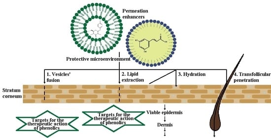
Liposomes may be considered among the first lipid-based nanosystems. After their discovery in the 1960s by Bangham, they were initially proposed as a model for biological membranes due to their compositional similarity. However, later in the 1970s, thanks to their excellent biocompatibility properties and entrapment ability, they were studied as potential drug delivery platforms [270,271]. Structurally, liposomes are spherical vesicles consisting of one or more phospholipid bilayers surrounding an inner aqueous compartment [272,273]. The vesicular structure, resulting from the amphipathic properties of the bilayer forming lipids, provides the opportunity to encapsulate hydrophobic and hydrophilic molecules [274]. The origin of the phospholipids (from various natural or synthetic sources) and their chemical structure influence liposomal properties and membrane fluidity. In their study, Jacquot et al. [275] investigated the effect of marine (salmon) and plant (rapeseed) isolated phospholipids on membrane fluidity and the mechanical properties of liposomal bilayers compared to dioleylphosphatidylcholine and dipalmitoylphosphatidylcholine-based membranes as references. The authors reported that the membrane fluidity was influenced by the saturation of the fatty acid chains; the highest values were obtained in the membranes based on dioleylphosphatidylcholine (unsaturated acyl chains) and lowest in the bilayers formed from dipalmitoylphosphatidylcholine (saturated acyl chains). Regarding mechanical properties, phase segregation was reported for the rapeseed membranes, whereas the unsaturated salmon bilayer was characterized by a homogenous structure. The rigidity of the liposomal bilayer may be further increased by the inclusion of cholesterol, which can fill the cavities resulting from the loose packing of phospholipids, thus improving liposomal in vitro and in vivo stability [276,277]. Liposomal physicochemical parameters (size, surface charge), membrane characteristics, and interaction between the encapsulated active agent and liposomal constituents, influence the mechanism and extent of the drug delivery process [278,279]. The appropriate size of liposomes for topical application is below 300 nm to reach deeper skin layers. However, vesicles with a size below 70 nm are characterized with maximum deposition in the epidermis as well the dermis [280]. Several mechanisms are proposed to explain the active agent transfer from liposomes to the skin, such as vesicle fusion with lipids of stratum corneum as a result of their similar structure; a fluidizing effect, leading to impaired skin integrity; intact liposomal penetration into different dermal layers (associated with their flexibility; possible alterations in size and structure); improved drug delivery through hair follicles or sweat ducts facilitated by liposomal vesicular structure, and free active agent penetration after its release from liposomes (Figure 1) [271,281,282]. To evaluate the influence of zeta potential on transdermal delivery, Park et al. [283] investigated resveratrol permeation from conventional liposomes as well from vesicles coated with chitosan. According to the authors, higher resveratrol skin deposition was estimated from the chitosan-coated vesicles due to the repulsive electrostatic interaction between cationic chitosan vesicles and negatively charged epidermal lipids. The incorporation of phenolic compounds in liposomes, as well their interaction with phospholipids, has been studied by many researchers. In their study Malekar et al. [274] investigated the localization of five chemically diverse phenolic compounds (raloxifene, garcinol, quercetin, trans-resveratrol, bisphenol A) in dipalmitoylphosphatidylcholine-based liposomal bilayer and their influence on colloidal stability. As reported by the authors, the phenolic compounds, localized in the central regions of the bilayer (resveratrol and quercetin), negatively influenced liposomal colloidal stability due to decreased contact with phosphate head groups. Inversely, phytochemicals in the glycerol region of the acyl chains (raloxifene, garcinol, bisphenol A) contributed to better stability thanks to the enhanced exposure with phosphate head groups or electrostatic repulsion forces. Phan et al. [92] studied the interaction mechanism of two different classes of polyphenols (flavonoids and trans-stilbenes) with liposomal membranes. The flavonoids’ gallate, galloyl, and hydroxyl groups are connected via hydrogen bonds with membrane lipids, leading to compact phospholipid assembly, reduced surface area, and forming a stiffer bilayer. The benzyl open ring structure of trans stilbenes, on the other site, determines its deeper intercalation into the hydrophobic bilayer, causing extension of the membrane area and enhancing its fluidity.
5.2. Solid Lipid Nanoparticles
Solid lipid nanoparticles, reported for the first time in the 1990s by Professor R.H. Müller and Professor M. Gasco, were proposed as an alternative approach to overcome the limitations associated with liposomes (e.g., phospholipid oxidation, costly materials, and production process, limited physical stability, difficulties in process scale-up) and polymeric nanoparticles (polymer toxic degradation process) [284,285]. As suggested by their name, SLNs are composed of individual or a mixture of lipids, solid at ambient and body temperature, dispersed in water or an aqueous phase containing surfactant [286]. Most commonly used lipids for SLNs preparation include triglycerides (trimyristin, tristearin), fatty acids (stearic, palmitic acid), waxes (beeswax, cetyl palmitate), and mono/di/triglycerides mixtures (glyceryl behenate—Compritol 888 ATO, glyceryl palmitostearate—Precirol ATO 5) [287]. Solid lipid nanoparticles can be suitable carriers for both hydrophobic and hydrophilic compounds. According to their structure and drug localization they can be classified as homogenous matrix models and core-shell models (drug-enriched shell and drug-enriched core) [288]. In the first case, the active agent is molecularly dispersed within the matrix or present as amorphous clusters. Homogenous matrix SLNs are obtained by cold or hot (when encapsulating highly lipophilic molecules) homogenization methods. In the second model, an outer shell containing the active agent surrounds a lipid core. The specific morphology of drug-enriched shell nanoparticles results from phase separation during the cooling phase; initially, the lipid in the center precipitates, shaping the inner, compound-free compartment. However, at the same time, drug concentration in the residual melted lipid increases and, after solidification, a drug-enriched shell is formed. In the third model SLNs, due to drug super-saturation in the lipid melt, a crystallization of the active agent is observed before the crystallization of the lipids, resulting in a drug-enriched core enclosed by a lipid (drug-free) shell [288,289,290]. The type of SLNs and their composition and physicochemical parameters affect their skin permeation. Due to the lipid nature of SLNs, as possible mechanisms of skin penetration (analogous to liposomes) the fusion of nanoparticles with lipids of stratum corneum, the lipid fluidizing properties of lipids, and transfollicular transfer are proposed. However, as a specific penetration mechanism, characteristic of SLNs may indicate their occlusive effect. Thanks to their large surface area and nanosized dimensions, they possess occlusive characteristics leading to improved skin hydration and enhanced penetration into dermal layers (Figure 1) [289,291]. In their study Kakkar et al. [292] used tetrahydrocurcumin-loaded SLNs characterized by sufficient occlusivity and anti-inflammatory effects. Their further incorporation in hydrogel formulation led to seventeen times higher skin permeation ability compared to plain tetrahydrocurcumin gel. Regarding their structure, suitable features for topical application include the drug-enriched shell nanoparticles, providing rapid drug release, which along with the occlusive effect is a favorable characteristic when increased drug penetration is necessary [288,293]. The disadvantages of SLNs, such as the tendency for drug expulsion or low drug loading capacity due to the ideal crystalline structure of the lipids, provide possibilities for further development, including the development of second-generation nanoparticles and structured lipid carriers (NLCs), which overcome the limitations mentioned above [284,294].
5.3. Nanostructured Lipid Carriers
Nanostructured lipid carriers are composed of solid and liquid lipids dispersed in the aqueous phase, stabilized by surfactants [295]. The inclusion of a liquid lipid in their composition disrupts the highly ordered crystalline structure characteristic of the SLNs. It leads to a less organized lipid matrix, providing space for more drug accumulation [296,297]. NLCs may be categorized into three types: imperfect crystal, amorphous and multiple types, by the lipid blending ratio and their method of preparation. Type I NLCs are prepared from lipids (predominantly solid, with small amount of oil phase), differing in their structure concerning chain length or saturation, leading to the formation of a disordered “imperfect” lipid matrix characterized by high encapsulation capacity. An amorphous structure, indicative of the second type of NLCs, is obtained by including specific lipids to the composition so the crystallization after cooling can be prevented. Consequently, leakage of the active agent also is minimized. The multiple model NLCs contain a significant amount of liquid oil phase in their composition, which facilitates a phase separation during the formulation process leading to nanosized liquid oil compartments among the solid lipid matrix. The solid matrix may be referred to as a barrier, providing a controlled release of the active agent, whereas the liquid lipids ensure better solubility of included lipophilic molecules and therefore determine higher drug encapsulation [284,293,298,299]. The effects of the included liquid lipid in the NLCs composition, as well as the selected oil phase on the encapsulation efficiency and antioxidant activity of the phenolic compound sesamol, was investigated by Puglia et al. [300]. The authors developed two different NLC formulations composed of the same solid lipid (Compritol®888 ATO) with varying liquid phases (Miglyol 812 or sesame oil), as well one model Compritol®888 ATO-based SLNs for reference. Sesamol encapsulation was higher in the NLCs formulations than the SLNs, with the highest entrapment efficiency values (>90%) and improved antioxidant activity for the formulation containing sesame oil as the liquid oil phase. The observed results may be attributed to the structural similarity between the active agent and selected oil phase, which determines the potent synergic effect. NLC composition also influences the occlusive effect and their dermal/transdermal delivery. In their study, Loo et al. [301] investigated the influence of lipid concentration (20% and 30%), solid lipid/oil ratio, and additives (lecithin, propylene glycol) on skin hydration and transepidermal water loss. According to the authors, NLCs with higher lipid content (30%), high solid lipid concentration (90%), and additives (slightly favorable outcomes in case of lecithin) were characterized with good occlusive properties and led to improved skin hydration and reduction of transepidermal water loss during the seven-day study period. Regarding particle size, another important factor determining the extent of skin permeation, both NLCs and SLNs may be prepared in the appropriate form for dermal application range (100–500 nm) via a high-pressure homogenization (hot and cold) method, which is also suitable for large scale production [289,298]. There is some controversial literature regarding the possibility of encapsulating temperature-sensitive compounds (such as some phenolic compounds) via the hot method. However, this technique is considered applicable due to the short heating time, except for highly temperature-sensitive, hydrophilic molecules, which might migrate to the aqueous phase during homogenization [288]. The mechanisms by which NLCs improve drug permeation through the skin are similar to those, discussed for SLNs (Figure 1).
5.4. Nanoemulsions
Nanoemulsions are isotropic colloidal dispersions composed of water and oil stabilized using surfactant/cosurfactant. One of the liquids is dispersed into nanosized droplets, usually between 20 and 200 nm [302]. The tiny droplet size causes their transparent/translucent appearance (analogical to microemulsions). However, differences between these two systems may result from the surfactant concentrations used (ca. 20% in microemulsions, vs. 3–10% in nanoemulsions), as well their dissimilar thermodynamic stability (nanoemulsions are thermodynamically unstable, while microemulsions are thermodynamically stable) [303,304]. Nanoemulsions are an attractive drug delivery system for topical application due to their miniature droplet size, homogenous size distribution, and large surface area, which ensure their uniform spreading onto the skin surface that facilitates drug penetration [305]. Similar to the other lipid-based nanosystems, an inverse relationship between the size of carriers and their transdermal penetration has been reported. According to Su et al. [306], who investigated transport through the skin of nanoemulsions loaded with environment-responsive and fluorescent dyes, formulations with droplets size of 80 nm can diffuse (but not penetrate) into the uncompromised epidermis and pass through canals of hair follicles, in contrast to larger (500 nm) sized formulations. The appropriate size of nanoemulsions is recommended to be below 50 nm to achieve an efficient transdermal delivery [307]. Other important factors affecting transdermal transportation of nanoemulsions are the formulation composition and type of emulsion (w/o or o/w). The oil phase of the systems may be composed of fatty acids (e.g., oleic acid), esters of fatty acids and alcohols oils (isopropyl myristate/GRAS certified), triglycerides (triacetin/GRAS certified), as well nonpolar essential oils or lipid-soluble vitamins, among others [304,308]. Liu et al. [309] investigated the influence of different oil phases (eutectic mixture of menthol and camphor or isopropyl myristate) on transdermal delivery of glabridin-loaded nanoemulsions. According to the performed skin permeation studies, the formulation composed of binary eutectic mixture led to three times higher skin permeation of glabridin (compared to isopropyl myristate nanoemulsion), which was seven times higher than the isoflavane solution. Depending on the type of nanoemulsion, different permeation mechanisms were discussed. In the case of encapsulation of hydrophilic molecules in w/o nanoemulsions, transdermal transportation may be facilitated as a result of the solubilizing properties of the included surfactants on stratum corneum, and delivery via the pore pathway/hair follicles canals for large molecules. Regarding transdermal delivery of hydrophobic molecules from o/w nanoemulsions, active agent permeation may be achieved due to disruption of the stratum corneum (by creating permeable pathways as a result of fluidization of cell membranes and extracellular spaces), or improved permeation characteristics due to skin hydration (Figure 1) [307,310].
Various examples supporting the beneficial effects of lipid-based nanosystems concerning their improved physicochemical properties, or overcoming technological/biopharmaceutical limitations of different phenolic compounds, are presented in Table 2.
The beneficial effects observed after incorporating phenolic phytochemicals in lipid-based nanocarriers, such as improved solubility, stability and skin permeation/penetration, are a prerequisite for further research, development, and industrial application. In Table 3 are presented some cosmetic products based on lipid nanocarriers encapsulating various phenolic compounds. The observed favorable outcomes may be described as synergetic; on the one side they result from the well-known antioxidant, antiaging or skin whitening properties of the phenolic phytochemicals [330], and on the other side results may be attributed to the penetration-enhancing properties or occlusive effects of the lipid nanocarriers.
6. Conclusions
The numerous beneficial effects characteristic of phytochemicals with phenolic structures, such as anti-inflammatory, antioxidant, antiproliferative, and antiaging activities, determine their broad utilization potential in pharmaceutics and the cosmetic industry. However, these advantageous features cannot be fully exploited due to unfavorable physicochemical or pharmacokinetic characteristics (i.e., poor solubility, stability, bioavailability). Lipid-based nanosystems, such as liposomes, solid lipid nanoparticles, nanostructured lipid carriers and nanoemulsions, represent a successful approach to overcome these limitations and improve their dermal/transdermal delivery. This review thoroughly discusses the physicochemical properties and mechanism of actions of various classes of phenolic compounds regarding their dermal application. Examples of their incorporation in different lipid nanocarriers, as well a summary of the obtained results, are also provided. According to the data, encapsulation of phenolic compounds in lipid-based nanosystems for topical application leads to improved solubility, stability, skin permeation capability and therapeutic performance in general.
Author Contributions
Conceptualization, V.A. and N.I.; methodology, V.G. and N.I.; investigation, N.I., Y.S. and V.G.; writing—original draft preparation V.G. and N.I.; writing—review and editing, V.A.; visualization, Y.S.; supervision, V.A.; project administration, V.A.; funding acquisition, V.A. All authors have read and agreed to the published version of the manuscript.
Funding
This work was funded by Medical University of Varna, Fund “Nauka” Project № 18027.
Institutional Review Board Statement
Not applicable.
Informed Consent Statement
Not applicable.
Data Availability Statement
Data sharing not applicable.
Acknowledgments
The authors would like to express their acknowledgement to the supporters: The Medical University of Varna.
Conflicts of Interest
The authors declare no conflict of interest.
References
- Del Rio, D.; Rodriguez-Mateos, A.; Spencer, J.P.E.; Tognolini, M.; Borges, G.; Crozier, A. Dietary (Poly)phenolics in Human Health: Structures, Bioavailability, and Evidence of Protective Effects Against Chronic Diseases. Antioxid. Redox Signal. 2013, 18, 1818–1892. [Google Scholar] [CrossRef] [Green Version]
- Działo, M.; Mierziak, J.; Korzun, U.; Preisner, M.; Szopa, J.; Kulma, A. The Potential of Plant Phenolics in Prevention and Therapy of Skin Disorders. Int. J. Mol. Sci. 2016, 17, 160. [Google Scholar] [CrossRef] [PubMed] [Green Version]
- Lorenzo, J.M.; Estévez, M.; Barba, F.J.; Thirumdas, R.; Franco, D.; Munekata, P.E.S. Polyphenols: Bioaccessibility and bioavailability of bioactive components. In Innovative Thermal and Non-Thermal Processing, Bioaccessibility and Bioavailability of Nutrients and Bioactive Compounds, 1st ed.; Barba, F., Saraiva, J.M.A., Cravotto, G., Lorenzo, J., Eds.; Elsevier: Amsterdam, The Netherlands, 2019; pp. 309–322. [Google Scholar]
- Martins, N.; Barros, L.; Ferreira, I.C.F.R. In vivo antioxidant activity of phenolic compounds: Facts and gaps. Trends Food Sci. Technol. 2016, 48, 1–12. [Google Scholar] [CrossRef] [Green Version]
- Mehta, J.P.; Parmar, P.H.; Vadia, S.H.; Patel, M.K.; Tripathi, C.B. In-vitro antioxidant and in-vivo anti-inflammatory activities of aerial parts of Cassia species. Arab. J. Chem. 2017, 10, S1654–S1662. [Google Scholar] [CrossRef] [Green Version]
- Miklasińska-Majdanik, M.; Kępa, M.; Wojtyczka, R.D.; Idzik, D.; Wąsik, T. Phenolic Compounds Diminish Antibiotic Resistance of Staphylococcus Aureus Clinical Strains. Int. J. Environ. Res. Public Health 2018, 15, 2321. [Google Scholar] [CrossRef] [Green Version]
- Tungmunnithum, D.; Thongboonyou, A.; Pholboon, A.; Yangsabai, A. Flavonoids and Other Phenolic Compounds from Medicinal Plants for Pharmaceutical and Medical Aspects: An Overview. Medicines 2018, 5, 93. [Google Scholar] [CrossRef]
- Kaurinovic, B.; Vastag, G. Flavonoids and Phenolic Acids as Potential Natural Antioxidants. In Antioxidants; Shalaby, E., Ed.; IntechOpen: London, UK, 2019; pp. 1–20. [Google Scholar]
- Zillich, O.V.; Schweiggert-Weisz, U.; Eisner, P.; Kerscher, M. Polyphenols as active ingredients for cosmetic products. Int. J. Cosmet. Sci. 2015, 37, 455–464. [Google Scholar] [CrossRef] [PubMed]
- Panzella, L.; Napolitano, A. Natural and Bioinspired Phenolic Compounds as Tyrosinase Inhibitors for the Treatment of Skin Hyperpigmentation: Recent Advances. Cosmetics 2019, 6, 57. [Google Scholar] [CrossRef] [Green Version]
- Przybylska-Balcerek, A.; Stuper-Szablewska, K. Phenolic acids used in the cosmetics industry as natural antioxidants. Eur. J. Med. Technol. 2019, 4, 24–32. [Google Scholar]
- Panzella, L. Natural Phenolic Compounds for Health, Food and Cosmetic Applications. Antioxidants 2020, 9, 427. [Google Scholar] [CrossRef]
- Boo, Y.C. Can Plant Phenolic Compounds Protect the Skin from Airborne Particulate Matter? Antioxidants 2019, 8, 379. [Google Scholar] [CrossRef] [Green Version]
- Kumar, N.; Goel, N. Phenolic acids: Natural versatile molecules with promising therapeutic applications. Biotechnol. Rep. 2019, 24, e00370. [Google Scholar] [CrossRef] [PubMed]
- Schempp, C.M.; Müller, K.A.; Winghofer, B.; Schöpf, E.; Simon, J.C. Johanniskraut (Hypericum perforatum L.) Eine Pflanze mit Relevanz für die Dermatologie. Hautarzt 2002, 53, 316–321. [Google Scholar] [CrossRef]
- Arct, J.; Pytkowska, K. Flavonoids as components of biologically active cosmeceuticals. Clin. Dermatol. 2008, 26, 347–357. [Google Scholar] [CrossRef] [PubMed]
- Cosme, P.; Rodríguez, A.B.; Espino, J.; Garrido, M. Plant Phenolics: Bioavailability as a Key Determinant of Their Potential Health-Promoting Applications. Antioxidants 2020, 9, 1263. [Google Scholar] [CrossRef] [PubMed]
- Soto, M.; Falqué, E.; Domínguez, H. Relevance of Natural Phenolics from Grape and Derivative Products in the Formulation of Cosmetics. Cosmetics 2015, 2, 259–276. [Google Scholar] [CrossRef] [Green Version]
- Naik, A.; Kalia, Y.N.; Guy, R.H. Transdermal drug delivery: Overcoming the skin’s barrier function. Pharm. Sci. Technol. Today 2000, 3, 318–326. [Google Scholar] [CrossRef]
- Arct, J.; Gronwald, M.; Kasiura, K. Possibilities for the prediction of an active substance penetration through epidermis. IFSCC Mag. 2001, 4, 179–183. [Google Scholar]
- Arct, J.; Oborska, A.; Mojski, M.; Binkowska, A.; Świdzikowska, B. Common cosmetic hydrophilic ingredients as penetration modifiers of flavonoids. Int. J. Cosmet. Sci. 2002, 24, 357–366. [Google Scholar] [CrossRef] [Green Version]
- Chuang, S.-Y.; Lin, Y.-K.; Lin, C.-F.; Wang, P.-W.; Chen, E.-L.; Fang, J.-Y. Elucidating the Skin Delivery of Aglycone and Glycoside Flavonoids: How the Structures Affect Cutaneous Absorption. Nutrients 2017, 9, 1304. [Google Scholar] [CrossRef] [Green Version]
- Fang, Z.; Bhandari, B. Encapsulation of polyphenols—A review. Trends Food Sci. Technol. 2010, 21, 510–523. [Google Scholar] [CrossRef]
- Mahdavi, S.A.; Jafari, S.M.; Ghorbani, M.; Assadpoor, E. Spray-drying Microencapsulation of Anthocyanins by Natural Biopolymers: A Review. Dry. Technol. 2014, 32, 509–518. [Google Scholar] [CrossRef]
- Kosović, E.; Topiař, M.; Cuřínová, P.; Sajfrtová, M. Stability testing of resveratrol and viniferin obtained from Vitis vinifera L. by various extraction methods considering the industrial viewpoint. Sci. Rep. 2020, 10, 5564. [Google Scholar] [CrossRef] [Green Version]
- Dai, L.; Li, Y.; Kong, F.; Liu, K.; Si, C.; Ni, Y. Lignin-Based Nanoparticles Stabilized Pickering Emulsion for Stability Improvement and Thermal-Controlled Release of trans-Resveratrol. ACS Sustain. Chem. Eng. 2019, 7, 13497–13504. [Google Scholar] [CrossRef]
- Kumar, R.; Kaur, K.; Uppal, S.; Mehta, S.K. Ultrasound processed nanoemulsion: A comparative approach between resveratrol and resveratrol cyclodextrin inclusion complex to study its binding interactions, antioxidant activity and UV light stability. Ultrason. Sonochem. 2017, 37, 478–489. [Google Scholar] [CrossRef] [PubMed]
- Lima, A.M.; Pizzol, C.D.; Monteiro, F.B.F.; Creczynski-Pasa, T.B.; Andrade, G.P.; Ribeiro, A.O.; Perrusi, J.R. Hypericin encapsulated in solid lipid nanoparticles: Phototoxicity and photodynamic efficiency. J. Photochem. Photobiol. B Biol. 2013, 125, 146–154. [Google Scholar] [CrossRef]
- Youssef, T.; Fadel, M.; Fahmy, R.; Kassab, K. Evaluation of hypericin-loaded solid lipid nanoparticles: Physicochemical properties, photostability and phototoxicity. Pharm. Dev. Technol. 2012, 17, 177–186. [Google Scholar] [CrossRef]
- Füller, J.; Kellner, T.; Gaid, M.; Beerhues, L.; Müller-Goymann, C.C. Stabilization of hyperforin dicyclohexylammonium salt with dissolved albumin and albumin nanoparticles for studying hyperforin effects on 2D cultivation of keratinocytes in vitro. Eur. J. Pharm. Biopharm. 2018, 126, 115–122. [Google Scholar] [CrossRef] [PubMed]
- Orth, H.C.J.; Rentel, C.; Schmidt, P.C. Isolation, Purity Analysis and Stability of Hyperforin as a Standard Material from Hypericum perforatum L. J. Pharm. Pharmacol. 1999, 51, 193–200. [Google Scholar] [CrossRef] [PubMed]
- Koyu, H.; Haznedaroglu, M.Z. Investigation of impact of storage conditions on Hypericum perforatum L. dried total extract. J. Food Drug Anal. 2015, 23, 545–551. [Google Scholar] [CrossRef] [Green Version]
- Park, S.N.; Lee, M.H.; Kim, S.J.; Yu, E.R. Preparation of quercetin and rutin-loaded ceramide liposomes and drug-releasing effect in liposome-in-hydrogel complex system. Biochem. Biophys. Res. Commun. 2013, 435, 361–366. [Google Scholar] [CrossRef]
- Kwon, H.-J.; Hwang, J.; Lee, J.; Chae, S.-K.; Lee, J.-H.; Kim, J.-H.; Hwang, K.-S.; Kim, E.-C.; Park, Y.-D. Analysis and investigation of chemical stability on phenolic compounds in Zanthoxylum schinifolium-containing dentifrices. J. Liq. Chromatogr. Relat. Technol. 2014, 37, 1685–1701. [Google Scholar] [CrossRef]
- Ramešová, Š.; Sokolová, R.; Degano, I.; Bulíčková, J.; Žabka, J.; Gál, M. On the stability of the bioactive flavonoids quercetin and luteolin under oxygen-free conditions. Anal. Bioanal. Chem. 2012, 402, 975–982. [Google Scholar] [CrossRef] [PubMed]
- Luo, X.; Guan, R.; Chen, X.; Liu, M.; Hao, Y.; Jiang, H. Optimized Preparation of Catechin Nanoliposomes by Orthogonal Design and Stability Study. Adv. J. Food Sci. Technol. 2014, 6, 921–925. [Google Scholar] [CrossRef]
- Latos-Brozio, M.; Masek, A. Natural Polymeric Compound Based on High Thermal Stability Catechin from Green Tea. Biomolecules 2020, 10, 1191. [Google Scholar] [CrossRef] [PubMed]
- Li, N.; Taylor, L.S.; Ferruzzi, M.G.; Mauer, L.J. Kinetic Study of Catechin Stability: Effects of pH, Concentration, and Temperature. J. Agric. Food Chem. 2012, 60, 12531–12539. [Google Scholar] [CrossRef]
- Jensen, J.S.; Wertz, C.F.; O’Neill, V.A. Preformulation Stability of trans-Resveratrol and trans-Resveratrol Glucoside (Piceid). J. Agric. Food Chem. 2010, 58, 1685–1690. [Google Scholar] [CrossRef]
- Lin, C.-F.; Leu, Y.-L.; Al-Suwayeh, S.A.; Ku, M.-C.; Hwang, T.-L.; Fang, J.-Y. Anti-inflammatory activity and percutaneous absorption of quercetin and its polymethoxylated compound and glycosides: The relationships to chemical structures. Eur. J. Pharm. Sci. 2012, 47, 857–864. [Google Scholar] [CrossRef] [PubMed]
- Křen, V. Glycoside vs. Aglycon: The Role of Glycosidic Residue in Biological Activity. In Glycoscience, 2nd ed.; Fraser-Reid, B.O., Tatsuta, K., Thiem, J., Eds.; Springer: Berlin/Heidelberg, Germany, 2008; pp. 2589–2644. [Google Scholar]
- Choi, S.-J.; Tai, B.H.; Cuong, N.M.; Kim, Y.-H.; Jang, H.-D. Antioxidative and anti-inflammatory effect of quercetin and its glycosides isolated from mampat (Cratoxylum formosum). Food Sci. Biotechnol. 2012, 21, 587–595. [Google Scholar] [CrossRef]
- Rha, C.-S.; Jeong, H.W.; Park, S.; Lee, S.; Jung, Y.S.; Kim, D.-O. Antioxidative, Anti-Inflammatory, and Anticancer Effects of Purified Flavonol Glycosides and Aglycones in Green Tea. Antioxidants 2019, 8, 278. [Google Scholar] [CrossRef] [PubMed] [Green Version]
- Munin, A.; Edwards-Lévy, F. Encapsulation of Natural Polyphenolic Compounds; A Review. Pharmaceutics 2011, 3, 793–829. [Google Scholar] [CrossRef] [Green Version]
- Belščak-Cvitanović, A.; Stojanović, R.; Manojlović, V.; Komes, D.; Cindrić, I.J.; Nedović, V.; Bugarski, B. Encapsulation of polyphenolic antioxidants from medicinal plant extracts in alginate–chitosan system enhanced with ascorbic acid by electrostatic extrusion. Food Res. Int. 2011, 44, 1094–1101. [Google Scholar] [CrossRef]
- Tylkowski, B.; Tsibranska, I. Polyphenols encapsulation—Application of innovation technologies to improve stability of natural products. In Microencapsulation; Giamberini, M., Prieto, S.F., Tylkowski, B., Eds.; De Gruyter: Berlin, Germany; Boston, MA, USA, 2015; pp. 97–114. [Google Scholar]
- Kumar, R. Lipid-Based Nanoparticles for Drug-Delivery Systems. In Nanocarriers for Drug Delivery; Mohapatra, S.S., Ranjan, S., Dasgupta, N., Mishra, R.K., Thomas, S., Eds.; Elsevier: Amsterdam, The Netherlands, 2019; pp. 249–284. [Google Scholar]
- Sengar, V.; Jyoti, K.; Jain, U.K.; Katare, O.P.; Chandra, R.; Madan, J. Lipid nanoparticles for topical and transdermal delivery of pharmaceuticals and cosmeceuticals. In Lipid Nanocarriers for Drug Targeting, 1st ed.; Grumezescu, A., Ed.; Elsevier: Amsterdam, The Netherlands, 2018; pp. 413–436. [Google Scholar]
- Kakadia, P.; Conway, B. Lipid nanoparticles for dermal drug delivery. Curr. Pharm. Des. 2015, 21, 2823–2829. [Google Scholar] [CrossRef] [PubMed] [Green Version]
- Ganesan, P.; Narayanasamy, D. Lipid nanoparticles: Different preparation techniques, characterization, hurdles, and strategies for the production of solid lipid nanoparticles and nanostructured lipid carriers for oral drug delivery. Sustain. Chem. Pharm. 2017, 6, 37–56. [Google Scholar] [CrossRef]
- Bhattacharya, A.; Sood, P.; Citovsky, V. The roles of plant phenolics in defence and communication during Agrobacterium and Rhizobium infection. Mol. Plant. Pathol. 2010, 11, 705–719. [Google Scholar] [CrossRef]
- Patil, V.M.; Masand, N. Anticancer Potential of Flavonoids: Chemistry, Biological Activities, and Future Perspectives. In Studies in Natural Products Chemistry, 1st ed.; Atta-ur-Rahman, Ed.; Elsevier: Amsterdam, The Netherlands, 2018; Volume 59, pp. 401–430. [Google Scholar]
- Zuiter, A.S. Proanthocyanidin: Chemistry and Biology: From Phenolic Compounds to Proanthocyanidins. In Reference Module in Chemistry, Molecular Sciences and Chemical Engineering; Elsevier: Amsterdam, The Netherlands, 2014; pp. 1–29. [Google Scholar]
- Santos-Sánchez, N.F.; Salas-Coronado, R.; Villanueva-Cañongo, C.; Hernández-Carlos, B. Antioxidant Compounds and Their Antioxidant Mechanism. In Antioxidants; Shalaby, E., Ed.; IntechOpen: London, UK, 2019; pp. 1–28. [Google Scholar]
- Snezhkina, A.V.; Kudryavtseva, A.V.; Kardymon, O.L.; Savvateeva, M.V.; Melnikova, N.V.; Krasnov, G.S.; Dmitriev, A.A. ROS Generation and Antioxidant Defense Systems in Normal and Malignant Cells. Oxid. Med. Cell. Longev. 2019, 2019, 1–17. [Google Scholar] [CrossRef]
- Cherrak, S.A.; Mokhtari-Soulimane, N.; Berroukeche, F.; Bensenane, B.; Cherbonnel, A.; Merzouk, H.; Elhabiri, M. In Vitro Antioxidant versus Metal Ion Chelating Properties of Flavonoids: A Structure-Activity Investigation. PLoS ONE 2016, 11, e0165575. [Google Scholar] [CrossRef]
- Hussain, T.; Tan, B.; Yin, Y.; Blachier, F.; Tossou, M.C.B.; Rahu, N. Oxidative Stress and Inflammation: What Polyphenols Can Do for Us? Oxid. Med. Cell. Longev. 2016, 2016, 1–9. [Google Scholar] [CrossRef] [Green Version]
- Heim, K.E.; Tagliaferro, A.R.; Bobilya, D.J. Flavonoid antioxidants: Chemistry, metabolism and structure-activity relationships. J. Nutr. Biochem. 2002, 13, 572–584. [Google Scholar] [CrossRef]
- Kumar, S.; Pandey, A.K. Chemistry and Biological Activities of Flavonoids: An Overview. Sci. World J. 2013, 2013, 1–16. [Google Scholar] [CrossRef] [Green Version]
- Gijsman, P. Polymer Stabilization. In Handbook of Environmental Degradation of Materials, 2nd ed.; Kutz, M., Ed.; Elsevier: Amsterdam, The Netherlands, 2012; pp. 673–714. [Google Scholar]
- Okayama, Y. Oxidative Stress in Allergic and Inflammatory Skin Diseases. Curr. Drug Targets Inflamm. Allergy 2005, 4, 517–519. [Google Scholar] [CrossRef] [PubMed]
- Yahfoufi, N.; Alsadi, N.; Jambi, M.; Matar, C. The Immunomodulatory and Anti-Inflammatory Role of Polyphenols. Nutrients 2018, 10, 1618. [Google Scholar] [CrossRef] [PubMed] [Green Version]
- Havermann, S.; Büchter, C.; Koch, K.; Wätjen, W. Role of Oxidative Stress in the Process of Carcinogenesis. In Studies on Experimental Toxicology and Pharmacology. Oxidative Stress in Applied Basic Research and Clinical Practice, 1st ed.; Roberts, S.M., Kehrer, J.P., Klotz, L.-O., Eds.; Humana Press: Totowa, NJ, USA, 2015; pp. 173–198. [Google Scholar]
- Harrison, D.; Griendling, K.K.; Landmesser, U.; Hornig, B.; Drexler, H. Role of oxidative stress in atherosclerosis. Am. J. Cardiol. 2003, 91, 7A–11A. [Google Scholar] [CrossRef]
- Giesey, R.L.; Mehrmal, S.; Uppal, P.; Delost, G. The Global Burden of Skin and Subcutaneous Disease: A Longitudinal Analysis from the Global Burden of Disease Study From 1990–2017. SKIN J. Cutan. Med. 2021, 5, 125–136. [Google Scholar] [CrossRef]
- Karimkhani, C.; Dellavalle, R.P.; Coffeng, L.E.; Flohr, C.; Hay, R.J.; Langan, S.M.; Nsoesie, E.O.; Ferrari, A.J.; Erskine, H.E.; Silverberg, J.I.; et al. Global Skin Disease Morbidity and Mortality: An Update From the Global Burden of Disease Study 2013. JAMA Dermatol. 2017, 153, 406–412. [Google Scholar] [CrossRef] [PubMed]
- Flohr, C.; Hay, R. Putting the burden of skin diseases on the global map. Br. J. Dermatol. 2021, 184, 189–190. [Google Scholar] [CrossRef]
- Cushnie, T.P.T.; Lamb, A.J. Antimicrobial activity of flavonoids. Int. J. Antimicrob. Agents 2005, 26, 343–356. [Google Scholar] [CrossRef]
- Tabassum, N.; Hamdani, M. Plants used to treat skin diseases. Pharmacogn. Rev. 2014, 8, 52–60. [Google Scholar] [CrossRef] [Green Version]
- Gottlieb, A.B. Therapeutic options in the treatment of psoriasis and atopic dermatitis. J. Am. Acad. Dermatol. 2005, 53, S3–S16. [Google Scholar] [CrossRef]
- Hajar, T.; Gontijo, J.R.V.; Hanifin, J.M. New and developing therapies for atopic dermatitis. An. Bras. Dermatol. 2018, 93, 104–107. [Google Scholar] [CrossRef] [Green Version]
- Richmond, J.M.; Harris, J.E. Immunology and Skin in Health and Disease. Cold Spring Harb. Perspect. Med. 2014, 4, a015339. [Google Scholar] [CrossRef] [PubMed] [Green Version]
- Dainichi, T.; Hanakawa, S.; Kabashima, K. Classification of inflammatory skin diseases: A proposal based on the disorders of the three-layered defense systems, barrier, innate immunity and acquired immunity. J. Dermatol. Sci. 2014, 76, 81–89. [Google Scholar] [CrossRef] [PubMed] [Green Version]
- Gunter, N.V.; Teh, S.S.; Lim, Y.M.; Mah, S.H. Natural Xanthones and Skin Inflammatory Diseases: Multitargeting Mechanisms of Action and Potential Application. Front. Pharmacol. 2020, 11, 594202. [Google Scholar] [CrossRef] [PubMed]
- Schwingen, J.; Kaplan, M.; Kurschus, F.C. Review-Current Concepts in Inflammatory Skin Diseases Evolved by Transcriptome Analysis: In-Depth Analysis of Atopic Dermatitis and Psoriasis. Int. J. Mol. Sci. 2020, 21, 699. [Google Scholar] [CrossRef] [Green Version]
- Giang, J.; Seelen, M.A.J.; van Doorn, M.B.A.; Rissmann, R.; Prens, E.P.; Damman, J. Complement Activation in Inflammatory Skin Diseases. Front. Immunol. 2018, 9, 639. [Google Scholar] [CrossRef]
- Cetin, E.D.; Savk, E.; Uslu, M.; Eskin, M.; Karul, A. Investigation of the Inflammatory Mechanisms in Alopecia Areata. Am. J. Dermatopathol. 2009, 31, 53–60. [Google Scholar] [CrossRef]
- Woo, Y.; Lim, J.; Cho, D.; Park, H. Rosacea: Molecular Mechanisms and Management of a Chronic Cutaneous Inflammatory Condition. Int. J. Mol. Sci. 2016, 17, 1562. [Google Scholar] [CrossRef] [Green Version]
- Richmond, J.M.; Frisoli, M.L.; Harris, J.E. Innate immune mechanisms in vitiligo: Danger from within. Curr. Opin. Immunol. 2013, 25, 676–682. [Google Scholar] [CrossRef] [Green Version]
- Neagu, M.; Constantin, C.; Caruntu, C.; Dumitru, C.; Surcel, M.; Zurac, S. Inflammation: A key process in skin tumorigenesis. Oncol. Lett. 2019, 17, 4068–4084. [Google Scholar] [CrossRef] [Green Version]
- de Oliveira, R.G., Jr.; Ferraz, C.A.A.; e Silva, M.G.; de Lavor, É.M.; Rolim, L.A.; de Lima, J.T.; Fleury, A.; Picot, L.; de Souza Siqueira Quintans, J.; Quintans, L.J., Jr.; et al. Flavonoids: Promising Natural Products for Treatment of Skin Cancer (Melanoma). In Natural Products and Cancer Drug Discovery; Badria, F.A., Ed.; Humana Press: Totowa, NJ, USA, 2017; pp. 161–210. [Google Scholar]
- Katiyar, S.K.; Afaq, F.; Perez, A.; Mukhtar, H. Green tea polyphenol (-)-epigallocatechin-3-gallate treatment of human skin inhibits ultraviolet radiation-induced oxidative stress. Carcinogenesis 2001, 22, 287–294. [Google Scholar] [CrossRef]
- Afaq, F.; Syed, D.N.; Malik, A.; Hadi, N.; Sarfaraz, S.; Kweon, M.-H.; Khan, N.; Zaid, M.A.; Mukhtar, H. Delphinidin, an Anthocyanidin in Pigmented Fruits and Vegetables, Protects Human HaCaT Keratinocytes and Mouse Skin Against UVB-Mediated Oxidative Stress and Apoptosis. J. Investig. Dermatol. 2007, 127, 222–232. [Google Scholar] [CrossRef] [Green Version]
- Papuc, C.; Goran, G.V.; Predescu, C.N.; Nicorescu, V.; Stefan, G. Plant Polyphenols as Antioxidant and Antibacterial Agents for Shelf-Life Extension of Meat and Meat Products: Classification, Structures, Sources, and Action Mechanisms. Compr. Rev. Food Sci. Food Saf. 2017, 16, 1243–1268. [Google Scholar] [CrossRef] [PubMed] [Green Version]
- Borges, A.; Ferreira, C.; Saavedra, M.J.; Simões, M. Antibacterial Activity and Mode of Action of Ferulic and Gallic Acids Against Pathogenic Bacteria. Microb. Drug Resist. 2013, 19, 256–265. [Google Scholar] [CrossRef] [PubMed]
- Anderson, J.C.; Headley, C.; Stapleton, P.D.; Taylor, P.W. Synthesis and antibacterial activity of hydrolytically stable (−)-epicatechin gallate analogues for the modulation of β-lactam resistance in Staphylococcus aureus. Bioorganic Med. Chem. Lett. 2005, 15, 2633–2635. [Google Scholar] [CrossRef] [PubMed] [Green Version]
- Zhao, W.-H.; Hu, Z.-Q.; Okubo, S.; Hara, Y.; Shimamura, T. Mechanism of Synergy between Epigallocatechin Gallate and β-Lactams against Methicillin-Resistant Staphylococcus aureus. Antimicrob. Agents Chemother. 2001, 45, 1737–1742. [Google Scholar] [CrossRef] [PubMed] [Green Version]
- Khameneh, B.; Iranshahy, M.; Soheili, V.; Fazly Bazzaz, B.S. Review on plant antimicrobials: A mechanistic viewpoint. Antimicrob. Resist. Infect. Control. 2019, 8, 118. [Google Scholar] [CrossRef] [Green Version]
- Sudano Roccaro, A.; Blanco, A.R.; Giuliano, F.; Rusciano, D.; Enea, V. Epigallocatechin-Gallate Enhances the Activity of Tetracycline in Staphylococci by Inhibiting Its Efflux from Bacterial Cells. Antimicrob. Agents Chemother. 2004, 48, 1968–1973. [Google Scholar] [CrossRef] [Green Version]
- Zhao, W.-H.; Hu, Z.-Q.; Hara, Y.; Shimamura, T. Inhibition of Penicillinase by Epigallocatechin Gallate Resulting in Restoration of Antibacterial Activity of Penicillin Against Penicillinase-Producing Staphylococcus Aureus. Antimicrob. Agents Chemother. 2002, 46, 2266–2268. [Google Scholar] [CrossRef] [Green Version]
- Qin, R.; Xiao, K.; Li, B.; Jiang, W.; Peng, W.; Zheng, J.; Zhou, H. The Combination of Catechin and Epicatechin Gallate from Fructus Crataegi Potentiates β-Lactam Antibiotics Against Methicillin-Resistant Staphylococcus Aureus (Mrsa) In Vitro and In Vivo. Int. J. Mol. Sci. 2013, 14, 1802–1821. [Google Scholar] [CrossRef] [Green Version]
- Phan, H.T.T.; Yoda, T.; Chahal, B.; Morita, M.; Takagi, M.; Vestergaard, M.C. Structure-dependent interactions of polyphenols with a biomimetic membrane system. Biochim. Biophys. Acta Biomembr. 2014, 1838, 2670–2677. [Google Scholar] [CrossRef] [PubMed] [Green Version]
- Wu, T.; He, M.; Zang, X.; Zhou, Y.; Qiu, T.; Pan, S.; Xu, X. A structure–activity relationship study of flavonoids as inhibitors of E. coli by membrane interaction effect. Biochim. Biophys. Acta Biomembr. 2013, 1828, 2751–2756. [Google Scholar] [CrossRef] [Green Version]
- Donadio, G.; Mensitieri, F.; Santoro, V.; Parisi, V.; Bellone, M.L.; De Tommasi, N.; Izzo, V.; Dal Piaz, F. Interactions with Microbial Proteins Driving the Antibacterial Activity of Flavonoids. Pharmaceutics 2021, 13, 660. [Google Scholar] [CrossRef] [PubMed]
- Pinho, E.; Ferreira, I.C.F.R.; Barros, L.; Carvalho, A.M.; Soares, G.; Henriques, M. Antibacterial Potential of Northeastern Portugal Wild Plant Extracts and Respective Phenolic Compounds. BioMed Res. Int. 2014, 2014, 1–8. [Google Scholar] [CrossRef] [Green Version]
- Cushnie, T.P.T.; Lamb, A.J. Assessment of the antibacterial activity of galangin against 4-quinolone resistant strains of Staphylococcus aureus. Phytomedicine 2006, 13, 187–191. [Google Scholar] [CrossRef] [PubMed]
- Przybyłek, I.; Karpiński, T.M. Antibacterial Properties of Propolis. Molecules 2019, 24, 2047. [Google Scholar] [CrossRef] [PubMed] [Green Version]
- Park, S.; Yi, Y.; Lim, M.H. Reactivity of Flavonoids Containing a Catechol or Pyrogallol Moiety with Metal-Free and Metal-Associated Amyloid-β. Bull. Korean Chem. Soc. 2020, 42, 17–24. [Google Scholar] [CrossRef]
- Barvinchenko, V.M.; Lipkovska, N.O.; Fedyanina, T.V.; Pogorelyi, V.K. Physico-chemical Properties of Supramolecular Complexes of Natural Flavonoids with Biomacromolecules. In Nanomaterials and Supramolecular Structures; Shpak, A.P., Gorbyk, P.P., Eds.; Springer: Dordrecht, The Netherlands, 2009; pp. 281–291. [Google Scholar]
- Uivarosi, V.; Munteanu, A.C.; Sharma, A.; Singh Tuli, H. Metal Complexation and Patent Studies of Flavonoid. In Current Aspects of Flavonoids: Their Role in Cancer Treatment; Singh Tuli, H., Ed.; Springer: Singapore, 2019; pp. 39–89. [Google Scholar]
- Wang, S.-X.; Zhang, F.-J.; Feng, Q.-P.; Li, Y.-L. Synthesis, characterization, and antibacterial activity of transition metal complexes with 5-hydroxy-7,4-dimethoxyflavone. J. Inorg. Biochem. 1992, 46, 251–257. [Google Scholar] [CrossRef]
- Kutluay, S.B.; Doroghazi, J.; Roemer, M.E.; Triezenberg, S.J. Curcumin inhibits herpes simplex virus immediate-early gene expression by a mechanism independent of p300/CBP histone acetyltransferase activity. Virology 2008, 373, 239–247. [Google Scholar] [CrossRef] [Green Version]
- Balasubramanyam, K.; Varier, R.A.; Altaf, M.; Swaminathan, V.; Siddappa, N.B.; Ranga, U.; Kundu, T.K. Curcumin, a Novel p300/CREB-binding Protein-specific Inhibitor of Acetyltransferase, Represses the Acetylation of Histone/Nonhistone Proteins and Histone Acetyltransferase-dependent Chromatin Transcription. J. Biol. Chem. 2004, 279, 51163–51171. [Google Scholar] [CrossRef] [Green Version]
- Šudomová, M.; Hassan, S.T.S. Nutraceutical Curcumin with Promising Protection against Herpesvirus Infections and Their Associated Inflammation: Mechanisms and Pathways. Microorganisms 2021, 9, 292. [Google Scholar] [CrossRef]
- Flores, D.J.; Lee, L.H.; Adams, S.D. Inhibition of Curcumin-Treated Herpes Simplex Virus 1 and 2 in Vero Cells. Adv. Microbiol. 2016, 6, 276–287. [Google Scholar] [CrossRef] [Green Version]
- Bernard, F.X.; Sablé, S.; Cameron, B.; Provost, J.; Desnottes, J.F.; Crouzet, J.; Blanche, F. Glycosylated flavones as selective inhibitors of topoisomerase IV. Antimicrob. Agents Chemother. 1997, 41, 992–998. [Google Scholar] [CrossRef] [PubMed] [Green Version]
- Barbieri, R.; Coppo, E.; Marchese, A.; Daglia, M.; Sobarzo-Sánchez, E.; Nabavi, S.F.; Nabavi, S.M. Phytochemicals for human disease: An update on plant-derived compounds antibacterial activity. Microbiol. Res. 2017, 196, 44–68. [Google Scholar] [CrossRef] [PubMed]
- Liu, M.-H.; Otsuka, N.; Noyori, K.; Shiota, S.; Ogawa, W.; Kuroda, T.; Hatano, T.; Tsuchiya, T. Synergistic Effect of Kaempferol Glycosides Purified from Laurus nobilis and Fluoroquinolones on Methicillin-Resistant Staphylococcus aureus. Biol. Pharm. Bull. 2009, 32, 489–492. [Google Scholar] [CrossRef] [Green Version]
- Xie, Y.; Yang, W.; Tang, F.; Chen, X.; Ren, L. Antibacterial Activities of Flavonoids: Structure-Activity Relationship and Mechanism. Curr. Med. Chem. 2014, 22, 132–149. [Google Scholar] [CrossRef] [PubMed]
- Ninfali, P.; Antonelli, A.; Magnani, M.; Scarpa, E.S. Antiviral Properties of Flavonoids and Delivery Strategies. Nutrients 2020, 12, 2534. [Google Scholar] [CrossRef]
- Adamczak, A.; Ożarowski, M.; Karpiński, T.M. Antibacterial Activity of Some Flavonoids and Organic Acids Widely Distributed in Plants. J. Clin. Med. 2020, 9, 109. [Google Scholar] [CrossRef] [PubMed] [Green Version]
- Anani, K.; Adjrah, Y.; Ameyapoh, Y.; Karou, S.D.; Agbonon, A.; de Souza, C.; Gbeassor, M. Effects of hydroethanolic extracts of Balanites aegyptiaca (L.) Delile (Balanitaceae) on some resistant pathogens bacteria isolated from wounds. J. Ethnopharmacol. 2015, 164, 16–21. [Google Scholar] [CrossRef]
- Saddiqe, Z.; Naeem, I.; Maimoona, A. A review of the antibacterial activity of Hypericum perforatum L. J. Ethnopharmacol. 2010, 131, 511–521. [Google Scholar] [CrossRef]
- Wölfle, U.; Seelinger, G.; Schempp, C. Topical Application of St. John’s Wort (Hypericum perforatum). Planta Med. 2013, 80, 109–120. [Google Scholar] [CrossRef] [Green Version]
- Feyzioğlu, B.; Demircili, M.E.; Özdemir, M.; Doğan, M.; Baykan, M.; Baysal, B. Antibacterial effect of hypericin. Afr. J. Microbiol. Res. 2013, 7, 979–982. [Google Scholar]
- Fritz, D.; Venturi, C.R.; Cargnin, S.; Schripsema, J.; Roehe, P.M.; Montanha, J.A.; von Poser, G.L. Herpes virus inhibitory substances from Hypericum connatum Lam., a plant used in southern Brazil to treat oral lesions. J. Ethnopharmacol. 2007, 113, 517–520. [Google Scholar] [CrossRef] [PubMed]
- Wen, S.; Zhang, J.; Yang, B.; Elias, P.M.; Man, M.-Q. Role of Resveratrol in Regulating Cutaneous Functions. Evid. Based Complement. Altern. Med. 2020, 2020, 1–20. [Google Scholar] [CrossRef] [Green Version]
- Chan, M.M.-Y. Antimicrobial effect of resveratrol on dermatophytes and bacterial pathogens of the skin. Biochem. Pharmacol. 2002, 63, 99–104. [Google Scholar] [CrossRef]
- He, M.; Min, J.-W.; Kong, W.-L.; He, X.-H.; Li, J.-X.; Peng, B.-W. A review on the pharmacological effects of vitexin and isovitexin. Fitoterapia 2016, 115, 74–85. [Google Scholar] [CrossRef]
- Man, M.-Q.; Yang, B.; Elias, P.M. Benefits of Hesperidin for Cutaneous Functions. Evid. Based Complement. Altern. Med. 2019, 2019, 1–19. [Google Scholar] [CrossRef] [Green Version]
- Köksal Karayıldırım, Ç. Characterization and in vitro Evolution of Antibacterial Efficacy of Novel Hesperidin Microemulsion. CBUJOS 2017, 13, 943–947. [Google Scholar] [CrossRef]
- Yadav, M.K.; Chae, S.-W.; Im, G.J.; Chung, J.-W.; Song, J.-J. Eugenol: A Phyto-Compound Effective against Methicillin-Resistant and Methicillin-Sensitive Staphylococcus aureus Clinical Strain Biofilms. PLoS ONE 2015, 10, e0119564. [Google Scholar] [CrossRef] [Green Version]
- Guimarães, I.; Baptista-Silva, S.; Pintado, M.; Oliveira, A.L. Polyphenols: A Promising Avenue in Therapeutic Solutions for Wound Care. Appl. Sci. 2021, 11, 1230. [Google Scholar] [CrossRef]
- Thang, P.T.; Patrick, S.; Teik, L.S.; Yung, C.S. Anti-oxidant effects of the extracts from the leaves of Chromolaena odorata on human dermal fibroblasts and epidermal keratinocytes against hydrogen peroxide and hypoxanthine-xanthine oxidase induced damage. Burns 2001, 27, 319–327. [Google Scholar] [CrossRef]
- Bahramsoltani, R.; Farzaei, M.H.; Rahimi, R. Medicinal plants and their natural components as future drugs for the treatment of burn wounds: An integrative review. Arch. Dermatol. Res. 2014, 306, 601–617. [Google Scholar] [CrossRef]
- Skórkowska-Telichowska, K.; Kulma, A.; Żuk, M.; Czuj, T.; Szopa, J. The Effects of Newly Developed Linen Dressings on Decubitus Ulcers. J. Palliat. Med. 2012, 15, 146–148. [Google Scholar] [CrossRef] [Green Version]
- Reinke, J.M.; Sorg, H. Wound Repair and Regeneration. Eur. Surg. Res. 2012, 49, 35–43. [Google Scholar] [CrossRef]
- Mittal, M.; Siddiqui, M.R.; Tran, K.; Reddy, S.P.; Malik, A.B. Reactive Oxygen Species in Inflammation and Tissue Injury. Antioxid. Redox Signal. 2014, 20, 1126–1167. [Google Scholar] [CrossRef] [Green Version]
- Abdulkhaleq, L.A.; Assi, M.A.; Abdullah, R.; Zamri-Saad, M.; Taufiq-Yap, Y.H.; Hezmee, M.N.M. The crucial roles of inflammatory mediators in inflammation: A review. Vet. World 2018, 11, 627–635. [Google Scholar] [CrossRef] [PubMed] [Green Version]
- Chen, D.; Hou, Q.; Zhong, L.; Zhao, Y.; Li, M.; Fu, X. Bioactive Molecules for Skin Repair and Regeneration: Progress and Perspectives. Stem Cells Int. 2019, 2019, 1–13. [Google Scholar] [CrossRef] [PubMed] [Green Version]
- Eun, C.-H.; Kang, M.-S.; Kim, I.-J. Elastase/Collagenase Inhibition Compositions of Citrus unshiu and Its Association with Phenolic Content and Anti-Oxidant Activity. Appl. Sci. 2020, 10, 4838. [Google Scholar] [CrossRef]
- Chen, L.-Y.; Cheng, H.-L.; Kuan, Y.-H.; Liang, T.-J.; Chao, Y.-Y.; Lin, H.-C. Therapeutic Potential of Luteolin on Impaired Wound Healing in Streptozotocin-Induced Rats. Biomedicines 2021, 9, 761. [Google Scholar] [CrossRef]
- Thring, T.S.; Hili, P.; Naughton, D.P. Anti-collagenase, anti-elastase and anti-oxidant activities of extracts from 21 plants. BMC Complement. Altern. Med. 2009, 9, 27. [Google Scholar] [CrossRef] [Green Version]
- Fujii, T.; Wakaizumi, M.; Ikami, T.; Saito, M. Amla (Emblica officinalis Gaertn.) extract promotes procollagen production and inhibits matrix metalloproteinase-1 in human skin fibroblasts. J. Ethnopharmacol. 2008, 119, 53–57. [Google Scholar] [CrossRef]
- Wittenauer, J.; Mäckle, S.; Sußmann, D.; Schweiggert-Weisz, U.; Carle, R. Inhibitory effects of polyphenols from grape pomace extract on collagenase and elastase activity. Fitoterapia 2015, 101, 179–187. [Google Scholar] [CrossRef]
- Abdul Karim, A.; Azlan, A.; Ismail, A.; Hashim, P.; Abd Gani, S.S.; Zainudin, B.H.; Abdullah, N.A. Phenolic composition, antioxidant, anti-wrinkles and tyrosinase inhibitory activities of cocoa pod extract. BMC Complement. Altern. Med. 2014, 14, 381. [Google Scholar] [CrossRef] [PubMed] [Green Version]
- Pientaweeratch, S.; Panapisal, V.; Tansirikongkol, A. Antioxidant, anti-collagenase and anti-elastase activities of Phyllanthus emblica, Manilkara zapota and silymarin: An in vitro comparative study for anti-aging applications. Pharm. Biol. 2016, 54, 1865–1872. [Google Scholar] [CrossRef] [PubMed] [Green Version]
- Fadhel Abbas Albaayit, S.; Abba, Y.; Rasedee, A.; Abdullah, N. Effect of Clausena excavata Burm. f. (Rutaceae) leaf extract on wound healing and antioxidant activity in rats. Drug Des. Dev. Ther. 2015, 9, 3507–3518. [Google Scholar]
- Geethalakshmi, R.; Sakravarthi, C.; Kritika, T.; Arul Kirubakaran, M.; Sarada, D.V.L. Evaluation of antioxidant and wound healing potentials of Sphaeranthus amaranthoides Burm.f. Biomed. Res. Int. 2013, 2013, 1–7. [Google Scholar] [CrossRef] [Green Version]
- Zofia, N.-Ł.; Martyna, Z.-D.; Aleksandra, Z.; Tomasz, B. Comparison of the Antiaging and Protective Properties of Plants from the Apiaceae Family. Oxid. Med. Cell. Longev. 2020, 2020, 1–16. [Google Scholar] [CrossRef]
- Dudonné, S.; Poupard, P.; Coutière, P.; Woillez, M.; Richard, T.; Mérillon, J.-M.; Vitrac, X. Phenolic Composition and Antioxidant Properties of Poplar Bud (Populus nigra) Extract: Individual Antioxidant Contribution of Phenolics and Transcriptional Effect on Skin Aging. J. Agric. Food Chem. 2011, 59, 4527–4536. [Google Scholar] [CrossRef]
- Dudonné, S.; Coutière, P.; Woillez, M.; Merillon, J.-M.; Vitrac, X. DNA macroarray study of skin aging-related genes expression modulation by antioxidant plant extracts on a replicative senescence model of human dermal fibroblasts. Phytother. Res. 2011, 25, 686–693. [Google Scholar] [CrossRef]
- Blom van Staden, A.; Lall, N. Medicinal Plants as Alternative Treatments for Progressive Macular Hypomelanosis. In Medicinal Plants for Holistic Health and Well-Being; Lall, N., Ed.; Elsevier: Amsterdam, The Netherlands, 2018; pp. 145–182. [Google Scholar]
- Liu, W.-S.; Kuan, Y.-D.; Chiu, K.-H.; Wang, W.-K.; Chang, F.-H.; Liu, C.-H.; Lee, C.-H. The Extract of Rhodobacter sphaeroides Inhibits Melanogenesis through the MEK/ERK Signaling Pathway. Mar. Drugs 2013, 11, 1899–1908. [Google Scholar] [CrossRef] [Green Version]
- Parvez, S.; Kang, M.; Chung, H.-S.; Cho, C.; Hong, M.-C.; Shin, M.-K.; Bae, H. Survey and mechanism of skin depigmenting and lightening agents. Phytother. Res. 2006, 20, 921–934. [Google Scholar] [CrossRef]
- Chai, W.-M.; Lin, M.-Z.; Wang, Y.-X.; Xu, K.-L.; Huang, W.-Y.; Pan, D.-D.; Zou, Z.-R.; Peng, Y.-Y. Inhibition of tyrosinase by cherimoya pericarp proanthocyanidins: Structural characterization, inhibitory activity and mechanism. Food Res. Int. 2017, 100, 731–739. [Google Scholar] [CrossRef] [PubMed]
- Song, W.; Zhu, X.-F.; Ding, X.-D.; Yang, H.-B.; Qin, S.-T.; Chen, H.; Wei, S.-D. Structural features, antioxidant and tyrosinase inhibitory activities of proanthocyanidins in leaves of two tea cultivars. Int. J. Food Prop. 2016, 20, 1348–1358. [Google Scholar] [CrossRef] [Green Version]
- Li, H.-R.; Habasi, M.; Xie, L.-Z.; Aisa, H.A. Effect of Chlorogenic Acid on Melanogenesis of B16 Melanoma Cells. Molecules 2014, 19, 12940–12948. [Google Scholar] [CrossRef] [PubMed] [Green Version]
- Hariharan, V.; Toole, T.; Klarquist, J.; Mosenson, J.; Longley, B.J.; Le Poole, I.C. Topical application of bleaching phenols; in-vivo studies and mechanism of action relevant to melanoma treatment. Melanoma Res. 2011, 21, 115–126. [Google Scholar] [CrossRef] [PubMed] [Green Version]
- Morgan, A.M.A.; Jeon, M.N.; Jeong, M.H.; Yang, S.Y.; Kim, Y.H. Chemical Components from the Stems of Pueraria lobata and Their Tyrosinase Inhibitory Activity. Nat. Prod. Sci. 2016, 22, 111–116. [Google Scholar] [CrossRef] [Green Version]
- Chung, K.W.; Jeong, H.O.; Lee, E.K.; Kim, S.J.; Chun, P.; Chung, H.Y.; Moon, H.R. Evaluation of Antimelanogenic Activity and Mechanism of Galangin In Silico and In Vivo. Biol. Pharm. Bull. 2018, 41, 73–79. [Google Scholar] [CrossRef] [Green Version]
- Solimine, J.; Garo, E.; Wedler, J.; Rusanov, K.; Fertig, O.; Hamburger, M.; Atanassov, I.; Butterweck, V. Tyrosinase inhibitory constituents from a polyphenol enriched fraction of rose oil distillation wastewater. Fitoterapia 2016, 108, 13–19. [Google Scholar] [CrossRef]
- Kim, D.H.; Lee, J.H. Comparative evaluation of phenolic phytochemicals from perilla seeds of diverse species and screening for their tyrosinase inhibitory and antioxidant properties. S. Afr. J. Bot. 2019, 123, 341–350. [Google Scholar] [CrossRef]
- Tanaka, Y.; Suzuki, M.; Kodachi, Y.; Nihei, K. Molecular design of potent, hydrophilic tyrosinase inhibitors based on the natural dihydrooxyresveratrol skeleton. Carbohydr. Res. 2019, 472, 42–49. [Google Scholar] [CrossRef]
- Zuo, A.-R.; Dong, H.-H.; Yu, Y.-Y.; Shu, Q.-L.; Zheng, L.-X.; Yu, X.-Y.; Cao, S.-W. The antityrosinase and antioxidant activities of flavonoids dominated by the number and location of phenolic hydroxyl groups. Chin. Med. 2018, 13, 51. [Google Scholar] [CrossRef] [PubMed] [Green Version]
- Crespo, M.I.; Chabán, M.F.; Lanza, P.A.; Joray, M.B.; Palacios, S.M.; Vera, D.M.A.; Carpinella, M.C. Inhibitory effects of compounds isolated from Lepechinia meyenii on tyrosinase. Food Chem. Toxicol. 2019, 125, 383–391. [Google Scholar] [CrossRef]
- Akaberi, M.; Emami, S.A.; Vatani, M.; Tayarani-Najaran, Z. Evaluation of Antioxidant and Anti-Melanogenic Activity of Different Extracts of Aerial Parts of N. Sintenisii in Murine Melanoma B16F10 Cells. Iran. J. Pharm. Res. 2018, 17, 225–235. [Google Scholar]
- Demirkiran, O.; Sabudak, T.; Ozturk, M.; Topcu, G. Antioxidant and Tyrosinase Inhibitory Activities of Flavonoids from Trifolium nigrescens Subsp. petrisavi. J. Agric. Food Chem. 2013, 61, 12598–12603. [Google Scholar] [CrossRef]
- Uesugi, D.; Hamada, H.; Shimoda, K.; Kubota, N.; Ozaki, S.; Nagatani, N. Synthesis, oxygen radical absorbance capacity, and tyrosinase inhibitory activity of glycosides of resveratrol, pterostilbene, and pinostilbene. Biosci. Biotechnol. Biochem. 2017, 81, 226–230. [Google Scholar] [CrossRef] [Green Version]
- Hu, Z.-M.; Zhou, Q.; Lei, T.-C.; Ding, S.-F.; Xu, S.-Z. Effects of hydroquinone and its glucoside derivatives on melanogenesis and antioxidation: Biosafety as skin whitening agents. J. Dermatol. Sci. 2009, 55, 179–184. [Google Scholar] [CrossRef] [PubMed]
- Kammeyer, A.; Willemsen, K.J.; Ouwerkerk, W.; Bakker, W.J.; Ratsma, D.; Pronk, S.D.; Smit, N.P.M.; Luiten, R.M. Mechanism of action of 4-substituted phenols to induce vitiligo and antimelanoma immunity. Pigment. Cell Melanoma Res. 2019, 32, 540–552. [Google Scholar] [CrossRef] [PubMed] [Green Version]
- Draelos, Z.D.; Deliencourt-Godefroy, G.; Lopes, L. An effective hydroquinone alternative for topical skin lightening. J. Cosmet. Dermatol. 2020, 19, 3258–3261. [Google Scholar] [CrossRef] [PubMed]
- Gandhi, V.; Verma, P.; Naik, G. Exogenous ochronosis After Prolonged Use of Topical Hydroquinone (2%) in a 50-Year-Old Indian Female. Indian J. Dermatol. 2012, 57, 394–395. [Google Scholar] [CrossRef]
- Park, J.; Park, J.H.; Suh, H.-J.; Lee, I.C.; Koh, J.; Boo, Y.C. Effects of resveratrol, oxyresveratrol, and their acetylated derivatives on cellular melanogenesis. Arch. Dermatol. Res. 2014, 306, 475–487. [Google Scholar] [CrossRef]
- Gianfaldoni, S.; Tchernev, G.; Lotti, J.; Wollina, U.; Satolli, F.; Rovesti, M.; França, K.; Lotti, T. Unconventional Treatments for Vitiligo: Are They (Un) Satisfactory? Open Access Maced. J. Med. Sci. 2018, 6, 170–175. [Google Scholar] [CrossRef] [Green Version]
- Rashighi, M.; Harris, J.E. Vitiligo Pathogenesis and Emerging Treatments. Dermatol. Clin. 2017, 35, 257–265. [Google Scholar] [CrossRef] [PubMed] [Green Version]
- Shivasaraun, U.V.; Sureshkumar, R.; Karthika, C.; Puttappa, N. Flavonoids as adjuvant in psoralen based photochemotherapy in the management of vitiligo/leucoderma. Med. Hypotheses 2018, 121, 26–30. [Google Scholar] [CrossRef]
- Gianfaldoni, S.; Wollina, U.; Tirant, M.; Tchernev, G.; Lotti, J.; Satolli, F.; Rovesti, M.; França, K.; Lotti, T. Herbal Compounds for the Treatment of Vitiligo: A Review. Open Access Maced. J. Med. Sci. 2018, 6, 203–207. [Google Scholar] [CrossRef] [Green Version]
- Asawanonda, P.; Klahan, S.O. Tetrahydrocurcuminoid Cream Plus Targeted Narrowband UVB Phototherapy for Vitiligo: A Preliminary Randomized Controlled Study. Photomed. Laser Surg. 2010, 28, 679–684. [Google Scholar] [CrossRef]
- Jeong, Y.-M.; Choi, Y.-G.; Kim, D.-S.; Park, S.-H.; Yoon, J.-A.; Kwon, S.-B.; Park, E.-S.; Park, K.-C. Cytoprotective effect of green tea extract and quercetin against hydrogen peroxide-induced oxidative stress. Arch. Pharm. Res. 2005, 28, 1251–1256. [Google Scholar] [CrossRef] [PubMed]
- Guan, C.; Xu, W.; Hong, W.; Zhou, M.; Lin, F.; Fu, L.; Liu, D.; Xu, A. Quercetin attenuates the effects of H2O2 on endoplasmic reticulum morphology and tyrosinase export from the endoplasmic reticulum in melanocytes. Mol. Med. Rep. 2015, 11, 4285–4290. [Google Scholar] [CrossRef] [Green Version]
- ICH Harmonised Tripartite Guideline: Photosafety Evaluation of Pharmaceuticals S10. 2013. Available online: https://database.ich.org/sites/default/files/S10_Guideline.pdf (accessed on 14 June 2021).
- Learn, D.B.; Donald, F.P.; Sambuco, C.P. Photosafety: Current Methods and Future Direction. In A Comprehensive Guide to Toxicology in Preclinical Drug Development; Faqi, A.S., Ed.; Elsevier: Amsterdam, The Netherlands, 2013; pp. 395–422. [Google Scholar]
- Li, X.; An, R.; Liang, K.; Wang, X.; You, L. Phototoxicity of traditional chinese medicine (TCM). Toxicol. Res. 2018, 7, 1012–1019. [Google Scholar] [CrossRef] [PubMed] [Green Version]
- Kim, K.; Park, H.; Lim, K.-M. Phototoxicity: Its Mechanism and Animal Alternative Test Methods. Toxicol. Res. 2015, 31, 97–104. [Google Scholar] [CrossRef] [PubMed] [Green Version]
- Costa, A.; Bonner, M.Y.; Arbiser, J.L. Use of Polyphenolic Compounds in Dermatologic Oncology. Am. J. Clin. Dermatol. 2016, 17, 369–385. [Google Scholar] [CrossRef] [Green Version]
- Jiang, A.-J.; Jiang, G.; Li, L.-T.; Zheng, J.-N. Curcumin induces apoptosis through mitochondrial pathway and caspases activation in human melanoma cells. Mol. Biol. Rep. 2015, 42, 267–275. [Google Scholar] [CrossRef]
- Abusnina, A.; Keravis, T.; Yougbaré, I.; Bronner, C.; Lugnier, C. Anti-proliferative effect of curcumin on melanoma cells is mediated by PDE1A inhibition that regulates the epigenetic integrator UHRF1. Mol. Nutr. Food Res. 2011, 55, 1677–1689. [Google Scholar] [CrossRef]
- Attoub, S.; Hassan, A.H.; Vanhoecke, B.; Iratni, R.; Takahashi, T.; Gaben, A.-M.; Bracke, M.; Awad, S.; John, A.; Kamalboor, H.A.; et al. Inhibition of cell survival, invasion, tumor growth and histone deacetylase activity by the dietary flavonoid luteolin in human epithelioid cancer cells. Eur. J. Pharmacol. 2011, 651, 18–25. [Google Scholar] [CrossRef] [PubMed]
- Tan, Z.; Zhang, Y.; Deng, J.; Zeng, G.; Zhang, Y. Purified Vitexin Compound 1 Suppresses Tumor Growth and Induces Cell Apoptosis in a Mouse Model of Human Choriocarcinoma. Int. J. Gynecol. Cancer 2012, 22, 360–366. [Google Scholar] [CrossRef] [PubMed]
- Lim, Y.C.; Lee, S.-H.; Song, M.H.; Yamaguchi, K.; Yoon, J.-H.; Choi, E.C.; Baek, S.J. Growth inhibition and apoptosis by (−)-epicatechin gallate are mediated by cyclin D1 suppression in head and neck squamous carcinoma cells. Eur. J. Cancer 2006, 42, 3260–3266. [Google Scholar] [CrossRef] [PubMed] [Green Version]
- Ji, B.-C.; Hsu, W.-H.; Yang, J.-S.; Hsia, T.-C.; Lu, C.-C.; Chiang, J.-H.; Yang, J.-L.; Lin, C.-H.; Lin, J.-J.; Wu Suen, L.-J.; et al. Gallic Acid Induces Apoptosis via Caspase-3 and Mitochondrion-Dependent Pathways in Vitro and Suppresses Lung Xenograft Tumor Growth in Vivo. J. Agric. Food Chem. 2009, 57, 7596–7604. [Google Scholar] [CrossRef]
- Kim, G.C.; Choi, D.S.; Lim, J.S.; Jeong, H.C.; Kim, I.R.; Lee, M.H.; Park, B.S. Caspases-dependent Apoptosis in Human Melanoma Cell by Eugenol. Korean J. Anat. 2006, 39, 245–253. [Google Scholar]
- Yang, G.; Fu, Y.; Malakhova, M.; Kurinov, I.; Zhu, F.; Yao, K.; Li, H.; Chen, H.; Li, W.; Lim, D.Y.; et al. Caffeic Acid Directly Targets ERK1/2 to Attenuate Solar UV-Induced Skin Carcinogenesis. Cancer Prev. Res. 2014, 7, 1056–1066. [Google Scholar] [CrossRef] [Green Version]
- Wan, S.B.; Chen, D.; Ping Dou, Q.; Hang Chan, T. Study of the green tea polyphenols catechin-3-gallate (CG) and epicatechin-3-gallate (ECG) as proteasome inhibitors. Bioorg. Med. Chem. 2004, 12, 3521–3527. [Google Scholar] [CrossRef]
- Pettinari, A.; Amici, M.; Cuccioloni, M.; Angeletti, M.; Fioretti, E.; Eleuteri, A.M. Effect of Polyphenolic Compounds on the Proteolytic Activities of Constitutive and Immuno-Proteasomes. Antioxid. Redox Signal. 2006, 8, 121–129. [Google Scholar] [CrossRef] [PubMed]
- Chen, D.; Daniel, K.G.; Chen, M.S.; Kuhn, D.J.; Landis-Piwowar, K.R.; Ping Dou, Q. Dietary flavonoids as proteasome inhibitors and apoptosis inducers in human leukemia cells. Biochem. Pharmacol. 2005, 69, 1421–1432. [Google Scholar] [CrossRef]
- Dikshit, P.; Goswami, A.; Mishra, A.; Chatterjee, M.; Ranjan Jana, N. Curcumin induces stress response, neurite outgrowth and prevent NF-κB activation by inhibiting the proteasome function. Neurotox. Res. 2006, 9, 29–37. [Google Scholar] [CrossRef] [PubMed]
- Mena, S.; Rodriguez, M.L.; Ponsoda, X.; Estrela, J.M.; Jäättela, M.; Ortega, A.L. Pterostilbene-Induced Tumor Cytotoxicity: A Lysosomal Membrane Permeabilization-Dependent Mechanism. PLoS ONE 2012, 7, e44524. [Google Scholar] [CrossRef] [PubMed]
- Chen, C.-L.; Chen, Y.; Tai, M.-C.; Liang, C.-M.; Lu, D.-W.; Chen, J.-T. Resveratrol inhibits transforming growth factor-β2-induced epithelial-to-mesenchymal transition in human retinal pigment epithelial cells by suppressing the Smad pathway. Drug Des. Dev. Ther. 2017, 11, 163–173. [Google Scholar] [CrossRef] [PubMed] [Green Version]
- Ren, Z.; Shen, J.; Mei, X.; Dong, H.; Li, J.; Yu, H. Hesperidin inhibits the epithelial to mesenchymal transition induced by transforming growth factor-β1 in A549 cells through Smad signaling in the cytoplasm. Braz. J. Pharm. Sci. 2019, 55, e18172. [Google Scholar] [CrossRef]
- Kalinowska, M.; Gryko, K.; Wróblewska, A.M.; Jabłońska-Trypuć, A.; Karpowicz, D. Phenolic content, chemical composition and anti-/pro-oxidant activity of Gold Milenium and Papierowka apple peel extracts. Sci. Rep. 2020, 10, 14951. [Google Scholar] [CrossRef]
- Jomová, K.; Hudecova, L.; Lauro, P.; Simunkova, M.; Alwasel, S.H.; Alhazza, I.M.; Valko, M. A Switch between Antioxidant and Prooxidant Properties of the Phenolic Compounds Myricetin, Morin, 3’,4’-Dihydroxyflavone, Taxifolin and 4-Hydroxy-Coumarin in the Presence of Copper(II) Ions: A Spectroscopic, Absorption Titration and DNA Damage Study. Molecules 2019, 24, 4335. [Google Scholar] [CrossRef] [Green Version]
- Kyselova, Z. Toxicological aspects of the use of phenolic compounds in disease prevention. Interdiscip. Toxicol. 2011, 4, 173–183. [Google Scholar] [CrossRef]
- Lein, A.; Oussoren, C. Dermal. In Practical Pharmaceutics; Bouwman-Boer, Y., Fenton-May, V., Le Brun, P., Eds.; Springer: Cham, Switzerland, 2015; pp. 229–263. [Google Scholar]
- Block, L.H. Medicated topicals. In The science and practice of Pharmacy, 21st ed.; Troy, D., Ed.; Lippincott Williams & Wilkins: Philadelphia, PA, USA, 2005; pp. 871–888. [Google Scholar]
- Makuch, E.; Nowak, A.; Günther, A.; Pełech, R.; Kucharski, Ł.; Duchnik, W.; Klimowicz, A. Enhancement of the antioxidant and skin permeation properties of eugenol by the esterification of eugenol to new derivatives. AMB Express 2020, 10, 187. [Google Scholar] [CrossRef]
- Günther, A.; Makuch, E.; Nowak, A.; Duchnik, W.; Kucharski, Ł.; Pełech, R.; Klimowicz, A. Enhancement of the Antioxidant and Skin Permeation Properties of Betulin and Its Derivatives. Molecules 2021, 26, 3435. [Google Scholar] [CrossRef]
- Walters, K.A.; Lane, M.E. Dermal and Transdermal Drug Delivery Systems. In Dermal Drug Delivery, 1st ed; Ghosh, T.K., Ed.; CRC Press: Boca Raton, FL, USA, 2020; pp. 1–60. [Google Scholar]
- Chen, J.; Yang, J.; Ma, L.; Li, J.; Shahzad, N.; Kim, C.K. Structure-antioxidant activity relationship of methoxy, phenolic hydroxyl, and carboxylic acid groups of phenolic acids. Sci. Rep. 2020, 10, 2611. [Google Scholar] [CrossRef]
- Zhang, C.L.; Fan, J. Application of Hypericin in Tumor Treatment and Diagnosis. J. Int. Pharm. Res. Int. 2012, 39, 402–408. [Google Scholar]
- Intagliata, S.; Modica, M.N.; Santagati, L.M.; Montenegro, L. Strategies to Improve Resveratrol Systemic and Topical Bioavailability: An Update. Antioxidants 2019, 8, 244. [Google Scholar] [CrossRef] [PubMed] [Green Version]
- Kozak, W.; Rachon, J.; Daśko, M.; Demkowicz, S. Selected Methods for the Chemical Phosphorylation and Thiophosphorylation of Phenols. Asian J. Org. Chem. 2018, 7, 314–323. [Google Scholar] [CrossRef]
- N’Da, D. Prodrug Strategies for Enhancing the Percutaneous Absorption of Drugs. Molecules 2014, 19, 20780–20807. [Google Scholar] [CrossRef] [PubMed] [Green Version]
- Williamson, G.; Plumb, G.W.; Garcia-Conesa, M.T. Glycosylation, Esterification, and Polymerization of Flavonoids and Hydroxycinnamates: Effects on Antioxidant Properties. In Plant Polyphenols 2, 1st ed.; Gross, G.G., Hemingway, R.W., Yoshida, T., Branham, S.J., Eds.; Springer: Boston, MA, USA, 1999; pp. 483–494. [Google Scholar]
- Nowak, A.; Cybulska, K.; Makuch, E.; Kucharski, Ł.; Różewicka-Czabańska, M.; Prowans, P.; Czapla, N.; Bargiel, P.; Petriczko, J.; Klimowicz, A. In Vitro Human Skin Penetration, Antioxidant and Antimicrobial Activity of Ethanol-Water Extract of Fireweed (Epilobium angustifolium L.). Molecules 2021, 26, 329. [Google Scholar] [CrossRef] [PubMed]
- Alonso, C.; Rubio, L.; Touriño, S.; Martí, M.; Barba, C.; Fernández-Campos, F.; Coderch, L.; Luís Parra, J. Antioxidative effects and percutaneous absorption of five polyphenols. Free Radic. Biol. Med. 2014, 75, 149–155. [Google Scholar] [CrossRef]
- Ng, T.B.; Wong, J.H.; Tam, C.; Liu, F.; Cheung, C.F.; Ng, C.C.W.; Tse, R.; Tse, T.F.; Chan, H. Methyl Gallate as an Antioxidant and Anti-HIV Agent. In HIV/AIDS: Oxidative Stress and Dietary Antioxidants, 1st ed.; Preedy, V.R., Watson, R.R., Eds.; Elsevier: Amsterdam, The Netherlands, 2018; pp. 161–168. [Google Scholar]
- Mota, F.L.; Queimada, A.J.; Pinho, S.P.; Macedo, E.A. Aqueous Solubility of Some Natural Phenolic Compounds. Ind. Eng. Chem. Res. 2008, 47, 5182–5189. [Google Scholar] [CrossRef] [Green Version]
- Badhani, B.; Sharma, N.; Kakkar, R. Gallic acid: A versatile antioxidant with promising therapeutic and industrial applications. RSC Adv. 2015, 5, 27540–27557. [Google Scholar] [CrossRef]
- ChemSpider. Search and Share Chemistry. Ellagic Acid. Available online: http://www.chemspider.com/Chemical-Structure.4445149.html (accessed on 10 July 2021).
- Evtyugin, D.D.; Magina, S.; Evtuguin, D.V. Recent Advances in the Production and Applications of Ellagic Acid and Its Derivatives. A Review. Molecules 2020, 25, 2745. [Google Scholar] [CrossRef]
- Bala, I.; Bhardwaj, V.; Hariharan, S.; Kumar, M.N.V.R. Analytical methods for assay of ellagic acid and its solubility studies. J. Pharm. Biomed. Anal. 2006, 40, 206–210. [Google Scholar] [CrossRef]
- Simić, A.Z.; Verbić, T.Ž.; Sentić, M.N.; Vojić, M.P.; Juranić, I.O.; Manojlović, D.D. Study of ellagic acid electro-oxidation mechanism. Monatsh. Chem. 2012, 144, 121–128. [Google Scholar] [CrossRef]
- National Library of Medicine. National Center for Biotechnology Information. PubChem. Compound Summary. 4-Hydroxycinnamic Acid. Available online: https://pubchem.ncbi.nlm.nih.gov/compound/4-Hydroxycinnamic-acid#section=LogP (accessed on 11 July 2021).
- Yu, Z.; Wang, Y.; Zhu, M.; Zhou, L. Measurement and Correlation of Solubility and Thermodynamic Properties of Vinpocetine in Nine Pure Solvents and (Ethanol + Water) Binary Solvent. J. Chem. Eng. Data 2018, 64, 150–160. [Google Scholar] [CrossRef]
- Beltrán, J.L.; Sanli, N.; Fonrodona, G.; Barrón, D.; Özkan, G.; Barbosa, J. Spectrophotometric, potentiometric and chromatographic pKa values of polyphenolic acids in water and acetonitrile–water media. Anal. Chim. Acta 2003, 484, 253–264. [Google Scholar] [CrossRef]
- Paracatu, L.C.; Faria, C.M.Q.G.; Quinello, C.; Rennó, C.; Palmeira, P.; Zeraik, M.L.; de Fonseca, L.M.; Ximenes, V.F. Caffeic Acid Phenethyl Ester: Consequences of Its Hydrophobicity in the Oxidative Functions and Cytokine Release by Leukocytes. Evid. Based Complement. Altern. Med. 2014, 2014, 1–13. [Google Scholar] [CrossRef] [Green Version]
- National Library of Medicine. National Center for Biotechnology Information. PubChem. Ferulic Acid. Available online: https://pubchem.ncbi.nlm.nih.gov/compound/Ferulic-acid#section=LogP0.0 (accessed on 14 July 2021).
- FOODB. Showing Compound Chlorogenic acid (FDB002582). Available online: https://foodb.ca/compounds/FDB002582 (accessed on 14 July 2021).
- Šeruga, M.; Tomac, I. Electrochemical Behaviour of Some Chlorogenic Acids and Their Characterization in Coffee by Square-Wave Voltammetry. Int. J. Electrochem. Sci. 2014, 9, 6134–6154. [Google Scholar]
- Hansch, C.; Leo, A.; Hoekman, D.H. Exploring QSAR—Hydrophobic, Electronic, and Steric Constants, 1st ed.; American Chemical Society: Washington, DC, USA, 1995; Volume 2, p. 20. [Google Scholar]
- Cavender, F.L.; O’Donohue, J. Phenol and Phenolics. In Patty’s Toxicology, 6th ed.; Bingham, E., Cohrssen, B., Eds.; John Wiley & Sons: Hoboken, NJ, USA, 2012; pp. 243–349. [Google Scholar]
- Zahid, M.; Grampp, G.; Mansha, A.; Bhatti, I.A.; Asim, S. Absorption and Fluorescence Emission Attributes of a Fluorescent dye: 2,3,5,6-Tetracyano-p-Hydroquinone. J. Fluoresc. 2013, 23, 829–837. [Google Scholar] [CrossRef] [PubMed]
- Dias, N.C.; Nawas, M.I.; Poole, C.F. Evaluation of a reversed-phase column (Supelcosil LC-ABZ) under isocratic and gradient elution conditions for estimating octanol–water partition coefficients. Analyst 2003, 128, 427–433. [Google Scholar] [CrossRef] [PubMed]
- Yalkowsky, S.H.; He, Y.; Jain, P. Handbook of Aqueous Solubility Data, 2nd ed.; CRC Press: Boca Raton, FL, USA, 2010; p. 687. [Google Scholar]
- Kortum, G.; Vogel, W.; Andrussow, K. Disssociation constants of organic acids in aqueous solution. In Pure and Applied Chemistry; Burrows, H.D., Stohner, J., Eds.; De Gruyter: Berlin, Germany; Boston, MA, USA, 1960; Volume 1, pp. 187–536. [Google Scholar]
- Rothwell, J.A.; Day, A.J.; Morgan, M.R.A. Experimental Determination of Octanol−Water Partition Coefficients of Quercetin and Related Flavonoids. J. Agric. Food Chem. 2005, 53, 4355–4360. [Google Scholar] [CrossRef]
- Wang, M.; Firrman, J.; Liu, L.; Yam, K. A Review on Flavonoid Apigenin: Dietary Intake, ADME, Antimicrobial Effects, and Interactions with Human Gut Microbiota. BioMed Res. Int. 2019, 2019, 1–18. [Google Scholar] [CrossRef]
- National Library of Medicine. National Center for Biotechnology Information. PubChem. Apigenin. Available online: https://pubchem.ncbi.nlm.nih.gov/compound/Apigenin (accessed on 15 July 2021).
- de Matos, A.M.; Martins, A.; Man, T.; Evans, D.; Walter, M.; Oliveira, M.C.; López, Ó.; Fernandez-Bolaños, J.G.; Dätwyler, P.; Ernst, B.; et al. Design and Synthesis of CNS-targeted Flavones and Analogues with Neuroprotective Potential Against H2O2- and Aβ1-42-Induced Toxicity in SH-SY5Y Human Neuroblastoma Cells. Pharmaceuticals 2019, 12, 98. [Google Scholar] [CrossRef] [PubMed] [Green Version]
- Costa, E.C.; Menezes, P.M.N.; de Almeida, R.L.; Silva, F.S.; de Araújo Ribeiro, L.A.; de Silva, J.A.; de Oliveira, A.P.; da Cruz Araújo, E.C.; Rolim, L.A.; Nunes, X.P. Inclusion of vitexin in β-cyclodextrin: Preparation, characterization and expectorant/antitussive activities. Heliyon 2020, 6, e05461. [Google Scholar] [CrossRef]
- Chemical Book. Vitexin. Available online: https://www.chemicalbook.com/ChemicalProductProperty_EN_CB3119208.htm (accessed on 17 July 2021).
- FOODB. Showing Compound Luteolin (FDB013255). Available online: https://foodb.ca/compounds/FDB013255 (accessed on 17 July 2021).
- Deng, S.-P.; Yang, Y.-L.; Cheng, X.-X.; Li, W.-R.; Cai, J.-Y. Synthesis, Spectroscopic Study and Radical Scavenging Activity of Kaempferol Derivatives: Enhanced Water Solubility and Antioxidant Activity. Int. J. Mol. Sci. 2019, 20, 975. [Google Scholar] [CrossRef] [Green Version]
- Herrero-Martínez, J.M.; Sanmartin, M.; Rosés, M.; Bosch, E.; Ràfols, C. Determination of dissociation constants of flavonoids by capillary electrophoresis. Electrophoresis 2005, 26, 1886–1895. [Google Scholar] [CrossRef]
- Lončarić, A.; Lamas Castro, J.P.; Guerra, E.; Lores, M. Increasing water solubility of Quercetin by increasing the temperature. In Proceedings of the 15th Instrumental Analysis Conference/Expoquimia, Barcelona, Spain, 3–5 October 2017. [Google Scholar]
- Srinivas, K.; King, J.W.; Howard, L.R.; Monrad, J.K. Solubility and solution thermodynamic properties of quercetin and quercetin dihydrate in subcritical water. J. Food Eng. 2010, 100, 208–218. [Google Scholar] [CrossRef]
- Pedriali, C.A.; Fernandes, A.U.; de Cássia Bernusso, L.; Polakiewicz, B. The synthesis of a water-soluble derivative of rutin as an antiradical agent. Quím. Nova 2008, 31, 2147–2151. [Google Scholar] [CrossRef] [Green Version]
- Topolewski, P.; Zommer-Urbańska, S. Spectrophotometric investigation of protolytic equilibria of rutin. Microchim. Acta 1989, 97, 75–80. [Google Scholar] [CrossRef]
- Srirangam, R.; Majumdar, S. Passive asymmetric transport of hesperetin across isolated rabbit cornea. Int. J. Pharm. 2010, 394, 60–67. [Google Scholar] [CrossRef] [PubMed] [Green Version]
- Majumdar, S.; Srirangam, R. Solubility, Stability, Physicochemical Characteristics and In Vitro Ocular Tissue Permeability of Hesperidin: A Natural Bioflavonoid. Pharm. Res. 2008, 26, 1217–1225. [Google Scholar] [CrossRef] [PubMed] [Green Version]
- Serra, H.; Mendes, T.; Bronze, M.R.; Simplício, A.L. Prediction of intestinal absorption and metabolism of pharmacologically active flavones and flavanones. Bioorg. Med. Chem. 2008, 16, 4009–4018. [Google Scholar] [CrossRef] [PubMed]
- Poaty, B.; Dumarçay, S.; Perrin, D. New lipophilic catechin derivatives by oxa-Pictet-Spengler reaction. Eur. Food Res. Technol. 2009, 230, 111–117. [Google Scholar] [CrossRef]
- Matsubara, T.; Wataoka, I.; Urakawa, H.; Yasunaga, H. High-Efficient Chemical Preparation of Catechinone Hair Dyestuff by Oxidation of (+)-Catechin in Water/Ethanol Mixed Solution. Sen’i Gakkaishi 2014, 70, 19–22. [Google Scholar] [CrossRef] [Green Version]
- Chen, J.; Zhang, L.; Li, C.; Chen, R.; Liu, C.; Chen, M. Lipophilized Epigallocatechin Gallate Derivative Exerts Anti-Proliferation Efficacy through Induction of Cell Cycle Arrest and Apoptosis on DU145 Human Prostate Cancer Cells. Nutrients 2020, 12, 92. [Google Scholar] [CrossRef] [PubMed] [Green Version]
- Zhang, X.; Wang, J.; Hu, J.-M.; Huang, Y.-W.; Wu, X.-Y.; Zi, C.-T.; Wang, X.-J.; Sheng, J. Synthesis and Biological Testing of Novel Glucosylated Epigallocatechin Gallate (EGCG) Derivatives. Molecules 2016, 21, 620. [Google Scholar] [CrossRef] [PubMed] [Green Version]
- Muzolf-Panek, M.; Gliszczyńska-Świgło, A.; Szymusiak, H.; Tyrakowska, B. The influence of stereochemistry on the antioxidant properties of catechin epimers. Eur. Food Res. Technol. 2012, 235, 1001–1009. [Google Scholar] [CrossRef] [Green Version]
- Priyadarsini, K.I. The Chemistry of Curcumin: From Extraction to Therapeutic Agent. Molecules 2014, 19, 20091–20112. [Google Scholar] [CrossRef] [Green Version]
- Shin, G.H.; Li, J.; Cho, J.H.; Kim, J.T.; Park, H.J. Enhancement of Curcumin Solubility by Phase Change from Crystalline to Amorphous in Cur-TPGS Nanosuspension. J. Food Sci. 2016, 81, N494–N501. [Google Scholar] [CrossRef] [PubMed]
- Yang, S.-C.; Tseng, C.-H.; Wang, P.-W.; Lu, P.-L.; Weng, Y.-H.; Yen, F.-L.; Fang, J.-Y. Pterostilbene, a Methoxylated Resveratrol Derivative, Efficiently Eradicates Planktonic, Biofilm, and Intracellular MRSA by Topical Application. Front. Microbiol. 2017, 8, 1103. [Google Scholar] [CrossRef] [Green Version]
- Robinson, K.; Mock, C.; Liang, D. Pre-formulation studies of resveratrol. Drug Dev. Ind. Pharm. 2015, 41, 1464–1469. [Google Scholar] [CrossRef] [PubMed]
- López-Nicolás, J.M.; García-Carmona, F. Aggregation State and pKaValues of (E)-Resveratrol As Determined by Fluorescence Spectroscopy and UV−Visible Absorption. J. Agric. Food Chem. 2008, 56, 7600–7605. [Google Scholar] [CrossRef] [PubMed]
- Jürgenliemk, G.; Nahrstedt, A. Dissolution, solubility and cooperativity of phenolic compounds from Hypericum perforatum L. in aqueous systems. Pharmazie 2008, 58, 200–203. [Google Scholar]
- Zhang, J.; Gao, L.; Hu, J.; Wang, C.; Hagedoorn, P.-L.; Li, N.; Zhou, X. Hypericin: Source, Determination, Separation, and Properties. Sep. Purif. Rev. 2020, 1–10. [Google Scholar] [CrossRef]
- Leonhartsberger, J.G.; Falk, H. The Protonation and Deprotonation Equilibria of Hypericin Revisited. Monatsh. Chem. 2002, 133, 167–172. [Google Scholar] [CrossRef]
- National Library of Medicine. National Center for Biotechnology Information. PubChem. Hyperforin. Available online: https://pubchem.ncbi.nlm.nih.gov/compound/Hyperforin (accessed on 19 July 2021).
- Hadzhiiliev, V.; Dimov, D. Separate isolation of hyperforin from hypericum perforatum (St. John’s Wort) pursuant to the coefficents LOG Kow, PKa and densities of the included compounds. Trakia J. Sci. 2015, 13, 19–23. [Google Scholar] [CrossRef]
- Cao, H.; Saroglu, O.; Karadag, A.; Diaconeasa, Z.; Zoccatelli, G.; Conte-Junior, C.A.; Gonzalez-Aguilar, G.A.; Ou, J.; Bai, W.; Zamarioli, C.M.; et al. Available technologies on improving the stability of polyphenols in food processing. Food Front. 2021, 2, 109–139. [Google Scholar] [CrossRef]
- Esparza, I.; Cimminelli, M.J.; Moler, J.A.; Jiménez-Moreno, N.; Ancín-Azpilicueta, C. Stability of Phenolic Compounds in Grape Stem Extracts. Antioxidants 2020, 9, 720. [Google Scholar] [CrossRef]
- Nuutila, A.M.; Kammiovirta, K.; Oksman-Caldentey, K.-M. Comparison of methods for the hydrolysis of flavonoids and phenolic acids from onion and spinach for HPLC analysis. Food Chem. 2002, 76, 519–525. [Google Scholar] [CrossRef]
- Ali, A.; Chong, C.H.; Mah, S.H.; Abdullah, L.C.; Choong, T.S.Y.; Chua, B.L. Impact of Storage Conditions on the Stability of Predominant Phenolic Constituents and Antioxidant Activity of Dried Piper betle Extracts. Molecules 2018, 23, 484. [Google Scholar] [CrossRef] [PubMed] [Green Version]
- Pignatello, R.; Pecora, T.M.G.; Cutuli, G.G.; Catalfo, A.; De Guidi, G.; Ruozi, B.; Tosi, G.; Cianciolo, S.; Musumeci, T. Antioxidant activity and photostability assessment of trans-resveratrol acrylate microspheres. Pharm. Dev. Technol. 2018, 24, 222–234. [Google Scholar] [CrossRef]
- Dodangeh, M.; Tang, R.-C.; Gharanjig, K. Improving the photostability of curcumin using functional star-shaped polyamidoamine dendrimer: Application on PET. Mater. Today Commun. 2019, 21, 100620. [Google Scholar] [CrossRef]
- Mihara, S.; Shibamoto, T. Photochemical reactions of eugenol and related compounds: Synthesis of new flavor chemicals. J. Agric. Food Chem. 1982, 30, 1215–1218. [Google Scholar] [CrossRef]
- Dall’Acqua, S.; Miolo, G.; Innocenti, G.; Caffieri, S. The Photodegradation of Quercetin: Relation to Oxidation. Molecules 2012, 17, 8898–8907. [Google Scholar] [CrossRef] [PubMed] [Green Version]
- Chaaban, H.; Ioannou, I.; Paris, C.; Charbonnel, C.; Ghoul, M. The photostability of flavanones, flavonols and flavones and evolution of their antioxidant activity. J. Photochem. Photobiol. A Chem. 2017, 336, 131–139. [Google Scholar] [CrossRef]
- Iglesias, J.; Pazos, M.; Lois, S.; Medina, I. Contribution of Galloylation and Polymerization to the Antioxidant Activity of Polyphenols in Fish Lipid Systems. J. Agric. Food Chem. 2010, 58, 7423–7431. [Google Scholar] [CrossRef] [PubMed] [Green Version]
- Vinardell, M.P.; Mitjans, M. Nanocarriers for Delivery of Antioxidants on the Skin. Cosmetics 2015, 2, 342–354. [Google Scholar] [CrossRef] [Green Version]
- Shade, C.W. Liposomes as Advanced Delivery Systems for Nutraceuticals. Integr. Med. 2016, 15, 33–36. [Google Scholar]
- Pierre, M.B.R.; dos Santos Miranda Costa, I. Liposomal systems as drug delivery vehicles for dermal and transdermal applications. Arch. Dermatol. Res. 2011, 303, 607–621. [Google Scholar] [CrossRef]
- Ibaraki, H.; Kanazawa, T.; Oogi, C.; Takashima, Y.; Seta, Y. Effects of surface charge and flexibility of liposomes on dermal drug delivery. J. Drug Deliv. Sci. Technol. 2019, 50, 155–162. [Google Scholar] [CrossRef]
- Akbarzadeh, A.; Rezaei-Sadabady, R.; Davaran, S.; Joo, S.W.; Zarghami, N.; Hanifehpour, Y.; Samiei, M.; Kouhi, M.; Nejati-Koshki, K. Liposome: Classification, preparation, and applications. Nanoscale Res. Lett. 2013, 8, 102. [Google Scholar] [CrossRef] [PubMed] [Green Version]
- Malekar, S.A.; Sarode, A.L.; Bach, A.C., II; Worthen, D.R. The Localization of Phenolic Compounds in Liposomal Bilayers and Their Effects on Surface Characteristics and Colloidal Stability. AAPS PharmSciTech 2016, 17, 1468–1476. [Google Scholar] [CrossRef] [PubMed]
- Jacquot, A.; Francius, G.; Razafitianamaharavo, A.; Dehghani, F.; Tamayol, A.; Linder, M.; Arab-Tehrany, E. Morphological and Physical Analysis of Natural Phospholipids-Based Biomembranes. PLoS ONE 2014, 9, e107435. [Google Scholar] [CrossRef] [PubMed] [Green Version]
- Bozzuto, G.; Molinari, A. Liposomes as nanomedical devices. Int. J. Nanomed. 2015, 10, 975–999. [Google Scholar] [CrossRef] [PubMed] [Green Version]
- Zoabi, A.; Touitou, E.; Margulis, K. Recent Advances in Nanomaterials for Dermal and Transdermal Applications. Colloids Interfaces 2021, 5, 18. [Google Scholar] [CrossRef]
- Figueroa-Robles, A.; Antunes-Ricardo, M.; Guajardo-Flores, D. Encapsulation of phenolic compounds with liposomal improvement in the cosmetic industry. Int. J. Pharm. 2021, 593, 120125. [Google Scholar] [CrossRef]
- Chinnagounder Periyasamy, P.; Leijten, J.C.H.; Dijkstra, P.J.; Karperien, M.; Post, J.N. Nanomaterials for the Local and Targeted Delivery of Osteoarthritis Drugs. J. Nanomater. 2012, 2012, 1–13. [Google Scholar] [CrossRef]
- Verma, D.D.; Verma, S.; Blume, G.; Fahr, A. Particle size of liposomes influences dermal delivery of substances into skin. Int. J. Pharm. 2003, 258, 141–151. [Google Scholar] [CrossRef]
- Hua, S. Lipid-based nano-delivery systems for skin delivery of drugs and bioactives. Front. Pharmacol. 2015, 6, 219. [Google Scholar] [CrossRef]
- Zeb, A.; Arif, S.T.; Malik, M.; Shah, F.A.; Din, F.U.; Qureshi, O.S.; Lee, E.-S.; Lee, G.-Y.; Kim, J.-K. Potential of nanoparticulate carriers for improved drug delivery via skin. J. Pharm. Investig. 2018, 49, 485–517. [Google Scholar] [CrossRef] [Green Version]
- Park, S.N.; Jo, N.R.; Jeon, S.H. Chitosan-coated liposomes for enhanced skin permeation of resveratrol. J. Ind. Eng. Chem. 2014, 20, 1481–1485. [Google Scholar] [CrossRef]
- Mishra, V.; Bansal, K.; Verma, A.; Yadav, N.; Thakur, S.; Sudhakar, K.; Rosenholm, J. Solid Lipid Nanoparticles: Emerging Colloidal Nano Drug Delivery Systems. Pharmaceutics 2018, 10, 191. [Google Scholar] [CrossRef] [PubMed] [Green Version]
- Mohammadi-Samani, S.; Ghasemiyeh, P. Solid lipid nanoparticles and nanostructured lipid carriers as novel drug delivery systems: Applications, advantages and disadvantages. Res. Pharm. Sci. 2018, 13, 288–303. [Google Scholar] [CrossRef]
- Czajkowska-Kośnik, A.; Szekalska, M.; Winnicka, K. Nanostructured lipid carriers: A potential use for skin drug delivery systems. Pharmacol. Rep. 2019, 71, 156–166. [Google Scholar] [CrossRef] [PubMed]
- Attama, A.A.; Momoh, M.A.; Builders, P.F. Lipid Nanoparticulate Drug Delivery Systems: A Revolution in Dosage Form Design and Development. In Recent Advances in Novel Drug Carrier Systems; Sezer, A.D., Ed.; IntechOpen: London, UK, 2012; pp. 107–140. [Google Scholar]
- Müller, R.H.; Radtke, M.; Wissing, S.A. Solid lipid nanoparticles (SLN) and nanostructured lipid carriers (NLC) in cosmetic and dermatological preparations. Adv. Drug Deliv. Rev. 2002, 54, S131–S155. [Google Scholar] [CrossRef]
- Liu, M.; Wen, J.; Sharma, M. Solid Lipid Nanoparticles for Topical Drug Delivery: Mechanisms, Dosage Form Perspectives, and Translational Status. Curr. Pharm. Des. 2020, 26, 3203–3217. [Google Scholar] [CrossRef] [PubMed]
- Balamurugan, K.; Chintamani, P. Lipid nano particulate drug delivery: An overview of the emerging trend. Pharma Innov. J. 2018, 7, 779–789. [Google Scholar]
- Wissing, S.; Lippacher, A.; Müller, R. Investigations on the occlusive properties of solid lipid nanoparticles (SLN). J. Cosmet. Sci. 2001, 52, 313–324. [Google Scholar] [PubMed]
- Kakkar, V.; Kaur, I.P.; Kaur, A.P.; Saini, K.; Singh, K.K. Topical delivery of tetrahydrocurcumin lipid nanoparticles effectively inhibits skin inflammation: In vitro and in vivo study. Drug Dev. Ind. Pharm. 2018, 44, 1701–1712. [Google Scholar] [CrossRef]
- Borges, A.; de Freitas, V.; Mateus, N.; Fernandes, I.; Oliveira, J. Solid Lipid Nanoparticles as Carriers of Natural Phenolic Compounds. Antioxidants 2020, 9, 998. [Google Scholar] [CrossRef]
- Costa, C.P.; Barreiro, S.; Moreira, J.N.; Silva, R.; Almeida, H.; Sousa Lobo, J.M.; Silva, A.C. In Vitro Studies on Nasal Formulations of Nanostructured Lipid Carriers (NLC) and Solid Lipid Nanoparticles (SLN). Pharmaceuticals 2021, 14, 711. [Google Scholar] [CrossRef]
- Garcês, A.; Amaral, M.H.; Sousa Lobo, J.M.; Silva, A.C. Formulations based on solid lipid nanoparticles (SLN) and nanostructured lipid carriers (NLC) for cutaneous use: A review. Eur. J. Pharm. Sci. 2018, 112, 159–167. [Google Scholar] [CrossRef]
- Bhise, K.; Kashaw, S.K.; Sau, S.; Iyer, A.K. Nanostructured lipid carriers employing polyphenols as promising anticancer agents: Quality by design (QbD) approach. Int. J. Pharm. 2017, 526, 506–515. [Google Scholar] [CrossRef] [PubMed]
- Tichota, D.; Silva, A.C.; Sousa Lobo, J.M.; Amaral, M.H. Design, characterization, and clinical evaluation of argan oil nanostructured lipid carriers to improve skin hydration. Int. J. Nanomedicine 2014, 9, 3855–3864. [Google Scholar] [PubMed] [Green Version]
- Battaglia, L.; Ugazio, E. Lipid Nano- and Microparticles: An Overview of Patent-Related Research. J. Nanomater. 2019, 2019, 1–22. [Google Scholar] [CrossRef] [Green Version]
- Jaiswal, P.; Gidwani, B.; Vyas, A. Nanostructured lipid carriers and their current application in targeted drug delivery. Artif. Cells Nanomed. Biotechnol. 2016, 44, 27–40. [Google Scholar] [CrossRef]
- Puglia, C.; Lauro, M.; Offerta, A.; Crascì, L.; Micicchè, L.; Panico, A.; Bonina, F.; Puglisi, G. Nanostructured Lipid Carriers (NLC) as Vehicles for Topical Administration of Sesamol: In Vitro Percutaneous Absorption Study and Evaluation of Antioxidant Activity. Planta Med. 2016, 83, 398–404. [Google Scholar] [CrossRef] [PubMed] [Green Version]
- Loo, C.H.; Basri, M.; Ismail, R.; Lau, H.L.N.; Tejo, B.A.; Kanthimathi, M.S.; Hassan, H.A.; Choo, Y. Effect of compositions in nanostructured lipid carriers (NLC) on skin hydration and occlusion. Int. J. Nanomedicine 2013, 8, 13–22. [Google Scholar] [CrossRef] [Green Version]
- Jaiswal, M.; Dudhe, R.; Sharma, P.K. Nanoemulsion: An advanced mode of drug delivery system. 3 Biotech. 2014, 5, 123–127. [Google Scholar] [CrossRef] [PubMed] [Green Version]
- Che Marzuki, N.H.; Wahab, R.A.; Abdul Hamid, M. An overview of nanoemulsion: Concepts of development and cosmeceutical applications. Biotechnol. Biotechnol. Equip. 2019, 33, 779–797. [Google Scholar] [CrossRef] [Green Version]
- Nastiti, C.; Ponto, T.; Abd, E.; Grice, J.; Benson, H.; Roberts, M. Topical Nano and Microemulsions for Skin Delivery. Pharmaceutics 2017, 9, 37. [Google Scholar] [CrossRef]
- Ugur Kaplan, A.B.; Cetin, M.; Orgul, D.; Taghizadehghalehjoughi, A.; Hacımuftuoglu, A.; Hekimoglu, S. Formulation and in vitro evaluation of topical nanoemulsion and nanoemulsion-based gels containing daidzein. J. Drug Deliv. Sci. Technol. 2019, 52, 189–203. [Google Scholar] [CrossRef]
- Su, R.; Fan, W.; Yu, Q.; Dong, X.; Qi, J.; Zhu, Q.; Zhao, W.; Wu, W.; Chen, Z.; Li, Y.; et al. Size-dependent penetration of nanoemulsions into epidermis and hair follicles: Implications for transdermal delivery and immunization. Oncotarget 2017, 8, 38214–38226. [Google Scholar] [CrossRef] [Green Version]
- Rai, V.K.; Mishra, N.; Yadav, K.S.; Yadav, N.P. Nanoemulsion as pharmaceutical carrier for dermal and transdermal drug delivery: Formulation, development, stability issues, basic considerations and applications. J. Control. Release 2018, 270, 203–225. [Google Scholar] [CrossRef] [PubMed]
- Aswathanarayan, J.B.; Vittal, R.R. Nanoemulsions and Their Potential Applications in Food Industry. Front. Sustain. Food Syst. 2019, 3, 95. [Google Scholar] [CrossRef] [Green Version]
- Liu, C.; Hu, J.; Sui, H.; Zhao, Q.; Zhang, X.; Wang, W. Enhanced skin permeation of glabridin using eutectic mixture-based nanoemulsion. Drug Deliv. Transl. Res. 2017, 7, 325–332. [Google Scholar] [CrossRef] [PubMed]
- Shaker, D.S.; Ishak, R.A.H.; Ghoneim, A.; Elhuoni, M.A. Nanoemulsion: A Review on Mechanisms for the Transdermal Delivery of Hydrophobic and Hydrophilic Drugs. Sci. Pharm. 2019, 87, 17. [Google Scholar] [CrossRef] [Green Version]
- Ghanbarzadeh, S.; Hariri, R.; Kouhsoltani, M.; Shokri, J.; Javadzadeh, Y.; Hamishehkar, H. Enhanced stability and dermal delivery of hydroquinone using solid lipid nanoparticles. Colloids Surf. B. Biointerfaces 2015, 136, 1004–1010. [Google Scholar] [CrossRef] [PubMed]
- Wen, A.-H.; Choi, M.-K.; Kim, D.-D. Formulation of Liposome for topical delivery of arbutin. Arch. Pharm. Res. 2006, 29, 1187–1192. [Google Scholar] [CrossRef]
- de Lourdes Reis Giada, M. Food Phenolic Compounds: Main Classes, Sources and Their Antioxidant Power. In Oxidative Stress and Chronic Degenerative Diseases—A Role for Antioxidants; Morales-Gonzalez, J.A., Ed.; IntechOpen: London, UK, 2013; pp. 87–112. [Google Scholar]
- Mostafa, M.; Alaaeldin, E.; Aly, U.F.; Sarhan, H.A. Optimization and Characterization of Thymoquinone-Loaded Liposomes with Enhanced Topical Anti-inflammatory Activity. AAPS PharmSciTech 2018, 19, 3490–3500. [Google Scholar] [CrossRef]
- Kakkar, S.; Bais, S. A Review on Protocatechuic Acid and Its Pharmacological Potential. Int. Sch. Res. Notices 2014, 2014, 1–9. [Google Scholar] [CrossRef] [Green Version]
- Daré, R.G.; Costa, A.; Nakamura, C.V.; Truiti, M.C.T.; Ximenes, V.F.; Lautenschlager, S.O.S.; Sarmento, B. Evaluation of lipid nanoparticles for topical delivery of protocatechuic acid and ethyl protocatechuate as a new photoprotection strategy. Int. J. Pharm. 2020, 582, 119336. [Google Scholar] [CrossRef]
- Harwansh, R.K.; Mukherjee, P.K.; Bahadur, S.; Biswas, R. Enhanced permeability of ferulic acid loaded nanoemulsion based gel through skin against UVA mediated oxidative stress. Life Sci. 2015, 141, 202–211. [Google Scholar] [CrossRef]
- Katuwavila, N.P.; Perera, A.D.L.C.; Karunaratne, V.; Amaratunga, G.A.J.; Karunaratne, D.N. Improved Delivery of Caffeic Acid through Liposomal Encapsulation. J. Nanomater. 2016, 2016, 1–7. [Google Scholar] [CrossRef]
- Garg, A.; Singh, S. Targeting of eugenol-loaded solid lipid nanoparticles to the epidermal layer of human skin. Nanomedicine 2014, 9, 1223–1238. [Google Scholar] [CrossRef]
- Tsai, M.-J.; Huang, Y.-B.; Fang, J.-W.; Fu, Y.-S.; Wu, P.-C. Preparation and Characterization of Naringenin-Loaded Elastic Liposomes for Topical Application. PLoS ONE 2015, 10, e0131026. [Google Scholar] [CrossRef] [Green Version]
- Durán, N.; Costa, A.F.; Stanisic, D.; Bernardes, J.S.; Tasic, L. Nanotoxicity and Dermal Application of Nanostructured Lipid Carrier Loaded with Hesperidin from Orange Residue. J. Phys. Conf. Ser. 2019, 1323, 012021. [Google Scholar] [CrossRef]
- Nemitz, M.C.; von Poser, G.L.; Teixeira, H.F. In vitro skin permeation/retention of daidzein, genistein and glycitein from a soybean isoflavone rich fraction-loaded nanoemulsions and derived hydrogels. J. Drug Deliv. Sci. Technol. 2019, 51, 63–69. [Google Scholar] [CrossRef]
- Chou, T.-H.; Liang, C.-H. The Molecular Effects of Aloe-Emodin (AE)/Liposome-AE on Human Nonmelanoma Skin Cancer Cells and Skin Permeation. Chem. Res. Toxicol. 2009, 22, 2017–2028. [Google Scholar] [CrossRef] [PubMed]
- Campani, V.; Marchese, D.; Pitaro, M.T.; Pitaro, M.; Grieco, P.; De Rosa, G. Development of a liposome-based formulation for vitamin K1 nebulization on the skin. Int. J. Nanomedicine 2014, 9, 1823–1832. [Google Scholar] [PubMed] [Green Version]
- Pleguezuelos-Villa, M.; Nácher, A.; Hernández, M.J.; Ofelia Vila Buso, M.A.; Ruiz Sauri, A.; Díez-Sales, O. Mangiferin nanoemulsions in treatment of inflammatory disorders and skin regeneration. Int. J. Pharm. 2019, 564, 299–307. [Google Scholar] [CrossRef] [PubMed]
- Gugleva, V.; Zasheva, S.; Hristova, M.; Andonova, V. Topical use of resveratrol: Technological aspects. Pharmacia 2020, 67, 89–94. [Google Scholar] [CrossRef]
- Gokce, E.; Korkmaz, E.; Dellera, E.; Sandri, G.; Bonferoni, M.C.; Ozer, O. Resveratrol-loaded solid lipid nanoparticles versus nanostructured lipid carriers: Evaluation of antioxidant potential for dermal applications. Int. J. Nanomedicine 2012, 7, 1841–1850. [Google Scholar] [CrossRef] [Green Version]
- Sirerol, J.A.; Feddi, F.; Mena, S.; Rodriguez, M.L.; Sirera, P.; Aupí, M.; Pérez, S.; Asensi, M.; Ortega, A.; Estrela, J.M. Topical treatment with pterostilbene, a natural phytoalexin, effectively protects hairless mice against UVB radiation-induced skin damage and carcinogenesis. Free Radic. Biol. Med. 2015, 85, 1–11. [Google Scholar] [CrossRef] [PubMed]
- Singh Hallan, S.; Sguizzato, M.; Pavoni, G.; Baldisserotto, A.; Drechsler, M.; Mariani, P.; Esposito, E.; Cortesi, R. Ellagic Acid Containing Nanostructured Lipid Carriers for Topical Application: A Preliminary Study. Molecules 2020, 25, 1449. [Google Scholar] [CrossRef] [PubMed] [Green Version]
- Ferreira, M.S.; Magalhães, M.C.; Oliveira, R.; Sousa-Lobo, J.M.; Almeida, I.F. Trends in the Use of Botanicals in Anti-Aging Cosmetics. Molecules 2021, 26, 3584. [Google Scholar] [CrossRef] [PubMed]
- SESDERMA Listening to Your Skin. Available online: https://www.sesderma.com/eu_en/home (accessed on 23 July 2021).
- M.Y.R. COSMETICS SOLUTION Innovative Organization. Available online: https://www.myrcosmeticssolution.com/ (accessed on 23 July 2021).
- VITACOS Corporation. Available online: http://www.vitacos.co.kr/english/main.php?m1=44&m2=46&m3=&board_mode=list&board_no=19&board_search_keyword=&board_page=1&board_search_head_word=&board_mode=view&board_no=20 (accessed on 23 July 2021).
Figure 1.
Possible mechanisms of drug permeation enhancement through stratum corneum by lipid-based nanoparticles/vesicles.
Figure 1.
Possible mechanisms of drug permeation enhancement through stratum corneum by lipid-based nanoparticles/vesicles.


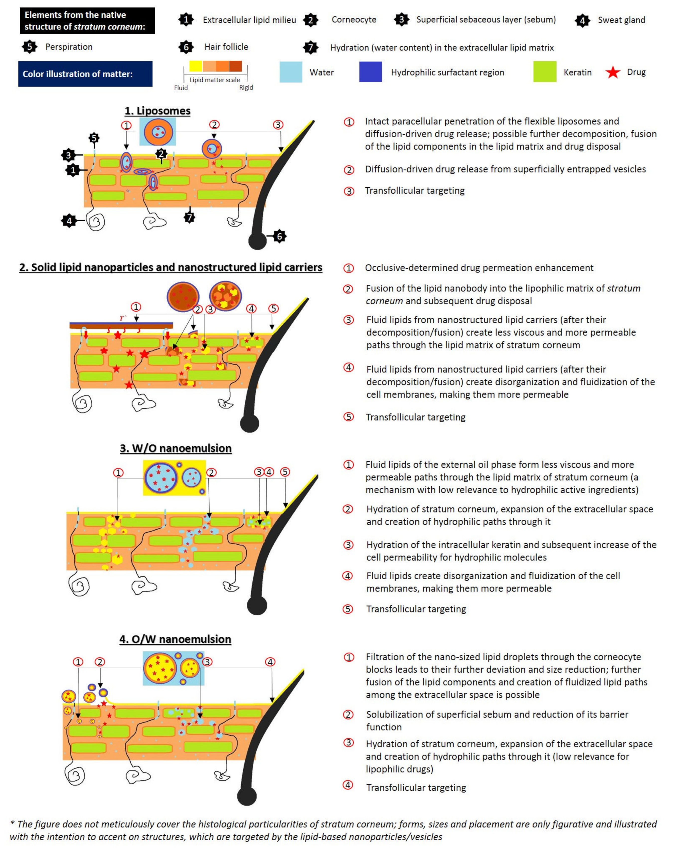
Table 1.
Physico-chemical characteristics of selected phenolic compounds.
| Phenolic Class | Phenolic Subclass | Phenolic Compound | Molecular Weight (g/mol) | Partition Coefficient (Log P) | Solubility in Water at 25 °C (mg/mL) | Ionizable Groups with Corresponding pKa Values |
|---|---|---|---|---|---|---|
| Simple phenols and derivatives | Phenolic acids (hydroxybenzoic acid derivatives) |  Gallic acid | 170.12 | −0.28 [208] | 14.7 [209] | pKa1 = 4.51 pKa2 = 8.7 pKa3 = 11.4 pKa4 > 13 [210] |
 Ellagic acid (ellagitannin, dimer of gallic acid) | 390.12 | 1.37 [211] | 0.0097 [212,213] (at 37 °C) | pKa1 = 5.42 pKa2 = 6.76 [214] | ||
| Phenolic acids (hydroxycinnamic acid derivatives) |  p-Coumaric acid | 164.05 | 1.46 [215] | 0.01 [216] | pKa1 = 4.92 pKa2 = 9.28 [217] | |
 Caffeic acid | 180.16 | 1.15 [218] | 0.98 [209] | pKa1 = 4.83 pKa2 = 8.90 pKa3 = 10.28 [217] | ||
 Ferulic acid | 194.18 | 1.51 [219] | 0.78 [209] | pKa1 = 4.66 pKa2 = 9.09 [217] | ||
 Chlorogenic acid | 354.31 | −0.75 [218] | 3.44 * [220] | pKa1 = 3.50 pKa2 = 8.42 pKa3 = 11.00 [221] | ||
| Other simple phenols |  Hydroquinone | 110.11 | 0.59 [222] | 7.20 [223] | pKa1 = 9.85 pKa2 = 11.40 [224] | |
 Eugenol | 164.20 | 2.49 [225] | 2.46 [226] | pKa = 10.19 [227] | ||
| Flavonoids | Flavones |  Apigenin | 270.05 | 2.92 [228] | 0.00135 [229] | pKa1 = 7.12 pKa2 = 8.10 [230] |
 Vitexin (Apigenin-8-C-glucoside) | 432.38 | 0.1 * [231] | 0.0762 [232] | pKa1 = 6.27 * [233] ** | ||
 Luteolin | 286.24 | 3.22 [228] | 0.14 * [234] | pKa1 = 6.57 * [234] ** | ||
| Flavonols |  Kaemferol | 286.23 | 3.11 [228] | 0.113 [235] (at 30 °C) | pKa1 = 6.96 pKa2 = 8.78 pKa3 = 10.60 [236] | |
 Quercetin | 302.24 | 1.82 [228] | 0.0004 [237] −0.002 [238] | pKa1 = 7.10 pKa2 = 9.09 pKa3 = 11.12 [236] | ||
 Rutin (Quercetin-3-O-rutinoside) | 610.52 | 0.76 [22] | 0.125 [239] | pKa1 = 2.92 pKa2 = 6.72 pKa3 = 8.26 pKa4 = 12.57 [240] | ||
| Flavanones |  Hesperitin | 302.27 | 2.9 [241] | 0.01572 [241] | pKa1 = 7.55 * pKa2 = 8.50 * pKa3 = 9.65 * | |
 Hesperidin (Hesperitin- 7-(6-O-(alpha-l-rhamnopyranosyl)-beta-d-glucopyranosyl) | 610.19 | 1.78 [242] | 0.00495 [242] | pKa1 = 10.0 pKa2 > 11.5 [243] | ||
| Flavan-3-ols |  Catechin | 290.26 | 0.41 [244] | 7.66 [245] | pKa1 = 8.68 pKa2 = 9.70 pKa3 = 11.55 [236] | |
 Epigallocatechin gallate | 458.37 | 0.46 [246] | 16.05 [247] | pKa1 = 7.75 pKa2 = 8.00 [248] | ||
| Curcuminoids |  Curcumin (keto form) | 368.38 | 3.0 [249] | 0.0006 [250] | pKa1 = 7.7–8.5 pKa2 = 8.5–10.4 pKa3 = 9.5–10.7 [249] | |
| Stilbenes |  Resveratrol | 228.25 | 3.09 * [251] | 0.05 [252] | pKa1 = 8.8 pKa2 = 9.8 pKa3 = 11.4 [253] | |
| Anthraquinones |  Hypericin | 504.44 | 3.43 [254] | Practically insoluble in water [255] ** | pKa1 = 2.00 pKa2 = 11.00 [256] | |
| Phloroglucinols |  Hyperforin | 536.78 | 13.17 [257] | 2.34 × 10⁻¹² [257] | pKa = 6.32 * [258] | |
* calculated value; ** information (or further information) was not found.
Table 2.
Phenolic compounds, encapsulated in lipid-based nanosystems for dermal. application.
| Main Class | Active Agent | Technological/Biopharmaceutical Issue | Lipid-Based Nanosystem/ Lipid Carrier | Obtained Results | References |
|---|---|---|---|---|---|
| Simple phenols and derivatives | Hydroquinone | Tendency to oxidation; hydrophilic structure hindering its topical application; side-effects due to systemic absorption | SLNs/Precirol® ATO5 |
| [311] |
| Arbutin | Highly hydrophilic compound; limited skin permeation | Liposomes/ Soybean phosphatidylcholine; cholesterol |
| [312,313] | |
| Thymoquinone (benzo-quinone) | Thermo/photosensitivity; hydrophobicity | Liposomes/ Phospholipon 90H®; cholesterol |
| [314] | |
| Protocatechuic acid; Ethyl protocatechuate (phenolic acid and derivative) | Sparingly hydrosolubilty (1:50); skin irritating properties; photosensitivity | SLNs/ Precirol ATO®5; NLCs/ Miglyol®810 N: Precirol ATO®5 3:7 |
| [315,316] | |
| Ferulic acid (phenolic acid) | Poor water solubility; low stability | Nanoemulsion/ Isostearyl isosearate |
| [317] | |
| Caffeic acid (hydroxyl-cinnamic acid) | Limited skin permeation | Liposomes/ egg phosphatidyl-choline; cholesterol |
| [318] | |
| Eugenol (Clove oil); (phenylpropene) | Predisposition to oxidation | SLNs/Stearic acid, Compritol® |
| [319] | |
| Flavanoids | Naringin (flavanone) | Limited aqueous solubility; poor oral bioavailability | Liposomes/ Epikuron-200; cholesterol, Tween 80 |
| [320] |
| Hesperidin (flavanone glycoside) | Poor aqueous solubility and bioavailability | NLCs/ Cupuaçu butter, buriti oil |
| [321] | |
| Isoflavone-aglycon-rich fraction (genistein, daidzein, glycitein) | Limited aqueous solubility | Nanoemulsion/ Egg lecithin (Lipoid E-80®), medium-chain triglycerides |
| [322] | |
| Anthraquinones | Aloe -emodin | Hydrophobic compound, crystallizes in water | Liposomes/ Hydrogenated soybean phosphatidyl-choline, cholesterol |
| [323] |
| Naphthoquinones | Vitamin K1 (phylloquinone) | Highly lipophilic compound; photosensitivity | Liposomes/ Soy phosphatidyl-choline, α-tocopherol |
| [324] |
| Xanthones | Mangiferin | Poor water solubility (0.111 mg/mL); low bioavailability | Nanoemulsion/ Almond oil, Lipoid ®S75 |
| [325] |
| Stilbenes | Resveratrol | Low aqueous solubility; photosensitivity | SLNs/ Compritol 888 ATO NLCs/ Compritol 888 ATO; Miglyol oil |
| [326,327] |
| Pterostilbene | None; favorable characteristics compared to resveratrol (i.e., increased lipophilicity, membrane permeability and bioavailability) | Liposomes/ Lecithin |
| [328] | |
| Tannins | Ellagic acid | Low aqueous solubility and permeability | NLCs/ Tristearin, Miglyol oil |
| [329] |
Table 3.
Cosmetic products containing phenolic phytochemicals formulated via nanotechnological approach.
Table 3.
Cosmetic products containing phenolic phytochemicals formulated via nanotechnological approach.
| Brand | Product | Phenolic Compounds | Lipid-Based Nanosystem | Benefits |
|---|---|---|---|---|
| Sesderma [331] | Sodyses Repair gel | Resveratrol, quercetin | Liposomes |
|
| Factor G Renew Rejuvenating serum | Quercetin, pterostilbene | Liposomes |
| |
| Hidroquin Whitening gel | Ferulic acid, Arbutin | Liposomes |
| |
| Reti Age Eye contour gel | Pterostilbene | Liposomes |
| |
| Kojicol Plus (+Kojic acid) Skin lightener cream | α-Arbutin, | Liposomes |
| |
| M.Y.R. [332] | Curcumin liposome Melasma and acne cream | Curcumin | Liposomes |
|
| Vitacos [333] | NanoVital Vitanics Whitening essence | Arbutin | Nanoemulsion |
|
| Dr. Theiss Medipharma Cosme-tics [298] | Olivenöl Anti Falten Pflege-konzentrat | Olea europaea oil | NLCs |
|
Publisher’s Note: MDPI stays neutral with regard to jurisdictional claims in published maps and institutional affiliations. |
© 2021 by the authors. Licensee MDPI, Basel, Switzerland. This article is an open access article distributed under the terms and conditions of the Creative Commons Attribution (CC BY) license (https://creativecommons.org/licenses/by/4.0/).
Share and Cite
MDPI and ACS Style
Gugleva, V.; Ivanova, N.; Sotirova, Y.; Andonova, V. Dermal Drug Delivery of Phytochemicals with Phenolic Structure via Lipid-Based Nanotechnologies. Pharmaceuticals 2021, 14, 837. https://doi.org/10.3390/ph14090837
AMA Style
Gugleva V, Ivanova N, Sotirova Y, Andonova V. Dermal Drug Delivery of Phytochemicals with Phenolic Structure via Lipid-Based Nanotechnologies. Pharmaceuticals. 2021; 14(9):837. https://doi.org/10.3390/ph14090837
Chicago/Turabian StyleGugleva, Viliana, Nadezhda Ivanova, Yoana Sotirova, and Velichka Andonova. 2021. "Dermal Drug Delivery of Phytochemicals with Phenolic Structure via Lipid-Based Nanotechnologies" Pharmaceuticals 14, no. 9: 837. https://doi.org/10.3390/ph14090837
Note that from the first issue of 2016, this journal uses article numbers instead of page numbers. See further details here.