
Field Study on the Prevalence of Ovine Footrot, Contagious Ovine Digital Dermatitis, and Their Associated Bacterial Species in Swedish Sheep Flocks

Abstract
:1. Introduction
2. Materials and Methods
2.1. Study Farms and Period
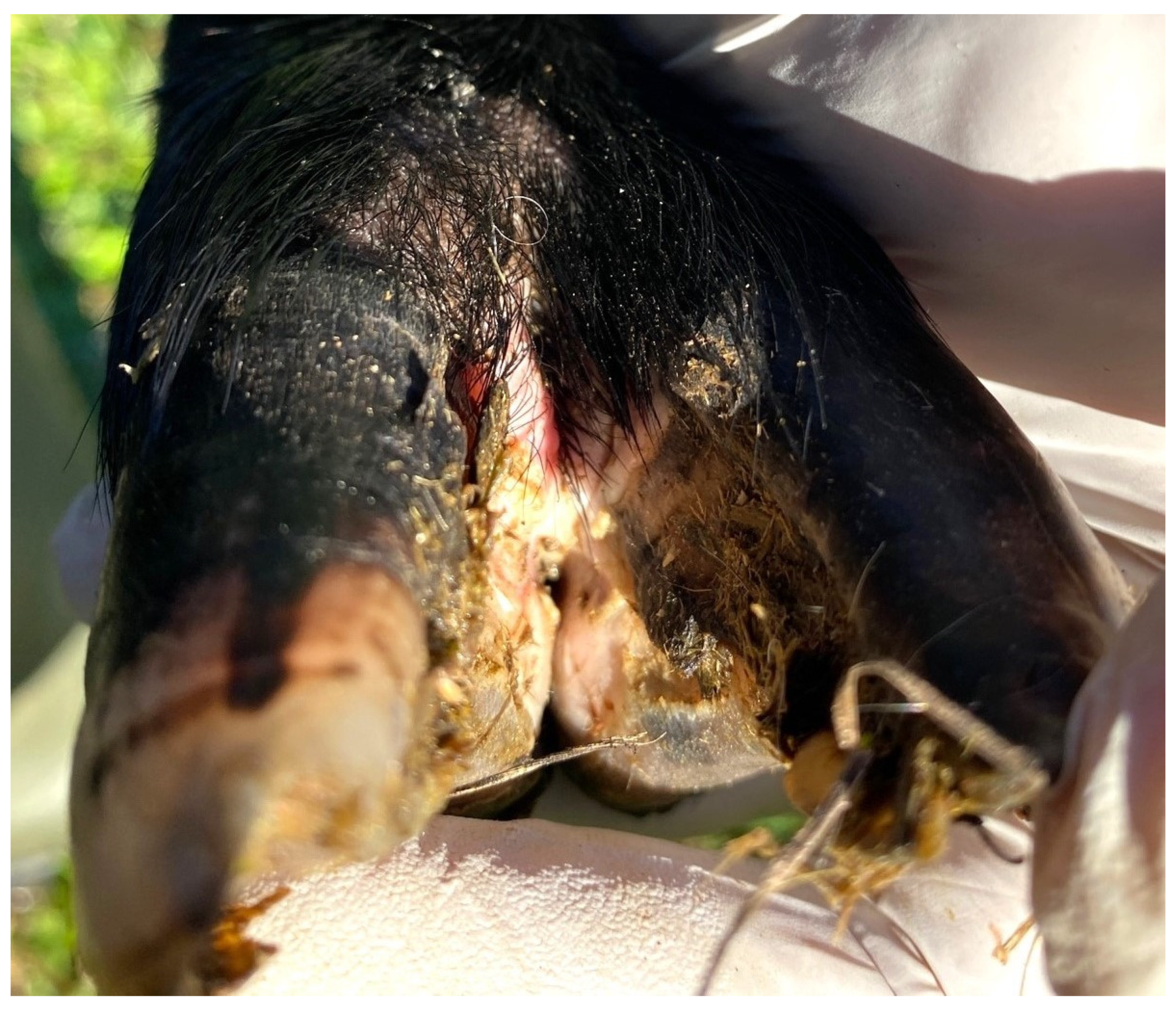
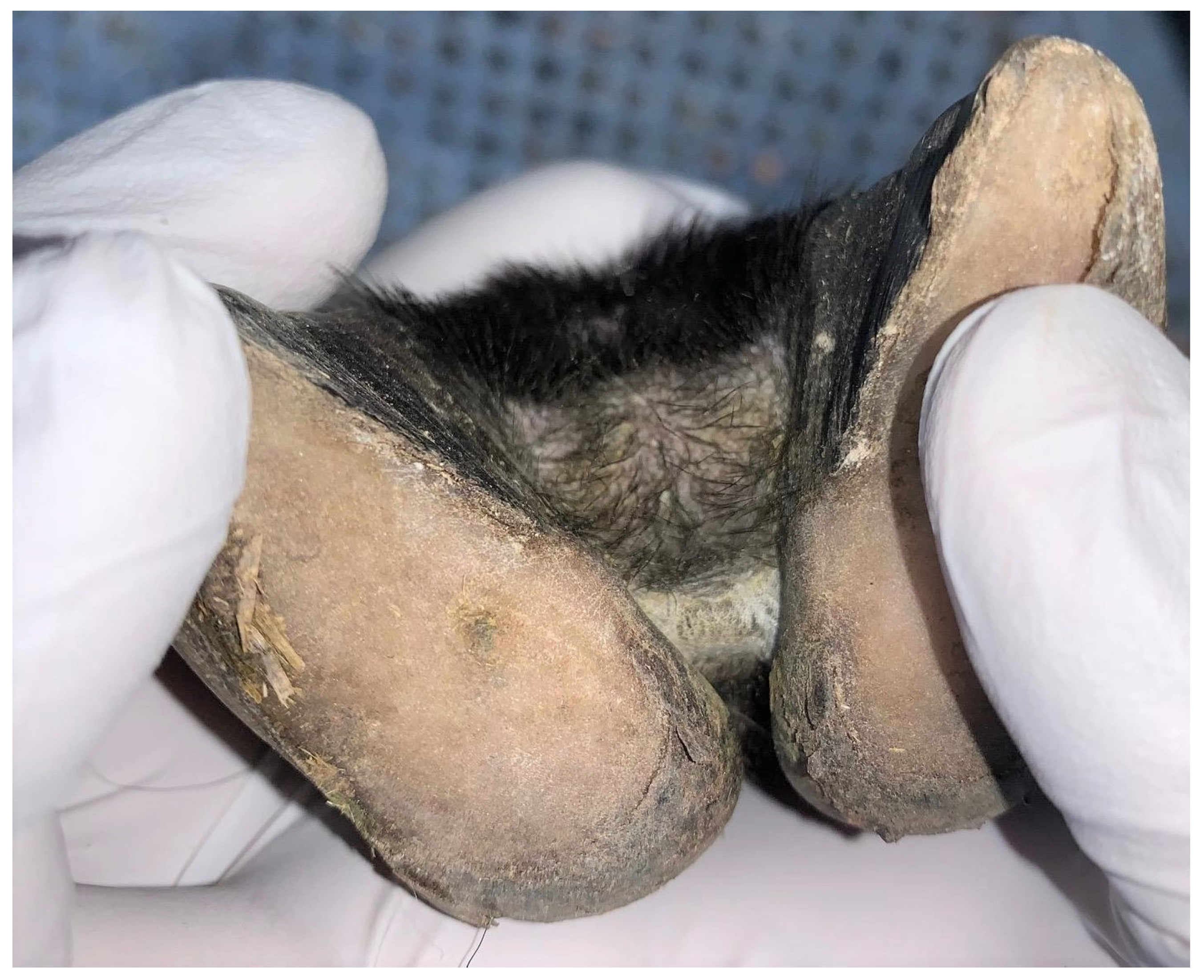
2.2. Sampling, Photography, and Lesion Assessment of Sheep Feet
2.3. Bacterial DNA Extraction and Real-Time PCR
2.4. Statistical Analysis
3. Results
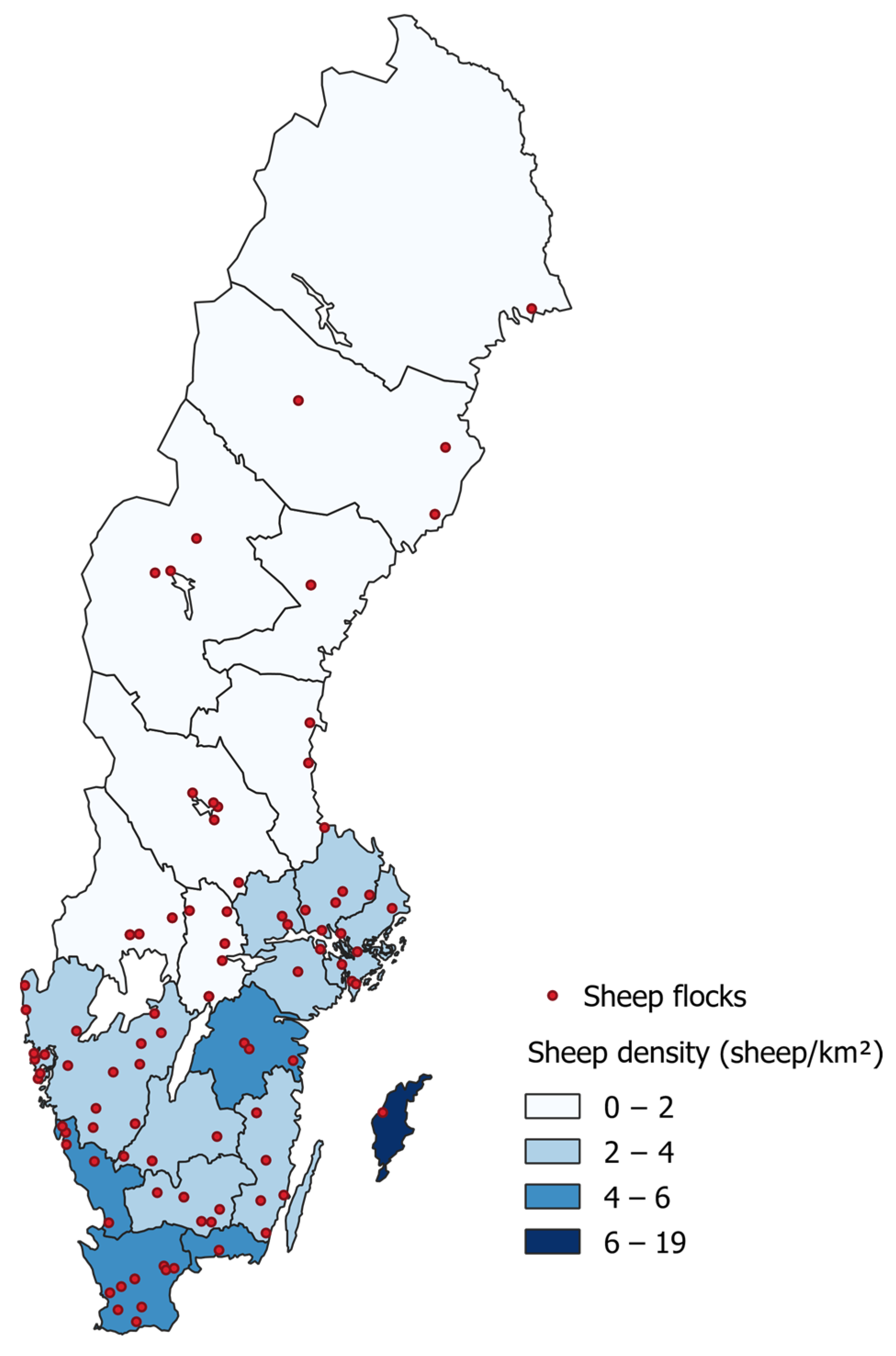
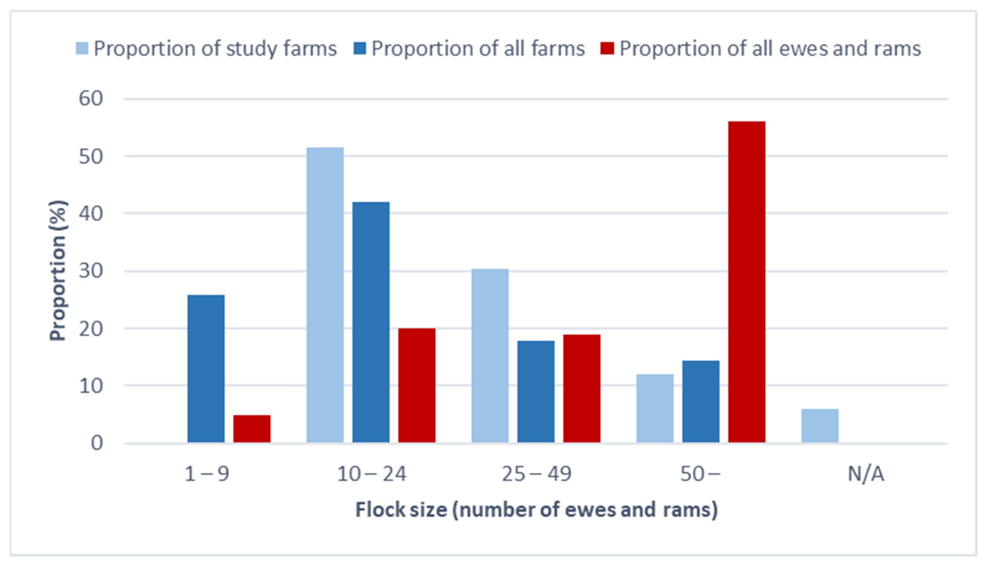
3.1. Study Farms
3.2. Assessment of Photographs
3.3. Real-Time PCR Analysis of Swab Samples
4. Discussion
Author Contributions
Funding
Institutional Review Board Statement
Informed Consent Statement
Data Availability Statement
Acknowledgments
Conflicts of Interest
References
- Beveridge, W.I.B. Foot-rot in sheep: A transmissible disease due to infection with Fusiformis nodosus (n.sp.). In Studies on Its Cause, Epidemiology and Control; Bulletin No. 140; Council for Scientific and Industrial Research: Melbourne, Australia, 1941; pp. 1–56. [Google Scholar]
- Harwood, D.G.; Cattell, J.H.; Lewis, C.J.; Naylor, R. Virulent foot rot in sheep. Vet. Rec. 1997, 140, 687. [Google Scholar]
- Sayers, G.; Marques, P.X.; Evans, N.J.; O’Grady, L.; Doherty, M.L.; Carter, S.D.; Nally, J.E. Identification of spirochetes associated with contagious ovine digital dermatitis. J. Clin. Microbiol. 2009, 47, 1199–1201. [Google Scholar] [CrossRef] [PubMed]
- Sullivan, L.E.; Clegg, S.R.; Angell, J.W.; Newbrook, K.; Blowey, R.W.; Carter, S.D.; Bell, J.; Duncan, J.S.; Grove-White, D.H.; Murray, R.D.; et al. High-level association of bovine digital dermatitis Treponema spp. with contagious ovine digital dermatitis lesions and presence of Fusobacterium necrophorum and Dichelobacter nodosus. J. Clin. Microbiol. 2015, 53, 1628–1638. [Google Scholar] [CrossRef]
- Duncan, J.S.; Angell, J.W.; Richards, P.; Lenzi, L.; Staton, G.J.; Grove-White, D.; Clegg, S.; Oikonomou, G.; Carter, S.D.; Evans, N.J. The dysbiosis of ovine foot microbiome during the development and treatment of contagious ovine digital dermatitis. Anim. Microbiome 2021, 3, 19. [Google Scholar] [CrossRef]
- Staton, G.J.; Angell, J.W.; Grove-White, D.; Clegg, S.R.; Carter, S.D.; Evans, N.J.; Duncan, J.S. Contagious Ovine Digital Dermatitis: A Novel Bacterial Etiology and Lesion Pathogenesis. Front. Vet. Sci. 2021, 8, 722461. [Google Scholar] [CrossRef] [PubMed]
- Stewart, D.J.; Peterson, J.E.; Vaughan, J.A.; Clark, B.L.; Emery, D.L.; Caldwell, J.B.; Kortt, A.A. The pathogenicity and cultural characteristics of virulent, intermediate and benign strains of Bacteroides nodosus causing ovine foot-rot. Aust. Vet. J. 1986, 63, 317–326. [Google Scholar] [CrossRef] [PubMed]
- Stäuble, A.; Steiner, A.; Frey, J.; Kuhnert, P. Simultaneous detection and discrimination of virulent and benign Dichelobacter nodosus in sheep of flocks affected by foot rot and in clinically healthy flocks by competitive real-time PCR. J. Clin. Microbiol. 2014, 52, 1228–1231. [Google Scholar] [CrossRef] [PubMed]
- Frosth, S.; König, U.; Nyman, A.K.; Pringle, M.; Aspán, A. Characterisation of Dichelobacter nodosus and detection of Fusobacterium necrophorum and Treponema spp. in sheep with different clinical manifestations of footrot. Vet. Microbiol. 2015, 179, 82–90. [Google Scholar] [CrossRef]
- Clifton, R.; Giebel, K.; Liu, N.L.B.H.; Purdy, K.J.; Green, L.E. Sites of persistence of Fusobacterium necrophorum and Dichelobacter nodosus: A paradigm shift in understanding the epidemiology of footrot in sheep. Sci. Rep. 2019, 9, 14429. [Google Scholar] [CrossRef]
- Tan, Z.L.; Nagaraja, T.G.; Chengappa, M.M. Fusobacterium necrophorum infections: Virulence factors, pathogenic mechanism and control measures. Vet. Res. Commun. 1996, 20, 113–140. [Google Scholar]
- Olofsson, A.; Bergsten, C.; Björk Averpil, H. Smittsam klövsjukdom hos får diagnostiserad för första gången i Sverige (Infectious claw disease diagnosed for the first time in Sweden). Sven. Vet. 2005, 11, 11–14, (In Swedish with English Summary). [Google Scholar]
- Bernhard, M.; Frosth, S.; König, U. First report on outbreaks of contagious ovine digital dermatitis in Sweden. Acta Vet. Scand. 2021, 63, 29. [Google Scholar] [CrossRef]
- Farm and Animal Health. Klövkontrollen Får—2022; Farm and Animal Health: Sweden, 2023; Available online: https://www.gardochdjurhalsan.se/wp-content/uploads/2023/04/9115-klovkontrollen-pop-sammanfattning-2022.pdf (accessed on 16 June 2023).
- König, U.; Nyman, A.K.; de Verdier, K. Prevalence of footrot in Swedish slaughter lambs. Acta Vet. Scand. 2011, 53, 27. [Google Scholar] [CrossRef]
- Rosander, A.; Albinsson, R.; König, U.; Nyman, A.; Frosth, S. Prevalence of bacterial species associated with ovine footrot and contagious ovine digital dermatitis in Swedish slaughter lambs. Acta Vet. Scand. 2022, 64, 6. [Google Scholar] [CrossRef] [PubMed]
- Duncan, J.S.; Angell, J.W.; Carter, S.D.; Evans, N.J.; Sullivan, L.E.; Grove-White, D.H. Contagious ovine digital dermatitis: An emerging disease. Vet. J. 2014, 201, 265–268. [Google Scholar] [CrossRef] [PubMed]
- Daniel, W.W. Biostatistics: A Foundation for Analysis in the Health Sciences, 7th ed.; Wiley: New York, NY, USA, 1999. [Google Scholar]
- Swedish Board of Agriculture. Agricultural Statistical Compilation; Swedish Board of Agriculture: Jönköping, Sweden, 2022. Available online: https://jordbruksverket.se/9322.html (accessed on 25 March 2023).
- Stewart, D.J.; Claxton, P.D. Ovine foot rot: Clinical diagnosis and bacteriology. In Australian Standard Diagnostic Techniques for Animal Diseases; Corner, L.A., Bagust, T.J., Eds.; CSIRO: East Melbourne, Australia, 1993; pp. 1–27. [Google Scholar]
- Angell, J.W.; Blundell, R.; Grove-White, D.H.; Duncan, J.S. Clinical and radiographic features of contagious ovine digital dermatitis and a novel lesion grading system. Vet. Rec. 2015, 176, 544. [Google Scholar] [CrossRef] [PubMed]
- Frosth, S.; Slettemeås, J.S.; Jørgensen, H.J.; Angen, Ø.; Aspán, A. Development and comparison of a real-time PCR assay for detection of Dichelobacter nodosus with culturing and conventional PCR: Harmonisation between three laboratories. Acta Vet. Scand. 2012, 54, 6. [Google Scholar] [CrossRef]
- Jensen, A.; Hagelskjaer Kristensen, L.; Prag, J. Detection of Fusobacterium necrophorum subsp. funduliforme in tonsillitis in young adults by real-time PCR. Clin. Microbiol. Infect. 2007, 13, 695–701. [Google Scholar] [CrossRef]
- Strub, S.; van der Ploeg, J.R.; Nuss, K.; Wyss, C.; Luginbuhl, A.; Steiner, A. Quantitation of Guggenheimella bovis and treponemes in bovine tissues related to digital dermatitis. FEMS Microbiol. Lett. 2007, 269, 48–53. [Google Scholar] [CrossRef]
- Frosth, S.; Eriksson, H.K.; Rosander, A. Development of a multiplex quantitative PCR assay for simultaneous detection of Treponema phagedenis, Treponema pedis, Treponema medium, and ‘Treponema vincentii’ and evaluation on bovine digital dermatitis biopsies. Vet. Res. Commun. 2023. [Google Scholar] [CrossRef]
- Storms, J.; Wirth, A.; Vasiliadis, D.; Brodard, I.; Hamann-Tholken, A.; Ambros, C.; Moog, U.; Jores, J.; Kuhnert, P.; Distl, O. Prevalence of Dichelobacter nodosus and Ovine Footrot in German Sheep Flocks. Animals 2021, 11, 1102. [Google Scholar] [CrossRef]
- Frosth, S.; König, U.; Nyman, A.K.; Aspán, A. Sample pooling for real-time PCR detection and virulence determination of the footrot pathogen Dichelobacter nodosus. Vet. Res. Commun. 2017, 41, 189–193. [Google Scholar] [CrossRef] [PubMed]
- Moore, L.J.; Wassink, G.J.; Green, L.E.; Grogono-Thomas, R. The detection and characterisation of Dichelobacter nodosus from cases of ovine footrot in England and Wales. Vet. Microbiol. 2005, 108, 57–67. [Google Scholar] [CrossRef] [PubMed]
- Maboni, G.; Frosth, S.; Aspán, A.; Tötemeyer, S. Ovine footrot: New insights into bacterial colonisation. Vet. Rec. 2016, 179, 228. [Google Scholar] [CrossRef] [PubMed]
- Ardüser, F.; Moore-Jones, G.; Gobeli Brawand, S.; Durr, S.; Steiner, A.; Ryser-Degiorgis, M.P.; Zanolari, P. Dichelobacter nodosus in sheep, cattle, goats and South American camelids in Switzerland-Assessing prevalence in potential hosts in order to design targeted disease control measures. Prev. Vet. Med. 2020, 178, 104688. [Google Scholar] [CrossRef]
- Frosth, S. Dichelobacter nodosus and Footrot in Swedish Sheep. Increased Knowledge and Improved Laboratory Diagnostics. Ph.D. Thesis, Swedish University of Agricultural Sciences, Uppsala, Sweden, 2016. [Google Scholar]
- Evans, N.J.; Timofte, D.; Isherwood, D.R.; Brown, J.M.; Williams, J.M.; Sherlock, K.; Lehane, M.J.; Murray, R.D.; Birtles, R.J.; Hart, C.A.; et al. Host and environmental reservoirs of infection for bovine digital dermatitis treponemes. Vet. Microbiol. 2012, 156, 102–109. [Google Scholar] [CrossRef]


| Footrot Score 0 (%) | Footrot Score 1 (%) | Footrot Score 2 (%) | Footrot Score Unknown (%) | Total | |
|---|---|---|---|---|---|
| Number of sheep | 273 (91.9) | 5 (1.7) | 2 (0.7) | 17 (5.7) | 297 |
| Number of sheep with D. nodosus | 12 (4.0) | 3 (1.0) | 2 (0.7) | 0 (0) | 17 |
| Number of sheep with F. necrophorum | 3 (1.0) | 1 (0.3) | 0 (0) | 0 (0) | 4 |
| Number of sheep with Treponema spp. | 178 (60.0) | 5 (1.7) | 2 (0.7) | 8 (2.7) | 193 |
| Footrot Score 0 (%) | Footrot Score 1 (%) | Footrot Score 2 (%) | Footrot Score Unknown (%) | Total | |
|---|---|---|---|---|---|
| Number of sheep flocks | 90 (90.1) | 3 (3.0) | 2 (2.0) | 4 (4.0) | 99 |
| Number of sheep flocks with D. nodosus | 6 (6.1) | 1 (1.0) | 2 (2.0) | 0 (0) | 9 |
| Number of sheep flocks with F. necrophorum | 2 (2.0) | 1 (1.0) | 0 (0) | 0 (0) | 3 |
| Number of sheep flocks with Treponema spp. | 75 (75.8) | 3 (3.0) | 2 (2.0) | 2 (2.0) | 82 |
Disclaimer/Publisher’s Note: The statements, opinions and data contained in all publications are solely those of the individual author(s) and contributor(s) and not of MDPI and/or the editor(s). MDPI and/or the editor(s) disclaim responsibility for any injury to people or property resulting from any ideas, methods, instructions or products referred to in the content. |
© 2023 by the authors. Licensee MDPI, Basel, Switzerland. This article is an open access article distributed under the terms and conditions of the Creative Commons Attribution (CC BY) license (https://creativecommons.org/licenses/by/4.0/).
Share and Cite
Rosander, A.; Mourath, S.; König, U.; Nyman, A.; Frosth, S. Field Study on the Prevalence of Ovine Footrot, Contagious Ovine Digital Dermatitis, and Their Associated Bacterial Species in Swedish Sheep Flocks. Pathogens 2023, 12, 1224. https://doi.org/10.3390/pathogens12101224
Rosander A, Mourath S, König U, Nyman A, Frosth S. Field Study on the Prevalence of Ovine Footrot, Contagious Ovine Digital Dermatitis, and Their Associated Bacterial Species in Swedish Sheep Flocks. Pathogens. 2023; 12(10):1224. https://doi.org/10.3390/pathogens12101224
Chicago/Turabian StyleRosander, Anna, Sophia Mourath, Ulrika König, Ann Nyman, and Sara Frosth. 2023. "Field Study on the Prevalence of Ovine Footrot, Contagious Ovine Digital Dermatitis, and Their Associated Bacterial Species in Swedish Sheep Flocks" Pathogens 12, no. 10: 1224. https://doi.org/10.3390/pathogens12101224