

The Extraordinary Case of a Woman with a 30-Year-Long Diffuse Leishmaniasis Cured with One Single Ampoule of Intranasal Pentavalent Antimoniate
 , , , , , , , , , and
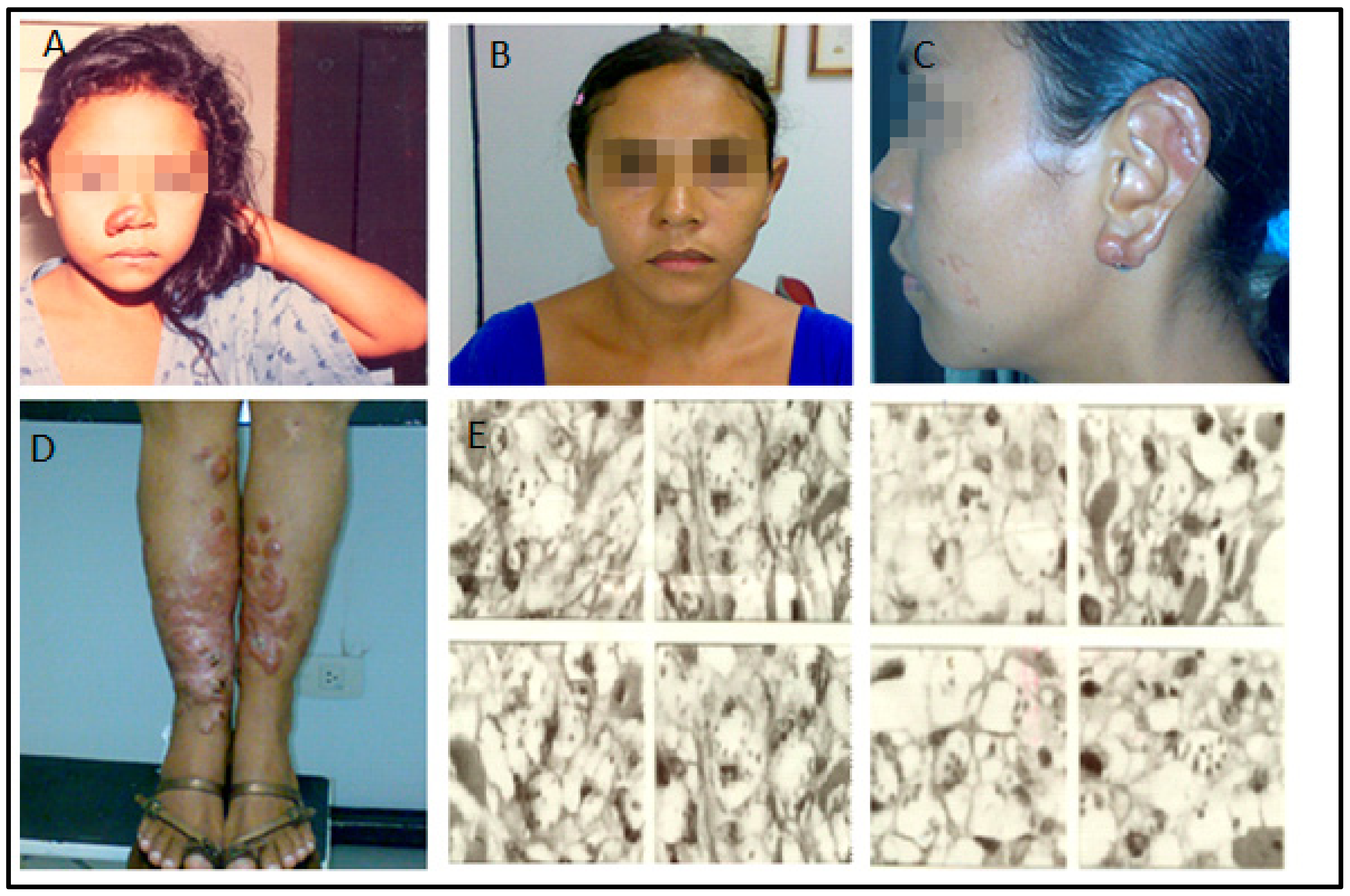
Abstract
:1. Introduction
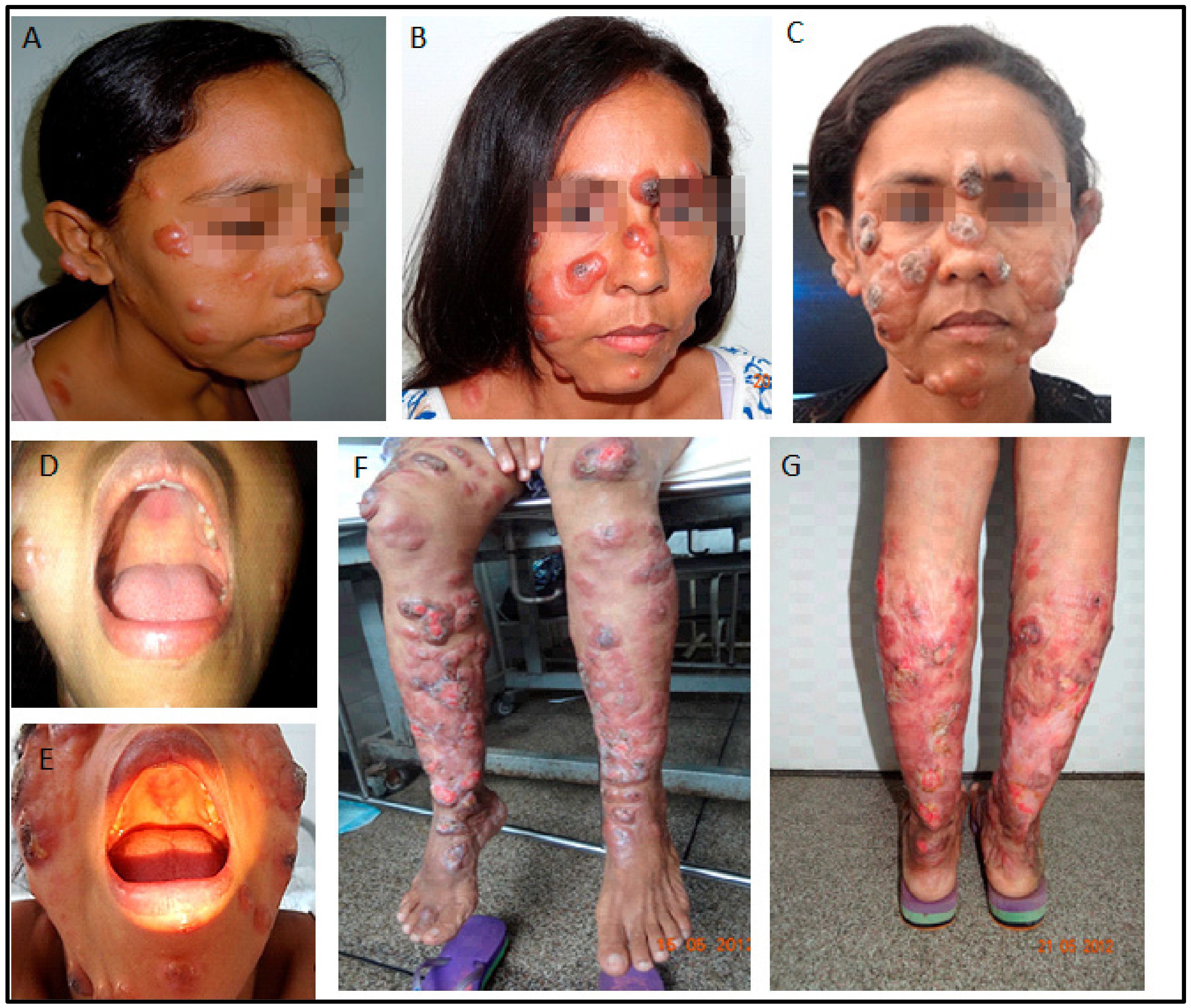
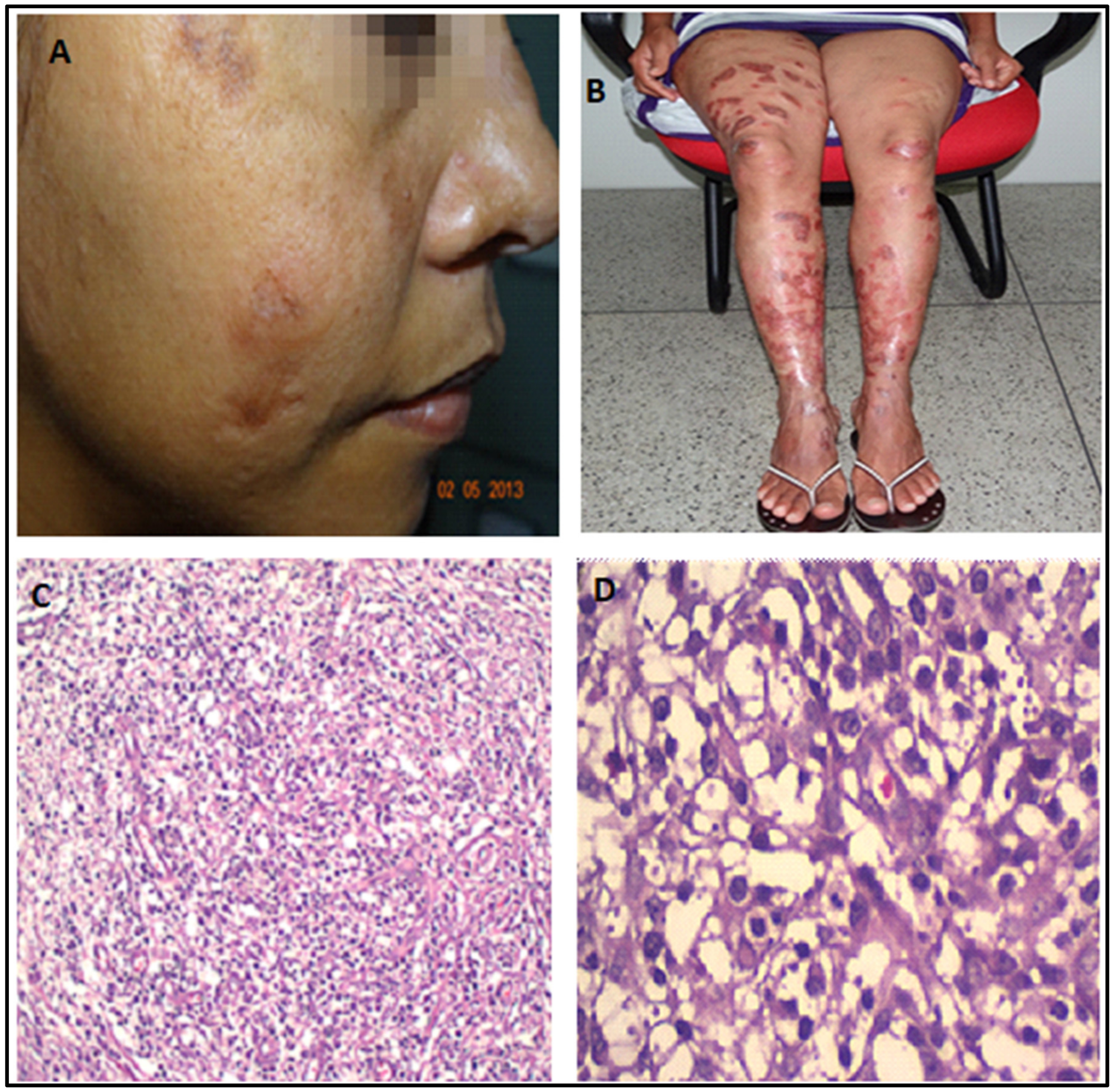
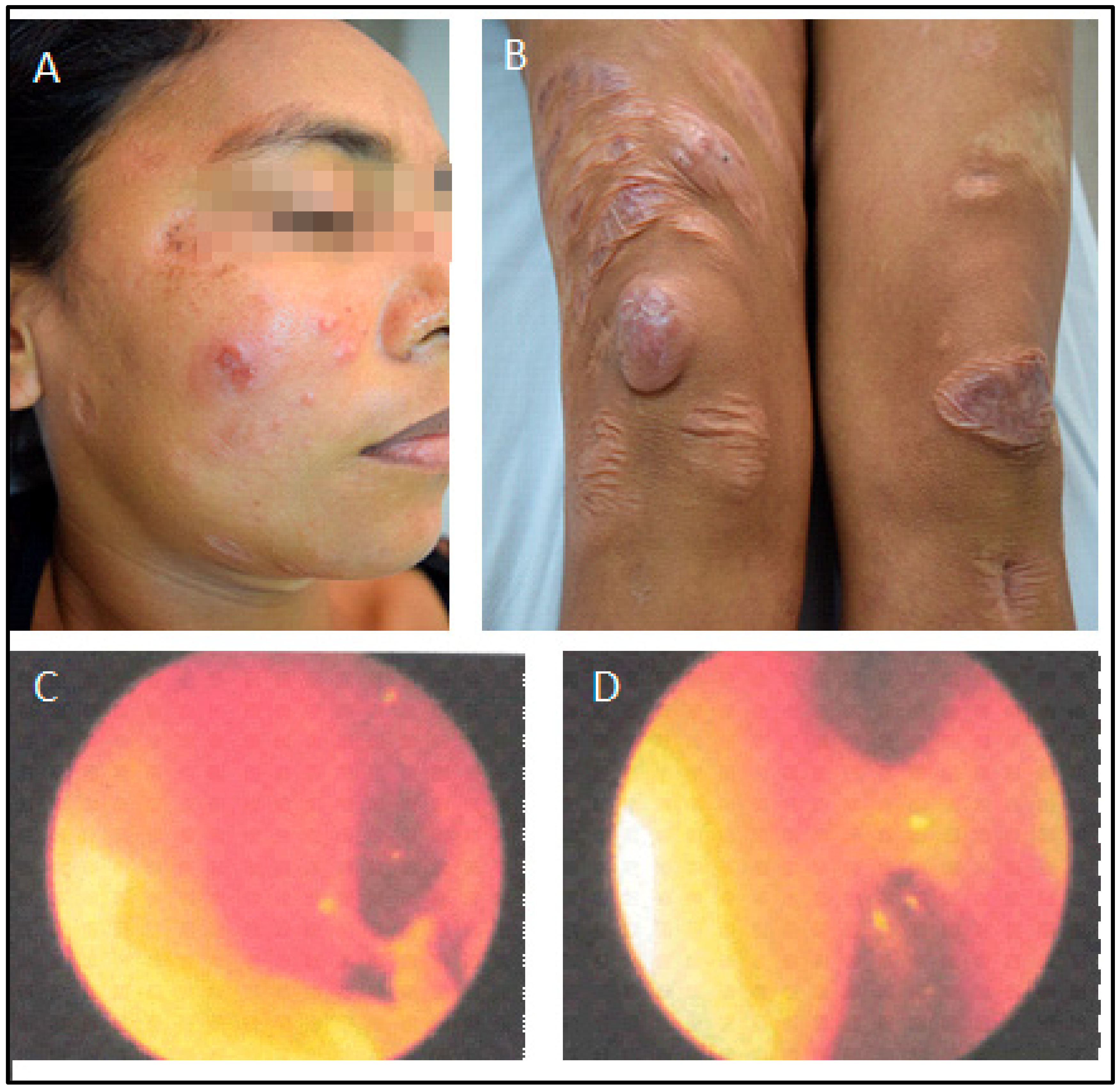
2. Case Report
3. Discussion
Supplementary Materials
Author Contributions
Funding
Institutional Review Board Statement
Informed Consent Statement
Data Availability Statement
Acknowledgments
Conflicts of Interest
References
- Espinosa, O.A.; Serrano, M.G.; Camargo, E.P.; Teixeira, M.M.G.; Shaw, J.J. An appraisal of the taxonomy and nomenclature of trypanosomatids presently classified as Leishmania and Endotrypanum. Parasitology 2018, 145, 430–442. [Google Scholar] [CrossRef]
- Silveira, F.T.; Lainson, R.; Shaw, J.J.; De Souza, A.A.; Ishikawa, E.A.; Braga, R.R. Cutaneous leishmaniasis due to Leishmania (Leishmania) amazonensis in Amazonian Brazil, and the significance of a negative Montenegro skin-test in human infections. Trans. R. Soc. Trop. Med. Hyg. 1991, 85, 735–738. [Google Scholar] [CrossRef]
- Coelho, A.C.; Trinconi, C.T.; Costa, C.H.N.; Uliana, S.R.B. In Vitro and In Vivo Miltefosine Susceptibility of a Leishmania amazonensis Isolate from a Patient with Diffuse Cutaneous Leishmaniasis: Follow-Up. PLoS Negl. Trop. Dis. 2016, 10, e0004720. [Google Scholar] [CrossRef] [PubMed] [Green Version]
- Costa, J.M.L.; Saldanha, A.C.R.; Silva, A.C.d.M.e.; Serra Neto, A.; Galvão, C.E.S.; Silva, C.D.M.P.; Silva, A.R.D. Estado atual da leishmaniose cutânea difusa (LCD) no Estado do Maranhão: II. aspectos epidemiológicos, clínico-evolutivos. Rev. Soc. Bras. Med. Trop. 1992, 25, 115–123. [Google Scholar] [CrossRef] [PubMed] [Green Version]
- Lainson, R.; Shaw, J.J.; Silveira, F.T.; Souza, A.A.A.d.; Braga, R.R.; Ishikawa, E.A.Y. The dermal leishmaniases of Brazil, with special reference to the eco-epidemiology of the disease in Amazonia. Memórias Inst. Oswaldo Cruz 1994, 89, 435–443. [Google Scholar] [CrossRef]
- Silveira, F.T.; Lainson, R.; Corbett, C.E. Clinical and immunopathological spectrum of American cutaneous leishmaniasis with special reference to the disease in Amazonian Brazil: A review. Memórias Inst. Oswaldo Cruz 2004, 99, 239–251. [Google Scholar] [CrossRef]
- Silveira, F.T.; Lainson, R.; Corbett, C.E. Further observations on clinical, histopathological, and immunological features of borderline disseminated cutaneous leishmaniasis caused by Leishmania (Leishmania) amazonensis. Memórias Inst. Oswaldo Cruz 2005, 100, 525–534. [Google Scholar] [CrossRef] [Green Version]
- Bonney, E.A. Immune Regulation in Pregnancy. Obstet. Gynecol. Clin. 2016, 43, 679–698. [Google Scholar] [CrossRef] [PubMed] [Green Version]
- WHO Expert Committee on the Control of the Leishmaniases & World Health Organization. Control of the Leishmaniases; World Health Organization: Geneva, Switzerland, 2010. [Google Scholar]
- Isaacs, M.J.; Hossler, E.W. Cutaneous leishmaniasis. Cutis 2015, 96, 408–409. [Google Scholar]
- França-Costa, J.; Van Weyenbergh, J.; Boaventura, V.S.; Luz, N.F.; Malta-Santos, H.; Oliveira, M.C.S.; Santos de Campos, D.C.; Saldanha, A.C.; Dos-Santos, W.L.; Bozza, P.T.; et al. Arginase I, Polyamine, and Prostaglandin E2 Pathways Suppress the Inflammatory Response and Contribute to Diffuse Cutaneous Leishmaniasis. J. Infect. Dis. 2015, 211, 426–435. [Google Scholar] [CrossRef] [Green Version]
- Ponte-Sucre, A.; Gamarro, F.; Dujardin, J.C.; Barrett, M.P.; López-Vélez, R.; García-Hernández, R.; Pountain, A.W.; Mwenechanya, R.; Papadopoulou, B. Drug resistance and treatment failure in leishmaniasis: A 21st century challenge. PLoS Negl. Trop. Dis. 2017, 11, e0006052. [Google Scholar] [CrossRef] [Green Version]
- Barroso, D.H.; Falcão, S.D.A.C.; Motta, J.D.O.C.D.; Sevilha dos Santos, L.; Takano, G.H.S.; Gomes, C.M.; Favali, C.B.F.; De Lima, B.D.; Sampaio, R.N.R. PD-L1 May Mediate T-Cell Exhaustion in a Case of Early Diffuse Leishmaniasis Caused by Leishmania (L.) amazonensis. Front. Immunol. 2018, 9, 1021. [Google Scholar] [CrossRef] [PubMed]
- Miggelbrink, A.M.; Jackson, J.D.; Lorrey, S.J.; Srinivasan, E.S.; Waibl-Polania, J.; Wilkinson, D.S.; Fecci, P.E. CD4 T-Cell Exhaustion: Does It Exist and What Are Its Roles in Cancer? Clin. Cancer Res. 2021, 27, 5742–5752. [Google Scholar] [CrossRef] [PubMed]
- Hernández-Ruiz, J.; Salaiza-Suazo, N.; Carrada, G.; Escoto, S.; Ruiz-Remigio, A.; Rosenstein, Y.; Zentella, A.; Becker, I. CD8 Cells of Patients with Diffuse Cutaneous Leishmaniasis Display Functional Exhaustion: The Latter Is Reversed, In Vitro, by TLR2 Agonists. PLoS Negl. Trop. Dis. 2010, 4, e871. [Google Scholar] [CrossRef] [PubMed] [Green Version]
- Silveira, F.T.; Lainson, R.; de Castro Gomes, C.M.; Laurenti, M.D.; Corbett, C.E.P. Immunopathogenic competences of Leishmania (V.) braziliensis and L. (L.) amazonensis in American cutaneous leishmaniasis. Parasite Immunol. 2009, 31, 423–431. [Google Scholar] [CrossRef]
- Van Griensven, J.; Gadisa, E.; Aseffa, A.; Hailu, A.; Beshah, A.M.; Diro, E. Treatment of Cutaneous Leishmaniasis Caused by Leishmania aethiopica: A Systematic Review. PLoS Negl. Trop. Dis. 2016, 10, e0004495. [Google Scholar] [CrossRef] [Green Version]
- Sampaio, R.N.R.; Ferreira, M.F.; Martins, S.S.; Motta, J.D.O.C.d. Successful treatment of diffuse cutaneous leishmaniasis caused by Leishmania amazonensis. An. Bras. Dermatol. 2021, 96, 602–604. [Google Scholar] [CrossRef]
- Bartlett, J.A.; van der Voort Maarschalk, K. Understanding the Oral Mucosal Absorption and Resulting Clinical Pharmacokinetics of Asenapine. AAPS PharmSciTech 2012, 13, 1110–1115. [Google Scholar] [CrossRef] [Green Version]
- Campos, M.B.; Lima, L.V.d.R.; de Lima, A.C.S.; Vasconcelos dos Santos, T.; Ramos, P.K.S.; Gomes, C.M.D.C.; Silveira, F.T. Toll-like receptors 2, 4, and 9 expressions over the entire clinical and immunopathological spectrum of American cutaneous leishmaniasis due to Leishmania (V.) braziliensis and Leishmania (L.) amazonensis. PLoS ONE 2018, 13, e0194383. [Google Scholar] [CrossRef]
- Christensen, S.M.; Belew, A.T.; El-Sayed, N.M.; Tafuri, W.L.; Silveira, F.T.; Mosser, D.M. Host and parasite responses in human diffuse cutaneous leishmaniasis caused by L. amazonensis. PLoS Negl. Trop. Dis. 2019, 13, e0007152. [Google Scholar] [CrossRef] [Green Version]
- Collier, J.L.; Weiss, S.A.; Pauken, K.E.; Sen, D.R.; Sharpe, A.H. Not-so-opposite ends of the spectrum: CD8+ T cell dysfunction across chronic infection, cancer and autoimmunity. Nat. Immunol. 2021, 22, 809–819. [Google Scholar] [CrossRef] [PubMed]
- Zaric, M.; Becker, P.D.; Hervouet, C.; Kalcheva, P.; Doszpoly, A.; Blattman, N.A.; O’Neill, L.; Yus, B.I.; Cocita, C.; Kwon, S.Y.; et al. Skin immunisation activates an innate lymphoid cell-monocyte axis regulating CD8+ effector recruitment to mucosal tissues. Nat. Commun. 2019, 10, 2214. [Google Scholar] [CrossRef] [PubMed] [Green Version]


Disclaimer/Publisher’s Note: The statements, opinions and data contained in all publications are solely those of the individual author(s) and contributor(s) and not of MDPI and/or the editor(s). MDPI and/or the editor(s) disclaim responsibility for any injury to people or property resulting from any ideas, methods, instructions or products referred to in the content. |
© 2023 by the authors. Licensee MDPI, Basel, Switzerland. This article is an open access article distributed under the terms and conditions of the Creative Commons Attribution (CC BY) license (https://creativecommons.org/licenses/by/4.0/).
Share and Cite
Gonçalves, S.V.C.B.; Costa, D.L.; Cantinho-Junior, J.d.J.; Vieira-Junior, J.N.; Ishikawa, E.A.Y.; Costa, R.N.; Costa-Filho, A.C.G.; Araújo, R.d.C.; Uliana, S.R.B.; Yasunaka, J.K.U.Y.; et al. The Extraordinary Case of a Woman with a 30-Year-Long Diffuse Leishmaniasis Cured with One Single Ampoule of Intranasal Pentavalent Antimoniate. Pathogens 2023, 12, 890. https://doi.org/10.3390/pathogens12070890
Gonçalves SVCB, Costa DL, Cantinho-Junior JdJ, Vieira-Junior JN, Ishikawa EAY, Costa RN, Costa-Filho ACG, Araújo RdC, Uliana SRB, Yasunaka JKUY, et al. The Extraordinary Case of a Woman with a 30-Year-Long Diffuse Leishmaniasis Cured with One Single Ampoule of Intranasal Pentavalent Antimoniate. Pathogens. 2023; 12(7):890. https://doi.org/10.3390/pathogens12070890
Chicago/Turabian StyleGonçalves, Sheila V. C. B., Dorcas L. Costa, João da J. Cantinho-Junior, José N. Vieira-Junior, Edna A. Y. Ishikawa, Rubens N. Costa, Antônio C. G. Costa-Filho, Ronald da C. Araújo, Silvia R. B. Uliana, Jenicer K. U. Y. Yasunaka, and et al. 2023. "The Extraordinary Case of a Woman with a 30-Year-Long Diffuse Leishmaniasis Cured with One Single Ampoule of Intranasal Pentavalent Antimoniate" Pathogens 12, no. 7: 890. https://doi.org/10.3390/pathogens12070890