
Tailoring Mesoporous Silica-Coated Silver Nanoparticles and Polyurethane-Doped Films for Enhanced Antimicrobial Applications
 , , , , ,
  and
and 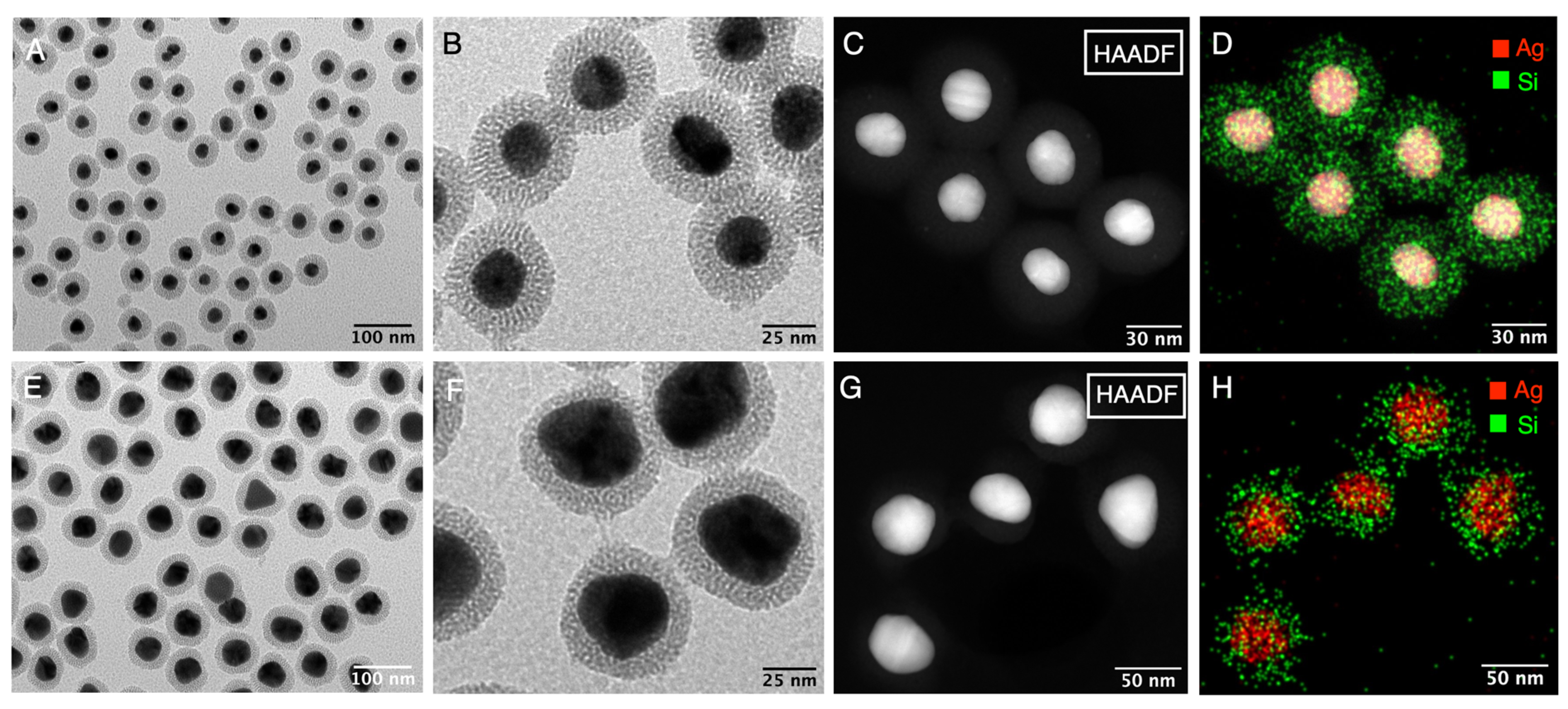
Abstract
:1. Introduction
2. Materials and Methods
2.1. Materials
2.2. Instrumentation
2.3. Methods
2.3.1. Synthesis of AgNPs
2.3.2. Synthesis of AuNPs
2.3.3. Functionalization of AgNPs and AuNPs with AMP
2.3.4. Mesoporous Silica Coating of AgNPs or AuNPs
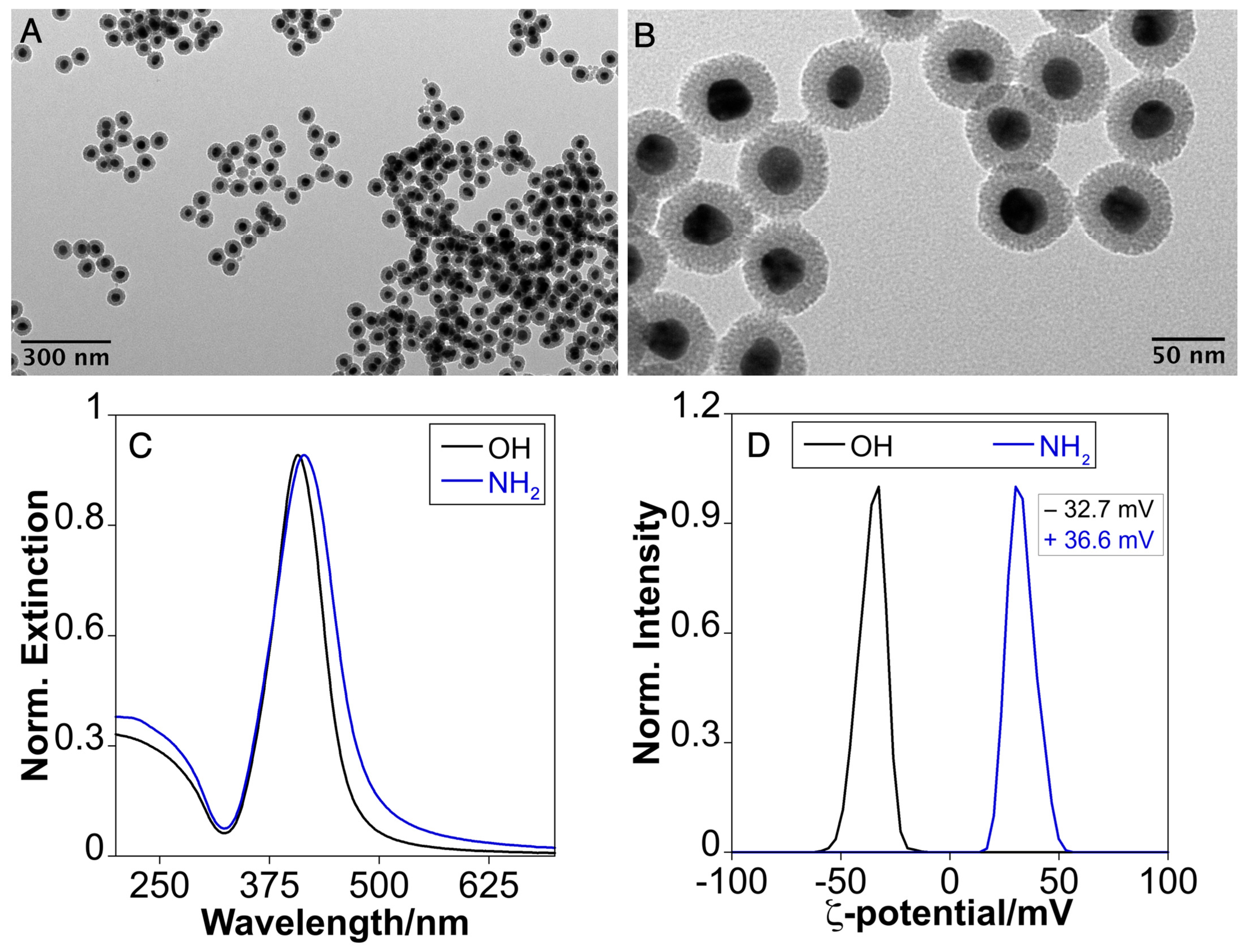
2.3.5. Amine Derivatization of Ag(28)@mSiO2-OH to Ag(28)@mSiO2-NH2
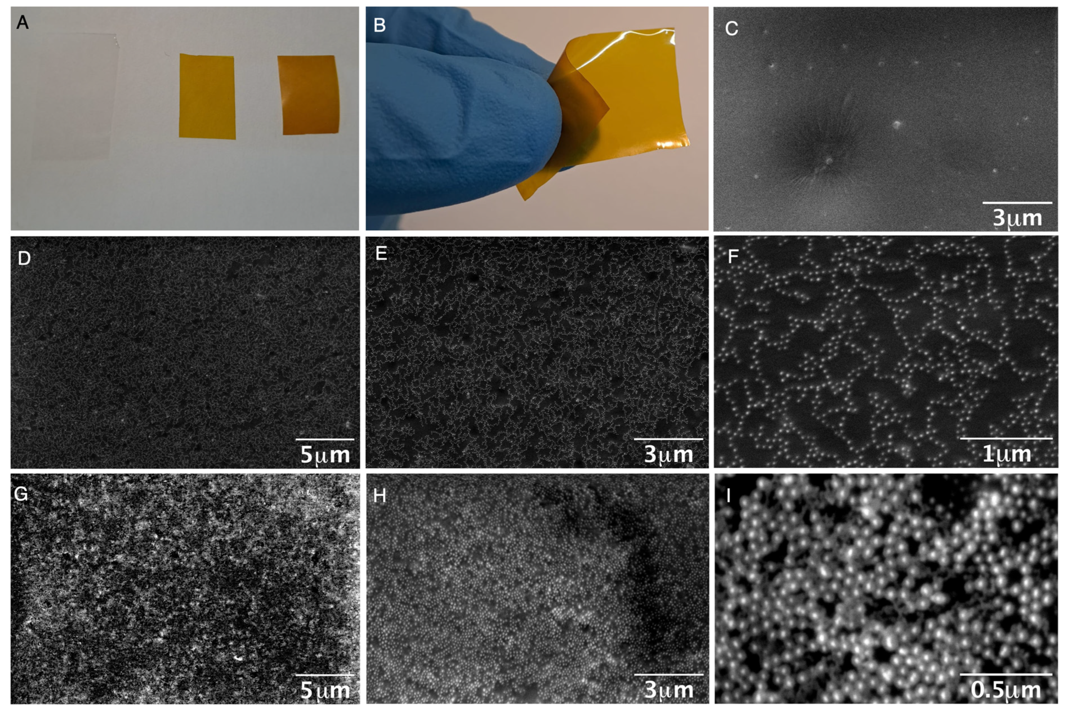
2.3.6. Ag(28)@mSiO2-NH2 Doped Polymer Synthesis
2.3.7. Antimicrobial Assays
3. Results and Discussion
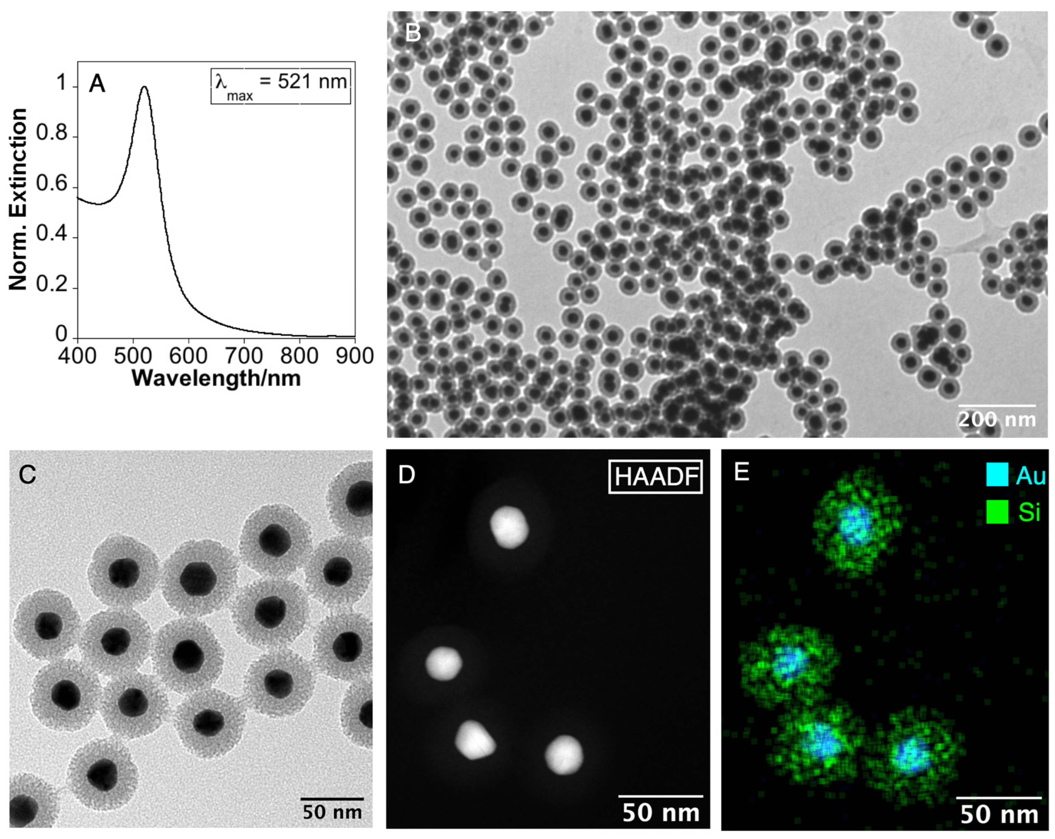
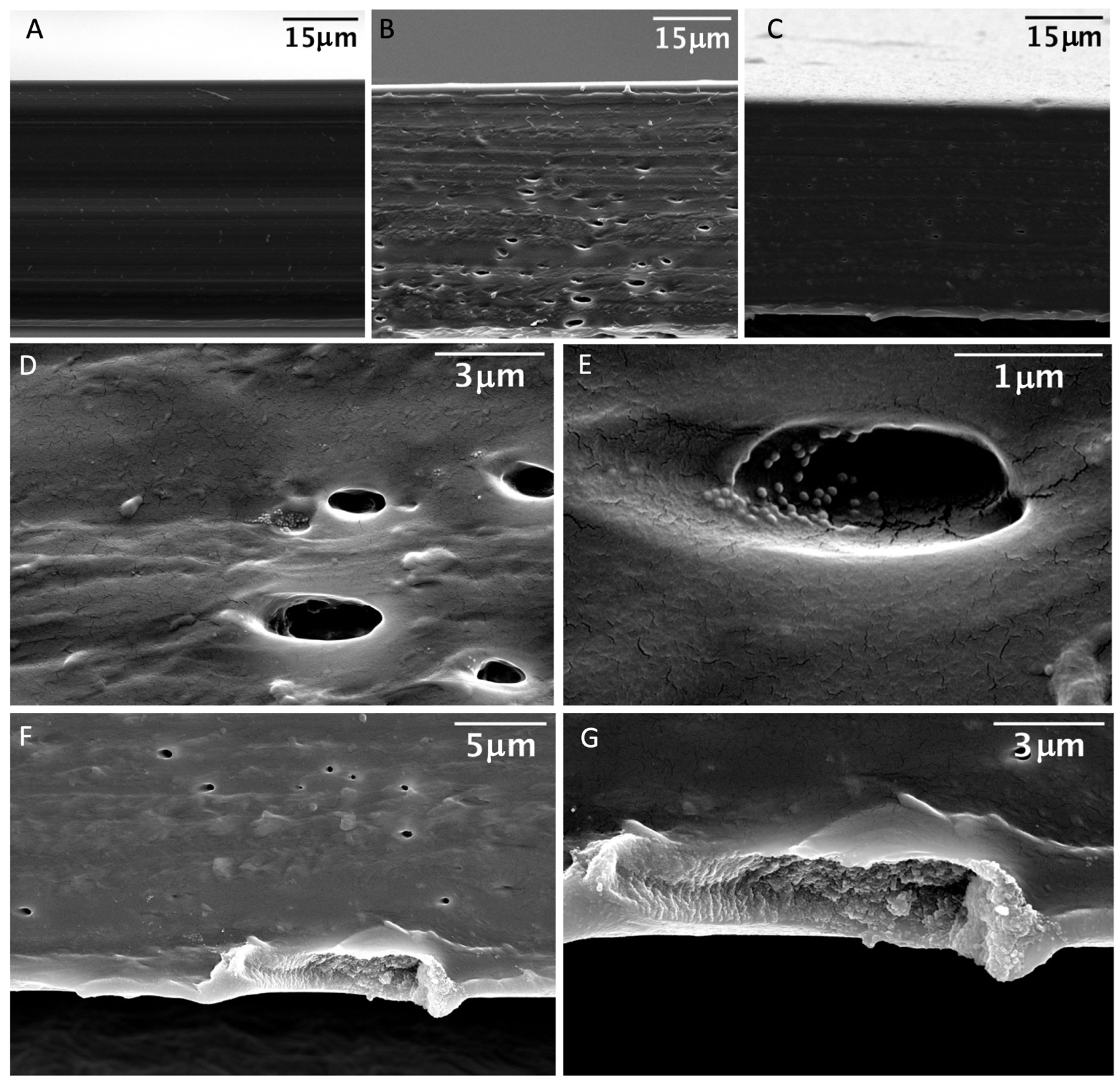
3.1. Synthesis and Characterization of Mesoporous Silica Coating Ag and Au NPs
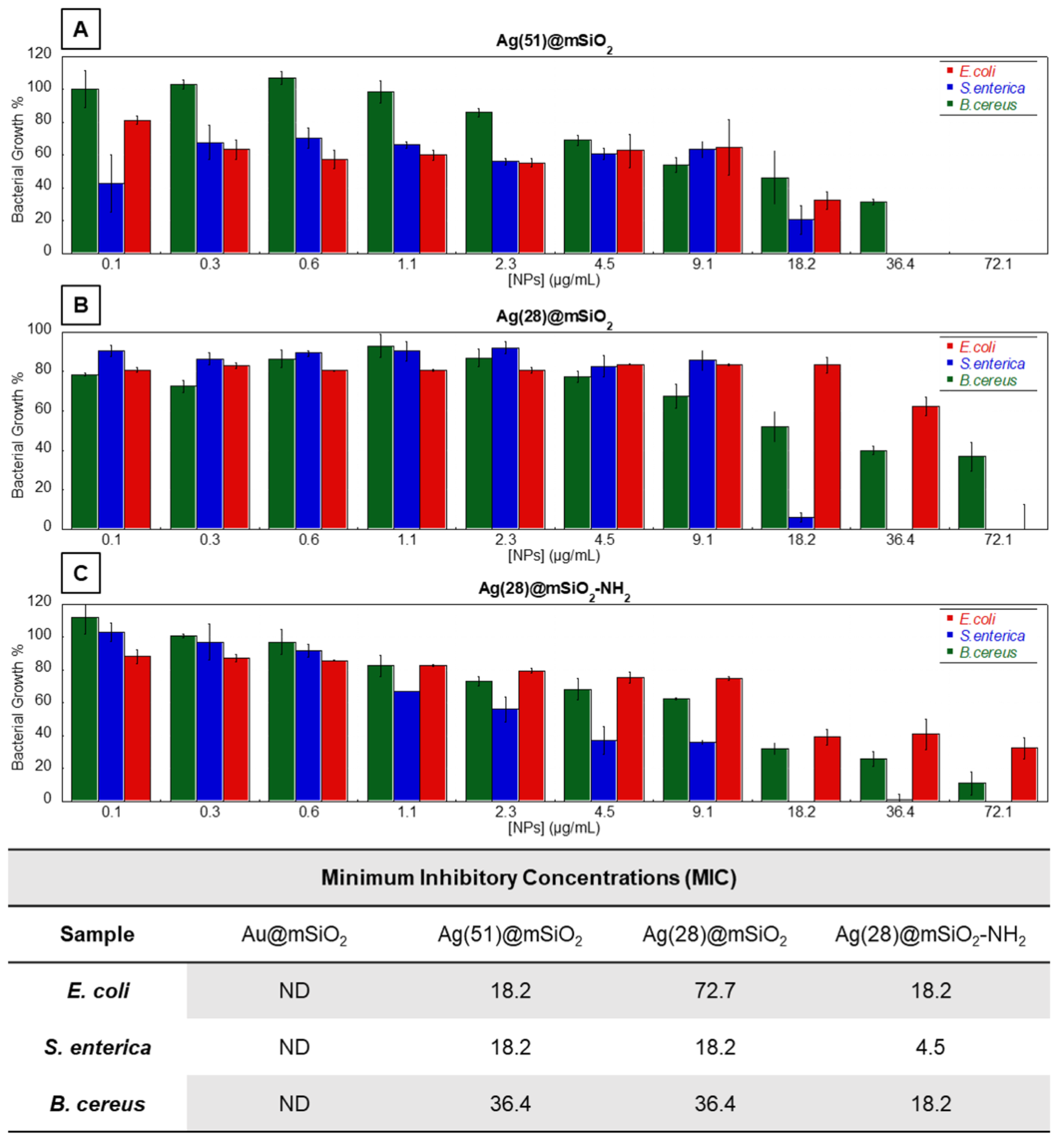
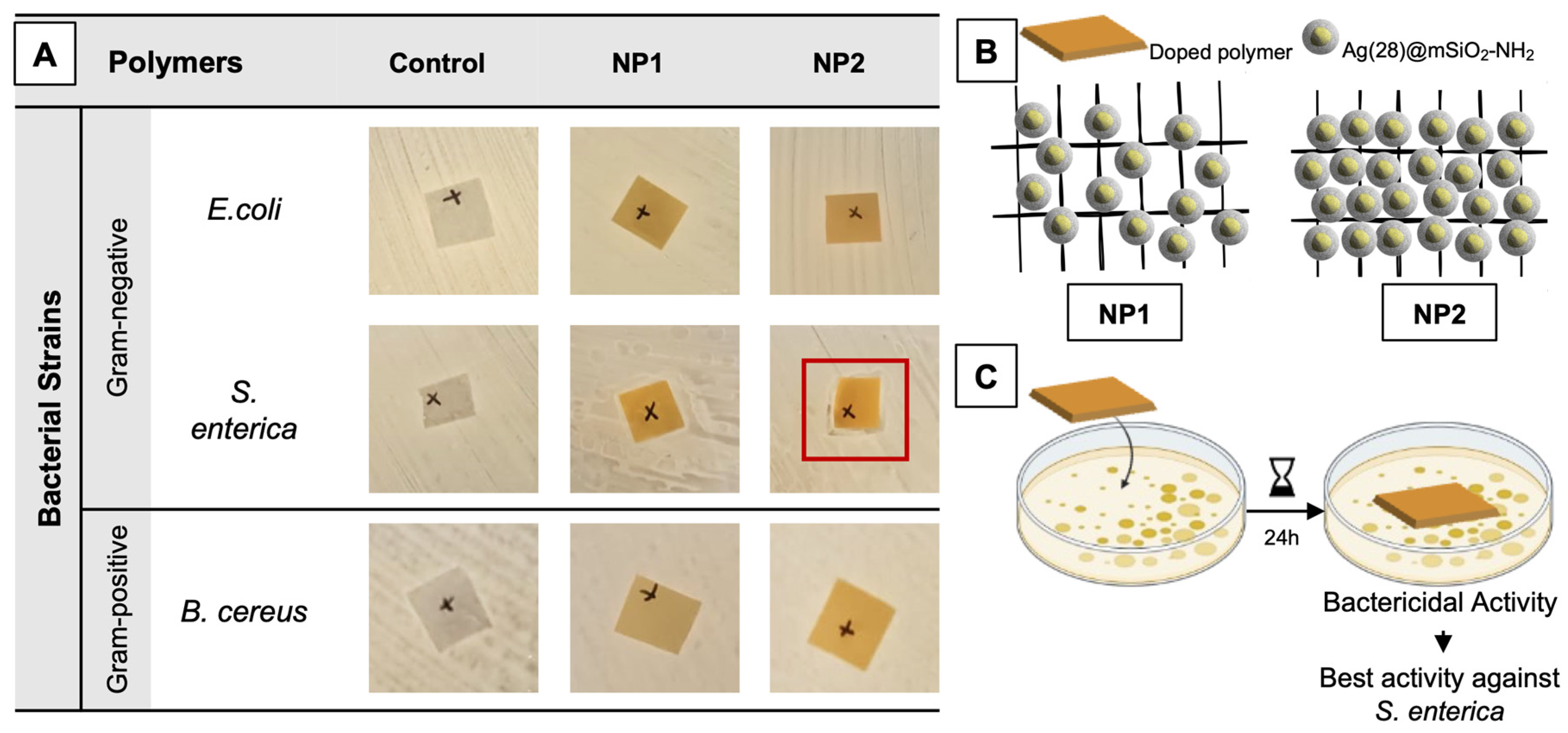
3.2. Antimicrobial Assays
4. Conclusions
Supplementary Materials
Author Contributions
Funding
Data Availability Statement
Conflicts of Interest
References
- Brown, E.D.; Wright, G.D. Antibacterial Drug Discovery in the Resistance Era. Nature 2016, 529, 336–343. [Google Scholar] [CrossRef]
- Kaper, J.B.; Nataro, J.P.; Mobley, H.L.T. Pathogenic Escherichia coli. Nat. Rev. Microbiol. 2004, 2, 123–140. [Google Scholar] [CrossRef] [PubMed]
- Blair, J.M.A.; Webber, M.A.; Baylay, A.J.; Ogbolu, D.O.; Piddock, L.J.V. Molecular Mechanisms of Antibiotic Resistance. Nat. Rev. Microbiol. 2015, 13, 42–51. [Google Scholar] [CrossRef] [PubMed]
- Wales, A.D.; Allen, V.M.; Davies, R.H. Chemical Treatment of Animal Feed and Water for the Control of Salmonella. Foodborne Pathog. Dis. 2010, 7, 3–15. [Google Scholar] [CrossRef] [PubMed]
- Nowack, B.; Krug, H.F.; Height, M. 120 Years of Nanosilver History: Implications for Policy Makers. Environ. Sci. Technol. 2011, 45, 7593–7595. [Google Scholar] [CrossRef]
- Menichetti, A.; Mavridi-Printezi, A.; Mordini, D.; Montalti, M. Effect of Size, Shape and Surface Functionalization on the Antibacterial Activity of Silver Nanoparticles. J. Funct. Biomater. 2023, 14, 244. [Google Scholar] [CrossRef] [PubMed]
- Mishra, A.; Pradhan, D.; Halder, J.; Biswasroy, P.; Rai, V.K.; Dubey, D.; Kar, B.; Ghosh, G.; Rath, G. Metal Nanoparticles against Multi-Drug-Resistance Bacteria. J. Inorg. Biochem. 2022, 237, 111938. [Google Scholar] [CrossRef] [PubMed]
- Xing, M.; Ge, L.; Wang, M.; Li, Q.; Li, X.; Ouyang, J. Nanosilver Particles in Medical Applications: Synthesis, Performance, and Toxicity. Int. J. Nanomed. 2014, 9, 2399. [Google Scholar] [CrossRef] [PubMed]
- Tang, S.; Zheng, J. Antibacterial Activity of Silver Nanoparticles: Structural Effects. Adv. Healthc. Mater. 2018, 7, 1701503. [Google Scholar] [CrossRef]
- Morones, J.R.; Elechiguerra, J.L.; Camacho, A.; Holt, K.; Kouri, J.B.; Ramírez, J.T.; Yacaman, M.J. The Bactericidal Effect of Silver Nanoparticles. Nanotechnology 2005, 16, 2346–2353. [Google Scholar] [CrossRef]
- Djafari, J.; Fernández-Lodeiro, C.; Fernández-Lodeiro, A.; Silva, V.; Poeta, P.; Igrejas, G.; Lodeiro, C.; Capelo, J.L.; Fernández-Lodeiro, J. Exploring the Control in Antibacterial Activity of Silver Triangular Nanoplates by Surface Coating Modulation. Front. Chem. 2019, 7, 677. [Google Scholar] [CrossRef]
- Fernández-Lodeiro, C.; Tambosi, R.; Fernández-Lodeiro, J.; Fernández-Lodeiro, A.; Nuti, S.; Ouchane, S.; Kébaïli, N.; Pérez-Juste, J.; Pastoriza-Santos, I.; Lodeiro, C. Adenosine-Monophosphate-Assisted Homogeneous Silica Coating of Silver Nanoparticles in High Yield. Nanomaterials 2023, 13, 2788. [Google Scholar] [CrossRef]
- Zheng, K.; Setyawati, M.I.; Leong, D.T.; Xie, J. Antimicrobial Silver Nanomaterials. Coord. Chem. Rev. 2018, 357, 1–17. [Google Scholar] [CrossRef]
- Zhang, X.F.; Liu, Z.G.; Shen, W.; Gurunathan, S. Silver Nanoparticles: Synthesis, Characterization, Properties, Applications, and Therapeutic Approaches. Int. J. Mol. Sci. 2016, 17, 1534. [Google Scholar] [CrossRef]
- Takeda, E.; Xu, W.; Terakawa, M.; Niidome, T. Tailored Structure and Antibacterial Properties of Silica-Coated Silver Nanoplates by Pulsed Laser Irradiation. ACS Omega 2022, 7, 7251–7256. [Google Scholar] [CrossRef]
- Fahmy, H.M.; Mosleh, A.M.; Elghany, A.A.; Shams-Eldin, E.; Abu Serea, E.S.; Ali, S.A.; Shalan, A.E. Coated Silver Nanoparticles: Synthesis, Cytotoxicity, and Optical Properties. RSC Adv. 2019, 9, 20118–20136. [Google Scholar] [CrossRef] [PubMed]
- Kankala, R.K.; Zhang, H.; Liu, C.; Kanubaddi, K.R.; Lee, C.; Wang, S.; Cui, W.; Santos, H.A.; Lin, K.; Chen, A. Metal Species–Encapsulated Mesoporous Silica Nanoparticles: Current Advancements and Latest Breakthroughs. Adv. Funct. Mater. 2019, 29, 1902652. [Google Scholar] [CrossRef]
- Yang, Y.; Zhang, M.; Song, H.; Yu, C. Silica-Based Nanoparticles for Biomedical Applications: From Nanocarriers to Biomodulators. Acc. Chem. Res. 2020, 53, 1545–1556. [Google Scholar] [CrossRef] [PubMed]
- Frickenstein, A.N.; Hagood, J.M.; Britten, C.N.; Abbott, B.S.; McNally, M.W.; Vopat, C.A.; Patterson, E.G.; MacCuaig, W.M.; Jain, A.; Walters, K.B.; et al. Mesoporous Silica Nanoparticles: Properties and Strategies for Enhancing Clinical Effect. Pharmaceutics 2021, 13, 570. [Google Scholar] [CrossRef] [PubMed]
- Wang, Y.; Zhao, Q.; Han, N.; Bai, L.; Li, J.; Liu, J.; Che, E.; Hu, L.; Zhang, Q.; Jiang, T.; et al. Mesoporous Silica Nanoparticles in Drug Delivery and Biomedical Applications. Nanomed. Nanotechnol. Biol. Med. 2015, 11, 313–327. [Google Scholar] [CrossRef]
- Rancan, F.; Gao, Q.; Graf, C.; Troppens, S.; Hadam, S.; Hackbarth, S.; Kembuan, C.; Blume-Peytavi, U.; Rühl, E.; Lademann, J.; et al. Skin Penetration and Cellular Uptake of Amorphous Silica Nanoparticles with Variable Size, Surface Functionalization, and Colloidal Stability. ACS Nano 2012, 6, 6829–6842. [Google Scholar] [CrossRef]
- Ertem, E.; Gutt, B.; Zuber, F.; Allegri, S.; Le Ouay, B.; Mefti, S.; Formentin, K.; Stellacci, F.; Ren, Q. Core–Shell Silver Nanoparticles in Endodontic Disinfection Solutions Enable Long-Term Antimicrobial Effect on Oral Biofilms. ACS Appl. Mater. Interfaces 2017, 9, 34762–34772. [Google Scholar] [CrossRef]
- Fathima, H.; Paul, L.; Thirunavukkuarasu, S.; Thomas, K.G. Mesoporous Silica-Capped Silver Nanoparticles for Sieving and Surface-Enhanced Raman Scattering-Based Sensing. ACS Appl. Nano Mater. 2020, 3, 6376–6384. [Google Scholar] [CrossRef]
- Wang, Y.; Ding, X.; Chen, Y.; Guo, M.; Zhang, Y.; Guo, X.; Gu, H. Antibiotic-Loaded, Silver Core-Embedded Mesoporous Silica Nanovehicles as a Synergistic Antibacterial Agent for the Treatment of Drug-Resistant Infections. Biomaterials 2016, 101, 207–216. [Google Scholar] [CrossRef] [PubMed]
- Neethu, S.; Midhun, S.J.; Sunil, M.A.; Soumya, S.; Radhakrishnan, E.K.; Jyothis, M. Efficient Visible Light Induced Synthesis of Silver Nanoparticles by Penicillium Polonicum ARA 10 Isolated from Chetomorpha Antennina and Its Antibacterial Efficacy against Salmonella Enterica Serovar Typhimurium. J. Photochem. Photobiol. B Biol. 2018, 180, 175–185. [Google Scholar] [CrossRef] [PubMed]
- Omnexus. Comprehensive Guide on Thermoplastic Polyurethanes (TPU). Available online: https://omnexus.specialchem.com/selection-guide/thermoplastic-polyurethanes-tpua (accessed on 28 February 2024).
- Backes, E.H.; Harb, S.V.; Pinto, L.A.; de Moura, N.K.; de Melo Morgado, G.F.; Marini, J.; Passador, F.R.; Pessan, L.A. Thermoplastic Polyurethanes: Synthesis, Fabrication Techniques, Blends, Composites, and Applications. J. Mater. Sci. 2024, 59, 1123–1152. [Google Scholar] [CrossRef]
- Miranda, C.; Castaño, J.; Valdebenito-Rolack, E.; Sanhueza, F.; Toro, R.; Bello-Toledo, H.; Uarac, P.; Saez, L. Copper-Polyurethane Composite Materials: Particle Size Effect on the Physical-Chemical and Antibacterial Properties. Polymers 2020, 12, 1934. [Google Scholar] [CrossRef] [PubMed]
- Zhou, X.; Wu, M.; Fu, L.; Liao, M.; Deng, L.; Wang, L.; Wang, H.; Xiang, Y.; Chen, S. A Facile Preparation Process of Polyhexamethylene Biguanide/Cellulose/Polylactic Acid Fiber Composite Antibacterial Paper. Ind. Crops Prod. 2023, 191, 115980. [Google Scholar] [CrossRef]
- Zhong, Y.; Zhang, T.; Zhang, W.; Wang, G.; Zhang, Z.; Zhao, P.; Liu, X.; Li, H. Antibacterial Castor Oil-Based Waterborne Polyurethane/Gelatin Films for Packaging of Strawberries. Food Packag. Shelf Life 2023, 36, 101055. [Google Scholar] [CrossRef]
- Wu, Z.; Zhang, Z.; Song, X.; Peng, W.; Zhao, X.; Zhao, H.; Liang, D.; Huang, C.; Duan, Q. A Silver Nanoparticles-Polylactic Acid Microspheres/Polylactic Acid-Thermoplastic Polyurethane Nanofibers Hierarchical Antibacterial Film. Ind. Crops Prod. 2024, 207, 117773. [Google Scholar] [CrossRef]
- Bastús, N.G.; Merkoçi, F.; Piella, J.; Puntes, V. Synthesis of Highly Monodisperse Citrate-Stabilized Silver Nanoparticles of up to 200 Nm: Kinetic Control and Catalytic Properties. Chem. Mater. 2014, 26, 2836–2846. [Google Scholar] [CrossRef]
- Ojea-Jiménez, I.; Bastús, N.G.; Puntes, V. Influence of the Sequence of the Reagents Addition in the Citrate-Mediated Synthesis of Gold Nanoparticles. J. Phys. Chem. C 2011, 115, 15752–15757. [Google Scholar] [CrossRef]
- Fernández-Lodeiro, C.; Fernández-Lodeiro, J.; Carbó-Argibay, E.; Lodeiro, C.; Pérez-Juste, J.; Pastoriza-Santos, I. The Versatility of Fe(II) in the Synthesis of Uniform Citrate-Stabilized Plasmonic Nanoparticles with Tunable Size at Room Temperature. Nano Res. 2020, 13, 2351–2355. [Google Scholar] [CrossRef]
- Fernández-Lodeiro, A.; Djafari, J.; Fernández-Lodeiro, J.; Duarte, M.P.; Mauricio, E.M.; Capelo-Martínez, J.L.; Lodeiro, C. Synthesis of Mesoporous Silica Coated Gold Nanorods Loaded with Methylene Blue and Its Potentials in Antibacterial Applications. Nanomaterials 2021, 11, 1338. [Google Scholar] [CrossRef]
- Rowe, L.R.; Chapman, B.S.; Tracy, J.B. Understanding and Controlling the Morphology of Silica Shells on Gold Nanorods. Chem. Mater. 2018, 30, 6249–6258. [Google Scholar] [CrossRef]
- Yamamoto, E.; Shimojima, A.; Wada, H.; Kuroda, K. Mesoporous Silica Nanoparticles with Dispersibility in Organic Solvents and Their Versatile Surface Modification. Langmuir 2020, 36, 5571–5578. [Google Scholar] [CrossRef] [PubMed]
- Jouault, N.; Zhao, D.; Kumar, S.K. Role of Casting Solvent on Nanoparticle Dispersion in Polymer Nanocomposites. Macromolecules 2014, 47, 5246–5255. [Google Scholar] [CrossRef]
- Wang, Y.; Liu, S.; Ding, K.; Zhang, Y.; Ding, X.; Mi, J. Quaternary Tannic Acid with Improved Leachability and Biocompatibility for Antibacterial Medical Thermoplastic Polyurethane Catheters. J. Mater. Chem. B 2021, 9, 4746–4762. [Google Scholar] [CrossRef] [PubMed]
- Veloso-Fernández, A.; Laza, J.M.; Ruiz-Rubio, L.; Martín, A.; Taguado, M.; Benito-Vicente, A.; Martín, C.; Vilas, J.L. Towards a New Generation of Non-Cytotoxic Shape Memory Thermoplastic Polyurethanes for Biomedical Applications. Mater. Today Commun. 2022, 33, 104730. [Google Scholar] [CrossRef]
- Reinhard, R.G.; Kalinowski, R.M.; Bodnaruk, P.W.; Eifert, J.D.; Boyer, R.R.; Duncan, S.E.; Bailey, R.H. Fate of Listeria on Various Food Contact and Noncontact Surfaces When Treated with Bacteriophage. J. Food Saf. 2020, 40, e12775. [Google Scholar] [CrossRef]
- Ilhan, I.; Kaya, M.; Turan, D.; Gunes, G.; Guner, F.S.; Kılıç, A. Thermoresponsive Polyurethane Films for Packaging Applications: Effects of Film Formulation on Their Properties. Food Packag. Shelf Life 2021, 29, 100695. [Google Scholar] [CrossRef]
- Vasconcelos, B.; Vediappan, K.; Oliveira, J.C.; Fonseca, C. Mechanically Robust Silver Coatings Prepared by Electroless Plating on Thermoplastic Polyurethane. Appl. Surf. Sci. 2018, 443, 39–47. [Google Scholar] [CrossRef]
- Villani, M.; Bertoglio, F.; Restivo, E.; Bruni, G.; Iervese, S.; Arciola, C.R.; Carulli, F.; Iannace, S.; Bertini, F.; Visai, L. Polyurethane-Based Coatings with Promising Antibacterial Properties. Materials 2020, 13, 4296. [Google Scholar] [CrossRef] [PubMed]
- Liu, Y.; Li, Q.; Liu, H.; Cheng, H.; Yu, J.; Guo, Z. Antibacterial Thermoplastic Polyurethane Electrospun Fiber Mats Prepared by 3-Aminopropyltriethoxysilane-Assisted Adsorption of Ag Nanoparticles. Chin. J. Polym. Sci. 2017, 35, 713–720. [Google Scholar] [CrossRef]
- Liu, M.; Liu, T.; Chen, X.; Yang, J.; Deng, J.; He, W.; Zhang, X.; Lei, Q.; Hu, X.; Luo, G.; et al. Nano-Silver-Incorporated Biomimetic Polydopamine Coating on a Thermoplastic Polyurethane Porous Nanocomposite as an Efficient Antibacterial Wound Dressing. J. Nanobiotechnol. 2018, 16, 89. [Google Scholar] [CrossRef] [PubMed]
- Marcelo, G.A.; Duarte, M.P.; Oliveira, E. Gold@mesoporous Silica Nanocarriers for the Effective Delivery of Antibiotics and By-Passing of β-Lactam Resistance. SN Appl. Sci. 2020, 2, 1354. [Google Scholar] [CrossRef]
- Kiran, J.U.; Roners, J.P.; Mathew, S. XPS and Thermal Studies of Silver Doped SiO2 Matrices for Plasmonic Applications. Mater. Today Proc. 2020, 33, 1263–1267. [Google Scholar] [CrossRef]
- García, M.A.; García-Heras, M.; Cano, E.; Bastidas, J.M.; Villegas, M.A.; Montero, E.; Llopis, J.; Sada, C.; De Marchi, G.; Battaglin, G.; et al. Photoluminescence of Silver in Glassy Matrices. J. Appl. Phys. 2004, 96, 3737–3741. [Google Scholar] [CrossRef]
- Reidy, B.; Haase, A.; Luch, A.; Dawson, K.; Lynch, I. Mechanisms of Silver Nanoparticle Release, Transformation and Toxicity: A Critical Review of Current Knowledge and Recommendations for Future Studies and Applications. Materials 2013, 6, 2295–2350. [Google Scholar] [CrossRef]
- Abbasi, M.; Gholizadeh, R.; Kasaee, S.R.; Vaez, A.; Chelliapan, S.; Fadhil Al-Qaim, F.; Deyab, I.F.; Shafiee, M.; Zareshahrabadi, Z.; Amani, A.M.; et al. An Intriguing Approach toward Antibacterial Activity of Green Synthesized Rutin-Templated Mesoporous Silica Nanoparticles Decorated with Nanosilver. Sci. Rep. 2023, 13, 5987. [Google Scholar] [CrossRef]
- Liu, Q.; Zhang, Y.; Huang, J.; Xu, Z.; Li, X.; Yang, J.; Huang, H.; Tang, S.; Chai, Y.; Lin, J.; et al. Mesoporous Silica-Coated Silver Nanoparticles as Ciprofloxacin/SiRNA Carriers for Accelerated Infected Wound Healing. J. Nanobiotechnol. 2022, 20, 386. [Google Scholar] [CrossRef]
- Huq, M.A.; Akter, S. Bacterial Mediated Rapid and Facile Synthesis of Silver Nanoparticles and Their Antimicrobial Efficacy against Pathogenic Microorganisms. Materials 2021, 14, 2615. [Google Scholar] [CrossRef]
- Dias de Emery, B.; Zottis Chitolina, G.; Qadir, M.I.; Quedi Furian, T.; Apellanis Borges, K.; de Souza Moraes, H.L.; Pippi Salle, C.T.; Pinheiro do Nascimento, V. Antimicrobial and Antibiofilm Activity of Silver Nanoparticles against Salmonella Enteritidis. Braz. J. Microbiol. 2023, 54, 285–292. [Google Scholar] [CrossRef]
- Avila-Calderón, E.D.; del Socorro Ruiz-Palma, M.; Aguilera-Arreola, M.G.; Velázquez-Guadarrama, N.; Ruiz, E.A.; Gomez-Lunar, Z.; Witonsky, S.; Contreras-Rodríguez, A. Outer Membrane Vesicles of Gram-Negative Bacteria: An Outlook on Biogenesis. Front. Microbiol. 2021, 12, 557902. [Google Scholar] [CrossRef]
- Kleanthous, C.; Armitage, J.P. The Bacterial Cell Envelope. Philos. Trans. R. Soc. B Biol. Sci. 2015, 370, 20150019. [Google Scholar] [CrossRef] [PubMed]
- Yin, I.X.; Zhang, J.; Zhao, I.S.; Mei, M.L.; Li, Q.; Chu, C.H. The Antibacterial Mechanism of Silver Nanoparticles and Its Application in Dentistry. Int. J. Nanomed. 2020, 15, 2555–2562. [Google Scholar] [CrossRef] [PubMed]
- Nam, S.; Park, B.; Condon, B.D. Water-Based Binary Polyol Process for the Controllable Synthesis of Silver Nanoparticles Inhibiting Human and Foodborne Pathogenic Bacteria. RSC Adv. 2018, 8, 21937–21947. [Google Scholar] [CrossRef] [PubMed]
- Gabrielyan, L.; Badalyan, H.; Gevorgyan, V.; Trchounian, A. Comparable Antibacterial Effects and Action Mechanisms of Silver and Iron Oxide Nanoparticles on Escherichia Coli and Salmonella typhimurium. Sci. Rep. 2020, 10, 13145. [Google Scholar] [CrossRef] [PubMed]
- Bruna, T.; Maldonado-Bravo, F.; Jara, P.; Caro, N. Silver Nanoparticles and Their Antibacterial Applications. Int. J. Mol. Sci. 2021, 22, 7202. [Google Scholar] [CrossRef]


Disclaimer/Publisher’s Note: The statements, opinions and data contained in all publications are solely those of the individual author(s) and contributor(s) and not of MDPI and/or the editor(s). MDPI and/or the editor(s) disclaim responsibility for any injury to people or property resulting from any ideas, methods, instructions or products referred to in the content. |
© 2024 by the authors. Licensee MDPI, Basel, Switzerland. This article is an open access article distributed under the terms and conditions of the Creative Commons Attribution (CC BY) license (https://creativecommons.org/licenses/by/4.0/).
Share and Cite
Nuti, S.; Fernández-Lodeiro, A.; Galhano, J.; Oliveira, E.; Duarte, M.P.; Capelo-Martínez, J.L.; Lodeiro, C.; Fernández-Lodeiro, J. Tailoring Mesoporous Silica-Coated Silver Nanoparticles and Polyurethane-Doped Films for Enhanced Antimicrobial Applications. Nanomaterials 2024, 14, 462. https://doi.org/10.3390/nano14050462
Nuti S, Fernández-Lodeiro A, Galhano J, Oliveira E, Duarte MP, Capelo-Martínez JL, Lodeiro C, Fernández-Lodeiro J. Tailoring Mesoporous Silica-Coated Silver Nanoparticles and Polyurethane-Doped Films for Enhanced Antimicrobial Applications. Nanomaterials. 2024; 14(5):462. https://doi.org/10.3390/nano14050462
Chicago/Turabian StyleNuti, Silvia, Adrián Fernández-Lodeiro, Joana Galhano, Elisabete Oliveira, Maria Paula Duarte, José Luis Capelo-Martínez, Carlos Lodeiro, and Javier Fernández-Lodeiro. 2024. "Tailoring Mesoporous Silica-Coated Silver Nanoparticles and Polyurethane-Doped Films for Enhanced Antimicrobial Applications" Nanomaterials 14, no. 5: 462. https://doi.org/10.3390/nano14050462