Novel Therapeutic Delivery of Nanocurcumin in Central Nervous System Related Disorders
by
 , , , and
Elisa Panzarini
1 ,
,
Stefania Mariano
1,
Stefano Tacconi
1, 
Elisabetta Carata
1,
Ada Maria Tata
2 and
Luciana Dini
2,3,* 1
Departament of Biological and Environmental Sciences and Technologies (Di.S.Te.B.A.), University of Salento, 73100 Lecce, Italy
2
Departament of Biology and Biotechnology “C. Darwin”, Sapienza University of Rome, 00185 Rome, Italy
3
CNR Nanotec, Campus Ecotekne, University of Salento, 73100 Lecce, Italy
*
Author to whom correspondence should be addressed.
Nanomaterials 2021, 11(1), 2; https://doi.org/10.3390/nano11010002
Submission received: 30 November 2020
/
Revised: 17 December 2020
/
Accepted: 18 December 2020
/
Published: 22 December 2020
(This article belongs to the Special Issue Nanonutraceuticals Delivery)
Abstract
:Nutraceuticals represent complementary or alternative beneficial products to the expensive and high-tech therapeutic tools in modern medicine. Nowadays, their medical or health benefits in preventing or treating different types of diseases is widely accepted, due to fewer side effects than synthetic drugs, improved bioavailability and long half-life. Among herbal and natural compounds, curcumin is a very attractive herbal supplement considering its multipurpose properties. The potential effects of curcumin on glia cells and its therapeutic and protective properties in central nervous system (CNS)-related disorders is relevant. However, curcumin is unstable and easily degraded or metabolized into other forms posing limits to its clinical development. This is particularly important in brain pathologies determined blood brain barrier (BBB) obstacle. To enhance the stability and bioavailability of curcumin, many studies focused on the design and development of curcumin nanodelivery systems (nanoparticles, micelles, dendrimers, and diverse nanocarriers). These nanoconstructs can increase curcumin stability, solubility, in vivo uptake, bioactivity and safety. Recently, several studies have reported on a curcumin exosome-based delivery system, showing great therapeutical potential. The present work aims to review the current available data in improving bioactivity of curcumin in treatment or prevention of neurological disorders.
1. Introduction
Natural products have been used for a long time and are still used by people for health improvement and for the treatment of various diseases. In 1989, Dr Stephan De Felice, President of the Foundation for Innovation in Medicine (FIM), coined the word nutraceuticals by the combination of “nutrition” and “pharmaceutical” and defined its concept as “a food or part of a food that provides medical or health benefits, including disease prevention and/or treatment” [1]. Thus, nutraceuticals are all the substances extracted from natural sources, derivatives from human metabolism (dehydroepiandrosterone or DHEA, S-adenosylmethionine or SAMe, carnitine, creatine, coenzyme Q10, lipoic acid, melatonin) and bioactive plant dietary components (polyphenols, saponins, probiotics, phytoestrogens, dietary fibers, carotenoids) that preserve their original properties without any chemical manipulation. Nutraceuticals are marketed as dietary supplements (e.g., vitamins, minerals, co-enzyme Q, carnitine), functional foods (e.g., yogurts, cereals, snacks, etc.) and medicinal foods (e.g., health bars with added medications, transgenic cows and lacto-ferrin for immune enhancement, etc.) that have health or medical benefits in disease prevention and treatment [2]. Health benefits of nutraceuticals rely on good bioavailability, long half-life and absence of side effects with respect to synthetic drugs, beside the enhancement of physiological processes, for example, immune response [3].
Among the large amount of nutraceuticals, considerable attention is given to turmeric derivatives due to a plethora of biological benefits (i.e., antioxidant, anti-inflammatory, anti-cancer, anti-growth, anti-arthritic, anti-atherosclerotic, anti-depressant, anti-aging, anti-diabetic, anti-microbial, wound healing, memory-enhancing, chemopreventive, chemosensitization and radiosensitization) [4,5,6]. Curcumin is the most widely studied turmeric derivative and represents 60–70% of the curcuminoids extracted from the rhizome of Curcuma longa [7]. Curcumin, with its characteristic yellow color, is a polyphenol compound with numerous bioactive properties for which it has been used for multiple applications in traditional medicine for more than 2000 years [8]. Given the many biological targets of curcumin, its effects in different biological pathways and processes have been well characterized, including adhesion molecules, transcription factors, growth factors, inflammatory mediators, apoptotic regulators, enzymes, kinases, membrane receptors and antioxidant systems [9]. Together with the fact that curcumin has little to no side effects, neuroprotective properties are also reported. Therefore, there is a particular growing interest about curcumin’s impact in central nervous system (CNS)-related neurodegenerative disorders, such as Alzheimer’s and Parkinson’s diseases, and brain malignancies, such as glioblastoma multiforme (GBM) [10,11].
Despite these amazing properties, the utility of curcumin is greatly hindered by its weak absorption, rapid metabolism, systemic elimination, degree of water solubility and limited blood brain barrier (BBB) permeability [12]. Therefore, various strategies have been proposed to overcome these limitations, like the use of adjuvants, nanomaterials, curcumin phospholipid complexes and curcumin formulated with various oils or metabolism inhibitors [13,14,15,16].
One of the emerging strategies in terms of improvement of the bioavailability and efficacy of curcumin is the development of nano-delivery. Indeed, many curcumin nano-delivery systems are currently under investigation. Recently, exosomes, nano-sized vesicles physiologically produced by cells, have been successfully proposed as curcumin delivering systems for the treatment of CNS-related pathologies, for their excellent biodegradability, biocompatibility and ability to cross the BBB [17].
The consumption of nutraceuticals is growing strongly in Europe, USA and Asia, mainly in the form of functional beverages, to contribute to the welfare of the increasing elderly population and, thus, to counter the increase in metabolic diseases. The most competitive companies involved world wide in the nutraceutical markets are exploring new field of application and new technological process of preparation to improve activities of compounds. Here, the current state of the art in nano-sized constructs able to improve bioactivity of curcumin in nervous system disorders is reviewed.
2. Bioactivity and Health Benefits of Curcumin
Curcumin was firstly described by Vogel and Pelletier in 1815, as a mix of resin and turmeric oil, while the pure form was obtained in 1842 by Vogel Jr. The chemical structure of curcumin was defined 68 years later by Milobedzka and Lampe in (1E, 6E)-1,7-bis (4-hydroxy-3 methoxyphenyl) [14,18]. Curcumin belongs to the polyphenols family, and it represents the main curcuminoid of turmeric Curcuma longa extract [19]. The typical composition of commercial curcumin is a combination of curcumin (~77%), demethoxy curcumin (~17%) and bisdemethoxy curcumin (~3%). Curcumin finds large use as food ingredient, as cosmetic industrial dye and in the formulation of medicinal products (e.g., to relieve muscle pain, inflammation, rheumatoid arthritis, inflammatory and gastrointestinal disorders, intermittent fever and leukoderma).
Important biotechnological applications of curcumin are widely used in the food industry where it is used as a spice or food additive, at a dose of 5–500 mg/kg, to ameliorate foodstuffs’ palatability and storage stability. The European Union has authorized the use of curcumin as food color with the name of CI 75300, Natural Yellow 3 or diferuloylmethane, and with the E100 code [20]. Curcumin is an efficient food preservative in its ability to suppress lipid peroxidation, suggesting a role as a possible natural preservative [21]. At a concentration of 1–2%, curcumin showed antimicrobial activity preserving stored chicken meal from contamination for about 90 days [22]. In the cosmetics sector curcumin is widely used for its antioxidant and anti-inflammatory activity and skin lightening property. In fact, in in vitro curcumin treated cells, including epidermal and dermal layers, inhibition of collagenase, hyaluronidase and elastase was demonstrated [23]. Altered skin pigmentation (such as solar elastoses, solar lentigines, actinic keratosis) caused by photodamage is reduced after curcumin treatment [23]. Curcumin essential oil has dermo-protective properties and stabilizes fatty components of cosmetics; it is, thus, used in the formulations of soaps and cosmetics, and to prevent the rancidity of lipids [24]. The yellow color of curcumin makes it an ideal natural dye in hair coloring moisturizers. The numerous recognized health benefits of curcumin, namely, anti-diabetic, anti-tumor and hepato-, neuro- and cardiovascular protective effects, depend on its metabolism that affects antioxidant, anti-inflammatory and immunoregulatory activities [25]. Curcumin can protect against inflammation by decreasing levels of pro-inflammatory cytokines, such as tumor necrosis alpha (TNFα), interleukin-6 (IL6) and interleukin 1β (IL1β), increasing levels of the anti-inflammatory cytokines interleukin-10 (IL10) and transforming growth factor- beta (TGFβ) [26] and by acting on NF-kB and peroxisome proliferator-activated receptor-gamma (PPAR-γ) pathways and myeloid differentiation protein 2-TLR4 co-receptor (TLR4-MD2) signaling [27,28,29].
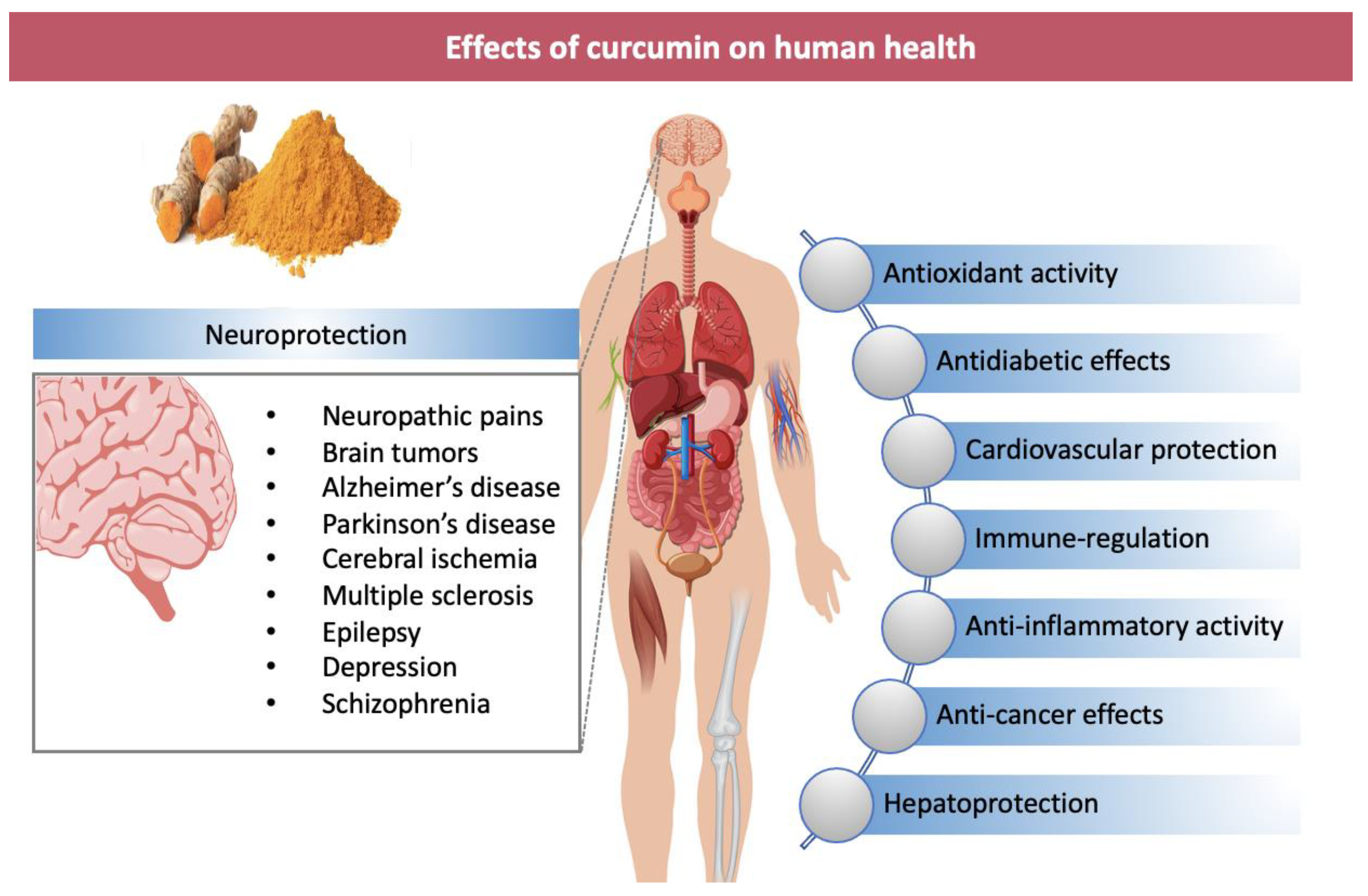
Finally, numerous preclinical studies have suggested neuroprotection properties of curcumin in treating Alzheimer’s disease (AD), Parkinson’s disease (PD), amyotrophic lateral sclerosis (ALS), multiple sclerosis (MS), epilepsy, stroke, traumatic brain injury, spinal cord injury, depression, dementia, schizophrenia and brain tumors [10,11,12] (Figure 1).
The imbalance between reactive oxygen species (ROS) production and antioxidant enzymes activity causes oxidative stress, which is associated with various chronic diseases. The presence of an electron-donating phenolic hydroxyl group in the chemical structure of curcumin confers antioxidant activity [30]. The scavenger activity of curcumin reduces oxidative stress and prevents ROS formation, namely, superoxide and nitric oxide radicals and hydrogen peroxide [31]. Curcumin prevents oxidative damage by blocking nuclear factor kB (NF-kB) activation. In human L02 hepatocytes pretreated with curcumin ROS formation caused by quinocetone was inhibited [32]. Curcumin administration restores the DNA-methyltransferase function in diabetic mice and inhibits hyperglycemia-induced ROS production and, in obese patients, reduces oxidative stress [33,34,35]. The activity of several antioxidant enzymes, (paraoxonase 1 arylesterase, catalase, CAT; glutathione S-transferase, GST; glutathione peroxidase, GSH-Px; superoxide dismutase, SOD; heme oxygenase-1, OH-1) is enhanced by curcumin that, by reducing lipid peroxidation, protect against carcinogenesis processes [36,37,38]. It should be kept in mind that the antioxidant effect of curcumin depends on the dose used and on the presence in culture medium of metal ions and that an antioxidant in vitro effect could not have the same in vivo effect [39,40,41].
Curcumin is also beneficial for the immune system by interacting with immune cells (T and B lymphocytes, macrophages, dendritic cells and natural killer cells) by modulating IgG, IgM, IgA and immune mediators (IL-1β, IL-4, IL-6, IL-17A, TNF-α) and thus, protecting against immune-related diseases [42]. It has been demonstrated that curcumin reduces the number of neutrophils and eosinophils and increases the amounts of lymphocytes. For example, curcumin regulates Th1/Th2 balance in ovalbumin-sensitized rats by stimulating Th1 cells and inhibiting Th2 cells [43]. Curcumin stimulates differentiation of bone marrow-derived mesenchymal stem cells towards anti-inflammatory M2 macrophages, providing a favorable microenvironment for adult full-thickness cutaneous wound healing [44]. Moreover, curcumin decreases macrophage infiltration and inhibits NF-kB pathway in macrophages [45].
Several chronic diseases, such as autoimmune, cardiovascular, endocrine, neurodegenerative and cancer, are characterized by a chronic inflammatory outbreak closely related to oxidative stress [25]. The effectiveness of curcumin in the prevention and treatment of these diseases, as well as metabolic, neurological, skin and infectious diseases, has been demonstrated in several preclinical and clinical studies [46].
3. Curcumin and CNS-Related Pathologies
3.1. Neurodegenerative Pathologies
In the last decades, a lot of attention was paid to increasing chronic neurodegenerative disorders. The population affected grows progressively and the impact of these diseases on patients’ lives, on society and the economy is more relevant [47]. Growing evidence indicates that oxidative stress contributes to the etiology and the progression of nervous system disorders, such as AD, PD, ALS and MS [48]. These pathologies are characterized by the progressive loss of neurons in different areas of the CNS, leading to cognitive, sensory and motor dysfunctions [49]. Other pathological features of neurodegenerative diseases are the accumulation of aggregated proteins, depletion of endogenous antioxidant enzyme activity, mitochondrial dysfunction and increasing neuroinflammation [50]. The link between aging and neuroinflammation has largely emerged and implicates the aberrant modulation of multiple sets of genes and proteins, such as an increased activation of microglia and astrocytes by both NF-kB and cyclooxygenase-2 (COX2) and inducible nitric oxide synthase (iNOS) levels; this, in turn, induces the release of pro-inflammatory cytokines, leading to neuronal death and subsequent cognitive deficits [51].
Considering these aspects, the identification of new molecules and drugs able to prevent, delay or alleviate cognitive impairment characterizing these pathologies appears of great clinical relevance.
Several preclinical studies have suggested beneficial roles for curcumin as an adjuvant therapy in free radical-based diseases [52,53]. As demonstrated in mice, curcumin is metabolized to form several metabolites with relevant biological effects. For its anti-inflammatory, antioxidant, antiproliferative and antimicrobial properties, curcumin has been proposed as an adjuvant treatment of several disorders, including spinal cord injury as well as for neurodegenerative diseases [54,55].
Of particular interest are the potential effects of curcumin in the regulation of nervous system activity. Curcumin can impair extrapyramidal symptoms and increased HO-1 expression through Akt/Nrf2 phosphorylation in the substantia nigra pars compacta of rats treated with rotenone, a pharmacological tool able to destroy dopaminergic neurons and therefore, used to induce experimental Parkinson’s disease (PD) [56]. Curcumin displayed a neuroprotective action by reducing protein misfolding and aggregation through upregulation of heat shock proteins such as Hsp90, Hsp70, Hsp60 and Hsp40 in mice models [57].
Considering its ability to interfere with the apoptotic pathways, curcumin was also demonstrated to exert neuroprotective actions in rats that underwent ischemia/reperfusion injury [58]. In addition, curcumin increased antioxidant molecules GSH and enzymes such as CAT and SOD. It was found that curcumin acts also as ROS scavenging, disrupting amyloid plaques and exhibiting anti-inflammatory and anti-apoptotic effects [59]. The impact of curcumin on neural stems cells and other neural cells is also relevant. In fact, several studies reported the capacity of curcumin to protect neural stem cells [60].
Despite these claimed properties, the poor intestinal absorption, structural instability, limited blood brain barrier (BBB) penetration and rapid degradation of curcumin, limits the potential as a therapeutic agent in clinical trials [61].
3.2. Brain Tumors
Gliomas are the most common primary brain tumors in humans, resulting from glial precursors or astrocytes transformation. GBM is the most frequent, aggressive (grade IV) form of glioma, frequently occurring in the brain or in the spinal cord.
The cellular and molecular origin of GBM is not yet completely known; however, the dysregulation of cellular signaling pathways and several genetic mutations were described as being involved in the regulation of cancer progression, invasion and metastasis formation. The overexpression of epidermal growth factor receptor (EGFR), the hyper-activation of PI3 kinase pathway, mutation of p53 genes or PTEN were reported in GBM formation [89,90].
Despite the progressions in neurosurgery, pharmacological therapy, immune therapy and radiation, the patient’s survival is still poor [91,92].
Interestingly natural compounds and their metabolites received much attention as promising therapeutic agents for the treatment of several human malignancies. Considering the abundant amounts of lipids in brain, the lipophilic nature of curcumin presents a good absorption, availability and stability in the CNS [11].
First, curcumin can inhibit proliferation and induce apoptosis of GBM cell line. The mechanisms require the p53 and caspase 3 activation or decreasing of anti-apoptotic genes, including NF-kB and Bcl2 [93,94]. The decreased expression of bcl-2, and DNA repair enzymes, such as O-6-Methylguanine-DNA Methyltransferase (MGMT), lead to reduced resistance of glioma cells against radiation and chemotherapeutic agents; this suggests that curcumin may be a potential useful adjuvant for common chemotherapeutic agents and radiation [95,96]. It has also been reported that curcumin inhibits migration and invasion in GBM cell line U87MG by modulation of matrix metalloproteinases (MMPs) and decreasing the expression of fascin, a protein involved in F actin aggregation [97].
The GBM cell survival curcumin-induced results decreased in relation to increased ROS production, caspases activation and mitochondrial membrane permeability alteration [98].
Curcumin can also induce autophagy by suppressing the Akt/mammalian target of rapamycin and activates the kinase pathways regulated by extracellular signals [11]. Interestingly curcumin inhibits the viability and proliferation of glioblastoma stem-like cells, considered the subpopulation responsible of GBM recurrence and formation [99,100,101].
In Table 4, the numerous studies on the role of curcumin as potential anti-tumor agent against GBM, are reported.
4. Nanotechnological-Based Strategies for Curcumin Delivery and Blood-Brain Barrier Crossing
4.1. Preparation and Formulation of Nanocurcumin
The development of nanotechnology-based applications in the health sector offers innovative therapeutic and diagnostic opportunities to address medical needs. In parallel, a need for a specific regulatory framework is growing; no regulatory practice exists for nanotechnology-enabled health products. However, due to the increased complexity of such products and their size-related properties, due to the fast progress in the field and the lack of robust datasets, the question remains whether the identified safety and efficacy requirements of the products are sufficient for a reliable characterization, assessment and market. Thus, new state-of-the-art methods, instruments, approaches or tools must be developed to sufficiently prove their reliability and relevance for the given purpose. Although the preparation methods are not the subject of this work, a brief summary of the main new methods of nano curcumin preparation along with advantages and disadvantages are reported in Table 5. The nanotechnology methods, reviewed by Rai et al., [117], developed to enhance the activity of nanocurcumin are various: coacervation, nanoprecipitation, spray drying, single emulsion, solvent evaporation, microemulsion, wet milling, thin film hydration, solid dispersion, emulsion polymerization, ionic gelation, ultrasonication, antisolvent precipitation and Fessi methods.
Depending on the method of preparation, nanoparticles, nanospheres or nanocapsules can be obtained. Nanocapsules are systems in which the drug is confined to a cavity surrounded by a unique polymer membrane, while nanospheres are matrix systems in which the drug is physically and uniformly dispersed [118,119]. The field of polymer nanoparticles is quickly expanding and playing an important role in a wide spectrum of areas [120]. The polymeric nanoparticles (size between 10–1000 nm) are prepared from biocompatible and biodegradable polymers where the drug is dissolved, entrapped, encapsulated or attached to a nanoparticle matrix.
Curcumin’s low solubility and bioavailability hinder the great properties of this compound and compromise its use in the biomedical field. This limit could be overcome by designing new methods of administration to stabilize the molecule and increase its bioavailability, by reducing its metabolism and increasing the retention time in the bloodstream. In fact, encapsulation of curcumin into nano-carriers (i.e., liposomes, engineered nanoparticles or exosomes, EXOs) and used in preclinical studies for different pathologies, demonstrated an improved efficacy in comparison to free molecule [121,122]. Nano-encapsulation of curcumin improves its stability, avoids the enzymatic and pH degradation, leading to increased half-life [123].
Many different nanotechnology-based strategies (e.g., polymers, liposomes, hydrogels, adjuvants and nanoparticles) have been designed in the last few decades with the aim of efficient curcumin loading, prolonging the time of properties retention, and of inactivation or hydrolysis protection [124]. These nano encapsulated curcumin with improved properties have been tested in vitro, in in vivo experiments and in pre-clinical trials as reported in Table 6.
An interesting strategy is represented by the amorphous solid dispersions (ASD), a modification of the solid state that improves the bioavailability by increasing the rate of dissolution. This technique is based on the incorporation of water-insoluble compounds into a hydrophilic carrier matrix. Zhang et al. [148] demonstrated that ASD forms of curcumin increased cytotoxicity and membrane permeability of U87 glioblastoma cells. In addition, in in vivo studies, the bioavailability of this formulation was 19-fold more efficient compared to free curcumin.
The oral bioavailability of hydroxypropyl methylcellulose-based solid dispersion of curcumin (DW-CUR 20) evaluated in in vivo, exceeded the limits of curcumin in pure form. In a mouse model of acute liver injury induced by tert-butyl hydroperoxide, oral administration of DW-CUR 20 was found to be highly hepatoprotective, as demonstrated by the improvement of the histological liver damage [149].
When curcumin is in form of nanosuspension, in other words, a nanosystem containing only pure drug crystals with a surfactant agent as stabilizer, the saturation and dissolution solubility is higher as the surface area increases. There are two approaches for nanosuspension preparation: top-down and bottom-up [150,151]. In the top-down preparation, particles with a micrometric size are reduced to nano size, while in the bottom-up method the nanosuspension, with a defined size, is formed by precipitation, crystallization, spray drying, et cetera of solutions [152]. The few reports on curcumin nanosuspensions are mainly focused on improved oral delivery. Different surfactants have been used as stabilizers that played a crucial role in the preparation of curcumin nanosuspensions. Curcumin solid lipid nanoparticles (Cur-SLNs) formulate with P-gp modulator excipients, TPGS (D-α-Tocopherol polyethylene glycol 1000 succinate) and Brij78 (that is a docosaethylene glycol mono octadecyl ether), enhance the solubility and bioavailability of curcumin. Again, curcumin nanosuspension prepared by precipitation method with sodium lauryl sulphate and polyvinylpyrrolidone k-60 showed an increased oral solubility, stability and dissolution rate with respect to pure curcumin [153]. High speed homogenization for the nanosuspension formulation by using Arabic gum as a natural polymeric surfactant was reported [154].
Although some polymeric systems are highly biocompatible, adverse long-term administration effects, due to the interaction between nanomaterials and biological systems, could be possible. A valid alternative is the use of lipid-based nanoparticles. They are usually glyceride derivatives that are easily metabolized and their advantage derives from high biocompatibility and biodegradation of lipids.
Liposomes are spherical vesicular systems composed by one or more layers of phospholipids and have an internal aqueous space where the drug can be loaded [155]. De Leo et al. [156] developed a liposome-based formulation for the delivery of curcumin through the colon, in other words, liposome particles with a polymer coating sensitive to pH changes of the gastrointestinal tract. In this study, curcumin was encapsulated in small unilamellar vesicles by micelle-to-vesicle transition method in a simple and organic solvent-free way. Curcumin-loaded liposomes can also be synthesized to provide insights into the influence of thermodynamic characteristics and permeation/dermal penetration of vesicles [157].
Indeed, many other approaches have been investigated as strategies to enhance the therapeutic potential of curcumin, like nanoemulsion, polymeric nanoparticles, microemulsion and miscellaneous nanosystems [158,159,160,161]. The ever increasing need to produce biocompatible nanomaterials and in compliance with the green circular economy, an adequate number of works have developed different bio-based curcumin nanocarriers with specific characteristics and applications, that are summarized in Table 7, modified from [162].
4.2. Efficacy of Nano-Encapsulation of Curcumin in Crossing BBB
The poor penetration of curcumin through the BBB together with its chemical-physical characteristics, represent important limitations for the application of these new systems in brain pathologies [163]. The BBB between the blood and the CNS has the physiological function of regulating ionic homeostasis, molecules exchange and cell infiltration, thus preserving the brain microenvironment and protecting the CNS from pathogens or circulating toxins [164,165]. Under pathological conditions, when primary tumors or metastasis grow beyond 1–2 mm in diameter, the BBB is structurally and functionally compromised and becomes blood brain tumor barrier (BBTB) [166]. The exchange of molecules through the BBB is limited by the presence of tight junctions between endothelial cells (ECs), the lack of fenestrations and the low rate of pinocytosis which, in turn, limit the passage through the transcellular way [167,168]. In addition, the expression on ECs of various transporters and multidrug resistance-related proteins (MRPs) can further restrict the entry of many molecules [169].
The structure, function and permeability of BBB can be altered not only by brain cancer, as the GBM, but also in other pathological conditions like MS, epilepsy, acquired immunodeficiency syndrome (AIDS), dementia and stroke [166,167,168,169,170,171]. Although numerous strategies have been designed for crossing the BBB, there are still several concerns that make brain drug delivery difficult. The use of engineered nanocarriers that can protect the drug and allow its transport through the BBB seems to be a winning strategy [172,173]. Nanotechnology allows synthesizing nanodevices with several advantages: (a) selective drug targeting to the diseased tissues; (b) accumulation of higher concentrations of drugs; (c) increase of vascular permeability; (d) BBB crossing; (e) enhancement of drug efficacy and reduction of drug toxicity [174,175,176,177].
For a correct drug-delivery crossing the BBB by using nanofabricated systems, several factors have to be considered such as size, charge, biocompatibility, stability in the blood circulation and controlled release capacity. For example, it has been established that nanoparticles (NPs) with a size smaller than 200 nm and with a positive surface charge cross the BBB more easily [178]. Studies on animal models of CNS diseases favor the use of NPs with a diameter between 50 and 100 nm. No less important is the specificity of nanosystems for BBB, generally determined by functionalizing them with specific proteins. For example, the use of cell penetrating peptides (CPP), penetratin and Tat protein for functionalizing NPs appear to be promising for this purpose. To increase BBB crossing, nanoparticles’ surfaces can be decorated with different ligands for diverse targets such as GLUT1 or albumin transporters, Lf receptors, LRP1 (targeted by angiopep-2) or Tf receptors [179]. In addition, the use of lipophilic and small-sized nanosystems appears to be advantageous, consider their ability to cross the BBB by simple passive diffusion [180]. Last but not least is the morphology of NPs (spherical, cubic, rod-shaped, etc.), which can influence their cellular uptake [181].
The final pathway of nanoparticle entry into the endothelial cells by transcytosis is determined by the initial step of endocytosis. The negative charge of the outer membrane of the endothelial cells affects the entry of the NPs; in fact, positively charged NPs use the adsorbing transcytosis pathway more easily than neutral or negatively charged ones that show a reduced protein adsorption, leading to longer circulation times. This issue can be circumvented by coating the nanoparticles with compounds such as Polyethylene Glycol (PEG) that generates a steric barrier around the nanoparticles avoiding opsonization and their subsequent elimination by the mononuclear phagocyte system [178].
A short review on some of the therapeutic properties of nanomaterials in drug delivery to CNS with patents is reported in Saeedi et al., [180].
Nanoparticles (NPs) can cross the BBB through receptor-mediated transcytosis (RMT), adsorptive-mediated transcytosis (AMT) and by enhanced permeability and retention (EPR) due to compromised tight junctions [173,182,183]. The NPs uptake via RMT needs (i) receptors on ECs of the adluminal (blood) side, (ii) endocytic vesicles moving through cytoplasm of ECs and (iii) the exocytosis of NPs at the abluminal (brain) side. Many receptors can mediate BBB crossing of NPs: transferrin receptor (TR), lactoferrin receptor (LR), insulin receptor (IR), low-density lipoprotein receptor related proteins (LRP), diphtheria toxin receptor, etc. [184,185,186,187,188]. This multivalent system for the treatment of neurodegenerative diseases leads to high local concentration of NPs on brain capillaries and accelerates the BBTB crossing. In AMT, cationic NPs are endocytosed by ECs that present a positive electric charge at their luminal surface and then exocytosed at the abluminal side. The efficacy of the transcytosis transfer via AMT is much higher than RMT, thanks to a greater binding capacity of AMT and to the possible conjugation of NPs with cationic proteins, polyamines or cell penetrating peptides that reinforce the interactions of NPs and ECs [183,189,190,191,192].
In Figure 2, the main types of nanostructured curcumin, the BBB structure and the crossing pathways are reported.
4.3. Efficacy of Nanoencapsulated Curcumin in the Treatment of Neurodegenerative Diseases
Despite the few in vitro, in vivo experiments and clinical studies, evidences of the bioactive role of different nanoencapsulated curcumin in the prevention and treatment of various CNS-related diseases suggest nanocurcumin as a novel strategy for neurodegenerative AD and PD. Meng et al. [195] investigated the efficacy of a new nanostructured low-density lipoprotein transporter, modified with curcumin loaded lactoferrin for a targeted brain release regulating AD progression. This 100 nm sized nanocurcumin formulation was able to cross the BBB and effectively release curcumin. To examine the effect of nanocurcumin on neuronal loss, 42 amino acid forms of amyloid β peptide (Aβ1-42) were injected bilaterally into the dorsal hippocampi of rats. Histopathological analysis demonstrated the ameliorative effect of the nanostructure on neuronal damage. In addition, plasma levels of malondialdehyde (MDA), an important indicator of lipid peroxidation, decreased in the nanocurcumin treated groups, confirming the BBB crossing and effectiveness in reducing the oxidative damages [73].
In another study, a novel brain-targeting nanoparticle, poly(lactide-co-glycolide)-block-poly(ethylene glycol) (PLGA-PEG) conjugated with B6 peptide and loaded with curcumin (150 nm PLGA-PEG-B6/Cur), was used in in vitro and in vivo tests and demonstrated that nanoparticles could narrow the diameter of curcumin increasing its cellular uptake and blood compatibility [196]. Furthermore, an ex vivo analysis also demonstrated the reduction of hippocampal β-amyloid formation and deposits with a hyper-phosphorylation of tau proteins [197].
The epigenetic regulatory role of patented 120 nm liposomal curcumin formulation, Lipocurc, is expressed by inhibition of DNA methyltransferases (DNMTs), regulation of histone modifications and of miRNAs, binding to DNA and interacting with transcriptional factors in the DJ-1 KO rat model of PD [198]. The DJ-1 knock-out (DJ-1 KO) transgenic rat model of PD was used to test the action of the subchronic treatment with Lipocurc of the transgenic PD rat model, improved motor impairment and reduced apoptosis. Lactoferrin-curcumin nanoparticles played a protective role against rotenone-induced toxicity in SK-N-SH neuroblastoma cells more efficiently than curcumin alone [199].
It is well established that oxidative stress plays a pivotal role in the pathology of PD and it is associated with nitric oxide production and mitochondrial dysfunctions [194]. Encapsulated curcumin in 100 nm sized alginate nanoparticles have been demonstrated to reduce oxidative stress and apoptosis in a transgenic Drosophila model [200]. The use of 11.3 nm alginate-curcumin nanoparticles as diet supplement for 24 d in a PD Drosophila model caused a strong reduction in oxidative stress-related lipid peroxidation [201].
4.4. Efficacy of Nanoencapsulated Curcumin in Glioblastoma Treatment
Numerous studies have reported on the use of curcumin as adjuvant to the GBM treatments [202]. In different experimental models of GBM (HSR-GBM11, JHH-GBM14, hU251MG, U87MG, GL261, F98, C6, N2a GBM cells) treated with different nano encapsulations of curcumin (50 nm polymeric curcumin NanoCur; methoxy polyethylene glycole-poly caprolactone, mPEG-PCL; solid lipid particles, SLCP; curcumin-loaded lipid-core nanocapsules, C-LNCs) obtained common results like the efficient entry by endocytosis, the increase of apoptosis, autophagy, arrest of a G2/M cell cycle, cell proliferation or GBM neurospheres formation and anti-tumorigenesis effects, which were better than for free curcumin [93,94,203,204,205]. An interesting approach, coupling antibody-conjugated biodegradable Poly (D, l-lactic-co-glycolic acid) nanoparticles (250 nm PLGA NPs) to enhance the photodynamic efficiency of curcumin on DKMG/EGFRvIII cells (EGFRvIII overexpressed human glioblastoma cell line), is very efficient at inducing curcumin release, cell internalization, cytotoxicity and photo-toxicity in GBM cells [206].
Furthermore, 80 mg of curcumin encapsulated into nano micelles is able to suppress U373 cell growth by modulation of Wnt and NF-κB pathways, and determined early G2/M cell cycle arrest followed by an increased sub-G1 and cell death induction [207]. Another novel nano encapsulation of curcumin, called dendrosomal curcumin (DNC), inhibits U87MG cell proliferation in a time- and dose-dependent manner that, when synergistically administered with p53 overexpression, enhances the number of apoptotic cells. Similar accumulation of apoptotic U87MG in SubG1 phase in a time- and dose-dependent manner was also observed with spherical 142 nm size DNC, by downregulation of octamer binding protein 4 (OCT4) and SRY (sex determining region Y)-box 2 (SOX-2) transcription factors [208]. The simultaneous p53 overexpression and DNC administration enhanced growth arrest and DNA damage (GADD45) gene expression and reduced NF-κB and c-Myc expression [209].
In a study of thirteen glioblastoma patients orally administered with 70 mg of 142 nm micellar curcuminoids (57.4 mg curcumin, 11.2 mg demethoxycurcumin (DMC), and 1.4 mg bis-demethoxycurcumin (BDMC)) three times per day for 4 days (total amount of 689 mg curcumin, 134 mg DMC and 17 mg BDMC) before surgery, led to quantifiable curcuminoids inside glioblastomas and may alter intratumoral energy metabolism [210].
Compared to free curcumin, in vivo treatment of mice with 196 nm C-LNCs results in a greater decrease in brain tumor size and prolonged survival [211]. Antibody-linked curcumin or phytosomal curcumin (curcuminoids (CC)) formulation prepared by binding a defined mass of CC to an equimolar mass of phosphatidylcholine that wrap and protect the CC hydrophobic domains around allowing in vivo higher bioavailability and stability) treatment causes complete remission in about 60% of GBM mice models, probably by inducing the shift of macrophages from the tumor-promoting M2 type to tumoricidal M1 type, the activation of NK cells and apoptotic cell death [212,213,214].
Finally, nano encapsulation of curcumin in polysaccharide matrices (diameters range 210–240 nm) based on hyaluronic acid (HA), chitosan hydrochloride (CSH) and curcumin-lactoferrin conjugated (Lf-Cur-PSNPs) show good BBB penetration. Lf-Cur-PSNPs are preferentially taken up by brain capillary endothelial cells and, after crossing the BBB, remain intact and more effective in targeting C6 glioma cells. Polyelectrolyte complex nanoparticles (PENPs) based on hyaluronic acid/chitosan (HA/CS) as carriers for water-insoluble curcumin as therapeutic carrier for brain gliomas, showed strong dose dependent cytotoxicity and high performance uptake by C6 glioma cells, through active endocytosis, macropinocytosis and clathrin-, caveolae- and CD44-mediated endocytosis [215].
4.5. Exosomes as Novel Delivery System for Curcumin
In recent years, novel nano-sized natural vesicles, the exosomes (EXOs), are receiving a lot of interest for their cell targeting potential, biocompatibility, long circulation time, capability to cross biological barriers and to penetrate into deep tissue [216,217,218].
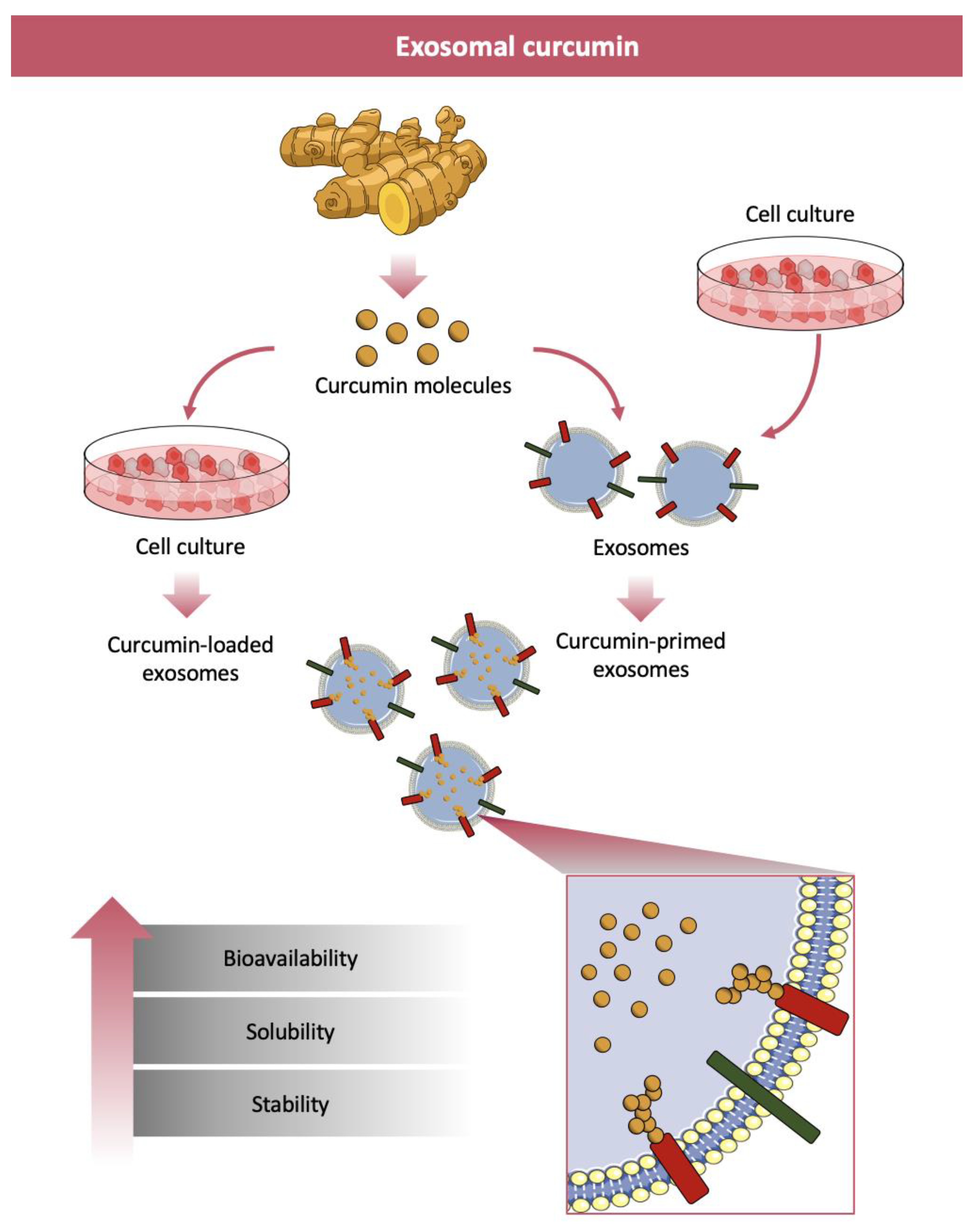
EXOs (30–150 nm size) are the smallest members of the extracellular vesicle (EV) family [219]. EXOs are formed by inward budding of the endosomal membranes of intraluminal vesicles (ILVs) to form multivesicular bodies (MVBs). After maturation, MVBs fuse with the plasma membrane and release ILVs in the form of EXOs into the extracellular environment, where they can mediate intercellular communication, exchange materials with other cells, eliminate unwanted cell products and mediate immune surveillance [220]. EXOs are composed of proteins, lipids and nucleic acids (DNA, mRNA, miRNA, lncRNA, etc.) stored during formation. EXOs are present in body fluids, such as blood, cerebrospinal fluid, amniotic fluid, urine, milk, etc., and participate in various physiological and pathological functions [221]. Recently, the presence of EXO-like vesicles, whose biogenesis is still unknown, have also been described in plants [222]. EXOs can be isolated, purified and characterized, following procedures that are continuously developing in parallel with the technologies for their loading with bioactive compounds [223,224].
Therapeutic drugs are loaded into EXOs by an active or passive method, with different loading yields and stabilities. Sonication, extrusion, electroporation or freeze-thaw cycles methods that disrupt EXOs membrane, restored after the loading process, are the main methods for active cargo loading [225]. Active loading (or EXOs loading) increases cargo up to 11 times compared to passive loading [226], but for the intrinsic characteristics of the process the native EXOs structure and targeting features are damaged. Passive cargo loading (or EXOs primed) consists of incubation of bioactive compounds with isolated EXOs or incubation of bioactive compounds with donor cells before vesicle isolation [225]. The first passive encapsulation process is based on the molecule diffusion through EXOs lipid membrane and strictly depends on the hydrophobic or hydrophilic properties of molecules to be loaded. The second passive encapsulation process is less common and consists, first, in the incubation of cargo with donor cells followed by isolation of EXOs engineered with the specific cargo at the cellular level under natural conditions [225]. In Figure 3 the synthesis of exosomes-loaded and exosomes-primed curcumin and the benefits in terms of bioavailability, solubility and stability achieved in curcumin delivery are reported.
Two recently published reviews discuss the use and applications of plant and animal derived EXOs that could potentially solve most of the problems of the existing nano-delivery systems [222,223].
EXOs can bind hydrophobic molecules that, as a consequence, increase their transport and their bioavailability allowing an efficient cellular uptake. Curcumin, that is a hydrophobic molecule, can be bound and transported by EXOs. This incorporation increases curcumin solubility, stability and bioavailability when compared to others and thus encourages the use of EXOs for therapeutic purposes [227,228,229,230,231,232,233].
Both EXOs curcumin loaded or primed have in vitro and in vivo therapeutic effects: anti-hypocholesterolemia, anticancer and anti-inflammatory (in cancer, ischemia-reperfusion injury and ischemic stroke). Table 8 reports a summary of anticancer effects of exosomes encapsulated curcumin.
Despite the mechanisms of how EXOs reaches the brain still being unknown, they are considered promising therapeutic agents for brain protection. Indeed, intranasal administration of EXOs-encapsulated curcumin in LPS-induced brain inflammation and in autoimmune encephalomyelitis in C57BL/6j induced a reduction in the number of activated inflammatory microglial cells [234]. The treatment with EXOs-encapsulated curcumin of stroke injury in type-1 diabetic mice ameliorates neurodegeneration, promotes neuronal survival and restores cognitive function by reducing infarct volume, edema and vascular damage [235]. In a model of mouse cerebral ischemia, treatment with EXOs-encapsulated curcumin reduced pro-inflammatory cytokines TNF-α, IL-1β and IL-6 levels and efficiently suppressed inflammation and cellular apoptosis in the lesion region of the ischemic brain [236]. Curcumin primed EXOs, prepared by incubating mouse brain endothelial cells with 7.5 µM curcumin for 72 h protected brain endothelial cell layer permeability by decreasing oxidative stress and mitigating impaired junction proteins [237]. Curcumin-loaded exosomes, isolated from mouse embryonic stem cells and administered nasally to ischemia-injured mice, was distributed into all brain regions and improved the neurological score after three days of treatment. In addition, the treatment restored vascular endothelial tight (claudin-5 and occludin) and adherent (VE-cadherin) junction proteins, suggesting that combining the potentials of embryonic stem cell exosomes and curcumin can help neurovascular restoration following ischemia-reperfusion injury in mice [238].

Table 8.
In vitro studies on exosomes encapsulated curcumin effect in cancer.
| Disease | Exosomes Origin | Exosomes Target | Outcomes | Ref. |
|---|---|---|---|---|
| Pancreatic cancer | PANC-1 cells, MIA PaCa-2 cells | PANC-1 cells MIA PaCa-2 cells | Cell death induction | [239] |
| Lung, breast, and cervical cancers | Raw bovine milk | H1299, A549 lung cancer cells MDA-MB-231, T47D breast cancer cells, HeLa cervical cancer cells | Antiproliferative, anti-inflammatory, and antitumor activities | [227] |
| Breast cancer | TS/A, 4T.1, B16 tumor cells | Murine mammary adenocarcinoma of spontaneous BALB/c origin | Anticancer properties by inhibition of NK cell activity | [240] |
| Lung cancer | H1299 | TCF21, BEAS-2B, A549, PC9, H1299Mouse lung cancer cells | Anti-cancer function by downregulating DNMT1, thereby upregulating TCF21 | [241] |
| Chronic myelogenous leukemia | K562, LAMA84 | CML mouse model | Cur-Exo containing miR-21 indicated an antineoplastic effect in chronic myeloid leukemia | [242] |
| Chronic myelogenous leukemia | K562, LAMA84 | CML mouse model | Angiogenic effect | [243,244] |
| Lymphoma Mammary gland cancer, Colon cancer | Grapefruit | HUVEC/human and T-lymphoma EL4 cells, 4T1 and 4TO7 breast cancer cell lines, NMuMG mammary gland epithelial cells, CT26 colon cancer/mouse | Inhibition of breast tumor and colon tumor growth | [245] |
5. Conclusions
Nanotechnology is fundamental in developing new strategies aimed to improve bioavailability, stability and targeting of nutraceuticals, for those, like curcuminoids, that possess low absorption and are rapidly metabolized and eliminated. Among the nutraceuticals, the pleiotropic biological activities of curcumin hold great promise for application in the clinics and for the global nutraceutical products market, that is estimated will grow about 7.2% in the next five years. The antioxidant, anti-inflammatory and anticancer activities of curcumin provide diverse health benefits against a wide spectrum of human diseases. Therefore, companies involved world wide in the nutraceuticals markets are developing new technological strategies to improve the beneficial effects for human health. In this review, we have discussed the advantageous use of curcumin against the central nervous system related disorders as well as the problems connected to its poor aqueous solubility and low bioavailability and to the fact that under various conditions curcumin is unstable, easily degraded or metabolized. For this purpose, in the field of neurological disorders (neurodegenerative diseases and cancer), we have further discussed the scientifically verified benefits of curcumin impaired by low brain bioavailability due to the limited blood-brain barrier permeability. Then, different kinds of curcumin nano-carriers (such as nanoparticles, micelles, dendrimers, conjugation with other materials, bio-based nanomaterials) are discussed along with their characteristic features like enhanced stability, bioavailability, solubility, in vivo uptake, bioactivity and safety. Among the recent research directions, a special focus has been directed at EXOs for their excellent properties, in other words, high loading efficiency and exact offloading process. Overall, the nanocurcumin delivery may lead to a significant improvement of its efficacy in CNS related disorders. However, in future, further in vivo and clinical investigations will help to achieve a safe and beneficial administration to AD, PD, ALS and cancer patients.
Funding
This research received no external funding.
Conflicts of Interest
The authors declare no conflict of interest.
References
- DeFelice, S.L. The Nutraceutical Revolution: Fueling a Powerful, New International Market; The Foundation for Innovation in Medicine: Mountside, NJ, USA, 1989. [Google Scholar]
- Chauhan, B.; Kumar, G.; Kalam, N.; Ansari, S.H. Current concepts and prospects of herbal nutraceutical: A review. J. Adv. Pharm. Technol. Res. 2013, 4, 4–8. [Google Scholar] [PubMed]
- Gupta, S.; Chauhan, D.; Mehla, K.; Sood, P.; Nair, A. An overview of nutraceuticals: Current scenario. J. Basic Clin. Pharm. 2010, 1, 55–62. [Google Scholar] [PubMed]
- Aggarwal, B.B.; Gupta, S.C.; Sung, B. Curcumin: An orally bioavailable blocker of TNF and other pro-inflammatory biomarkers. Br. J. Pharmacol. 2013, 169, 1672–1692. [Google Scholar] [CrossRef] [PubMed] [Green Version]
- Goel, A.; Aggarwal, B.B. Curcumin, the golden spice from Indian saffron, is a chemosensitizer and radiosensitizer for tumors and chemoprotector and radioprotector for normal organs. Nutr. Cancer 2010, 62, 919–930. [Google Scholar] [CrossRef]
- Gupta, S.C.; Kim, J.H.; Kannappan, R.; Reuter, S.; Dougherty, P.M.; Aggarwal, B.B. Role of nuclear factor κB-mediated inflammatory pathways in cancer-related symptoms and their regulation by nutritional agents. Exp. Biol. Med. 2011, 236, 658–671. [Google Scholar] [CrossRef] [Green Version]
- Tyagi, A.K.; Prasad, S.; Yuan, W.; Li, S.; Aggarwal, B.B. Identification of a novel compound (β-sesquiphellandrene) from turmeric (Curcuma longa) with anticancer potential: Comparison with curcumin. Investig. New Drugs 2015, 33, 1175–1186. [Google Scholar] [CrossRef]
- Hatcher, H.; Planalp, R.; Cho, J.; Torti, F.M.; Torti, S.V. Curcumin: From ancient medicine to current clinical trials. Cell. Mol. Life Sci. 2008, 65, 1631–1652. [Google Scholar] [CrossRef]
- Xu, X.Y.; Meng, X.; Li, S.; Gan, R.Y.; Li, Y.; Li, H.B. Bioactivity, health benefits, and related molecular mechanisms of curcumin: Current progress, challenges, and perspectives. Nutrients 2018, 10, 1553. [Google Scholar] [CrossRef] [Green Version]
- Maiti, P.; Dunbar, G.J. Use of curcumin, a natural polyphenol for targeting molecular pathways in treating age-related neurodegenerative diseases. Int. J. Mol. Sci. 2018, 19, 1637. [Google Scholar] [CrossRef] [PubMed] [Green Version]
- Shabaninejad, Z.; Pourhanifeh, M.H.; Movahedpour, A.; Mottaghi, R.; Nickdasti, A.; Mortezapour, E.; Shafiee, A.; Hajighadimi, S.; Moradizarmehri, S.; Sadeghian, M.; et al. Therapeutic potentials of curcumin in the treatment of glioblstoma. Eur. J. Med. Chem. 2020, 188, 112040. [Google Scholar] [CrossRef]
- Bhat, A.; Mahalakshmi, A.M.; Ray, B.; Tuladhar, S.; Hediyal, T.A.; Manthiannem, E.; Padamati, J.; Chandra, R.; Chidambaram, S.B.; Sakharkar, M.K. Benefits of curcumin in brain disorders. BioFactors 2019, 45, 666–689. [Google Scholar] [CrossRef] [PubMed]
- Anand, P.; Kunnumakkara, A.B.; Newman, R.A.; Aggarwal, B.B. Bioavailability of curcumin: Problems and promises. Mol. Pharm. 2007, 4, 807–818. [Google Scholar] [CrossRef] [PubMed]
- Anand, P.; Sundaram, C.; Jhurani, S.; Kunnumakkara, A.B.; Aggarwal, B.B. Curcumin and cancer: An “old-age” disease with an “age-old” solution. Cancer Lett. 2008, 267, 133–164. [Google Scholar] [CrossRef] [PubMed]
- Goel, A.; Kunnumakkara, A.B.; Aggarwal, B.B. Curcumin as “Curecumin”: From kitchen to clinic. Biochem. Pharmacol. 2008, 75, 787–809. [Google Scholar] [CrossRef] [Green Version]
- Nair, H.B.; Sung, B.; Yadav, V.R.; Kannappan, R.; Chaturvedi, M.M.; Aggarwal, B.B. Delivery of antiinflammatory nutraceuticals by nanoparticles for the prevention and treatment of cancer. Biochem. Pharmacol. 2010, 80, 1833–1843. [Google Scholar] [CrossRef] [PubMed] [Green Version]
- Huyan, T.; Li, H.; Peng, H.; Chen, J.; Yang, R.; Zhang, W.; Li, Q. Extracellular Vesicles—Advanced nanocarriers in cancer therapy: Progress and achievements. Int. J. Nanomed. 2020, 15, 6485–6502. [Google Scholar] [CrossRef]
- Gupta, S.C.; Patchva, S.; Koh, W.; Aggarwal, B.B. Discovery of curcumin, a component of golden spice, and its miraculous biological activities. Clin. Exp. Pharmacol. Physiol. 2012, 39, 283–299. [Google Scholar] [CrossRef]
- Nelson, K.M.; Dahlin, J.L.; Bisson, J.; Graham, J.; Pauli, G.F.; Walters, M.A. The essential medicinal chemistry of curcumin. J. Med. Chem. 2017, 60, 1620–1637. [Google Scholar] [CrossRef]
- Sharifi-Rad, J.; Rayess, Y.E.; Rizk, A.A.; Sadaka, C.; Zgheib, R.; Zam, W.; Sestito, S.; Rapposelli, S.; Neffe-Skocińska, K.; Zielińska, D.; et al. Turmeric and its major compound curcumin on health: Bioactive effects and safety profiles for food, pharmaceutical, biotechnological and medicinal applications. Front. Pharmacol. 2020, 11, 01021. [Google Scholar] [CrossRef]
- Abdeldaiem, M. Use of yellow pigment extracted from turmeric (Curcuma longa) rhizomes powder as natural food preservative. Am. J. Food Sci. Technol. 2014, 2, 36–47. [Google Scholar]
- Gul, P.; Bakht, J. Antimicrobial activity of turmeric extract and its potential use in food industry. J. Food Sci. Technol. 2015, 52, 2272–2279. [Google Scholar] [CrossRef] [PubMed] [Green Version]
- Koo, E.; Kimbal, A.B.; Wanner, M. Cosmeceuticals. In Rook’s Textbook of Dermatology, 9th ed.; Griffiths, C., Barker, J., Bleiker, T., Chalmers, R., Creamer, D., Eds.; John Wiley & Sons: Oxford, UK, 2016; p. 156. [Google Scholar]
- Sasikumar, B. Genetic resources of Curcuma: Diversity, characterization and utilization. Plant Genet. Resour. 2005, 3, 230–251. [Google Scholar] [CrossRef]
- Pulido-Moran, M.; Moreno-Fernandez, J.; Ramirez-Tortosa, C.; Ramirez-Tortosa, M.C. Curcumin and health. Molecules 2016, 21, 264. [Google Scholar] [CrossRef] [PubMed]
- Ferreira, V.H.; Nazli, A.; Dizzell, S.E.; Mueller, K.; Kaushic, C. The anti-inflammatory activity of curcumin protects the genital mucosal epithelial barrier from disruption and blocks replication of HIV-1 and HSV-2. PLoS ONE 2015, 10, e0124903. [Google Scholar] [CrossRef] [Green Version]
- Antoine, F.; Girard, D. Curcumin increases gelatinase activity in human neutrophils by a p38 mitogen-activated protein kinase (MAPK)-independent mechanism. J. Immunotoxicol. 2015, 12, 188–193. [Google Scholar] [CrossRef] [Green Version]
- Li, H.Y.; Yang, M.; Li, Z.; Meng, Z. Curcumin inhibits angiotensin II-induced inflammation and proliferation of rat vascular smooth muscle cells by elevating PPAR-gamma activity and reducing oxidative stress. Int. J. Mol. Med. 2017, 39, 1307–1316. [Google Scholar] [CrossRef] [Green Version]
- Zhang, Y.L.; Liu, Z.G.; Wu, J.Z.; Bai, B.; Chen, H.J.; Xiao, Z.X.; Chen, L.F.; Zhao, Y.J.; Lum, H.; Wang, Y.; et al. New MD2 inhibitors derived from curcumin with improved anti-inflammatory activity. Eur. J. Med. Chem. 2018, 148, 291–305. [Google Scholar] [CrossRef]
- Zheng, Q.T.; Yang, Z.H.; Yu, L.Y.; Ren, Y.Y.; Huang, Q.X.; Liu, Q.; Ma, X.Y.; Chen, Z.K.; Wang, Z.B.; Zheng, X. Synthesis and antioxidant activity of curcumin analogs. J. Asian Nat. Prod. Res. 2017, 19, 489–503. [Google Scholar] [CrossRef]
- Tapia, E.; Sanchez-Lozada, L.G.; Garcia-Nino, W.R.; Garcia, E.; Cerecedo, A.; Garcia-Arroyo, F.E.; Osorio, H.; Arellano, A.; Cristobal-Garcia, M.; Loredo, M.L.; et al. Curcumin prevents maleate-induced nephrotoxicity: Relation to hemodynamic alterations, oxidative stress, mitochondrial oxygen consumption and activity of respiratory complex I. Free Radic. Res. 2014, 48, 1342–1354. [Google Scholar] [CrossRef]
- Dai, C.S.; Ciccotosto, G.D.; Cappai, R.; Tang, S.S.; Li, D.W.; Xie, S.L.; Xiao, X.L.; Velkov, T. Curcumin attenuates colistin-induced neurotoxicity in N2a cells via anti-inflammatory activity, suppression of oxidative stress, and apoptosis. Mol. Neurobiol. 2018, 55, 421–434. [Google Scholar] [CrossRef]
- Sahebkar, A.; Mohammadi, A.; Atabati, A.; Rahiman, S.; Tavallaie, S.; Iranshahi, M. Curcuminoids modulate pro-oxidantantioxidant balance but not the immune response to heat shock protein 27 and oxidized LDL in obese individuals. Phytother. Res. 2013, 27, 1883–1888. [Google Scholar] [CrossRef] [PubMed]
- Ganjali, S.; Sahebkar, A.; Mahdipour, E.; Jamialahmadi, K.; Torabi, S.; Akhlaghi, S. Investigation of the effects of curcumin on serum cytokines in obese individuals: A randomized controlled trial. Sci. World J. 2014, 2014, 898361. [Google Scholar] [CrossRef] [PubMed]
- Maugeri, A.; Mazzone, M.G.; Giuliano, F.; Vinciguerra, M.; Basile, G.; Barchitta, M.; Agodi, A. Curcumin modulates DNA methyltransferase functions in a cellular model of diabetic retinopathy. Oxid. Med. Cell. Longev. 2018, 2018, 5407482. [Google Scholar] [CrossRef] [PubMed]
- Iqbal, M.; Sharma, S.D.; Okazaki, Y.; Fujisawa, M.; Okada, S. Dietary supplementation of curcumin enhances antioxidant and phase II metabolizing enzymes in ddY male mice: Possible role in protection against chemical carcinogenesis and toxicity. Pharmacol. Toxicol. 2003, 92, 33–38. [Google Scholar] [CrossRef] [PubMed]
- El-Bahr, S.M. Effect of curcumin on hepatic antioxidant enzymes activities and gene expressions in rats intoxicated with aflatoxin b1. Phytother. Res. 2015, 29, 134–140. [Google Scholar] [CrossRef] [PubMed]
- Xu, D.P.; Li, Y.; Meng, X.; Zhou, T.; Zhou, Y.; Zheng, J.; Zhang, J.J.; Li, H.B. Natural antioxidants in foods and medicinal plants: Extraction, assessment and resources. Int. J. Mol. Sci. 2017, 18, 96. [Google Scholar] [CrossRef]
- Joe, B.; Vijaykumar, M.; Lokesh, B.R. Biological properties of curcumin-cellular and molecular mechanisms of action. Crit. Rev. Food Sci. Nutr. 2004, 44, 97–111. [Google Scholar] [CrossRef]
- Ahsan, H.; Hadi, S.M. Strand scission in DNA induced by curcumin in the presence of Cu(II). Cancer Lett. 1998, 124, 23–30. [Google Scholar] [CrossRef]
- Ahsan, H.; Parveen, N.; Khan, N.U.; Hadi, S.M. Pro-oxidant, anti-oxidant and cleavage activities on DNA of curcumin and its derivatives demethoxycurcumin and bisdemethoxycurcumin. Chem. Biol. Interact. 1999, 121, 161–175. [Google Scholar] [CrossRef]
- Abdollahi, E.; Momtazi, A.A.; Johnston, T.P.; Sahebkar, A. Therapeutic effects of curcumin in inflammatory and immune-mediated diseases: A nature-made jack-of-all-trades? J. Cell. Physiol. 2018, 233, 830–848. [Google Scholar] [CrossRef]
- Shakeri, F.; Boskabady, M.H. Anti-inflammatory, antioxidant, and immunomodulatory effects of curcumin in ovalbumin-sensitized rat. BioFactors 2017, 43, 567–576. [Google Scholar] [CrossRef] [PubMed]
- Yang, Z.; He, C.M.; He, J.Y.; Chu, J.; Liu, H.P.; Deng, X.Y. Curcumin-mediated bone marrow mesenchymal stem cell sheets create a favorable immune microenvironment for adult full-thickness cutaneous wound healing. Stem Cell. Res. Ther. 2018, 9, 21. [Google Scholar] [CrossRef] [PubMed]
- Liu, Y.; Chen, L.Y.; Shen, Y.; Tan, T.; Xie, N.Z.; Luo, M.; Li, Z.H.; Xie, X.Y. Curcumin ameliorates ischemia-induced limb injury through immunomodulation. Med. Sci. Monit. 2016, 22, 2035–2042. [Google Scholar] [CrossRef] [PubMed] [Green Version]
- Salehi, B.; Stojanović-Radić, Z.; Matejić, J.; Sharifi-Rad, M.; Anil Kumar, N.V.; Martins, N.; Sharifi-Rad, J. The therapeutic potential of curcumin: A review of clinical trials. Eur. J. Med. Chem. 2019, 163, 527–545. [Google Scholar] [CrossRef]
- Confaloni, A.; Tosto, G.; Tata, A.M. Promising therapies for Alzheimer’s disease. Curr. Pharm. Des. 2016, 22, 2050–2056. [Google Scholar] [CrossRef]
- Calabrese, V.; Cornelius, C.; Dinkova-Kostova, A.T.; Calabrese, E.J.; Mattson, M.P. Cellular stress responses, the hormesis paradigm, and vitagenes: Novel targets for therapeutic intervention in neurodegenerative disorders. Antioxid. Redox Signal. 2010, 13, 1763–1811. [Google Scholar] [CrossRef]
- Alexander, G.E. Biology of Parkinson’s disease: Pathogenesis and pathophysiology of a multisystem neurodegenerative disorder. Dialogues Clin. Neurosci. 2004, 6, 259–280. [Google Scholar]
- Maiti, P.; Paladugu, L.; Dunbar, G.L.J.B. Solid lipid curcumin particles provide greater anti-amyloid, anti-inflammatory and neuroprotective effects than curcumin in the 5xFAD mouse model of Alzheimer’s disease. BMC Neurosci. 2018, 19, 7. [Google Scholar] [CrossRef] [Green Version]
- Dantzer, R.; O’Connor, J.C.; Freund, G.G.; Johnson, R.W.; Kelley, K.W. From inflammation to sickness and depression: When the immune system subjugates the brain. Nat. Rev. Neurosci. 2008, 9, 46–56. [Google Scholar] [CrossRef] [Green Version]
- Tang, M.; Taghibiglou, C. The mechanisms of action of curcumin in Alzheimer’s disease. J. Alzheimers Dis. 2017, 58, 1003–1016. [Google Scholar] [CrossRef]
- Chainoglou, E.; Siskos, A.; Pontiki, E.; Hadjipavlou-Litina, D. Hybridization of curcumin analogues with cinnamic acid derivatives as multi-target agents against Alzheimer’s disease targets. Molecules 2020, 25, 4958. [Google Scholar] [CrossRef] [PubMed]
- Cornelius, C.; Trovato Salinaro, A.; Scuto, M.; Fronte, V.; Cambria, M.T.; Pennisi, M.; Bella, R.; Milone, P.; Graziano, A.; Crupi, R.; et al. Cellular stress response, sirtuins and UCP proteins in Alzheimer disease: Role of vitagenes. Immun. Ageing 2013, 10, 41. [Google Scholar] [CrossRef] [PubMed] [Green Version]
- Trovato Salinaro, A.; Cornelius, C.; Koverech, G.; Koverech, A.; Scuto, M.; Lodato, F.; Fronte, V.; Muccilli, V.; Reibaldi, M.; Longo, A.; et al. Cellular stress response, redox status, and vitagenes in glaucoma: A systemic oxidant disorder linked to Alzheimer’s disease. Front. Pharmacol. 2014, 5, 129. [Google Scholar] [CrossRef] [PubMed] [Green Version]
- Cui, Q.; Li, X.; Zhu, H. Curcumin ameliorates dopaminergic neuronal oxidative damage via activation of the Akt/Nrf2 pathway. Mol. Med. Rep. 2016, 13, 1381–1388. [Google Scholar] [CrossRef] [Green Version]
- Ma, Q.L.; Zuo, X.; Yang, F.; Ubeda, O.J.; Gant, D.J.; Alaverdyan, M.; Teng, E.; Hu, S.; Chen, P.P.; Maiti, P. Curcumin suppresses soluble tau dimers and corrects molecular chaperone, synaptic, and behavioral deficits in aged human tau transgenic mice. J. Biol. Chem. 2013, 288, 4056–4065. [Google Scholar] [CrossRef] [Green Version]
- Al-Omar, F.A.; Nagi, M.N.; Abdulgadir, M.M.; Al Joni, K.S.; Al-Majed, A.A. Immediate and delayed treatments with curcumin prevents forebrain ischemia-induced neuronal damage and oxidative insult in the rat hippocampus. Neurochem. Res. 2006, 31, 611–618. [Google Scholar] [CrossRef]
- Calabrese, V.; Guagliano, E.; Sapienza, M.; Mancuso, C.; Butterfield, D.A.; Stella, A.M. Redox regulation of cellular stress response in neurodegenerative disorders. Ital. J. Biochem. 2006, 55, 263–282. [Google Scholar]
- Kim, S.J.; Son, T.G.; Park, H.R.; Park, M.; Kim, M.S.; Kim, H.S.; Chung, H.Y.; Mattson, M.P.; Lee, J. Curcumin stimulates proliferation of embryonic neural progenitor cells and neurogenesis in the adult hippocampus. J. Biol. Chem. 2008, 283, 14497–14505. [Google Scholar] [CrossRef] [Green Version]
- Scuto, M.C.; Mancuso, C.; Tomasello, B.; Ontario, M.L.; Cavallaro, A.; Frasca, F.; Maiolino, L.; Trovato Salinaro, A.; Calabrese, E.J.; Calabrese, V. Curcumin, hormesis and the nervous system. Nutrients 2019, 11, 2417. [Google Scholar] [CrossRef] [Green Version]
- Tripanichkul, W.; Jaroensuppaperch, E.O. Ameliorating effects of curcumin on 6-OHDA-induced dopaminergic denervation, glial response, and SOD1 reduction in the striatum of hemiparkinsonian mice. Eur. Rev. Med. Pharmacol. Sci. 2013, 17, 1360–1368. [Google Scholar]
- Gui, H.Y.; Chen, R.N.; Peng, Y.; Hu, J.H.; Mao, Z.; Ning, R.; Shang, W.; Liu, W.; Xiong, J.; Hu, G.; et al. Curcumin protects against 1-methyl-4-phenylpyridinium ion- and lipopolysaccharide-induced cytotoxicities in the mouse mesencephalic astrocyte via inhibiting the cytochrome P450 2E1. Evid. Based Complement. Altern. Med. 2013, 2013, 523484. [Google Scholar] [CrossRef] [PubMed]
- He, X.J.; Uchida, K.; Megumi, C.; Tsuge, N.; Nakayama, H. Dietary curcumin supplementation attenuates 1-methyl-4-phenyl-1,2,3,6-tetrahydropyridine (MPTP) neurotoxicity in C57BL mice. J. Toxicol. Pathol. 2015, 28, 197–206. [Google Scholar] [CrossRef] [PubMed] [Green Version]
- Singh, P.K.; Kotia, V.; Ghosh, D.; Mohite, G.M.; Kumar, A.; Maji, S.K. Curcumin modulates alpha-synuclein aggregation and toxicity. ACS Chem. Neurosci. 2013, 4, 393–407. [Google Scholar] [CrossRef] [PubMed] [Green Version]
- Jiang, T.F.; Zhang, Y.J.; Zhou, H.Y.; Wang, H.M.; Tian, L.P.; Liu, J.; Ding, J.Q.; Chen, S. Curcumin ameliorates the neurodegenerative pathology in A53T alpha-synuclein cell model of Parkinson’s disease through the downregulation of mTOR/p70S6K signaling and the recovery of macroautophagy. J. Neuroimmune Pharmacol. 2013, 8, 356–369. [Google Scholar] [CrossRef]
- Jayaraj, R.L.; Elangovan, N.; Manigandan, K.; Singh, S.; Shukla, S. CNB-001 a novel curcumin derivative, guards dopamine neurons in MPTP model of Parkinson’s disease. BioMed Res. Int. 2014, 2014, 236182. [Google Scholar] [CrossRef] [Green Version]
- Wang, Y.L.; Ju, B.; Zhang, Y.Z.; Yin, H.L.; Liu, Y.J.; Wang, S.S.; Zeng, Z.L.; Yang, X.P.; Wang, H.T.; Li, J.F. Protective effect of curcumin against oxidative stress-induced injury in rats with Parkinson’s disease through the Wnt/β-catenin signaling pathway. Cell. Physiol. Biochem. 2017, 43, 2226–2241. [Google Scholar] [CrossRef]
- Wang, H.M.; Zhao, Y.X.; Zhang, S.; Liu, G.D.; Kang, W.Y.; Tang, H.D.; Ding, J.Q.; Chen, S.D. PPARγ agonist curcumin reduces the amyloid-β-stimulated inflammatory responses in primary astrocytes. J Alzheimers Dis. 2010, 20, 1189–1199. [Google Scholar] [CrossRef]
- Feng, H.L.; Fan, H.; Dang, H.Z.; Chen, X.P.; Ren, Y.; Yang, J.D.; Wang, P.W. Neuroprotective effect of curcumin to Aβ of double transgenic mice with Alzheimer’s disease. China J. Chin. Mater. Med. 2014, 39, 3846–3849. [Google Scholar]
- Lim, G.P.; Chu, T.; Yang, F.; Beech, W.; Frautschy, S.A.; Cole, G.M. The curry spice curcumin reduces oxidative damage and amyloid pathology in an Alzheimer transgenic mouse. J. Neurosci. 2001, 21, 8370–8377. [Google Scholar] [CrossRef]
- Bassani, T.B.; Turnes, J.M.; Moura, E.; Bonato, J.M.; Cóppola-Segovia, V.; Zanata, S.M.; Oliveira, R.; Vital, M. Effects of curcumin on short-term spatial and recognition memory, adult neurogenesis and neuroinflammation in a streptozotocin-induced rat model of dementia of Alzheimer’s type. Behav. Brain Res. 2017, 335, 41–54. [Google Scholar] [CrossRef]
- Ahmed, T.; Enam, S.A.; Gilani, A.H. Curcuminoids enhance memory in an amyloid-infused rat model of Alzheimer’s disease. Neuroscience 2010, 169, 1296–1306. [Google Scholar] [CrossRef] [PubMed]
- Garcia-Alloza, M.; Borrelli, L.A.; Rozkalne, A.; Hyman, B.T.; Bacskai, B.J. Curcumin labels amyloid pathology in vivo, disrupts existing plaques, and partially restores distorted neurites in an Alzheimer mouse model. J. Neurochem. 2007, 102, 1095–1104. [Google Scholar] [CrossRef] [PubMed]
- Huang, H.C.; Xu, K.; Jiang, Z.F. Curcumin-mediated neuroprotection against amyloid-beta-induced mitochondrial dysfunction involves the inhibition of GSK-3beta. J. Alzheimers Dis. 2012, 32, 981–996. [Google Scholar] [CrossRef] [PubMed]
- Rane, J.S.; Bhaumik, P.; Panda, D. Curcumin inhibits tau aggregation and disintegrates preformed tau filaments in vitro. J. Alzheimers Dis. 2017, 60, 999–1014. [Google Scholar] [CrossRef] [PubMed]
- Liu, Z.J.; Li, Z.H.; Liu, L.; Tang, W.X.; Wang, Y.; Dong, M.R.; Xiao, C. Curcumin attenuates beta-amyloid-induced neuroinflammation via activation of peroxisome proliferator-activated receptor-gamma function in a rat model of Alzheimer’s Disease. Front. Pharmacol. 2016, 7, 261. [Google Scholar] [CrossRef] [PubMed] [Green Version]
- Sundaram, J.R.; Poore, C.P.; Sulaimee, N.; Pareek, T.; Cheong, W.F.; Wenk, M.R.; Pant, H.C.; Frautschy, S.A.; Low, C.M.; Kesavapany, S. Curcumin ameliorates neuroinflammation, neurodegeneration, and memory deficits in p25 transgenic mouse model that bears hallmarks of Alzheimer’s disease. J. Alzheimers Dis. 2017, 60, 1429–1442. [Google Scholar] [CrossRef]
- Tai, Y.H.; Lin, Y.Y.; Wang, K.C.; Chang, C.L.; Chen, R.Y.; Wu, C.C.; Cheng, I.H. Curcuminoid submicron particle ameliorates cognitive deficits and decreases amyloid pathology in Alzheimer’s disease mouse model. Oncotarget 2018, 9, 10681–10697. [Google Scholar] [CrossRef] [Green Version]
- Natarajan, C.; Bright, J.J. Curcumin inhibits experimental allergic encephalomyelitis by blocking IL-12 signaling through janus kinase-STAT pathway in T lymphocytes. J. Immunol. 2002, 168, 6506–6513. [Google Scholar] [CrossRef] [Green Version]
- Kim, G.Y.; Kim, K.H.; Lee, S.H.; Yoon, M.S.; Lee, H.J.; Moon, D.O.; Park, Y.M. Curcumin inhibits immunostimulatory function of dendritic cells: MAPKs and translocation of NF- B as potential targets. J. Immunol. 2005, 174, 8116–8124. [Google Scholar] [CrossRef] [Green Version]
- Fahey, A.J.; Adrian Robins, R.; Constantinescu, C.S. Curcumin modulation of IFN-beta and IL-12 signaling and cytokine induction in human T cells. J. Cell. Mol. Med. 2007, 11, 1129–1137. [Google Scholar] [CrossRef]
- Chearwae, W.; Bright, J.J. 15-deoxy-Delta(12,14)-prostaglandin J(2) and curcumin modulate the expression of toll-like receptors 4 and 9 in autoimmune T lymphocyte. J. Clin. Immunol. 2008, 28, 558–570. [Google Scholar] [CrossRef] [PubMed]
- Zhang, Z.J.; Zhao, L.X.; Cao, D.L.; Zhang, X.; Gao, Y.J.; Xia, C. Curcumin inhibits LPS-induced CCL2 expression via JNK pathway in C6 rat astrocytoma cells. Cell. Mol. Neurobiol. 2012, 32, 1003–1010. [Google Scholar] [CrossRef] [PubMed]
- Agrawal, S.S.; Gullaiya, S.; Dubey, V.; Singh, V.; Kumar, A.; Nagar, A.; Tiwari, P. Neurodegenerative shielding by curcumin and its derivatives on brain lesions induced by 6-OHDA model of Parkinson’s disease in Albino Wistar rats. Card. Psych. Neur. 2012, 2012, 942981. [Google Scholar] [CrossRef] [PubMed]
- Tegenge, M.A.; Rajbhandari, L.; Shrestha, S.; Mithal, A.; Hosmane, S.; Venkatesan, A. Curcumin protects axons from degeneration in the setting of local neuroinflammation. Exp. Neurol. 2014, 253, 102–110. [Google Scholar] [CrossRef] [PubMed]
- Seyedzadeh, M.H.; Safari, Z.; Zare, A.; Gholizadeh Navashenaq, J.; Razavi, A.; Kardar, G.A.; Khorramizadeh, M.R. Study of curcumin immunomodulatory effects on reactive astrocyte cell function. Int. Immunopharmacol. 2014, 22, 230–235. [Google Scholar] [CrossRef]
- Mohajeri, M.; Sadeghizadeh, M.; Najafi, F.; Javan, M. Polymerized nanocurcumin attenuates neurological symptoms in EAE model of multiple sclerosis through down regulation of inflammatory and oxidative processes and enhancing neuroprotection and myelin repair. Neuropharmacology 2015, 99, 156–167. [Google Scholar] [CrossRef]
- Watanabe, K.; Tachibana, O.; Sato, K.; YoneKawa, Y.; Kleihues, P.; Ohgak, H. Overexpression of EGF receptor and p53 mutation are mutually exclusive in the evolutionary of primary and secondary glioblastomas. Brain Pathol. 1996, 6, 217–223. [Google Scholar] [CrossRef]
- Lino, M.; Merlo, A. PI3Kinase signaling in glioblastoma. J. Neuro Oncol. 2011, 103, 417–427. [Google Scholar] [CrossRef] [Green Version]
- Saadatpour, Z.; Bjorklund, G.; Chirumbolo, S.; Alimohammadi, M.; Ehsani, H.; Ebrahiminejad, H.; Pourghadamyari, H.; Baghaei, B.; Mirzaei, H.R.; Sahebkar, A.; et al. Cancer Molecular imaging and cancer gene therapy. Cancer Gene Ther. 2016. [Google Scholar] [CrossRef]
- Hanif, S.; Muhammad, P.; Chesworth, R.; Rehman, F.U.; Qian, R.J.; Zheng, M.; Shi, B.Y. Nanomedicine-based immunotherapy for central nervous system disorders. Acta Pharmacol. Sin. 2020, 41, 936–953. [Google Scholar] [CrossRef]
- Maiti, P.; Plemmons, A.; Gary, L. Combination treatment of berberine and solid lipid curcumin particles increased cell death and inhibited PI3K/Akt/mTOR pathway of human cultured glioblastoma cells more effectively than did individual treatments. PLoS ONE 2019, 14, e0225660. [Google Scholar] [CrossRef] [PubMed]
- Maiti, P.; Scott, J.; Sengupta, D.; Al-Gharaibeh, A.; Dunbar, G.L. Curcumin and solid lipid curcumin particles induce autophagy, but inhibit mitophagy and the PI3K-Akt/mTOR pathway in cultured glioblastoma cells. Int. J. Mol. Sci. 2019, 20, 399. [Google Scholar] [CrossRef] [PubMed] [Green Version]
- Dhandapani, K.M.; Mahesh, V.B.; Brann, D.W. Curcumin suppresses growth and chemoresistance of human glioblastoma cells via AP-1 and NFkappaB transcription factors. J. Neurochem. 2007, 102, 522–538. [Google Scholar] [CrossRef] [PubMed]
- Yin, S.; Du, W.; Wang, F.; Han, B.; Cui, Y.; Yang, D.; Chen, H.; Liu, D.; Liu, X.; Zhai, X.; et al. MicroRNA-326 sensitizes human glioblastoma cells to curcumin via the SHH/GLI1 signaling pathway. Cancer Biol. Ther. 2018, 19, 260–270. [Google Scholar] [CrossRef]
- Park, K.S.; Yoon, S.Y.; Park, S.H.; Hwang, J.H. Anti-migration and anti-invasion effects of curcumin via suppression of fascin expression in glioblastoma cells. Brain Tumor Res. Treat. 2019, 7, 16–24. [Google Scholar] [CrossRef]
- Reuter, S.; Eifes, S.; Dicato, M.; Aggarwal, B.B.; Diederich, M. Modulation of antiapoptotic and survival pathways by curcumin as a strategy to induce apoptosis in cancer cells. Biochem. Pharmacol. 2008, 76, 1340–1351. [Google Scholar] [CrossRef]
- Ambegaokar, S.S.; Wu, L.; Alamshahi, K.; Lau, J.; Jazayeri, L.; Chan, S.; Khanna, P.; Hsieh, E.; Timiras, P.S. Curcumin inhibits dose-dependently and time dependently neuroglial cell proliferation and growth. Neuroendocrinol. Lett. 2003, 24, 469. [Google Scholar]
- Perry, M.C.; Demeule, M.; Regina, A.; Moumdjian, R.; Eliveau, R.B. Curcumin inhibits tumor growth and angiogenesis in glioblastoma xenografts. Mol. Nutr. Food Res. 2010, 54, 1192–1201. [Google Scholar] [CrossRef]
- Klinger, N.V.; Mittal, S. Therapeutic potential of curcumin for the treatment of brain tumors. Oxid. Med. Cell Longev. 2016, 9324085. [Google Scholar] [CrossRef]
- Lee, J.E.; Yoon, S.S.; Moon, E.Y. Curcumin-induced autophagy augments its antitumor effect against A172 human glioblastoma cells. Biomol. Ther. 2019, 7, 484–491. [Google Scholar] [CrossRef]
- Huang, B.R.; Tsai, C.H.; Chen, C.C.; Way, T.D.; Kao, J.Y.; Liu, Y.S.; Lin, H.Y.; Lai, S.W.; Lu, D.Y. Curcumin promotes connexin 43 degradation and temozolomide-induced apoptosis in glioblastoma cells. Am. J. Chin. Med. 2019, 47, 657–674. [Google Scholar] [CrossRef] [PubMed]
- Garrido-Armas, M.; Corona, J.C.; Escobar, M.L.; Torres, V.; Ordonez-Romero, F.; Hernandez-Hernandez, A.; Arenas-Huertero, F. Paraptosis in human glioblastoma cell line induced by curcumin. Toxicol. In Vitro 2018, 51, 63–73. [Google Scholar] [CrossRef] [PubMed]
- Zhao, J.; Zhu, J.; Lv, X.; Xing, J.; Liu, S.; Chen, C.; Xu, Y. Curcumin potentiates the potent antitumor activity of ACNU against glioblastoma by suppressing the PI3K/AKT and NF-κB/COX-2 signaling pathways. Onco Targets Ther. 2017, 10, 5471–5482. [Google Scholar] [CrossRef] [PubMed] [Green Version]
- Wang, Y.; Ying, X.; Xu, H.; Yan, H.; Li, X.; Tang, H. The functional curcumin liposomes induce apoptosis in C6 glioblastoma cells and C6 glioblastoma stem cells in vitro and in animals. Int. J. Nanomed. 2017, 12, 1369–1384. [Google Scholar] [CrossRef] [PubMed] [Green Version]
- Gersey, Z.C.; Rodriguez, G.A.; Barbarite, E.; Sanchez, A.; Walters, W.M.; Ohaeto, K.C.; Komotar, R.J.; Graham, R.M. Curcumin decreases malignant characteristics of glioblastoma stem cells via induction of reactive oxygen species. BMC Cancer 2017, 17, 99. [Google Scholar] [CrossRef] [PubMed] [Green Version]
- Mukherjee, S.; Baidoo, J.; Fried, A.; Atwi, D.; Dolai, S.; Boockvar, J.; Symons, M.; Ruggieri, R.; Raja, K.; Banerjee, P. Curcumin changes the polarity of tumor-associated microglia and eliminates glioblastoma. Int. J. Cancer 2016, 139, 2838–2849. [Google Scholar] [CrossRef] [PubMed] [Green Version]
- Cheng, C.; Jiao, J.T.; Qian, Y.; Guo, X.Y.; Huang, J.; Dai, M.C.; Zhang, L.; Ding, X.P.; Zong, D.; Shao, J.F. Curcumin induces G2/M arrest and triggers apoptosis via FoxO1 signaling in U87 human glioma cells. Mol. Med. Rep. 2016, 13, 3763–3770. [Google Scholar] [CrossRef] [Green Version]
- Wu, H.; Liu, Q.; Cai, T.; Chen, Y.D.; Wang, Z.F. Induction of microRNA-146a is involved in curcumin-mediated enhancement of temozolomide cytotoxicity against human glioblastoma. Mol. Med. Rep. 2015, 12, 5461–5466. [Google Scholar] [CrossRef] [Green Version]
- Zanotto-Filho, A.; Braganhol, E.; Klafke, K.; Figueiró, F.; Terra, S.R.; Paludo, F.J.; Morrone, M.; Bristot, I.J.; Battastini, A.M.; Forcelini, C.M.; et al. Autophagy inhibition improves the efficacy of curcumin/temozolomide combination therapy in glioblastomas. Cancer Lett. 2015, 358, 220–231. [Google Scholar] [CrossRef] [Green Version]
- Senft, C.; Polacin, M.; Priester, M.; Seifert, V.; Kögel, D.; Weissenberger, J. The nontoxic natural compound Curcumin exerts anti-proliferative, anti-migratory, and anti-invasive properties against malignant gliomas. BMC Cancer 2010, 10, 491. [Google Scholar] [CrossRef] [Green Version]
- Su, C.C.; Wang, M.J.; Chiu, T.L. The anti-cancer efficacy of curcumin scrutinized through core signaling pathways in glioblastoma. Int. J. Mol. Med. 2010, 26, 217–224. [Google Scholar] [PubMed] [Green Version]
- Choi, B.H.; Kim, C.G.; Bae, Y.S.; Lim, Y.; Lee, Y.H.; Shin, S.Y. p21 Waf1/Cip1 expression by curcumin in U-87MG human glioma cells: Role of early growth response-1 expression. Cancer Res. 2008, 68, 1369–1377. [Google Scholar] [CrossRef] [PubMed] [Green Version]
- Karmakar, S.; Banik, N.L.; Patel, S.J.; Ray, S.K. Curcumin activated both receptor-mediated and mitochondria-mediated proteolytic pathways for apoptosis in human glioblastoma T98G cells. Neurosci. Lett. 2006, 407, 53–58. [Google Scholar] [CrossRef] [PubMed]
- Karmakar, S.; Banik, N.L.; Ray, S.K. Curcumin suppressed anti-apoptotic signals and activated cysteine proteases for apoptosis in human malignant glioblastoma U87MG cells. Neurochem. Res. 2007, 32, 2103–2113. [Google Scholar] [CrossRef]
- Rai, M.; Pandit, P.; Gaikwad, S.; Yadav, A.; Gade, A. Potential applications of curcumin and curcumin nanoparticles: From traditional therapeutics to modern nanomedicine. Nanotechnol. Rev. 2015, 4, 161–172. [Google Scholar] [CrossRef]
- Mora-Huertas, C.E.; Fessi, H.; Elaissari, A. Polymer-based nanocapsules for drug delivery. Int. J. Pharm. 2010, 385, 113–142. [Google Scholar] [CrossRef]
- Nagavarma, B.V.N.; Yadav, H.K.; Ayaz, A.V.L.S.; Vasudha, L.S.; Shivakumar, H.G. Different techniques for preparation of polymeric nanoparticles. Asian J. Pharm. Clin. Res. 2012, 5, 16–23. [Google Scholar]
- Hosokawa, M.; Nogi, K.; Naito, M.; Yokoyama, T. Nanoparticle Technology Handbook; Elsevier: Amsterdam, The Netherlands, 2007. [Google Scholar]
- Subramani, P.A.; Panati, K.; Narala, V.R. Curcumin nanotechnologies and its anticancer activity. Nutr. Cancer 2017, 69, 381–393. [Google Scholar] [CrossRef]
- Hussain, Z.; Thu, H.E.; Ng, S.F.; Khan, S.; Katas, H. Nanoencapsulation, an efficient and promising approach to maximize wound healing efficacy of curcumin: A review of new trends and state-of-the-art. Colloids Surf. B Biointerfaces 2017, 150, 223–241. [Google Scholar] [CrossRef]
- Szymusiak, M.; Hu, X.; Plata, P.A.L.; Ciupinski, P.; Wang, Z.J.; Liu, Y.J. Bioavailability of curcumin and curcumin glucuronide in the central nervous system of mice after oral delivery of nano-curcumin. Int. J. Pharm. 2016, 511, 415–423. [Google Scholar] [CrossRef] [Green Version]
- Masserini, M. Nanoparticles for brain drug delivery. ISRN Biochem. 2013, 21, 238428. [Google Scholar] [CrossRef] [PubMed] [Green Version]
- Tefas, L.R.; Sylvester, B.; Tomuta, I.; Sesarman, A.; Licarete, E.; Banciu, M.; Porfire, A. Development of antiproliferative long-circulating liposomes co-encapsulating doxorubicin and curcumin, through the use of a quality-by-design approach. Drug Des. Dev. Ther. 2017, 11, 1605–1621. [Google Scholar] [CrossRef] [PubMed] [Green Version]
- Sesarman, A.; Tefas, L.; Sylvester, B.; Licarete, E.; Rauca, V.; Luput, L.; Patras, L.; Banciu, M.; Porfire, A. Anti-angiogenic and anti-inflammatory effects of long-circulating liposomes co-encapsulating curcumin and doxorubicin on C26 murine colon cancer cells. Pharmacol. Rep. 2018, 70, 331–339. [Google Scholar] [CrossRef] [PubMed]
- Chen, Y.; Du, Q.; Guo, Q.; Huang, J.; Liu, L.; Shen, X.; Peng, J. A W/O emulsion mediated film dispersion method for curcumin encapsulated pH-sensitive liposomes in the colon tumor treatment. Drug Dev. Ind. Pharm. 2019, 45, 282–291. [Google Scholar] [CrossRef]
- Huo, X.; Zhang, Y.; Jin, X.; Li, Y.; Zhang, L. A novel synthesis of selenium nanoparticles encapsulated PLGA nanospheres with curcumin molecules for the inhibition of amyloid β aggregation in Alzheimer’s disease. J. Photochem. Photobiol. B 2019, 190, 98–102. [Google Scholar] [CrossRef]
- Reddy, A.S.; Lakshmi, B.A.; Kim, S.; Kim, J. Synthesis and characterization of acetyl curcumin-loaded core/shell liposome nanoparticles via an electrospray process for drug delivery, and theranostic applications. Eur. J. Pharm. Biopharm. 2019, 142, 518–530. [Google Scholar] [CrossRef]
- Vetha, B.S.S.; Kim, E.M.; Oh, P.S.; Kim, S.H.; Lim, S.T.; Sohn, M.H. Curcumin encapsulated micellar nanoplatform for blue light emitting diode induced apoptosis as a new class of cancer therapy. Macromol. Res. 2019, 27, 1179–1184. [Google Scholar] [CrossRef]
- Yang, X.; Li, Z.; Wang, N.; Li, L.; Song, L.; He, T.; Sun, L.; Wang, Z.; Wu, Q.; Luo, N.; et al. Curcumin-encapsulated polymeric micelles suppress the development of colon cancer in vitro and in vivo. Sci. Rep. 2015, 5, 10322. [Google Scholar] [CrossRef]
- Chang, T.; Trench, D.; Putnam, J.; Stenzel, M.H.; Lord, M.S. Curcumin-Loading-Dependent Stability of PEGMEMA-Based Micelles Affects Endocytosis and Exocytosis in Colon Carcinoma Cells. Mol. Pharm. 2016, 13, 924–932. [Google Scholar] [CrossRef]
- Javadi, S.; Rostamizadeh, K.; Hejazi, J.; Parsa, M.; Fathi, M. Curcumin mediated down-regulation of αV β3 integrin and up-regulation of pyruvate dehydrogenase kinase 4 (PDK4) in Erlotinib resistant SW480 colon cancer cells. Phytother. Res. 2018, 32, 355–364. [Google Scholar] [CrossRef] [Green Version]
- Chen, S.; Li, Q.; Li, H.; Yang, L.; Yi, J.Z.; Xie, M.; Zhang, L.M. Long-circulating zein-polysulfobetaine conjugate-based nanocarriers for enhancing the stability and pharmacokinetics of curcumin. Mater. Sci. Eng. C 2020, 109, 110636. [Google Scholar] [CrossRef]
- Ndong Ntoutoume, G.; Granet, R.; Mbakidi, J.P.; Brégier, F.; Léger, D.Y.; Fidanzi-Dugas, C.; Lequart, V.; Joly, N.; Liagre, B.; Chaleix, V.; et al. Development of curcumin-cyclodextrin/cellulose nanocrystals complexes: New anticancer drug delivery systems. Bioorg. Med. Chem. Lett. 2016, 26, 941–945. [Google Scholar] [CrossRef]
- Maria, D.N.; Mishra, S.R.; Wang, L.; Abd-Elgawad, A.H.; Soliman, O.A.; El-Dahan, M.S.; Jablonski, M.M. Water-soluble complex of curcumin with cyclodextrins: Enhanced physical properties for ocular drug delivery. Curr. Drug Deliv. 2017, 14, 875–886. [Google Scholar] [CrossRef]
- Guo, S. Encapsulation of curcumin into b-cyclodextrins inclusion: A review. In Proceedings of the 2nd International Conference on Biofilms (ChinaBiofilms 2019), Guangzhou, China, 10–13 October 2019; Xu, Z.B., Chen, D.Q., Liu, J.Y., Eds.; EDP Sciences: Les Ulis, France, 2019. [Google Scholar]
- Falconieri, M.C.; Adamo, M.; Monasterolo, C.; Bergonzi, M.C.; Coronnello, M.; Bilia, A.R. New dendrimer-based nanoparticles enhance curcumin solubility. Planta Med. 2017, 83, 420–425. [Google Scholar] [CrossRef]
- Elmi, T.; Shafiee Ardestani, M.; Hajialiani, F.; Motevalian, M.; Mohamadi, M.; Sadeghi, S.; Zamani, Z.; Tabatabaie, F. Novel chloroquine loaded curcumin based anionic linear globular dendrimer G2: A metabolomics study on Plasmodium falciparum in vitro using 1H NMR spectroscopy. Parasitology 2020, 147, 747–759. [Google Scholar] [CrossRef]
- Amanlou, N.; Parsa, M.; Rostamizadeh, K.; Sadighian, S.; Moghaddam, F. Enhanced cytotoxic activity of curcumin on cancer cell lines by incorporating into gold/chitosan nanogels. Mater. Chem. Phys. 2019, 226, 151–157. [Google Scholar] [CrossRef]
- Wang, Z.; Zhang, R.X.; Zhang, C.; Dai, C.; Ju, X.; He, R. Fabrication of stable and self-assembling rapeseed protein nanogel for hydrophobic curcumin delivery. J. Agric. Food Chem. 2019, 67, 887–894. [Google Scholar] [CrossRef] [PubMed]
- Priya, P.; Raj, R.M.; Vasanthakumar, V.; Raj, V. Curcumin-loaded layer-by-layer folic acid and casein coated carboxymethyl cellulose/casein nanogels for treatment of skin cancer. Arab. J. Chem. 2020, 13, 694–708. [Google Scholar] [CrossRef]
- Nambiar, S.; Osei, E.; Fleck, A.; Darko, J.; Mutsaers, A.J.; Wettig, S. Synthesis of curcumin-functionalized gold nanoparticles and cytotoxicity studies in human prostate cancer cell line. Appl. Nanosci. 2018, 8, 347–357. [Google Scholar] [CrossRef]
- Elbialy, N.S.; Abdelfatah, E.A.; Khalil, W.A. Antitumor activity of curcumin-green synthesized gold nanoparticles: In vitro study. BioNanoScience 2019, 9, 813–820. [Google Scholar] [CrossRef]
- Bhatt, H.; Rompicharla, S.; Komanduri, N.; Aashma, S.; Paradkar, S.; Ghosh, B.; Biswas, S. Development of curcumin-loaded solid lipid nanoparticles utilizing glyceryl monostearate as single lipid using qbd approach: Characterization and evaluation of anticancer activity against human breast cancer cell line. Curr. Drug Deliv. 2018, 15, 1271–1283. [Google Scholar] [CrossRef] [PubMed]
- Wang, W.; Chen, T.; Xu, H.; Ren, B.; Cheng, X.; Qi, R.; Liu, H.; Wang, Y.; Yan, L.; Chen, S.; et al. Curcumin-Loaded Solid Lipid Nanoparticles Enhanced Anticancer Efficiency in Breast Cancer. Molecules 2018, 23, 1578. [Google Scholar] [CrossRef] [PubMed] [Green Version]
- Fathy Abd-Ellatef, G.E.; Gazzano, E.; Chirio, D.; Hamed, A.R.; Belisario, D.C.; Zuddas, C.; Peira, E.; Rolando, B.; Kopecka, J.; Assem Said Marie, M.; et al. Curcumin-Loaded Solid Lipid Nanoparticles Bypass P-Glycoprotein Mediated Doxorubicin Resistance in Triple Negative Breast Cancer Cells. Pharmaceutics 2020, 12, 96. [Google Scholar] [CrossRef] [PubMed] [Green Version]
- Zhang, Q.; Polyakov, N.E.; Chistyachenko, Y.S. Preparation of curcumin self-micelle solid dispersion with enhanced bioavailability and cytotoxic activity by mechanochemistry. Drug Deliv. 2018, 25, 198–209. [Google Scholar] [CrossRef] [PubMed]
- Shin, M.S.; Yu, J.S.; Lee, J. A hydroxypropyl methylcellulose-based solid dispersion of curcumin with enhanced bioavailability and its hepatoprotective activity. Biomolecules 2019, 9, 281. [Google Scholar] [CrossRef] [PubMed] [Green Version]
- Kavitha, V.; Neethu, C.; Dineshkumar, B.; Krishnakumar, K.; John, A. Nanosuspension formulation: An improved drug delivery system. Nanosci. Nanotechnol. Int. J. 2014, 4, 1–5. [Google Scholar]
- Arunkumar, N.; Deecaraman, M.; Rani, C. Nanosuspension technology and its applications in drug delivery. Asian J. Pharm. 2014, 3, 168. [Google Scholar] [CrossRef]
- Yadollahi, R.; Vasilev, K.; Simovic, S. Nanosuspension technologies for delivery of poorly soluble drugs. J. Nanomater. 2015, 2015, 216375. [Google Scholar] [CrossRef]
- Kaur, J.; Bawa, P.; Rajesh, S.Y.; Sharma, P.; Ghai, D.; Jyoti, J. Formulation of curcumin nanosuspension using Box–Behnken design and study of impact of drying techniques on its powder characteristics. Asian J. Pharm. Clin. Res. 2017, 10, 43–51. [Google Scholar] [CrossRef] [Green Version]
- Duong, B.T.; Truong, H.N.; Nguyen, Q.A.P.; Nhan, L.T.H. Preparation of curcumin nanosuspension with gum arabic as a natural stabilizer: Process optimization and product characterization. Processes 2020, 8, 970. [Google Scholar] [CrossRef]
- Akbarzadeh, A.; Rezaei-Sadabady, R.; Davaran, S.; Joo, S.W.; Zarghami, N.; Hanifehpour, Y. Liposome: Classification, preparation, and applications. Nanoscale Res. Lett. 2013, 8, 102. [Google Scholar] [CrossRef] [PubMed] [Green Version]
- De Leo, V.; Milano, F.; Mancini, E.; Comparelli, R.; Giotta, L.; Nacci, A.; Longobardi, F.; Garbetta, A.; Agostiano, A.; Catucci, L. Encapsulation of curcumin-loaded liposomes for colonic drug delivery in a pH-responsive polymer cluster using a pH-driven and organic solvent-free process. Molecules 2018, 23, 739. [Google Scholar] [CrossRef] [PubMed] [Green Version]
- Campani, V.; Scotti, L.; Silvestri, T.; Biondi, M.; De Rosa, G. Skin permeation and thermodynamic features of curcumin-loaded liposomes. J. Mater. Sci. Mater. Med. 2020, 31, 18. [Google Scholar] [CrossRef] [PubMed]
- Shinde, R.L.; Devarajan, P.V. Docosahexaenoic acid-mediated, targeted and sustained brain delivery of curcumin microemulsion. Drug Deliv. 2017, 24, 152–161. [Google Scholar] [CrossRef] [Green Version]
- Razi, M.A.; Wakabayashi, R.; Tahara, Y.; Goto, M.; Kamiya, N. Genipin- stabilized caseinate-chitosan nanoparticles for enhanced stability and anti-cancer activity of curcumin. Colloids Surf. B Biointerfaces 2018, 164, 308–315. [Google Scholar] [CrossRef]
- Li, J.; Jiang, F.; Chi, Z.; Han, D.; Yu, L.; Liu, C. Development of Enteromorpha prolifera polysaccharide-based nanoparticles for delivery of curcumin to cancer cells. Int. J. Biol. Macromol. 2018, 112, 413–421. [Google Scholar] [CrossRef]
- Jintapattanakit, A.; Hasan, H.M.; Junyaprasert, V.B. Vegetable oil based nanoemulsions containing curcuminoids: Formation optimization by phase inversion temperature method. J. Drug Deliv. Sci. Technol. 2018, 44, 289–297. [Google Scholar] [CrossRef]
- Nasery, M.M.; Abadi, B.; Poormoghadam, D.; Zarrabi, A.; Keyhanvar, P.; Khanbabaei, H.; Ashrafizadeh, M.; Mohammadinejad, R.; Tavakol, S.; Sethi, G. Curcumin delivery mediated by bio-based nanoparticles: A review. Molecules 2020, 25, 689. [Google Scholar] [CrossRef] [Green Version]
- Yavarpour-Bali, H.; Ghasemi-Kasman, M.; Pirzadeh, M. Curcumin-loaded nanoparticles: A novel therapeutic strategy in treatment of central nervous system disorders. Int. J. Nanomed. 2019, 14, 4449–4460. [Google Scholar] [CrossRef] [Green Version]
- Abbott, N.J.; Patabendige, A.A.K.; Dolman, D.E.M.; Yusof, S.R.; Begley, D.J. Structure and function of the blood-brain barrier. Neurobiol. Dis. 2010, 37, 13–25. [Google Scholar] [CrossRef]
- Ghose, A.K.; Viswanadhan, V.N.; Wendoloski, J.J. A knowledge-based approach in designing combinatorial or medicinal chemistry libraries for drug discovery. 1. A qualitative and quantitative characterization of known drug databases. J. Comb. Chem. 1999, 1, 55–68. [Google Scholar] [CrossRef] [PubMed]
- Obermeier, B.; Daneman, R.; Ransohoff, R.M. Development, maintenance and disruption of the blood-brain barrier. Nat. Med. 2013, 19, 1584–1596. [Google Scholar] [CrossRef] [PubMed]
- Hawkins, B.T.; Davis, T.P. The blood-brain barrier/neurovascular unit in health and disease. Pharmacol. Rev. 2005, 57, 173–185. [Google Scholar] [CrossRef] [PubMed]
- Daneman, R. The blood-brain barrier in health and disease. Ann. Neurol. 2012, 72, 648–672. [Google Scholar] [CrossRef]
- Rip, J.; Schenk, G.J.; de Boer, A.G. Differential receptor-mediated drug targeting to the diseased brain. Expert Opin. Drug Deliv. 2009, 6, 227–237. [Google Scholar] [CrossRef] [PubMed]
- Abbott, N.J.; Rönnbäck, L.; Hansson, E. Astrocyte-endothelial interactions at the blood-brain barrier. Nat. Rev. Neurosci. 2006, 7, 41–53. [Google Scholar] [CrossRef] [PubMed]
- De Vries, H.E.; Kooij, G.; Frenkel, D.; Georgopoulos, S.; Monsonego, A.; Janigro, D. Inflammatory events at blood-brain barrier in neuroinflammatory and neurodegenerative disorders: Implications for clinical disease. Epilepsia 2012, 53, 45–52. [Google Scholar] [CrossRef] [Green Version]
- Tosi, G.; Fano, R.A.; Bondioli, L.; Badiali, L.; Benassi, R.; Rivasi, F.; Ruozi, B.; Forni, F.; Vandelli, M.A. Investigation on mechanisms of glycopeptide nanoparticles for drug delivery across the blood-brain barrier. Nanomedicine 2011, 6, 423–436. [Google Scholar] [CrossRef]
- Chen, Y.; Liu, L. Modern methods for delivery of drugs across the blood-brain barrier. Adv. Drug Deliv. Rev. 2012, 64, 640–665. [Google Scholar] [CrossRef]
- Vasir, J.K.; Reddy, M.K.; Labhasetwar, V.D. Nanosystems in drug targeting: Opportunities and challenges. Curr. Nanosci. 2005, 1, 47–64. [Google Scholar] [CrossRef]
- Maeda, H.; Wu, J.; Sawa, T.; Matsumura, Y.; Hori, K. Tumor vascular permeability and the EPR effect in macromolecular therapeutics: A review. J. Control. Release 2000, 65, 271–284. [Google Scholar] [CrossRef]
- Van Tellingen, O.; Yetkin-Arik, B.; de Gooijer, M.C.; Wesseling, P.; Wurdinger, T.; de Vries, H.E. Overcoming the blood-brain tumor barrier for effective glioblastoma treatment. Drug Resist. Updates 2015, 19, 1–12. [Google Scholar] [CrossRef] [PubMed]
- Feng, S.S.; Mu, L.; Win, K.Y.; Huang, G. Nanoparticles of biodegradable polymers for clinical administration of paclitaxel. Curr. Med. Chem. 2004, 11, 413–424. [Google Scholar] [CrossRef]
- Ceña, V.; Játiva, P. Nanoparticle crossing of blood-brain barrier: A road to new therapeutic approaches to central nervous system diseases. Nanomedicine 2018, 13, 1513–1516. [Google Scholar] [CrossRef] [PubMed] [Green Version]
- Bhaskar, S.; Tian, F.; Stoeger, T.; Kreyling, W.; de la Fuente, J.M.; Grazú, V.; Borm, P.; Estrada, G.; Ntziachristos, V.; Razansky, D. Multifunctional Nanocarriers for diagnostics, drug delivery and targeted treatment across blood-brain barrier: Perspectives on tracking and neuroimaging. Part. Fibre Toxicol. 2010, 7, 3. [Google Scholar] [CrossRef] [Green Version]
- Saeedi, M.; Eslamifar, M.; Khezri, K.; Dizaj, S.M. Applications of nanotechnology in drug delivery to the central nervous system. Biomed. Pharmacother. 2019, 111, 666–675. [Google Scholar] [CrossRef] [PubMed]
- Teleanu, D.M.; Negut, I.; Grumezescu, V.; Grumezescu, A.M.; Teleanu, R.I. Nanomaterials for Drug Delivery to the Central Nervous System. Nanomaterials 2019, 9, 371. [Google Scholar] [CrossRef] [Green Version]
- Pardridge, W.M. Drug delivery to the brain. J. Cereb. Blood Flow. Metab. 1997, 17, 713–731. [Google Scholar] [CrossRef]
- Hervé, F.; Ghinea, N.; Scherrmann, J.M. CNS delivery via adsorptive transcytosis. AAPS J. 2008, 10, 455–472. [Google Scholar] [CrossRef] [Green Version]
- Qian, Z.M.; Li, H.; Sun, H.; Ho, K. Targeted drug delivery via the transferrin receptor-mediated endocytosis pathway. Pharmacol. Rev. 2002, 54, 561–587. [Google Scholar] [CrossRef]
- Huang, R.; Ke, W.; Liu, Y.; Jiang, C.; Pei, Y. The use of lactoferrin as a ligand for targeting the polyamidoamine-based gene delivery system to the brain. Biomaterials 2008, 29, 238–246. [Google Scholar] [CrossRef] [PubMed]
- Demeule, M.; Currie, J.C.; Bertrand, Y.; Ché, C.; Nguyen, T.; Régina, A.; Gabathuler, R.; Castaigne, J.P.; Béliveau, R. Involvement of the low-density lipoprotein receptor-related protein in the transcytosis of the brain delivery vector angiopep-2. J. Neurochem. 2008, 106, 1534–1544. [Google Scholar] [CrossRef] [PubMed]
- Ulbrich, K.; Knobloch, T.; Kreuter, J. Targeting the insulin receptor: Nanoparticles for drug delivery across the blood-brain barrier (BBB). J. Drug Target. 2011, 19, 125–132. [Google Scholar] [CrossRef] [PubMed]
- Wang, P.; Xue, Y.; Shang, X.; Liu, Y. Diphtheria toxin mutant CRM197-mediated transcytosis across blood-brain barrier in vitro. Cell. Mol. Neurobiol. 2010, 30, 717–725. [Google Scholar] [CrossRef]
- Kumagai, A.K.; Eisenberg, J.B.; Pardridge, W.M. Absorptive-mediated endocytosis of cationized albumin and a beta-endorphin-cationized albumin chimeric peptide by isolated brain capillaries. Model system of blood-brain barrier transport. J. Biol. Chem. 1987, 262, 15214–15219. [Google Scholar]
- Poduslo, J.F.; Curran, G.L. Polyamine modification increases the permeability of proteins at the blood-nerve and blood-brain barriers. J. Neurochem. 1996, 66, 1599–1609. [Google Scholar] [CrossRef]
- Bickel, U.; Yoshikawa, T.; Pardridge, W.M. Delivery of peptides and proteins through the blood-brain barrier. Adv. Drug Deliv. Rev. 2001, 46, 247–279. [Google Scholar] [CrossRef]
- Huang, Y.; Jiang, Y.; Wang, H.; Wang, J.; Shin, M.C.; Byun, Y.; He, H.; Liang, Y.; Yang, V.C. Curb challenges of the “Trojan Horse” approach: Smart strategies in achieving effective yet safe cell-penetrating peptide-based drug delivery. Adv. Drug Deliv. Rev. 2013, 65, 1299–1315. [Google Scholar] [CrossRef] [Green Version]
- Daneman, R.; Zhou, L.; Kebede, A.A.; Barres, B.A. Pericytes are required for blood-brain barrier integrity during embryogenesis. Nature 2010, 468, 562–566. [Google Scholar] [CrossRef] [Green Version]
- Pardridge, W.M. Drug and gene targeting to the brain with molecular Trojan horses. Nat. Rev. Drug Discov. 2002, 1, 131–139. [Google Scholar] [CrossRef]
- Meng, F.; Asghar, S.; Gao, S. A novel LDL-mimic nanocarrier for the targeted delivery of curcumin into the brain to treat Alzheimer’s disease. Coll. Surf. B Biointerfaces 2015, 134, 88–97. [Google Scholar] [CrossRef] [PubMed]
- Fan, Y.; Chen, M.; Zhang, J.; Maincent, P.; Xia, X.; Wu, W. Updated progress of nanocarrier-based intranasal drug delivery systems for treatment of brain diseases. Crit. Rev. Ther. Drug Carr. Syst. 2018, 35, 433–467. [Google Scholar] [CrossRef] [PubMed]
- Fan, S.; Zheng, Y.; Liu, X.; Fang, W.; Chen, X.; Liao, W.; Jing, X.; Lei, M.; Tao, E.; Ma, Q.; et al. Curcumin-loaded PLGA-PEG nanoparticles conjugated with B6 peptide for potential use in Alzheimer’s disease. Drug Deliv. 2018, 25, 1091–1102. [Google Scholar] [CrossRef] [PubMed] [Green Version]
- Chiu, S.; Terpstra, K.J.; Bureau, Y.; Hou, J.; Raheb, H.; Cernvosky, Z.; Badmeav, V.; Copen, J.; Husni, M.; Woodbury-Farina, M. Liposomal-formulated curcumin [Lipocurc™] targeting HDAC (histone deacetylase) prevents apoptosis and improves motor deficits in Park 7 (DJ-1)-knockout rat model of Parkinson’s disease: Implications for epigenetics-based nanotechnology-driven drug platform. J. Complement. Integr. Med. 2013, 10, 75–88. [Google Scholar] [CrossRef]
- Bollimpelli, V.S.; Kumar, P.; Kumari, S.; Kondapi, A.K. Neuroprotective effect of curcumin-loaded lactoferrin nano particles against rotenone induced neurotoxicity. Neurochem. Int. 2016, 95, 37–45. [Google Scholar] [CrossRef]
- Yan, M.H.; Wang, X.; Zhu, X.J. Mitochondrial defects and oxidative stress in Alzheimer disease and Parkinson disease. Free Radic. Biol. Med. 2013, 62, 90–101. [Google Scholar] [CrossRef] [Green Version]
- Siddiqui, Y.H.; Khan, W.; Singh, B.R.; Naqvi, A.H. Synthesis of alginate-curcumin nanocomposite and its protective role in transgenic Drosophila model of Parkinson’s disease. ISRN Pharmacol. 2013, 2013, 794582. [Google Scholar]
- Ravindran, J.; Prasad, S.; Aggarwal, B.B. Curcumin and cancer cells: How many ways can curry kill tumor cells selectively? AAPS J. 2009, 11, 495–510. [Google Scholar] [CrossRef]
- Bisht, S.; Feldmann, G.; Soni, S.; Ravi, R.; Karikar, C.; Maitra, A.; Maitra, A. Polymeric nanoparticle-encapsulated curcumin (“nanocurcumin”): A novel strategy for human cancer therapy. J. Nanobiotechnol. 2007, 5, 3. [Google Scholar] [CrossRef] [Green Version]
- Shao, J.; Zheng, D.; Jiang, Z.; Xu, H.; Hu, Y.; Li, X.; Lu, X. Curcumin delivery by methoxy polyethylene glycol-poly(caprolactone) nanoparticles inhibits the growth of C6 glioma cells. Acta Biochim. Biophys. Sin. 2011, 43, 267–274. [Google Scholar] [CrossRef] [Green Version]
- Marslin, G.; Sarmento, B.F.; Franklin, G.; Martins, J.A.; Silva, C.J.; Gomes, A.F.; Sárria, M.P.; Coutinho, O.M.; Dias, A.C. Curcumin encapsulated into methoxy poly(ethylene glycol) poly(ε-caprolactone) nanoparticles increases cellular uptake and neuroprotective effect in glioma cells. Planta Med. 2017, 83, 434–444. [Google Scholar] [CrossRef] [PubMed] [Green Version]
- Jamali, Z.; Khoobi, M.; Hejazi, S.M.; Eivazi, N.; Abdolahpour, S.; Imanparast, F.; Moradi-Sardareh, H.; Paknejad, M. Evaluation of targeted curcumin (CUR) loaded PLGA nanoparticles for in vitro photodynamic therapy on human glioblastoma cell line. Photodiagn. Photodyn. Ther. 2018, 23, 190–201. [Google Scholar] [CrossRef] [PubMed]
- Hesari, A.; Rezaei, M.; Rezaei, M.; Dashtiahangar, M.; Fathi, M.; Rad, J.G.; Momeni, F.; Avan, A.; Ghasemi, F. Effect of curcumin on glioblastoma cells. J. Cell. Physiol. 2019, 234, 10281–10288. [Google Scholar] [CrossRef] [PubMed]
- Dützmann, S.; Schiborr, C.; Kocher, A.; Pilatus, U.; Hattingen, E.; Weissenberger, J.; Geßler, F.; Quick-Weller, J.; Franz, K.; Seifert, V.; et al. Intratumoral concentrations and effects of orally administered micellar curcuminoids in glioblastoma patients. Nutr. Cancer 2016, 68, 943–948. [Google Scholar] [CrossRef] [PubMed]
- Tahmasebi Mirgani, M.; Isacchi, B.; Sadeghizadeh, M.; Marra, F.; Bilia, A.R.; Mowla, S.J.; Najafi, F.; Babaei, E. Dendrosomal curcumin nanoformulation downregulates pluripotency genes via miR-145 activation in U87MG glioblastoma cells. Int. J. Nanomed. 2014, 9, 403–417. [Google Scholar]
- Keshavarz, R.; Bakhshinejad, B.; Babashah, S.; Baghi, N.; Sadeghizadeh, M. Dendrosomal nanocurcumin and p53 overexpression synergistically trigger apoptosis in glioblastoma cells. Iran. J. Basic Med. Sci. 2016, 19, 1353–1362. [Google Scholar] [PubMed]
- Zanotto-Filho, A.; Coradini, K.; Braganhol, E.; Schröder, R.; de Oliveira, C.M.; Simões-Pires, A.; Battastini, A.M.; Pohlmann, A.R.; Guterres, S.S.; Forcelini, C.M.; et al. Curcumin-loaded lipid-core nanocapsules as a strategy to improve pharmacological efficacy of curcumin in glioma treatment. Eur. J. Pharm. Biopharm. 2013, 83, 156–167. [Google Scholar] [CrossRef] [PubMed]
- Belcaro, G.; Hosoi, M.; Pellegrini, L.; Appendino, G.; Ippolito, E.; Ricci, A.; Ledda, A.; Dugall, M.; Cesarone, M.R.; Maione, C.; et al. A controlled study of a lecithinized delivery system of curcumin (Meriva®) to alleviate the adverse effects of cancer treatment. Phytother. Res. 2014, 28, 444–450. [Google Scholar] [CrossRef]
- Cuomo, J.; Appendino, G.; Dern, A.S.; Schneider, E.; McKinnon, T.P.; Brown, M.J.; Togni, S.; Dixon, B.M. Comparative absorption of a standardized curcuminoid mixture and its lecithin formulation. J. Nat. Prod. 2011, 74, 664–669. [Google Scholar] [CrossRef]
- Mukherjee, S.; Fried, A.; Hussaini, R.; White, R.; Baidoo, J.; Yalamanchi, S.; Banerjee, P. Phytosomal curcumin causes natural killer cell-dependent repolarization of glioblastoma (GBM) tumor-associated microglia/macrophages and elimination of GBM and GBM stem cells. J. Exp. Clin. Cancer Res. 2018, 37, 168. [Google Scholar] [CrossRef] [Green Version]
- Xu, Y.; Asghar, S.; Yang, L.; Li, H.; Wang, Z.; Ping, Q.; Xiao, Y. Lactoferrin-coated polysaccharide nanoparticles based on chitosan hydrochloride/hyaluronic acid/PEG for treating brain glioma. Carbohyder. Polym. 2017, 157, 419–428. [Google Scholar] [CrossRef] [PubMed]
- Yang, L.; Gao, S.; Asghar, S.; Liu, G.; Song, J.; Wang, X.; Ping, Q.; Zhang, C.; Xiao, Y. Hyaluronic acid/chitosan nanoparticles for delivery of curcuminoid and its in vitro evaluation in glioma cells. Int. J. Biol. Macromol. 2015, 72, 1391–1401. [Google Scholar] [CrossRef] [PubMed]
- Hood, J.L.; Wickline, S.A. A systematic approach to exosome based translational nanomedicine. Nanomed. Nanobiotechnol. 2012, 4, 458–467. [Google Scholar] [CrossRef] [PubMed]
- Cardoso, A.M.; Guedes, J.R.; Cardoso, A.L.; Morais, C.; Cunha, P.; Viegas, A.T.; Costa, R.; Jurado, A.; Pedroso de Lima, M.C. Recent Trends in Nanotechnology toward CNS Diseases: Lipid-Based Nanoparticles and Exosomes for Targeted Therapeutic Delivery. Int. Rev. Neurobiol. 2016, 130, 1–40. [Google Scholar] [PubMed]
- Vader, P.; Mol, E.A.; Pasterkamp, G.; Schiffelers, R.M. Extracellular vesicles for drug delivery. Adv. Drug Deliv. Rev. 2016, 106, 148–156. [Google Scholar] [CrossRef] [PubMed]
- Théry, C.; Witwer, K.W.; Aikawa, E.; Alcaraz, M.J.; Anderson, J.D.; Andriantsitohaina, R.; Antoniou, A.; Arab, T.; Archer, F.; Atkin-Smith, G.K.; et al. Minimal information for studies of extracellular vesicles 2018 (MISEV2018): A position statement of the International Society for Extracellular Vesicles and update of the MISEV2014 guidelines. J. Extracell. Vesicles 2018, 7, 1535750. [Google Scholar] [CrossRef] [Green Version]
- Van Niel, G.; D’Angelo, G.; Raposo, G. Shedding light on the cell biology of extracellular vesicles. Nat. Rev. Mol. Cell Biol. 2018, 19, 213–228. [Google Scholar] [CrossRef]
- Kowal, J.; Tkach, M.; Thery, C. Biogenesis and secretion of exosomes. Curr. Opin. Cell Biol. 2014, 29, 116–125. [Google Scholar] [CrossRef] [Green Version]
- Rome, S. Biological properties of plant-derived extracellular vesicles. Food Funct. 2019, 10, 529–538. [Google Scholar] [CrossRef]
- Akuma, P.; Okagu, O.D.; Udenigwe, C.C. Naturally occurring exosome vesicles as potential delivery vehicle for bioactive compounds. Front. Sustain. Food Syst. 2019, 3, 23. [Google Scholar] [CrossRef]
- Gandham, S.; Su, X.; Wood, J.; Nocera, A.L.; Alli, S.C.; Milane, L.; Zimmerman, A.; Amiji, M.; Ivanov, A.R. Technologies and standardization in research on extracellular vesicles. Trends Biotechnol. 2020, 38, 1066–1098. [Google Scholar] [CrossRef] [PubMed]
- Luan, X.; Sansanaphongpricha, K.; Myers, I.; Chen, H.; Yuan, H.; Sun, D. Engineering exosomes as refined biological nanoplatforms for drug delivery. Acta Pharmacol. Sin. 2017, 38, 754–763. [Google Scholar] [CrossRef] [PubMed] [Green Version]
- Fuhrmann, G.; Serio, A.; Mazo, M.; Nair, R.; Stevens, M.M. Active loading into extracellular vesicles significantly improves the cellular uptake and photodynamic effect of porphyrins. J. Control. Release 2015, 205, 35–44. [Google Scholar] [CrossRef] [PubMed]
- Aqil, F.; Munagala, R.; Jeyabalan, J.; Agrawal, A.K.; Gupta, R. Exosomes for the enhanced tissue bioavailability and efficacy of curcumin. AAPS J. 2017, 19, 1691–1702. [Google Scholar] [CrossRef] [PubMed]
- Kalani, A.; Kamat, P.K.; Kalani, K.; Tyagi, N. Epigenetic impact of curcumin on stroke prevention. Metab. Brain Dis. 2015, 30, 427–435. [Google Scholar] [CrossRef] [Green Version]
- Kalani, A.; Kamat, P.K.; Tyagi, N. Abstract T P89: Curcumin encapsulated stem cell exosomes attenuate ischemic brain injury in type 1 diabetic Akita mice. Stroke 2015, 46, ATP89. [Google Scholar]
- Mandal, S. Curcumin, a promising anti-cancer therapeutic: It’s bioactivity and development of drug delivery vehicles. Int. J. Drug Res. Technol. 2017, 6, 14. [Google Scholar]
- Sun, D.; Zhuang, X.; Xiang, X.; Liu, Y.; Zhang, S.; Liu, C. A novel nanoparticle drug delivery system: The anti-inflammatory activity of curcumin is enhanced when encapsulated in exosomes. Mol. Ther. 2010, 18, 1606. [Google Scholar] [CrossRef]
- Sun, D.; Zhuang, X.; Zhang, S.; Deng, Z.B.; Grizzle, W.; Miller, D.; Zhang, H.G. Exosomes are endogenous nanoparticles that can deliver biological information between cells. Adv. Drug Deliv. Rev. 2013, 65, 342–347. [Google Scholar] [CrossRef]
- Vashisht, M.; Rani, P.; Onteru, S.K.; Singh, D. Curcumin encapsulated in milk exosomes resists human digestion and possesses enhanced intestinal permeability in vitro. Appl. Biochem. Biotechnol. 2017, 183, 993–1007. [Google Scholar] [CrossRef]
- Zhuang, X.; Xiang, X.; Grizzle, W.; Sun, D.; Zhang, S.; Axtell, R.C. Treatment of brain inflammatory diseases by delivering exosome encapsulated anti-inflammatory drugs from the nasal region to the brain. Mol. Ther. 2011, 19, 1769. [Google Scholar] [CrossRef] [PubMed]
- Kalani, A.; Tyagi, N. Exosomes in neurological disease, neuroprotection, repair and therapeutics: Problems and perspectives. Neural Regen. Res. 2015, 10, 1565. [Google Scholar] [CrossRef] [PubMed]
- Tian, T.; Zhang, H.X.; He, C.P.; Fan, S.; Zhu, Y.L.; Qi, C.; Gao, J. Surface functionalized exosomes as targeted drug delivery vehicles for cerebral ischemia therapy. Biomaterials 2018, 150, 137–149. [Google Scholar] [CrossRef] [PubMed]
- Kalani, A.; Kamat, P.K.; Chaturvedi, P.; Tyagi, S.C.; Tyagi, N. Curcumin-primed exosomes mitigate endothelial cell dysfunction during hyperhomocysteinemia. Life Sci. 2014, 107, 1–7. [Google Scholar] [CrossRef] [PubMed] [Green Version]
- Kalani, A.; Chaturvedi, P.; Kamat, P.K.; Maldonado, C.; Bauer, P.; Joshua, I.G.; Tyagi, N. Curcumin-loaded embryonic stem cell exosomes restored neurovascular unit following ischemia-reperfusion injury. Int. J. Biochem. Cell Biol. 2016, 79, 360–369. [Google Scholar] [CrossRef] [PubMed] [Green Version]
- Osterman, C.J.; Lynch, J.C.; Leaf, P.; Gonda, A.; Ferguson Bennit, H.R.; Griffiths, D.; Wall, N.R. Curcumin modulates pancreatic adenocarcinoma cell-derived exosomal function. PLoS ONE 2015, 10, e0132845. [Google Scholar] [CrossRef] [Green Version]
- Zhang, H.G.; Kim, H.; Liu, C.; Yu, S.; Wang, J.; Grizzle, W.E.; Kimberly, R.P.; Barnes, S. Curcumin reverses breast tumor exosomes mediated immune suppression of NK cell tumor cytotoxicity. Biochim. Biophys. Acta 2007, 1773, 1116–1123. [Google Scholar] [CrossRef] [Green Version]
- Wu, H.; Zhou, J.; Zeng, C.; Wu, D.; Mu, Z.; Chen, B.; Xie, Y.; Ye, Y.; Liu, J. Curcumin increases exosomal TCF21 thus suppressing exosome-induced lung cancer. Oncotarget 2016, 7, 87081–87090. [Google Scholar] [CrossRef] [Green Version]
- Taverna, S.; Giallombardo, M.; Pucci, M.; Flugy, A.; Manno, M.; Raccosta, S.; Rolfo, C.; De Leo, G.; Alessandro, R. Curcumin inhibits in vitro and in vivo chronic myelogenous leukemia cells growth: A possible role for exosomal disposal of miR-21. Oncotarget 2015, 6, 21918–21933. [Google Scholar] [CrossRef] [Green Version]
- Taverna, S.; Fontana, S.; Monteleone, F.; Pucci, M.; Saieva, L.; De Caro, V.; Cardinale, V.G.; Giallombardo, M.; Vicario, E.; Rolfo, C.; et al. Curcumin modulates chronic myelogenous leukemia exosomes composition and affects angiogenic phenotype via exosomal miR-21. Oncotarget 2016, 7, 30420–30439. [Google Scholar] [CrossRef]
- Wang, Q.; Ren, Y.; Mu, J.; Egilmez, N.K.; Zhuang, X.; Deng, Z.; Zhang, L.; Yan, J.; Miller, D.; Zhang, H.G. Grapefruit-derived nanovectors use an activated leukocyte trafficking pathway to deliver therapeutic agents to inflammatory tumor sites. Cancer Res. 2015, 75, 2520–2529. [Google Scholar] [CrossRef] [PubMed] [Green Version]
Figure 1.
Biological activities of curcumin and related benefits on health. Curcumin finds application in management of central nervous system pathologies (neurodegenerative disorders, brain cancer and health mental illness).
Figure 1.
Biological activities of curcumin and related benefits on health. Curcumin finds application in management of central nervous system pathologies (neurodegenerative disorders, brain cancer and health mental illness).

Figure 2.
Main types of curcumin nanocarriers, structure of blood brain barrier (BBB) and BBB crossing modality are reported. BBB is composed of endothelial cells (ECs), pericytes (PCs) and astrocytes, whose end-feet cover the basal lamina of the brain capillaries [166,193]. There are several transport routes divided in diffusion and transcytosis by which small soluble molecules and endogenous macromolecules move across the BBB, respectively. Hydrophilic molecules and lipophilic substances diffuse between the cells through the tight junctions or move through the cells by dissolving in the plasma membrane [173]. In addition, carrier mediated transports in which, for example, SLC2/Glut1 glucose and LAT1 amino acid carriers are present, which mediate the uptake of essential nutrients [169]. Transcytosis involves endocytic vesicles shuttling from the luminal cell side to the abluminal side where the molecules are released by exocytosis. The transcytosis process can be specific or receptor-mediated (RMT) [194] and nonspecific or adsorptive-mediated (AMT) [183]. An example of regulated receptor mediated transport is represented by the entry into the ECs of transferrin, insulin and lipoprotein [195].

Figure 3.
Synthesis of exosomes-loaded and exosomes-primed curcumin and benefits in terms of bioavailability, solubility and stability achieved in curcumin delivery.
Figure 3.
Synthesis of exosomes-loaded and exosomes-primed curcumin and benefits in terms of bioavailability, solubility and stability achieved in curcumin delivery.

Table 1.
In vitro and in vivo studies of curcumin on Parkinson’s disease.
| Cell Line /Animal Model | Route of Treatment Dose Duration | Outcomes | Ref. |
|---|---|---|---|
| ICR mice | Intravenous injection 200 mg/kg 7 days | Increase in SOD1 expression; inhibition of glial response; reduction in the activation of astrocytes | [62] |
| C57BL/6J mice derived primary mesencephalic astrocyte | Diet 40 mg/kg 7 weeks | Reduction of ROS; inhibition of CYP2E1 activity; enhancement of GDNF and TGFβ1 expression | [63] |
| C57BL mice | Diet 0.5% or 2.0% (w/w) 7 weeks | Block of the neurotoxicity of MPTP in the nigrostriatal dopaminergic system; increased expression of GDNF and TGFβ1 | [64] |
| SH-SY5Y neuronal cells | 100, 200, and 300 μM curcumin for 20 h | Modulation of α-synuclein aggregation and toxicity | [65] |
| SH-SY5Y neuronal cells | 30 μM 1 day | Neuroprotection by inducing macroautophagy | [66] |
| C57BL/6 mice | Intraperitoneal injection 24 mg/kg 7 days | Neuroprotection by enhancement of monoamine transporter expressions and cosseted mitochondria | [67] |
| Sprague-Dawley (SD) rats | Gavage 10 and 15 μmol/L 3 weeks | Activation of the Wnt/β-catenin signaling pathway; increase in glutathione peroxidase and superoxide dismutase | [68] |
Table 2.
In vitro and in vivo studies of curcumin on Alzheimer’s disease.
| Cell Line /Animal Model | Route of Treatment Dose Duration | Outcomes | Ref. |
|---|---|---|---|
| APPV717I transgenic mice. | Diet 40 mg/kg 4 weeks | Reduction of amyloid-β protein and inhibition of inflammation | [69] |
| APPswe/PS1dE9dtg mice | Diet high (400 mg/kg medium (200 mg/kg) low (100 mg/kg) 6 months | Reduction of the expressions of hippocampal Aβ40, Aβ42 and ADDLs | [70] |
| hTau transgenic mice | Diet 500 ppm 19–20 months | Reduction of soluble Tau oligomers and Fyn, perhaps through increasing HSP70, HSP90, and HSC70. Improvement of the excitatory synaptic profile. | [57] |
| Tg2576 | Diet low (160 ppm) high (5000 ppm) 6 months | Decrease of insoluble β-amyloid (Aβ), soluble Aβ, and plaque. Suppression of microgliosis | [71] |
| Wistar rats | Gavage 25, 50,100 mg/kg 30 days | Increase of neurogenesis Decrease of neuroinflammation | [72] |
| Amyloid-beta (Abeta) peptide-infused rats | Diet 3 mg/kg | Spatial memory enhancement | [73] |
| APPswe/PS1dE9 mice | Intravenous 7.5 mg/kg/day 7 days | Reverse in existing amyloid pathology and associated neurotoxicity; Prevention of oxidative stress, inflammation and neurotoxicity | [74] |
| SH-SY5Y neuronal cells | Neuroprotection against Aβ-induced mitochondrial metabolic deficiency; abnormal alteration of oxidative stress | [75] | |
| Rats primary adult and fetal neurons | 3.3 ± 0.4 and 8 ± 1 μM | Inhibition of oligomerization of tau and disaggregation of tau filaments | [76] |
| APPswe/PS1Δ9 mice | Intraperitoneal injection 50 mg/kg 4 weeks | Reduction of activation of microglia and astrocytes cytokine production and inhibition of nuclear factor kappa B (NF-κB) signaling pathway | [77] |
| Tg2576 mice | Diet 4 g/kg 12 weeks | Suppression of neuroinflammation via regulation of cPLA2/LPC signaling pathways and inhibition the cytokine IL-1 and GFAP | [78] |
| APP transgenic mice (line J20) | Gavage 0.75 mg/mL 90 days | Enhancement of amyloid clearance and modulation of neuroinflammation | [79] |
Table 3.
In vitro and in vivo studies of curcumin on multiple sclerosis.
| Cell Line /Animal Model | Route of Treatment Dose Duration | Outcomes | Ref. |
|---|---|---|---|
| SJL/ J mice | Intravenous injection 50 or 100 μg 25 days | Amelioration of severity and duration of clinical paralysis Decrease of inflammation and demyelination in the CNS | [80] |
| MBP-immune spleen cells | 20 μg/mL | Decrease of proliferation of Th1 cell, IFN-∂ and IL-12 production | [80] |
| C57BL/6 and BALB/c generated DC | 25 μM | Decrease of expression of CD80, CD86, and MHC class II molecules Inhibition of the maturation of DC, secretion of IL-12 and Th1 activation | [81] |
| Peripheral blood mononuclear cells (PBMC) | 20 μg/mL 18 h | Decrease of IL-12 -induced STAT4 phosphorylation, IFN-∂ production, and IL-12 Rβ1 and β2 expression increase of IFN-β-induced STAT4 phosphorylation, IFN-α-induced IL-10, and IFNAR1 expression | [82] |
| C57BL/6 and SJL/ J mice | Diet 100 μg 14 days | Amelioration of symptoms and intensity Inhibition of neural antigen-specific T cell response | [83] |
| C6 rat astrocytoma cells | 2.5, 10, 25 μM 30 min | Decrease of expression of CCL2 mRNA and protein Downregulation of CCL2 expression | [84] |
| Albino Wistar rats | Oral 60 mg/kg 3 weeks | Protection against neuronal degeneration | [85] |
| Primary microglia cultures from P3-P6 Sprague-Dawley rats | Protect axons from NO-mediated degeneration | [86] | |
| U373-MG human astrocytes | 0, 2.5, 5 μM | Decrease of release of IL-6 and MMP-9 Downregulation of MCP-1 mRNA expression | [87] |
| Lumbar spinal cord | 12.5 mg/kg | Decrease of demyelination, immune cells infiltration, IL-17, NF-κB, and TNF-α Receptor expression increase of expression of IL-4 and foxp3 | [88] |
Table 4.
Anti-tumor effects of curcumin on glioblastoma cell lines.
| Cell Line | Dose | Outcomes | Ref. |
|---|---|---|---|
| A172 | 10 µM | Autophagy induction | [102] |
| U87MG | 10 µM | Migration and invasion decrease | [97] |
| U87MG, U251MG | 10 µM | Apoptosis induction | [103] |
| U87MG, GL261,P98, C6-glioma N2a | 25 µM | Autophagy induction | [93,94] |
| A172 | 50 µM | Apoptosis induction | [104] |
| U118MG, U87MG, U251MG | 20 µM | Proliferation and migration inhibition Apoptosis induction | [105] |
| C6 | 10 µM | Tumor growth inhibition Apoptosis induction | [106] |
| Glio 3, 4, 9, 11, 14 | 25 µM | Proliferation inhibition ROS induction | [107] |
| U87MG, U251MG | 10, 20, 40 µM | Proliferation and migration inhibition Apoptosis induction | [96] |
| GB3B, GB4B, GB8G | 46.4 µM | Cell death induction | [108] |
| U87MG | 20, 40 µM | Cell cycle arrest and apoptosis induction Proliferation inhibition | [109] |
| U87MG | 20, 100 µM | Proliferation inhibition Apoptosis induction | [110] |
| U373MG | 50 µM | Proliferation and migration inhibition Cell death induction | [111] |
| A172, MZ-18, MZ-54, Mz-256, Mz-304 | 10, 20, 50 µM | Proliferation, migration and invasion inhibition | [112] |
| DBTRG | 10, 20, 30 µg/mL | Cell cycle arrest Apoptosis induction | [113] |
| U87MG, C6 | 5, 10, 20 µM/L | Cell cycle arrest Proliferation inhibition Apoptosis induction | [114] |
| T98G, U87MG, T67, C6 | 25–50 µM/L | Cell growth and chemotherapy resistance suppression | [95] |
| U87MG, T98G | 25, 50 µM | Apoptosis induction | [115,116] |
Table 5.
Main procedures for obtaining nanocurcumin formulations, their advantages, disadvantages and main features, obtained by summarizing data reported in [118].
Table 5.
Main procedures for obtaining nanocurcumin formulations, their advantages, disadvantages and main features, obtained by summarizing data reported in [118].
| Method | Procedure | Advantages | Disadvantages | Size Range (nm) | Encapsulation Efficiency (%) | Release Efficiency (%) |
|---|---|---|---|---|---|---|
| Coacervation technique | Dissolution of polymers in organic solvent. Suspension of curcumin, stirring and mixing. Centrifugation | Inexpensive Absence of hazardous solvents | Require large amounts of solvent | 87–600 | 45 | 90 after 10 d |
| Nanoprecipitation | Dissolution of polymers in organic solvent. Suspension of curcumin, stirring and mixing in water. | Facility to develop nanoparticles in one step, not much expense is involved, low electric power is required, and it is fast | The success of preparing nanoparticles is restricted only to a narrow region of the polymer/solvent/anti-solvent composition map | 125–500 | 90 | 90 after 12 h |
| Spray-drying method | Curcumin and polymers are dissolved in the same mixture of solvents. Evaporation of solvents by hot air flow. | Rapid, continuous, cost-effective, reproducible, single-step and scalable process | Low yield | 125–750 | 90 | 90 after 8 h |
| Solvent evaporation method | Preparation of a solution consisting of polymer and curcumin. Evaporation of solvent used for dissolving curcumin. | Prevention of thermal deposition by using low temperature in evaporation step | Expensive use of reagents. Time-consuming evaporation process | 90–120 | 80 | 80 after 72 h |
| Microemulsion | Stirring on surfactant and adding of curcumin along oil and water. | Very high increase of curcumin biological activity. Easy method | Very sensible to temperature and pH variation during synthesis | 2–100 | 80 | 90 after 8 h |
| Emulsion polymerization method | Surfactant is dissolved in pure water by ultrasonication. Curcumin is dissolved in organic solvent and added to the surfactant | Fast and readily scalable method | Not reported | 85–200 | 70 | 90 after 24 h |
Table 6.
Summary of some types of nano encapsulation of curcumin.
| Type of Nanocarrier, Form and Size (nm) | Type of Disease | Outcomes | References |
|---|---|---|---|
| Globular liposomes 25–205 | Breast, colorectal and lung cancer Melanoma Renal ischemia Malaria | High curcumin solubility, stability and tissue distribution. Increased anti tumor and anti angiogenesis effect. Anti malarial effect | [125,126,127,128,129,130] |
| Spherical micelles 10–100 | Lung, colorectal and breast cancer | High curcumin solubility, bioavailability. Anti oxidative and anti tumor effect. | [131,132,133,134] |
| Cyclic cyclodextrins 150–500 | Bowel disease Breast, lung, pancreas and prostate cancer | High curcumin solubility, bioavailability. Enhanced anti proliferative effect. Anti cancer and anti inflammatory effect. | [135,136,137] |
| Globular dendrimers 15–150 | Breast and colon cancer | High curcumin stability. Enhanced anti proliferative and anti cancer effect. | [138,139] |
| Cross-linked polymer network nanogel 10–200 | Melanoma Breast, pancreas, colorectal and skin cancer | High curcumin bioavailability, half-life and controlled release. Anti cancer effect. | [140,141,142] |
| Globular gold nanoparticles 200–250 | Prostate and colorectal cancer. | High curcumin stability. Enhanced anti proliferative and anti cancer effect. | [143,144] |
| Spherical solid lipid nanoparticles 50–1000 | Cerebral ischemia Colitis Allergy Breast cancer | Prolonged blood circulation Anti inflammatory effect Improved brain delivery | [145,146,147] |
Table 7.
In vivo and in vitro applications of bio-based curcumin nanocarriers.
| In Vitro/In Vivo /Simulated System | Properties | Nanocarrier |
|---|---|---|
| HCT116 cells | Protection of antioxidant properties Cytotoxicity, cell cycle arrest | Cu loading EWP Cu-PECs |
| Caco-2 cells | Improved permeability efficiency | Cu-ACRU/CS NPs |
| Improved hydrophobic drug delivery | Cu-loaded RBA-CS NPs | |
| Improved antioxidant activity, stability | Cu-loaded BSA-dextran NPs | |
| Improved stability and bioaccessibility | CDG-CANPs | |
| HeLa cells | Improved solubility and therapeutic efficacy | Cu-loaded gel-mPEG nanogels |
| Improved bioavailability, uptake and controlled release. Anticancer potential | OSA starch loaded nano Cu | |
| HeLa cells H9c2 cells | Enhanced cytotoxicity | Cu-Alg NPs |
| HaCaT cells | Improved drug release and transdermal penetration | Cu-Chitosan NPs |
| Improved uptake | Cu-CS/Alg NPs | |
| L929 cells MCF-7 cells | Killing of cancer cells Inhibition of microbial growth | ANC NPs |
| HCT116 cells MCF-7 cells | Improved antiproliferative activity | SSPS NPs |
| MCF-7 cells | Improved therapeutic efficacy | BSA@CUR NPs |
| Kelly cells | Improved cytotoxicity | Cu-loaded silk fibroin NPs |
| A549 cells | Improved uptake and cytotoxicity | Cu cross-linked HAS NPs |
| In vitro model of osteoarthritis | Improved cyto and hemo compatibility | Cu plus SFNs |
| Rats | Improved plasma circulation time | Cu-loaded silk NPs |
| Improved anti-bacterial growth and hair follicles growth | Cu-loaded film | |
| Mice | Reduction of pro-inflammatory cytokines in skin | Cu-NLCs |
| Murine model of melanoma | Improved survival and reduction of tumor size | Cu-loaded BSA NPs |
| Simulated GIT | Improved protection of hydrophobic drug | Cu-zein/rhamnolipid complexes |
| Simulated gastro-intestinal digestion | Improved stability and control release | Zein-HA NPs |
| Improved antioxidant activity | Pectin coated CZ NPs | |
| Simulated gastric and intestinal fluids | Improved encapsulation efficiency | Starch NPs |
| GIT model | To be exploited for functional food and beverage | Cu-loaded zein NPs |
Abbreviations: HAS, human serum albumin; BSA, bovine serum albumin; NPs, nanoparticles; HA, hyaluronic acid; Apt-HSA/CCM, aptamer-decorated curcumin-loaded human serum albumin; SSPS, soluble soybean polysaccharide; Cu-ACRU/CS, curcumin-loaded acylated cruciferin/charged chitosan; CDG-CANPs, curcumin diethyl diglutarate-loaded chitosan/alginate NPs; Cu-AlgNP, curcumin loaded alginate NP; Cu-CS/Alg NPs, curcumin-loaded chitosan/alginate NPs; CMC, carboxymethyl cellulose; Cu-NLCs curcumin loaded nanostructured lipid carriers; ANC, aminated nanocellulose; EWP, egg white protein; PECs, polyelectrolyte complexes; SC, sodium caseinate; SA, sodium alginate; GIT, gastro-intestinal tract.
Publisher’s Note: MDPI stays neutral with regard to jurisdictional claims in published maps and institutional affiliations. |
© 2020 by the authors. Licensee MDPI, Basel, Switzerland. This article is an open access article distributed under the terms and conditions of the Creative Commons Attribution (CC BY) license (http://creativecommons.org/licenses/by/4.0/).
Share and Cite
MDPI and ACS Style
Panzarini, E.; Mariano, S.; Tacconi, S.; Carata, E.; Tata, A.M.; Dini, L. Novel Therapeutic Delivery of Nanocurcumin in Central Nervous System Related Disorders. Nanomaterials 2021, 11, 2. https://doi.org/10.3390/nano11010002
AMA Style
Panzarini E, Mariano S, Tacconi S, Carata E, Tata AM, Dini L. Novel Therapeutic Delivery of Nanocurcumin in Central Nervous System Related Disorders. Nanomaterials. 2021; 11(1):2. https://doi.org/10.3390/nano11010002
Chicago/Turabian StylePanzarini, Elisa, Stefania Mariano, Stefano Tacconi, Elisabetta Carata, Ada Maria Tata, and Luciana Dini. 2021. "Novel Therapeutic Delivery of Nanocurcumin in Central Nervous System Related Disorders" Nanomaterials 11, no. 1: 2. https://doi.org/10.3390/nano11010002
Note that from the first issue of 2016, this journal uses article numbers instead of page numbers. See further details here.