Article

Phenolic Compound, Antioxidant, Antibacterial, and In Silico Studies of Extracts from the Aerial Parts of Lactuca saligna L.

1
Biology, Environment and Health Team, Faculty of Sciences and Technologies, Moulay Ismail University, Meknes 50070, Morocco
2
Team of Microbiology and Health, Laboratory of Chemistry-Biology Applied to the Environment, Faculty of Sciences, Moulay Ismail University, Meknes 50070, Morocco
3
Biochemistry of Natural Substances, Faculty of Science and Techniques, Moulay Ismail University, Errachdia 50003, Morocco
4
Department of Chemical, Biological, Pharmaceutical and Environmental Sciences, University of Messina, 98168 Messina, Italy
5
Institute of Anorganic and Analytical Chemistry, University of Münster, Corrensstraße 48, 48149 Münster, Germany
6
Oasis System Research Unit, Regional Center of Agricultural Research of Errachidia, National Institute of Agricultural Research, P.O. Box 415, Rabat 10090, Morocco
7
Laboratory of Engineering and Applied Technologies, Higher School of Technology, M’ghila Campus, Sultan Moulay Slimane University, Beni Mellal 23000, Morocco
8
Bioactive Molecules, Health and Biotechnology, Centre of Technology and Transformation, Faculty of Sciences, Moulay Ismail University, Meknes 50070, Morocco
9
Food Science Department, College of Agriculture, University of Basrah, Basrah 61004, Iraq
10
Department of Biomedical, Dental, Morphological and Functional Imaging Sciences, University of Messina, 98125 Messina, Italy
*
Authors to whom correspondence should be addressed.
Molecules 2024, 29(3), 596; https://doi.org/10.3390/molecules29030596
Submission received: 19 December 2023 / Revised: 19 January 2024 / Accepted: 23 January 2024 / Published: 25 January 2024
(This article belongs to the Special Issue Exclusive Feature Papers in Natural Products Chemistry 2.0)

Abstract

:
Medicinal plants are considered a major source for discovering novel effective drugs. To our knowledge, no studies have reported the chemical composition and biological activities of Moroccan Lactuca saligna extracts. In this context, this study aims to characterize the polyphenolic compounds distributed in hydro-methanolic extracts of L. saligna and evaluate their antioxidant and antibacterial activities; in addition, in silico analysis based on molecular docking and ADMET was performed to predict the antibacterial activity of the identified phenolic compounds. Our results showed the identification of 29 among 30 detected phenolic compounds with an abundance of dicaffeoyltartaric acid, luteolin 7-glucoronide, 3,5-di-O-caffeoylquinic acid, and 5-caffeoylquinic acid with 472.77, 224.30, 196.79, and 171.74 mg/kg of dried extract, respectively. Additionally, antioxidant activity assessed by DPPH scavenging activity, ferric reducing antioxidant power (FRAP) assay, and ferrous ion-chelating (FIC) assay showed interesting antioxidant activity. Moreover, the results showed remarkable antibacterial activity against Escherichia coli, Salmonella typhimurium, Pseudomonas aeruginosa, Enterococcus faecalis, Staphylococcus aureus, and Listeria monocytogenes with minimum inhibitory concentrations between 1.30 ± 0.31 and 10.41 ± 0.23 mg/mL. Furthermore, in silico analysis identified three compounds, including Apigenin 7-O-glucuronide, Quercetin-3-O-glucuronide, and 3-p-Coumaroylquinic acid as potent candidates for developing new antibacterial agents with acceptable pharmacokinetic properties. Hence, L. saligna can be considered a source of phytochemical compounds with remarkable activities, while further in vitro and in vivo studies are required to explore the main biological activities of this plant.

1. Introduction

In recent years, there has been growing interest in exploring natural sources of bioactive compounds with potential health benefits [1,2]. Plants have been recognized as rich reservoirs of various phytochemicals, including phenolic compounds, which have attracted attention for their diverse biological activities [3,4]. Evaluating their antioxidant and antimicrobial properties is crucial to harness their therapeutic potential effectively. Among the wide variety of plant species, Lactuca saligna, commonly known as wild lettuce, stands out as a promising candidate for investigation.
L. saligna, a member of the Asteraceae family, is widespread throughout the Mediterranean basin and extends into the Caucasus and temperate Europe as far as central Germany and southern Russia, as well as Iraq, Iran, Saudi Arabia, and northern Africa [5,6]. In Morocco, it is widely distributed in different ecological niches and has been traditionally used in folk medicine for its potential therapeutic benefits. However, a comprehensive investigation of the phenolic composition, antioxidant potential, antibacterial activities, and molecular interactions of extracts from the aerial part of L. saligna is still lacking.
Phenolic compounds, as secondary metabolites in plants, have been extensively studied for their antioxidant properties and play a pivotal role in combating diseases associated with oxidative stress [7,8,9]. Removal of reactive oxygen species (ROS) involves a variety of mechanisms, including both enzymatic processes (catalase, peroxidases, superoxide dismutase, glutathione reductase, and minerals, which act as enzymatic cofactors, such as copper, iron, and zinc) and non-enzymatic pathways (vitamins B, C, and E as well as phenolic compounds, flavonoids, carotenoids, and α-tocopherols) [10,11,12]. In response to the potential side effects associated with synthetic antioxidants such as butylated hydroxytoluene (BHT), butylated hydroxyanisole (BHA), propyl gallate (PG), ethylenediaminetetraacetic acid (EDTA), and nordihydroguaiaretic acid (NDGA), there is a growing demand for plant-based alternatives [13,14]. The safer nature of plant-based antioxidants is driving their increased use, in line with a broader trend towards natural and sustainable approaches to health and well-being. This paradigm shift highlights the importance of exploring and incorporating the rich array of bioactive compounds found in plants as effective alternatives in the prevention and management of oxidative stress-related diseases.
The global health threat posed by the emergence of multidrug-resistant bacteria is becoming increasingly critical, rendering antibiotics less effective in combating infections [15]. As a response to this growing crisis, numerous plant-derived natural products have surfaced as potent and promising antimicrobials [16,17,18]. In this context, polyphenols derived from medicinal and food herbs have gained prominence as a potential source of effective antioxidant and antimicrobial agents [19]. Scientists and consumers alike are increasingly drawn to polyphenols due to their abundance in our diet, notable antioxidant properties, and crucial role in preventing various diseases associated with oxidative stress, including cancer, cardiovascular diseases, and neurodegenerative conditions [19,20]. The rising interest in polyphenols stems from their recognized capacity to act as antioxidants through mechanisms such as reducing agents, hydrogen donors, singlet oxygen quenchers, and metal chelators [21,22]. This multifaceted role positions phenols as key players in the pursuit of novel strategies for disease prevention and underscores their potential as valuable contributors to global health initiatives.
Recently, in silico studies, including molecular docking and virtual screening, have become indispensable tools in modern drug discovery [23]. These computational approaches can provide insights into the potential interactions between bioactive compounds and target proteins, aiding in the prediction of their pharmacological activities. Integrating in silico studies into the investigation of L. saligna extracts can enhance our understanding of the molecular mechanisms underlying their bioactivity.
The present study aims to characterize the phenolic composition of extracts from the aerial parts of L. saligna, evaluate their antioxidant potential and antibacterial activities, and employ in silico methods to predict the interactions between the identified phenolic compounds and selected target proteins of bacteria.

2. Results and Discussion

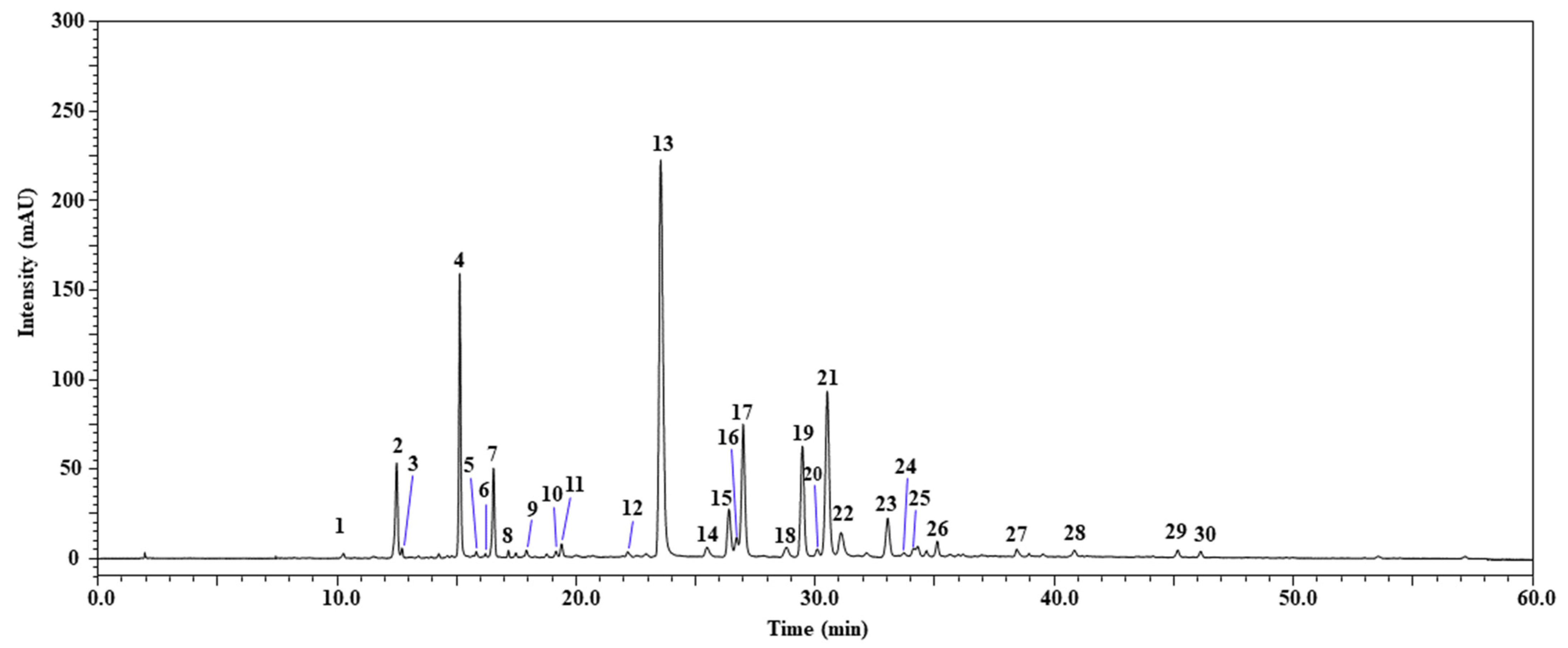
2.1. Identification of Phenolic Compounds by HPLC-PDA/ESI-MS

The HPLC-PDA/ESI-MS analysis of the phenolic compounds in the studied extracts of L. saligna aerial parts led to the detection of 30 compounds (Figure 1); 29 were positively identified according to retention times, λmax, mass spectrometry, and literature data, while 1 compound remained unknown (Table 1). The identified compounds were assigned to flavonoids (such as quercetin derivatives, apigenin derivatives, luteolin and its derivatives, genistein, and chrysoeriol) and phenolic acids (caffeic acid and its derivatives, caffeoylmalic acid, caffeoyltartaric acid, dicaffeoyltartaric acid isomers, caffeoylquinic acid isomers, 3-p-Coumaroylquinic acid, di-Hydroxybenzoic acid-hexoside, and Caffeoylferuloylquinic acid). Additionally, the results showed that the most abundant compounds were dicaffeoyltartaric acid (472.77 mg/kg of dried extract), followed by luteolin 7-glucoronide (224.30 mg/kg of dried extract), 3,5-di-O-caffeoylquinic acid (196.79 mg/kg of dried extract), and 5-caffeoylquinic acid (171.74 mg/kg of dried extract). To our knowledge, this study is the first to pinpoint phenolic compounds in L. saligna extract. Nevertheless, prior studies have detected these compounds within other species of the genus Lactuca. Ilgün et al. [24] studied methanolic latex extract from L. saligna in Turkey and showed its richness in Lactucin, with 13.94970 ± 0.24 mgstd/glatex. In addition, an Egyptian study revealed the presence of hexacosan-1ol, germanicol, taraxasterol, and moretenol in the aerial parts of L. saligna [25]. Hence, it is noted that little is known about the chemical composition of L. saligna extract.

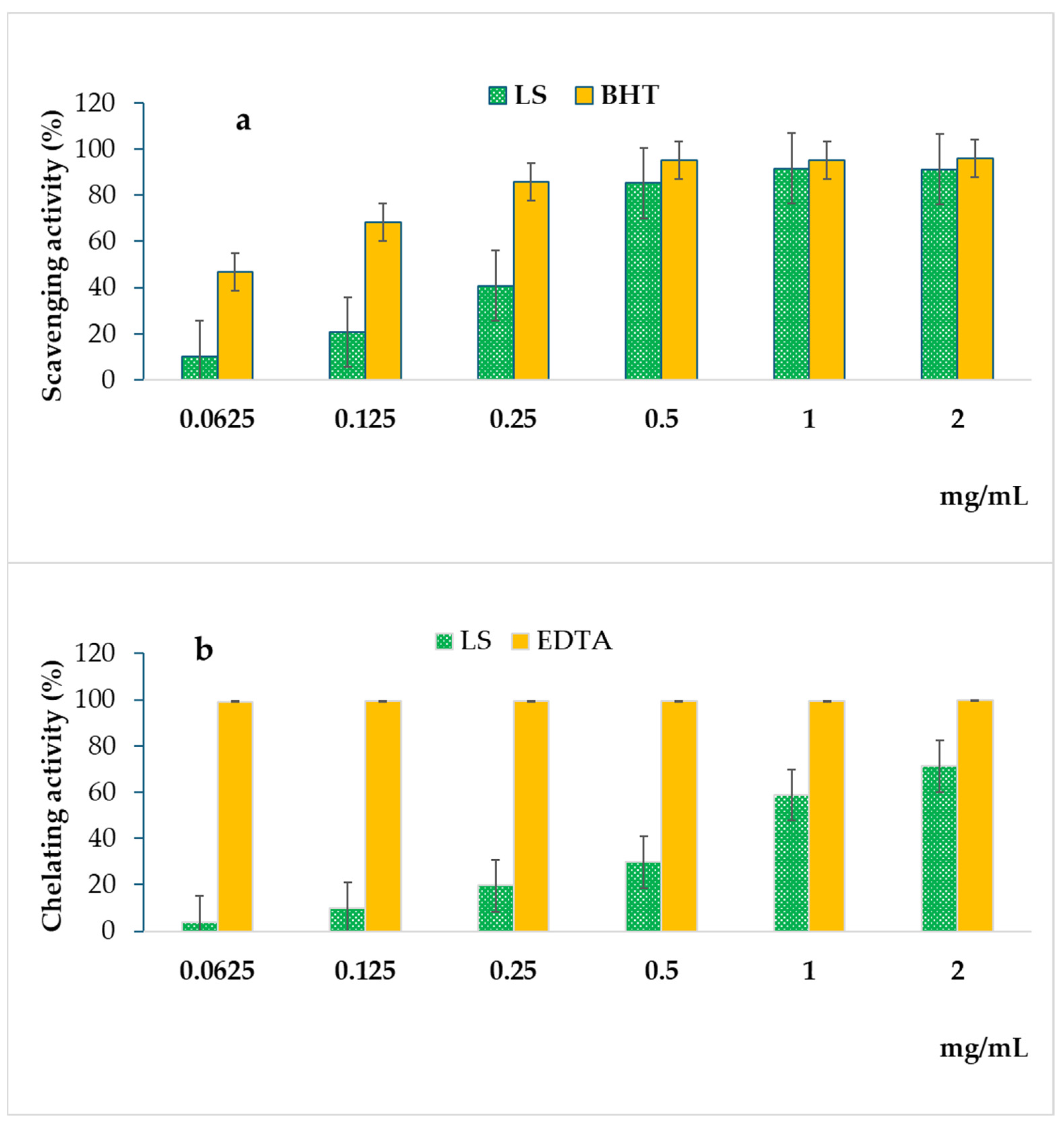
2.2. Antioxidant Activity

The antioxidant potential of extracts obtained from the aerial parts of L. saligna was determined through three distinct in vitro experimental methods, namely, DPPH scavenging activity, ferric reducing antioxidant power (FRAP) assay, and ferrous ion-chelating (FIC) assay. The use of various methods aimed to encompass diverse antioxidant mechanisms present in the plant extract. The obtained results are summarized in Figure 2 and Table 2.
In the DPPH assay, the results revealed a noteworthy radical scavenging activity exhibited by extract derived from L. saligna. This activity proved to be highly comparable to that of the standard antioxidant, BHT (butylated hydroxytoluene), particularly within the concentration range of 0.5 to 2 mg/mL (Figure 2a). The observed trend showcased a substantial efficacy of L. saligna in neutralizing the stable free radical DPPH, implying a potent capacity for mitigating oxidative stress. Quantitatively, the calculated IC50 values further underscored the remarkable performance of L. saligna extract in the DPPH assay. In this assay, L. saligna showed an interesting antioxidant capacity, with an IC50 of 0.2969 ± 0.012 mg/mL compared to the standard BHT with an IC50 of 0.0656 ± 0.008 mg/mL (p < 0.05) (Table 2). Additionally, the ferric reducing antioxidant power (FRAP) assay showed that L. saligna extract exhibits a remarkable antioxidant capacity of 13.952 ± 0.2477 ASE/mL compared to that found in the reference standard (BHT) with a value of 1.131 ± 0.037 ASE/mL (p < 0.05). Furthermore, the ferrous ion chelating activity assay showed that L. saligna extract has lower antioxidant activity, with an IC50 of 1.4219 ± 0.0034 mg/mL compared to the reference standard EDTA (ethylenediaminetetraacetic acid) with an IC50 of 0.0067 ± 0.0003 mg/mL (p < 0.05) (Figure 2b; Table 2). Consequently, these findings suggest that extract obtained from L. saligna holds considerable potential as a natural source of antioxidants, supporting its utilization as a promising candidate for developing nature-based antioxidants for various health and industrial purposes.
To our knowledge, no previous studies have assessed the antioxidant activity of L. saligna extracts, especially in Morocco. Notably, our results emphasize the importance of understanding the nuanced antioxidant mechanisms exhibited by plant extracts. While L. saligna demonstrated robust radical scavenging activity in the DPPH assay and remarkable performance in the FRAP assay, its performance in the ferrous ion chelating activity assay indicated a less pronounced ability to bind and sequester ferrous ions compared to the reference standard EDTA. Such findings contribute to a comprehensive understanding of the antioxidant profile of L. saligna extract, providing valuable information for potential applications and further exploration of its biological activities.
The noteworthy antioxidant activity of L. saligna extract can be linked to its chemical composition, notably to its phenolic compounds [32]. In this regard, previous studies have demonstrated the antioxidant activity of dicaffeoyltartaric acid [33], luteolin 7-glucuronide [34], 3,5-di-O-caffeoylquinic acid [35], and 5-caffeoylquinic acid [36], which were identified as major phenolic compounds in this study. In addition, flavonoids such as Quercetin derivatives, Apigenin derivatives, Genistein, and Chrysoeriol have shown effective antioxidant activity [37,38,39,40]. Nevertheless, endogenous phenolic compounds may act in synergy to enhance the overall antioxidant activity of extract.

2.3. Antibacterial Activity

The antibacterial activity of L. saligna extract was assessed against six bacterial strains, including Gram-negative and Gram-positive bacteria, by the determination of MIC and MBC values. The obtained results are summarized in Table 3. Our findings based on the determination of MIC values showed that L. saligna extract is more effective against Gram-positive bacteria (MIC values between 1.30 ± 0.31 and 5.20 ± 0.13 mg/mL) than Gram-negative bacteria (MIC values between 5.20 ± 0.19 and 10.41 ± 0.23 mg/mL). Moreover, the determination of MBC and the ratio between MBC and MIC showed that the studied extract had a bactericidal effect against Gram-positive bacteria and a bacteriostatic effect against Gram-negative bacteria (Table 3).
To our knowledge, no previous studies have assessed the antibacterial activity of L. saligna extract. Nonetheless, the reason behind it acting differently on Gram-positive and Gram-negative bacteria could stem from distinctions in the cellular structures of these bacteria. Gram-positive bacteria are characterized by a robust layer of peptidoglycan in their cell walls, whereas Gram-negative bacteria possess a thinner peptidoglycan layer in addition to an outer phospholipidic membrane. Similarly, previous studies have demonstrated the high antibacterial activity of plant extract and essential oils against Gram-positive compared to Gram-negative bacteria [8,41].
Notably, the antibacterial activity of plant extracts can be correlated to their chemical composition, with the presence of polyphenolic compounds being a key factor in this correlation [41,42]. In fact, the effective antibacterial activity of L. saligna extract against both Gram-positive and Gram-negative bacteria may be attributed to its richness in polyphenolic compounds, particularly those belonging to the classes of flavonoids and phenolic acids [43]. Bajko et al. showed that 5-O-caffeoylquinic acid has interesting antibacterial activity against both Gram-positive and Gram-negative bacteria, with MICs between 5 and 10 mg/mL [44]. In addition, studies have proven the antibacterial activity of quercetin, apigenin, luteolin, and caffeic acid and their derivative compounds [45,46,47,48]. Interestingly, the mode of action of plant compounds on bacteria is complex and may vary depending on different factors. Hence, it was reported that plant-derived compounds may induce cell wall and membrane rupture, leading to an increase in cellular permeability, the inhibition of proteins involved in septum formation, DNA segregation and cell division, the inhibition of the expression of respiratory chain complex proteins, intracellular ATP depletion, and the disruption of metabolic pathways [41]. Furthermore, plant compounds may exert simultaneous effects at various cellular sites, occasionally demonstrating a synergistic interaction between different compounds that enhances the antibacterial activity of plant extracts.

2.4. In Silico Analysis

2.4.1. Virtual Screening

In this study, four molecular compounds, including Apigenin 7-O-glucuronide, Quercetin-3-O-glucuronide, Chrysoeriol, and 3-p-Coumaroylquinic acid were found to be active against the six bacterial targets. The results of the virtual screening of phenolic compounds extracted from the aerial parts of L. saligna are presented in Table 4.

2.4.2. ADMET Analysis

Absorption, distribution, metabolism, excretion, and toxicity (ADMET) play a key role in drug discovery. In our study, the prediction of ADMET properties was performed using the admtsar and pKCSM web servers. The prediction of ADMET parameters is listed in Table 5. This method is very important for selecting the best molecules that we can use as drug candidates. We were interested in Apigenin 7-O-glucuronide, Quercetin-3-O-glucuronide, Chrysoeriol, and 3-p-Coumaroylquinic acid, the most potent inhibitors in the data set. As a result, we observed that these compounds had high calculated values for intestinal absorption—more than 80%, except for Chrysoeriol, which showed an intestinal absorption of less than 30%, which indicated low absorbance. This indicates that these molecules can be readily absorbed from the gut and circulate in the blood [49]. Distribution analysis showed that the molecules Apigenin 7-O-glucuronide, Quercetin-3-O-glucuronide, and 3-p-Coumaroylquinic acid are poorly distributed in the brain, with values below 1 [50]. Similarly, metabolizing enzymes are the focus of the main Phase I study in drug discovery. Cytochrome P450 (CYP) includes both substrate and inhibitory enzymes; the most important P450 cytochromes are CYP 2D6 and CYP 3A4 and they are involved in the metabolism of almost half of the drugs currently in use. The results obtained indicate that Apigenin 7-O-glucuronide, Quercetin-3-O-glucuronide, and 3-p-Coumaroylquinic acid are non-substrates and non-inhibitors of the CYP 2D6 enzyme. However, they are substrates and inhibitors of the CYP 3A4 enzyme.

2.4.3. Molecular Docking

Molecular docking techniques are generally applied to define the binding mechanisms between ligands and receptors. The target ligand is docked to the active site to verify the accuracy of the molecular docking. The molecules with the best virtual screening scores for the six bacteria were docked to the same active sites. Drugs with docking scores between (−98.32 kcal/mol, −102.63 kcal/mol, −88.32 kcal/mol) were selected as promising compounds for P. aeroginosa; (−100.1 kcal/mol, −92.16 kcal/mol, −71.21 kcal/mol) were selected for E. coli; (−88.25 kcal/mol, −93.48 kcal/mol, −78.18 kcal/mol) were selected for S. typhimurium; (−102.36 kcal/mol, −93.16 kcal/mol, −80.63 kcal/mol) were selected for S. aureus; (−86.25 kcal/mol, −85.47 kcal/mol, −69.22 kcal/mol) were selected for E. faecalis; (−89.03 kcal/mol, −98.62 kcal/mol, −66.61 kcal/mol) were selected for L. monocytogenes. The total energy hydrogen bonds (HBonds) and other interactions of the three selected compounds are shown in Table 3.
The results illustrated in Table 3 show that the compounds Quercetin-3-O-glucuronide, Apigenin 7-O-glucuronide, and 3-p Coumaroylquinic acid are stabilized in the pockets of receptors 1U1Z, 1FJ4, 6IE9, 3JOJ, 6QXS, and 1AOD by various interactions with very low binding affinities of (−7.6, −6.8, −6.5 kcal/mol), (−8.7, −7.9, −7.1 kcal/mol), (−8.4, −8.7, −7.7 kcal/mol), (−8.8, −9.1, −7.6 kcal/mol), (−7.8, −7.7, −7.7 kcal/mol), and (−7.4, −7.8, −6.8 kcal/mol), respectively. Predicted docking results for these three compounds are shown in Supplementary Figures S1–S6.
The molecular docking proved the antibacterial activity of hydro-methanolic extracts of L. saligna. Furthermore, the strong binding affinity exhibited by compounds such as Quercetin-3-O-glucuronide, Apigenin 7-O-glucuronide, and 3-p Coumaroylquinic acid to the specific proteins of the investigated bacteria positions them as promising candidates for the development of novel antibacterial drugs. Nevertheless, additional in vitro and in vivo studies are required.

3. Materials and Methods

3.1. Chemicals and Reagents

LC-MS grade methanol, formic acid, acetonitrile, and water were purchased from Merk Life Science (Merk KGaA, Darmstadt, Germany). 2,2 diphenyl-1 picrylhydrazyl (DPPH), ethylenediaminetetraacetic acid (EDTA), butylated hydroxytoluene (BHT), potassium ferrycyanide [K3Fe (CN)6], ferric chloride (FeCl3), and trichloroacetic acid were supplied by Carlo Erba (Milan, Italy).

3.2. Plant Materials and Phenolic Compound Extraction

The aerial parts of L. saligna were harvested in June 2022 from the middle Atlas of Morocco (Ifrane region). The collected plant was identified by the botanist Professor Rahou Abdelilah from the Faculty of Sciences of Meknes, Moulay Ismail University, and confirmed by Professor Ibn Tattou Mohammed at the Scientific Institute of Rabat (Morocco). The plant sample was protected from light and dried at room temperature for 15 days; then, it was crushed and stored at +4 °C until use. The extraction of phenolic compounds was carried out according to our previously published protocol [8].

3.3. Polyphenolic Compound Analysis by HPLC-PDA/ESI-MS

The analysis of polyphenolic compounds was performed using high-performance liquid chromatography coupled with a photodiode array detector and electrospray ionization-mass spectrometry (HPLC-PDA-ESI/MS) (Shimadzu, Kyoto, Japan). Chromatographic separation was carried out on an Ascentis Express C18 column (150 × 4.6 mm, 2.7 µm; Merck Life Science, Merck KGaA, Darmstadt, Germany) using as a mobile phase 0.1% (v/v) acid formic in water (mobile phase A) and 0.1% (v/v) acid formic in acetonitrile (mobile phase B). The gradient elution applied was as follows: 0–5 min (5% B), 5–15 min (10% B), 15–30 min (20% B), 30–60 min (50% B), and 60 min (100% B) at a flow rate of 0.8 mL/min. The column temperature was 30 °C and the injection volume was 5 µL. UV detection wavelengths were in the range of λ = 100–400 nm. Negative-ion mass spectra were set as follows: scan range, m/z 100–800; nebulizing gas (N2) flow rate, 1.5 L/min; drying gas (N2) flow rate, 15 L/min; interface temperature, 350 °C. LabSolutions software ver. 5.92 (Shimadzu, Kyoto, Japan) was used to control the LC-PDA-ESI-MS system and the data processing. The identification of phenolic compounds was performed by a comparison of retention times and UV-visible and mass spectra of unknown peaks with the literature data.

3.4. Antioxidant Activity

The determination of the antioxidant activity of L. saligna extract was performed based on three different mechanisms, including radical scavenging of DPPH, reducing power (FRAP), and ferrous ions chelating capacity. BHT and EDTA were used as standards for this study.

3.4.1. DPPH Assay

The DPPH scavenging activity of L. saligna extract was assessed according to the method described previously [8]. Briefly, 0.5 mL of each concentration of plant extract (0.0625–2 mg/mL) and the standard BHT were mixed with 3 mL of a 0.1 mM methanol DPPH solution and stored in the dark for 20 min. After that, the absorbance of the mixture was determined at 517 nm using a spectrophotometer (UV-1601, Shimadzu). Then, the percentage (%) of radical scavenging activity was calculated using the following formula:
Radical scavenging activity percentage (%) = ((A0 − Ac)/A0) × 100
where A0 is the DPPH absorbance without the sample and Ac is the absorbance in the presence of the sample or standard. The IC50 values were calculated as the concentration of extract causing a 50% inhibition of DPPH radical; a lower IC50 value corresponded to a higher antioxidant activity of extracts. The experiments were performed in triplicates.

3.4.2. FRAP Assay

The ability to convert Fe3+ to Fe2+ was used to assess the ferric reducing capacity of L. saligna extract, according to the previously described method [51], with few modifications. Briefly, a mixture containing 2.5 mL of phosphate buffer (0.2 M, pH 6.6) and 2.5 mL of 1% potassium ferrycyanide [K3Fe (CN)6] was prepared and then 1 mL of each sample concentration (0.0625–2 mg/mL) was added. The obtained mixture was incubated at 50 °C for 20 min and then 2.5 mL of 10% trichloroacetic acid was added before the mixture was centrifuged at 3000 rpm for 10 min. Afterward, 2.5 mL of the supernatant was mixed with 2.5 mL of distilled water and 0.5 mL of 0.1% ferric chloride (FeCl3) and incubated in the dark for 10 min before measuring the absorbance at 700 nm. The experiments were carried out in triplicates and the obtained results were presented as mean absorbance values ± standard deviation (SD) and acid equivalent (ASE/mL) ± SD.

3.4.3. Ferrous Ions (Fe2+) Chelating Activity

The inhibition of Fe2+–ferrozine complex formation was used to measure the ferrous ion Fe2+ chelating activity of L. saligna extract according to the method described previously [8]. Briefly, 1 mL of each concentration of extract (0.0625–2 mg/mL) was mixed with 0.5 mL of methanol and 0.05 mL of 2 mM FeCl2; then, the reaction was started by adding 0.1 mL of 5 mM ferrozine and the mixture was incubated in the dark for 10 min at room temperature before measuring the absorbance at 562 nm. EDTA was used as the standard for this assay. The inhibition of ferrozine–(Fe2+) complex formation percentage was determined according to the following formula:
Inhibition of ferrozine–Fe2+) complex formation (%) = ((A0 − Ac)/A0) × 100
where A0 is the absorbance of the control and Ac is the absorbance in the presence of the sample or standard. The IC50 was determined as mean ± SD.

3.5. Antibacterial Activity

3.5.1. Bacterial Strains and Culture

To assess the antibacterial activity of L. saligna extract, six bacterial species were used in this study, including Gram-negative bacteria—Escherichia coli, Salmonella typhimurium, and Pseudomonas aeruginosa—and Gram-positive bacteria—Enterococcus faecalis, Staphylococcus aureus, and Listeria monocytogenes. Bacterial strains were prepared by sub-culturing a loopful from the frozen stock culture (−80 °C) on Tryptone Soy Yeast Extract Agar (TSYEA; Biolife, Milan, Italy) followed by incubation at 37 °C for 24 h.

3.5.2. The Determination of MIC and MBC

The minimum inhibitory concentrations (CMIs) and minimum bactericidal concentrations (MBCs) were assessed by microdilution assay [51]. In flat-bottom 96-well microplates, the first column wells were used to prepare a mixture of 50 μL of sterile, distilled water and 50 μL of extract (500 mg/mL). From the first well plate, a series of two-fold dilutions were prepared in sterile, distilled water. Afterward, 50 μL of Tryptone Soy Yeast Extract Broth and 50 μL of bacterium suspensions (108 cfu/mL) were added to each well. The positive control contained Tryptone Soy Yeast Extract Broth and a bacterial suspension while the negative control contained Tryptone Soy Yeast Extract Broth without a bacterial suspension. The microplates were incubated at 37 °C for 24 h and then 40 μL of TTC (2,3,5-triphenyl tetrazolium chloride) was added to each well and re-incubated at 37 °C for 30 min. The lowest concentration of extract that did not show bacterial growth was considered as MIC. However, the determination of MBC was performed by sub-culturing 5 μL of wells that did not show bacterial growth on the Tryptone Soy Yeast Extract Agar followed by incubation at 37 °C for 24 h, and the concentrations that did not show any growth of colonies on the media were considered as MBC. Furthermore, the bacteriostatic or bactericidal effect of the extract was determined by calculating the MBC/MIC ratio; if this ratio was below 4, the effect was bactericidal, and if it was greater than 4, the effect was bacteriostatic [52].

3.6. In Silico Analysis

3.6.1. Compounds and Bioinformatics Tools

The phenolic compounds identified in L. saligna extract by HPLC-PDA/ESI-MS were used for this analysis. The molecular structure of each compound was retrieved from the PubChem database (https://pubchem.ncbi.nlm.nih.gov/, accessed on 9 November 2023), and the optimized compounds were prepared by converting them to pdb format and submitting them in .pdbqt format using Autodock software (http://vina.scripps.edu/, accessed on 9 November 2023). Moreover, RCSB PDB (https://www.rcsb.org/, accessed on 10 November 2023), PyMOL (https://pymol.org/2/, accessed on 15 November 2023), and Discovery Studio (https://www.3ds.com/products/biovia/discovery-studio, accessed on 16 November 2023), were used in this study.

3.6.2. Protein Preparation and Active Site Prediction

The receptors,—(FabZ) (3R)-hydroxyacylacyl carrier protein dehydratase (FabZ) (PDB ID: 1U1Z) for P. aeruginosa [53], thymidylate synthase (EfTS) (PDB ID: 6QXS) for E. faecalis [54], beta-ketoacyl-[acyl carrier protein] synthase (PDB ID: 1fj4) for E. coli [55], phosphatidylinositol-specific phospholipase C (PDB ID: 1AOD) for L. monocytogenes [56], RamR PDB (6IE9) for S. typhimurium [57], and the UDP-N-acetyl-mannosamine dehydrogenase Cap5O (PDB ID: 3JOL) for S. aureus [58]—were retrieved from the protein database (PDB) and processed by removing water molecules and any small molecules loaded with the target receptor using PyMOL software, then submitted to autodock tool software to locate the active site in the processed receptor and convert it to (.pdbqt) format by autodock vina for docking calculations.

3.6.3. Structure-Based Virtual Screening

The software iGEMDOCK (Generic Evolution Method for Docking) version 2.1 was used to perform high-speed virtual screening. In silico screening of 29 identified phenolic compounds in L. saligna extract was performed using PDB codes (ID: 1U1Z) for P. aeruginosa, (ID: 6QXS) for E. faecalis, (ID: 1fj4) for E. coli, (ID: 1AOD) for L. monocytogenes, (ID: 6IE9) for S. typhimurium, and (ID: 3JOL) for S. aureus. The screening score, which was calculated from the total energy calculations (Total energy = VdW + HBond + electrostatic), was calculated using iGEMDOCK v2.1.11. The standard parameters used for screening, namely, population size, generations, and number of solutions, were set to 300, 70, and 2, respectively. The energy-based results were analyzed, and 4 potential inhibitors were selected based on their stability for more detailed analyses.

3.6.4. ADMET Analysis

As part of the process of developing a new antimicrobial drug, it is essential to assess the pharmacologically active substance, and this assessment was carried out in silico using ADMET (absorption, distribution, metabolism, excretion, and toxicity) analysis; ADMET parameters were calculated using admetASR 2.0 and pkCSM (https://biosig.lab.uq.edu.au/pkcsm/ accessed on 20 November 2023).

3.6.5. Molecular Docking

The anchoring process involved extracting the co-crystallized reference ligand and water molecules from the crystal structure. Polar hydrogen atoms were added. Throughout the anchoring process, the protein was maintained in a rigid state, while the ligand was allowed to be extremely flexible. The ligands (Apigenin 7-O-glucuronide, Quercetin-3-O-glucuronide, 3-p-Coumaroylquinic) with the best pharmacokinetic properties and virtual screening results were anchored to ((3R)-hydroxyacylacyl) carrier protein dehydratase for P. aeruginosa (PDB ID: 1U1Z), thymidylate synthase (EfTS) (PDB ID: 6QXS) for E. faecalis, beta-ketoacyl-[acyl carrier protein] synthase (PDB ID: 1fj4) for E. coli, UDP-N-acetyl-mannosamine dehydrogenase Cap5O (PDB ID: 3JOL) for S. aureus, and phosphatidylinositol-specific phospholipase C (PDB 1AOD) for L. monocytogenes (Figures S1–S6). Autodock vina was used to generate binding positions for the bioactive ligands in the active sites of the six targets. Once docking was complete, ligand placement was used to obtain the minimum binding energy. Discovery Studio and PyMOL were used to visualize the results. The type of interactions established by each molecule in the active sites was also compared.

3.7. Statistical Analysis

Statistical analysis was performed using SPSS software (SPSS version 22, IBM Corp, Armonk, NY, USA). Data were subjected to an analysis of variance, followed by Duncan’s New Multiple Range Test (DMRT). All experiments were performed in triplicate and the differences were considered significant at p < 0.05.

4. Conclusions

This study reported, for the first time, the polyphenolic compound composition and antioxidant and antibacterial activities of aerial-part extracts of Moroccan L. saligna. Interestingly, L. saligna can be considered an interesting source of secondary metabolites, where HPLC-PDA/ESI-MS identified 29 among 30 detected compounds. Additionally, hydro-methanolic extracts of L. saligna exhibited high antioxidant activity and remarkable antibacterial activity against both Gram-positive and Gram-negative bacteria. Furthermore, in silico analysis showed that among the 29 identified compounds, Apigenin 7-O-glucuronide, Quercetin-3-O-glucuronide, and 3-p-Coumaroylquinic acid exhibit high binding affinity scores and establish stable interactions. Consequently, our study showed that L. saligna is a potent source of biomolecules and could be used as a sustainable source for developing drugs with interesting antioxidant and antibacterial activities.

Supplementary Materials

The following supporting information can be downloaded at https://www.mdpi.com/article/10.3390/molecules29030596/s1. Figure S1. 2D view of the conformations of different interactions between the three inhibitors in the 1U1Z active site; Figure S2. 2D view of the conformations of different interactions between the three inhibitors in the 1FJ4 active site; Figure S3. 2D view of the conformations of different interactions between the three inhibitors in the 6IE9 active site; Figure S4. 2D view of the conformations of different interactions between the three inhibitors in the 3JOL active site; Figure S5. 2D view of the conformations of different interactions between the three inhibitors in the 6QXS active site; Figure S6. 2D view of the conformations of different interactions between the three inhibitors in the 1AOD active site.

Author Contributions

Conceptualization, F.R.F. and F.C.; methodology, A.B. and F.C.; software, A.B., S.M., T.B. and Y.O.E.M.; validation, A.B., A.A.B. and K.S.; investigation, A.B., N.M. and P.O.; resources, F.C.; data curation, F.C.; writing—original draft preparation, A.B., S.M., A.E.-D. and S.C.; writing—review and editing, F.C., K.S. and A.B.A.; supervision, F.C. and F.R.F.; project administration, F.C. All authors have read and agreed to the published version of the manuscript.

Funding

This research received no external funding.

Institutional Review Board Statement

Not applicable.

Informed Consent Statement

Not applicable.

Data Availability Statement

Data are contained within the article and Supplementary Materials.

Acknowledgments

The authors are thankful to Shimadzu and Merck Life Science Corporations for their continuous support.

Conflicts of Interest

The authors declare no conflicts of interest.

References

  1. Sen, T.; Samanta, S.K. Medicinal Plants, Human Health and Biodiversity: A Broad Review. Adv. Biochem. Eng. Biotechnol. 2014, 147, 59–110. [Google Scholar] [CrossRef]
  2. Jamila, F.; Mostafa, E. Ethnobotanical Survey of Medicinal Plants Used by People in Oriental Morocco to Manage Various Ailments. J. Ethnopharmacol. 2014, 154, 76–87. [Google Scholar] [CrossRef]
  3. Chroho, M.; Aazza, M.; Bouymajane, A.; El Majdoub, Y.O.; Cacciola, F.; Mondello, L.; Zair, T.; Bouissane, L. HPLC-PDA/ESI-MS Analysis of Phenolic Compounds and Bioactivities of the Ethanolic Extract from Flowers of Moroccan Anacyclus clavatus. Plants 2022, 11, 3423. [Google Scholar] [CrossRef]
  4. Chlif, N.; Bouymajane, A.; Oulad El Majdoub, Y.; Diouri, M.; Rhazi Filali, F.; Bentayeb, A.; Altemimi, A.B.; Mondello, L.; Cacciola, F. Phenolic Compounds, in Vivo Anti-Inflammatory, Analgesic and Antipyretic Activities of the Aqueous Extracts from Fresh and Dry Aerial Parts of Brocchia cinerea (Vis.). J. Pharm. Biomed. Anal. 2022, 213, 114695. [Google Scholar] [CrossRef] [PubMed]
  5. Lebeda, A.; Dolezalová, I.; Feráková, V.; Astley, D. Geographical Distribution of Wild Lactuca Species (Asteraceae, Lactuceae). Bot. Rev. 2004, 70, 328–356. [Google Scholar] [CrossRef]
  6. Lebeda, A.; Křístková, E.; Kitner, M.; Mieslerová, B.; Pink, D.A. Wild Lactuca Saligna: A Rich Source of Variation for Lettuce Breeding. In Enhancing Crop Genepool Use: Capturing Wild Relative and Landrace Diversity for Crop Improveme; CABI: Wallingford, UK, 2016; pp. 32–46. [Google Scholar]
  7. Khan, M.I.R.; Khan, N.A. Reactive Oxygen Species and Antioxidant Systems in Plants: Role and Regulation under Abiotic Stress; Springer: Singapore, 2017; ISBN 9789811052545. [Google Scholar]
  8. Bouymajane, A.; Rhazi Filali, F.; Oulad El Majdoub, Y.; Ouadik, M.; Abdelilah, R.; Cavò, E.; Miceli, N.; Taviano, M.F.; Mondello, L.; Cacciola, F. Phenolic Compounds, Antioxidant and Antibacterial Activities of Extracts from Aerial Parts of Thymus zygis Subsp. gracilis, Mentha suaveolens and Sideritis incana from Morocco. Chem. Biodivers. 2022, 19, e202101018. [Google Scholar] [CrossRef]
  9. Souza, M.P.; de Lima, B.R.; Sá, I.S.C.; de Freitas, F.A. Bioactive Compounds Isolated from Amazonian Fruits and Their Possible Applications. Stud. Nat. Prod. Chem. 2023, 79, 205–239. [Google Scholar] [CrossRef]
  10. Mehta, S.K.; Gowder, S.J.T.; Mehta, S.K.; Gowder, S.J.T. Members of Antioxidant Machinery and Their Functions. In Basic Principles and Clinical Significance of Oxidative Stress; IntechOpen: River, Croatia, 2015; ISBN 978-953-51-2200-5. [Google Scholar]
  11. Ndhlala, A.R.; Moyo, M.; Van Staden, J. Natural Antioxidants: Fascinating or Mythical Biomolecules? Molecules 2010, 15, 6905. [Google Scholar] [CrossRef]
  12. Moussa, Z.; Judeh, Z.M.A.; Ahmed, S.A.; Moussa, Z.; Judeh, Z.M.A.; Ahmed, S.A. Nonenzymatic Exogenous and Endogenous Antioxidants. In Free Radical Medicine and Biology; IntechOpen: River, Croatia, 2019; ISBN 978-1-78985-144-1. [Google Scholar]
  13. Pedro, A.C.; Paniz, O.G.; Fernandes, I.d.A.A.; Bortolini, D.G.; Rubio, F.T.V.; Haminiuk, C.W.I.; Maciel, G.M.; Magalhães, W.L.E. The Importance of Antioxidant Biomaterials in Human Health and Technological Innovation: A Review. Antioxidants 2022, 11, 1644. [Google Scholar] [CrossRef] [PubMed]
  14. Brewer, M.S. Natural Antioxidants: Sources, Compounds, Mechanisms of Action, and Potential Applications. Compr. Rev. Food Sci. Food Saf. 2011, 10, 221–247. [Google Scholar] [CrossRef]
  15. Levy, S.B.; Bonnie, M. Antibacterial Resistance Worldwide: Causes, Challenges and Responses. Nat. Med. 2004, 10, S122–S129. [Google Scholar] [CrossRef]
  16. Khameneh, B.; Iranshahy, M.; Soheili, V.; Fazly Bazzaz, B.S. Review on Plant Antimicrobials: A Mechanistic Viewpoint. Antimicrob. Resist. Infect. Contr. 2019, 8, 1–28. [Google Scholar] [CrossRef]
  17. Anand, U.; Jacobo-Herrera, N.; Altemimi, A.; Lakhssassi, N. A Comprehensive Review on Medicinal Plants as Antimicrobial Therapeutics: Potential Avenues of Biocompatible Drug Discovery. Metabolites 2019, 9, 258. [Google Scholar] [CrossRef]
  18. Brown, E.D.; Wright, G.D. Antibacterial Drug Discovery in the Resistance Era. Nature 2016, 529, 336–343. [Google Scholar] [CrossRef]
  19. Cheynier, V.; Tomas-Barberan, F.A.; Yoshida, K. Polyphenols: From Plants to a Variety of Food and Nonfood Uses. J. Agric. Food Chem. 2015, 63, 7589–7594. [Google Scholar] [CrossRef] [PubMed]
  20. Sadiki, F.Z.; Bouymajane, A.; Sbiti, M.; Channaoui, S.; Micalizzi, G.; Cacciola, F.; Dugo, P.; Mondello, L.; El Idrissi, M. Chemical Profile, Antibacterial, Antioxidant and Insecticidal Properties of the Essential Oil from Tetraclinis articulata (Vahl) Masters Cones. J. Essent. Oil Res. 2022, 34, 383–393. [Google Scholar] [CrossRef]
  21. Ji, M.; Gong, X.; Li, X.; Wang, C.; Li, M. Advanced Research on the Antioxidant Activity and Mechanism of Polyphenols from Hippophae Species—A Review. Molecules 2020, 25, 917. [Google Scholar] [CrossRef] [PubMed]
  22. Kim, H.S.; Quon, M.J.; Kim, J.-A. New Insights into the Mechanisms of Polyphenols beyond Antioxidant Properties; Lessons from the Green Tea Polyphenol, Epigallocatechin 3-Gallate. Redox Biol. 2014, 2, 187–195. [Google Scholar] [CrossRef] [PubMed]
  23. Shaker, B.; Ahmad, S.; Lee, J.; Jung, C.; Na, D. In Silico Methods and Tools for Drug Discovery. Comput. Biol. Med. 2021, 137, 104851. [Google Scholar] [CrossRef] [PubMed]
  24. Ilgün, S.; Akkol, E.K.; Ilhan, M.; Polat, D.Ç.; Kılıç, A.B.; Coşkun, M.; Sobarzo-Sánchez, E. Sedative Effects of Latexes Obtained from Some Lactuca L. Species Growing in Turkey. Molecules 2020, 25, 1587. [Google Scholar] [CrossRef] [PubMed]
  25. El-Fattah, H.A.; Khalil, A.T.; Mansour, E.S.; Waight, E.S. Triterpenes and Triterpene Esters from Lactuca saligna. Int. J. Pharmacogn. 1992, 30, 21–26. [Google Scholar] [CrossRef]
  26. Viacava, G.E.; Roura, S.I.; Berrueta, L.A.; Iriondo, C.; Gallo, B.; Alonso-Salces, R.M. Characterization of Phenolic Compounds in Green and Red Oak-Leaf Lettuce Cultivars by UHPLC-DAD-ESI-QToF/MS Using MSE Scan Mode. J. Mass. Spectrom. 2017, 52, 873–902. [Google Scholar] [CrossRef] [PubMed]
  27. Ribas-Agustí, A.; Gratacós-Cubarsí, M.; Sárraga, C.; García-Regueiro, J.A.; Castellari, M. Analysis of Eleven Phenolic Compounds Including Novel P-Coumaroyl Derivatives in Lettuce (Lactuca sativa L.) by Ultra-High-Performance Liquid Chromatography with Photodiode Array and Mass Spectrometry Detection. Phytochem. Anal. 2011, 22, 555–563. [Google Scholar] [CrossRef] [PubMed]
  28. Abu-Reidah, I.M.; Contreras, M.M.; Arráez-Román, D.; Segura-Carretero, A.; Fernández-Gutiérrez, A. Reversed-Phase Ultra-High-Performance Liquid Chromatography Coupled to Electrospray Ionization-Quadrupole-Time-of-Flight Mass Spectrometry as a Powerful Tool for Metabolic Profiling of Vegetables: Lactuca Sativa as an Example of Its Application. J. Chromatogr. A 2013, 1313, 212–227. [Google Scholar] [CrossRef]
  29. Pepe, G.; Sommella, E.; Manfra, M.; De Nisco, M.; Tenore, G.C.; Scopa, A.; Sofo, A.; Marzocco, S.; Adesso, S.; Novellino, T.; et al. Evaluation of Anti-Inflammatory Activity and Fast UHPLC–DAD–IT-TOF Profiling of Polyphenolic Compounds Extracted from Green Lettuce (Lactuca sativa L.; Var. Maravilla de Verano). Food Chem. 2015, 167, 153–161. [Google Scholar] [CrossRef]
  30. Jeong, S.W.; Kim, G.S.; Lee, W.S.; Kim, Y.H.; Kang, N.J.; Jin, J.S.; Lee, G.M.; Kim, S.T.; Abd El-Aty, A.M.; Shim, J.H.; et al. The Effects of Different Night-Time Temperatures and Cultivation Durations on the Polyphenolic Contents of Lettuce: Application of Principal Component Analysis. J. Adv. Res. 2015, 6, 493–499. [Google Scholar] [CrossRef]
  31. Qin, X.X.; Zhang, M.Y.; Han, Y.Y.; Hao, J.H.; Liu, C.J.; Fan, S.X. Beneficial Phytochemicals with Anti-Tumor Potential Revealed through Metabolic Profiling of New Red Pigmented Lettuces (Lactuca sativa L.). Int. J. Mol. Sci. 2018, 19, 1165. [Google Scholar] [CrossRef]
  32. Kalisz, S.; Oszmiański, J.; Kolniak-Ostek, J.; Grobelna, A.; Kieliszek, M.; Cendrowski, A. Effect of a Variety of Polyphenols Compounds and Antioxidant Properties of Rhubarb (Rheum rhabarbarum). LWT 2020, 118, 108775. [Google Scholar] [CrossRef]
  33. Liu, G.; Zhu, W.; Li, S.; Zhou, W.; Zhang, H.; Wang, J.; Liu, X.; Zhang, J.; Liang, L.; Xu, X. Antioxidant Capacity and Interaction of Endogenous Phenolic Compounds from Tea Seed Oil. Food Chem. 2022, 376, 131940. [Google Scholar] [CrossRef]
  34. Ng, T.B.; Liu, F.; Wang, Z.T. Antioxidative Activity of Natural Products from Plants. Life Sci. 2000, 66, 709–723. [Google Scholar] [CrossRef]
  35. Makori, S.I.; Mu, T.H.; Sun, H.N. Physicochemical Properties, Antioxidant Activities, and Binding Behavior of 3,5-Di-O-Caffeoylquinic Acid with Beta-Lactoglobulin Colloidal Particles. Food Chem. 2021, 347, 129084. [Google Scholar] [CrossRef]
  36. Chen, X.Q.; Yang, J.H.; Cho, S.S.; Kim, J.H.; Xu, J.Q.; Seo, K.; Ki, S.H. 5-Caffeoylquinic Acid Ameliorates Oxidative Stress-Mediated Cell Death via Nrf2 Activation in Hepatocytes. Pharm. Biol. 2020, 58, 999–1005. [Google Scholar] [CrossRef]
  37. Mishra, B.; Priyadarsini, K.I.; Kumar, M.S.; Unnikrishnan, M.K.; Mohan, H. Effect of O-Glycosilation on the Antioxidant Activity and Free Radical Reactions of a Plant Flavonoid, Chrysoeriol. Bioorg Med. Chem. 2003, 11, 2677–2685. [Google Scholar] [CrossRef]
  38. Lesjak, M.; Beara, I.; Simin, N.; Pintać, D.; Majkić, T.; Bekvalac, K.; Orčić, D.; Mimica-Dukić, N. Antioxidant and Anti-Inflammatory Activities of Quercetin and Its Derivatives. J. Funct. Foods 2018, 40, 68–75. [Google Scholar] [CrossRef]
  39. Ganai, A.A.; Farooqi, H. Bioactivity of Genistein: A Review of In Vitro and In Vivo Studies. Biomed. Pharmacother. 2015, 76, 30–38. [Google Scholar] [CrossRef]
  40. Zhao, Y.; Zhang, X.; Zhang, N.; Zhou, Q.; Fan, D.; Wang, M. Lipophilized Apigenin Derivatives Produced during the Frying Process as Novel Antioxidants. Food Chem. 2022, 379, 132178. [Google Scholar] [CrossRef] [PubMed]
  41. Álvarez-Martínez, F.J.; Barrajón-Catalán, E.; Herranz-López, M.; Micol, V. Antibacterial Plant Compounds, Extracts and Essential Oils: An Updated Review on Their Effects and Putative Mechanisms of Action. Phytomedicine 2021, 90, 153626. [Google Scholar] [CrossRef] [PubMed]
  42. Efenberger-Szmechtyk, M.; Nowak, A.; Czyzowska, A. Plant Extracts Rich in Polyphenols: Antibacterial Agents and Natural Preservatives for Meat and Meat Products. Crit. Rev. Food Sci. Nutr. 2021, 61, 149–178. [Google Scholar] [CrossRef]
  43. Farhadi, F.; Khameneh, B.; Iranshahi, M.; Iranshahy, M. Antibacterial Activity of Flavonoids and Their Structure–Activity Relationship: An Update Review. Phytother. Res. 2019, 33, 13–40. [Google Scholar] [CrossRef] [PubMed]
  44. Bajko, E.; Kalinowska, M.; Borowski, P.; Siergiejczyk, L.; Lewandowski, W. 5-O-Caffeoylquinic Acid: A Spectroscopic Study and Biological Screening for Antimicrobial Activity. LWT-Food Sci. Technol. 2016, 65, 471–479. [Google Scholar] [CrossRef]
  45. Alizadeh, S.R.; Ebrahimzadeh, M.A. Quercetin Derivatives: Drug Design, Development, and Biological Activities, a Review. Eur. J. Med. Chem. 2022, 229, 114068. [Google Scholar] [CrossRef]
  46. Liu, R.; Zhang, H.; Yuan, M.; Zhou, J.; Tu, Q.; Liu, J.J.; Wang, J. Synthesis and Biological Evaluation of Apigenin Derivatives as Antibacterial and Antiproliferative Agents. Molecules 2013, 18, 11496–11511. [Google Scholar] [CrossRef]
  47. Lv, P.C.; Li, H.Q.; Xue, J.Y.; Shi, L.; Zhu, H.L. Synthesis and Biological Evaluation of Novel Luteolin Derivatives as Antibacterial Agents. Eur. J. Med. Chem. 2009, 44, 908–914. [Google Scholar] [CrossRef]
  48. Khan, F.; Bamunuarachchi, N.I.; Tabassum, N.; Kim, Y.M. Caffeic Acid and Its Derivatives: Antimicrobial Drugs toward Microbial Pathogens. J. Agric. Food Chem. 2021, 69, 2979–3004. [Google Scholar] [CrossRef] [PubMed]
  49. Norinder, U.; Bergström, C.A.S. Prediction of ADMET Properties. ChemMedChem 2006, 1, 920–937. [Google Scholar] [CrossRef]
  50. En-Nahli, F.; Hajji, H.; Ouabane, M.; Aziz Ajana, M.; Sekatte, C.; Lakhlifi, T.; Bouachrine, M. ADMET Profiling and Molecular Docking of Pyrazole and Pyrazolines Derivatives as Antimicrobial Agents. Arab. J. Chem. 2023, 16, 105262. [Google Scholar] [CrossRef]
  51. Ed-Dra, A.; Filai, F.R.; Bou-Idra, M.; Zekkori, B.; Bouymajane, A.; Moukrad, N.; Benhallam, F.; Bentayeb, A. Application of Mentha Suaveolens Essential Oil as an Antimicrobial Agent in Fresh Turkey Sausages. J. Appl. Biol. Biotechnol. 2018, 6, 7–12. [Google Scholar] [CrossRef]
  52. Bouymajane, A.; Filali, F.R.; Ed-Dra, A.; Aazza, M.; Nalbone, L.; Giarratana, F.; Alibrando, F.; Miceli, N.; Mondello, L.; Cacciola, F. Chemical Profile, Antibacterial, Antioxidant, and Anisakicidal Activities of Thymus zygis subsp. gracilis Essential Oil and Its Effect against Listeria monocytogenes. Int. J. Food Microbiol. 2022, 383, 109960. [Google Scholar] [CrossRef]
  53. Kimber, M.S.; Martin, F.; Lu, Y.; Houston, S.; Vedadi, M.; Dharamsi, A.; Fiebig, K.M.; Schmid, M.; Rock, C.O. The Structure of (3R)-Hydroxyacyl-Acyl Carrier Protein Dehydratase (FabZ) from Pseudomonas aeruginosa. J. Biol. Chem. 2004, 279, 52593–52602. [Google Scholar] [CrossRef] [PubMed]
  54. Pozzi, C.; Ferrari, S.; Luciani, R.; Tassone, G.; Costi, M.P.; Mangani, S. Structural Comparison of Enterococcus faecalis and Human Thymidylate Synthase Complexes with the Substrate DUMP and Its Analogue FdUMP Provides Hints about Enzyme Conformational Variabilities. Molecules 2019, 24, 1257. [Google Scholar] [CrossRef]
  55. Price, A.C.; Choi, K.H.; Heath, R.J.; Li, Z.; White, S.W.; Rock, C.O. Inhibition of β-Ketoacyl-Acyl Carrier Protein Synthases by Thiolactomycin and Cerulenin: Structure and Mechanism. J. Biol. Chem. 2001, 276, 6551–6559. [Google Scholar] [CrossRef] [PubMed]
  56. Moser, J.; Gerstel, B.; Meyer, J.E.W.; Chakraborty, T.; Wehland, J.; Heinz, D.W. Crystal Structure of the Phosphatidylinositol-Specific Phospholipase C from the Human Pathogen Listeria monocytogenes. J. Mol. Biol. 1997, 273, 269–282. [Google Scholar] [CrossRef]
  57. Yamasaki, S.; Nakashima, R.; Sakurai, K.; Baucheron, S.; Giraud, E.; Doublet, B.; Cloeckaert, A.; Nishino, K. Crystal Structure of the Multidrug Resistance Regulator RamR Complexed with Bile Acids. Sci. Rep. 2019, 9, 177. [Google Scholar] [CrossRef]
  58. Gruszczyk, J.; Fleurie, A.; Olivares-Illana, V.; Béchet, E.; Zanella-Cleon, I.; Moréra, S.; Meyer, P.; Pompidor, G.; Kahn, R.; Grangeasse, C.; et al. Structure Analysis of the Staphylococcus aureus UDP-N-Acetyl-Mannosamine Dehydrogenase Cap5O Involved in Capsular Polysaccharide Biosynthesis. J. Biol. Chem. 2011, 286, 17112–17121. [Google Scholar] [CrossRef]
Figure 1. Characterization of phenolic compounds in hydro-methanolic aerial part extract of L. saligna acquired at 330 nm using HPLC-PDA/ESI-MS. Peak identification as in Table 1.
Molecules 29 00596 g001
Figure 2. Free radical scavenging activity (a) and ferrous ion chelating activity (b) of the hydromethanolic extract obtained from the aerial parts of L. saligna.
Molecules 29 00596 g002
Table 1. Identification of phenolic compounds in hydromethanolic aerial part extract of L. saligna using HPLC-PDA/ESI-MS.
CompoundstR (min)UVmax (nm)[M − H] m/zPhenolic Content *References
1di-Hydroxybenzoic acid-hexoside10.30314315-[26]
2Caffeoyltartaric acid12.5132731176.39[27]
3Caffeic acid-hexoside12.732893419.96[26]
45-Caffeoylquinic acid15.15298, 325353171.74[27]
5Quercetin hexose-glucuronide15.833426392.05[26]
6Quercetin-O-di-hexoside16.22339625, 3011.38[26]
7Caffeic acid16.57294, 32317966.05[28]
8Apigenin glucoside17.183554311.03[28]
93-p-Coumaroylquinic acid17.9331133711.70[26]
10Caffeoylmalic acid19.183262959.40[27]
11Caffeoylferuloylquinic acid19.4132536716.27[29]
12Quercetin-3-O-glucuronide22.19279, 341477, 3013.33[26]
13Dicaffeoyltartaric acid23.55299, 328473, 311, 179472.77[27]
14Dicaffeoyltartaric acid isomer25.48299, 328473, 311, 17916.79[27]
15Quercetin hexose26.40254, 350463, 303+33.96[28]
16di-4-Hydroxyphenylacetyl-hexose26.71347447-[28]
17Luteolin 7-glucoronide27.00252, 347461224.30[28]
18p-Coumaroylcaffeoyltartaric acid28.7832045717.88[27,28]
19Quercetin 6-acetyl-3-O-glucoside29.47255, 354505, 30182.36[30]
20Quercetin malonylglucoside30.09363549, 505, 303+8.56[27]
213,5-di-O-Caffeoylquinic acid30.51297, 326515, 353, 179196.79[26]
22Quercetin 3-O-rhamnoside31.07331447, 30117.96[28]
23Apigenin 7-O-glucuronide33.03267, 334445, 2698.89[28]
24Luteolin 7-glucoronide33.703414617.92[28]
253,5-di-O-Caffeoylquinic acid isomer34.1132651513.43[26]
26Apigenin 7-O-glucoside35.10267, 282, 3464313.59[31]
27Unknown38.42331473, 269--
28Luteolin40.8535028513.20[28]
29Genistein45.163312699.63[31]
30Chrysoeriol46.123452991.52[31]
* Phenolic content in dried extract (mg/kg); (-) fragments observed; (+) detected in positive ionization mode.
Table 2. Antioxidant activity of hydromethanolic extract obtained from the aerial part of L. saligna.
SamplesDPPH Assay
IC50 (mg/mL)
FRAP Assay
ASE/mL
FIC Assay
IC50 (mg/mL)
L. saligna extract0.297 ± 0.012 a13.952 ± 0.248 a1.422 ± 0.003 a
BHT0.065 ± 0.008 b1.131 ± 0.037 bND
EDTANDND0.007 ± 0.000 b
Values are expressed as the mean ± SD (n = 3). Different letters within the same column indicate significant differences between mean values (one-way ANOVA followed by Duncan’s New Multiple Range Test, p < 0.05). ND: not determined.
Table 3. MIC and MBC (mg/mL) exhibited by hydromethanolic extracts obtained from the aerial part of L. saligna against pathogenic bacteria. Results presented as mean ± SD.
BacteriaGram TypeMICMBCMBC/MICEffect
Escherichia coli10.41 ± 0.2383.33 ± 0.128Bacteriostatic
Pseudomonas aeruginosa5.20 ± 0.1983.33 ± 0.2016Bacteriostatic
Salmonella typhimurium10.41 ± 0.14166.66 ± 0.1216Bacteriostatic
Listeria monocytogenes+5.20 ± 0.1310.83 ± 0.122Bactericidal
Enterococcus faecalis+5.20 ± 0.2210.83 ± 0.152Bactericidal
Staphylococcus aureus+1.30 ± 0.315.20 ± 0.164Bactericidal
Table 4. Docking results showing the binding affinities of phytocompounds and the hydrogen interactions established with amino acids (Figures S1–S6).
Total Energy
kcal/mol
Binding Affinity
kcal/mol
Hydrogen Bonds
Pseudomonas aeroginosa
Apigenin 7-O-glucuronide−98.32−7.6Trp(60), Tyr(93), Cys(79)
Quercetin-3-O-glucuronide−102.63−6.8Tyr(75), Thy(115), Ser(129), Tyr(47)
3-p-Coumaroylquinic acid−88.32−6.5Tyr(93), Ser(129)
Escherichia coli
Apigenin 7-O-glucuronide−100.1−8.7Asn(396)
Quercetin-3-O-glucuronide−92.16−7.9Leu(9), Asp(25), Asn(17), Glu(80)
3-p-Coumaroylquinic acid−71.21−7.1Gly(391), Thr(302), Gly(205), Met(204), Val(270)
Salmonella Typhimurium
Apigenin 7-O-glucuronide−88.25−8.4Thr(85), Cys(67), Asp(152)
Quercetin-3-O-glucuronide−93.48−8.7Ser(137), Thr(85), Asp(124)
3-p-Coumaroylquinic acid−78.18−7.7Asp(30), Thr(35), Ser(103)
Staphylococcus aureus
Apigenin 7-O-glucuronide−102.36−8.8Thr(82), Cys(258), Asn(84), Asp(30)
Quercetin-3-O-glucuronide−93.16−9.1Asn(84), Thr(119), Val(80), Ala(79), Glu(151)
3-p-Coumaroylquinic acid−80.63−7.6Ser(137), Thr(85), Tyr(59)
Enterococcus faecalis
Apigenin 7-O-glucuronide−86.25−7.8Glu(59), Pro(195), Ser(218)
Quercetin-3-O-glucuronide−85.47−7.7Ile(80), His(198), Asn(228)
3-p-Coumaroylquinic acid−69.22−6.7Asp(30), Thr(35), Ser(103)
Listeria monocytogenes
Quercetin-3-O-glucuronide−89.03−7.4Asp(229), Thr(34)
Apigenin 7-O-glucuronide−98.62−7.8Asn(207), Leu(62), Glu(128)
3-p-Coumaroylquinic acid−66.61−6.8Asp(204), Asn(207)
Table 5. The results of the ADMET test with pKCSM of potent antibacterial compounds from L. saligna extract.
CompoundsApigenin 7-O-glucuronideQuercetin-3-O-glucuronideChrysoeriol3-p-Coumaroylquinic Acid
Absorption and Distribution
Blood–Brain Barrier−1.305−1.322−0.943−1.16
Human Gut Absorption67702982
Substrat glycoprotéine P----
Inhibitor of Glycoprotein P----
Metabolism
CYP450 2D6 SubstrateNoNoNoNo
CYP450 3A4 SubstrateYesYesYesYes
CYP450 2D6 InhibitorNoNoNoNo
CYP3A4 InhibitorsNoNoNoNo
Excretion and Toxicity
HepatotoxicityNoNoNoNo
CarcinogensNoNoNoNo
AMES MutagenicityNoNoNoNo
Disclaimer/Publisher’s Note: The statements, opinions and data contained in all publications are solely those of the individual author(s) and contributor(s) and not of MDPI and/or the editor(s). MDPI and/or the editor(s) disclaim responsibility for any injury to people or property resulting from any ideas, methods, instructions or products referred to in the content.

