Article

Antioxidant, Volatile Compounds; Antimicrobial, Anti-Inflammatory, and Dermatoprotective Properties of Cedrus atlantica (Endl.) Manetti Ex Carriere Essential Oil: In Vitro and In Silico Investigations

1
Laboratory of Microbial Biotechnology and Bioactive Molecules, Sciences and Technologies Faculty, Sidi Mohamed Ben Abdellah University, Imouzzer Road, Fez 30000, Morocco
2
High Institute of Nursing Professions and Health Techniques Casablanca, Casablanca 20250, Morocco
3
Department of Biology, College of Sciences, Princess Nourah bint Abdulrahman University, Riyadh 11671, Saudi Arabia
4
Department of Science Laboratories, College of Science and Arts, Qassim University, Ar Rass 51921, Saudi Arabia
5
LIMAS, Faculty of Sciences Dhar El Mahraz, Sidi Mohamed Ben Abdellah University, Fez 30050, Morocco
6
Department of Laboratory Medicine, Faculty of Applied Medical Sciences, Umm Al-Qura University, Makkah 21955, Saudi Arabia
7
Faculty of Data Science and Information Technology, INTI International University, Nilai 71800, Malaysia
8
Biochemistry Department College of Science University of Jeddah, Jeddah 80203, Saudi Arabia
9
Laboratory of Human Pathologies Biology, Department of Biology, Faculty of Sciences, Mohammed V University, Rabat 10106, Morocco
*
Authors to whom correspondence should be addressed.
Molecules 2023, 28(15), 5913; https://doi.org/10.3390/molecules28155913
Submission received: 15 March 2023 / Revised: 9 April 2023 / Accepted: 12 April 2023 / Published: 6 August 2023

Abstract

:
Cedrus atlantica (Endl.) Manetti ex Carriere is an endemic tree possessing valuable health benefits which has been widely used since time immemorial in international traditional pharmacopoeia. The aim of this exploratory investigation is to determine the volatile compounds of C. atlantica essential oils (CAEOs) and to examine their in vitro antimicrobial, antioxidant, anti-inflammatory, and dermatoprotective properties. In silico simulations, including molecular docking and pharmacokinetics absorption, distribution, metabolism, excretion, and toxicity (ADMET), and drug-likeness prediction were used to reveal the processes underlying in vitro biological properties. Gas chromatography–mass spectrophotometry (GC-MS) was used for the chemical screening of CAEO. The antioxidant activity of CAEO was investigated using four in vitro complementary techniques, including ABTS and DPPH radicals scavenging activity, ferric reductive power, and inhibition of lipid peroxidation (β-carotene test). Lipoxygenase (5-LOX) inhibition and tyrosinase inhibitory assays were used for testing the anti-inflammatory and dermatoprotective properties. GC-MS analysis indicated that the main components of CAEO are β-himachalene (28.99%), α-himachalene (14.43%), and longifolene (12.2%). An in vitro antimicrobial activity of CAEO was examined against eleven strains of Gram-positive bacteria (three strains), Gram-negative bacteria (four strains), and fungi (four strains). The results demonstrated high antibacterial and antifungal activity against ten of them (>15 mm zone of inhibition) using the disc-diffusion assay. The microdilution test showed that the lowest values of MIC and MBC were recorded with the Gram-positive bacteria in particular, which ranged from 0.0625 to 0.25 % v/v for MIC and from 0.5 to 0.125 % v/v for MBC. The MIC and MFC of the fungal strains ranged from 0.5 to 4.0% (MIC) and 0.5 to 8.0% v/v (MFC). According to the MBC/MIC and MFC/MIC ratios, CAEO has bactericidal and fungicidal activity. The results of the in vitro antioxidant assays revealed that CAEO possesses remarkable antioxidant activity. The inhibitory effects on 5-LOX and tyrosinase enzymes was also significant (p < 0.05). ADMET investigation suggests that the main compounds of CAEO possess favorable pharmacokinetic properties. These findings provide scientific validation of the traditional uses of this plant and suggest its potential application as natural drugs.

1. Introduction

Historically, since the appearance of man on earth, people have used medicinal plants to improve their health or treat illnesses. There is archaeological evidence dating back 60,000 years ago in Iraq which indicates that humans used medicinal herbs such as hollyhock (Alcea rosea) to heal some diseases [1,2]. Recently, the interest in medicinal plants has been revived. Global studies have been carried out to confirm their effectiveness, and some of the results have sparked the development of plant-based medications. The annual market value of items made from medicinal plants surpasses USD 100 billion worldwide, and the trend in medicinal plant consumption continues to rise rapidly [3,4]. This is due to three main reasons. First, more than 80% of world populations use medicinal plants in primary health care [1,5]. Second, in many instances, modern medicine has failed to treat infectious and chronic illnesses. It has been reported that antibiotic-resistant pathogens have become a serious and worldwide concern; the excessive use of antibiotics in both the animal and therapeutic sectors, as well as the resulting selection pressure, are the major contributors to the development of antibiotic-resistant infections [6]. In addition, various researchers have cited that oxidative stresses and free radical damage of cells are increasing, which contributes to the epidemiology of many chronic ailments, including cardiovascular and inflammatory disease, diabetes, and cancer, and these have been exacerbated by growing pollution levels in our environment. Thus, plant antioxidants defend against tissue damage caused by free radicals and may play a significant role by restricting the production of radicals, scavenging them, or enhancing their breakdown [7,8]. The third reason for using medicinal plants in health sector is that medicinal plants are abundant, affordable, have fewer side effects than synthetic pharmaceuticals, and do not contaminate the environment through waste disposal [9,10].
Cedrus atlantica (Endl.) Manetti ex Carriere is an endemic tree to Morocco’s Rif and the Middle Atlas Mountains [11]. The essential oils of C. atlantica serve in the synthesis of different products, such as perfumes and certain hygiene products [12]. As evidenced in the literature, the chemical composition of C. atlantica EO showed a plethora of bioactive compounds, belonging to different chemical classes, including oxygenated sesquiterpenes and sesquiterpene hydrocarbons. The major compounds encountered were β-himachalene, α-atlanton, calamenene, 9-iso-thujopsanone, δ-cadinene, cedroxyde, iso-cedranol, γ-himachalene, cedranone, cedrol, caryophyllene, deodarnone, and himachalol [13,14,15,16]. In fact, the qualitative and/or quantitative amount of these phytochemicals are variable according to several intrinsic and extrinsic factors, such as plant origin, harvest time, soil composition (Zn, Fe, Cu), climatic conditions, extraction, and processing methods [11,17]. Moreover, this chemical polymorphism could also be genetically determined [17]. These events may modulate the synthesis and secretion of volatile constituents by activating some key enzymes. C. atlantica EO is known by its valuable healthy benefits and pharmacological activities, including antioxidant, antibacterial, antifungal, analgesic, and insecticidal properties [18,19,20,21]. These effects are mainly attributed to the above-mentioned volatile compounds, which may act alone or together. The present exploration was carried out to valorize the therapeutic values of a known Moroccan endemic iconic tree; Cedrus atlantica. In this investigation, we aimed to determine the volatile compounds of Cedrus atlantica essential oil as well as its in vitro antibacterial, and antifungal, antioxidant, anti-inflammatory, and dermatoprotective properties. In silico simulations, including molecular docking and pharmacokinetics absorption, distribution, metabolism, excretion, and toxicity (ADMET), and drug-likeness prediction were used to reveal the processes underlying the in vitro biological properties.

2. Results and Discussion

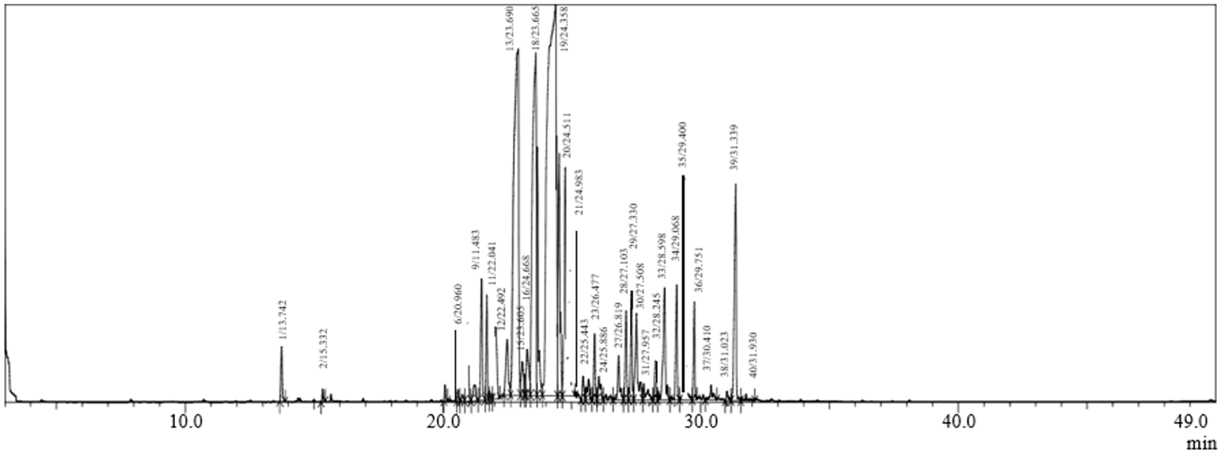
2.1. Chemical Composition

The chemical composition data of CAEO, along with the percentage of each identified compound, the molecular formula, structural subclass, and retention index (RI) values are presented in Table 1 and Figure 1. Thirty volatile compounds were identified in CAEO EO by GC–MS, representing 98.89 % of the total of this oil. The chemical composition of CAEO is dominated by sesquiterpene hydrocarbons (77.9%) and oxygenated sesquiterpenes (15.92%). Moreover, the main bioactive compounds detected in CAEO are β -Himachalene (28.99%), α-Himachalene (14.43%), and Longifolene (12.2%).
Numerous investigations examined the chemical components of CAEO in various Moroccan districts, including the province of Ifrane (Itzer and Senoual forests) [20], which contains β-himachalene (27.67–44.23%), followed by α-himachalene (12.2–16.69%), trans-Cadina-1(6),4-diene (11.27–8.45%), and 6-camphenol (4.54–3.16%) as major components, with seventy components represented mainly by sesquiterpene hydrocarbons (24–45.5%) and oxygenated hydrocarbons (13.63%–26%). The chemical components of cedar wood EO has been elucidated in different areas around the world, including Algeria [24], Lebanon [25], France [26], and Morocco [12]. These investigations showed significant differences in the chemical composition of CAEO. Indeed, the chemical profile of the samples collected in France and Lebanon, in which α-pinene and himachalol were found as the main components, was different from those of Algerian and Moroccan sawdust EOs, which consist mostly of hemichalene isomers. Comparable results have been reported in an earlier investigation by Başer and Demircakmak [27] who demonstrated the abundance of himachalene α, β and γ isomers (58.6%) in Cedrus libani EO from Antalya, Turkey.
Some studies have shown that the chemical profile of EOs varies according to many extrinsic and intrinsic factors, such as the geographical locations, the development stage of the plant, soil composition, harvesting period, storage process, the plant parts used, growth, and geoclimatic conditions [17,28,29].
Definitely, the variability in chemical profile of Eos is influenced by many factors mentioned earlier in this paper. These factors can influence and control the biosynthesis of secondary metabolites through the induction and/or the repression of the key enzyme genes. This process can be linked to specific epigenetic regulation, including DNA methylation, histone modifications, and chromatin remodeling [30].

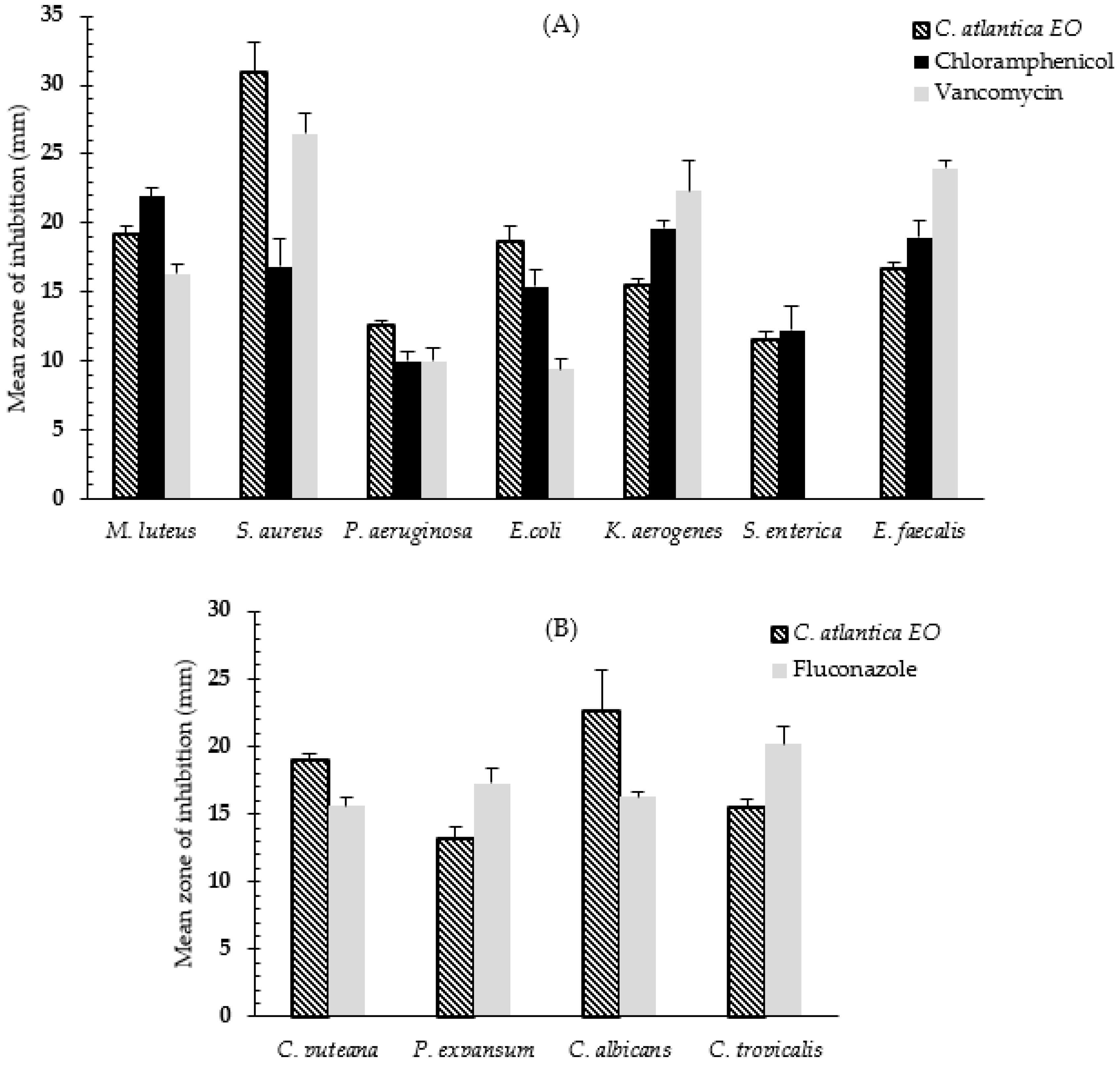
2.2. Antimicrobial Activity

To determine the antibacterial activity of CAEO, the agar disc-diffusion technique was used. Figure 2A summarizes the antibacterial activity, whereas Figure 2B summarizes the antifungal activity. The results of this test can be interpreted based on the width of the inhibitory zone: the EO activity is categorized as low activity at 10 mm, moderate activity at >10 to 15 mm, and high activity at >15 mm [31]. Therefore, CAEO showed the highest antibacterial activity against Staphylococcus aureus (30.98 ± 2.12 mm), followed by Micrococcus luteus (19.21 ± 0.51 mm), Escherichia coli (18.65 ± 1.18 mm), Enterococcus faecalis (16.74 ± 0.35 mm), Pseudomonas aeruginosa (16.0.4 ± 0.57 mm), and Klebsiella aerogenes (15.53 ± 0.49 mm), respectively. Moderate activity was recorded against Salmonella enterica (11.50 ± 0.62 mm).
The Gram-positive bacteria were the most susceptible, and the results were statistically significant (ANOVA, p < 0.05) compared to the reference antibiotics (Figure 2A). Regarding the antifungal potential, the disc diffusion results showed that CAEO has high antifungal activity against Candida albicans (22.65 ± 2.93 mm), followed by Coniophora puteana (18.98 ± 0.53 mm), and Candida tropicalis (15.50 ± 0.62 mm), respectively. However, moderate activity of CAEO was observed against Penicillium expansum (13.21 ± 0.87 mm). The results were significant and comparable to the referenced antifungal drug (Figure 2B).
The broth microdilution method was used to determine MIC, MBC, and MFC values, which are shown in Table 2 and Table 3. Obviously, the lowest values of MIC and MBC were recorded with the Gram-positive bacteria (M. luteus, S. aureus, and E. faecalis), which ranged from 0.0625 to 0.25 % v/v for MIC and from 0.5 to 0.125% v/v for MBC, respectively. The MIC and MBC values for the Gram-negative bacteria were between 0.25 and 0.25 % for E. coli and K. aerogenes, 0.5 and 2.0% for P. aeruginosa, and 1.0 and 1.0% for S. enterica. These results support the findings of the disc-diffusion method (Table 2). For the fungal strains, the lowest MIC and MFC values were recorded with C. tropicalis (MIC and MFC = 0.5% v/v), followed by P. expansum (MIC and MFC = 1.0% v/v), C albicans (MIC = 1.0 and MFC = 2.0% v/v), and C. puteana (MIC = 4.0 and MFC = 8.0% v/v) confirming the noticeable antifungal efficacy of CAEO (Table 3). The MIC, MBC, and MFC results were highly effective and competitive with the referenced antibiotics. Moreover, MBC/MIC and MFC/MIC ratios revealed that CAEO have a bactericidal and fungicidal mechanism. This conclusion came from the fact that antimicrobial agents can be categorized as bactericidal or fungicidal if the ratio of the MBC/MIC to the MFC/MIC is lower than or equal to 4.0 and it is feasible to achieve concentrations of the tested agent that kill 99.9% of the organisms treated. If these ratios are greater than 4.0, it may not be feasible to provide doses of the tested agent adequate to kill 99.9% of the microorganisms, and the agent is deemed bacteriostatic [1,32].
The results of the disc-diffusion method reported high antibacterial activity against S. aureus, M. luteus, E. coli, E. faecalis, P. aeruginosa, and K. aerogenes. However, moderate activity against S. enterica was recorded (Figure 2A). The current findings are consistent with many previous studies which confirmed the antibacterial and antifungal efficacy of CAEO against various microbial strains [33,34,35]. The antimicrobial effect of EOs may be affected by an array of parameters, including climatic factors, geographical location, harvesting times, soil characteristics, and growth cycle stage, making it difficult to standardize the chemical composition of EOs in order to manufacture a drug [36]. As a result, repeated investigations of the same plant are critical.
The broth microdilution method that was used to determine MIC, MBC, and MFC values is recommended by researchers to confirm the antimicrobial properties of essential oils due to its precision, simplicity, and resource and time savings [37]. According to our results, low values of MIC, MBC, and MFC have been reported with CAEO against tested bacterial and fungal strains to varying degrees, which were competitive with the tested antibiotics. This highlights two critical points: the first reflects the relative deterioration in antibiotic efficacy, and the second demonstrates the importance and efficacy of some essential oils as a potential alternative and promising source of novel antimicrobial drugs. Our results were in line with earlier studies that found CAEO to be very effective against five different fungal strains, including Aspergillus niger, Thielavia hyalocarpa, Penicillium commune, Penicillium expansum, and Penicillium crustosum, with MIC values between 0.5 and 1.0% v/v and MBCs between less than 8.0 and 8.0% v/v [33].
Our investigation also confirms the frequently reported finding that some bioactive EOs tend to be more efficient against Gram-positive bacteria than Gram-negative bacteria [38,39,40], suggesting that the target site of the EOs are mainly the cell wall and cell membrane. Our investigation revealed that CAEO have bactericidal and fungicidal mechanism. Bactericidal and fungicidal agents directly kill the microbial cell while bacteriostatic and fungistatic agents are able to inhibit the growth of the microbial cell. The common belief is that bactericidal agents eradicate pathogens rapidly. In clinical practice, however, there are no discernible distinctions between bactericidal and bacteriostatic drugs [41,42], although this information provides a theoretical basis to understand the mode of action of EOs and its implications for pharmaceutical formulation and drug discovery. Finally, it is crucial to seek alternatives to antibiotics from several sources, such as essential oils. Antibiotic resistance is a developing problem, and more research into effective antimicrobials is needed to counteract it [43].

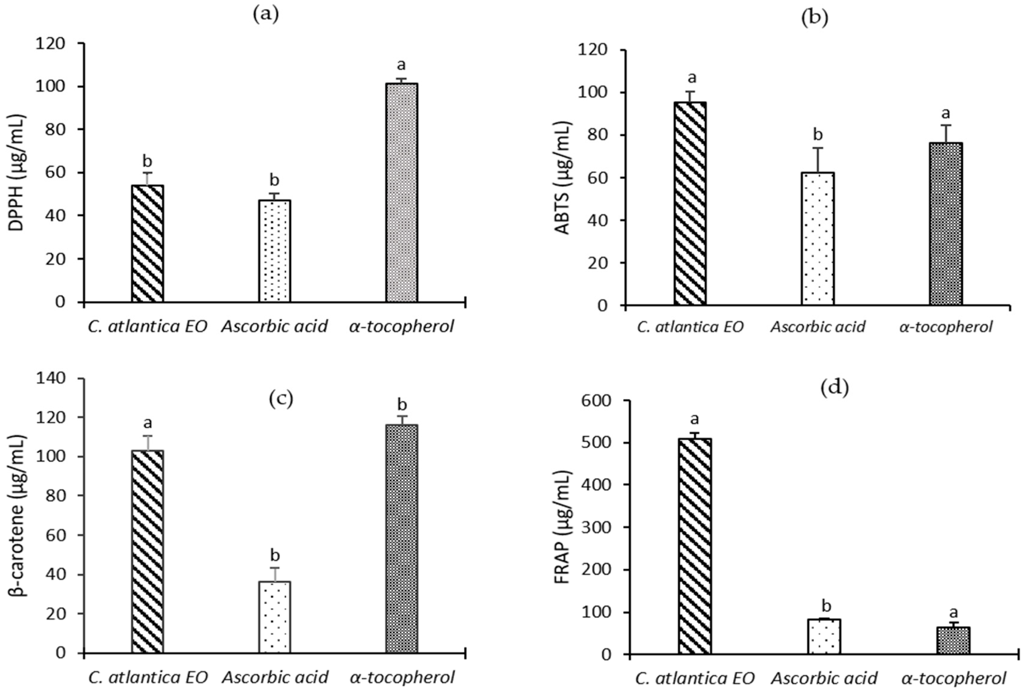
2.3. Antioxidant Activity

Essential oils are a complex of bioactive molecules displaying various antioxidant effects in several biological systems. In this work, we have investigated the antioxidant potential of CAEO using four complementary in vitro tests, namely DPPH, ABTS, ferric reductive power, and the ß-carotene bleaching method. As reported in Figure 3, CAEO exhibits significant antioxidant activities as compared to the standard antioxidants ascorbic acid and α-tocopherol, which were used as controls (p < 0.05). CAEO exerts strong scavenging activity against DPPH and ABTS radical with IC50 values of 54.19 ± 5.86 and 54.19 ± 5.86 µg/mL, respectively. Based on the results obtained by FRAP technique, it was reported that CAEO has a promising ability to reduce the ferric ion, with an EC50 value of 509.50 ± 12.58 µg/mL. However, this reducing power effect is still less effective when compared to the positive controls ascorbic acid (IC50 = 82.55 ± 2.58 µg/mL) and α-tocopherol (IC50 = 64.73 ± 9.97 µg/mL). Moreover, as evidenced by the β-carotene-bleaching test, CAEO significantly prevents lipid peroxidation with IC50 = 103.13 ± 7.26 µg/mL (p < 0.05).
Taken together, CAEO displays significant antioxidant properties by targeting different mechanisms, suggesting its potential application as a natural preservative and antioxidant. Our findings are consistent with those obtained in the literature, including the work of Jaouadi et al. [20]. The results indicated that CAEO extracted from wood tar exerts promising DPPH-scavenging and ferric ion-reductive ability with IC50 values of 126 µg/mL and 143 µg/mL, respectively. Moreover, in their recent investigation, Kačániová and colleagues [44] demonstrated the highest inhibitory effect of CAEO against the DPPH radical. These findings may be related to the phenolic content existing in the volatile compounds of this plant [8], while these properties are not only reflected by a single bioactive component, but are also ascribed to the high amount of sesquiterpene hydrocarbons (77.9%) and oxygenated sesquiterpenes (15%) present in the CAEO. In fact, positive correlation has been established between the antioxidant potential of a given sample and the phenolic content, which allows them to prevent lipid, DNA, and other macromolecules from oxidation, and mitigate the harmful effects induced by the production of reactive oxygen species (ROS) [8,45]. This event may prevent the occurrence and the pathogenesis of several chronic diseases, including diabetes, cancer, heart disease, and neurological degeneration.

2.4. Anti-Inflammatory Activity

The extreme generation of inflammatory mediators can lead to many diseases, such as cancer, cardiovascular issues, stroke, and neurodegenerative disorders [46]. Lipoxygenases (LOXs) are monomeric proteins that engender the oxidation of polyunsaturated fatty acids, especially linoleic and arachidonic acid, to generate hydroperoxides. LOX products can be transformed into other derivatives, playing a key role in inflammation [47]. Therefore, the regression of LOX activity can moderate inflammatory process.
In this study, we considered the anti-inflammatory activity of CAEO using the 5-LOX enzyme inhibition assay. As can be observed in Table 4, the CAEO possesses a considerable inhibitory effect of the enzyme with important IC50 value of 36.42 ± 0.103 µg/mL which is close to that of the reference compound (IC50 of quercetine = 21.31 ± 0.017 µg/mL). Based on these findings, we can deduce that CAEO exhibits potent anti-inflammatory action.
Indeed, few reports have examined the anti-inflammatory properties of C. atlantica essential oil. Recently, Al Kamaly et al. [11] revealed that the essential oil of Moroccan Cedrus atlantica (Middle Atlas) is able to inhibit 98.36% of paw edema induced with carrageenan with a concentration of 50 mg/kg. Another work proposed that the inhalation of CAEO can relieve postoperative pain in Swiss male mice by stimulating the serotonergic, noradrenergic, opioidergic, and dopaminergic systems [21].
Additionally, some reports indicated that extracts from other Cedrus species, particularly Cedrus deodara, Cedrus libani, and Cedrus brevifolia, exert in vitro and in vivo anti-inflammatory action by inhibiting COX-2/TNF-α/NF-κB activation, repressing the lipoxygenase activity, and preventing linoleic acid and lipid peroxidation [48,49,50,51].
This anti-inflammatory ability could be assigned to bioactive substances comprised in the EOs. Interestingly, Elias et al. [52] showed that the 2-Himachelen-7-ol compound, isolated from Cedrus libani volatile oil, displays strong anti-inflammatory power in formalin-provoked paw edema, in addition to dose dependent suppression of cyclooxygenase-2 (COX-2) protein expression in rat monocytes [52].

2.5. Dermatoprotective Activity

Epidermis aging is the main process that induces dryness, toughness, and pigmentation inequality (hyper- or hypo-pigmentation). Tyrosinase is a metalo-oxidase enzyme implied in the development of melanogenesis in mammals. Indeed, this enzyme generates the oxidation of monophenols and o-diphenols into reactive o-quinones in the initial step of melanogenesis. Hence, tyrosinase inhibition can be a crucial dermatoprotective pathway [53,54].
To estimate the dermatoprotective effect of the essential oil obtained from C. atlantica, the inhibition of tyrosinase activity was analyzed. At the best of our knowledge, the current study is the first one concerning the tyrosinase enzyme inhibitory activity of CAEO. Table 4 provides the IC50 values of the CAEO and quercetin (the standard compound). Our results showed that CAEOs exhibit significant inhibition with an IC50 of 141.103 ± 0.06 μg/mL, which is slightly higher than IC50 of quercetin (93.27 ± 0.021 μg/mL).
Importantly, Heinrich and his colleagues [55] reported in their review that the essential oil of C. deodara is mainly used to treat dermatological complications in India, Nepal, and Pakistan [55]. Based on the complexity of the EO compositions, the inhibition of tyrosinase activity is mostly ascribed to a synergistic interaction of their components with the enzyme [56]. In addition, several studies revealed that some plants from Pinaceae family, especially Morus alba, Pinus thunbergii, Pinus sylvestris, C. deodara, and Larix kaempferi, own important dermatoprotective properties (anti-melanogenic, anti-tyrosinase, anti-elastase, hyaluronidase, and anti-browning properties) [57,58,59,60,61].

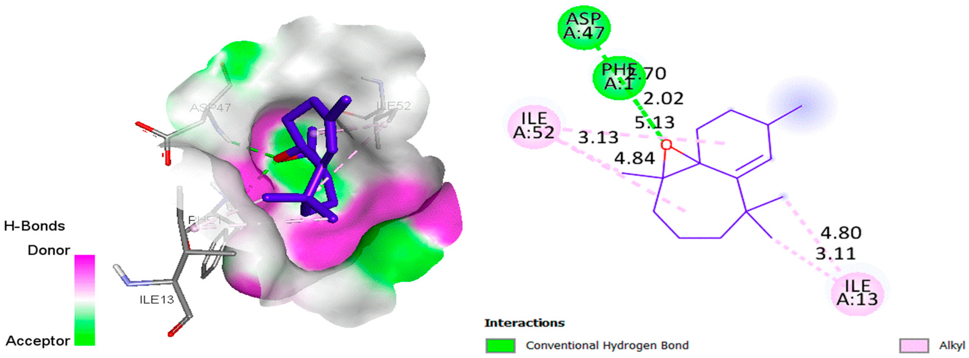
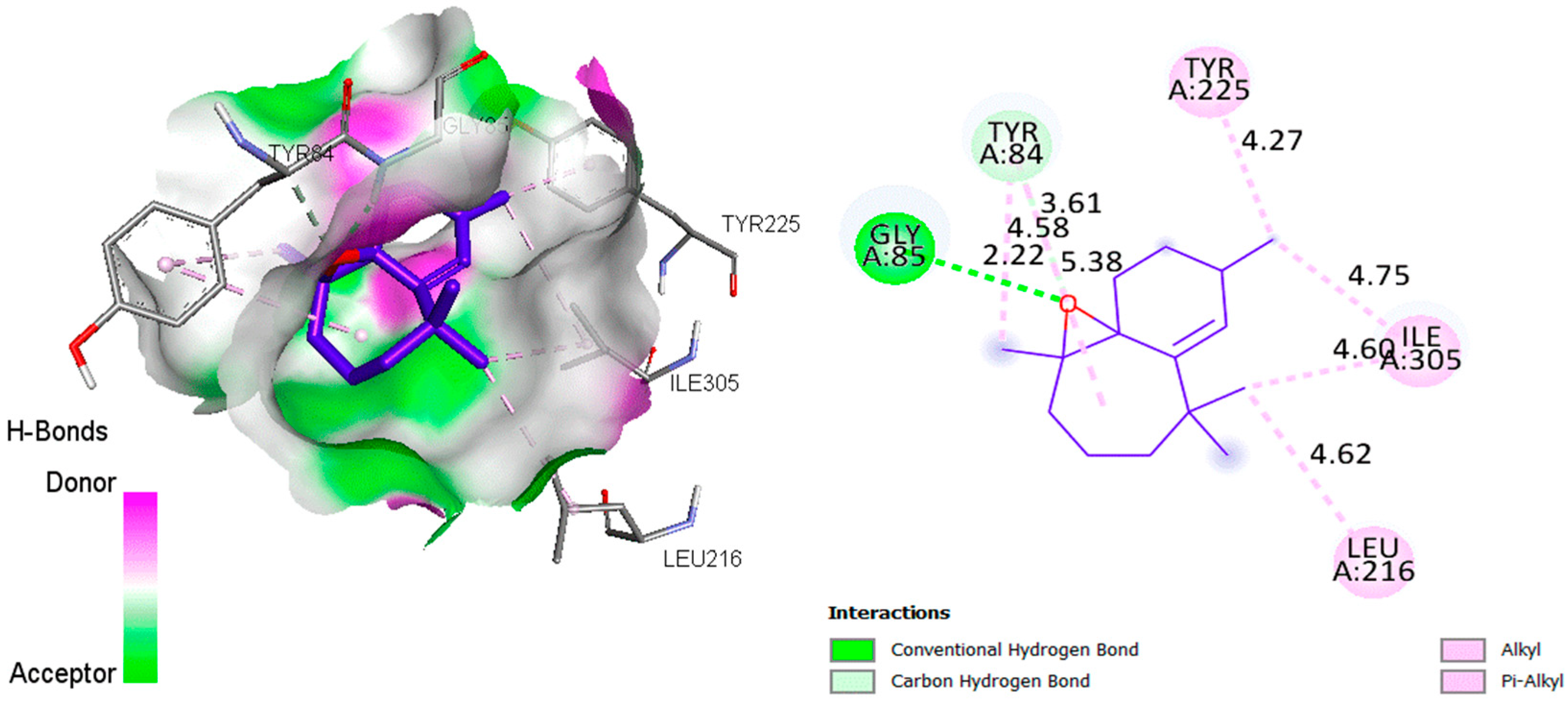
2.6. Molecular Docking Analysis

The aim of this in silico study was to identify the interaction modes of essential oils with the active sites of bacterial and fungal proteins, by using molecular docking to visualize the intermolecular interactions. The analysis revealed that the active site pocket in 4XO8 [62] was formed by PHE1, ASP47, ASP54, GLN133, ASN135, and ASP140, while the crucial sites in 1ZAP [63] were found to be GLY34, TYR84, GLY85, ASP218, THR221, and ILE305. The hydrogen-bonding interactions between the (+)-β-Himachalene oxide and the Escherichia coli as well as Candida albicans proteins are visualized in 2D using Figure 4 and Figure 5, respectively.
For the Escherichia coli protein (Figure 4), it was observed during docking analysis that there was a two-strong hydrogen bonding interaction between the O atom of the (+)-β-Himachalene oxide and NH site of PHE1 as well as ASP47, with a distance of 2.020 Å and 2.703 Å, respectively. Additionally, there were several Alkyl–Alkyl interactions with the amino acids ILE13 and ILE52. When it comes to the Candida albicans protein (Figure 5), the O atom of (+)-β-Himachalene oxide was also found to be involved in a strong hydrogen-bonding interaction with the NH site of GLY85 at a distance of 2.223 Å, along with multiple Alkyl-Alkyl and Pi-alkyl interactions with several other amino acids. Thus, the (+)-β-Himachalene oxide compound formed hydrogen bonding interactions with the most important key residues in the active site of both Escherichia coli and Candida albicans proteins. It is worth noting that the presence of hydrogen bonds strengthened the binding of essential oil compounds to receptors, allowing the compounds to have strong inhibitory effects on receptor proteins. This molecular docking study showed in silico the targeted active site and required mode of interaction against bacterial and fungal receptors.

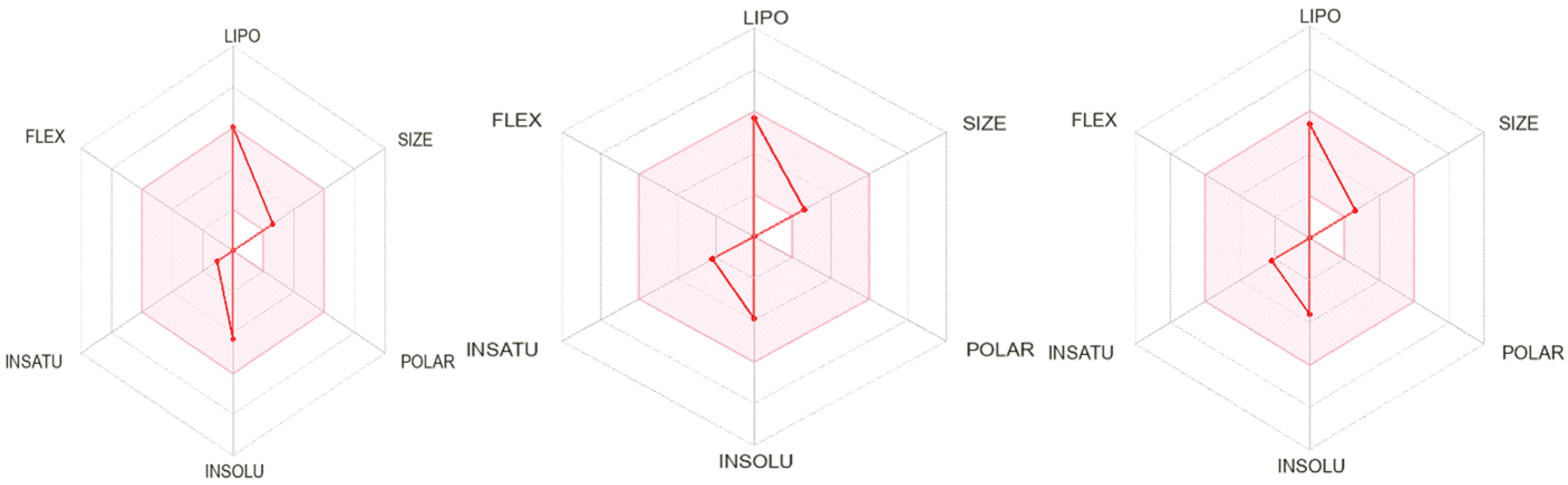
2.7. ADMET Prediction and Drug Likeness

The feasibility of using bioactive compounds as drugs against bacterial and fungal infections was evaluated by predicting their ADMET pharmacokinetic parameters. Table 5 and Table 6 present the results of in silico predictions of ADMET and drug likeness properties, respectively.
For the ADMET prediction analyses in Table 5, a value below 30% for absorption suggests poor intestinal absorption. Thus, all compounds showed a higher value (94%), indicating good intestinal absorption. For the blood–brain barrier (BBB), a compound with a LogBB < −1 is expected to have poor distribution to the brain, while a LogBB > 0.3 is likely to cross the BBB. Similarly, a compound with a LogPS > −2 is considered capable of penetrating the central nervous system (CNS), whereas a LogPS < −3 will find it difficult to move into the CNS. Thus, all the compounds have excellent potential for crossing barriers. Enzymatic metabolism is the process by which drugs are chemically transformed in the human body, and it plays a crucial role in the metabolic stability of drugs. The liver contains several cytochrome P450 enzymes, including CYP1A2, CYP2C19, CYP2C9, CYP2D6, and CYP3A4, which are the major drug-metabolizing enzymes responsible for biotransforming more than 90% of drugs. When these metabolic enzymes are inhibited, it can lead to an increase in the concentration of active drugs in the body. In this study, the primary human enzymes responsible for metabolizing drugs used to treat bacterial and fungal infections are CYP1A2 and CYP3A4 [64,65]. We designed several compounds, most of which were found to be substrates or inhibitors of CYP3A4 and CYP1A2. We observed that all compounds displayed low total clearance values, indicating potential accumulation and persistence of the drugs in the body, we found no evidence of toxicity. Overall, these results suggest that all the compounds possess favorable pharmacokinetic properties.
Based on the results presented in Table 6, we evaluated the drug similarity of the three compounds using four filters: Lipinski, Ghose, Veber, and Egan. All tested compounds were found to meet all drug similarity rules. However, they all displayed a violation of Lipinski’s rules (MLogP (Moriguchi’s logP) > 4.15). We also established bioavailability scores for each molecule by evaluating six parameters: lipophilicity, molecular weight, insolubility, establishment, polarity, and flexibility (Figure 6).
All three compounds demonstrated a high bioavailability score of 0.55. They all exhibited very high lipophilicity scores, which can be attributed to their failure to pass the Lipinski five rule (MLogP > 4.15). Furthermore, the flexibility and polarity scores were both zero, indicating that all three compounds should be orally bioavailable.

3. Materials and Methods

3.1. Reagents

NaCl, p-iodonitrotetrazoliumchloride, lipoxygenase (5-LOX), tyrosinase, 1,1-diphenyl-2-picrylhydrazyl (DPPH), α-tocopherol, potassium ferricyanide K3Fe(CN)6, methanol, acid 2,2′-azino-bis (3-éthylbenzothiazoline-6-sulphonique (ABTS), ascorbic acid, trichloroacetic acid (TCA), ferric chloride, β-carotene, chloroform, tween-80, L-DOPA, linoleic acid, ethanol, and quercetin were procured from Sigma-Aldrich. Potato dextrose agar (PDA), luria-Bertani (LB) agar, DMSO, chloramphenicol, vancomycin, and fluconazole were purchased from labKem, Barcelona, Spain and Biokar Diagnostics, Beauvais, France. All used elements were of analytical grade.

3.2. Plant Materiel and EO Extractions

Cedrus atlantica (Endl.) Manetti ex Carriere, wood was harvested from its wild habitat in the Azrou region (Middle Atlas Mountains, Morocco) (33°26′0″ N 5°13′0″ W) in March 2022. Botanical authenticity was performed at the Scientific Institute, Mohammed V University in Rabat, Morocco, under voucher specimen RAB 113587. The extraction procedure of C. atlantica essential oil (CAEO) was performed via hydro-distillation using Clevenger-type apparatus. Concisely, 50 g of the dry wood was in placed in water and boiled for three hours. The obtained oil was recuperated and kept at a temperature of 4 °C until the upcoming assays.

3.3. Gas Chromatography–Mass Spectrometry (GC–MS) Analysis

Volatile compounds of C. atlantica EO were analyzed with a Hewlett–Packard Gas Chromatographer HP 6890 coupled with a mass spectrometer (MS) HP5973 model, equipped with an HP-5MS (5% phenylmethyl siloxane) capillary column (30 m × 0.25 mm × film thickness 0.25 µm). The column temperature was programmed at 50 °C for 5 min and 200 °C with a 4 °C/min rate. Helium served as a carrier gas at 1.5 mL/min flow rate. The samples were injected in a split mode with a ratio of 1:50. MS was identified through electron ionization (EI) at an ionization voltage of 70 eV, using a spectral scan range of 40–450 m/z. This apparatus was controlled by a computer system type ”HP ChemStation”, which allowed us to monitor MS and total ions gas chromatography (GC-TIC) analysis. CAEO compounds were identified by establishing their retention index (RI) following the Van Den Dool method [66] (Determined using n-alkanes (C9-C31) series), and also by computer matching of their MS identities with the recorded data library (Wiley 09, Nist 2002). Finally, the chemical characterization was completed by matching the fragmentation patterns of MS with those published in the literature.

3.4. Antimicrobial Activity

3.4.1. Tested Microorganisms

In order to evaluate the antimicrobial potential of CAEO, eleven microbial strains were used in the current investigation, including three Gram positive bacteria: Micrococcus luteus ATTC 14452, Staphylococcus aureus ATCC 29213, Enterococcus faecalis (Clinical isolate), four Gram-negative bacteria: Escherichia coli ATCC 25922, Salmonella enterica serotype Typhi, Pseudomonas aeruginosa ATCC 27853, Klebsiella aerogenes ATCC 13048, and four fungal strains: Coniophora puteana (ATCC 9351), Penicillium expansum (food-spoilage isolate), Candida albicans (Clinical isolates), and Candida tropicalis (Clinical isolates). The source of all strains was the Laboratory of Microbial Biotechnology and Bioactive Molecules at the Faculty of Sciences in Fez, Morocco. Bacterial and fungal cultures were revitalized by aplying a looped needle containing the culture onto the agar surface using nutritional agar (NA) media for bacteria and potato dextrose agar (PDA) media for fungi. Then the cultures were incubated at 30–37 °C for 24 h for bacteria and 48–72 h for fungi. Fresh bacterial and fungal cultures were used to generate bacterial and fungal suspensions, which were then suspended in 5 mL of sterile physiological NaCl solution, and the turbidity was measured using a standard of 0.5 McFarland. For antibacterial screening, a final bacterial density of around 106 CFU/mL for bacteria and about 104 to 105 CFU/mL for fungi were used in the experiments in compliance with the standards of the National Committee for Clinical Laboratory Standards, United States National Committee for Clinical Laboratory Standards, United States [22].

3.4.2. Disc-Diffusion Method

The antimicrobial activity of CAEO was examined by the agar disc-diffusion method with slight modifications [23]. In brief, the culture suspension was sown on extract peptone dextrose (YPD) agar for fungi and Luria–Bertani (LB) agar medium for bacteria. Before being placed on an agar plate, each of the 6 mm diameter sterile paper discs were saturated with 10 µL of pure EO. The positive controls for bacteria were chloramphenicol and vancomycin (10 µg/disc), whereas the positive control for fungi was fluconazole (10 µg/disc). Bacteria were incubated on plates for 24 h at 30–35 °C, whereas fungi were incubated on plates for 48–72 h at 25 °C. The inhibitory zones’ widths were measured in millimeters after incubation, and the findings were provided as the mean ± standard deviation for three separate tests.

3.4.3. Minimum Inhibitory Concentration

The minimum inhibitory concentration (MIC) of CAEO was determined by using a method that has been published before, although with a few modifications [67]. In a nutshell, EO concentrations were prepared in two-fold serial dilutions that ranged from 4.0 to 0.0625 % (v/v). EOs were diluted in broth medium (extract–peptone–dextrose broth for fungi and Luria–Bertani broth for bacteria) containing 5% DMSO and were then tested in sterile 96-well plates by adding 190 µL of each dilution in each single well. After that, 10 µL of the bacterial culture that had been adjusted to McFarland beforehand were poured into each well. Serial two-fold dilutions of antibiotics (chloramphenicol, vancomycin, and fluconazole) were made in a range of 256.0–2.0 µg/mL and served as positive controls. After that, the 96-well plates that had been prepared were left in the incubator for 24 h at 30–35 °C for bacteria or for 48–72 h at 25 °C for fungi. A medium with 5% DMSO but no microbial suspension was used as a negative control for the experiment. After incubation, 50 μL of p-iodo-nitro-tetrazolium chloride (0.2 mg/mL) was injected into each micro-well to evaluate the growth of the bacteria (growth indicator). The highest sample dilution at which the yellow-to-pink color shift could still be seen was used to calculate the MIC.

3.4.4. MBC and MFC Assay

After the MIC test, the minimum bactericidal concentration (MBC) for bacteria and the minimum fungicidal concentration (MFC) for fungi were determined using agar plates [23]. In summary, 50 µL was pipetted from each MIC tube and dispersed over plates containing the suitable medium (YPD agar for fungi and LB for bacteria), which was then incubated under the optimal conditions (24 h at 30–35 °C for bacteria or 48–72 h at 25 °C for fungi). The plates were examined for microbial growth after incubation. The minimal growth concentration (MBC/MFC) was defined as the MIC at which no growth was detectable. In addition, the MBC/MIC and MFC/MIC ratios were calculated to identify the possible mechanism of the examined EO.

3.5. Antioxidant Assays

The in vitro antioxidant activities of C. atlantica EO were investigated using four complementary techniques, including ABTS and DPPH radicals scavenging activity, ferric reductive power and β-carotene-linoleic acid bleaching assay.

3.5.1. DPPH Radical Scavenging Assay

The stable radical 1,1-diphenyl-2-picrylhydrazyl (DPPH) was used to examine the antiradical activity of CAEO using a slightly reformed version of Bouyahya et al.’s method [17]. Briefly, a 700 µL aliquot of DPPH solution (0.004%) was added to 100 µL of C. atlantica EO (solubilized in methanol) at various concentrations. After, the obtained solution was incubated at room temperature for 25 min in a dark place. Then, the absorbance was read at 517 nm. The experiment was performed in triplicate and IC50 values were calculated based on inhibition curves and presented as means ± SD. Ascorbic acid (E300) and α-tocopherol (E307) were used as reference free-radical scavengers.

3.5.2. ABTS Scavenging Assay

The discoloration test of ABTS+ was performed as previously described in the literature [67]. Concisely, radical cation (ABTS+) was produced by mixing equal aliquots of 7 mM of ABTS solution and 2.45 mM of potassium persulfate solution. The mixture was incubated in a dark place at 25 °C for 14–16 h. Then, the obtained ABTS+ solution was diluted with methanol until accomplishing an absorbance of 0.7 (±0.03) at 734 nm. Afterwards, 2 mL of the prepared ABTS∙+ was added to 200 μL and then incubated for 3 min. The absorbance was read at 734 nm and the antioxidant potential of CAEO was reported as IC50 ± SD (n = 3). Ascorbic acid and α-tocopherol were used as controls.

3.5.3. Ferric-Reducing Antioxidant Power (FRAP) Assay

The reductive potential of CAEO was evaluated using the method adopted by Jaouadi et al. [20], with slight changes. In brief, equal aliquots of 1% of potassium ferricyanide K3Fe(CN)6 solution and the phosphate buffer solution (0.2 M, pH 6.6) were mixed with CAEO at various concentrations. Then, the obtained solution was incubated in a water-bath at 55 °C for 20 min. To stop the reaction, a volume of 1.25 mL of 10% trichloroacetic acid (TCA) was added and the solution was centrifuged at 3500 r/min for 7 min. Next, 1.25 mL of the supernatant was mixed with 1.25 mL of H2O2 and 250 µL of ferric chloride (0.1%). The absorbance was read at 700 nm; ascorbic acid and α-tocopherol were used as standard. The reductive ability was established as an IC50 value (μg/mL).

3.5.4. Inhibition of Lipid Peroxidation

The inhibition of the lipid peroxidation capacity was investigated by the β-carotene-linoleic acid test according to the procedure indicated by Gulluce et al. [68]. Briefly, the stock solution of β-carotene/linoleic acid was prepared as follows: 1 mg of β-carotene was solubilized in 5 mL of chloroform, then 10 mg of linoleic acid and 100 mg of Tween-80 were added to the β-carotene solution. The chloroform was evaporated using rotary evaporator at 45 °C and 100 rpm; subsequently, 50 mL of distilled water was added to the residue. A volume of 1 mL β-carotene solution was then mixed with 100 µL of CAEO at various concentrations. The test tubes were incubated at boiling water at 50 °C for 100 min. The variation of β-carotene absorbance was followed at 470 nm against a blank.
The antioxidant properties were established as terms of the residual color inhibition relative to the control using the following equation:
I (%) = 100 = (Abs (t = 100 min)/Abs (t = 0)) × 100
where Abs (t = 100 min): is the absorbance of β-carotene after 100 min of experiments residual in the CAEO and Abs (t = 0) is the absorbance of β-carotene at the starting time of the assay.

3.6. In Vitro Anti-Inflammatory Assay

The in vitro anti-inflammatory activity of CAEO was determined by the Lipoxygenase (5-LOX) inhibition technique, following the linoleic acid oxidation at 234 nm as described elsewhere [69]. In short, 20 µL of CAEO (dissolved in ethanol) and 20 µL of 5-LOX from glycine max (100 U/mL) were first mixed with 0.2 mL of phosphate buffer (0.1 M, pH 9), then the solution was incubated at 25 °C for 6 min. Afterwards, 20 µL of linoleic acid (4.18 mM in ethanol) was added to the mixture and followed for 3 min at 234 nm. The data were expressed as IC50 ± SEM of three independent measurements. Quercetin was used as a standard compound.

3.7. Dermatoprotective Activity

The tyrosinase inhibitory activity was carried out to assess the dermatoprotective potential of CAEO according to the previous reported technique [17], with slight changes. In brief, CAEO at 20 μL was added to 0.1 mL of tyrosinase solution (333 U/mL, 50 mM phosphate buffer at pH 6.5) and kept at 37 °C for 10 min. Next, 0.3 mL of the substrates L-DOPA (5 mM) were added. After 30–40 min of incubation at 37 °C, the absorbance was read at 510 nm using UV-Vis 1240 spectrophotometer. The data were used for expression of dermatoprotective activity as half inhibitory concentrations (IC50) for three independent experiments. Quercetin was used as a standard reference.

3.8. Molecular Docking

Molecular docking was employed to investigate the interaction between essential oils and the active site of target proteins, and to identify the key structural requirements based on binding affinity [70]. The 3D crystal structures of the target proteins, Escherichia coli (PDB ID: 4XOB) [62] and Candida albicans (PDB ID: 1ZAP) [63], were retrieved from the Protein Data Bank (PDB) database (https://www.rcsb.org/ (accessed on 17 January 2023). To prepare the protein structures, the Discovery Studio version 4.1 software was utilized to eliminate water molecules, ligands, and non-protein components. Subsequently, to analyze the ligand–protein interactions, we utilized AutoDock 4.2 and the AUTOGRID algorithm [71] to create a 3D grid and measure the energies of the interactions. The center grid box size was set to (−20.461, −10.721, and −4.502) for 4XOB and (8.775, 24.999 and 2.583) for 1ZAP to position the ligand in the complexes. The resulting docked ligand conformations were analyzed using 2D and 3D visualizations in Discovery Studio to investigate the binding interactions.

3.9. In Silico Pharmacokinetics ADMET and Drug-Likeness Prediction

Computer technology has had a profound impact on drug discovery, enabling the development of new drug candidates with greater efficiency and accuracy [72]. In silico studies provide valuable insights into ADMET [73] pharmacokinetic parameters, including absorption, distribution, metabolism, excretion, and toxicity. This approach employs pharmacokinetic parameters and drug similarity to perform preliminary assessments during drug discovery. With the aid of the online tool pkCSM [74], we were able to determine a compound’s absorption potential in the human small intestine, distribution in the body, biotransformation, elimination, and toxicity levels. Consequently, computational technology plays a vital role in evaluating ADMET pharmacokinetic parameters. To evaluate the drug likeness of the compounds, we utilized rule-based filters from Lipinski [75], Ghose [76], Veber [77], and Egan [78]. These filters assess various parameters, including molecular weight, number of hydrogen bond donors and acceptors, log P, and the number of rotatable bonds. We utilized the SwissADME online tool to perform this assessment [79], allowing us to efficiently predict the potential of the compound to become a drug candidate.

3.10. Statistical Analysis

All experiments were executed by three independent tests (n = 3) and the obtained data were established as mean ± standard deviations (SD). The data analyses was carried out by GraphPad prism 9 and XLSTAT statistics software v. 2016 and the means were compared adopting one-way analysis of variance (ANOVA), followed by Tukey test. A p value of <0.05 was considered statistically significant.

4. Conclusions

Cedrus atlantica is an endemic tree possessing valuable health benefits which has been widely used in traditional medicine since ancient times/ Here, C. atlantica essential oil has been found to have promising pharmacological properties, with different biological effects such as antibacterial, antifungal, antioxidant, anti-inflammatory, and dermatoprotective activities. As evidenced by GC-MS investigation, these effects are probably related to various bioactive compounds identified in the volatile part of C. atlantica. ADMET simulation suggests that the main compounds of CAEO possess favorable pharmacokinetic properties. Furthermore, considerable attention should be given to the application of CAEO as a promising natural agent in many industries. Indeed, this oil could be applied as active packaging (i.e., as films and coatings) in the food industry. The CAEOs may also be used as biopesticides in the agricultural industry due to their biodegradable and eco-friendly properties. Furthermore, they could represent powerful biomedical applications as nanodelivery systems in medical and pharmaceutical industries. However, further in vivo and clinical studies are strongly recommended to confirm the pharmacological effects of this plant, and the evaluation of its toxicity is also crucial in order to verify its safety.

Author Contributions

Conceptualization, N.E.H., K.F.-B. and H.N.M.; data curation, N.E.H. and N.B.; formal analysis, H.N.M., A.B., K.F.-B., N.E.H., S.H.A.-M. and E.M.A.; funding acquisition; K.W.G.; investigation, N.E.H., N.B. and M.J.; methodology, H.N.M., N.E.H. and A.B., resources, N.E.H., H.N.M., N.B., A.Q. and H.A.; software, N.E.H., H.H., N.B. and M.J.; supervision, H.N.M., A.B. and K.F.-B.; validation A.A.-F., K.W.G., N.E.H., H.N.M. and A.B.; visualization, A.B. and S.H.A.-M.; writing—original draft, N.E.H., N.B. E.M.A., H.H. and M.J.; writing—review and editing, K.F.-B., H.N.M., A.B. and S.H.A.-M. All authors have read and agreed to the published version of the manuscript.

Funding

This research received no external funding.

Institutional Review Board Statement

Not applicable.

Informed Consent Statement

Not applicable.

Data Availability Statement

Not applicable.

Acknowledgments

Princess Nourah bint Abdulrahman University Researchers Supporting Project number (PNURSP2023R158), Princess Nourah bint Abdulrahman University, Riyadh, Saudi Arabia.

Conflicts of Interest

The authors declare no conflict of interest.

References

  1. Abdallah, E.M. Antibacterial Activity of Hibiscus sabdariffa L. Calyces against Hospital Isolates of Multidrug Resistant Acinetobacter Baumannii. J. Acute Dis. 2016, 5, 512–516. [Google Scholar] [CrossRef] [Green Version]
  2. Cowan, M.M. Plant Products as Antimicrobial Agents. Clin. Microbiol. Rev. 1999, 12, 564–582. [Google Scholar] [CrossRef] [PubMed] [Green Version]
  3. Chaughule, R.S.; Barve, R.S. Role of Herbal Medicines in the Treatment of Infectious Diseases. Vegetos 2023, 1–11. [Google Scholar] [CrossRef] [PubMed]
  4. Sofowora, A.; Ogunbodede, E.; Onayade, A. The Role and Place of Medicinal Plants in the Strategies for Disease Prevention. Afr. J. Tradit. Complement. Altern. Med. 2013, 10, 210–229. [Google Scholar] [CrossRef] [PubMed]
  5. Mlilo, S.; Sibanda, S. An Ethnobotanical Survey of the Medicinal Plants Used in the Treatment of Cancer in Some Parts of Matebeleland, Zimbabwe. South Afr. J. Bot. 2022, 146, 401–408. [Google Scholar] [CrossRef]
  6. Borghesi, A.; Stronati, M. Superbugs and Antibiotics in the Newborn. J. Pediatr. Neonatal Individ. Med. (JPNIM) 2015, 4, e040253. [Google Scholar]
  7. Kıran, T.R.; Otlu, O.; Karabulut, A.B. Oxidative Stress and Antioxidants in Health and Disease. LaboratoriumsMedizin 2023, 47, 1–11. [Google Scholar] [CrossRef]
  8. Ak, T.; Gülçin, I. Antioxidant and Radical Scavenging Properties of Curcumin. Chemico-Biol. Interact. 2008, 174, 27–37. [Google Scholar] [CrossRef]
  9. Salmerón-Manzano, E.; Garrido-Cardenas, J.A.; Manzano-Agugliaro, F. Worldwide Research Trends on Medicinal Plants. Int. J. Environ. Res. Public Health 2020, 17, 3376. [Google Scholar] [CrossRef]
  10. Boy, H.I.A.; Rutilla, A.J.H.; Santos, K.A.; Ty, A.M.T.; Alicia, I.Y.; Mahboob, T.; Tangpoong, J.; Nissapatorn, V. Recommended Medicinal Plants as Source of Natural Products: A Review. Digit. Chin. Med. 2018, 1, 131–142. [Google Scholar] [CrossRef]
  11. Al Kamaly, O.; Saleh, A.; Al Sfouk, A.; Alanazi, A.S.; Parvez, M.K.; Ousaaid, D.; Assouguem, A.; Mechchate, H.; Bouhrim, M. Cedrus atlantica (Endl.) Manetti Ex Carrière Essential Oil Alleviates Pain and Inflammation with No Toxicity in Rodent. Processes 2022, 10, 581. [Google Scholar] [CrossRef]
  12. Uehara, A.; Tommis, B.; Belhassen, E.; Satrani, B.; Ghanmi, M.; Baldovini, N. Odor-Active Constituents of Cedrus atlantica Wood Essential Oil. Phytochemistry 2017, 144, 208–215. [Google Scholar] [CrossRef] [PubMed]
  13. Derwich, E.; Benziane, Z.; Boukir, A. Chemical Composition and in Vitro Antibacterial Activity of the Essential Oil of Cedrus atlantica. Int. J. Agric. Biol. 2010, 12, 381–385. [Google Scholar]
  14. Salhi, N.; Fidah, A.; Rahouti, M.; Ismaili, M.R.; Kabouchi, B.; Famiri, A. Preservative Effect of Tetraclinis Articulata and Cedrus atlantica Wood Extractives against Fungal Decay. Madera y Bosques 2020, 26, 1–10. [Google Scholar] [CrossRef]
  15. Fidah, A.; Salhi, N.; Rahouti, M.; Kabouchi, B.; Ziani, M.; Aberchane, M.; Famiri, A. Natural Durability of Cedrus atlantica Wood Related to the Bioactivity of Its Essential Oil against Wood Decaying Fungi. Maderas. Cienc. Tecnol. 2016, 18, 567–576. [Google Scholar] [CrossRef] [Green Version]
  16. Ainane, A.; Khammour, F.; Charaf, S.; Elabboubi, M.; Elkouali, M.; Talbi, M.; Benhima, R.; Cherroud, S.; Ainane, T. Chemical Composition and Insecticidal Activity of Five Essential Oils: Cedrus atlantica, Citrus limonum, Rosmarinus officinalis, Syzygium aromaticum and Eucalyptus globules. Mater. Today Proc. 2019, 13, 474–485. [Google Scholar] [CrossRef]
  17. Bouyahya, A.; Et-Touys, A.; Abrini, J.; Talbaoui, A.; Fellah, H.; Bakri, Y.; Dakka, N. Lavandula Stoechas Essential Oil from Morocco as Novel Source of Antileishmanial, Antibacterial and Antioxidant Activities. Biocatal. Agric. Biotechnol. 2017, 12, 179–184. [Google Scholar] [CrossRef]
  18. Prabuseenivasan, S.; Jayakumar, M.; Ignacimuthu, S. In Vitro Antibacterial Activity of Some Plant Essential Oils. BMC Complement. Altern. Med. 2006, 6, 1–8. [Google Scholar] [CrossRef] [Green Version]
  19. Satrani, B.; Aberchane, M.; Farah, A.; Chaouch, A.; Talbi, M. Composition Chimique et Activité Antimicrobienne Des Huiles Essentielles Extraites Par Hydrodistillation Fractionnée Du Bois de Cedrus atlantica Manetti. Acta Bot. Gall. 2006, 153, 97–104. [Google Scholar] [CrossRef]
  20. Jaouadi, I.; Cherrad, S.; Bouyahya, A.; Koursaoui, L.; Satrani, B.; Ghanmi, M.; Chaouch, A. Chemical Variability and Antioxidant Activity of Cedrus atlantica Manetti Essential Oils Isolated from Wood Tar and Sawdust. Arab. J. Chem. 2021, 14, 103441. [Google Scholar] [CrossRef]
  21. Martins, D.F.; Emer, A.A.; Batisti, A.P.; Donatello, N.; Carlesso, M.G.; Mazzardo-Martins, L.; Venzke, D.; Micke, G.A.; Pizzolatti, M.G.; Piovezan, A.P. Inhalation of Cedrus atlantica Essential Oil Alleviates Pain Behavior through Activation of Descending Pain Modulation Pathways in a Mouse Model of Postoperative Pain. J. Ethnopharmacol. 2015, 175, 30–38. [Google Scholar] [CrossRef] [PubMed] [Green Version]
  22. Reisner, B.S.; Woods, G.L. Laboratory for Antimicrobial Methods Susceptibility Testing. Antimicrob./Anti-Infect. Mater. Princ. Appl. 1999, 292. [Google Scholar]
  23. Al-Mijalli, S.H.; Assaggaf, H.; Qasem, A.; El-Shemi, A.G.; Abdallah, E.M.; Mrabti, H.N.; Bouyahya, A. Antioxidant, Antidiabetic, and Antibacterial Potentials and Chemical Composition of Salvia Officinalis and Mentha Suaveolens Grown Wild in Morocco. Adv. Pharmacol. Pharm. Sci. 2022, 2022, 2844880. [Google Scholar] [CrossRef] [PubMed]
  24. Boudarene, L.; Rahim, L.; Baaliouamer, A.; Meklati, B.Y. Analysis of Algerian Essential Oils from Twigs, Needles and Wood of Cedrus atlantica G. Manetti by GC/MS. J. Essent. Oil Res. 2004, 16, 531–534. [Google Scholar] [CrossRef]
  25. Saab, A.M.; Harb, F.Y.; Koenig, W.A. Essential Oil Components in Heart Wood of Cedrus Libani and Cedrus atlantica from Lebanon. Minerva Biotecnol. 2005, 17, 159. [Google Scholar]
  26. Paoli, M.; Nam, A.-M.; Castola, V.; Casanova, J.; Bighelli, A. Chemical Variability of the Wood Essential Oil of Cedrus atlantica Manetti from Corsica. Chem. Biodivers. 2011, 8, 344–351. [Google Scholar] [CrossRef] [PubMed]
  27. Başer, K.H.C.; Demircakmak, B. The Essential Oil of Taurus Cedar (Cedrus libani A. Rich): Recent Results. Chem. Nat. Compd. 1995, 31, 16–20. [Google Scholar] [CrossRef]
  28. El Hachlafi, N.; Benkhaira, N.; Ferioun, M.; Kandsi, F.; Jeddi, M.; Chebat, A.; Addi, M.; Hano, C.; Fikri-Benbrahim, K. Moroccan Medicinal Plants Used to Treat Cancer: Ethnomedicinal Study and Insights into Pharmacological Evidence. Evid.-Based Complement. Altern. Med. 2022, 2022, 1645265. [Google Scholar] [CrossRef] [PubMed]
  29. El Hachlafi, N.; Chebat, A.; Fikri-Benbrahim, K. Ethnopharmacology, Phytochemistry, and Pharmacological Properties of Thymus Satureioides Coss. Evid.-Based Complement. Altern. Med. 2021, 2021, 6673838. [Google Scholar] [CrossRef] [PubMed]
  30. Vriet, C.; Hennig, L.; Laloi, C. Stress-Induced Chromatin Changes in Plants: Of Memories, Metabolites and Crop Improvement. Cell. Mol. Life Sci. 2015, 72, 1261–1273. [Google Scholar] [CrossRef] [PubMed]
  31. ALrajhi, M.; Al-Rasheedi, M.; Eltom, S.E.M.; Alhazmi, Y.; Mustafa, M.M.; Ali, A.M. Antibacterial Activity of Date Palm Cake Extracts (Phoenix dactylifera). Cogent Food Agric. 2019, 5, 1625479. [Google Scholar] [CrossRef]
  32. Davis, J.L. Pharmacologic Principles. Equine Intern. Med. 2018, 4, 79–137. [Google Scholar]
  33. Bennouna, F.; Lachkar, M.; El Abed, S.; Saad, I. Cedrus atlantica Essential Oil: Antimicrobial Activity and Effect on the Physicochemical Properties of Cedar Wood Surface. Moroc. J. Biol. 2020, 16, 35–45. [Google Scholar]
  34. Benouaklil, F.; HAMAIDI-CHERGUI, F.; Hamaidi, M.S.; Saidi, F. Chemical Composition and Antimicrobial Properties Of Algerian Cedrus atlantica M. Essential Oils. Rev. Agrobiol. 2017, 7, 355–362. [Google Scholar]
  35. Rhafouri, R.; Strani, B.; Zair, T.; Ghanmi, M.; Aafi, A.; El Omari, M.; Bentayeb, A. Chemical Composition, Antibacterial and Antifungal Activities of the Cedrus atlantica (Endl.) Manettiex Carriè Re Seeds Essential Oil. Mediterr. J. Chem. 2014, 3, 1034–1043. [Google Scholar] [CrossRef]
  36. Dhifi, W.; Bellili, S.; Jazi, S.; Bahloul, N.; Mnif, W. Essential Oils’ Chemical Characterization and Investigation of Some Biological Activities: A Critical Review. Medicines 2016, 3, 25. [Google Scholar] [CrossRef] [Green Version]
  37. Vanegas, D.; Abril-Novillo, A.; Khachatryan, A.; Jerves-Andrade, L.; Peñaherrera, E.; Cuzco, N.; Wilches, I.; Calle, J.; León-Tamariz, F. Validation of a Method of Broth Microdilution for the Determination of Antibacterial Activity of Essential Oils. BMC Res. Notes 2021, 14, 1–7. [Google Scholar] [CrossRef]
  38. Çelebi, Ö.; Fidan, H.; Iliev, I.; Petkova, N.; Dincheva, I.; Gandova, V.; Stankov, S.; Stoyanova, A. Chemical Composition, Biological Activities, and Surface Tension Properties of Melissa officinalis L. Essential Oil. Turk. J. Agric. For. 2023, 47, 67–78. [Google Scholar] [CrossRef]
  39. Sharma, A.D.; Kaur, I. Chemical Profile and In-Silico Docking Studies on Bioactives from Essential Oil of Cymbopogan pendulus Targeting Penicillin Binding Proteins (PBPs) in Bacteria. Biol. Med. Nat. Prod. Chem. 2023, 12, 225–232. [Google Scholar]
  40. Walasek-Janusz, M.; Grzegorczyk, A.; Zalewski, D.; Malm, A.; Gajcy, S.; Gruszecki, R. Variation in the Antimicrobial Activity of Essential Oils from Cultivars of Lavandula angustifolia and L.× Intermedia. Agronomy 2022, 12, 2955. [Google Scholar] [CrossRef]
  41. Finberg, R.W.; Moellering, R.C.; Tally, F.P.; Craig, W.A.; Pankey, G.A.; Dellinger, E.P.; West, M.A.; Joshi, M.; Linden, P.K.; Rolston, K.V. The Importance of Bactericidal Drugs: Future Directions in Infectious Disease. Clin. Infect. Dis. 2004, 39, 1314–1320. [Google Scholar] [CrossRef] [PubMed] [Green Version]
  42. Nemeth, J.; Oesch, G.; Kuster, S.P. Bacteriostatic versus Bactericidal Antibiotics for Patients with Serious Bacterial Infections: Systematic Review and Meta-Analysis. J. Antimicrob. Chemother. 2015, 70, 382–395. [Google Scholar] [CrossRef] [PubMed] [Green Version]
  43. Abdallah, E.M. Plants: An Alternative Source for Antimicrobials. J. Appl. Pharm. Sci. 2011, 6, 16–20. [Google Scholar]
  44. Kačániová, M.; Galovičová, L.; Valková, V.; Ďuranová, H.; Štefániková, J.; Čmiková, N.; Vukic, M.; Vukovic, N.L.; Kowalczewski, P.Ł. Chemical Composition, Antioxidant, In Vitro and In Situ Antimicrobial, Antibiofilm, and Anti-Insect Activity of Cedar atlantica Essential Oil. Plants 2022, 11, 358. [Google Scholar] [CrossRef] [PubMed]
  45. Aruoma, O.I. Free Radicals, Oxidative Stress, and Antioxidants in Human Health and Disease. J. Am. Oil Chem. Soc. 1998, 75, 199–212. [Google Scholar] [CrossRef] [PubMed]
  46. Gilbert, N.C.; Newcomer, M.E.; Werz, O. Untangling the Web of 5-Lipoxygenase-Derived Products from a Molecular and Structural Perspective: The Battle between pro-and Anti-Inflammatory Lipid Mediators. Biochem. Pharmacol. 2021, 193, 114759. [Google Scholar] [CrossRef]
  47. Lončarić, M.; Strelec, I.; Moslavac, T.; Šubarić, D.; Pavić, V.; Molnar, M. Lipoxygenase Inhibition by Plant Extracts. Biomolecules 2021, 11, 152. [Google Scholar] [CrossRef]
  48. Chen, Y.; Chi, L.; Liang, X.; Shi, Y.; Wu, T.; Ye, M.; Han, P.; Lin, L.; Zhang, L.; Xu, P. Essential Oils of Cedrus deodara Leaves Exerting Anti-Inflammation on TPA-Induced Ear Edema by Inhibiting COX-2/TNF-α/NF-ΚB Activation. J. Essent. Oil Bear. Plants 2020, 23, 422–431. [Google Scholar] [CrossRef]
  49. Karrat, L.; Abajy, M.Y.; Nayal, R. Investigating the Anti-Inflammatory and Analgesic Properties of Leaves Ethanolic Extracts of Cedrus libani and Pinus brutia. Heliyon 2022, 8, e09254. [Google Scholar] [CrossRef]
  50. Douros, A.; Hadjipavlou-Litina, D.; Nikolaou, K.; Skaltsa, H. The Occurrence of Flavonoids and Related Compounds in Cedrus brevifolia A. Henry Ex Elwes & A. Henry Needles. Inhibitory Potencies on Lipoxygenase, Linoleic Acid Lipid Peroxidation and Antioxidant Activity. Plants 2017, 7, 1. [Google Scholar] [CrossRef] [Green Version]
  51. Cretu, E.; Trifan, A.; Aprotosoaie, A.C.; Miron, A. 15-Lipoxygenase Inhibition, Superoxide and Hydroxyl Radicals Scavenging Activities of Cedrus brevifolia Bark Extracts. Rev. Med. Chir. Soc. Med. Nat. Iasi. 2013, 117, 250–256. [Google Scholar] [PubMed]
  52. Elias, A.; Shebaby, W.N.; Nehme, B.; Faour, W.; Bassil, B.S.; El Hakim, J.; Iskandar, R.; Dib-Jalbout, N.; Mroueh, M.; Daher, C. In Vitro and In Vivo Evaluation of the Anticancer and Anti-Inflammatory Activities of 2-Himachelen-7-Ol Isolated from Cedrus libani. Sci. Rep. 2019, 9, 12855. [Google Scholar] [CrossRef] [PubMed] [Green Version]
  53. Bouyahya, A.; Lagrouh, F.; El Omari, N.; Bourais, I.; El Jemli, M.; Marmouzi, I.; Salhi, N.; Faouzi, M.E.A.; Belmehdi, O.; Dakka, N. Essential Oils of Mentha viridis Rich Phenolic Compounds Show Important Antioxidant, Antidiabetic, Dermatoprotective, Antidermatophyte and Antibacterial Properties. Biocatal. Agric. Biotechnol. 2020, 23, 101471. [Google Scholar] [CrossRef]
  54. Marmouzi, I.; Kharbach, M.; El Jemli, M.; Bouyahya, A.; Cherrah, Y.; Bouklouze, A.; Vander Heyden, Y.; Faouzi, M.E.A. Antidiabetic, Dermatoprotective, Antioxidant and Chemical Functionalities in Zizyphus lotus Leaves and Fruits. Ind. Crops Prod. 2019, 132, 134–139. [Google Scholar] [CrossRef]
  55. Heinrich, M.; Jiang, H.; Scotti, F.; Booker, A.; Walt, H.; Weckerle, C.; Maake, C. Medicinal Plants from the Himalayan Region for Potential Novel Antimicrobial and Anti-Inflammatory Skin Treatments. J. Pharm. Pharmacol. 2021, 73, 956–967. [Google Scholar] [CrossRef]
  56. Cheraif, K.; Bakchiche, B.; Gherib, A.; Bardaweel, S.K.; Çol Ayvaz, M.; Flamini, G.; Ascrizzi, R.; Ghareeb, M.A. Chemical Composition, Antioxidant, Anti-Tyrosinase, Anti-Cholinesterase and Cytotoxic Activities of Essential Oils of Six Algerian Plants. Molecules 2020, 25, 1710. [Google Scholar] [CrossRef] [Green Version]
  57. Chaita, E.; Lambrinidis, G.; Cheimonidi, C.; Agalou, A.; Beis, D.; Trougakos, I.; Mikros, E.; Skaltsounis, A.-L.; Aligiannis, N. Anti-Melanogenic Properties of Greek Plants. A Novel Depigmenting Agent from Morus Alba Wood. Molecules 2017, 22, 514. [Google Scholar] [CrossRef] [Green Version]
  58. Yang, H.; Wang, Z.; Song, W.; Zhao, Z.; Zhao, Y. Isolation of Proanthocyanidins from Pinus Thunbergii Needles and Tyrosinase Inhibition Activity. Process Biochem. 2021, 100, 245–251. [Google Scholar] [CrossRef]
  59. Yu, Z.-L.; Zhang, Z.; Zeng, W.-C. Investigation of Antibrowning Activity of Pine Needle (Cedrus deodara) Extract with Fresh-Cut Apple Slice Model and Identification of the Primary Active Components. Eur. Food Res. Technol. 2014, 239, 669–678. [Google Scholar] [CrossRef]
  60. Kakumu, Y.; Yamauchi, K.; Mitsunaga, T. Identification of Chemical Constituents from the Bark of Larix kaempferi and Their Tyrosinase Inhibitory Effect. Holzforschung 2019, 73, 637–643. [Google Scholar] [CrossRef]
  61. Chiocchio, I.; Mandrone, M.; Sanna, C.; Maxia, A.; Tacchini, M.; Poli, F. Screening of a Hundred Plant Extracts as Tyrosinase and Elastase Inhibitors, Two Enzymatic Targets of Cosmetic Interest. Ind. Crops Prod. 2018, 122, 498–505. [Google Scholar] [CrossRef]
  62. Yakovenko, O.; Sharma, S.; Forero, M.; Tchesnokova, V.; Aprikian, P.; Kidd, B.; Mach, A.; Vogel, V.; Sokurenko, E.; Thomas, W.E. FimH Forms Catch Bonds That Are Enhanced by Mechanical Force Due to Allosteric Regulation. J. Biol. Chem. 2008, 283, 11596. [Google Scholar] [CrossRef] [PubMed] [Green Version]
  63. Abad-Zapatero, C.; Goldman, R.; Muchmore, S.W.; Hutchins, C.; Stewart, K.; Navaza, J.; Payne, C.D.; Ray, T.L. Structure of a Secreted Aspartic Protease from c. Albicans Complexed with a Potent Inhibitor: Implications for the Design of Antifungal Agents. Protein Sci. A Publ. Protein Soc. 1996, 5, 640–652. [Google Scholar] [CrossRef] [PubMed] [Green Version]
  64. Yamada, M.; Hatsuta, K.; Niikawa, M.; Imaishi, H. Detoxification of Aflatoxin B1 Contaminated Maize Using Human CYP3A4. J. Microbiol. Biotechnol. 2020, 30, 1207–1213. [Google Scholar] [CrossRef]
  65. Niwa, T.; Inoue-Yamamoto, S.; Shiraga, T.; Takagi, A. Effect of Antifungal Drugs on Cytochrome P450 (CYP) 1A2, CYP2D6, and CYP2E1 Activities in Human Liver Microsomes. Biol. Pharm. Bull. 2005, 28, 1813–1816. [Google Scholar] [CrossRef] [Green Version]
  66. Santiuste, J.M.; Tarján, G.; Ullrich, E.; Takács, J.M. Contribution to Linearly Programmed Temperature Gas Chromatography: Further Application of the Van Den Dool–Kratz Equation, and a New Utilization of the Sadtler Retention Index Library. J. Chromatogr. A 2008, 1181, 103–115. [Google Scholar] [CrossRef]
  67. Benkhaira, N.; Koraichi, S.I.; Fikri-Benbrahim, K. In Vitro Methods to Study Antioxidant and Some Biological Activities of Essential Oils: A Review. Biointerface Res. Appl. Chem. 2022, 12, 3332. [Google Scholar]
  68. Gulluce, M.; Sahin, F.; Sokmen, M.; Ozer, H.; Daferera, D.; Sokmen, A.; Polissiou, M.; Adiguzel, A.; Ozkan, H. Antimicrobial and Antioxidant Properties of the Essential Oils and Methanol Extract from Mentha longifolia L. Ssp. longifolia. Food Chem. 2007, 103, 1449–1456. [Google Scholar] [CrossRef]
  69. Al-Mijalli, S.H.; Mrabti, N.N.; Ouassou, H.; Sheikh, R.A.; Abdallah, E.M.; Assaggaf, H.; Bakrim, S.; Alshahrani, M.M.; Awadh, A.A.A.; Qasem, A.; et al. Phytochemical Variability, In Vitro and In Vivo Biological Investigations, and In Silico Antibacterial Mechanisms of Mentha piperita Essential Oils Collected from Two Different Regions in Morocco. Foods 2022, 11, 3466. [Google Scholar] [CrossRef]
  70. Hadni, H.; Bakhouch, M.; Elhallaoui, M. 3D-QSAR, Molecular Docking, DFT and ADMET Studies on Quinazoline Derivatives to Explore Novel DHFR Inhibitors. J. Biomol. Struct. Dyn. 2021. [Google Scholar] [CrossRef]
  71. Morris, G.M.; Huey, R.; Lindstrom, W.; Sanner, M.F.; Belew, R.K.; Goodsell, D.S.; Olson, A.J. AutoDock4 and AutoDockTools4: Automated Docking with Selective Receptor Flexibility. J. Comput. Chem. 2009, 30, 2785–2791. [Google Scholar] [CrossRef] [PubMed] [Green Version]
  72. Hadni, H.; Fitri, A.; Benjelloun, A.T.; Benzakour, M.; Mcharfi, M. Evaluation of Flavonoids as Potential Inhibitors of the SARS-CoV-2 Main Protease and Spike RBD: Molecular Docking, ADMET Evaluation and Molecular Dynamics Simulations. J. Indian Chem. Soc. 2022, 99, 100697. [Google Scholar] [CrossRef]
  73. Ferreira, L.L.G.; Andricopulo, A.D. ADMET Modeling Approaches in Drug Discovery. Drug Discov. Today 2019, 24, 1157–1165. [Google Scholar] [CrossRef] [PubMed]
  74. Pires, D.E.V.; Blundell, T.L.; Ascher, D.B. PkCSM: Predicting Small-Molecule Pharmacokinetic and Toxicity Properties Using Graph-Based Signatures. J. Med. Chem. 2015, 58, 4066–4072. [Google Scholar] [CrossRef] [PubMed]
  75. Lipinski, C.A.; Lombardo, F.; Dominy, B.W.; Feeney, P.J. Experimental and Computational Approaches to Estimate Solubility and Permeability in Drug Discovery and Development Settings. Adv. Drug Deliv. Rev. 1997, 23, 3–25. [Google Scholar] [CrossRef]
  76. Ghose, A.K.; Viswanadhan, V.N.; Wendoloski, J.J. A Knowledge-Based Approach in Designing Combinatorial or Medicinal Chemistry Libraries for Drug Discovery. 1. A Qualitative and Quantitative Characterization of Known Drug Databases. J. Comb. Chem. 1999, 1, 55–68. [Google Scholar] [CrossRef]
  77. Veber, D.F.; Johnson, S.R.; Cheng, H.Y.; Smith, B.R.; Ward, K.W.; Kopple, K.D. Molecular Properties That Influence the Oral Bioavailability of Drug Candidates. J. Med. Chem. 2002, 45, 2615–2623. [Google Scholar] [CrossRef]
  78. Egan, W.J.; Merz, K.M.; Baldwin, J.J. Prediction of Drug Absorption Using Multivariate Statistics. J. Med. Chem. 2000, 43, 3867–3877. [Google Scholar] [CrossRef]
  79. Daina, A.; Michielin, O.; Zoete, V. SwissADME: A Free Web Tool to Evaluate Pharmacokinetics, Drug-Likeness and Medicinal Chemistry Friendliness of Small Molecules. Sci. Rep. 2017, 7, 42717. [Google Scholar] [CrossRef] [Green Version]
Figure 1. Chromatogram of gas chromatography (GC) analysis of C. atlantica EO.
Molecules 28 05913 g001
Figure 2. Antimicrobial activity of CAEO against (A) bacteria and (B) fungal strains compared to commercialized drugs (chloramphenicol, vancomycin, and fluconazole) using disc-diffusion method. Results are expressed as means ± standard deviation (SD) of three independent measurements; diameter of inhibition zone including disc diameter of 6 mm.
Molecules 28 05913 g002
Figure 3. Antioxidant activities of CAEO (a) IC50 of DPPH assay, (b) IC50 of ABTS assay, (c) IC50 of β-carotene bleaching test, (d) EC50 of reducing power. Data with the same letter in the same test presents a non-significant difference by Tukey’s multiple range test (ANOVA, p < 0.05). The results are expressed as means ± SD of three independent measurements.
Molecules 28 05913 g003
Figure 4. 2D and 3D visualization of the interaction types between (+)-β-Himachalene oxide with Escherichia coli 4XO8 (binding energy −6.4 kcal/mol).
Molecules 28 05913 g004
Figure 5. 2D and 3D visualization of the interaction types between the (+)-β-Himachalene oxide with Candida albicans 1ZAP (binding energy −6.7 kcal/mol).
Molecules 28 05913 g005
Figure 6. Bioavailability radars for similarity between drugs and potential inhibitors.
Molecules 28 05913 g006
Table 1. Chemical composition of C. atlantica EO identified using GC-MS.
No. a nCompounds bMolecular FormulaRI cRI lit d%Relative Peak AreaIdentification
Cedrus atlantica EO
1Limona ketoneC9H14O110911050.58MS, IR
2p-MethylacetophenoneC9H10O 114211420.14MS, IR
3α-LongipineneC15H24134713470.16MS, IR
4YlangeneC15H24122112190.24MS, IR
5α-CopaeneC15H24137513760.11MS, IR
6Eudesma-2,4,11-trieneC15H22149714971.38MS, IR
7Isovalencenyl formateC16H24O2178217860.39MS, IR
8β—PanasinseneC15H24 141114131.61MS, IR
9LongifoleneC15H241398139812.2MS, IR
10Himachala-2,4-dieneC15H24149914951.35MS, IR
11VestitenoneC12H18O137113731.15MS, IR
12α-HimachaleneC15H241475147514.43MS, IR
13Himachalene-1,4-dieneC15H24149914990.22MS, IR
14γ-himachalene C15H24149915000.99MS, IR
15α-cedreneC15H24140314042.90MS, IR
16β-HimachaleneC15H241505150128.99MS, IR
17δ-CadineneC15H24146914683.65MS, IR
18α-BisaboleneC15H24151815217.71MS, IR
19α-calacoreneC15H20154715470.37MS, IR
20Himachalene oxideC15H22O 155115510.77MS, IR
21LongiborneolC15H26O159315920.71MS, IR
22β-Himachalene oxideC15H24O161016101.18MS, IR
23IsolongifololC15H26O173317331.40MS, IR
24Di-epi-1,10-cubenolC15H26O161516111.70MS, IR
25HimachalolC15H26O164816470.85MS, IR
26Allo-himachalolC15H26O167416792.27MS, IR
27(Z)-γ-AtlantoneC15H22O169816991.52MS, IR
28DeodaroneC15H24O2178117804.18MS, IR
29(Z)-α-AtlantoneC15H22O 170317034.81MS, IR
30Aromadendrene oxideC15H24O164216420.44MS, IR
Total identified % 98.40 %
Monoterpene hydrocarbons -
Oxygenated monoterpenes-
Sesquiterpene hydrocarbons77.9
Oxygenated sesquiterpenes15.92
Ketones0.72
Other4.03
a In order of elution on HP-5 ms, b compounds identified based on RI and MS. c Retention index calculated from alkanes series on HP-5 MS capillary column (C9-C31). d Retention index from data libraries (NIST) [22,23].
Table 2. MIC, MBC and MBC/MIC, values of CAEO against bacterial strains.
Bacterial StrainC. atlantica EO
% v/v
Chloramphenicol
µg/mL
Vancomycin
µg/mL
MICMBCMBC/MICMICMBCMBC/MICMICMBCMBC/MIC
S. aureus
ATCC 29213
0.125 0.1251.02.02.01.02.08.04.0
M. luteus
ATTC 14452
0.06250.1252.032.064.02.01.02.02.0
E. faecalis
(Clinical isolate)
0.250.52.08.016.02.08.016.02.0
E. coli
ATCC 25922
0.250.251.064.064.01.032.032.01.0
S. enterica
serotype Typhi
1.01.01.016.016.01.0256.0256.01.0
P. aeruginosa
ATCC 27853
0.52.04.016.016.01.032.032.01.0
K. aerogenes
ATCC 13048
0.250.251.032.032.01.016.032.02.0
MIC: Minimum inhibitory concentration in % (v/v), MBC: minimum bactericidal concentration in % (v/v). Chloramphenicol and vancomycin were used as standard drugs. Final bacterial density was around 106 CFU/mL.
Table 3. MIC, MFC and MFC/MIC, values of CAEO against fungal strains.
Fungal Strains C. atlantica EO (% v/v)Fluconazole (µg/mL)
MICMFCMFC/MICMICMFCMFC/MIC
C. albicans1.02.02.08.08.01.0
C. tropicalis0.50.51.01.01.01.0
P. expansum
(Food-spoilage isolate)
1.01.01.016.016.01.0
C. puteana
(ATCC 9351)
4.08.02.032.064.02.0
MIC: Minimum inhibitory concentration in % (v/v), MFC: minimum fungicidal concentration in % (v/v). Fluconazole was used as standard.
Table 4. In vitro anti-inflammatory and dermatoprotective activities of CAEO.
AssayCAEO (IC50 µg/mL)Quercetin (IC50 µg/mL)
5-Lipoxygenase36.42 ± 0.10321.31 ± 0.017
Tyrosinase141.103 ± 0.0693.27 ± 0.021
Values are mean ± SEM (n = 3).
Table 5. In silico ADMET prediction of the potential inhibitors.
CompoundsAbsorptionDistributionMetabolismExcretionToxicity
Intestinal
Absorption
(Human)
VDss
(Human)
BBB PermeabilityCNS PermeabilitySubstrateInhibitorTotal
Clearance
AMES
Toxicity
CYP
2D63A41A22C192C92D63A4
Numeric (% Absorbed)Numeric
(Log L/kg)
Numeric (Log BB)Numeric (Log PS)Categorical (Yes/No)Numeric (Log
ml/min/kg)
Categorical (Yes/No)
Longifolene95.7670.7810.808−1.949NoYesNoNoNoNoNo0.901No
α-Himachalene94.5560.6480.731−2.322NoNoYesYesYesNoNo1.1No
β-Himachalene94.4650.6570.718−2.322NoNoYesNoYesNoNo1.089No
Table 6. Drug likeness and bioavailability score predictions of the potential inhibitors.
CompoundsDrug Likeness
LipinskiGhoseVeberEganBioavailability Score
LongifoleneYes (1 violation)YesYesYes0.55
α-HimachaleneYes (1 violation)YesYesYes0.55
β-HimachaleneYes (1 violation)YesYesYes0.55
Disclaimer/Publisher’s Note: The statements, opinions and data contained in all publications are solely those of the individual author(s) and contributor(s) and not of MDPI and/or the editor(s). MDPI and/or the editor(s) disclaim responsibility for any injury to people or property resulting from any ideas, methods, instructions or products referred to in the content.

