Article

Chemical Composition and Antioxidant Potential of Five Algae Cultivated in Fully Controlled Closed Systems

1
CBQF—Centro de Biotecnologia e Química Fina—Laboratório Associado, Escola Superior de Biotecnologia, Universidade Católica Portuguesa, Rua Diogo Botelho 1327, 4169-005 Porto, Portugal
2
LAQV/REQUIMTE, Department of Chemistry, University of Aveiro, 3810-193 Aveiro, Portugal
3
CICECO—Aveiro Institute of Materials, Department of Materials and Ceramic Engineering, University of Aveiro, 3810-193 Aveiro, Portugal
4
AlgaPlus, Travessa Alexandre da Conceição s/n, 3830-196 Ílhavo, Portugal
*
Author to whom correspondence should be addressed.
Molecules 2023, 28(12), 4588; https://doi.org/10.3390/molecules28124588
Submission received: 17 April 2023 / Revised: 2 June 2023 / Accepted: 3 June 2023 / Published: 6 June 2023
(This article belongs to the Special Issue Marine Natural Products in Food Chemistry)

Abstract

:
In this study, the chemical composition and antioxidant profile of five edible macroalgae, Fucus vesiculosus, Palmaria palmata, Porphyra dioica, Ulva rigida, and Gracilaria gracilis, cultivated in fully controlled closed systems, were determined. Protein, carbohydrates, and fat contents ranged between 12.4% and 41.8%, 27.6% and 42.0%, and 0.1% and 3.4%, respectively. The tested seaweeds presented considerable amounts of Ca, Mg, K, Mn, and Fe, which reinforce their favorable nutritional profile. Regarding their polysaccharide composition, Gracilaria gracilis and Porphyra dioica were rich in sugars common to agar-producing red algae, and Fucus vesiculosus was composed mainly of uronic acids, mannose, and fucose, characteristic of alginate and fucoidans, whereas rhamnose and uronic acid, characteristic of ulvans, predominated in Ulva rigida. Comparatively, the brown F. vesiculosus clearly stood out, presenting a high polysaccharide content rich in fucoidans, and higher total phenolic content and antioxidant scavenging activity, determined by DPPH and ABTS. The remarkable potential of these marine macroalgae makes them excellent ingredients for a wide range of health, food, and industrial applications.

1. Introduction

Algae are photosynthetic eukaryotes that play a crucial role in the ecosystem, possessing a diverse biology and rich evolutionary history with important participation in the shaping of the planet’s atmosphere, and even nowadays the production of oxygen by algae is about 50% of all oxygen produced [1,2,3]. Seaweeds have been on earth far beyond the dawn of mankind and have had varying degrees of influence on human societies across history [2]. Nowadays, scientific data are opening the path for the full valorization of algae, showing their remarkable potential for a wide range of health and industrial applications [4,5].
The marine environment is an untapped source of unique and efficient compounds, where algae stand out as a valuable source of chemical compounds such as polysaccharides, proteins, minerals, enzymes, glycoproteins, polyunsaturated fatty acids, sulfolipids, phenolics, terpenoids, and other secondary metabolites [6,7,8]. Seaweed compounds have the advantages of being of natural origin and, given their antioxidant, antibacterial, anticoagulant, and antitumor activities, their ingestion may impact positively the health of individuals and reduce the risk of developing certain pathologies such as cancer, type 2 diabetes, cardiovascular diseases and related risk factors, and neurological diseases, among others [8,9,10,11,12,13,14].
Because of their remarkable advantages, macroalgae are excellent for use as ingredients in the development of innovative functional foods, dietary supplements, nutraceuticals, and even pharmaceuticals. In fact, in the year 2017, the algae product market was valued at USD 3.78 billion, being expected to achieve a value of USD 5.17 billion by 2023 [15,16,17]. This clearly highlights increasing consumer demand for foods and algae-derived products that combine natural and sustainable ingredients, high nutritional value, and health benefits [4,16,18,19].
Algae grow by adapting to environmental stress conditions such as sunlight, salinity, temperature, carbon dioxide supply (CO2), and others [20]. Because environmental and seasonal conditions are extremely variable and typically very harsh, wild algae’s chemical composition diverges greatly even within the same species [20,21]. Cultivation of algae in fully controlled systems offers the advantage of controlling development and processing conditions, creating a sustainable biomass product with controlled quality and biological properties [22]. This type of production provides algae biomass with improved quality standards, higher productivity, and ensures that the culture is not contaminated by undesirable microorganisms [22]. Furthermore, farmed seaweed contributes to sustainability development goals, since it does not require great agricultural areas, as plant-based proteins do, and may be used directly as an ingredient for food, feed, or supplement applications.
Characterization of seaweed in terms of chemical compounds and biological activities is an essential step required for the validation and valorization of algae for further nutritional, biotechnological, medical, and industrial applications. Therefore, the main objective of the present study was to determine the chemical composition and antioxidant activity of five different edible species of algae produced in fully controlled closed systems located on the western coast of the Iberian Peninsula: Fucus vesiculosus (brown algae), Palmaria palmata, Porphyra dioica, Gracilaria gracilis (red algae), and Ulva rigida (green algae). This study strongly contributes to generating an open mapping of land-based, integrated multi-trophic aquaculture (IMTA)-farmed seaweed composition, all edible and with tremendous potential for health, food, industrial, and biotechnological applications.

2. Results and Discussion

2.1. Proximate Composition

The chemical profile of the five previously dried farmed seaweeds under analysis is presented in Table 1. The contrasting profiles that are observed for the different seaweeds highlight the different taxonomic and physiological characteristics that are being covered with specimens from all three main algae groups.
Algae biomass presents high water content, which constitutes a good environment for enzymatic activity, and microbial growth, which leads to high perishability [23]. Drying of these marine ingredients is an excellent strategy to further develop industrial, pharmaceutical, and biotechnological applications. This process consists of removing water and consequently reducing moisture, leading to product stabilization and longer shelf life [23]. Despite having undergone a similar drying process, the seaweed specimens varied significantly (p < 0.05) in their final moisture contents, which ranged from 3.2 g/100 g dry seaweed in the red seaweed P. palmata, followed closely by G. gracilis (4.1 g/100 g dry seaweed), to 16.2 g/100 g dry seaweed in the green seaweed U. rigida. The brown macroalgae F. vesiculosus presented an intermediate moisture content of 12.4 g/100 g dry seaweed (Table 1).
Algae are considered a valuable source of protein, yet their contents vary according to species and farming conditions. Protein content was statistically different among all species (p < 0.05). The brown algae F. vesiculosus revealed contents of 12.4 g/100 g dry seaweed, followed by 19.5 g/100 g dry seaweed in the green alga U. rigida, and the three highest values (two- to three-fold higher) were registered in the red algae species (highest value of 41.8 g/100 g dry seaweed in P. Palmata). These results are consistent with the results found in scientific literature. Studies performed by Wells et al. [19] reported that red and green algae generally presented higher protein levels compared to brown algae, and Cherry et al. (2019) in their review reported protein content ranges consistent with those found in this study, namely, from 0.67% to 45.0% in red seaweeds, from 5.02% to 19.66% in brown seaweeds, and from 3.42% to 29.80% in green seaweeds [18,19]. Such variability is understandable since harvested seaweeds derive from different growth process (cultivation versus wild collection), lifecycle, seasonality (temperature and light intensity), geographical distribution, and ecological conditions (available nutrients, salinity). All such parameters have a great impact on the chemical composition of seaweed, including protein content. Notably, the red seaweeds in the present study, which derived from integrated multi-trophic aquaculture systems, where growth is performed under controlled conditions, presented higher protein values than those previously described by Rodrigues et al. (2015) for wild G. gracilis (20.2%) harvested in Buarcos bay on the Central West Coast of Portugal, by Fernández-Segovia, Lerma-García, Fuentes, and Barat (2018) for P. dioica (approximately 22%), collected in the Atlantic coastal region of Galicia (Spain), and by Bjarnadóttir et al. (2018) for P. Palmata (24.8%) harvested in Skjerstadfjorden, Bodø, Norway [13,24,25]. In contrast, protein values are below those previously described by Gadberry et al. (2018) for U. rigida (29.7%) produced in land-based cultivation systems, and yet almost equivalent to those presented by Lorenzo et al. (2017) for F. vesiculosus (12.99%) collected in the Galician coast (Spain) [13,24,25,26,27].
The macroalgae studied in the present work, especially G. gracilis, P. dioica, and P. palmata (red species), are excellent sources of alternative high-quality protein with levels comparable to those present in meat, eggs, soybean, or milk [28]. Therefore, these organisms could be part of the solution for the need for alternative protein sources and imperative increase in food production by around 70%, in order to meet the demands of a growing population that is expected to increase 2.3 billion people by 2050 [28]. Seaweed production has nutritional and productivity effectiveness advantages in comparison with traditional high-protein crops such as soybean and pulse legumes [28,29]. Macroalgae can develop without the need for fresh water or arable land and possess a higher protein yield per unit area in comparison with terrestrial crops (2.5–7.5 tons/Ha/year in comparison with 0.6–1.2 tons/Ha/year and 1–2 tons/Ha/year, for soybean and pulse legumes, respectively) [28,29]. In short, seaweeds are definitely a good source of high-quality and sustainably produced protein that should be implemented in healthy diets and in the development of novel food products in the near future [16].
Total carbohydrate content ranged from 9.3 g/100 g dry seaweed in the red seaweed P. palmata to 30.2 g/100 g dry seaweed in the brown algae F. vesiculosus. The proximate composition of this macronutrient varied greatly among species, with higher values being found in the brown and green species analyzed (30.2 g/100 g and 27.9 g/100 g dry seaweed, respectively). Among the red algae, P. dioica and G. gracilis presented very similar values (29.8 and 26.8 g/100 g dry seaweed), and P. palmata the lowest value among seaweed species (9.3 g/100 g dry seaweed) (Table 1). Carbohydrate composition of seaweeds includes important and biologically active molecules, including polysaccharides [30], a source of dietary fibers, whose characterization will be presented and discussed in the following sections. Dietary fibers are important nutritional compounds since they cannot be completely degraded by human digestive enzymes, with benefits for human gut microbiota and overall health.
Fat content in seaweeds is naturally low, and those analyzed in this study were no exception. The fat content of our macroalgae ranged from 0.078 g/100 g dry seaweed in the green alga U. rigida to 3.4 g/100 g dry seaweed in the brown alga F. vesiculosus. Among the red seaweed species assessed, P. dioica registered the highest total fat values (1.59 g/100 g dry seaweed). Results were statistically significantly different among all seaweeds except for comparisons between G. gracilis and P. palmata (red seaweed) and P. palmata and U. rigida (red and green seaweeds) (Table 1). Given their high nutritional value, and low-calorie and low-fat profiles, algae are an excellent asset to be incorporated into healthy balanced diets [31]. Furthermore, their versatility of use enables them to be consumed dried, fresh, pickled, cooked, or as a component of food products such as healthy snacks, soups, and bread, among others, which corroborates even further the potential of these organisms to be included in the daily diet of consumers [16,31]. The results obtained in the present research are quite similar to those obtained by Rodrigues et al. (2015) for G. gracilis collected from Buarcos bay on the Central West Coast of Portugal (0.60 g/100 g dry seaweed) and Lorenzo et al. (2017) for F. vesiculosus (3.75 g/100 g dry seaweed) collected on the Galician coast (Spain) [13,27]. In contrast, Gadberry et al. (2018) obtained higher lipid contents for U. rigida (2.97 g/100 g dry seaweed) produced in land-based cultivation systems, as well as Mæhre, Malde, Eilertsen, and Elvevoll (2014) for P. palmata (1.4 g/100 g dry seaweed) harvested off the coast of Norway [26,32]. Lower lipid contents were described by Fernández-Segovia et al. (2018) for P. dioica (0.08 g/100 g dry seaweed), harvested in Skjerstadfjorden, Bodø, Norway [24].
Ash contents were quite similar among the tested seaweeds, ranging from 23.41 g/100 g dry seaweed for the red macroalgae P. dioica to 29.8 g/100 g dry seaweed in the brown algae F. vesiculosus. Statistically significant differences were registered between all species for this parameter, except for comparisons between F. vesiculosus and G. gracilis and between P. palmata and U. rigida (Table 1). Our results were quite similar to the results obtained by Rodrigues et al. (2015) for G. gracilis (24.8 g/100 g dry seaweed) and by Fernández-Segovia et al. (2018) for P. dioica (24 g/100 g dry seaweed) [13,24]. In contrast, Lorenzo et al. (2017) obtained lower results in comparison with our analysis for F. vesiculosus (20.7/100 g dry seaweed) and Gadberry et al. (2018) and Mæhre et al. (2014) for U. rigida (32 g/100 g dry seaweed) and P. palmata, respectively [26,27,32]. These differences in ash contents among the same species were probably due to different growing conditions such as sunlight, salinity, temperature, and carbon dioxide supply (CO2), among others [20]. In fact, all algae samples from these previous studies were harvested from the wild, with the exception of those performed by Gadberry et al. (2018), in which algae were also grown in land-based cultivation systems. In this case, it should be highlighted that growth conditions greatly varied from those used for the algae studied herein [26].

2.2. Elemental Composition of Seaweeds

Minerals are essential for the human body. As such, their adequate dietary intake greatly contributes to the prevention of chronic and degenerative diseases such as cancer, neurological, and cardiovascular diseases and premature aging [33,34,35]. In addition to their macronutrient value, algae can also deliver a considerable amount of highly valuable elements, which reinforces their potential to be incorporated into the development of functional foods, pharmaceuticals, and nutraceuticals [5,36]. Seaweed mineral content is very variable and is tremendously influenced by several exogenous and endogenous factors such as environmental conditions, morphological features, and geographic location [37,38]. Among the tested five seaweed species, the most representative micronutrients detected were calcium (Ca), potassium (K), sodium (Na), magnesium (Mg), iron (Fe), and manganese (Mn). Phosphorus (P) and zinc (Zn) were consistently present in all the seaweed species, but in lower amounts in comparison with the previously mentioned elements. The full results are shown in Table 2.
Sodium was the most representative mineral in overall quantitative terms for F. vesiculosus and P. dioica (47.64 and 42.56 mg/g dry seaweed, respectively), and values differed statistically (p < 0.05) between all algae species. Furthermore, brown and red algae species presented higher amounts of Na compared to the green algae (Table 2). Regarding K content, this was the most abundant element present in the G. gracilis, P. palmata (red species), and U. rigida (green species) seaweeds, ranging from 13.64 mg/g dry seaweed in U. rigida to 89.20 mg/g dry seaweed in the red seaweed G. gracilis. Additionally, K contents were statistically different among all species (p < 0.05), except for comparisons between F. vesiculosus (brown algae) and one of the red seaweed species, P. palmata (Table 2). High Na intake is closely related with high blood pressure (hypertension), a primary risk factor for cardiovascular disease, and as such a diet low in Na and high in K is widely recommended as an effective preventive strategy [39,40]. Several studies have established that Na-to-K intake ratio (Na:K) is a more relevant predictor of hypertension than the analysis of each of these micronutrients alone [39,40,41]. Foods with molar ratios of Na:K close to 1.0 are considered beneficial for human health, and as such their consumption should be privileged [42]. Regarding the tested seaweeds, G. gracilis and P. palmata (red species) presented Na:K ratios below 1.0, (0.50 and 0.85, respectively), whereas F. vesiculosus, P. dioica, and U. rigida obtained ratios above 1.0 (2.19, 2.52, and 1.31, respectively). Although F. vesiculosus, P. dioica, and U. rigida presented higher Na:K ratios, these results are still below the results registered for cheese (4.94), preserved and tinned fish (3.64), processed meat (2.77), and pizza, crackers, and other salty snacks (3.31) [43]. Furthermore, the salty taste of G. gracilis and P. palmata makes these marine ingredients even more suitable candidates for salt substitutes to be used in the reformulation of different food categories where salt reduction means not only is Na intake reduced, but K intake is enhanced together with that of other important minerals [13,16].
Calcium values were statistically different among all species (p < 0.05), and it was within the red algae species group that the minimum and maximum values were reported, ranging from 2.25 mg/g dry seaweed in P. dioica to 15.91 mg/g dry seaweed in P. palmata. Between the brown and green algae species, F. vesiculosus (brown algae) registered more than double the Ca content of U. rigida (green algae) (Table 2). This mineral is the most prevalent in the human body and its adequate intake is closely related to bone mineral density [44,45]. Furthermore, recent randomized controlled trials have also established a relationship between Ca intake and a reduced risk of high blood pressure, preeclampsia, or colorectal adenomas [44]. Macroalgae in the present study deliver good amounts of Ca that should be further explored in the development of alternative Ca supplements in the future targeting osteoporosis prevention and other low-Ca-intake-related diseases. Furthermore, macroalgae Ca content is higher than that of oranges, carrots, potatoes, and apples [38]. Regarding this mineral’s bioavailability in algae, it widely varies depending on the macroalgae species, food matrix, and other factors [5,46]. For example, the macroalgae L. digitata is rich in alginates that retain divalent cations, which include Ca, leading to poor bioavailability [38]. The same is expected to occur with F. vesiculosus, also a rich source of alginate [38]. On the other hand, the bioavailability of AAA Ca, a calcium supplement in oyster shell and the seaweed Sargassum fusiformis, has been reported to be very high [38].
Magnesium is an essential element for the human body, being a cofactor for more than 300 metabolic reactions such as DNA and RNA synthesis, protein synthesis, and cellular energy production and storage, among many others [47]. Furthermore, Mg is extremely important for overall health and its deficiency is closely related with the development of several chronic diseases such as neurological conditions, cerebrovascular accidents, and migraine headaches [47,48,49]. Magnesium concentrations ranged from 2.22 mg/g dry seaweed in the red macroalgae P. palmata to 18.60 mg/g dry seaweed in the green algae U. rigida, more than double the amount found in the red and brown species groups (Table 2). Notably, Mg concentrations were statistically similar (p > 0.05) among species from the same group (Rhodophyta) but statistically different (p < 0.05) between species from different groups, except for those reported for P. dioica and F. vesiculosus (red and brown algae, respectively). High Ca-to-Mg-intake ratios have been associated with increased risk of cardiovascular disease, colorectal and prostate cancer, and overall cancer mortality [50,51]. Current evidence suggests that a 1.70 to 2.60 range of Ca:Mg ratio can lead to reduction in disease risk [51]. However, this evidence is still limited because only a few studies have analyzed dietary Ca:Mg ratios <1.70 as well as >2.60 [51]. Furthermore, these benefits may be dependent on specific health outcomes and gender [51]. Macroalgae from the present study presented Ca:Mg ratios that ranged from 0.19 in the green algae U. rigida to 7.16 in the red algae P. palmata (Table 2). None of the five studied macroalgae are within the optimum Ca:Mg range of 1.70–2.60, and as such they should be incorporated into food matrixes that can help to balance Ca:Mg ratio.
The macroalgae species tested herein were shown to be an important source of several trace elements. Regarding recommended dietary intake (RDI) contribution, Fe and Mn amounts were the most relevant across all the species tested. Values for Fe ranged from 0.46 mg/g dry seaweed in P. palmata to 1.65 mg/g dry seaweed in G. gracilis. This means that a consumption of 10 g of G. gracilis provides 117.9% of the RDI of this micronutrient, and as such, this seaweed is an excellent choice for consumers. This considerable amount of Fe can be extremely useful to incorporate into plant-based diets targeting specific dietary habits such as vegetarians and vegans, who generally have a low intake of this micronutrient [52]. Manganese values ranged from 0.06 mg/g dry seaweed in the green seaweed U. rigida to 0.29 mg/g dry seaweed in the brown seaweed F. vesiculosus. Interestingly, four out of the five algae tested presented Mn amounts very near or even above the RDI for this micronutrient. In particular, a consumption of 10 g of F. vesiculosus or G. gracilis provides 145.0% and 120.0% RDI, respectively (Table 2). Manganese insufficient dietary intake leads to several health problems such as poor bone formation and skeletal defects, altered carbohydrate and lipid metabolism, or abnormal glucose tolerance. Nevertheless, the deficiency in this micronutrient is extremely rare due to several available dietary sources, and as such it has only been reported under experimental settings [53]. Although the seaweed species analyzed herein have considerable amounts of Mn, their consumption is unlikely to impose any danger for consumers. The tolerable upper intake level for this micronutrient is 9–11 mg/day for adults and its absorption is tightly regulated in the gut—in fact, toxicity from dietary exposure has not yet been reported in the scientific literature [53].

2.3. Dietary Fiber Determination and Polysaccharide Characterization

Diet composition, daily dietary intake, and acute dietary changes have a big impact on modulating the microbial composition of the gut [54]. In fact, scientists are currently recognizing that diet is a key environmental factor for modulation of gastrointestinal microbiota composition and metabolic function, and that the consumption of specific dietary ingredients such as fibers is an excellent form to benefit human gut microbiota and overall health since it is correlated with metabolic, immunologic, and protective functions in the human organism [54,55,56]. In this context, seaweeds are excellent sources of potential prebiotic fibers such as fucoidans, alginates, carrageenans, ulvans, and exopolysaccharides that are not digested, yet on reaching the colon are selectively fermented by beneficial colonic microbiota [56].
Total dietary fiber of tested seaweeds ranged from 15.0 g/100 g dry seaweed in the red algae G. gracilis to 37.0 g/100 g dry seaweed in the brown seaweed F. vesiculosus, with values being statistically different in all comparisons between species, except for comparisons between P. palmata and U. rigida (p < 0.05). Analysis of intra-species results shows that the brown and green algae, F. vesiculosus and U. rigida, possess total fiber contents higher than the three tested red algae species (P. palmata, P. dioica, and G. gracilis). Regarding the soluble fiber contents, values ranged between 7.1 and 20.4 g/100 g dry seaweed, both values being found within the red seaweed species, namely, P. palmata and P. dioica, respectively. Values were statistically different among all species (p < 0.05), except for comparisons between G. gracilis (red algae) and U. rigida (green algae) and P. palmata and U. rigida. The complete results are shown in Table 3.
Dietary fiber of macroalgae contains several polysaccharides of high relevance for food, biomedical research, and therapeutic applications such as agars, ulvans, and fucoidans [57,58]. Sulfated polysaccharides, in particular, exhibit many biological activities such as antitumor, immunomodulatory, anticoagulant, and anti-mutagenic, among other relevant biological activities [5,7].
A very characteristic and highly relevant polysaccharide composition was found for each of the tested species. The brown seaweed F. vesiculosus revealed a total sugar content of 22%, which was mainly composed by uronic acids (UA, 42 mol%), mannose (Man, 21 mol%), and fucose (16 mol%) (Table 4). This composition is characteristic of Fucus seaweed, containing alginate (composed by mannuronic and guluronic acids) and fucoidan, composed by fucose and mannose [59,60]. Ulva rigida revealed a total sugar content of 28% composed mainly of glucose (Glc, 37 mol%) and containing a similar amount of rhamnose and uronic acid (23 mol% and 28 mol%, respectively), characteristic of ulvan, the main soluble polysaccharide of this seaweed [58]. These sulfated polysaccharides, as fucoidans and ulvans, have been widely researched due to their biological properties, namely, the immunomodulatory, hemostasis, pathogen inhibition, anti-inflammatory capacity, and antitumoral, being interesting compounds for application in health-related areas [61].
The red seaweed P. palmata had a lower total sugar content (9%), composed mainly by xylose (57 mol%) and galactose (22 mol%), with minor contents of UA, Glc, and Man (Table 4). This seaweed is reported to be constituted by mixed-linked xylans, xylogalactans, and acid xylomannans [60,61,62]. The total sugar content of P. dioica and G. gracilis were 30% and 27%, respectively. These seaweeds showed a sugar composition common for agar-producing red algae, with galactose as the main sugar residue (54 mol% and 34 mol%, respectively), as well as the presence of 3,6-anhydrogalactose (14 mol% and 25 mol%, respectively) and a naturally methyl-esterified sugar, 6-O-Me-galactose (11 mol% and 7 mol%, respectively) [63]. The complete results are shown in Table 4. Studies have been demonstrating the tremendous probiotic effects of galactose and some of its degradation products such as 3,6-anhydrogalactose and 6-O-Me-galactose [64,65]. In addition, 3,6-anhydrogalactose exhibited anti-cancer activity against human colon HCT-116 cells [65]. Galactose and 3,6-anhydrogalactose also demonstrated tremendous potential to be used as an ingredient for the cosmeceutical and food industries [66]. As demonstrated by Xie et al. (2020), polysaccharides from Gracilaria chouae, Porphyra haitanensis, and Gracilaria blodgettii as well as their degradation products showed dose-dependent tyrosinase inhibitory activity that ranged between 24.2% and 26.8% [66]. Furthermore, the degradation products showed a much higher tyrosinase inhibition than native polysaccharides [66].

2.4. Fatty Acid Composition

Fatty acids comprise several important and diverse functions in cells functioning, which range from structural integrity of cell membranes to signaling molecules and suppliers of energy [67]. By influencing cell properties, fatty acids lead to the production of biologically active substances, altered metabolism, gene expression, and hormone responsiveness, influencing human health and well-being, physiological function, and disease risk [68]. In particular, Omega-3 and Omega-6 polyunsaturated fatty acids (PUFA) have been of special interest given their scientific evidenced benefits for overall human health and prevention of several chronic diseases [69,70,71,72]. The tested seaweeds revealed a qualitative profile that ranged between the saturated myristic fatty acid (C14:0) and omega-3 eicosapentaenoic acid (EPA; C20:5 n-3) with total fatty acid contents ranging from 4.10 µg/mg dry seaweed in the red seaweed P. palmata to 26.39 µg/mg dry seaweed in the brown seaweed F. vesiculosus. However, red and green macroalgae registered similar total fatty acid contents (4.10 to 6.99 µg/mg dry seaweed in the red species versus 5.91 µg/mg dry seaweed for the green U. rigida), and the brown macroalgae F. vesiculosus registered an almost four-fold higher content with 26.39 µg/mg dry seaweed (Table 5). Palmitic acid (C16:0) was the predominant fatty acid among the three red macroalgae (G. gracilis, P. palmata, and P. dioica) and the green macroalga U. rigida (3.66, 1.99, 3.38, and 2.94 µg/mg dry seaweed, respectively), whereas for F. vesiculosus, although it also presented a considerable amount of palmitic acid (24.2% of total fatty acid content), its main compound was the monounsaturated oleic acid (C18:1 c9)—33.5% of total fatty acid content. These results agree with those found in scientific literature. Several studies have previously detected palmitic acid as the main component of several seaweed species [13,32,73,74,75,76]. Furthermore, Lorenzo et al. (2017) have previously characterized F. vesiculosus fatty acid profiles, pinpointing oleic and palmitic acids as its main components (19.94% and 14.66% of total fatty acid content, respectively) [27]. Furthermore, F. vesiculosus is distinct in its polyunsaturated fatty acid content, being the only source of eicosapentaenoic acid (EPA, C20:5 n-3) among the species studied, except for P. palmata, where five-fold lower content was detected.
The MUFA/SFA ratio ranged from 0.13 for the red macroalgae P. dioica to 0.85 for the brown macroalgae F. vesiculosus. Among the macroalgae species, the three red macroalgae P. dioica, G. gracilis, and P. palmata, presented the lowest MUFA/SFA ratios, followed by the green U. rigida and brown F. vesiculosus (Table 5). As expected, an inverse trend was observed for the SFA/MUFA ratio (Table 5). For the SFA/(MUFA + PUFAS) ratio, F. vesiculosus presented the lowest value with 0.81 followed by the green macroalgae U. rigida (1.81) and the three red macroalgae G. gracilis, P. palmata, and P. dioica (2.08, 2.18, and 5.50, respectively) (Table 5). In what concerns fatty acid composition, F. vesiculosus presents a more valuable nutritional profile in comparison with the other macroalgae studied. This brown macroalgae presents the highest MUFA/SFA ratio as well as the lowest SFA/MUFA and SFA/(MUFA + PUFAS) ratios.

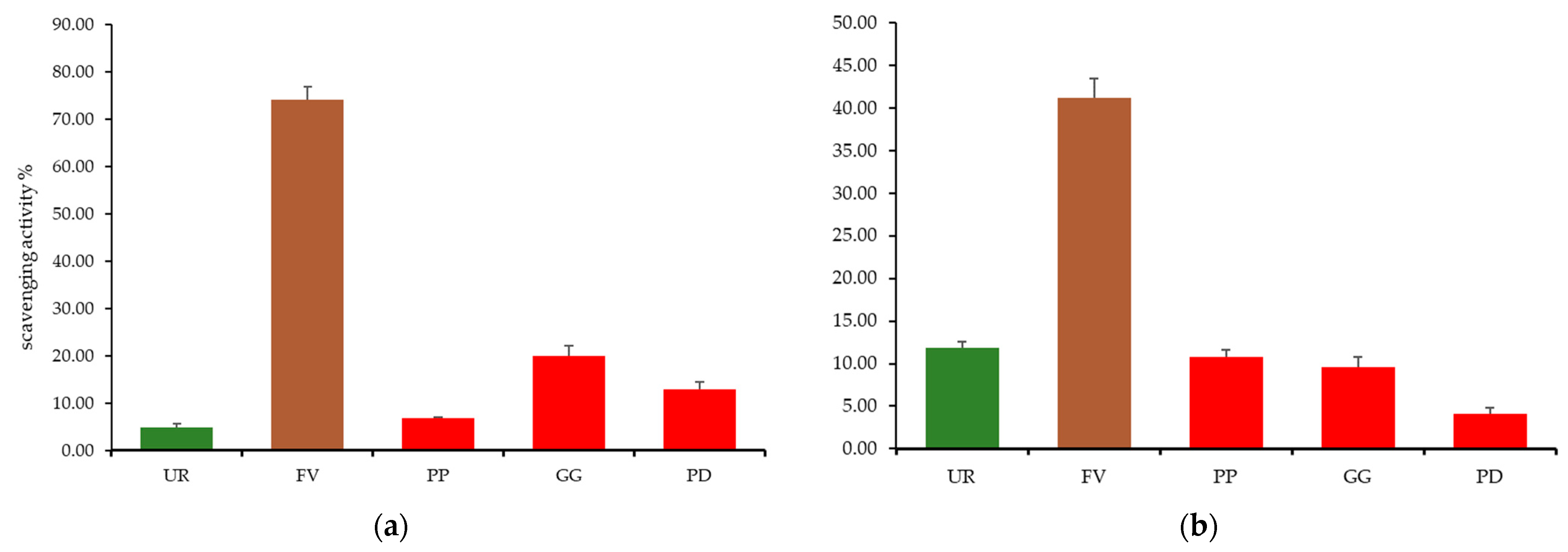
2.5. Total Phenolic Content, Free Radical Scavenging Activity, and Total Antioxidant Activity

Total phenolic content (TPC) of macroalgae ranged from 3.27 mg GAE/g dry seaweed for the green seaweed U. rigida to 10.89 mg GAE/g dry seaweed for the brown seaweed F. vesiculosus. Red seaweeds P. palmata, G. gracilis, and P. dioica presented higher total phenolic contents than the green counterparts but clearly below those presented by the brown species with 5.52, 4.41, and 3.87 mg GAE/g dry seaweed, respectively. Statistical differences in TPC (p < 0.05) were reported between F. vesiculosus and all the remaining species and between U. rigida and P. palmata (Table 6). The presence of phenolic compounds in macroalgae, which may include flavonols, catechins, flavonol glycosides, and in particular, phlorotannins (that can only be found in brown macroalgae species), have shown a noteworthy contribution to cancer, diabetes mellitus, and neurodegenerative and cardiovascular disease prevention, and continue to be the object of novel green extraction technologies and subsequent mechanistic and biological validation [5,77,78,79,80,81]. Currently, at least 301 metabolites harbored in marine macroalgae, including polyphenols, as well as sterols, carotenoids, vitamins, and several others, are known as a source of antioxidant activity [14,82,83]. It should be noted that this myriad of compounds are often multifunctional, expressing not only antioxidant or other previously mentioned biological properties but also technological traits (colorants, preservatives) to replace synthetic counterparts. This function versatility as well as high biodiversity increase the relevance of marine algae as good sources of such natural compounds for food and biomedical applications. This high interest is further supported by the advantageous growth conditions of algae and ease of handling, in comparison to other important plant-based sources of compounds with reported antioxidant activity, since not only are they are highly productive but they also do not need arable land for growth, contributing to sustainable production ecosystems [12,82].
The total antioxidant and DPPH free radical activities of seaweed methanolic extracts are shown in Table 6. Interestingly, a good correlation between TPC of the tested seaweeds and corresponding antioxidant capacity is observed. The higher the TPC, the lower the DPPH antioxidant activity expressed as Trolox equivalents and the higher the percentage scavenging activity. Ulva rigida, which registered the lowest TPC, registered a DPPH activity of 0.129 mg Trolox equiv/g dry seaweed (Table 6) and a percentage scavenging activity of only 9.7% (Figure 1), whereas F. vesiculosus, revealing the highest TPC showed, by far (at least four-fold), the lowest DPPH activity (0.033 mg Trolox equiv/g dry seaweed) and the highest percentage scavenging activity (74.1%). The tested red seaweed species showed intermediate values for both DPPH activity (Table 6) and percentage scavenging activity (Figure 1) which were statistically different (p < 0.05) from the brown species tested but similar between P. palmata and P. dioica, with p > 0.05. A similar tendency regarding the brown macroalgae highest antioxidant activity was observed by Tenorio-Rodriguez et al. (2017) when comparing 17 macroalgae ethanolic extracts (six Rhodophyta, four Chlorophyta, and seven Ochrophyta) collected at Baja California Peninsula, México [84]. Highest mean total phenolic content was registered for the brown macroalgae group (176.5 µg/GAE g dry weight), followed by red macroalgae (45.6 µg/GAE g dry weight) and green macroalgae (32.7 µg/GAE g dry weight) [84]. A similar tendency was observed regarding DPPH scavenging activity (higher values obtained for the brown macroalgae Cystoseira osmundacea—67.9%) and nitric oxide scavenging activity (higher values obtained for the brown macroalgae E. arborea, P. concrecens, and D. delicatula—reductions of 63.2%, 60.6%, and 59.8%, respectively), as well as for ferric reducing antioxidant power (FRAP) assay (brown macroalgae group had the highest value (4.6 ± 3.5 µM FeSO4 µg−1) followed by red macroalgae (3.1 µM FeSO4 µg−1) and green macroalgae (2.6 µM FeSO4 µg−1) [84].
In what concerns total antioxidant capacity measured by the ABTS assay, the values reported followed a similar trend to that observed for the DPPH scavenging capacity. Fucus vesiculosus registered a total antioxidant capacity of 8.20 mg Ascorbic Acid equiv/g dry seaweed (Table 6), corresponding to a 40.3% ABTS scavenging activity. The remaining tested species revealed at least four-fold lower scavenging capacities. However, in this case, the green macroalgae U. rigida registered the second-highest scavenging activity value with 11.5%, followed by the three red macroalgae P. palmata, G. gracilis, and P. dioica with 10.6%, 9.3%, and 3.8%, respectively. F. vesiculosus results were similar to those reported for the brown macroalgae Laminaria bongardiana water extract (0.03 mg Ascorbic Acid/gdry algae) but lower than those obtained for both ethanolic or water extracts of other brown macroalgae species such as Laminaria cichorioides, Kjellmaniella crassifolia, Undaria pinnatifida, Costaria costata, and Sargassum pallidum [77]. In what concerns the red macroalgae species P. dioica, G. gracilis, and P. palmata, total phenolic content values were above those obtained for ultrasound-assisted extracts of Gelidium sesquipedale (ranging from 0.42 to 2.52 mg GAE/g dw) but lower regarding DPPH results (ranging from 0.35 to 0.49 Trolox equiv/g dw) [85]. Total phenolic content for the green U. rigida species were above those obtained for crude hydroalcoholic extract and crude aqueous fraction of the green macroalgae Codium fragile (2.202 and 0.298 mg GAE/g dw, respectively) but below those obtained for Codium fragile ethyl acetate fraction (22.381 mg GAE/g dw) and crude hydroalcoholic extract, ethyl acetate, and aqueous fractions of the green Cladophora rupestris (20.179, 21.726, and 15.833 mg GAE/g dw, respectively) [86].
With growing consumer demand and potential industrial applications, natural sources of antioxidant compounds such as the tested macroalgae herein are extremely valuable [16,19,87]. Furthermore, biomedical research has been associating macroalgae antioxidant capacity with benefits for several types of cancer, neurodegenerative diseases, and other pathologies [8,12,88,89,90,91].

3. Materials and Methods

3.1. Specimens of Seaweeds

Specimens of red algae, Rhodophyta (Palmaria palmata, Porphyra dioica, and Gracilaria gracilis), green algae, Chlorophyta (Ulva rígida), and brown algae, Phaeophyceae (Fucus vesiculosus), were obtained from land-based fully controlled cultivation systems under the integrated multi-trophic aquaculture (IMTA) sustainable concept and provided by ALGAplus®—a company specialized in the production of seaweeds and their commercialization for food and cosmetic applications, based in Portugal (Aveiro district). The landbased open IMTA farming system of ALGAplus® is located at the coastal lagoon of Ria de Aveiro. All macroalgae were harvested in January 2018. Algae taxonomic classification were based in the Algaebase [92]. All macroalgae were provided in dried powder with less than 1.0 mm particle size and were used for further experiments.

3.2. Chemical Characterization of Seaweeds

3.2.1. Proximate Composition

Moisture and ash contents were determined according to the AOAC methods (1990).
Total fat content was determined by Soxhlet extraction and nitrogen content by the Kjeldahl method, where protein is calculated by multiplying the nitrogen content by 6.25 [93,94]. Total sugar content was calculated by the sum of all monosaccharides determined, as described in Section 2.3. Total dietary fiber, insoluble dietary fiber, and soluble dietary fiber in seaweeds were determined by the method of Goering and Van Soest, 1975 [95].

3.2.2. Elemental Composition

Elemental composition was determined following the method described by Rodrigues et al. (2015) with some modifications [13]. First, a microwave-assisted acid digestion procedure was performed as proposed by Speedwave MW-3+ (Berghof, Berchtesgaden, Germany) for dried plant samples in dried seaweed samples. A sample with up to 0.25 g dry seaweed was placed in the digestion vessel and added with 6 mL of concentrated nitric acid and 1 mL of hydrogen peroxide. The vessels were capped and placed in a microwave pressure digestor Speedwave MWS-3+ (Berghof) and subjected to microwave radiation at 20 bar according to the following program: room temperature was raised first to 130 °C at 22 °C/min and 30% of irradiation power, then to 160 °C at 6 °C/min and 40% of irradiation power, remaining 5 min at this temperature, and to 170 °C at 5 °C/min and 50% of irradiation power, remaining 5 min at this temperature. The cooling process consisted of decreasing temperature first to 100 °C for 4 min and then to room temperature. After cooling, acid digests were made up to 50 mL with Milli-Q water. Two replicates were performed for each seaweed sample as well as blanks.
The content of each element is expressed as the mean plus standard deviation. The elemental composition was determined using an inductively coupled plasma (ICP) optical emission spectrometer model Optima™ 7000 DV ICP-OES (Dual View, PerkinElmer Life and Analytical Sciences, Shelton, CT, USA) with radial plasma configuration following the method previously described by Rodrigues et al. (2015). The accuracy of the method (microwave acid digestion and ICP-OES analysis) was assessed by analysis of certified reference material NIES-03 (Seaweed Chlorella; LGC standards, UK). Three replicates of reference material were subject to microwave digestion and analyzed three times by ICP-OES. Recovery ranged between 89% and 116%.
To calculate individual molar Na:K ratios, contents of sodium and potassium (mg/10 g dry seaweed) of each sample were converted in millimoles (mmol) using the conversion 23 mg sodium = 1 mmol sodium and 39 mg potassium = 1 mmol potassium [42].

3.2.3. Analysis of Fatty Acids

Fatty acid analysis was performed according to the procedure described by Fontes, Pimentel, Rodríguez-Alcalá, and Gomes, (2018), with some modifications [96]. To 100 mg seaweed sample, 200 µL of tritridecanoin (Sigma-Aldrich, St. Louis, MO, USA) was added. Then, 2.26 mL of methanol (Fisher Chemical, Fair Lawn, NJ, USA) and 800 µL of hexane (Sigma-Aldrich, St. Louis, MO, USA) followed by 240 µL of sodium methoxide (Sigma-Aldrich, St. Louis, MO, USA) were added. Samples were vortexed and incubated at 80 °C for 10 min. After cooling in ice, 1.25 mL of DMF (Sigma-Aldrich, St. Louis, MO, USA) and 1.25 mL of sulfuric acid (Fluka chemicals and reagents, Buchs, Switzerland) were added. Samples were vortexed and incubated at 60 °C for 30 min and 1 mL of hexane was added. Finally, after cooling, samples were vortexed and centrifuged (1250× g; 18 °C; 5 min). The upper layer containing methyl esters (FAME) was collected for further analysis. Samples were analyzed in a gas chromatograph HP6890A (Hewlett-Packard, Avondale, PA, USA) equipped with a flame-ionization detector (GLC-FID) and a BPX70 capillary column (60 m × 0.32 mm × 0.25 μm; SGE Europe Ltd., Courtaboeuf, France) following the chromatographic conditions described by Fontes et al. (2018) [97].

3.3. Polysaccharide Characterization

Neutral sugar analysis was performed following the procedure described by Coimbra, Waldron, and Selvendran, [98], except for red macroalgae Palmaria palmata, Porphyra dioica, and Gracilaria gracilis, for which monosaccharide residues were obtained after acid reductive hydrolysis [99]. For all samples, the monosaccharides obtained after hydrolysis were derivatized to alditol acetates and analyzed by GC-FID, using 2-deoxyglucose (Sigma-Aldrich, St. Louis, MO, USA) as internal standard. Uronic acids (UA) were determined by a modification of the 3-phenylphenol colorimetric method [100].

3.4. Determination of Total Phenolic Content and Antioxidant Activity

3.4.1. Extract Preparation

Antioxidant extraction was performed according to Marinho, Sørensen, Safafar, Pedersen, and Holdt, 2019, with some modifications [101]. Each seaweed dried powder (1 g) was weighed into a centrifuge tube and 25 mL of methanol was added. Tubes were placed in a sonicator for 30 min. Each sample was centrifuged (2164× g for 10 min) and the supernatant was collected into a new tube. The pellet was resuspended, and the extraction process was repeated once. Extractions were performed for each of the three sampling replicates (n = 3).

3.4.2. Phenolic Content

Determination of total phenolic content was performed by the method of Folin–Ciocalteu as previously described by Rodrigues, Sousa, et al., 2015, using gallic acid (LabChem, Zelienople, PA, USA) as standard [102]. Results are expressed as micrograms of gallic acid equivalents per gram of dry seaweed weight. The total polyphenol content of 2 mL was determined by colorimetry at 720 nm. Three true replicates were performed and analyzed in triplicate (n = 9).

3.4.3. DPPH Free Radical Scavenging Activity

The DPPH free radical scavenging activity was determined according to the method described by Suresh et al., 2013, using Trollox (Sigma-Aldrich, St. Louis, MO, USA) as standard [103]. An aliquot (0.1 mL) of each extract (2 mg of lyophilized solids/mL) was added to 3.0 mL of a 0.1 mM ethanolic DPPH solution. After incubation for 30 min at 30 °C in the dark, the absorbance was measured at 517 nm. Three true replicates were performed and analyzed in triplicate (n = 9). Results were expressed as equivalent concentration of trollox (mg trollox equiv/g dry seaweed), whereas the percentage of scavenging activity was also calculated using the following formula:
S c a v e n g i n g % = 1 A s a m p l e A b l a n k A C o n t r o l × 100

3.4.4. Total Antioxidant Capacity

The total antioxidant capacity of extracts solutions was measured according to method described by Gião et al., 2007 [104]. To 2 mL of diluted ABTS solution 120 μL of extract was added and the absorbance at 734 nm of three true replicates was measured using ascorbic acid (Sigma-Aldrich, St. Louis, MO, USA) as standard; analyses were performed in triplicate. Results were expressed as equivalent concentration of ascorbic acid (mg ascorbic acid equiv/g dry seaweed). The percentage of scavenging activity was also determined using the following formula:
S c a v e n g i n g % = A A B T S + A s a m p l e A A B T S + × 100

3.5. Statistical Analysis

Results are reported as mean values ± standard deviations. Statistical analysis was performed using IBM SPSS Statistics version 25 for Microsoft windows. Normality and homogeneity were examined and one-way ANOVA with the Holm-–Sidak test for post hoc analyses was applied to evaluate statistical differences between seaweeds (p < 0.05).

4. Conclusions

Results of the present study show that the tested macroalgae species, obtained from land-based fully controlled cultivation systems, possess an important nutritional profile, being a rich source of high-quality protein and dietary fiber, and providing considerable amounts of essential elements such as Ca, Mg, K, Mn, and Fe important in health promotion and disease prevention. Furthermore, these marine macroalgae are also an excellent source of different kinds of bioactive substances, including sulphated polysaccharides and phenolic compounds with significant antioxidant activity. Comparatively, among the five algae species tested, the brown macroalgae F. vesiculosus clearly stood out, presenting high polysaccharide content, rich in fucoidans and alginates, the highest phenolic content, as well as significant DPPH and ABTS scavenging activities. This study strongly contributes to generating an open mapping of land-based, integrated multi-trophic aquaculture (IMTA)-farmed seaweed composition. Furthermore, we demonstrated that the extraordinary nutritional and bioactive potential of these marine macroalgae can make them excellent ingredients for a wide range of food, cosmetic, and pharmaceutical industry applications. To further prove such potential, future studies should be conducted to explore the bioavailability and determine the in vivo bioactivity of these compounds, opening new research and industrial opportunities.

Author Contributions

Conceptualization, P.N., A.P.-M., A.C.F. and A.M.G.; methodology, P.N., A.C.F. and A.M.G.; software, P.N.; validation, P.N., A.C.F. and A.M.G.; formal analysis, P.N., A.C.F. and A.M.G.; investigation, P.N., É.M. and C.N.; resources, A.C.F., A.M.G., C.N. and M.A.C.; data curation, P.N.; writing—original draft preparation, P.N.; writing—review and editing, P.N., A.P.-M., A.C.F. and A.M.G.; supervision, A.C.F., A.M.G., C.N. and M.A.C.; project administration, A.C.F. and A.M.G.; funding acquisition, A.M.G. and M.A.C.; microalga production and characterization, H.A. All authors have read and agreed to the published version of the manuscript.

Funding

The authors gratefully acknowledge the financial support of “Fundo Europeu de Desenvolvimento Regional” (FEDER) through the research project VALORMAR: Valorizacão Integral dos Recursos Marinhos: Potencial, Inovação Tecnológica e Novas Aplicações (POCI-024517-FEDER) and Paulo Nova’s individual FCT PhD research grant (SFRH/BD/05747/2020). We would also like to thank the scientific collaboration under the FCT project UID/Multi/50016/2020 and from FCT/MEC (PIDDAC)—IF/00588/2015 and within the scope of the project CICECO—Aveiro Institute of Materials (UIDB/50011/2020 and LA/P/0006/2020) and LAQV-REQUIMTE (UIDP/50006/2020), financed by national funds through the FCT/MEC (PIDDAC). C.N. thanks national funds (OE) through FCT, I.P., in the scope of the framework contract foreseen in numbers 4, 5, and 6 of Article 23 of the Decree Law 57/2016 of August 29, changed by Law 57/2017 of July 19.

Institutional Review Board Statement

Not applicable.

Informed Consent Statement

Not applicable.

Data Availability Statement

The data are available from the corresponding author.

Conflicts of Interest

The authors declare no conflict of interest.

Sample Availability

Samples of the compounds are available from the authors.

References

  1. Chapman, R.L. Algae: The world’s most important “plants”—An introduction. Mitig. Adapt. Strateg. Glob. Chang. 2013, 18, 5–12. [Google Scholar] [CrossRef]
  2. Pérez-Lloréns, J.L.; Mouritsen, O.G.; Rhatigan, P.; Cornish, M.L.; Critchley, A.T. Seaweeds in mythology, folklore, poetry, and life. J. Appl. Phycol. 2020, 32, 3157–3182. [Google Scholar] [CrossRef]
  3. Umen, J.; Coelho, S. Algal Sex Determination and the Evolution of Anisogamy. Annu. Rev. Microbiol. 2019, 73, 267–291. [Google Scholar] [CrossRef] [PubMed]
  4. Gutiérrez-Rodríguez, A.G.; Juárez-Portilla, C.; Olivares-Bañuelos, T.; Zepeda, R.C. Anticancer activity of seaweeds. Drug Discov. Today 2018, 23, 434–447. [Google Scholar] [CrossRef] [PubMed]
  5. Dini, I. The Potential of Algae in the Nutricosmetic Sector. Molecules 2023, 28. [Google Scholar] [CrossRef]
  6. Freitas, A.C.; Pereira, L.; Rodrigues, D.; Carvalho, A.; Panteleitchouk, T.; Gomes, A.; Duarte, A. Marine Functional Foods. In Springer Handbook of Marine Biotechnology; Springer: Berlin/Heidelberg, Germany, 2015; pp. 969–994. ISBN 978-3-642-53970-1. [Google Scholar]
  7. Rodrigues, D.; Costa-pinto, A.R.; Sousa, S.; Vasconcelos, M.W.; Pintado, M.M.; Pereira, L.; Rocha-santos, T.A.P. Sargassum muticum and Osmundea pinnatifida Enzymatic Extracts: Chemical, Structural, and Cytotoxic Characterization. Mar. Drugs 2019, 17, 209. [Google Scholar] [CrossRef]
  8. Alves, C.; Silva, J.; Pinteus, S.; Gaspar, H.; Alpoim, M.C.; Botana, L.M.; Pedrosa, R. From marine origin to therapeutics: The antitumor potential of marine algae-derived compounds. Front. Pharmacol. 2018, 9, 777. [Google Scholar] [CrossRef]
  9. Admassu, H.; Gasmalla, M.A.A.; Yang, R.; Zhao, W. Identification of Bioactive Peptides with α-Amylase Inhibitory Potential from Enzymatic Protein Hydrolysates of Red Seaweed (Porphyra spp.). J. Agric. Food Chem. 2018, 66, 4872–4882. [Google Scholar] [CrossRef]
  10. Caporgno, M.P.; Mathys, A. Trends in Microalgae Incorporation Into Innovative Food Products With Potential Health Benefits. Front. Nutr. 2018, 5, 58. [Google Scholar] [CrossRef]
  11. Hamed, I.; Özogul, F.; Özogul, Y.; Regenstein, J.M. Marine Bioactive Compounds and Their Health Benefits: A Review. Compr. Rev. Food Sci. Food Saf. 2015, 14, 446–465. [Google Scholar] [CrossRef]
  12. Nova, P.; Pimenta-martins, A.; Silva, J.L.; Machado, A.; Gomes, A.M.; Freitas, A.C. Health benefits and bioavailability of marine resources components that contribute to health—What’ s new ? Crit. Rev. Food Sci. Nutr. 2020, 60, 3680–3692. [Google Scholar] [CrossRef] [PubMed]
  13. Rodrigues, D.; Freitas, A.C.; Pereira, L.; Rocha-Santos, T.A.P.; Vasconcelos, M.W.; Roriz, M.; Rodríguez-Alcalá, L.M.; Gomes, A.M.P.; Duarte, A.C. Chemical composition of red, brown and green macroalgae from Buarcos bay in Central West Coast of Portugal. Food Chem. 2015, 183, 197–207. [Google Scholar] [CrossRef] [PubMed]
  14. Nova, P.; Gomes, A.M.; Costa-Pinto, A.R. It comes from the sea: Macroalgae-derived bioactive compounds with anti-cancer potential. Crit. Rev. Biotechnol. 2023, 12, 1310. [Google Scholar] [CrossRef]
  15. Markets and Markets Algae Products Market| Size, Share and Market Forecast to 2023 | MarketsandMarkets. Available online: https://www.marketsandmarkets.com/Market-Reports/algae-product-market-250538721.html (accessed on 8 June 2020).
  16. Nova, P.; Martins, A.P.; Teixeira, C.; Abreu, H.; Silva, J.G.; Silva, A.M.; Freitas, A.C.; Gomes, A.M. Foods with microalgae and seaweeds fostering consumers health: A review on scientific and market innovations. J. Appl. Phycol. 2020, 32, 1789–1802. [Google Scholar] [CrossRef]
  17. Ścieszka, S.; Klewicka, E. Algae in food: A general review. Crit. Rev. Food Sci. Nutr. 2019, 59, 3538–3547. [Google Scholar] [CrossRef] [PubMed]
  18. Cherry, P.; O’hara, C.; Magee, P.J.; Mcsorley, E.M.; Allsopp, P.J. Risks and benefits of consuming edible seaweeds. Nutr. Rev. 2019, 77, 307–329. [Google Scholar] [CrossRef]
  19. Wells, M.L.; Potin, P.; Craigie, J.S.; Raven, J.A.; Merchant, S.S.; Helliwell, K.E.; Smith, A.G.; Camire, M.E.; Brawley, S.H. Algae as nutritional and functional food sources: Revisiting our understanding. J. Appl. Phycol. 2017, 29, 949–982. [Google Scholar] [CrossRef]
  20. Wang, H.M.D.; Li, X.C.; Lee, D.J.; Chang, J.S. Potential biomedical applications of marine algae. Bioresour. Technol. 2017, 244, 1407–1415. [Google Scholar] [CrossRef]
  21. Stengel, D.B.; Connan, S. Natural Products from Marine Algae: Methods and Protocols; Humana Press: Totowa, NJ, USA, 2015; ISBN 9781493926848. [Google Scholar]
  22. Christaki, E.; Bonos, E.; Florou-Paneri, P. Chapter 14—Innovative Microalgae Pigments as Functional Ingredients in Nutrition. In Handbook of Marine Microalgae: Biotechnology Advances; Kim, S.-K., Ed.; Academic Press: Boston, MA, USA, 2015; pp. 233–243. ISBN 978-0-12-800776-1. [Google Scholar]
  23. Neves, F.d.F.; Demarco, M.; Tribuzi, G. Drying and Quality of Microalgal Powders for Human Alimentation. In Microalgae—From Physiology to Application; Vítová, M., Ed.; IntechOpen: London, UK, 2019. [Google Scholar]
  24. Fernández-Segovia, I.; Lerma-García, M.J.; Fuentes, A.; Barat, J.M. Characterization of Spanish powdered seaweeds: Composition, antioxidant capacity and technological properties. Food Res. Int. 2018, 111, 212–219. [Google Scholar] [CrossRef]
  25. Bjarnadóttir, M.; Aðalbjörnsson, B.V.; Nilsson, A.; Slizyte, R.; Roleda, M.Y.; Hreggviðsson, G.Ó.; Friðjónsson, Ó.H.; Jónsdóttir, R. Palmaria palmata as an alternative protein source: Enzymatic protein extraction, amino acid composition, and nitrogen-to-protein conversion factor. J. Appl. Phycol. 2018, 30, 2061–2070. [Google Scholar] [CrossRef]
  26. Gadberry, B.A.; Colt, J.; Maynard, D.; Boratyn, D.C.; Webb, K.; Johnson, R.B.; Saunders, G.W.; Boyer, R.H. Intensive land-based production of red and green macroalgae for human consumption in the pacific northwest: An evaluation of seasonal growth, yield, nutritional composition, and contaminant levels. Algae 2018, 33, 109–125. [Google Scholar] [CrossRef]
  27. Lorenzo, J.M.; Agregán, R.; Munekata, P.E.S.; Franco, D.; Carballo, J.; Şahin, S.; Lacomba, R.; Barba, F.J. Proximate composition and nutritional value of three macroalgae: Ascophyllum nodosum, Fucus vesiculosus and Bifurcaria bifurcata. Mar. Drugs 2017, 15, 360. [Google Scholar] [CrossRef] [PubMed]
  28. Bleakley, S.; Hayes, M. Algal Proteins: Extraction, Application, and Challenges Concerning Production. Foods 2017, 6, 33. [Google Scholar] [CrossRef] [PubMed]
  29. Van Krimpen, M.; Bikker, P.; van der Meer, I.; van der Peet-Schwering, C.; Vereijken, J. Cultivation, Processing and Nutritional Aspects for Pigs and Poultry of European Protein Sources as Alternatives for Imported Soybean Products; Wageningen UR Livestock Research: Wageningen, The Netherlands, 2013. [Google Scholar]
  30. Cian, R.E.; Drago, S.R.; De Medina, F.S.; Martínez-Augustin, O. Proteins and carbohydrates from red seaweeds: Evidence for beneficial effects on gut function and microbiota. Mar. Drugs 2015, 13, 5358–5383. [Google Scholar] [CrossRef] [PubMed]
  31. Pereira, L. A review of the nutrient composition of selected edible seaweeds. In Seaweed: Ecology, Nutrient Composition and Medicinal Uses; Nova Science Publishers: Coimbra, Portugal, 2011; pp. 15–47. ISBN 978-1-61470-878-0. [Google Scholar]
  32. Mæhre, H.K.; Malde, M.K.; Eilertsen, K.E.; Elvevoll, E.O. Characterization of protein, lipid and mineral contents in common Norwegian seaweeds and evaluation of their potential as food and feed. J. Sci. Food Agric. 2014, 94, 3281–3290. [Google Scholar] [CrossRef]
  33. Meng, Y.; Sun, J.; Yu, J.; Wang, C.; Su, J. Dietary Intakes of Calcium, Iron, Magnesium, and Potassium Elements and the Risk of Colorectal Cancer: A Meta-Analysis. Biol. Trace Elem. Res. 2019, 189, 325–335. [Google Scholar] [CrossRef]
  34. Tako, E. Dietary trace minerals. Nutrients 2019, 11, 2823. [Google Scholar] [CrossRef]
  35. Sales, C.H.; Fontanelli, M.D.M.; Vieira, D.A.S.; Marchioni, D.M.; Fisberg, R.M. Inadequate dietary intake of minerals: Prevalence and association with socio-demographic and lifestyle factors. Br. J. Nutr. 2017, 117, 267–277. [Google Scholar] [CrossRef]
  36. Mišurcová, L.; Škrovánková, S.; Samek, D.; Ambrožová, J.; Machů, L. Health Benefits of Algal Polysaccharides in Human Nutrition; Academic Press: Burlington, ON, Canada, 2012; Volume 66, ISBN 9780123945976. [Google Scholar]
  37. Orsavova, J. Seaweed Minerals as Nutraceuticals. Adv. Food Nutr. Res. 2011, 64, 371–390. [Google Scholar] [CrossRef]
  38. Circuncisão, A.R.; Catarino, M.D.; Cardoso, S.M.; Silva, A.M.S. Minerals from macroalgae origin: Health benefits and risks for consumers. Mar. Drugs 2018, 16, 400. [Google Scholar] [CrossRef]
  39. Morrissey, E.; Giltinan, M.; Kehoe, L.; Nugent, A.P.; McNulty, B.A.; Flynn, A.; Walton, J. Sodium and potassium intakes and their ratio in adults (18–90 y): Findings from the Irish national adult nutrition survey. Nutrients 2020, 12, 938. [Google Scholar] [CrossRef] [PubMed]
  40. Iwahori, T.; Miura, K.; Ueshima, H. Time to consider use of the sodium-to-potassium ratio for practical sodium reduction and potassium increase. Nutrients 2017, 9, 700. [Google Scholar] [CrossRef] [PubMed]
  41. Stambler, J. The INTERSALT and implications Study: Background, methods, findings, and implications. Am. J. Clin. Nutr. 1997, 65, 626S–642S. [Google Scholar] [CrossRef] [PubMed]
  42. De Morais, I.L.; Lunet, N.; Albuquerque, G.; Gelormini, M.; Casal, S.; Damasceno, A.; Pinho, O.; Moreira, P.; Jewell, J.; Breda, J.; et al. The sodium and potassium content of the most commonly available street foods in Tajikistan and Kyrgyzstan in the context of the FEEDcities project. Nutrients 2018, 10, 98. [Google Scholar] [CrossRef] [PubMed]
  43. Malavolti, M.; Naska, A.; Fairweather-Tait, S.J.; Malagoli, C.; Vescovi, L.; Marchesi, C.; Vinceti, M.; Filippini, T. Sodium and potassium content of foods consumed in an italian population and the impact of adherence to a mediterranean diet on their intake. Nutrients 2021, 13, 2681. [Google Scholar] [CrossRef]
  44. Cormick, G.; Belizán, J.M. Calcium intake and health. Nutrients 2019, 11, 1606. [Google Scholar] [CrossRef]
  45. Theobald, H. Dietary calcium and health. Nutr Bull. 2005, 30, 237–277. [Google Scholar] [CrossRef]
  46. Shkembi, B.; Huppertz, T. Calcium absorption from food products: Food matrix effects. Nutrients 2022, 14, 180. [Google Scholar] [CrossRef]
  47. Volpe, S.L. Magnesium in Disease Prevention and Overall Health. Adv. Nutr. 2013, 4, 378S–383S. [Google Scholar] [CrossRef]
  48. Razzaque, M.S. Magnesium: Are we consuming enough? Nutrients 2018, 10, 1863. [Google Scholar] [CrossRef]
  49. Kirkland, A.E.; Sarlo, G.L.; Holton, K.F. The role of magnesium in neurological disorders. Nutrients 2018, 10, 730. [Google Scholar] [CrossRef]
  50. Chen, F.; Du, M.; Blumberg, J.B.; Chui, K.K.H.; Ruan, M.; Rogers, G.; Shan, Z.; Zeng, L.; Zhang, F.F. Association Among Dietary Supplement Use, Nutrient Intake, and Mortality Among U.S. Adults. Ann. Intern. Med. 2019, 170, 604–613. [Google Scholar] [CrossRef] [PubMed]
  51. Costello, R.B.; Rosanoff, A.; Dai, Q.; Saldanha, L.G.; Potischman, N.A. Perspective: Characterization of Dietary Supplements Containing Calcium and Magnesium and Their Respective Ratio-Is a Rising Ratio a Cause for Concern? Adv. Nutr. 2021, 12, 291–297. [Google Scholar] [CrossRef] [PubMed]
  52. Sliwinska, A.; Luty, J.; Aleksandrowicz-Wrona, E.; Małgorzewicz, S. Iron status and dietary iron intake in vegetarians. Adv. Clin. Exp. Med. 2018, 27, 1383–1389. [Google Scholar] [CrossRef] [PubMed]
  53. Aschner, M.; Erikson, K. Manganese. Adv. Nutr. 2017, 8, 520–521. [Google Scholar] [CrossRef]
  54. Leeming, E.R.; Johnson, A.J.; Spector, T.D.; Roy, C.I.L. Effect of diet on the gut microbiota: Rethinking intervention duration. Nutrients 2019, 11, 2862. [Google Scholar] [CrossRef]
  55. Holscher, H.D. Dietary fiber and prebiotics and the gastrointestinal microbiota. Gut Microbes 2017, 8, 172–184. [Google Scholar] [CrossRef]
  56. De Jesus Raposo, M.F.; De Morais, A.M.M.B.; De Morais, R.M.S.C. Emergent sources of prebiotics: Seaweeds and microalgae. Mar. Drugs 2016, 14, 27. [Google Scholar] [CrossRef]
  57. Patil, N.P.; Le, V.; Sligar, A.D.; Mei, L.; Chavarria, D.; Yang, E.Y.; Baker, A.B. Algal Polysaccharides as Therapeutic Agents for Atherosclerosis. Front. Cardiovasc. Med. 2018, 5, 153. [Google Scholar] [CrossRef]
  58. Kidgell, J.T.; Magnusson, M.; de Nys, R.; Glasson, C.R.K. Ulvan: A systematic review of extraction, composition and function. Algal. Res. 2019, 39, 101422. [Google Scholar] [CrossRef]
  59. Catarino, M.D.; Silva, A.M.S.; Cardoso, S.M. Phycochemical constituents and biological activities of Fucus spp. Mar. Drugs 2018, 16, 249. [Google Scholar] [CrossRef] [PubMed]
  60. Allsopp, P.; Crowe, W.; Bahar, B.; Harnedy, P.A.; Brown, E.S.; Taylor, S.S.; Smyth, T.J.; Soler-Vila, A.; Magee, P.J.; Gill, C.I.R.; et al. The effect of consuming Palmaria palmata-enriched bread on inflammatory markers, antioxidant status, lipid profile and thyroid function in a randomised placebo-controlled intervention trial in healthy adults. Eur. J. Nutr. 2016, 55, 1951–1962. [Google Scholar] [CrossRef] [PubMed]
  61. Khan, H.; Saeedi, M.; Nabavi, S.M.; Mubarak, M.S.; Bishayee, A. Glycosides from Medicinal Plants as Potential Anticancer Agents: Emerging Trends Towards Future Drugs. Curr. Med. Chem. 2019, 26, 2389–2406. [Google Scholar] [CrossRef] [PubMed]
  62. Viana, A.G.; Noseda, M.D.; Gonçalves, A.G.; Duarte, M.E.R.; Yokoya, N.; Matulewicz, M.C.; Cerezo, A.S. β-D-(1→4), β-D-(1→3) “mixed linkage” xylans from red seaweeds of the order Nemaliales and Palmariales. Carbohydr. Res. 2011, 346, 1023–1028. [Google Scholar] [CrossRef] [PubMed]
  63. Alba, K.; Kontogiorgos, V. Seaweed Polysaccharides (Agar, Alginate Carrageenan); Elsevier: Amsterdam, The Netherlands, 2018; ISBN 9780128140451. [Google Scholar]
  64. Yun, E.J.; Yu, S.; Park, N.J.; Cho, Y.; Han, N.R.; Jin, Y.S.; Kim, K.H. Metabolic and enzymatic elucidation of cooperative degradation of red seaweed agarose by two human gut bacteria. Sci. Rep. 2021, 11, 13955. [Google Scholar] [CrossRef] [PubMed]
  65. Yun, E.J.; Yu, S.; Kim, Y.A.; Liu, J.J.; Kang, N.J.; Jin, Y.S.; Kim, K.H. In vitro prebiotic and anti-colon cancer activities of agar-derived sugars from red seaweeds. Mar. Drugs 2021, 19, 213. [Google Scholar] [CrossRef]
  66. Xie, X.T.; Zhang, X.; Liu, Y.; Chen, X.Q.; Cheong, K.L. Quantification of 3,6-anhydro-galactose in red seaweed polysaccharides and their potential skin-whitening activity. 3 Biotech. 2020, 10, 189. [Google Scholar] [CrossRef]
  67. De Carvalho, C.C.C.R.; Caramujo, M.J. The various roles of fatty acids. Molecules 2018, 23, 2583. [Google Scholar] [CrossRef]
  68. Calder, P.C. Functional Roles of Fatty Acids and Their Effects on Human Health. J. Parenter. Enter. Nutr. 2015, 39, 18S–32S. [Google Scholar] [CrossRef]
  69. Sakamoto, A.; Saotome, M.; Iguchi, K.; Maekawa, Y. Marine-derived omega-3 polyunsaturated fatty acids and heart failure: Current understanding for basic to clinical relevance. Int. J. Mol. Sci. 2019, 20, 4025. [Google Scholar] [CrossRef]
  70. Saini, R.K.; Keum, Y.S. Omega-3 and omega-6 polyunsaturated fatty acids: Dietary sources, metabolism, and significance—A review. Life Sci. 2018, 203, 255–267. [Google Scholar] [CrossRef] [PubMed]
  71. Ranard, K.M.; Jeon, S.; Mohn, E.S.; Griffiths, J.C.; Johnson, E.J.; Erdman, J.W. Dietary guidance for lutein: Consideration for intake recommendations is scientifically supported. Eur. J. Nutr. 2017, 56, 37–42. [Google Scholar] [CrossRef] [PubMed]
  72. Li, X.; Bi, X.; Wang, S.; Zhang, Z.; Li, F.; Zhao, A.Z. Therapeutic potential of ω-3 polyunsaturated fatty acids in human autoimmune diseases. Front. Immunol. 2019, 10, 1169–1179. [Google Scholar] [CrossRef] [PubMed]
  73. Kendel, M.; Wielgosz-Collin, G.; Bertrand, S.; Roussakis, C.; Bourgougnon, N.B.; Bedoux, G. Lipid composition, fatty acids and sterols in the seaweeds ulva armoricana, and solieria chordalis from brittany (France): An analysis from nutritional, chemotaxonomic, and antiproliferative activity perspectives. Mar. Drugs 2015, 13, 5606–5628. [Google Scholar] [CrossRef] [PubMed]
  74. De Alencar, D.B.; Diniz, J.C.; Rocha, S.A.S.; Pires-Cavalcante, K.M.S.; De Lima, R.L.; De Sousa, K.C.; Freitas, J.O.; Bezerra, R.M.; Baracho, B.M.; Sampaio, A.H.; et al. Fatty acid composition from the marine red algae pterocladiella capillacea (S. G. gmelin) santelices & hommersand 1997 and osmundaria obtusiloba (C. agardh) R. E. norris 1991 and its antioxidant activity. An. Acad. Bras. Cienc. 2018, 90, 449–459. [Google Scholar] [CrossRef]
  75. El Maghraby, D.M.; Fakhry, E.M. Lipid content and fatty acid composition of Mediterranean macro-algae as dynamic factors for biodiesel production. Oceanologia 2015, 57, 86–92. [Google Scholar] [CrossRef]
  76. Rodrigues, D.; Freitas, A.C.; Queirós, R.; Rocha-Santos, T.A.P.; Saraiva, J.A.; Gomes, A.M.P.; Duarte, A.C. Bioactive Polysaccharides Extracts from Sargassum muticum by High Hydrostatic Pressure. J. Food Process. Preserv. 2017, 41, e12977. [Google Scholar] [CrossRef]
  77. Aminina, N.M.; Karaulova, E.P.; Vishnevskaya, T.I.; Yakush, E.V.; Kim, Y.K.; Nam, K.H.; Son, K.T. Characteristics of Polyphenolic Content in Brown Algae of the Pacific Coast of Russia. Molecules 2020, 25, 3909. [Google Scholar] [CrossRef]
  78. Barbosa, M.; Valentão, P.; Ferreres, F.; Gil-Izquierdo, Á.; Andrade, P.B. In vitro multifunctionality of phlorotannin extracts from edible Fucus species on targets underpinning neurodegeneration. Food Chem. 2020, 333, 127456. [Google Scholar] [CrossRef]
  79. Ford, L.; Theodoridou, K.; Sheldrake, G.N.; Walsh, P.J. A critical review of analytical methods used for the chemical characterisation and quantification of phlorotannin compounds in brown seaweeds. Phytochem. Anal. 2019, 30, 587–599. [Google Scholar] [CrossRef]
  80. Murray, M.; Dordevic, A.L.; Ryan, L.; Bonham, M.P. An emerging trend in functional foods for the prevention of cardiovascular disease and diabetes: Marine algal polyphenols. Crit. Rev. Food Sci. Nutr. 2018, 58, 1342–1358. [Google Scholar] [CrossRef] [PubMed]
  81. Mahato, N.; Sinha, M.; Sharma, K.; Koteswararao, R.; Cho, M.H. Modern Extraction and Purification Techniques for Obtaining High Purity Food-Grade Bioactive Compounds and Value-Added Co-Products from Citrus Wastes. Foods 2019, 8, 523. [Google Scholar] [CrossRef]
  82. Pruteanu, L.L.; Bailey, D.S.; Cristian, A. The Biochemistry and Effectiveness of Antioxidants in Food, Fruits, and Marine Algae. Antioxidants 2023, 12, 860. [Google Scholar] [CrossRef] [PubMed]
  83. Cunha, S.A.; Pintado, M.E. Bioactive peptides derived from marine sources: Biological and functional properties. Trends Food Sci. Technol. 2022, 119, 348–370. [Google Scholar] [CrossRef]
  84. Tenorio-Rodriguez, P.A.; Murillo-Álvarez, J.I.; Campa-Cordova, Á.I.; Angulo, C. Antioxidant screening and phenolic content of ethanol extracts of selected Baja California Peninsula macroalgae. J. Food Sci. Technol. 2017, 54, 422–429. [Google Scholar] [CrossRef]
  85. Castejón, N.; Parailloux, M.; Izdebska, A.; Lobinski, R.; Fernandes, S.C.M. Valorization of the red algae gelidium sesquipedale by extracting a broad spectrum of minor compounds using green approaches. Mar. Drugs 2021, 19, 574. [Google Scholar] [CrossRef] [PubMed]
  86. Surget, G.; Roberto, V.P.; Le Lann, K.; Mira, S.; Guérard, F.; Laizé, V.; Poupart, N.; Cancela, M.L.; Stiger-Pouvreau, V. Marine green macroalgae: A source of natural compounds with mineralogenic and antioxidant activities. J. Appl. Phycol. 2017, 29, 575–584. [Google Scholar] [CrossRef]
  87. Fradinho, P.; Flórez-Fernández, N.; Sousa, I.; Raymundo, A.; Domínguez, H.; Torres, M.D. Environmentally friendly processing of Laminaria ochroleuca for soft food applications with bioactive properties. J. Appl. Phycol. 2020, 32, 1455–1465. [Google Scholar] [CrossRef]
  88. Pereira, J.M.; Lopes-Rodrigues, V.; Xavier, C.P.R.; Lima, M.J.; Lima, R.T.; Ferreira, I.C.F.R.; Vasconcelos, M.H. An aqueous extract of Tuberaria lignosa inhibits cell growth, alters the cell cycle profile, and induces apoptosis of NCI-H460 tumor cells. Molecules 2016, 21, 595. [Google Scholar] [CrossRef]
  89. Lee, J.C.; Hou, M.F.; Huang, H.W.; Chang, F.R.; Yeh, C.C.; Tang, J.Y.; Chang, H.W. Marine algal natural products with anti-oxidative, anti-inflammatory, and anti-cancer properties. Cancer Cell Int. 2013, 13, 55. [Google Scholar] [CrossRef]
  90. Sajadimajd, S.; Momtaz, S.; Haratipour, P.; El-Senduny, F.F.; Panah, A.I.; Navabi, J.; Soheilikhah, Z.; Farzaei, M.H.; Rahimi, R. Molecular Mechanisms Underlying Cancer Preventive and Therapeutic Potential of Algal Polysaccharides. Curr. Pharm. Des. 2019, 25, 1210–1235. [Google Scholar] [CrossRef] [PubMed]
  91. Geisen, U.; Zenthoefer, M.; Peipp, M.; Kerber, J.; Plenge, J.; Managò, A.; Fuhrmann, M.; Geyer, R.; Hennig, S.; Adam, D.; et al. Molecular mechanisms by which a Fucus vesiculosus extract mediates cell cycle inhibition and cell death in pancreatic cancer cells. Mar. Drugs 2015, 13, 4470–4491. [Google Scholar] [CrossRef] [PubMed]
  92. Guiry, M.; Guiry, G.; AlgaeBase. World-Wide Electronic Publication, National University of Ireland, Galway. Available online: http://www.algaebase.org (accessed on 9 June 2020).
  93. Almeida, C.C.; Alvares, T.S.; Costa, M.P.; Conte-Junior, C.A. Protein and Amino Acid Profiles of Different Whey Protein Supplements. J. Diet. Suppl. 2016, 13, 313–323. [Google Scholar] [CrossRef] [PubMed]
  94. Al Juhaimi, F.; Özcan, M.M.; Ghafoor, K.; Babiker, E.E.; Hussain, S. Comparison of cold-pressing and soxhlet extraction systems for bioactive compounds, antioxidant properties, polyphenols, fatty acids and tocopherols in eight nut oils. J. Food Sci. Technol. 2018, 55, 3163–3173. [Google Scholar] [CrossRef] [PubMed]
  95. Goering, H.K.; Van Soest, P. Forage fiber analyses. US Dep. Agric.379 1975, 387–598. [Google Scholar]
  96. Voss, G.B.; Rodríguez-Alcalá, L.M.; Valente, L.M.P.; Pintado, M.M. Impact of different thermal treatments and storage conditions on the stability of soybean byproduct (okara). J. Food Meas. Charact. 2018, 12, 1981–1996. [Google Scholar] [CrossRef]
  97. Fontes, A.L.; Pimentel, L.; Rodríguez-Alcalá, L.M.; Gomes, A. Effect of Pufa Substrates on Fatty Acid Profile of Bifidobacterium breve Ncimb 702258 and CLA/CLNA Production in Commercial Semi-Skimmed Milk. Sci. Rep. 2018, 8, 15591. [Google Scholar] [CrossRef]
  98. Coimbra, M.A.; Waldron, K.W.; Selvendran, R.R. Isolation and characterisation of cell wall polymers from olive pulp (Olea europaea L.). Carbohydr. Res. 1994, 252, 245–262. [Google Scholar] [CrossRef]
  99. Stevenson, T.T.; Furneaux, R.H. Chemical methods for the analysis of sulphated galactans from red algae. Carbohydr. Res. 1991, 210, 277–298. [Google Scholar] [CrossRef]
  100. Blumenkrantz, N.; Asboe-Hansen, G. New method for quantitative determination of uronic acids. Anal. Biochem. 1973, 54, 484–489. [Google Scholar] [CrossRef]
  101. Marinho, G.S.; Sørensen, A.D.M.; Safafar, H.; Pedersen, A.H.; Holdt, S.L. Antioxidant content and activity of the seaweed Saccharina latissima: A seasonal perspective. J. Appl. Phycol. 2019, 31, 1343–1354. [Google Scholar] [CrossRef]
  102. Rodrigues, D.; Sousa, S.; Silva, A.; Amorim, M.; Pereira, L.; Rocha-Santos, T.A.P.; Gomes, A.M.P.; Duarte, A.C.; Freitas, A.C. Impact of enzyme- and ultrasound-assisted extraction methods on biological properties of red, brown, and green seaweeds from the Central West Coast of Portugal. J. Agric. Food Chem. 2015, 63, 3177–3188. [Google Scholar] [CrossRef] [PubMed]
  103. Suresh, V.; Senthilkumar, N.; Thangam, R.; Rajkumar, M.; Anbazhagan, C.; Rengasamy, R.; Gunasekaran, P.; Kannan, S.; Palani, P. Separation, purification and preliminary characterization of sulfated polysaccharides from Sargassum plagiophyllum and its in vitro anticancer and antioxidant activity. Process. Biochem. 2013, 48, 364–373. [Google Scholar] [CrossRef]
  104. Gião, M.S.; González-Sanjosé, M.L.; Rivero-Pérez, M.D.; Pereira, C.I.; Pintado, M.E.; Malcata, F.X. Infusions of Portuguese medicinal plants: Dependence of final antioxidant capacity and phenol content on extraction features. J. Sci. Food Agric. 2007, 87, 2638–2647. [Google Scholar] [CrossRef] [PubMed]
Figure 1. (a) DPPH free radical scavenging activity and (b) ABTS free radical scavenging activity of Ulva Rigida (UR), Fucus vesiculosus (FV), Palmaria palmata (PP), Gracilaria gracilis (GG), and Porphyra dioica (PD) seaweeds.
Molecules 28 04588 g001
Table 1. Proximate composition of seaweeds.
ParameterF. vesiculosus
Molecules 28 04588 i001
G. gracilis
Molecules 28 04588 i002
P. palmata
Molecules 28 04588 i003
P. dioica
Molecules 28 04588 i004
U. rigida
Molecules 28 04588 i005
% moisture (g/100 g *)12.4 ± 0.2 a4.1 ± 0.2 b3.2 ± 0.1 c10.4 ± 0.2 d16.2 ± 0.1 e
% protein (g/100 g *)12.4 ± 0.01 a28.8 ± 0.01 b41.8 ± 0.1 c35.7 ± 0.1 d19.5 ± 0.1 e
% sugars (g/100 g *)30.2 ± 1.3 26.8 ± 2.79.3 ± 0.1 29.8 ± 0.627.9 ± 0.4
% total fat (g/100 g *)3.4 ± 0.2 a0.68 ± 0.01 b0.32 ± 0.02 b,c1.59 ± 0.01 d 0.08 ± 0.004 c
% ash (g/100 g *)29.8 ± 0.1 a29.0 ± 0.2 a27.1 ± 0.6 b23.4 ± 0.03 c26.6 ± 0.4 b
* Dry seaweed. a–e, in a row: different letters indicate significant differences (p < 0.05) between species.
Table 2. Elemental composition of seaweeds and their percentage impact on daily nutrient intake.
Elementsmg/
day
F. vesiculosus
mg/10 g
Portion
%
R
D
I
G. gracilis
mg/10 g
Portion
%
R
D
I
P. palmata
mg/10 g
Portion
%
R
D
I
P. dioica
mg/10 g
Portion
%
R
D
I
U. rigida
mg/10 g
Portion
%
R
D
I
Calcium (Ca)80077.9 ± 1.6 a9.762.4 ± 0.3 b7.8159.1 ± 1.4 c19.922.5 ± 2.3 d2.835.4 ± 2.5 e4.4
Potassium (K)2000369.6 ± 5.5 a18.5892.0 ± 7.5 b44.6345.2 ± 7.2 a 17.3286.3 ± 4.3c14.3136.4 ± 7.4 d6.8
Sodium (Na)--476.4 ± 7.6 a--261.3 ± 1.7 b--175.9 ± 4.2 c--425.6 ± 6.2 d--105.7 ± 3.3 e--
Magnesium (Mg)37581.3 ± 2.4 a21.740.2 ± 1.4 b10.722.20 ± 0.04 b5.961.7 ± 2.3 a,b16.5186.0± c49.6
Phosphorus (P)70011.7 ± 0.2 a1.741.5 ± 1.7 b5.911.0 ± 0.18 a1.639.3 ± 1.4 c5.612.7 ± 0.2 d1.8
Iron (Fe)1410.3 ± 0.3 a73.616.5 ± 0.8 b117.94.6 ± 0.2 c32.96.2 ± 0.2 c44.413.1 ± 0.4 b93.6
Zinc (Zn)100.56 ± 0.5 a5.60.41 ± 0.03 b4.10.18 ± 0.01 c1.80.73 ± 0.02 d7.30.14 ± 0.02 c1.4
Copper (Cu)10.080 ± 0.001a8.00.26 ± 0.01 b26.00.050 ± 0.001a5.00.70 ± 0.03 c70.00.08 ± 0.01 a8.0
Manganese (Mn)22.94 ± 0.03 a145.02.40 ± 0.03 b120.02.10 ± 0.02 c105.01.90 ± 0.01 c95.60.60 ± 0.02 d30.0
Na/K ratio (mmol)--2.19--0.50--0.86--2.52--1.31--
Ca/Mg ratio (mg)--0.96--1.56--7.16--0.36--0.19--
%RDI Based on daily intake portion of 10 g of dry seaweed. a–e, in a row: different letters indicate significant differences (p < 0.05) between species.
Table 3. Total, soluble, and insoluble dietary fiber contents of seaweeds.
ParameterF. vesiculosusG. gracilisP. palmataP. dioicaU. rigida
% fiber (g/100 g *)Total37.0 ± 0.4 a15.0 ± 0.03 b26.5 ± 0.7 c21.7 ± 0.8 d27.3 ± 1.6 c
Soluble15.6 ± 1.1 a7.1 ± 0.1 b9.8 ± 0.4 c20.4 ± 0.9 d8.9 ± 1.4 b,c
Insoluble21.4 ± 1.6 a7.9 ± 0.2 b16.7 ± 0.8 c1.3 ± 0.05 d18.4 ± 0.2 c
* Dry seaweed. a–d in a row: different letters indicate significant differences (p < 0.05) between species.
Table 4. Monosaccharide composition (mol%) for all seaweed samples analyzed.
mol %F. vesiculosusU. rigidaP. palmataP. dioicaG. gracilis
Rha0.423.10.30.10.5
Fuc14.3--2.30.10.7
Rib2.10.21.10.70.6
Ara0.4--0.20.2--
3,6-AnGal----0.214.424.6
Xyl2.06.556.56.52.0
6-O-Me-Gal------10.97.3
4-O-Me-Gal--------4.5
Man17.21.33.37.40.4
Gal3.03.321.953.834.1
Glc9.437.36.02.619.3
UA51.228.48.13.35.9
Ara—arabinose, Fuc—fucose, Gal—galactose, Glc—glucose, Man—manose, Rha—rhamnose, Rib—ribose, UA—uronic acid, Xyl—xylose, 3,6-AnGal—3,6-anhydrogalactose, 4-O-Me-Gal—4-O-Me-galactose, 6-O-Me-Gal—6-O-Me-galactose.
Table 5. Fatty acid composition (µg/mg of dry weight) and total saturated, monounsaturated, and polyunsaturated fatty acid fractions of each seaweed species.
SeaweedF. vesiculosusG. gracilisP. palmataP. dioicaU. rigida
Fatty AcidsMeanSDMeanSDMeanSDMeanSDMeanSD
C14i0.29 a0.010.18 b,c0.020.12 c0.010.23 a,b<0.010.24 a,b 0.02
C143.76 a0.030.71 b0.010.54 c0.010.80 b0.010.08 d0.00
C150.17 <0.01----------------
C15:10.20 a0.010.13 a,b0.010.09 b0.010.17 a,b<0.010.18 a,b0.02
C166.39 a0.063.66 b0.021.99 c0.063.38 b,c0.032.94 c0.21
C16:1------------0.16<0.01----
C16:1 c70.12 a<0.010.08 b0.010.16 c<0.01--------
C16:1 c90.45 a<0.010.58 b0.01------------
C170.09 a<0.01--------0.65 b0.010.27 c0.02
C17:1 c100.09 a<0.01------------0.24 b0.02
C180.71 a0.010.16 b0.010.15 b0.010.11 c<0.010.14 b0.04
C18:1 t40.21 a<0.01------------0.16 a0.01
C18:1 c98.83 a0.050.66 b<0.010.71 b0.040.14 c<0.01----
C18:1 c110.17 a<0.010.22 b<0.010.17 a0.010.21 b<0.011.22 c0.11
C18:2 c9c121.38 a<0.010.10 b<0.010.07 b0.01----0.12 b0.02
C18:3 c9c12c150.91 a0.01------------0.18 b0.03
C200.17 a<0.01------------0.13 b0.02
C18:2 c9t110.07 a<0.01--------0.18 b<0.01----
C210.08<0.01----------------
C20:4 n61.63 a0.010.50 b0.05----0.07 c<0.01----
C220.14 a<0.01----------------
C20:5 n30.53 a<0.01----0.10 b0.01--------
Total µg/mg (dry seaweed)26.396.994.106.115.91
SFA11.794.722.815.173.80
MUFA10.081.661.120.681.80
PUFAS4.520.610.170.260.30
MUFA/SFA ratio0.850.350.400.130.47
SFA/MUFA ratio1.172.842.517.602.11
SFA/(MUFA + PUFAS) ratio0.812.082.185.501.81
Data expressed as mean (Mean; n = 2) and standard deviation (SD). SFA/MUFA/PUFA: total of saturated/monounsaturated/polyunsaturated fatty acids. a–c: in a row, different letters mean significant differences among seaweed species (p < 0.05).
Table 6. Total phenolic content, radical scavenging activity, and total antioxidant capacity of methanolic extracts from tested algae species.
Radical Scavenging
Activity
Total Antioxidant
Capacity
Total Phenolic Content
SEAWEEDDPPH, mg Trolox
equiv/g
Dry Algae Extract
ABTS, mg Ascorbic Acid
equiv/g
Dry Algae Extract
Folin–Ciocalteu, mg
Gallic acid equiv/g % Dry Algae Extract
F. vesiculosus0.033 ± 0.002 a8.20 ± 0.07 a10.89 ± 0.61 a
U. rigida0.129 ± 0.002 b9.89 ± 0.70 b3.27 ± 0.25 b
P. palmata0.127 ± 0.010 b8.35 ± 0.20 a5.52 ± 0.52 c
P. dioica0.126 ± 0.003 b9.67 ± 0.06 b3.87 ± 0.41 b,c
G. gracilis0.115 ± 0.005 c8.90 ± 0.11 c4.41 ± 0.21 b,c
a–c: in a row, different letters mean significant differences among seaweed species (p < 0.05).
Disclaimer/Publisher’s Note: The statements, opinions and data contained in all publications are solely those of the individual author(s) and contributor(s) and not of MDPI and/or the editor(s). MDPI and/or the editor(s) disclaim responsibility for any injury to people or property resulting from any ideas, methods, instructions or products referred to in the content.

