Article

Ulvophyte Green Algae Caulerpa lentillifera: Metabolites Profile and Antioxidant, Anticancer, Anti-Obesity, and In Vitro Cytotoxicity Properties

1
Department of Biological Sciences, State Islamic University of Sunan Kalijaga (UIN Sunan Kalijaga), Yogyakarta 55281, Indonesia
2
Department of Clinical Nutrition, Faculty of Medicine, Hasanuddin University, Makassar 90245, Indonesia
3
Medical School Department, Faculty of Medicine, Brawijaya University, Malang 65145, Indonesia
4
Department of Pathology, College of Korean Medicine, Kyung Hee University, Seoul 02447, Republic of Korea
5
Korean Medicine-Based Drug Repositioning Cancer Research Center, College of Korean Medicine, Kyung Hee University, Seoul 02447, Republic of Korea
6
Nutrition and Food, Faculty of Medicine, Sam Ratulangi University, Manado 95115, Indonesia
*
Author to whom correspondence should be addressed.
Molecules 2023, 28(3), 1365; https://doi.org/10.3390/molecules28031365
Submission received: 19 January 2023 / Revised: 26 January 2023 / Accepted: 27 January 2023 / Published: 31 January 2023
(This article belongs to the Special Issue Discovery of Bioactive Ingredients from Natural Products III)

Abstract

:
Marine algae have excellent bioresource properties with potential nutritional and bioactive therapeutic benefits, but studies regarding Caulerpa lentillifera are limited. This study aims to explore the metabolites profile and the antioxidant, anticancer, anti-obesity, and in vitro cytotoxicity properties of fractionated ethanolic extract of C. lentillifera using two maceration and soxhlet extraction methods. Dried simplicia of C. lentillifera was mashed and extracted in ethanol solvent, concentrated and evaporated, then sequentially partitioned with equal volumes of ethyl acetate and n-Hexane. Six samples were used in this study, consisting of ME (Maceration—Ethanol), MEA (Maceration—Ethyl Acetate), MH (Maceration—n-Hexane), SE (Soxhletation—Ethanol), SEA (Soxhletation—Ethyl Acetate), and SH (Soxhletation—n-Hexane). Non-targeted metabolomic profiling was determined using LC-HRMS, while antioxidant, anti-obesity, and anticancer cytotoxicity were determined using DPPH and ABTS, lipase inhibition, and MTT assay, respectively. This study demonstrates that C. lentillifera has several functional metabolites, antioxidant capacity (EC50 MH is very close to EC50 of Trolox), as well as anti-obesity properties (EC50 MH < EC50 orlistat, an inhibitor of lipid hydrolyzing enzymes), which are useful as precursors for new therapeutic approaches in improving obesity-related diseases. More interestingly, ME, MH, and SE are novel bioresource agents for anticancer drugs, especially for hepatoma, breast, colorectal, and leukemia cancers. Finally, C. lentillifera can be a nutraceutical with great therapeutic benefits.

1. Introduction

The algae group belonging to the genus Caulerpa, especially Caulerpa racemosa and Caulerpa lentillifera, are types of ulvophyte green algae that are widely consumed in the Pacific and Southeast Asian regions. Asia accounted for 97.4% of global seaweed production in 2019 (99.1% of cultivation), with seven of the ten largest-producing countries located in East or Southeast Asia [1,2]. Until now, the main aquaculture production that has high economic value is Caulerpa lentillifera, which is also traded internationally in Pacific countries [3,4]. Although the consumption of macroalgae is not as common in Europe as in Asia, microalgae have gained popularity due to their physiologically active components, earning them the nickname “new superfood” [5]. Groups of algae in the genus Caulerpa are recognized as functional food sources due to the presence of secondary metabolites. These algae are rich in carbohydrates, proteins, unsaturated fatty acids, and vitamin complexes, and have a much higher mineral content when compared to terrestrial vegetables [6,7,8,9]. However, the aquaculture-production potential of many varieties of Caulerpa sea grapes is rarely studied and has never been used in a high-density commercial-scale system [3]. The global harvest of macroalgae in 2013 was estimated at USD 6.7 billion, with more than 95% produced in sea-algae-farming countries [10]. About two-thirds of Indonesia’s territory is in the form of the sea, and it is famous as one of the world’s mega-diversity areas, with more than 555 macroalgae species having been identified in its ocean areas. In addition, most of Indonesia’s islands are located within the Coral Triangle, which has been identified as an area with a very high diversity of Caulerpa [11,12,13].
Caulerpa lentillifera originated in tropical areas around India and the Pacific Ocean, such as Indonesia, but today it can also be found in the Korean Peninsula due to changes in global temperatures. The genus Caulerpa is famous as an edible species that has a high nutritional content, such as minerals, dietary fibers, vitamin A, vitamin C, and several essential unsaturated fatty acids [14,15]. The genus Caulerpa has also been used to treat a wide variety of diseases. For instance, anti-inflammatory, antioxidant, antimicrobial, lipid-lowering, and anticancer properties, as well as cardiovascular protection, renal protection, hepatoprotection, and neuroprotection, are just a few of the previously described medicinal activities of seaweed [16]. Unlike terrestrial plants, marine algae have not been widely used as an alternative medicine or adjuvant to medicines. However, technological advances and innovative engineering have allowed organisms in the marine environment to be used for scientific experiments in the last 50 years [17].
Interestingly, natural populations of C. lentillifera have varying nutritional and biochemical properties due to environmental factors such as predation, sedimentation, salinity, temperature, pollution, and nutrients; thus, different geographical growing fields can contribute to varying levels of nutrients and secondary metabolites [11]. Despite its abundance, research on Indonesian C. lentillifera’s bioactive molecules profile and their direct biological activities is still limited. Due to the uniqueness of each alga and its particular characteristics, exploration of the multifunctional properties of these algae has become urgent. Therefore, in this study, two different extraction methods (maceration and soxhletation) were used, with each extraction using three solvents each having different polarities, namely n-Hexane (non-polar), ethyl acetate (semi-polar), and ethanol (polar), in order to elucidate different compounds on the basis of the degree of polarity of the compounds. To date, there have been no studies profiling and exploring the compounds or metabolites of Indonesian C. lentillifera, especially those related to bioactivity in various kinds of non-communicable diseases, such as cancer and obesity-related diseases. Therefore, this study urgently aimed to profile the metabolites and the antioxidant, anticancer, anti-obesity, and cytotoxicity properties of Indonesian C. lentillifera.

2. Results

2.1. Metabolites Profile of Caulerpa lentillifera via HPLC-ESI-HRMS/MS Analysis

Using two different ethanol extraction methods (maceration and soxhletation), as well as solvent types sequentially partitioned with equal volumes of n-Hexane and ethyl acetate, Caulerpa lentillifera samples with different metabolites profiles were successfully obtained and analyzed using non-targeted metabolomic profiling HPLC-ESI-HRMS/MS analysis (Table 1). The data shown in Table 1 are based on matches to the library of mzCloud Best Match (>99%) and are sorted according to their abundance.
The highest abundance of compounds was obtained from C. lentillifera ethanolic macerated-extract fraction (ME), with as many as eight metabolites. Furthermore, six compounds were obtained from MEA, five metabolites from MH, seven compounds from SE, six from SEA, and five from SH. The abundance and type of compounds obtained in this study depended on the extraction method and solvent used.

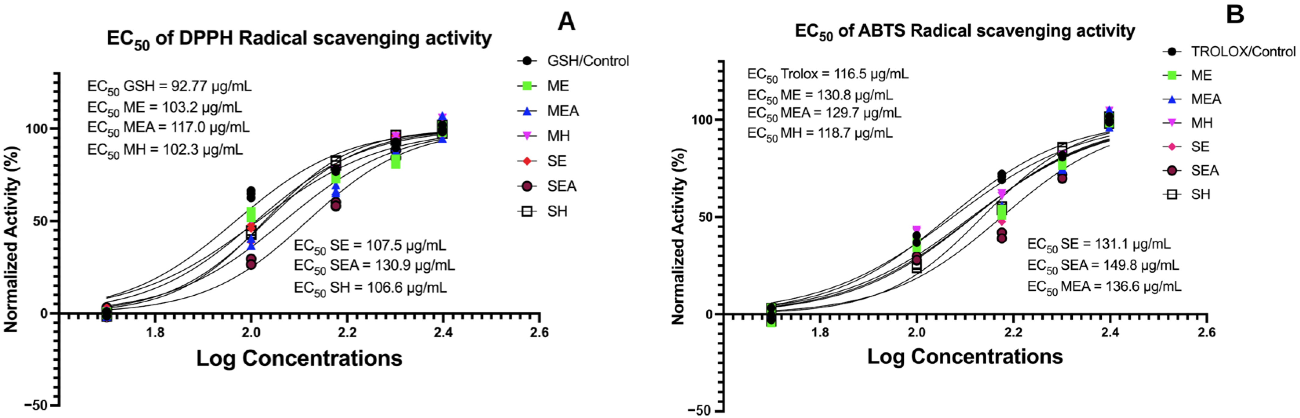
2.2. Radical Scavenging Activity (Antioxidant Properties) of Caulerpa lentillifera

The scavenging activity of free radicals determines antioxidant properties due to its reduction–oxidation (redox) properties. It is important to see the potential of Caulerpa lentillifera as a functional food candidate in reducing various diseases, including cancer and obesity, by improving oxidative stress. The antioxidant potential in this study was evaluated using DPPH and ABTS, and the antioxidant activity observed depended on the concentration dose used (Figure 1).
In the DPPH, it was seen that the antioxidant potential of C. lentillifera in ME, MH, SE, and SH had EC50 values of 103.2 μg/mL, 102.3 μg/mL, 107.5 μg/mL, and 106.6 μg/mL, respectively, compared to glutathione as a positive control (GSH), which showed an EC50 value of 92.77 μg/mL (Figure 1A). In the ABTS radical scavenging assay, it appears that C. lentillifera in MH has the potential to be an antioxidant compared to Trolox as a positive control (EC50 MH is very close to EC50 Trolox; Figure 1B).

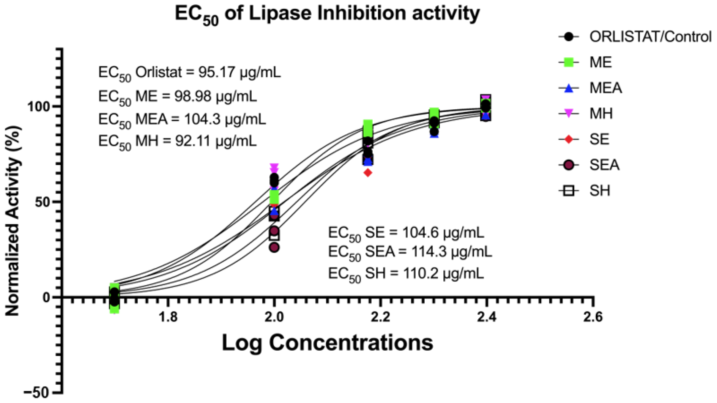
2.3. The Anti-obesity Potential of Caulerpa lentillifera via Lipase Inhibitory Activity

The inhibitory activity of lipase determines anti-obesity properties due to its inhibitory properties to lipid (lipase) hydrolyzing enzymes, and it is important to see the potential of Caulerpa lentillifera as a functional food candidate in reducing various diseases, including obesity. Anti-obesity potential in this study was evaluated using in vitro lipase inhibitory activity in a dose-dependent manner (Figure 2).
It was shown that C. lentillifera, especially in MH, shows more potent anti-obesity activity than the standard drug orlistat, which was a positive control (EC50 MH < EC50 orlistat). This is in line with the results in Figure 1B, which highlight MH having the highest antioxidant properties compared to other groups. Furthermore, the anti-obesity potential of C. lentillifera in ME, MEA, SE, SH, and SEA exhibited EC50 values of 98.98 μg/mL, 104.3 μg/mL, 104.6 μg/mL, 110.2 μg/mL, and 114.3 μg/mL, respectively, approaching the EC50 values of orlistat (95.17 μg/mL; Figure 2).

2.4. Cytotoxicity Properties of C. lentillifera

The LC50 value of C. lentillifera in a cytotoxicity test in normal cell lines is presented in Table 2. The LC50 sequence of samples that were lowest or showing the highest cytotoxicity to the lowest cytotoxicity is SE, ME, SEA, MH, MEA, and SH at 24 incubation hours; and MEA, SE, ME, MH, SEA, and SH at 48 h (Table 2).
In general, the LC50 value of C. lentillifera > 500 μg/mL, and this result suggested that C. lentillifera (ME, MEA, MH, SE, SEA, and SH) as an antioxidant with anti-obesity agents in the observed EC50 was safe. Furthermore, in terms of cytotoxicity, it was observed that C. lentillifera was safe to be potentially developed into various products, especially future functional food products.

2.5. Antiproliferative Activity of Caulerpa lentillifera

When considering the anticancer potential of C. lentillifera via proliferation inhibition of ethanolic extract and its fractions, ME, MH, and SE showed some potential against various observed cancer cells (Table 3). For example, ME (IC50: 160.80 μg/mL) and MH (IC50: 100.50 μg/mL) showed stronger activity on the KAIMRC1 cell line, while MH with IC50: 283.00 μg/mL showed high activity on the HEP G2 cell line, thus indicating its potential as an anticancer agent in hepatoma. Similarly, ME (IC50: 170.10 μg/mL), MH (IC50: 184.00 μg/mL), and SE (IC50: 160.50 μg/mL) showed high activity on the colorectal HCT-8 cell line. Furthermore, ME (IC50: 320.50 μg/mL), MH (IC50: 280.50 μg/mL), and SE (IC50: 104.10 μg/mL) also showed high activity on cell line MDA-MB-231, followed by ME (IC50: 100.90 μg/mL) and MH (IC50: 350.20 μg/mL), which showed high activity on the MCF-7 cell line. Even MH showed high activity against KG-1a, with IC50 of 340.50 μg/mL, thus showing its potential as an anticancer agent in breast cancer. Interestingly, ME with an IC50 of 315.50 μg/mL also showed high activity on the HL-60 leukemia cell line. As in the normal cell cytotoxicity shown in Table 2, IC50 on the normal/control cell lines (monocular blood/PBMC and epithelial) was more than 500 μg/mL, which indicated that it was safe.

3. Discussion

This study intended to explore the metabolites profile and the antioxidant, anti-obesity, anticancer, and in vitro cytotoxicity of fractionated ethanol extract of C. lentillifera, ulvophyte green algae. The Mediterranean diet that has been recognized worldwide is associated with a reduced risk of cancer, heart disease, obesity, stroke, and other non-communicable diseases [18,19]. This lower risk is universally ascribed to a high intake of vegetables and fruits and, thus, high consumption of phytochemicals, including marine products. This study explored Caulerpa lentillifera using two different ethanol extraction methods (maceration and soxhlet), and the highest abundance of compounds was obtained from C. lentillifera ethanolic macerated-extract fraction (ME) (Table 1). The main compound in the most abundant ME was 3-[3-(beta-d-Glucopyranosyloxy)-2-hydroxyphenyl]propanoic acid (C15H20O9), which in previous studies showed great activity as an anti-obesity agent and molecular antidiabetic [20]. In fact, in this study, many fatty acids and their derivatives were also found, such as α-linolenic acid (in ME), oleamide (in MH), α-eleostearic acid and palmitoleic acid (in SE), and γ-linolenic acid ethyl ester (in SH) (Table 1). Evidence from systematic review and meta-analysis suggests that consumption of fatty acids can lower the risk of cardiometabolic syndrome and restore imbalances in lipid and glucose metabolism, and thus may contribute to the prevention and treatment of cardiovascular diseases, including obesity [21]. Ouabain (in MEA) and cafestol (in SEA) were also observed in C. lentillifera. Cafestol can suppress the rapid growth and migration of cancer cells [22], while ouabain is mainly used in the treatment of congestive heart failure and arrhythmias [23], and even as an anticancer agent in melanoma [24].
Currently, there is great interest in the development of new nutraceuticals of natural origin, in particular marine bioresources for use in pharmaceuticals and/or as potential dietary supplements. This demonstrates the importance of C. lentillifera’s role in the development of future drugs and/or dietary supplements. It is even more interesting that its cytotoxicity properties are considered safe since the lethal concentration (LC50) is >500 μg/mL, implying fewer cytotoxic properties in normal cells (Table 2), based on National Cancer Institute criteria [25]. This is in line with similar studies on the cytotoxicity of subcritical water extraction of genus Caulerpa [26], which did not show significant cytotoxicity activity, and further findings from a review study by Aroyehun et al., (2020), which stated that the toxicity was minimal [27]. Therefore, further development of C. lentillifera products will be of interest to many scientists. In line with the finding by Syakilla et al. [5] that algae is a “new superfood”, recent research has also shown the potential of C. lentillifera as a promising agent against SARS-CoV-2 [28].
One of the most significant and most promising approaches to treatment for obesity and cancer is to fight cellular pro-oxidant states by suppressing the production of reactive oxygen species (ROS) and subsequent oxidative stress [29,30]. Natural antioxidants can present a viable therapeutic target in helping to correct the negative effects of obesity, cancer, and oxidative stress [31,32]. You et al. [33] have studied isolated dietary fiber from C. lentillifera for the first time to identify its potential anti-obesity activity. However, the study by You et al. [33] has not shown potential anti-obesity effects through the lipase inhibition pathway. This study demonstrates that C. lentillifera can be an antioxidant agent with anti-obesity effects since it has an antioxidant capacity (EC50 MH is very close to EC50 Trolox/control positive) followed by anti-obesity properties via lipase inhibition (EC50 MH < EC50 orlistat/control positive)—a useful precursor to new therapeutic approaches in improving obesity-related diseases. Lipase inhibitors are clinically used in obese individuals to improve lipid metabolism [34]. Lipase inhibitors, by inhibiting the absorption of fatty acids, reduce the buildup of free fatty acids, which also lowers LDL levels in serums [35]. Furthermore, cumulative evidence suggests that antioxidants can modulate target and dynamic cellular processes to correct redox imbalances in obesity and cancer [36,37]. Therefore, this study has succeeded in uncovering antioxidants and novel lipase inhibitors that have the potential to improve obesity, cancer, and related comorbidities. Furthermore, in addition to its potential as an antioxidant and anti-obesity agent, C. lentillifera has anticancer capacity, in particular in ME, MH, and SE as novel bioresource agents for anticancer drugs, especially for human hepatoma, breast, colorectal, and leukemia cancers. As regards anticancer activity, C. lentillifera has more broadly demonstrated its anticancer properties than other species, for example Caulerpa racemosa, which has only shown its best potential as a drug candidate for breast cancer [17].
Therefore, this original study is dedicated to providing a view of the latest findings on the best extraction techniques for the recovery of bioactive constituents and the chemical characterization of phytoextracts with valuable biological activity from C. lentillifera. Furthermore, it is hoped that further extensive antioxidant assays can complement the result of this publication, for example by exploring the use of the ESR spectrometer method in determining antioxidant activity. Moreover, the research that is being planned involves in vivo and in vitro bioassays and even human clinical trials to further evidence the biological activity of purified compounds and complex extracts and/or combined extracts derived from different matrices of C. lentillifera. In addition, it is hoped that, in the future, the presentation of data on the compounds examined in this study can be reproduced by other researchers using a computational approach, such as in silico (molecular docking and dynamics simulations).

4. Materials and Methods

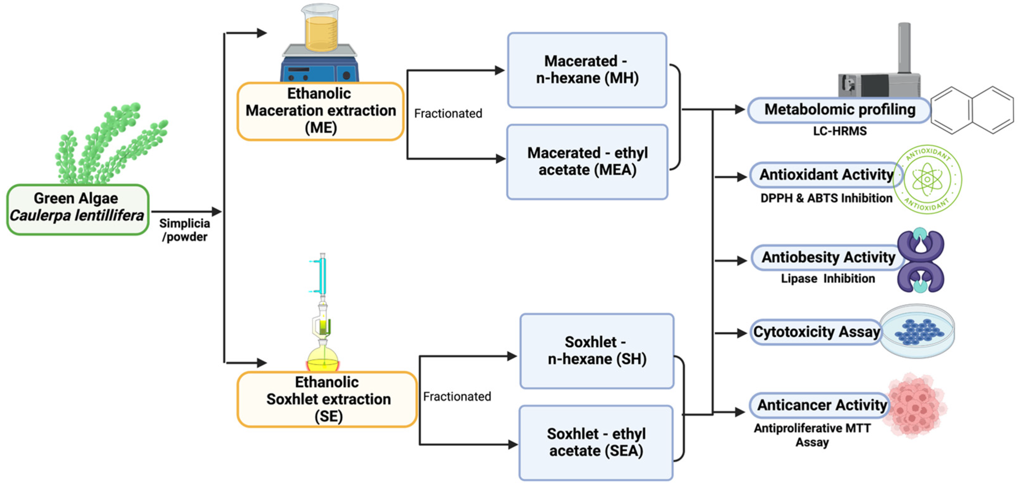
4.1. Preparation of Caulerpa lentillifera

Fresh green algae (Caulerpa lentillifera) or sea grapes were collected from the Caulerpa cultivation pond in Jepara Regency, Central Java Province, Indonesia (6.5868° S latitude, 110.6444° E longitude). The owner of the C. lentillifera pond and the local authorities approved the Caulerpa lentillifera collection sample. Botanical identification and authentication were confirmed at the Integrated Laboratory of the State Islamic University of Sunan Kalijaga (UIN Sunan Kalijaga), Yogyakarta-55281, Indonesia, in accordance with the National Center for Biotechnology Information (NCBI) Taxonomy ID: NCBI: txid148947 and Integrated Taxonomic Information System—Report ID 6973. The researchers (authors) state and confirm that all methods used in this study comply with relevant in vitro and algae research guidelines and regulations. Additionally, the entire body of C. lentillifera to be used was thoroughly washed so that any dirt adhering to it was removed and the plant became clean. C. lentillifera that had been washed, drained, and sun-dried for two to three days were then dried in an oven (IN55 Memmert Incubator; Schwabach, Germany) at 60 degrees Celsius. The dried C. lentillifera was then chopped into small pieces and pulverized in a blender to produce C. lentillifera simplicia powder. The dried simplicia was pulverized and then extracted using maceration and the soxhlet method (Figure 3).

4.2. Caulerpa lentillifera Extraction

The dried simplicia was extracted using either the hot or the cold method [11]. Maceration represents the cold way, while soxhlet represents the hot way.

4.2.1. Maceration Extraction Method (Cold Extraction)

One kilogram of sea grape powder (C. lentillifera) was placed in a dark bottle, to which was added 2 L of 96% ethanol solvent in a ratio of 1:2. The simplicia was soaked for three periods of 24 h. Every 1 × 24 h, the obtained filtrate was stirred occasionally and filtered with Whatman 41 paper, and the residue was re-macerated in a new 96% ethanol solvent. The extraction results were then concentrated using a rotary evaporator under low pressure (100 millibars) for 90 min, evaporated in a 40 °C oven, and sequentially partitioned with equal volumes of ethyl acetate and n-Hexane to yield a thick extract of C. lentillifera (Figure 3).

4.2.2. Soxhlet Extraction Method (Hot Extraction)

A soxhlet device was installed, then 50 g of sea grape powder (C. lentillifera) wrapped in filter paper was placed in a soxhlet tube (thimble). A total of 250 mL of a 96% ethanol solvent was added and divided into two parts: 150 mL was placed in the soxhlet flask (round base flask), and 100 mL was placed in the soxhlet tube to wet the simplicia. The proportion of simplicia to solvent was 1 to 5. The soxhletation procedure was performed at between 70 and 80 degrees Celsius. The extraction time was up to three cycles, and the extraction results were partitioned sequentially with equal volumes of ethyl acetate and n-Hexane (Figure 3).

4.3. Metabolomic Profiling Analysis

In accordance with Permatasari et al., (2022) [20], the untargeted metabolomics profiling test (to view the compounds profile) on the samples (ME, MEA, MH, SE, SEA, and SH; Figure 1) was performed by Laboratorium Sentral Ilmu Hayati (LSIH/Central Laboratory of Life Sciences, Brawijaya University, Malang-65145, Indonesia; ISO 9001:2008 and ISO 17025:2005), test number 041-LSIH-UB-LK. Then, 50 µL of samples were centrifuged for 2 min at 6000 rpm after being vortexed 30 times with ethanol (96%) in a volume of 2000 rpm. Before analysis, 0.22 m syringe filters were used to filter supernatants. The LC-HRMS system consisted of a Thermo Scientific Dionex (Waltham, MA, USA) Ultimate 3000 RSLC Nano High-Performance Liquid Chromatography (HPLC) instrument and a micro flow meter. The analytical column was a 1 mm × 1.9 m Hypersil GOLD aQ 50 column maintained at 30 °C. Solvents A and B contained 0.1% formic acid dissolved in water and 0.1% formic acid dissolved in acetonitrile, respectively. Separation was effected using a linear gradient at a flow rate of 40 L/min for 30 min. The Thermo Scientific Q Exact had a full-scan resolution of 70,000, a data-dependent MS2 resolution of 17,500, and a 30-min operation period in both positive and negative modes.

4.4. Antioxidant Activity by ABTS and DPPH Radical Scavenging Activity Assay (ABTS and DPPH Inhibition) (%)

The scavenging of 2,2′-Azino-bis(3-ethylbenzothiazoline-6-sulfonic acid) or diammonium salt radical cation (ABTS+; Sigma-Aldrich; Saint Louis, MO, USA) was based on the procedure outlined by Permatasari [38]. Potassium persulfate (2.4 mM) and ABTS (7 mM) were mixed in a 1:1 ratio, shielded from light with aluminum foil, and allowed to react for 14 h at 22 degrees Celsius. To obtain a working solution with an absorbance of 0.706 at 734 nm, the mixture was further diluted (e.g., 1 mL of the stock solution plus 60 mL of ethanol). For each test, a fresh working solution was formulated. The samples’ (ME, MEA, MH, SE, SEA, and SH) extracts (50, 100, 150, 200, and 250 µg/mL) were diluted with ABTS working solution (1 mL), and the absorbance at 734 nm was measured after 7 min. As a positive control, the known antioxidant molecule Trolox was utilized. The antioxidant activity in the (2,2-diphenyl-1-picrylhydrazyl radical scavenging activity) (DPPH) test was assayed in accordance with Permatasari’s procedure [38]. DPPH solution was prepared by dissolving 24 mg of DPPH in 100 mL of methanol. In the testing vial, a concentration of 50, 100, 150, 200, and 250 μg/mL of samples (ME, MEA, MH, SE, SEA, and SH) was added to the DPPH reagent (3 mL). The DPPH-samples mixture was then incubated at room temperature for 30 min. Change in the concentration of DPPH was observed based on 517 nm absorbance. Glutathione (GSH; Sigma-Aldrich, 354102) was used as a positive control.
To ensure the validity of the data results (ABTS and DPPH tests), each sample went through the above procedures thrice (triplicates test; n = 3).
Inhibition of DPPH and ABTS was expressed as a percentage and determined according to the formula below:
Inhibition   Activity   % = A 0 A 1 A 0 × 100 %
A0 = absorbance of blank; A1 = absorbance of standard or sample.
The half-maximal effective concentration ratio (EC50), which is defined as the sample concentration that results in a 50% decrease in the initial radical concentration, was used to express the radical scavenging ability of the samples and Trolox for ABTS, and the samples and glutathione for the DPPH assay.

4.5. Cytotoxicity Evaluation of Caulerpa lentillifera Using MTT Assay

A human Caucasian skin fibroblast cell line (Normal cell; Bud-8) was used to assess the cell viability [39]. The rate of Bud-8 cell-line proliferation following treatment was determined using a 3-(4,5-dimethylthiazol-2-yl)-2,5-diphenyl tetrazolium bromide (MTT) assay. Mitochondrial dehydrogenase can convert MTT to Formazan, a purple and water-insoluble compound, based on the viability of the cell. Cells were maintained in Dulbecco’s Modified Eagle’s Medium (DMEM), supplemented with 10% fetal bovine serum (FBS) and 1 × Penicillamine-Streptomycin-Neomycin (PSN). Then, 100 microliters of cells (4 × 10⁴ cells/mL) were seeded in a 96-well plate and incubated for 24 h at 37 °C with 5% carbon dioxide. After 24 h of incubation, 100 µL of samples containing 100, 200, 300, 400, and 500 µg/mL was added to 100 µL of cells (ME, MEA, MH, SE, SEA, and SH). The plate was incubated for 24 and 48 h at 37 °C with 5% CO2. After incubation, the morphology of the cells was examined under a microscope. Sigma supplied 20 microliters of a 5 mg/mL MTT solution per well plate. Following an additional two to four hours of incubation, the medium was drained from the plate. Crystals of formazan were dissolved in 100 µL of dimethyl sulfoxide (DMSO; Sigma). The percentage of viable cells and LC50 cells was estimated using the following formula:
%   Cell   Viability = A 1 A 0   ×   100 %
where A0 is absorbance control in cells given 1% DMSO and A1 is absorbance control in cells given the test sample.
Lethal concentration (LC50) is the lowest concentration of samples that inhibits 50% of cells. In general, a low LC50 value indicates high toxicity. Extracts with high LC50 are preferred for use due to their low toxicity effect on host cells [39].

4.6. Anticancer Evaluation of Caulerpa lentillifera via Antiproliferative Activity

The protocol study [17], in which antiproliferative experiments were conducted, was granted institutional review board (IRB) approval. The American Type Culture Collection (ATCC) supplied cell lines for colorectal cancer (HCT-8), human breast cancer (MCF-7), hepatic cancer (Hep G2), leukemia (lymphoblastic: K-562; acute myeloid leukemia: HL-60; erythroleukemia: KG-1a), breast epithelial cancer (MDA-MB-231), and normal breast epithelial cancer (MCF-10A). Primary blood mononuclear cells (PBMC) and the KAIMRC 1 cell line were cultured in the authors’ laboratory. Purchased from Sigma-Aldrich®, the positive control was mitoxantrone (C22H28N4O6; CID 4212).
In accordance with the manufacturer’s instructions, the CellTiter-Glo assay (Promega; Madison, WI, USA) was utilized to determine the effect of algal fractions on the proliferation of non-adherent cells. An EnVision (London, UK) plate reader was utilized to quantify luminescence, which was then normalized to average DMSO controls and expressed as a percentage. Each portion of the extracts had a concentration ranging from 0 to 250 µg/mL, and the cells were seeded in 96-well plates containing a growth medium. After incubating the cells at 37 °C for 24 h, CellTiter-Glo reagent was added to each well and stirred for 2 min. Then, luminescence was measured using the EnVision plate reader (Perkin Elmer; Buckinghamshire, UK). Each compound’s IC50 values (µg/mL), or half-maximal inhibitory concentrations, were calculated [17].
The MTT test was conducted using the aforementioned method. To assess the impact of the algal fractions on the proliferation of adherent cells, the MTT reagent (Sigma) was used in place of the CellTiter-Glo reagent. Utilizing the Spectra Max spectrophotometer from Invitrogen, absorbance was measured. It was then adjusted to average DMSO controls and given as a percentage [40].

4.7. In Vitro Anti-Obesity via Lipase Inhibition Assay (%)

Initially, crude pig-pancreatic lipase (PPL, 1 mg/mL) was dissolved in phosphate buffer (50 mM, pH 7) and then centrifuged at 12,000× g to remove insoluble components. To create an enzyme stock (0.1 mg/mL), the supernatant was diluted 10-fold with buffer. Prior research was used to evaluate the lipase inhibition potential [13]. A transparent 96-well microplate containing 100 µL of samples (ME, MEA, MH, SE, SEA, and SH) was combined with 20 µL of p-nitrophenyl butyrate (pNPB,10 mM in buffer) and incubated for 10 min at 37 °C. The outcome was compared to the reference drug orlistat, a well-known PPL inhibitor. At 405 nm, measurements were taken using a microplate reader. The unit of activity was calculated using the yield from the reaction rate of 1 mol of p-nitrophenol per minute at 37 °C. To measure the lipase inhibition activity, PPL activity was reduced in the test mixture by a specific amount. To ensure the validity of the study results, each sample was verified three times (in triplicate). The inhibitory data were obtained using the equation below.
Inhibition   of   Lipase   Activity   % = 100 B Bc A Ac × 100 %
A = activity without inhibitor; Ac—negative control without inhibitor; B—activity with inhibitor; Bc—negative control with inhibitor.

4.8. Management and Analysis of Data

For the metabolomic profiling study, compound data were sorted according to the Advanced Mass Spectral Database (M/Z cloud; https://www.mzcloud.org, accessed on 23 February 2022) best match criteria with a match rate of >99.0%. The statistical analysis was conducted using the MacBook version of GraphPad Prism 9 Software. Each EC50 and LC50 data set was generated utilizing nonlinear regression formulas to statistically evaluate data from the in vitro experiments, including antioxidant inhibition of ABTS, DPPH, cytotoxicity, and lipase inhibitory activities conducted three times. In addition, the calculated mean and standard deviation were displayed for all data.

5. Conclusions

On the basis of this study, and supported by some scientific evidence, Caulerpa lentillifera is an ulvophyte green alga that has several functional metabolites, antioxidant capacity, as well as anti-obesity properties, which are useful as precursors for new therapeutic approaches in improving obesity-related diseases. More interestingly, ME, MH, and SE are novel bioresource agents for anticancer agents, especially for hepatoma, breast, colorectal, and even leukemia cancers. Therefore, C. lentillifera has potential to be a functional food with strong therapeutic benefits and to be developed as a promising drug candidate in the future. However, in vivo trials and a clinical trial, which the authors are planning for the future, are urgently needed to confirm and validate these mechanisms.

6. Patents

The preparation method and formulation of C. lentillifera extract resulting from the work reported in this study have been registered as a patent in Indonesia with number P00202300540.

Author Contributions

Conceptualization, F.N. and B.K.; methodology, F.N., F.R.Q. and B.K.; software and visualization, F.N.; validation, B.K., N.A.T., N.M., S.K., M.M., J.C., M.C. and M.N.P.; formal analysis and supervision, B.K.; investigation, F.N.; data curation, F.N. and F.R.Q.; writing—original draft preparation, review, and editing, N.A.T., F.N., B.K., N.A.T., N.M., S.K., M.M., J.C., M.C., M.N.P. and F.R.Q. All authors have read and agreed to the published version of the manuscript.

Funding

This research was supported by the Basic Science Research Program through the National Research Foundation of Korea (NRF), funded by the Ministry of Education (NRF-2020R1I1A2066868); the National Research Foundation of Korea (NRF), grant funded by the Korea government (MSIT) (No. 2020R1A5A2019413); and a grant from the Korea Health Technology R&D Project through the Korea Health Industry Development Institute (KHIDI), funded by the Ministry of Health and Welfare, Republic of Korea (grant number: HF20C0038).

Institutional Review Board Statement

Not applicable.

Informed Consent Statement

Not applicable.

Data Availability Statement

The data sets generated and/or analyzed in this study are available in the manuscript or can be requested from the author (F.N.) upon reasonable request.

Conflicts of Interest

The authors declare no conflict of interest.

Sample Availability

Samples of the compounds are not available from the authors.

References

  1. Cai, J.; Lovatelli, A.; Aguilar-Manjarrez, J.; Cornish, L.; Dabbadie, L.; Desrochers, A.; Diffey, S.; Garrido, G.E.; Geehan, J.; Hurtado, A.; et al. Seaweeds and microalgae: An overview for unlocking their potential in global aquaculture development. FAO Fish. Aquac. Circ. 2021, 1229, 48. Available online: https://archimer.ifremer.fr/doc/00705/81738/ (accessed on 10 January 2023).
  2. Das, M.K.; Silpavathi, L.; Das, D. Pharmacognosy and In-vivo anticancer potential of an indigenous marine macroalga, Ulvafasciata Delile from Visakhapatnam coast, India. Orient. Pharm. Exp. Med. 2021, 21, 433–442. Available online: https://link.springer.com/article/10.1007/s13596-021-00594-3 (accessed on 10 January 2023). [CrossRef]
  3. Paul, N.A.; Neveux, N.; Magnusson, M.; de Nys, R. Comparative production and nutritional value of “sea grapes”—The tropical green seaweeds Caulerpa lentillifera and C. racemosa. J. Appl. Phycol. 2013, 26, 1833–1844. Available online: https://link.springer.com/article/10.1007/s10811-013-0227-9 (accessed on 10 January 2023). [CrossRef]
  4. Zhang, M.; Ma, Y.; Che, X.; Huang, Z.; Chen, P.; Xia, G.; Zhao, M. Comparative Analysis of Nutrient Composition of Caulerpa lentillifera from Different Regions. J. Ocean Univ. China 2019, 19, 439–445. [Google Scholar] [CrossRef]
  5. Syakilla, N.; George, R.; Chye, F.Y.; Pindi, W.; Mantihal, S.; Wahab, N.A.; Fadzwi, F.M.; Gu, P.H.; Matanjun, P. A Review on Nutrients, Phytochemicals, and Health Benefits of Green Seaweed, Caulerpa lentillifera. Foods 2022, 11, 2832. Available online: https://www.mdpi.com/1827680 (accessed on 10 January 2023). [CrossRef] [PubMed]
  6. Pooja, S. Algae used as Medicine and Food-A Short Review. J. Appl. Pharm. Sci. Res. 2014, 6, 33–35. [Google Scholar]
  7. Circuncisão, A.R.; Catarino, M.D.; Cardoso, S.M.; Silva, A.M.S. Minerals from Macroalgae Origin: Health Benefits and Risks for Consumers. Mar. Drugs 2018, 16, 400. Available online: https://www.mdpi.com/1660-3397/16/11/400/htm (accessed on 10 January 2023). [CrossRef] [PubMed] [Green Version]
  8. Permatasari, H.K.; Bulain, S.; Amar, N.; Azizah, M.R.; Muslim, F.Z.; Daud, V.P.A.; Nurkolis, F. Anticancer Properties of Caulerpa racemosa: A Review Study. Nutr. Clin. Y Diet. Hosp. 2022, 42, 110–121. Available online: https://revista.nutricion.org/index.php/ncdh/article/download/278/237 (accessed on 10 January 2023).
  9. Belyagoubi, L.; Belyagoubi-Benhammou, N.; Atik-Bekkara, F.; Abdelouahid, D.E. Influence of harvest season and different polarity solvents on biological activities, phenolic compounds and lipid-soluble pigment contents of Spirogyra sp. from Algeria. Orient. Pharm. Exp. Med. 2021, 22, 359–369. Available online: https://link.springer.com/article/10.1007/s13596-021-00551-0 (accessed on 10 January 2023). [CrossRef]
  10. Darmawan, M.; Zamani, N.P.; Irianto, H.E.; Madduppa, H. Molecular Characterization of Caulerpa racemosa (Caulerpales, Chlorophyta) from Indonesia Based on the Plastid tufA Gene. Squalen Bull. Mar. Fish. Postharvest Biotechnol. 2021, 16, 101–109. Available online: http://www.bbp4b.litbang.kkp.go.id/squalen-bulletin/index.php/squalen/article/view/588 (accessed on 10 January 2023). [CrossRef]
  11. Nurkolis, F.; Yusuf, V.M.; Yusuf, M.; Kusuma, R.J.; Gunawan, W.B.; Hendra, I.W.; Radu, S.; Taslim, N.A.; Mayulu, N.; Sabrina, N.; et al. Metabolomic Proling, In Vitro Antioxidant and Cytotoxicity Properties of Caulerpa racemosa: Functional Food of the Future from Algae. Res. Sq. 2022, 2158307, pp. 1–25. Available online: https://www.researchsquare.com/article/rs-2158307/latest.pdf (accessed on 10 January 2023).
  12. Kuswari, M.; Nurkolis, F.; Mayulu, N.; Ibrahim, F.M.; Taslim, N.A.; Wewengkang, D.S.; Sabrina, N.; Arifin, G.R.; Mantik, K.E.K.; Bahar, M.R.; et al. Sea grapes extract improves blood glucose, total cholesterol, and PGC-1α in rats fed on cholesterol- and fat-enriched diet. F1000Research 2021, 10, 718. [Google Scholar] [CrossRef] [PubMed]
  13. Permatasari, H.K.; Firani, N.K.; Prijadi, B.; Irnandi, D.F.; Riawan, W.; Yusuf, M.; Amar, N.; Chandra, L.A.; Yusuf, V.M.; Subali, A.D.; et al. Kombucha drink enriched with sea grapes (Caulerpa racemosa) as potential functional beverage to contrast obesity: An in vivo and in vitro approach. Clin. Nutr. ESPEN 2022, 49, 232–240. Available online: https://www.sciencedirect.com/science/article/pii/S2405457722002406 (accessed on 10 January 2023). [CrossRef]
  14. Sharma, B.R.; Rhyu, D.Y. Anti-diabetic effects of Caulerpa lentillifera: Stimulation of insulin secretion in pancreatic ß-cells and enhancement of glucose uptake in adipocytes. Asian Pac. J. Trop. Biomed. 2014, 4, 575–580. Available online: https://www.sciencedirect.com/science/article/pii/S2221169115301325 (accessed on 10 January 2023). [CrossRef] [PubMed] [Green Version]
  15. Manoppo, J.I.C.; Nurkolis, F.; Pramono, A.; Ardiaria, M.; Murbawani, E.A.; Yusuf, M.; Qhabibi, F.R.; Yusuf, V.M.; Amar, N.; Karim, M.R.A.; et al. Amelioration of obesity-related metabolic disorders via supplementation of Caulerpa lentillifera in rats fed with a high-fat and high-cholesterol diet. Front. Nutr. 2022, 9, 1010867. Available online: https://www.researchgate.net/profile/Faqrizal-Qhabibi/publication/363581598_Amelioration_of_Obesity-related_Metabolic_Disorders_Via_Supplementation_of_Caulerpa_lentillifera_in_Rats_Fed_With_a_High-fat_and_High-cholesterol_Diet/links/632365f30a70852150f7cd (accessed on 10 January 2023). [CrossRef] [PubMed]
  16. Shah, M.D.; Maran, B.A.V.; Shaleh, S.R.M.; Zuldin, W.H.; Gnanaraj, C.; Yong, Y.S. Therapeutic Potential and Nutraceutical Profiling of North Bornean Seaweeds: A Review. Mar. Drugs 2022, 20, 101. Available online: https://www.mdpi.com/1660-3397/20/2/101 (accessed on 10 January 2023). [CrossRef]
  17. Dissanayake, I.H.; Bandaranayake, U.; Keerthirathna, L.R.; Manawadu, C.; Silva, R.M.; Mohamed, B.; Rizwan, A.; Peiris, D.C. Integration of in vitro and in-silico analysis of Caulerpa racemosa against antioxidant, antidiabetic, and anticancer activities. Sci. Rep. 2022, 12, 20848. Available online: https://www.nature.com/articles/s41598-022-24021-y (accessed on 10 January 2023). [CrossRef] [PubMed]
  18. Muscogiuri, G.; Verde, L.; Sulu, C.; Katsiki, N.; Hassapidou, M.; Frias-Toral, E.; Cucalón, G.; Pazderska, A.; Yumuk, V.D.; Colao, A.; et al. Mediterranean Diet and Obesity-related Disorders: What is the Evidence? Curr. Obes. Rep. 2022, 11, 287–304. [Google Scholar] [CrossRef]
  19. Estruch, R.; Ros, E. The role of the Mediterranean diet on weight loss and obesity-related diseases. Rev. Endocr. Metab. Disord. 2020, 21, 315–327. [Google Scholar] [CrossRef]
  20. Permatasari, H.K.; Nurkolis, F.; Hardinsyah, H.; Taslim, N.A.; Sabrina, N.; Ibrahim, F.M.; Visnu, J.; Kumalawati, D.A.; Febriana, S.A.; Sudargo, T.; et al. Metabolomic Assay, Computational Screening, and Pharmacological Evaluation of Caulerpa racemosa as an Anti-obesity with Anti-aging by Altering Lipid Profile and Peroxisome Proliferator-Activated Receptor-γ Coactivator 1-α Levels. Front. Nutr. 2022, 9, 1412. Available online: https://www.frontiersin.org/articles/10.3389/fnut.2022.939073/full (accessed on 10 January 2023). [CrossRef]
  21. Fazelian, S.; Moradi, F.; Agah, S.; Hoseini, A.; Heydari, H.; Morvaridzadeh, M.; Omidi, A.; Pizarro, A.B.; Ghafouri, A.; Heshmati, J. Effect of omega-3 fatty acids supplementation on cardio-metabolic and oxidative stress parameters in patients with chronic kidney disease: A systematic review and meta-analysis. BMC Nephrol. 2021, 22, 160. [Google Scholar] [CrossRef]
  22. Iwamoto, H.; Izumi, K.; Natsagdorj, A.; Naito, R.; Makino, T.; Kadomoto, S.; Hiratsuka, K.; Shigehara, K.; Kadono, Y.; Narimoto, K.; et al. Coffee diterpenes kahweol acetate and cafestol synergistically inhibit the proliferation and migration of prostate cancer cells. Prostate 2018, 79, 468–479. [Google Scholar] [CrossRef] [PubMed]
  23. Botelho, A.F.M.; Miranda, A.L.S.; Freitas, T.G.; Milani, P.F.; Barreto, T.; Cruz, J.S.; Melo, M.M. Comparative Cardiotoxicity of Low Doses of Digoxin, Ouabain, and Oleandrin. Cardiovasc. Toxicol. 2020, 20, 539–547. [Google Scholar] [CrossRef] [PubMed]
  24. L’Hôte, V.; Courbeyrette, R.; Pinna, G.; Cintrat, J.C.; Le Pavec, G.; Delaunay-Moisan, A.; Mann, C.; Thuret, J.-Y. Ouabain and chloroquine trigger senolysis of BRAF-V600E-induced senescent cells by targeting autophagy. Aging Cell 2021, 20, e13447. [Google Scholar] [CrossRef] [PubMed]
  25. Niksic, H.; Becic, F.; Koric, E.; Gusic, I.; Omeragic, E.; Muratovic, S.; Miladinovic, B.; Duric, K. Cytotoxicity screening of Thymus vulgaris L. essential oil in brine shrimp nauplii and cancer cell lines. Sci. Rep. 2021, 11, 13178. Available online: https://www.nature.com/articles/s41598-021-92679-x (accessed on 10 January 2023). [CrossRef]
  26. Pangestuti, R.; Haq, M.; Rahmadi, P.; Chun, B.-S. Nutritional Value and Biofunctionalities of Two Edible Green Seaweeds (Ulva lactuca and Caulerpa racemosa) from Indonesia by Subcritical Water Hydrolysis. Mar. Drugs 2021, 19, 578. Available online: https://www.mdpi.com/1660-3397/19/10/578/htm (accessed on 10 January 2023). [CrossRef]
  27. Aroyehun, A.Q.B.; Razak, S.A.; Palaniveloo, K.; Nagappan, T.; Rahmah, N.S.N.; Jin, G.W.; Chellappan, D.K.; Chellian, J.; Kunnath, A.P. Bioprospecting Cultivated Tropical Green Algae, Caulerpa racemosa (Forsskal) J. Agardh: A Perspective on Nutritional Properties, Antioxidative Capacity and Anti-Diabetic Potential. Foods 2020, 9, 1313. Available online: https://www.mdpi.com/2304-8158/9/9/1313/htm (accessed on 10 January 2023). [CrossRef]
  28. You, Y.; Song, H.; Wang, L.; Peng, H.; Sun, Y.; Ai, C.; Wen, C.; Zhu, B.; Song, S. Structural characterization and SARS-CoV-2 inhibitory activity of a sulfated polysaccharide from Caulerpa lentillifera. Carbohydr. Polym. 2022, 280, 119006. [Google Scholar] [CrossRef]
  29. Udensi, U.K.; Tchounwou, P.B. Dual effect of oxidative stress on leukemia cancer induction and treatment. J. Exp. Clin. Cancer Res. 2014, 33, 106. [Google Scholar] [CrossRef] [Green Version]
  30. Arany, I.; Hall, S.; Reed, D.K.; Dixit, M. The pro-oxidant gene p66shc increases nicotine exposure-induced lipotoxic oxidative stress in renal proximal tubule cells. Mol. Med. Rep. 2016, 14, 2771–2777. Available online: https://www.spandidos-publications.com/mmr/14/3/2771 (accessed on 10 January 2023). [CrossRef] [Green Version]
  31. Takaki, A.; Kawai, D.; Yamamoto, K. Multiple hits, including oxidative stress, as pathogenesis and treatment target in non-alcoholic steatohepatitis (NASH). Int. J. Mol. Sci. 2013, 14, 20704–20728. Available online: https://www.mdpi.com/59072 (accessed on 10 January 2023). [CrossRef] [Green Version]
  32. Wang, Z.; Li, Z.; Ye, Y.; Xie, L.; Li, W. Oxidative stress and liver cancer: Etiology and therapeutic targets. Oxidative Med. Cell. Longev. 2016, 2016, 7891574. Available online: https://www.hindawi.com/journals/omcl/2016/7891574/ (accessed on 10 January 2023). [CrossRef] [PubMed] [Green Version]
  33. You, Y.; Song, H.; Yan, C.; Ai, C.; Tong, Y.; Zhu, B.; Song, S. Dietary fibers obtained from Caulerpa lentillifera prevent high-fat diet-induced obesity in mice by regulating the gut microbiota and metabolite profiles. Food Funct. 2022, 13, 11262–11272. Available online: https://pubs.rsc.org/en/content/articlehtml/2022/fo/d2fo01632j (accessed on 10 January 2023). [CrossRef] [PubMed]
  34. Patil, S.; Patil, M.; Maheshwari, V.L.; Patil, R.H. Pancreatic Lipase (PL) Inhibitors from Medicinal Plants and Their Potential Applications in the Management of Obesity. In Natural Products as Enzyme Inhibitors; Maheshwari, V.L., Patil, R.H., Eds.; Springer: Singapore, 2022; pp. 153–167. [Google Scholar]
  35. Liu, T.T.; Liu, X.T.; Chen, Q.X.; Shi, Y. Lipase Inhibitors for Obesity: A Review. Biomed. Pharmacother. 2020, 128, 110314. Available online: https://www.sciencedirect.com/science/article/pii/S0753332220305060 (accessed on 10 January 2023). [CrossRef] [PubMed]
  36. Hecker, L.; Logsdon, N.J.; Kurundkar, D.; Kurundkar, A.; Bernard, K.; Hock, T.; Meldrum, E.; Sanders, Y.Y.; Thannickal, V.J. Reversal of Persistent Fibrosis in Aging by Targeting Nox4-Nrf2 Redox Imbalance. Sci. Transl. Med. 2014, 6, 231ra47. Available online: https://www.science.org/doi/abs/10.1126/scitranslmed.3008182 (accessed on 10 January 2023). [CrossRef] [Green Version]
  37. Niforou, K.; Cheimonidou, C.; Trougakos, I.P. Molecular chaperones and proteostasis regulation during redox imbalance. Redox Biol. 2014, 2, 323–332. Available online: https://www.sciencedirect.com/science/article/pii/S2213231714000329 (accessed on 10 January 2023). [CrossRef] [Green Version]
  38. Permatasari, H.K.; Nurkolis, F.; Ben Gunawan, W.; Yusuf, V.M.; Yusuf, M.; Kusuma, R.J.; Sabrina, N.; Muharram, F.R.; Taslim, N.A.; Mayulu, N.; et al. Modulation of gut microbiota and markers of metabolic syndrome in mice on cholesterol and fat enriched diet by butterfly pea flower kombucha. Curr. Res. Food Sci. 2022, 5, 1251–1265. [Google Scholar] [CrossRef]
  39. Nemudzivhadi, V.; Masoko, P. In vitro assessment of cytotoxicity, antioxidant, and anti-inflammatory activities of Ricinus communis (euphorbiaceae) leaf extracts. Evid. Based Complement. Altern. Med. 2014, 2014, 625961. Available online: https://www.hindawi.com/journals/ecam/2014/625961/ (accessed on 10 January 2023). [CrossRef] [Green Version]
  40. Salawu, K.M.; Ajaiyeoba, E.O.; Ogbole, O.O.; Adeniji, J.A.; Faleye, T.C.; Agunu, A. Antioxidant, Brine Shrimp Lethality, and Antiproliferative Properties of Gel and Leaf Extracts of Aloe schweinfurthii and Aloe vera. J. Herbs Spices Med. Plants 2017, 23, 263–271. Available online: https://www.tandfonline.com/doi/abs/10.1080/10496475.2017.1318328 (accessed on 10 January 2023). [CrossRef]
Figure 1. Antioxidant capacity of C. lentillifera. (A) The half-maximal effective concentration (EC50) of DPPH radical scavenging activity. (B) The half-maximal effective concentration (EC50) of ABTS radical scavenging activity. ME: Maceration—Ethanol; MEA: Maceration—Ethyl Acetate; MH: Maceration—n-Hexane; SE: Soxhletation—Ethanol; SEA: Soxhletation—Ethyl Acetate; SH: Soxhletation—n-Hexane.
Molecules 28 01365 g001
Figure 2. The half-maximal effective concentration (EC50) of lipase inhibition activity. ME: Maceration—Ethanol; MEA: Maceration—Ethyl Acetate; MH: Maceration—n-Hexane; SE: Soxhletation—Ethanol; SEA: Soxhletation—Ethyl Acetate; SH: Soxhletation—n-Hexane.
Molecules 28 01365 g002
Figure 3. Methodical schematic of Caulerpa lentillifera study. ME: Maceration—Ethanol; MEA: Maceration—Ethyl Acetate; MH: Maceration—n-Hexane; SE: Soxhletation—Ethanol; SEA: Soxhletation—Ethyl Acetate; SH: Soxhletation—n-Hexane.
Molecules 28 01365 g003
Table 1. Metabolites profile observed in C. lentillifera from HPLC-ESI-HRMS/MS analysis.
SampleCompoundsMolecular
Formula
Retention Time (Min)Area Max (Peak Area)Observed MW HR-ESIMS m/z
ME3-[3-(beta-d-Glucopyranosyloxy)-2-hydroxyphenyl]propanoic acidC15H20O915.52,607,709,506.66344.11
CholineC5H13NO0.9481,702,868,432.32104.11
BetaineC5H11NO20.952815,230,823.19117.08
2-(1H-indol-3-yl)-3-[4-(trifluoromethyl)phenyl]acrylonitrileC18H11F3N210.763484,746,646.13312.09
2-(3,4-dihydroxyphenyl)acetamideC8H9NO30.931464,700,110.40167.06
IsoamylamineC5H13N0.916204,192,224.9387.1
Palmitoleic acidC16H30O215.894188,602,459.59254.22
α-Linolenic acidC18H30O220.04675,757,786.32278.44
MEA2,2,6,6-Tetramethyl-1-piperidinol (TEMPO)C9H19NO12.259312,358,366.23156.14
9-Oxo-10(E),12(E)-octadecadienoic acidC18H30O317.615128,180,581.48294.22
13-hydroperoxy-9Z,11E-octadecadienoic acidC18H32O417.427123,110,274.85312.23
ethyl 3-oxo-5,6-diphenyl-2,3- dihydropyridazine-4-carboxylateC19H16N2O317.35173,810,505.50320.12
OuabainC29H44O1220.51857,317,714.92584.66
(3S,3aR,4S,4aR,7aR,8R,9aR)-3,4a,8-
trimethyl-2,5-dioxo-
2H,3H,3aH,4H,4aH,5H,7aH,8H,9H,9aHazuleno[6,5-b]furan-4-yl 2-methylpropanoate
C19H26O514.87451,133,618.50334.18
MH5-(2-Thienyl)nicotinic acidC10H7NO2S0.927209,833,574.73205.02
OleamideC18H35NO21.564113,705,808.63281.27
4-{[(4,6-Dimethoxypyrimidin-2-yl)amino]methylidene}-2-phenyl-4,5-dihydro-1,3-oxazol-5-oneC16H14N4O411.58285,055,308.35326.1
1,2-dihydroxyheptadec-16-yn-4-yl acetateC19H34O415.66979,749,057.14326.25
AdenosineC10H13N5O41.02479,366,759.62267.1
SEBetaineC5H11NO20.902615,708,699.02117.08
dl-β-LeucineC6H13NO20.901104,452,391.68131.09
α-Eleostearic acidC18H30O216.92882,954,091.16278.22
CholineC5H13NO0.90662,987,796.05104.11
Palmitoleic acidC16H30O216.65437,079,182.87254.22
Stearoyl EthanolamideC20H41NO222.833,398,733.18309.3
LevalbuterolC13H21NO313.39825,493,790.28239.15
SEAHexadecanamideC16H33NO21.916353,793,967.91255.26
2,2,6,6-Tetramethyl-1-piperidinol (TEMPO)C9H19NO12.294274,478,961.83156.14
Ethyl palmitoleateC18H34O218.68175,543,295.89282.26
2-(3-Chloro-2-fluorophenyl)-2,3-dihydroisothiazol-3-oneC9H5ClFNOS0.83560,193,728.47228.98
CafestolC20H28O317.43439,577,349.70316.2
ShogaolC17H24O313.13216,983,601.06276.17
SHOctadec-9-ynoic acidC18H32O217.36718,964,856.34280.45
3,5-di-tert-Butyl-4-hydroxybenzaldehydeC15H22O216.95117,509,512.15234.16
Sphingosine (d18:1)C18H37NO220.01516,138,319.09299.5
γ-Linolenic acid ethyl esterC20H34O217.7213,897,675.48306.26
8Z,11Z,14Z-Eicosatrienoic acidC20H34O218.5488,618,378.15306.2545
ME: Maceration—Ethanol; MEA: Maceration—Ethyl Acetate; MH: Maceration—n-Hexane; SE: Soxhletation—Ethanol; SEA: Soxhletation—Ethyl Acetate; SH: Soxhletation—n-Hexane.
Table 2. LC50 value of C. lentillifera in a cytotoxicity test in BUD-8 normal cell lines.
Hours of IncubationLC50 (μg/mL)
MEMEAMHSESEASH
24 h2719.004677.874397.292111.904190.678445.30
48 h186713542090157534405000
ME: Maceration—Ethanol; MEA: Maceration—Ethyl Acetate; MH: Maceration—n-Hexane; SE: Soxhletation—Ethanol; SEA: Soxhletation—Ethyl Acetate; SH: Soxhletation—n-Hexane.
Table 3. IC50 values (μg/mL) exhibited by the C. lentillifera on different cancer (colorectal, hepatoma, breast, leukemia, and normal (monocular blood and epithelial) cell lines.
ExtractColorectalHepatomaBreast Cancer Cell LinesLeukemiaControl Cell Lines
HCT-8Hep G2KAIMR C1MDA-MB-231MCF-7KG-1aK-562HL-60Human EpithelialPBMC
ME170.101065.00160.80320.50100.90508.501500.30315.50850.904040.00
MEA540.60744.60885.011230.501800.301260.801328.502505.904040.603074.10
MH184.00283.00100.50280.50350.20340.502705.603805.505009.007080.00
SE160.50870.60640.60104.10840.60645.60954.601085.012008.003024.30
SEA587.50934.301084.002085.001509.002580.501934.303084.004031.104598.00
SH2003.503282.003056.003824.802506.002503.103203.003003.005056.005033.00
M/
Control
0.1880.1010.7980.6651.4090.1670.4581.0340.1340.204
ME: Maceration—Ethanol; MEA: Maceration—Ethyl Acetate; MH: Maceration—n-Hexane; SE: Soxhletation—Ethanol; SEA: Soxhletation—Ethyl Acetate; SH: Soxhletation—n-Hexane.
Disclaimer/Publisher’s Note: The statements, opinions and data contained in all publications are solely those of the individual author(s) and contributor(s) and not of MDPI and/or the editor(s). MDPI and/or the editor(s) disclaim responsibility for any injury to people or property resulting from any ideas, methods, instructions or products referred to in the content.

