

An Imidazo[1,5-a]pyridine Benzopyrylium-Based NIR Fluorescent Probe with Ultra-Large Stokes Shifts for Monitoring SO2
Department of Chemistry and Pharmaceutical Engineering, Shandong First Medical University & Shandong Academy of Medical Sciences, No. 619, Changcheng Road, Taian 271016, China
*
Authors to whom correspondence should be addressed.
†
These authors contributed equally to this work.
Molecules 2023, 28(2), 515; https://doi.org/10.3390/molecules28020515
Submission received: 8 December 2022
/
Revised: 25 December 2022
/
Accepted: 29 December 2022
/
Published: 5 January 2023
(This article belongs to the Special Issue Fluorescent Probes for Imaging and Diagnostics)
Abstract
:A mitochondria-targeted NIR probe based on the FRET mechanism was developed. It shows ultra-large Stokes shifts (460 nm) and emission shifts (285 nm). Furthermore, we also realized the imaging of SO2 in living SKOV-3 cells, zebrafish and living mice which may be useful for understanding the biological roles of SO2 in mitochondria and in vivo.
1. Introduction
Sulfur dioxide, a well-known atmospheric pollutant, has been regarded as a new possible gas transmitter following NO, CO and H2S [1,2,3,4]. It plays important roles in many physiological processes. SO2 can dissolve easily in water to form its derivatives bisulfite (HSO3−) and sulfite (SO32−), so the physiological functions of SO2 can be attributed to its derivatives (HSO3−/SO32−). However, a high level of endogenous SO2, generated by the oxidation of H2S and thiol-containing amino acids in mitochondria, may bring about neurological disorders, cancers and other diseases [5,6,7,8]. Hence, it is greatly important to establish sensitive and rapid methods for SO2 detection to further gain insight into its functions in biological systems, especially in mitochondria.
Recently, fluorescent probes have become a powerful tool in biological imaging owing to their simplicity, high selectivity and small cell damage [9,10,11,12]. Different from traditional intensity-based probes, ratiometric probes are independent of the probe concentration, environment and excitation intensity [13,14,15]. Besides the ICT (Intramiolecular Charge Transfer)-based ratiometric probes, fluorescence resonance energy transfer (FRET)-based ratiometric probes are the most widely designed and used (Table S1). Until now, numerous FRET-based SO2 probes have been designed and synthesized due to their large pseudo-Stokes shifts, avoiding interference of a biological background [16,17,18,19,20,21,22,23,24,25].
As classic fluorophores, hemicyanines have drawn increasing attention because of their simple synthesis and excellent response to SO2 [26]. Their derivatives were selected as acceptors to construct FRET probes [27,28]. However, the emission of the hemicyanines is around 600 nm, which seriously limits their application in vivo. Therefore, it is of significance to search for new fluorophores, especially with NIR emission, as acceptors.
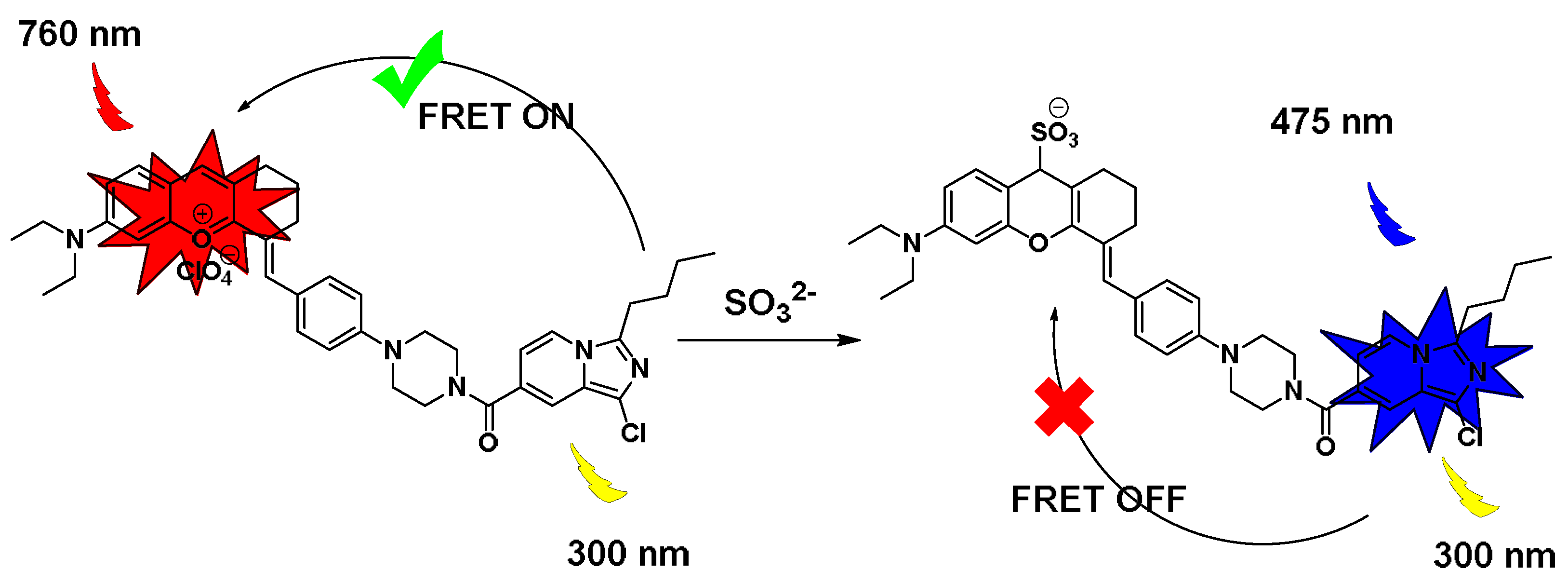
On the other hand, to build an effective FRET platform, the development of new fluorophores as donors whose emission overlaps well with the absorption of acceptors is essential. Owing to the good optical properties [29], imidazole[1,5-a]pyridines were selected as the donor to construct the FRET platform [30]. In addition, we chose benzopyran salt as the acceptor because of its NIR emission. Meanwhile, the benzopyran moiety could not only be used as a reactive site for the Michael addition reaction with SO2 to achieve detection purposes, but it could also target mitochondria due to positive electricity. Therefore, the designed probe IPB-RL-1 could successfully achieve its imaging of SO2 in mitochondria in SKOV-3 cells.
2. Results and Discussion
2.1. Synthesis of IPB-RL-1
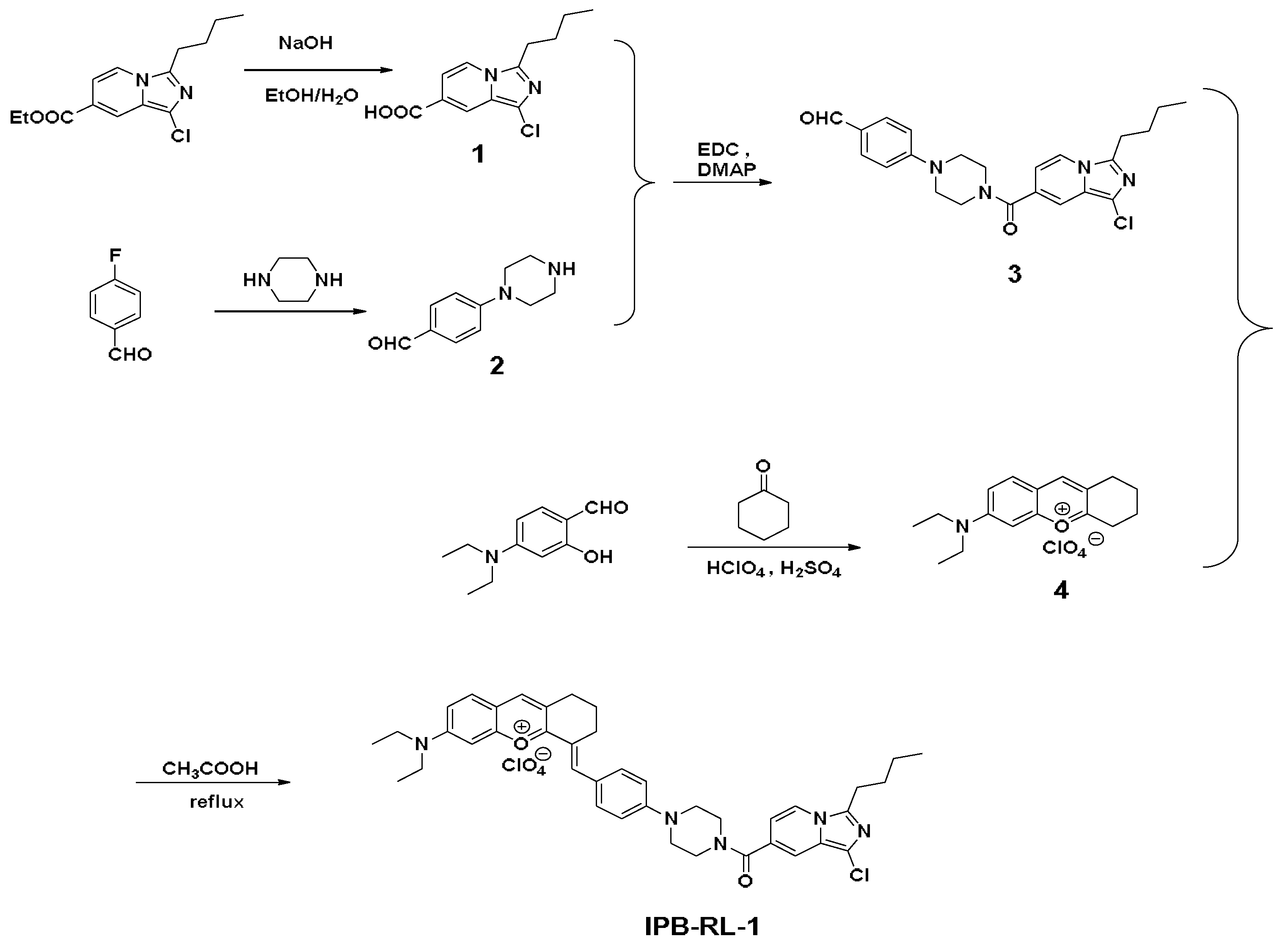
The probe IPB-RL-1 was easily prepared using a classic organic reaction, as shown in Scheme 1. The structure was confirmed by NMR and HRMS.
2.2. Optical Properties of IPB-RL-1
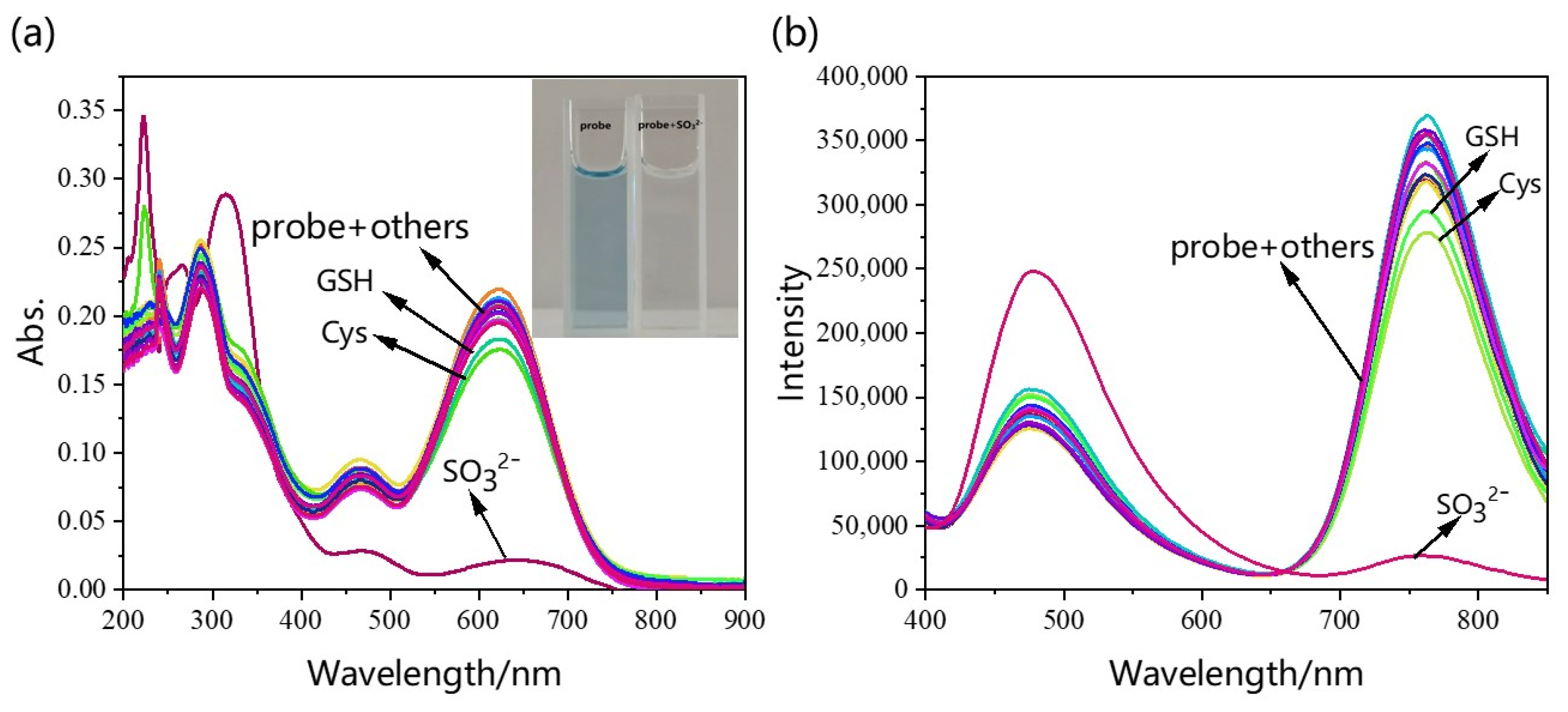
To examine the optical properties of IPB-RL-1, we first examined its selectivity. As shown in Figure 1a,b, there were no obvious changes in absorption and emission after the probe reacted with various ions (Br−, CH3COO−, Cl−, ClO4−, ClO−, F−, H2PO4−, HCO3−, HPO42−, HS−, I−, NO2−, NO3−, S2O82−, SO42−, GSH, and Cys). However, when SO32− was added, it was clearly observed by the naked eye that the probe solution changed from blue to colorless, and the fluorescence intensity was quenched at 760 nm, indicating that IPB-RL-1 showed good selectivity for the detection of SO32−. The anti-interference experiment (Figure S1) demonstrated that IPB-RL-1 had good anti-interference performance and could specifically detect SO32− even in the presence of other ions.
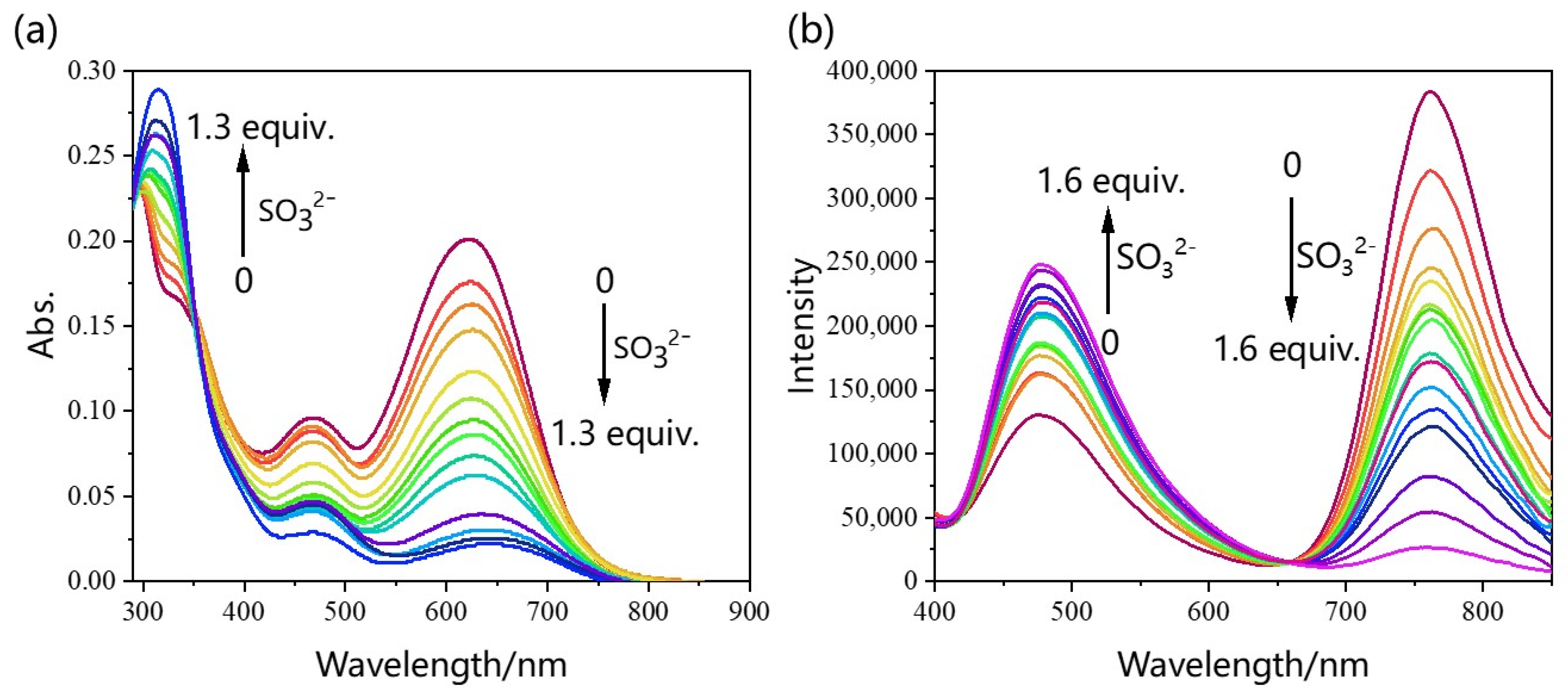
For a better application in living systems, UV–vis and fluorescence titration experiments were also carried out. As shown in Figure 2a, IPB-RL-1 has a strong UV–vis absorption peak at 620 nm in the solution of DMSO/PBS (V/V = 3/7). Yet, with the continuous addition of SO32−, the absorption peak at 620 nm decreased and the absorption peak at 310 nm increased. Meanwhile, the naked eye captured a rapid color change of the probe solution from blue to colorless. The near-infrared fluorescence emission peak at 760 nm decreased with the increase of SO32− while the emission peak at 470 nm increased (Figure 2b), which further confirmed that the FRET was turned off. In addition, an excellent linear correlation between the ratio F470/F760 and SO32− concentration was observed. The detection limit was calculated to be 0.98 μM using the linear regression curve (Figure S2) and LOD formula (LOD = 3 σ/k, σ is the standard deviation of the blank measurement, and k is the slope of the fluorescence emission ratio (I475/I760) and SO32− concentration). In the process of monitoring the reaction time between IPB-RL-1 and SO32−, the fluorescence intensity reached equilibrium (Figure S3) in a very short time (less than 10 s). These results indicated that IPB-RL-1 was suitable for further application in imaging in cells and in vivo.
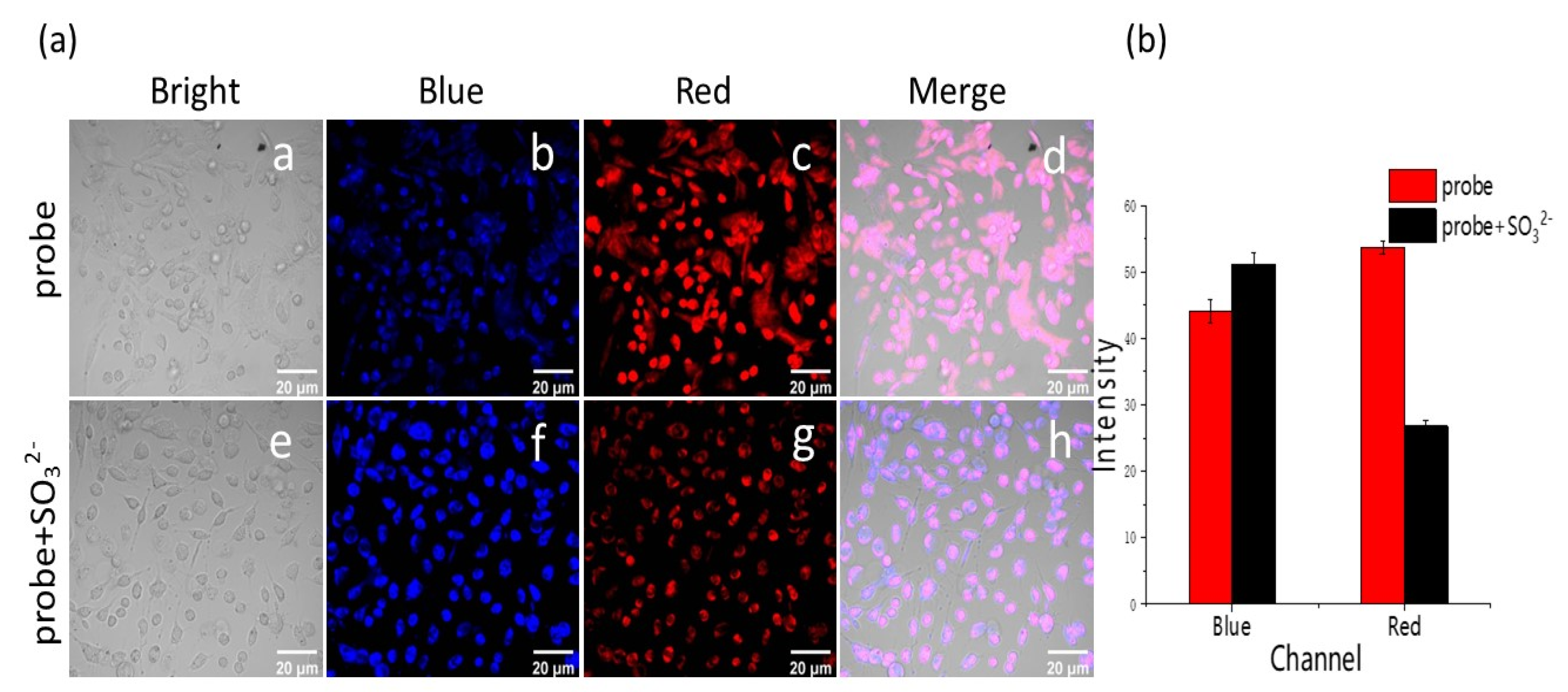
The results of the MTT (Methyl Thiazolyl Tetrazolium) experiment (Figure S4) showed that IPB-RL-1 had a lower cytotoxicity to SKOV-3 cells and could be used for further cell imaging experiments. In Figure 3, fluorescence in the red and blue channels were observed after SKOV-3 cells were incubated with the probe for 1 h. However, when the cells were incubated with the probe for 1 h and then incubated with SO32− for 20 min, the fluorescence in the blue channel was enhanced and the fluorescence in the red channel was significantly weakened, which suggested that probe IPB-RL-1 could be used to detect SO32− in SKOV-3 cells.
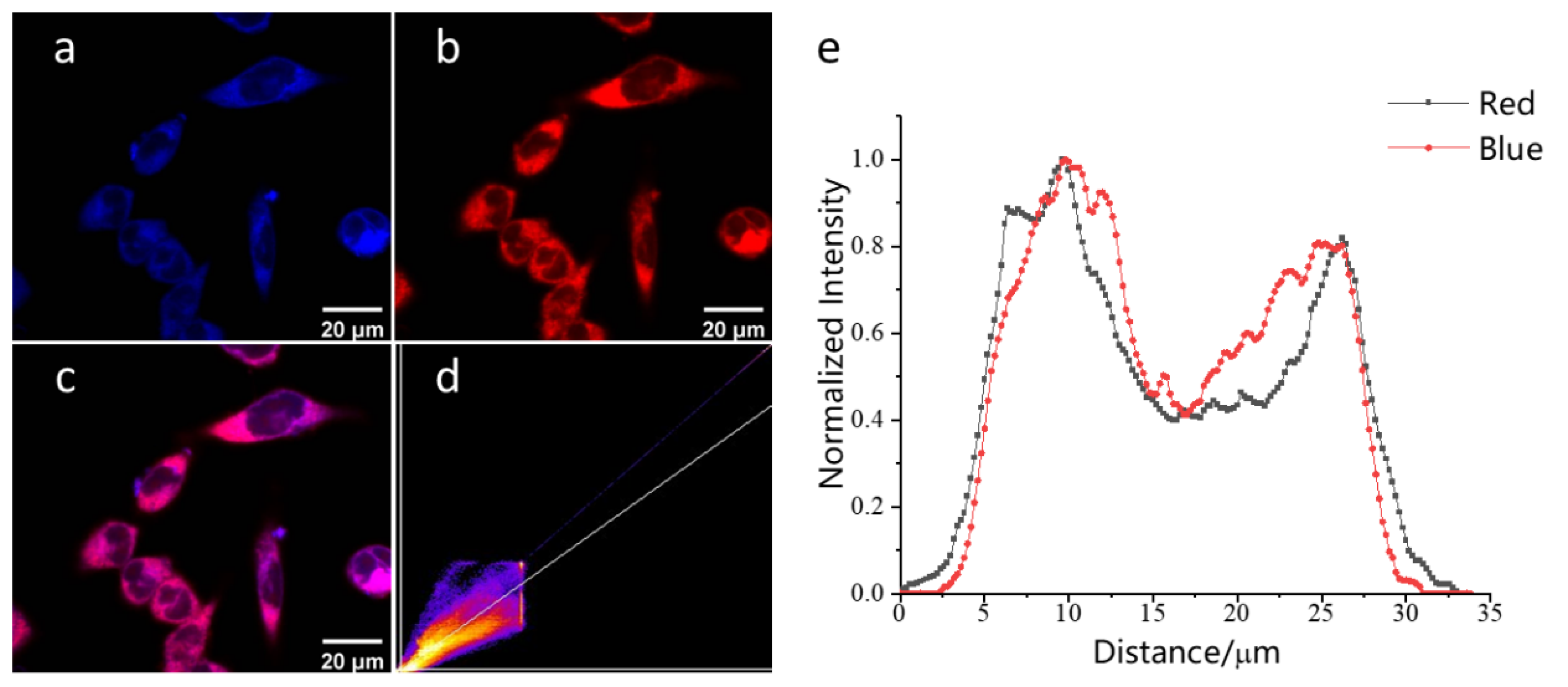
Next, since the benzopyran part of IPB-RL-1 is positively charged, the mitochondria-targeted experiment was tested. As shown in Figure 4, the red fluorescence of MitoTracker Red and the blue fluorescence of probe IPB-RL-1 overlap well (coefficient = 0.91).
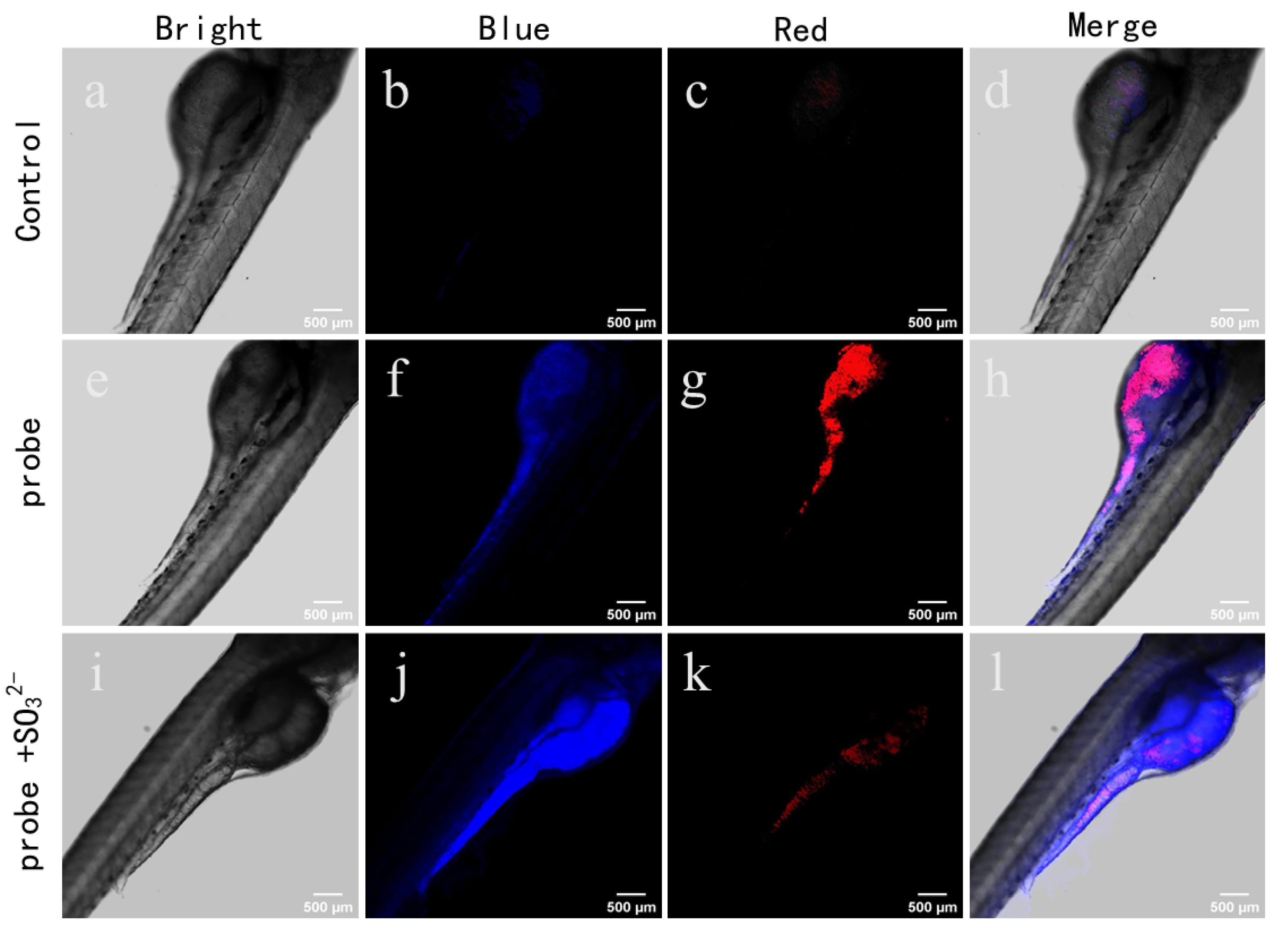
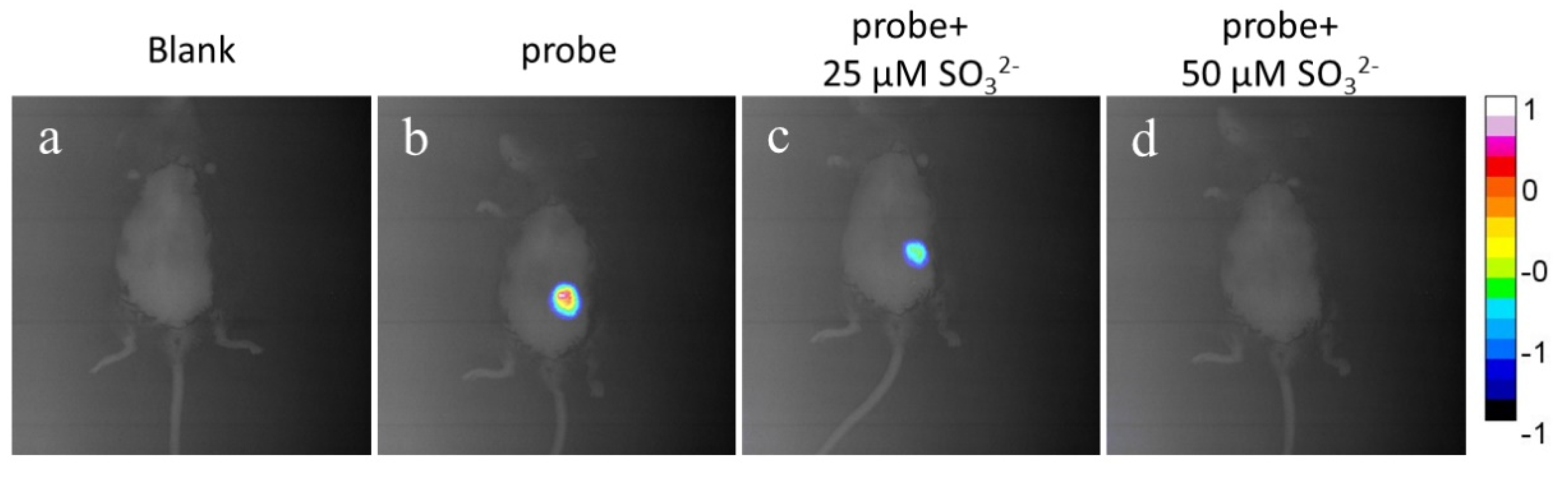
Owning to the excellent properties of IPB-RL-1 in cell imaging, its capability for the visualization of SO32− in zebrafish was examined. As depicted in Figure 5, weak blue and red fluorescent signals were observed in the control group. When the zebrafish were incubated with IPB-RL-1 for 1 h, the fluorescent signals became obviously strong both in the blue channel and the red channel. Yet, when the zebrafish were incubated with IPB-RL-1 for 1 h and then Na2SO3 for 30 min, the fluorescent signals in the red channel became obviously weak while there was no significant change in the blue channel. Therefore, we believe that IPB-RL-1 can effectively image in vivo. Hence, imaging in mice was conducted to further explore its application advantages. As NIR fluorescence emission is required for the experiments in vivo, only fluorescence changes in the 698–766 nm range were used. As shown in Figure 6b, obvious signals were observed after the probe was injected into mice for 5 min. However, with the increase in Na2SO3 concentration, the fluorescence signals gradually weakened (Figure 6c,d). As the response time is less than 5 min, it is very suitable for the real-time monitoring of SO32− in mice.
Based on the above results, we envisioned the mechanism of detection as follows (Scheme 2). At the excitation wavelength of 380 nm, the donor (imidazo[1,5-a]pyridine) transfers energy to the acceptor (benzopyran) and NIR fluorescence emission at 760 nm was observed. However, after the addition of SO32−, the reaction between SO32− and benzopyran breaks the π conjugate of benzopyran, resulting in the destruction of FRET, and thus, the energy of imidazo[1,5-a] pyridine cannot be transferred to the benzopyran. Therefore, the fluorescence emission at 760 nm disappeared and the emission at 475 nm increased. This is also confirmed by 1H NMR (Figure S5).
3. Experimental
Synthesis of the Probe IPB-RL-1
As demonstrated in Scheme 1, compounds 1–4 were synthesized according to the reported procedure [9,27].
Compound 3 (0.10 g, 0.24 mmol), compound 4 (0.10 g, 0.28 mmol) and CH3COOH (8 mL) were added to a 25 mL round-bottom flask. The mixture was heated to reflux for 3 h and then poured into water (100 mL). After being extracted with DCM (20 mL) three times, the combined organic solvent was removed under reduced pressure. The pure product was obtained by column chromatography (CH2Cl2:MeOH = 200:1). Black solid, 1H NMR (400 MHz, DMSO-d6) δ: 8.36 (s, 1H), 8.22 (d, J = 7.2 Hz, 1H), 8.00 (s, 1H), 7.78 (s, 1H), 7.59 (d, J = 8.8 Hz, 2H), 7.49 (s, 1H), 7.33 (dd, J = 9.2, 2.4 Hz, 1H), 7.19 (d, J = 2.4 Hz, 1H), 7.02 (d, J = 8.8 Hz, 2H), 6.70 (dd, J = 7.2, 1.6 Hz, 1H), 2.99–2.85 (m, 5H), 2.79 (s, 2H), 1.80 (m, 3H), 1.64 (m, 4H), 1.30 (m, 4H), 1.18 (m, 9H), 1.03–0.97 (m, 2H), 0.85 (m, 4H) ppm; 13C NMR (101 MHz, DMSO-d6) δ: 167.73, 164.09, 158.68, 155.65, 151.84, 138.94, 134.09, 132.07, 130.42, 128.61, 126.33, 125.51, 124.32, 123.74, 123.48, 122.83, 118.70, 118.15, 116.07, 114.57, 114.02, 112.81, 112.41, 45.83, 29.49, 29.12, 27.35, 25.65, 22.56, 22.20, 21.47, 14.42, 14.16, 13.02 ppm; HRMS: ([M]+) Calcd for C40H45ClN5O2: 622.3256; found: 622.3266.
4. Conclusions
In summary, a NIR ratiometric fluorescent probe IPB-RL-1 with an ultra-large Stokes shift (460 nm) that is superior to most reported probes has been developed. IPB-RL-1 shows high sensitivity and selectivity. Detection of SO2 in mitochondria in living SKOV-3 cells was also realized. Moreover, the probe was successfully used to detect SO2 in zebrafish which may be useful for the understanding of biological roles of SO2 in mitochondria and in vivo. However, due to the small overlap between donor emission and acceptor absorption of the probe IPB-RL-1, the fluorescence transfer efficiency is only 51%, which implies that in order to obtain a high fluorescence transfer efficiency, the overlap effect between donor emission and acceptor absorption, in addition to the distance between donor and acceptor, should be carefully considered for the FRET-based probe design in the future.
Supplementary Materials
Supplementary data associated with this article can be found in the online version. The following supporting information can be downloaded at: https://www.mdpi.com/article/10.3390/molecules28020515/s1, Figure S1: Ratiometric fluorescence responses F475/F760 of IPB-RL-1 upon the addition of 10 equiv. SO32− in the presence of 100 eq. background ions (1, probe; 2, SO42-; 3, Br−; 4, ACO−; 5, Cl−; 6, ClO−; 7, Cys; 8, ClO4−; 9, GSH; 10, F−; 11, H2PO4−; 12, HCO3−; 13, HPO42-; 14, HS−; 15, I−; 16, NO2−; 17, NO3−; 18, S2O82-; 19, SO32−); Figure S2: Relationship between fluorescence intensity ratio (F475/F760) and SO32− concentration; Figure S3: Time dependent increase of IPB-RL-1 fluorescence intensities after addition of SO32−; Figure S4: Cytotoxicity of IPB-RL-1; Figure S5: Normalized emission spectra of donor (compound 3) and normalized absorption spectra of IPB-RL-1; Figure S6: The emission spectrum of probe IPB-RL-1 and donor; Figures S7–S13: 1H NMR, 13C NMR, HRMS of related compounds; Table S1: Comparison with other probes [31,32,33,34,35,36,37,38,39,40,41,42,43,44,45,46,47,48,49,50,51,52,53,54,55,56,57].
Author Contributions
Conceptualization, R.C. and C.L.; methodology, C.L.; software, P.Z.; validation, R.C., C.L. and P.Z.; formal analysis, Y.G.; investigation, Y.G.; resources, Y.G.; data curation, K.Q.; writing—original draft preparation, K.Q.; writing—review and editing, Y.G.; visualization, Y.G.; supervision, Y.G. All authors have read and agreed to the published version of the manuscript.
Funding
This research was supported by the Natural Science Foundation of Shandong Province (ZR2021MB033), the Incubation Program of Youth Innovation of Shandong Province, and the Innovative Research Programs of Higher Education of Shandong Province (2019KJC009).
Institutional Review Board Statement
Not applicable.
Informed Consent Statement
Not applicable.
Data Availability Statement
The data that support the findings of this study are available from the corresponding author upon reasonable request.
Conflicts of Interest
The authors declare no conflict of interest.
References
- Lin, V.S.; Chen, W.; Xian, M.; Chang, C.J. Chemical probes for molecular imaging and detection of hydrogen sulfide and reactive sulfur species in biological systems. Chem. Soc. Rev. 2015, 44, 4596–4618. [Google Scholar] [CrossRef] [PubMed] [Green Version]
- Jiao, X.; Li, Y.; Niu, J.; Xie, X.; Wang, X.; Tang, B. Small-Molecule Fluorescent Probes for Imaging and Detection of Reactive Oxygen, Nitrogen, and Sulfur Species in Biological Systems. Anal. Chem. 2018, 90, 533–555. [Google Scholar] [CrossRef] [PubMed]
- Wu, L.; Sedgwick, A.C.; Sun, X.; Bull, S.D.; He, X.-P.; James, T.D. Reaction-Based Fluorescent Probes for the Detection and Imaging of Reactive Oxygen, Nitrogen, and Sulfur Species. Acc. Chem. Res. 2019, 52, 2582–2597. [Google Scholar] [CrossRef] [PubMed] [Green Version]
- Li, K.; Li, L.-L.; Zhou, Q.; Yu, K.-K.; Kim, J.S.; Yu, X.-Q. Reaction-based fluorescent probes for SO2 derivatives and their biological applications. Co-ord. Chem. Rev. 2019, 388, 310–333. [Google Scholar] [CrossRef]
- Liu, A.; Ji, R.; Shen, S.; Cao, X.; Ge, Y. A ratiometric fluorescent probe for sensing sulfite based on a pyrido[1,2-a]benzimidazole fluorophore. New J. Chem. 2017, 41, 10096–10100. [Google Scholar] [CrossRef]
- Ma, C.; Zhang, L.; Hou, L.; Chen, F.; Liu, A.; Ji, R.; Wang, Q.; Yuan, C.; Ge, Y. A fluorescent probe with a large Stokes shift for sensing sulfite in dry white wine based on pyrazolo[1,5-a]pyridine fluorophore. Tetrahedron Lett. 2021, 76, 153210. [Google Scholar] [CrossRef]
- Duan, G.Y.; Wang, H.; Sun, H.; Yuan, C.; Xu, Z.; Ge, Y.Q. Near-infrared TBET cassette with ultra large stokes shift and its application for SO2 imaging in cells. Chem. Eng. J. Adv. 2021, 8, 100141. [Google Scholar] [CrossRef]
- Cui, R.; Gao, Y.; Ge, H.; Shi, G.; Li, Y.; Liu, H.; Ma, C.; Ge, Y.; Liu, C. A turn-on fluorescent probe based on indolizine for the detection of sulfite. New J. Chem. 2022, 46, 8088–8093. [Google Scholar] [CrossRef]
- Zhang, W.; Huo, F.; Zhang, Y.; Chao, J.; Yin, C. Mitochondria-targeted NIR fluorescent probe for reversible imaging H2O2/SO2 redox dynamics in vivo. Sens. Actuators B 2019, 297, 126747. [Google Scholar] [CrossRef]
- Ren, H.; Huo, F.; Wu, X.; Liu, X.; Yin, C. An ESIPT-induced NIR fluorescent probe to visualize mitochondrial sulfur dioxide during oxidative stress in vivo. Chem. Commun. 2021, 57, 655–658. [Google Scholar] [CrossRef]
- He, L.; Yang, Y.; Lin, W. Rational Design of a Rigid Fluorophore–Molecular Rotor-Based Probe for High Signal-to-Background Ratio Detection of Sulfur Dioxide in Viscous System. Anal. Chem. 2019, 91, 15220–15228. [Google Scholar] [CrossRef] [PubMed]
- Gao, W.; Ma, Y.; Lin, W. Design of a FRET-based fluorescent probe for the reversible detection of SO2and formaldehyde in living cells and mice. New J. Chem. 2020, 44, 13654–13658. [Google Scholar] [CrossRef]
- Wu, L.; Huang, C.; Emery, B.P.; Sedgwick, A.C.; Bull, S.D.; He, X.-P.; Tian, H.; Yoon, J.; Sessler, J.L.; James, T.D. Förster resonance energy transfer (FRET)-based small-molecule sensors and imaging agents. Chem. Soc. Rev. 2020, 49, 5110–5139. [Google Scholar] [CrossRef] [PubMed]
- Yuan, L.; Lin, W.; Zheng, K.; Zhu, S. FRET-Based Small-Molecule Fluorescent Probes: Rational Design and Bioimaging Applications. Acc. Chem. Res. 2013, 46, 1462–1473. [Google Scholar] [CrossRef] [PubMed]
- Lee, M.H.; Kim, J.S.; Sessler, J.L. Small molecule-based ratiometric fluorescence probes for cations, anions, and biomolecules. Chem. Soc. Rev. 2015, 44, 4185–4191. [Google Scholar] [CrossRef] [Green Version]
- Tan, L.; Ding, H.; Chanmungkalakul, S.; Peng, L.; Yuan, G.; Yang, Q.; Liu, X.; Zhou, L. A smart TP-FRET-based ratiometric fluorescent sensor for bisulfite/formaldehyde detection and its imaging application. Sens. Actuators B 2021, 345, 130331. [Google Scholar] [CrossRef]
- Zhu, X.; Zhu, L.; Liu, H.-W.; Hu, X.; Peng, R.-Z.; Zhang, J.; Zhang, X.-B.; Tan, W. A two-photon fluorescent turn-on probe for imaging of SO2 derivatives in living cells and tissues. Anal. Chim. Acta 2016, 937, 136–142. [Google Scholar] [CrossRef]
- Shen, W.; Xu, H.; Feng, J.; Sun, W.; Hu, G.; Hu, Y.; Yang, W. A ratiometric and colorimetric fluorescent probe designed based on FRET for detecting SO32−/HSO3− in living cells and mice. Spectrochim. Acta Part A 2021, 263, 120183. [Google Scholar] [CrossRef]
- Liu, W.; Zhang, D.; Ni, B.; Li, J.; Weng, H.; Ye, Y. Mitochondria-targeted and FRET based ratiometric fluorescent probe for SO2 and its cell imaging. Sens. Actuators B 2019, 284, 330–336. [Google Scholar] [CrossRef]
- Zhang, W.; Huo, F.; Cheng, F.; Yin, C. Employing an ICT-FRET Integration Platform for the Real-Time Tracking of SO2 Metabolism in Cancer Cells and Tumor Models. J. Am. Chem. Soc. 2020, 142, 6324–6331. [Google Scholar] [CrossRef]
- Huang, Y.; Zhang, Y.; Huo, F.; Chao, J.; Yin, C. A dual-targeted organelles SO2 specific probe for bioimaging in related diseases and food analysis. Chem. Eng. J. 2022, 433, 133750. [Google Scholar] [CrossRef]
- Huang, M.-F.; Chen, L.-N.; Ning, J.-Y.; Wu, W.-L.; He, X.-D.; Miao, J.-Y.; Zhao, B.-X. A new lipid droplets-targeted fluorescence probe for specific detection of SO2 derivatives in living cells. Sens. Actuators B 2018, 261, 196–202. [Google Scholar] [CrossRef]
- Yan, Y.-H.; Cui, X.-L.; Li, Z.-Y.; Ding, M.-M.; Che, Q.-L.; Miao, J.-Y.; Zhao, B.-X.; Lin, Z.-M. A synergetic FRET/ICT platform-based fluorescence probe for ratiometric imaging of bisulfite in lipid droplets. Anal. Chim. Acta 2020, 1137, 47–55. [Google Scholar] [CrossRef]
- Sun, W.-X.; Li, N.; Li, Z.-Y.; Yuan, Y.-C.; Miao, J.-Y.; Zhao, B.-X.; Lin, Z.-M. A mitochondria-targeted ratiometric fluorescence probe for detection of SO2 derivatives in living cells. Dye. Pigment. 2020, 182, 108658. [Google Scholar] [CrossRef]
- Liu, F.-T.; Li, N.; Chen, Y.-S.; Yu, H.-Y.; Miao, J.-Y.; Zhao, B.-X. A quinoline-coumarin near-infrared ratiometric fluorescent probe for detection of sulfur dioxide derivatives. Anal. Chim. Acta 2022, 1211, 339908. [Google Scholar] [CrossRef]
- Sun, W.; Guo, S.; Hu, C.; Fan, J.; Peng, X. Recent Development of Chemosensors Based on Cyanine Platforms. Chem. Rev. 2016, 116, 7768–7817. [Google Scholar] [CrossRef]
- Zhang, D.; Liu, A.; Ji, R.; Dong, J.; Ge, Y. A mitochondria-targeted and FRET-based ratiometric fluorescent probe for detection of SO2 derivatives in water. Anal. Chim. Acta 2018, 1055, 133–139. [Google Scholar] [CrossRef]
- Song, G.; Liu, A.; Jiang, H.; Ji, R.; Dong, J.; Ge, Y. A FRET-based ratiometric fluorescent probe for detection of intrinsically generated SO2 derivatives in mitochondria. Anal. Chim. Acta 2018, 1053, 148–154. [Google Scholar] [CrossRef]
- Chen, F.; Liu, A.; Ji, R.; Xu, Z.; Dong, J.; Ge, Y. A FRET-based probe for detection of the endogenous SO2 in cells. Dye. Pigment. 2019, 165, 212–216. [Google Scholar] [CrossRef]
- Ge, Y.; Ji, R.; Shen, S.; Cao, X.; Li, F. A ratiometric fluorescent probe for sensing Cu2+ based on new imidazo[1,5-a]pyridine fluorescent dye. Sens. Actuators B 2017, 245, 875–881. [Google Scholar] [CrossRef]
- Yan, Y.; He, X.; Miao, J.; Zhao, B. A near-infrared and mitochondria-targeted fluorescence probe for ratiometric monitoring of sulfur dioxide derivatives in living cells. J. Mater. Chem. B. 2019, 7, 6585–6591. [Google Scholar] [CrossRef]
- Shen, R.; Qian, Y. A novel ratiometric fluorescent probe for specific detection of HSO3− at nanomolar level through 1,4-Michael addition. J. Photochem. Photobiol. A. 2020, 387, 112110. [Google Scholar] [CrossRef]
- Wu, W.; Ma, H.; Huang, M.; Miao, J.; Zhao, B. Mitochondria-targeted ratiometric fluorescent probe based on FRET for bisulfite. Sens. Actuators, B. 2017, 241, 239–244. [Google Scholar] [CrossRef]
- Yang, D.; Ning, J.; Wu, X.; Yao, W.; Shi, H.; Miao, J.; Zhao, B.; Lin, Z. Ratiometric fluorescence sensing of endogenous sulfur dioxide derivatives: Bio-imaging application in lipid droplets. Dyes Pigm. 2021, 192, 109457. [Google Scholar] [CrossRef]
- Li, D.; Wang, Z.; Cui, J.; Wang, X.; Miao, J.; Zhao, B. A new fluorescent probe for colorimetric and ratiometric detection of sulfur dioxide derivatives in liver cancer cells. Sci. Rep. 2017, 7, 45294. [Google Scholar] [CrossRef]
- Zhao, J.; Huang, L.; Yan, M.; Qu, Y.; Feng, H.; Sun, Y. A lysosome specific ratiometric fluorescent probe for detection of bisulfite ion based on hybrid coumarin-benzimidazolium compounds. Phosphorus. Sulfur. 2021, 196, 321–327. [Google Scholar] [CrossRef]
- Shen, R.; Qian, Y. A mitochondria-oriented fluorescent probe for ultrafast and ratiometric detection of HSO3− based on naphthalimide–hemicyanine. New J. Chem. 2019, 43, 7606–7612. [Google Scholar] [CrossRef]
- Wang, Y.; Meng, Q.; Zhang, R.; Jia, H.; Wang, C.; Zhang, Z. A mitochondria-targeted ratiometric probe for the fluorescent and colorimetric detection of SO2 derivatives in live cells. J. Lumin. 2017, 192, 297–302. [Google Scholar] [CrossRef] [Green Version]
- Liu, K.; Chen, Y.; Sun, H.; Wang, S.; Kong, F. Construction of a novel near-infrared fluorescent probe with multiple fluorescence emission and its application for SO2 derivative detection in cells and living zebrafish. J. Mater. Chem. B. 2018, 6, 7060–7065. [Google Scholar] [CrossRef]
- Yang, D.; He, X.; Wu, X.; Shi, H.; Miao, J.; Zhao, B.; Lin, Z. A novel mitochondria-targeted ratiometric fluorescent probe for endogenous sulfur dioxide derivatives as a cancer-detecting tool. J. Mater. Chem. B. 2020, 8, 5722–5728. [Google Scholar] [CrossRef]
- Yin, G.; Gan, Y.; Yu, T.; Niu, T.; Yin, P.; Chen, H.; Zhang, Y.; Li, H.; Yao, S. A dual-emission and mitochondria-targeted fluorescent probe for rapid detection of SO2 derivatives and its imaging in living cells. Talanta. 2019, 191, 428–434. [Google Scholar] [CrossRef]
- Huang, Y.; Zhang, Y.; Huo, F.; Yin, C. FRET-dependent single/two-channel switch endowing a dual detection for sulfite and its organelle targeting applications. Dyes Pigm. 2021, 184, 108869. [Google Scholar] [CrossRef]
- Wang, M.; Liu, Q.; Sun, X.; Zheng, S.; Ma, Y.; Wang, Y.; Yan, M.; Lu, Z.; Fan, C.; Lin, W. Ratiometric and reversible detection of endogenous SO2 and HCHO in living cells and mice by a near-infrared and dual-emission fluorescent probe. Sensors Actuators B Chem. 2021, 335, 129649. [Google Scholar] [CrossRef]
- Zhang, L.; Wang, Z.; Liu, J.; Miao, J.; Zhao, B. A rational design of ratiomet-ric fluorescent probes based on new ICT/FRET platform and imaging of endogenous sulfite in living cells. Sens. Actuators, B. 2017, 253, 19–26. [Google Scholar] [CrossRef]
- Song, G.; Luo, J.; Xing, X.; Ma, H.; Yang, D.; Cao, X.; Ge, Y.; Zhao, B. A ratiometric fluorescence probe for rapid detection of mitochondrial SO2 derivatives. New J. Chem. 2018, 42, 3063–3068. [Google Scholar] [CrossRef]
- Li, D.; Han, X.; Yan, Z.; Cui, Y.; Miao, J.; Zhao, B. A far-red ratiometric fluorescent probe for SO2 derivatives based on the ESIPT enhanced FRET platform with improved performance. Dyes Pigm. 2018, 151, 95–101. [Google Scholar] [CrossRef]
- Yan, Y.; Wu, Q.; Che, Q.; Ding, M.; Xu, M.; Miao, J.; Zhao, B.; Lin, Z. A mitochondria-targeted fluorescent probe for the detection of endogenous SO2 derivatives in living cells. Analyst. 2020, 145, 2937–2944. [Google Scholar] [CrossRef]
- Li, D.; Wang, Z.; Su, H.; Miao, J.; Zhao, B. Fluorescence detection of endogenous bisulfite in liver cancer cells using an effective ESIPT enhanced FRET platform. Chem. Commun. 2017, 53, 577–580. [Google Scholar] [CrossRef]
- Lu, Y.; Dong, B.; Song, W.; Sun, Y.; Mehmood, A.; Lin, W. A mitochondria-targeting ratiometric fluorescent probe for the detection of sulfur dioxide in living cells. New J. Chem. 2020, 44, 11988–11992. [Google Scholar] [CrossRef]
- Li, Z.; Cui, X.; Yan, Y.; Che, Q.; Miao, J.; Zhao, B.; Lin, Z. A novel endoplasmic reticulum-targeted ratiometric fluorescent probe based on FRET for the detection of SO2 derivatives. Dyes Pigm. 2021, 188, 109180. [Google Scholar] [CrossRef]
- Li, D.; Wang, Z.; Cao, X.; Cui, J.; Wang, X.; Cui, H.; Miao, J.; Zhao, B. A mitochondria-targeted fluorescent probe for ratiometric detection of endogenous sulfur dioxide derivatives in cancer cells. Chem. Commun. 2016, 52, 2760–2763. [Google Scholar] [CrossRef]
- Zhang, G.; Ji, R.; Kong, X.; Ning, F.; Liu, A.; Cui, J.; Ge, Y. A FRET based ratiometric fluorescent probe for detection of sulfite in food. RSC Adv. 2019, 9, 1147–1150. [Google Scholar] [CrossRef] [Green Version]
- Yang, Y.; He, L.; Xu, K.; Lin, W. Development of a mitochondria-targeted fluorescent probe for the ratiometric visualization of sulfur dioxide in living cells and zebrafish. Anal. Methods. 2019, 11, 3931–3935. [Google Scholar] [CrossRef]
- Shen, W.; Xu, H.; Feng, J.; Sun, W.; Hu, G.; Yang, W. A ratiometric and colorimetric fluorescent probe designed based on FRET for detecting SO32-/HSO3- in living cells and mice. Spectrochim. Acta, Part A. 2021, 263, 120183. [Google Scholar] [CrossRef]
- Yang, X.; Zhou, X.; Zhang, S.; Yang, Y.; Chen, J.; Guo, X.; Li, Z.; Yang, R.; Zhou, Y. A TP-FRET-based two-photon fluorescent probe for ratiometric visualization of endogenous sulfur dioxide derivatives in mitochondria of living cells and tissues. Chem. Commun. 2016, 52, 10289–10292. [Google Scholar] [CrossRef]
- Li, T.; Huo, F.; Chao, J.; Yin, C. Independent bi-reversible reactions and regulable FRET efficiency achieving real-time visualization of Cys metabolizing into SO2. Chem. Commun. 2020, 56, 11453–11456. [Google Scholar] [CrossRef]
- Chen, X.; Chen, Q.; He, D.; Yang, S.; Yang, Y.; Qian, J.; Long, L.; Wang, K. Mitochondria targeted and immobilized ratiometric NIR fluorescent probe for investigating SO2 phytotoxicity in plant mitochondria. Sens. Actuators, B. 2022, 370, 132433. [Google Scholar] [CrossRef]
Scheme 1.
Synthetic route of IPB-RL-1.

Figure 1.
(a) UV−vis and (b) fluorescence spectra in response to different ions (λex = 300 nm, slit: 5 nm/5 nm, 5 μM for fluorescence and 50 μM for UV−vis).
Figure 1.
(a) UV−vis and (b) fluorescence spectra in response to different ions (λex = 300 nm, slit: 5 nm/5 nm, 5 μM for fluorescence and 50 μM for UV−vis).

Figure 2.
(a) UV−vis and (b) fluorescence spectra of IPB-RL-1 upon the addition of SO32− (λex = 300 nm, slit: 5 nm/5 nm, 5 μM for fluorescence and 50 μM for UV−vis).
Figure 2.
(a) UV−vis and (b) fluorescence spectra of IPB-RL-1 upon the addition of SO32− (λex = 300 nm, slit: 5 nm/5 nm, 5 μM for fluorescence and 50 μM for UV−vis).

Figure 3.
(a) Imaging of IPB-RL-1 in SKOV-3 cells. The first row is the imaging of SKOV-3 cells incubated with IPB-RL-1 for 30 min, and the second row is the imaging of SKOV-3 cells incubated with IPB-RL-1 for 30 min and then treated with Na2SO3 for 20 min. (b) Comparison of the fluorescence intensity between the red channel and blue channel (λex: 405 nm; blue: λem = 420–520 nm; red: λem = 700–800 nm).

Figure 4.
(a) Fluorescence images of IPB-RL-1 in SKOV-3 cells (blue channel: 420–520 nm; λex: 405 nm). (b) Fluorescence images of Mito-Tracker Red CMXRos (red channel: 570–620 nm; λex: 561 nm). (c) Merged images of (a,b). (d) Images of co −location (co −location coefficient 0.91). (e) Fluorescence intensity of red channel and blue channel changes, respectively (λex: 405 nm).

Figure 5.
Fluorescence images of IPB-RL-1 in zebrafish ((a−d): the control group; (e−h): zebrafish incubated with IPB-RL-1 only; (i−l): zebrafish incubated with IPB-RL-1 for 1 h and then SO32− for 30 min; λex: 405 nm; blue: λem = 420–520 nm; red: λem = 700–800 nm).
Figure 5.
Fluorescence images of IPB-RL-1 in zebrafish ((a−d): the control group; (e−h): zebrafish incubated with IPB-RL-1 only; (i−l): zebrafish incubated with IPB-RL-1 for 1 h and then SO32− for 30 min; λex: 405 nm; blue: λem = 420–520 nm; red: λem = 700–800 nm).

Figure 6.
Fluorescence images of IPB-RL-1 in living mice. (a) Images of the mice only; (b) images after 100 μL of 50 μM IPB-RL-1 was injected into the mice for 5 min; (c) images after 100 μL of 25 μM SO32− was injected into the mice for 5 min; (d) images after re-injection of 100 μL 50 μM SO32− at the same location for 5 min (λex = 635 nm, λem = 698–766 nm).

Scheme 2.
Proposed mechanism.

Disclaimer/Publisher’s Note: The statements, opinions and data contained in all publications are solely those of the individual author(s) and contributor(s) and not of MDPI and/or the editor(s). MDPI and/or the editor(s) disclaim responsibility for any injury to people or property resulting from any ideas, methods, instructions or products referred to in the content. |
© 2023 by the authors. Licensee MDPI, Basel, Switzerland. This article is an open access article distributed under the terms and conditions of the Creative Commons Attribution (CC BY) license (https://creativecommons.org/licenses/by/4.0/).
Share and Cite
MDPI and ACS Style
Cui, R.; Liu, C.; Zhang, P.; Qin, K.; Ge, Y. An Imidazo[1,5-a]pyridine Benzopyrylium-Based NIR Fluorescent Probe with Ultra-Large Stokes Shifts for Monitoring SO2. Molecules 2023, 28, 515. https://doi.org/10.3390/molecules28020515
AMA Style
Cui R, Liu C, Zhang P, Qin K, Ge Y. An Imidazo[1,5-a]pyridine Benzopyrylium-Based NIR Fluorescent Probe with Ultra-Large Stokes Shifts for Monitoring SO2. Molecules. 2023; 28(2):515. https://doi.org/10.3390/molecules28020515
Chicago/Turabian StyleCui, Renle, Caihong Liu, Ping Zhang, Kun Qin, and Yanqing Ge. 2023. "An Imidazo[1,5-a]pyridine Benzopyrylium-Based NIR Fluorescent Probe with Ultra-Large Stokes Shifts for Monitoring SO2" Molecules 28, no. 2: 515. https://doi.org/10.3390/molecules28020515