Review

Antioxidant Cardioprotection against Reperfusion Injury: Potential Therapeutic Roles of Resveratrol and Quercetin

1
Molecular and Clinical Pharmacology Program, Faculty of Medicine, Institute of Biomedical Sciences, University of Chile, Santiago P.O. Box 70111, Chile
2
Indian Institute of Technology Gandhinagar, Palaj, Gandhinagar 382055, Gujarat, India
3
Department of Cardiovascular, Endocrine-Metabolic Diseases, and Aging, Italian National Institute of Health, 00161 Rome, Italy
4
Department of Physiology and Pharmacology “Vittorio Erspamer”, Sapienza University of Rome, 00161 Rome, Italy
*
Author to whom correspondence should be addressed.
Molecules 2022, 27(8), 2564; https://doi.org/10.3390/molecules27082564
Submission received: 4 March 2022 / Revised: 6 April 2022 / Accepted: 12 April 2022 / Published: 15 April 2022

Abstract

:
Ischemia-reperfusion myocardial damage is a paradoxical tissue injury occurring during percutaneous coronary intervention (PCI) in acute myocardial infarction (AMI) patients. Although this damage could account for up to 50% of the final infarct size, there has been no available pharmacological treatment until now. Oxidative stress contributes to the underlying production mechanism, exerting the most marked injury during the early onset of reperfusion. So far, antioxidants have been shown to protect the AMI patients undergoing PCI to mitigate these detrimental effects; however, no clinical trials to date have shown any significant infarct size reduction. Therefore, it is worthwhile to consider multitarget antioxidant therapies targeting multifactorial AMI. Indeed, this clinical setting involves injurious effects derived from oxygen deprivation, intracellular pH changes and increased concentration of cytosolic Ca2+ and reactive oxygen species, among others. Thus, we will review a brief overview of the pathological cascades involved in ischemia-reperfusion injury and the potential therapeutic effects based on preclinical studies involving a combination of antioxidants, with particular reference to resveratrol and quercetin, which could contribute to cardioprotection against ischemia-reperfusion injury in myocardial tissue. We will also highlight the upcoming perspectives of these antioxidants for designing future studies.

1. Introduction

Cardiovascular disease is the most common cause of morbidity and mortality worldwide, with 17.9 million deaths every year, according to updated WHO data from the year 2017, which represents 31% of all registered deaths in the world [1]. Among cardiovascular diseases, stroke and acute myocardial infarct (AMI) are the main causes of death, the latter being responsible for around 9 million deaths each year [2]. AMI is produced by either partial or complete occlusion of the coronary arterial circulation. It is mainly caused by atheromatous plaques that are vulnerable to rupture or erosion causing thrombotic alterations that block blood flow to cardiac tissue [3]. The current gold standard treatment for AMI is percutaneous coronary intervention (PCI), a procedure whose objective is to restore the arrival of blood flow to the ischemic heart tissue [4]. Paradoxically, the restoration of blood flow to ischemic myocardial tissue induces additional damage. Studies in animal models of AMI have suggested that myocardial reperfusion is responsible for up to 50% of the final infarct size [3]. One of the key factors involved is oxidative stress, which influences myocardial reperfusion via multiple pathological mechanisms. Consequently, the antioxidant defense system could become overwhelmed, thus resulting in oxidative damage and, ultimately, cell death [5]. Numerous attempts have been performed to reduce myocardial tissue damage after ischemia-reperfusion (I/R) by enhancing the antioxidant defense system with antioxidant treatments. Accordingly, the administration of molecules such as vitamin C, vitamin E, N-acetylcysteine, deferoxamine and polyphenols, among others, has been studied [6]. However, these therapies have led to suboptimal and inconsistent results. As a consequence, until today, there is no reliable therapy against I/R damage for these patients. It still remains a challenge to find an effective therapeutic strategy that can prevent such damage and decrease infarct size after reperfusion by PCI.
Here, it is noteworthy that although antioxidant therapy has not been successful enough to be implemented, most of the studies have been mainly based on the administration of the antioxidants as monotherapies. Nevertheless, it is well known that the pathophysiology of cardiac tissue damage caused by I/R is multifactorial, and multiple forms of ROS generation might contribute to this damage. Therefore, as an alternative strategy, we should consider synergistic antioxidant combination therapies for multitarget effects with minimized toxicity and high selectivity [7]. Therefore, it would be expected that the search for new associations of antioxidants would lead us to achieve an improvement in the pharmacological response.
To elucidate the translational potential of antioxidant therapies in the treatment of I/R injury after PCI, a detailed analysis of the available evidence with a focus on possible synergies between the different compounds is needed. Among the multiple natural available antioxidants, it has been evaluated the use of resveratrol, which is safe to use and effective on low doses, whose cardioprotective effect has been demonstrated in various studies performed in animal models, such as an I/R rat model, where a 21-day oral treatment with resveratrol decreased the incidence of atrioventricular block and lethality [8], and quercetin, which has been shown in various animal models to exhibit a variety of active biological functions, including anti-inflammatory, anticoagulation, and oxygen radical-scavenging activities as shown by the recent experiment in an animal model illustrating a reduction in the expression of various oxidant enzymes, thus decreasing oxidative stress [9].

1.1. Oxidative Stress

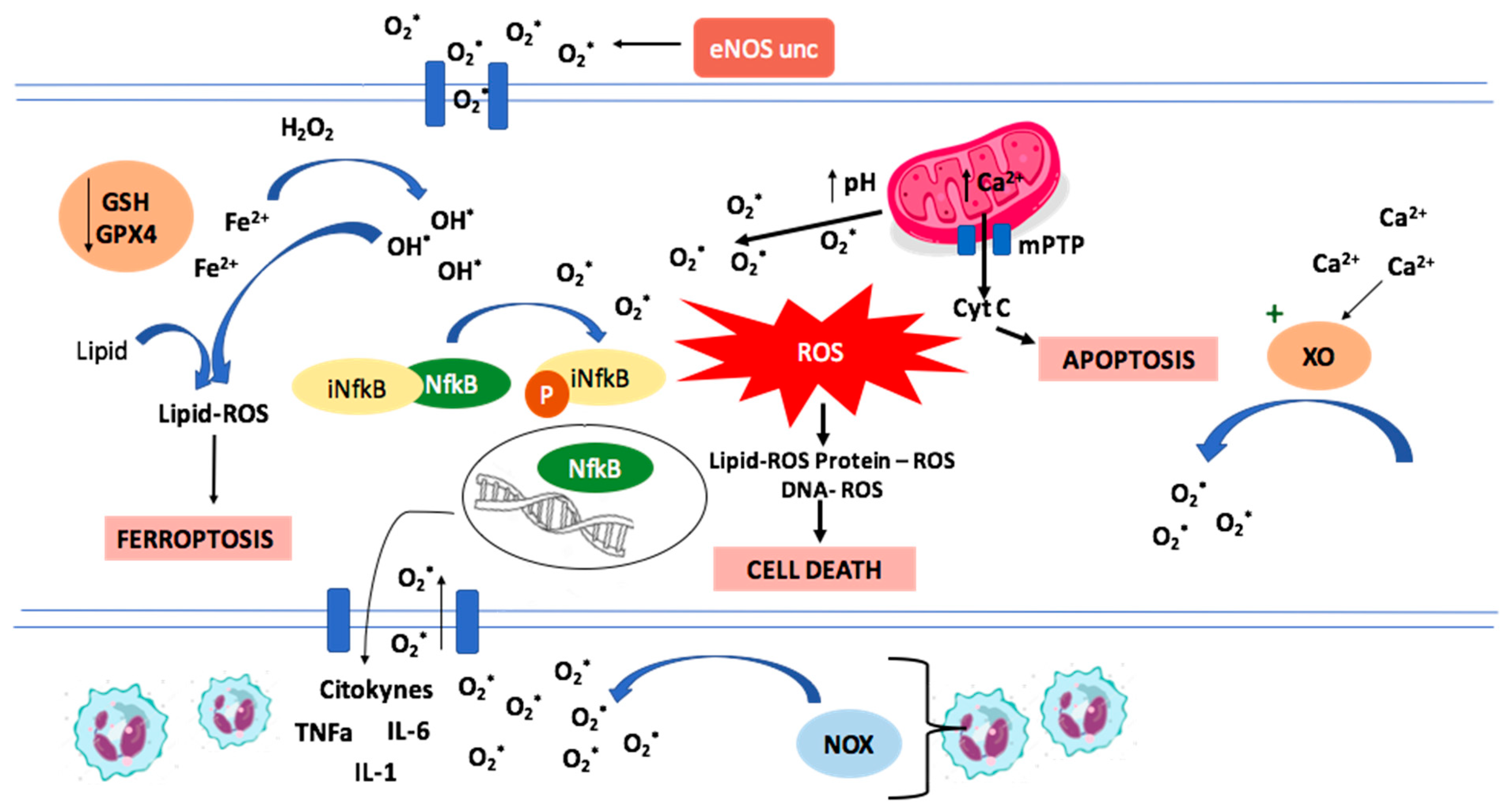
1.1.1. Oxidative Stress and ROS

Oxidative stress results from the imbalance between free radicals production and the antioxidant defense system [10]. Under oxidative stress conditions, two kinds of reactive species are produced, reactive oxygen species (ROS), such as radical anion superoxide, hydrogen peroxide, hydroxyl radical and reactive nitrogen species (RNS), which include nitric oxide radical, nitrogen dioxide radical, and peroxynitrite [11].
ROS can be formed by enzymatic or non-enzymatic mediated processes. Enzymatic ROS sources include reduced nicotinamide adenine dinucleotide phosphate oxidase (NADPH oxidase or NOX), xanthine oxidase (XO), uncoupled endothelial nitric oxide synthase (eNOS), among others [12]. In addition, an example of the non-enzymatic mechanisms of ROS production is the direct generation of hydroxyl radicals via Fenton and Haber–Weiss reactions, both of which depend on the availability of transition metal ions such as free iron in the form of labile iron pool (LIP). The hydroxyl radical is highly reactive and of low specificity, making it capable of damaging more biomolecules than any other ROS [13].

1.1.2. Mechanisms of Ischemia/Reperfusion Cellular Damage

The mechanisms accounting for I/R damage are depicted in Figure 1.

1.1.3. ROS Production in Ischemia/Reperfusion

The occlusion of a coronary artery branch inhibits blood flow to myocardial tissue causing regional ischemia [14]. During myocardial ischemia, the decrease in the oxygen supply in cardiac tissue causes a marked reduction of oxygen supply to the mitochondrial electron transport chain, leading to a shift from aerobic to anaerobic cellular respiration. Consequently, a decrease in ATP production ensues, accompanied by lactic acid accumulation and a decrease in cellular pH [15]. The pH acidification increases the Na+ influx through the Na+/H+ exchanger, and the lack of ATP decreases the activity of the Na+/K+ pump, leading to an increase in intracellular Na+ that activates the Ca2+/Na+ exchanger to function in a reverse direction, thus exchanging intracellular Na+ with extracellular Ca2+ [16]. As a result, there is an overload in intracellular Ca2+ that cannot be captured by the Ca2+ ATPase (SERCA) in the sarcoplasmic reticulum due to the lack of ATP [17]. The overload of intracellular Ca2+ induces enzymes changes. One of them is the conversion of xanthine dehydrogenase in endothelial cells to xanthine oxidase (XO), an enzyme that generates superoxide anion radicals, contributing to the induction of oxidative stress [18]. Furthermore, during cardiac ischemia, tetrahydrobiopterin, a cofactor of endothelial eNOS, is oxidized, causing eNOS to get uncoupled and produce superoxide instead of nitric oxide. These changes are related to a burst in ROS production that has been found once reperfusion has restored oxygen supply to the myocardial tissue [19]. Exacerbation of oxidative stress during reperfusion overwhelms the endogenous antioxidant defenses, causing direct damage by lipid peroxidation, DNA oxidation and protein carbonylation, effects that can progress until cell death [19,20]. The main effectors of this damage are peroxynitrite and hydroxyl radicals generated from Fenton/Haber–Weiss reactions and [19].
Other ROS sources in cardiac tissue exposed to ischemia-reperfusion are NADPH oxidase in neutrophils, mitochondrial electron transport chain (mETC), cytochrome P450, lipooxygenase (LOX), cyclooxygenase, and monoamine oxidase [21].

1.2. Intracellular Organelle Dysfunctions Associated with Ischemia/Reperfusion Injury

1.2.1. Endoplasmic Reticulum Stress (ERS)

Endoplasmic reticulum stress has been described as another potential mechanism of I/R injury. It results from the accumulation of unfolded proteins in the endoplasmic reticulum, leading to an imbalance in calcium ion homeostasis and the activation of the unfolded protein response (UPR), a signaling pathway aimed to counteract the stress conditions effects otherwise leading to cell apoptosis [22]. Protein kinase-like ER kinase (PERK) is one of the sensors of stress that activate and coordinate the cell response following UPR activation. In particular, PERK may participate via the ERS-PERK signaling pathway in mediating the translocation and activation of the pro-survival nuclear factor erythroid 2-related factor 2 (Nrf2), suggesting that its influence on cell survival may be closely associated with regulating Nrf2/ARE signaling pathway [23].

1.2.2. Mitochondrial Dysfunction

Mitochondrial permeability transition pore (mPTP) is a protein channel located in the inner mitochondrial membrane involved in necrotic and apoptotic cell death. When mPTP opens, it can induce the uncoupling of oxidative phosphorylation. Under ischemic conditions, the acid pH induced by lactate accumulation has an inhibitory effect on the mPTP, causing it to close [24]. Later, within the first few minutes of reperfusion [25], the intracellular acidic pH is restored to physiological values due to the reabsorption of lactate, and a mitochondrial Ca2+ overload ensues [26]. Furthermore, mPTP opens due to mitochondrial oxidative stress [27], uncoupling the oxidative phosphorylation and decreasing intracellular ATP availability, which can induce cell death through the mitochondrial pathway [26]. In addition, mPTP-induced cell death could be caused by the transport of molecules less than 1.5 kDa by mPTP, which collapses the mitochondrial membrane potential. Other consequences of mPTP opening are mitochondrial matrix swelling and the release of mitochondrial proteins, activating cell death mechanisms, such as apoptosis, necrosis, and necroptosis [28]. Moreover, the loss of mitochondrial potential generates a rise in ROS production [29].

1.2.3. Inflammation Mediated Damage

The mechanism of I/R injury is usually considered sterile. However, it displays a series of inflammatory reactions resembling infectious processes such as inducing cytokines and chemokines production with infiltration of immune system cells [30,31]. This inflammatory response is thought to function as a double-edged sword: on one side, it enables the removal of cell debris and promotes wound healing. Damage-associated molecular pattern (DAMP) mediated signaling can also exacerbate the inflammatory state in a disproportional matter, thereby leading to additional tissue damage. After reperfusion, neutrophil and monocyte become activated by many damage signals released by necrotic cells. The primary immune response expands tissue damage by inducing neutrophil, NLRP3-inflammasome [32] and Toll-like receptor 9-pathogen recognition receptor activation [33], which converge on the activation of the myeloid differentiation primary response gene 88 (MyD88) and nuclear factor-κB (NF-κB) pathways [3] which in turn polarize T lymphocyte towards a pro-inflammatory phenotype.
NF-κB proteins are a family of transcription factors that regulate the expression of genes involved in inflammation, immune response, cell proliferation, apoptosis [34] and activation of the mTOR pathway, leading to autophagy [35]. Reactive oxygen species can induce NF-κB activation through phosphorylation and degradation of NF-κB inhibitory cofactor IkBα, which allows NF-κB to translocate into the nucleus and modify cardiac tissue gene expression by up-regulation of genes involved in inflammatory and pro-fibrotic responses [36]. Neutrophils become important contributors to myocardial damage since they not only release proteolytic enzymes (hydrolases, metalloproteinases, and proteases) but also constitute a major ROS source to generate superoxide anions through NADPH oxidase (NOX) [37], causing damage to cardiomyocytes and endothelial cells. From a wound-healing perspective, after the inflammatory phase is dominated by pro-inflammatory T helper 1 lymphocytes, a second, anti-inflammatory reparative phase leading to wound healing and scar formation comes later. Cytokines have a key role in tissue repair. IL-6, IL-10, transforming growth factor-beta (TGF-β), and a subpopulation of T-lymphocytes known as ‘T Regulatory cells’ have all been associated with suppressing the pro-inflammatory response and steering the immune system towards repair and resolution following I/R [38,39]. Apoptotic neutrophils induce an anti-inflammatory phenotype in infiltrated macrophages upon their phagocytosis, which inhibits the macrophage proinflammatory tissue-damaging response and leads them to produce IL-10 and TGF-β [40,41], which dampening inflammation, as well as excessive tissue scarring during the reparative phase, thus inhibiting I/R-derived damage.

1.3. Mechanisms of Cell Death in Ischemia-Reperfusion

Ferroptosis

Ferroptosis is a significant mechanism of injury in myocardial infarction and can be a promising target to reduce infarct size [42]. Ferroptosis consists of an iron-dependent, non-apoptotic form of cell death [43]. It is a regulated form of necrosis that is driven by oxidative stress and is characterized by the accumulation of lipid peroxidation products and ROS derived from iron metabolism.
Glutathione peroxidase 4 (GPX4) is the key regulator of ferroptosis [43]. This enzyme transforms phospholipid hydroperoxides to lipid alcohols using reduced GSH, which inhibits ferroptosis and explains why it mostly happens when cellular GSH levels are low, or GPX4 is inhibited [44]. Inhibition of GPX4 activity can lead to the accumulation of lipid peroxides, which is a marker of ferroptosis [43]. During I/R, intracellular reduced glutathione (GSH) decreases, as well as the activity of GPX4. Thus, lipid peroxides cannot be reduced by GPX4, and Fe2+ oxidizes lipids in a Fenton-like manner, resulting in a large amount of ROS concentration, which promotes ferroptosis.
Thus, iron metabolism is one of the main pathways of ischemia-reperfusion injury involved in cell death [42]. In animal models of coronary artery ligation-induced myocardial I/R injury, it has been observed that ferroptosis inhibition may repair cell damage and infarct size [43]. Iron is transported inside cardiomyocytes after recognition by the transferrin receptor 1 (TfR1) [45], and Ferritin (FT) is the main iron storage protein [13]. It is of interest to remark that ferrous ionic free iron is a key ferroptosis inducer; therefore, this deleterious process could be inhibited by iron chelators [46].

1.4. Apoptosis

Studies performed in in vivo animal models have demonstrated the important role of apoptosis in cell death following I/R, particularly during the phase of reperfusion [47]. Energy restoration characterizing reperfusion promotes the apoptotic process. Experiments in isolated rat hearts demonstrated that mitochondrial dysfunction and caspase activation occur after global ischemia and that caspase activation is dependent on the time of ischemia [48]. The opening of the mPTP and the changes in the mitochondrial transmembrane potential promote the release of pro-apoptotic proteins such as cytochrome c to the cytosol (23), leading to apoptotic cell death [49].

1.5. Defense Mechanisms against Oxidative Stress

Antioxidant systems regulate redox homeostasis by controlling the intracellular ROS levels and their interaction with biological molecules. This system has enzymatic and non-enzymatic mechanisms involved. Enzymes constitute the first line of antioxidative cellular defense; they are superoxide dismutase (SOD), glutathione peroxidase (GPX) and catalase (CAT). Non-enzymatic antioxidants can be exogenous or endogenous and include molecules such as vitamin C, vitamin E, reduced glutathione (GSH), carotenoids, flavonoids and polyphenols, among many others [50,51].
Antioxidants act by many mechanisms. Some of them are ROS scavenging or of their precursors, inhibiting ROS production, attenuating the catalysis of ROS generation via chelating metal ions, enhancing endogenous antioxidant generation, repairing the oxidative damage inflicted on the macromolecules and reducing apoptotic cell death by up-regulating the anti-apoptotic gene Bcl-2 [50]. Antioxidant enzymes are encoded by several housekeeping genes largely controlled by Nrf2 [52].

1.6. Nrf2 Signalling Pathway

Nrf2 is a transcription factor considered an essential regulator for maintaining the redox balance [53] via inducing endogenous antioxidant enzymes in response to oxidative stress [54]. Kelch-like ECH-associated protein 1 (Keap1) is a cullin3-dependent E3 ubiquitin ligation substrate [55] and a suppressor protein that physically binds Nrf2 and retains it in the cytoplasm so that it can be rapidly ubiquitinated and go to proteasomal degradation by the Cu3-Rbk1 complex [56]. Under oxidative stress conditions, intracellular ROS levels increase, and Keap1 undergoes conformational changes [57] that impair its ability to interact with Nrf2, thus facilitating the complex dissociation and preventing Nrf2 proteasomal degradation. Consequently, Nrf2 translocates into the nucleus and binds to the antioxidant response elements (ARE), located in the promoter region of target genes encoding antioxidant proteins, thereby enhancing their expression [36,58].
It is important to address that Nrf2 is activated by phosphorylation and that different molecules can do this from different pathways, such as phosphatidylinositol 3- kinase (PI3K) [59], protein kinase C (PKC) [60] and c-Jun NH2 terminal protein kinase (JNK) [61] pathways. Meanwhile, glycogen synthase kinase 3β (GSK3β) promotes Nrf2 degradation independently from keap1 and Nrf2 translocation and inactivation in the nucleus [62]. Nrf2 participates in the regulation of cellular defense and mediates cellular repair, proliferation, and/or regeneration. In addition, Nrf2 protects the mitochondria against oxidative damage [63] and tissues from harm, exerting anti-inflammatory and anti-apoptotic effects [53]. As for the anti-apoptotic effect, Nrf2 is involved in the up-regulation of uncoupling protein (UCP)3 expression by binding to its promoter, and this signaling mechanism promotes cell survival under oxidative stress [54]. UCPs are inner membrane proteins of mitochondria that regulate the uncoupling of oxidative phosphorylation and protect against pro-oxidant mediators [64].
Nrf2 signaling may also be involved in autophagy regulation. Recent research shows an increased Nrf2 nuclear expression and a decreased cytosolic concentration in mice treated with rapamycin, a known autophagy inducer. Moreover, Nrf2-KO mice treated with rapamycin after AMI had increased levels of apoptosis, oxidative stress, and a bigger infarct size when compared to regular mice treated with rapamycin [65].
In summary, Nrf2/Keap1/ARE corresponds to a relevant signaling pathway that can attenuate myocardial infarct size after myocardial ischemia and reperfusion by up-regulation of antioxidant, anti-inflammatory, and autophagy mechanisms, which leads to a cardioprotective effect.

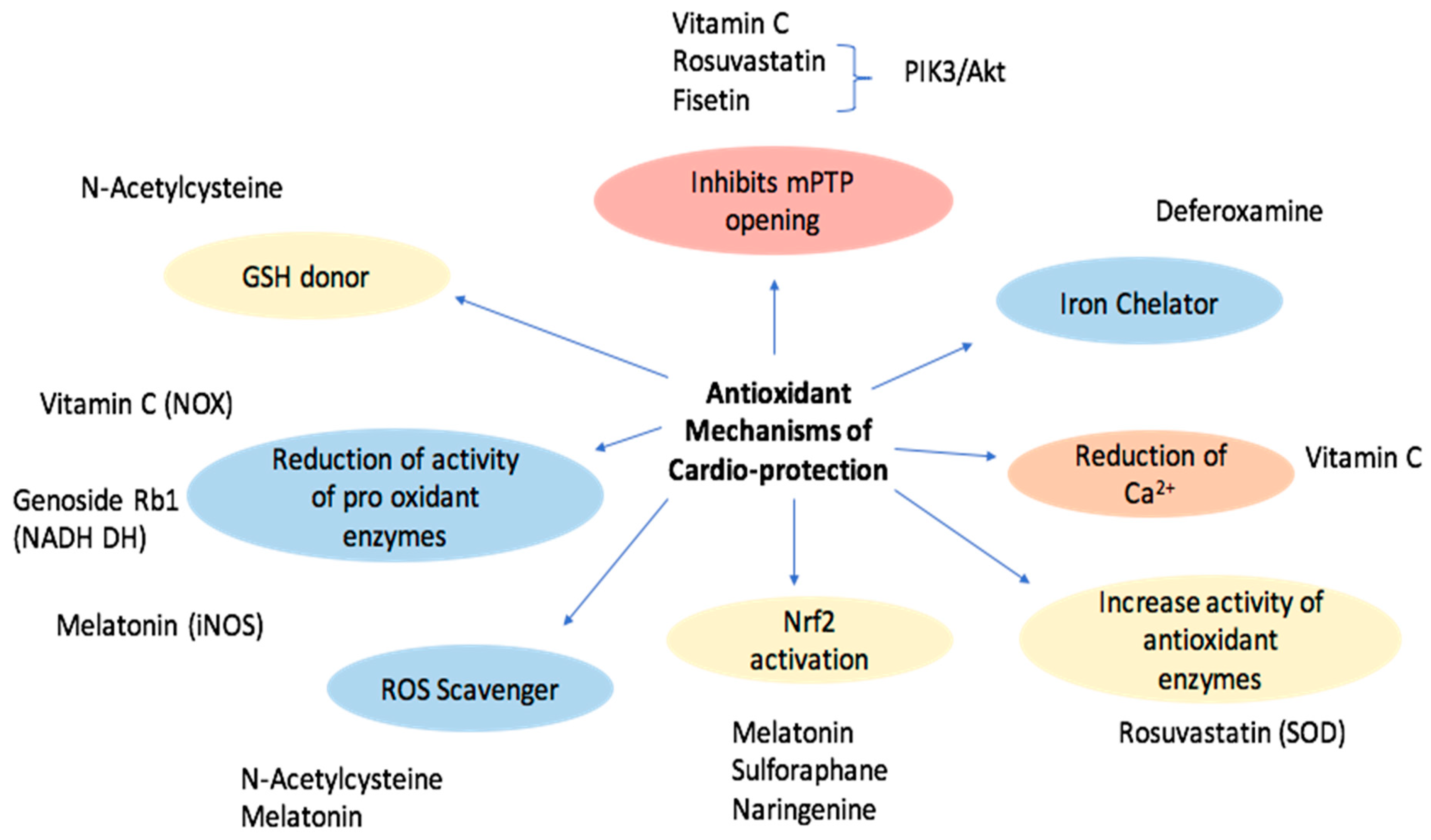
1.7. Exogenous Antioxidants with Reported Cardioprotective Effects
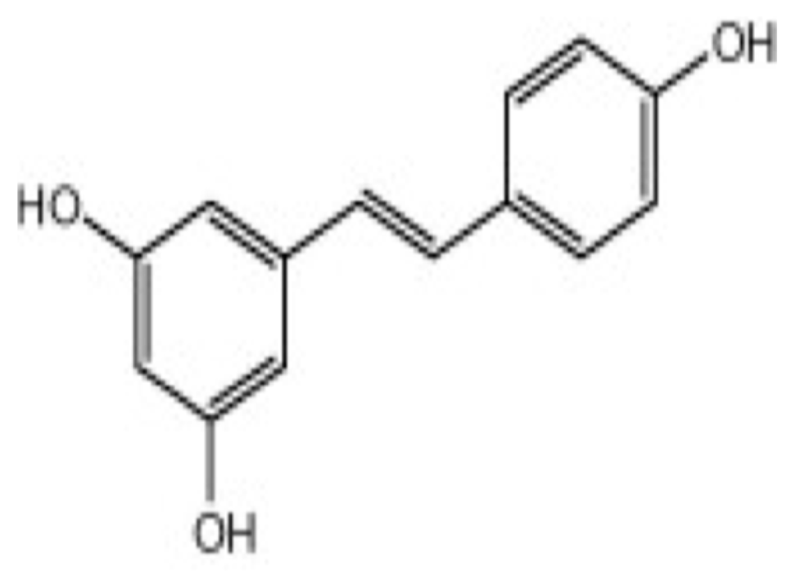
The potential usefulness of antioxidants as protective tools against I/R damage has attracted enormous interest. Clinical and experimental studies have been performed to determine the ability of natural or synthetic compounds with antioxidant and anti-inflammatory properties to counteract the mechanisms responsible for tissue alteration during I/R and to identify the involved molecular pathways [66]. Table 1 and Table 2 and Figure 2 summarize the molecules evaluated with respect to their cellular protective mechanisms.

1.8. Resveratrol

Resveratrol is a natural polyphenol with multiple biological activities [90]. It has a molecular weight of 228.25 g/mol [8] and exists in two isomeric forms (cis and trans), with the trans form (Figure 3) being the most common with more therapeutic benefits [91]. This antioxidant is found in many fruits and vegetables, such as grapes, Polygonum cuspidatum and mulberry [92].
Resveratrol became of interest during the early 1990s when a French paradox study mentioned that it was beneficial for health [91]. Since then, this molecule has been the subject of numerous studies, and many physiological effects have been assigned to it. Different studies have evidenced that it has various properties, including anti-inflammatory, antioxidant, anti-diabetic, antihypertensive, anti-cancerous and cardioprotective effects [90,92].

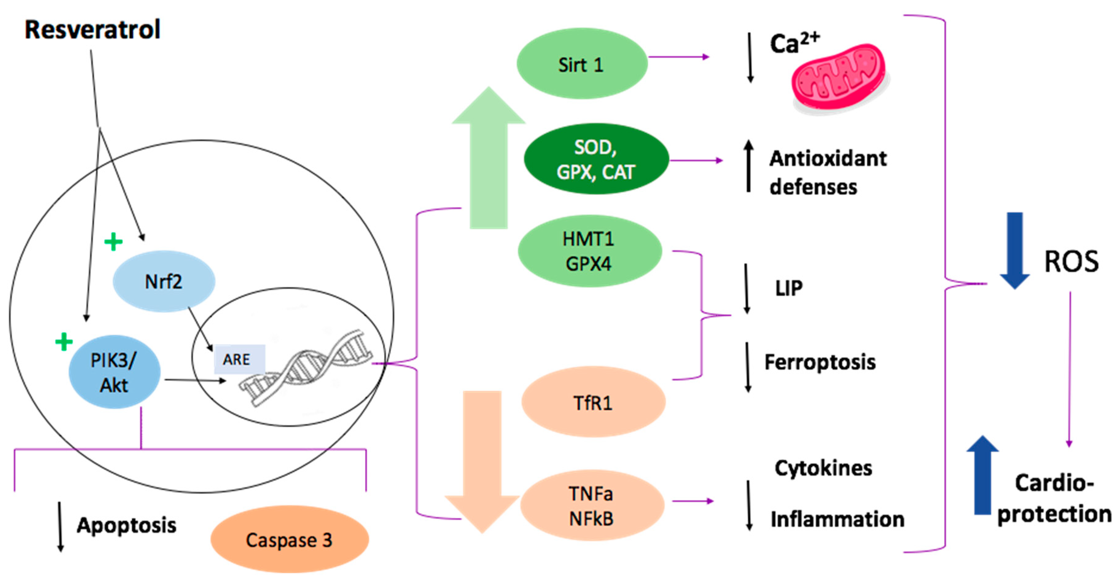
1.9. Resveratrol Cardioprotective Mechanisms

Regarding its cardioprotective action, several mechanisms have been designed attempting to understand which molecular pathways allow resveratrol to have an impact on cellular conditions related to myocardial damage due to I/R, such as oxidative stress, apoptosis, autophagy, hypoxia-induced endoplasmic reticulum stress, nitric oxide synthesis and others [93,94].
We will discuss some of the molecular pathways that have been described as the potential for resveratrol effect on cardiomyocytes. It is relevant that most studies have been performed in rat myocardial in vivo I/R models (Figure 4).

1.9.1. SIRT1 Activation

Silent information regulator factor 2-related enzyme 1 (SIRT1) consists of a nicotinamide adenine dinucleotide-dependent deacetylase that regulates oxidative stress-related proteins and other proteins that affect the function of cardiomyocytes. It was proposed that resveratrol activates SIRT1 leading to the activation of a cardioprotective response, reducing the Ca2+ overload in the cytosol and mitochondria [95]. According to this theory, a recent study showed that in a rat model of cardiac I/R, 21-day oral treatment with resveratrol decreased the incidence of atrioventricular block and lethality, thus suggesting the usefulness of this natural compound in the prevention of lethal cardiac arrhythmias after reperfusion [90]. Furthermore, it has been demonstrated that intraperitoneal injection of resveratrol in mice determines the increase of SIRT1 levels and a concomitant decrease of UCP-2 expression, thus protecting myocardial tissue against I/R injury in vivo [96].

1.9.2. Ferroptosis Inhibition

In a recent study carried out by Li et al. [89], in vitro experiments with H9c2 cells and a model of oxygen-glucose deprivation/reoxygenation (OGD/R) demonstrated that OGD/R-induced H9c2 cells showed increased cell death by ferroptosis. The addition of resveratrol in cell cultures reduced oxidative stress and the content of Fe2+. In vivo experiments performed by the same authors in rats ligated and perfused by the left anterior descending branch showed that when resveratrol was administered at the concentration of 50 mg/kg for 14 days, oxidative stress and the content of Fe2+ decreased again. The effect was explained by changes in the regulation of gene expression, in which resveratrol down-regulated transferrin receptor 1 expression and up-regulated the expression of ferritin heavy chain 1 and GPX4, leading to inhibition of ferroptosis and potentiation of the antioxidant defenses, respectively. The consequent reduction in oxidative stress is evidenced by a reduction in malondialdehyde (MDA) production [89].

1.9.3. Attenuation of Inflammation and Apoptosis

There are multiple ways in which resveratrol might inhibit apoptosis. Yu et al. [97] studied rats the antioxidant and anti-apoptotic effects of resveratrol in myocardial I/R injury. Compared to the control, the resveratrol group showed decreased ROS levels, increased catalase and glutathione peroxidase activities, and increased cell viability by apoptosis inhibition. Further, a decrease in lactate dehydrogenase (LDH) and plasma creatine kinase MB (CK-MB) levels were found. They suggested that these effects were mediated by the activation of the PI3K/AKT signaling pathway, which down-regulates the expression of apoptotic genes and regulates cell proliferation [98,99]. Apparently, PI3K/AKT signaling pathway is inhibited during the I/R process, resulting in potent activation of apoptosis and less viable tissue [97,100]. Moreover, it has been suggested that resveratrol attenuates apoptosis by reducing caspase 3 expression [101]. In another study, Hu et al. [102] indicated that resveratrol attenuates necroptosis after ischemia-reperfusion by inhibition of the tumor necrosis factor-alpha (TNF-α)/receptor-interacting protein kinase 1 (RIP1)/RIP3/mixed-lineage kinase domain-like (MLKL) signaling pathway. The results were that after treatment with different resveratrol concentrations, the expressions of TNF-α, RIP1, RIP3, and p-MLKL/MLKL decreased, as well as necroptosis. Under those circumstances, cell viability increased. All these effects were seen in dose-dependent manners [102]. Xing et al. [103] measured the levels of serum interleukin-1β (IL-1β), IL-6, and TNF-a in rat myocardial tissue during I/R injury. They also measured the expressions of mRNA and proteins of Toll-like receptor 4 (TLR4), NF-κB, p65, IL-1β, IL-6, and TNF-α. Resveratrol treatment resulted in a significant reduction of serum IL-1β, TNF-α, and IL-6, TLR4, NF-κB, p65, IL-1β, IL-6, and TNF-α mRNA and protein expressions in cardiac tissue cells [103].

1.9.4. Nrf2 Activation

As indicated before, Nrf2 up-regulates the expression of proteins involved in the antioxidant response. Using an in vitro hypoxia–reoxygenation (HR) model on HUVEC, it has been demonstrated that the increased cell viability and the reduced apoptotic rate and oxidative stress observed in cells treated with resveratrol were significantly reversed by knocking down Nrf2 [104]. Furthermore, in a rat model of lower-extremity I/R, treatment with resveratrol improved the apoptotic state of the femoral artery and reduced oxidative stress; this effect was abolished by the co-administration of an inhibitor of Nrf2 [104]. These results indicate that resveratrol might attenuate the I/R injury damage through up-regulating Keap1/Nrf2 signaling, mainly for its effects on oxidative stress. This could increase cell viability and decrease the apoptotic rate [104].

1.9.5. Reduced Mitochondrial Dysfunction

A recent study demonstrated that the addition of resveratrol in an in vitro I/R model obtained with primary rat cardiomyocytes decreased lactate dehydrogenase and creatine kinase MB release and ROS production and increased cell viability and catalase and glutathione peroxidase activities [97]. Moreover, resveratrol significantly increases the activity of mitochondrial superoxide dismutase and reduces the levels of malondialdehyde, indicating reduced oxidative damage to the mitochondria. The effects observed after resveratrol addition were reversed using PI3K siRNA, thus suggesting that the protective effect of resveratrol on cardiomyocytes under I/R conditions can be due to PI3K/AKT signaling pathway activation [97].

1.9.6. Other Pathways

It has also been proposed that resveratrol could reduce oxidative stress and protect myocardial cells via activating the VEGF-β/antioxidant signaling pathway [105]. Particularly, experiments conducted on rat hearts and on H9c2 cells showed that after treatment with resveratrol, a marked improvement of left ventricular function and a reduction of infarct size ex vivo, and decreased cell death and apoptosis of H9c2 cells during I/R occurred. The treatment with resveratrol was associated with the up-regulation of VEGF-β mRNA and protein levels, which caused the activation of Akt and the inhibition of GSK3β. The inhibition of VEGF-β inhibited the cardioprotective effects of resveratrol [105]. Of interest, the treatment of cardiomyocytes with isorhapontigenin (ISO), a resveratrol analog, was able to counteract angiotensin (Ang) II-induced cardiac hypertrophy by inhibiting Ang-II-dependent phosphorylation of PKC, Erk1/2, JNK, and p38. The same authors also observed that pretreatment with ISO down-modulated Ang II-mediated NF-κB activation by affecting the degradation and phosphorylation of IκBα [106].

1.9.7. Considerations of Safety in the Use of Resveratrol

Resveratrol is one of the potential therapeutic agents for the prevention of I/R heart tissue damage, but there are some issues that must be addressed. Shaito et al. [91] reviewed the potential adverse effects of the use of resveratrol. Resveratrol showed a beneficial effect at low doses, and the optimal dosage with safer and higher potency is still under research. Resveratrol presents a hormetic effect, and high doses have toxic effects, including inhibition of p450 cytochromes, which leads to interaction with several drugs and pro-oxidant effects [107]. We should pay attention to this dose-dependent effect on the redox state (antioxidant at low doses and pro-oxidant at high ones), given the fact that we are focused on an antioxidant activity [91].
Furthermore, studies usually focus on the short-term outcomes of resveratrol administration. Therefore, long-term effects are widely unknown. Additionally, there is not enough information about interactions between resveratrol and other therapies and little research about its pharmacokinetics, especially regarding absorption and bioavailability [91]. Referring to resveratrol water solubility, it is 0.03 mg/mL [108], which leads to low bioavailability. Nevertheless, different formulations have been designed to overcome this limitation, which will be discussed below.

1.9.8. Associations between Resveratrol and Other Antioxidants
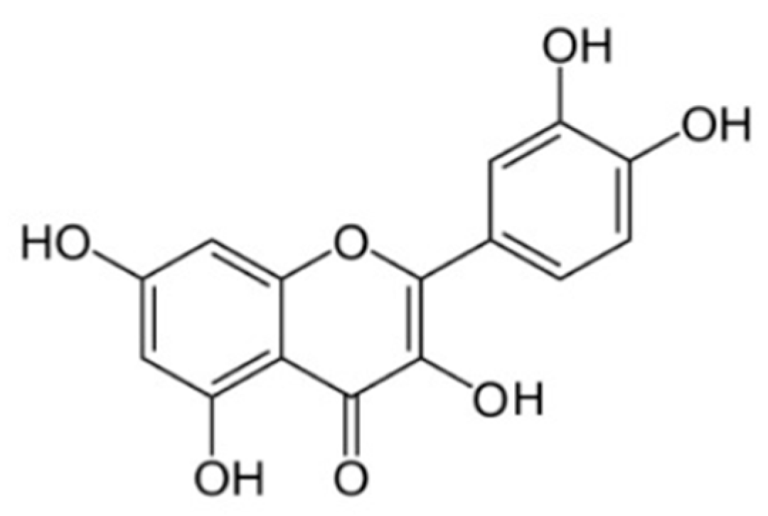
Different pathways and mechanisms participate in the generation of I/R injury. Even though resveratrol interferes with many of those mechanisms to reduce and/or prevent I/R damage, others are not covered by it. Therefore, it is reasonable to think that it would be beneficial to propose an association of resveratrol with other natural antioxidants (Table 1), looking for a synergic effect able to reduce the damage as much as possible. Until now, associations of resveratrol with other antioxidants or clinical drugs have not been studied. As a result, in the search for the most beneficial multitarget therapeutic scheme, we propose that there could be a synergistic effect between vitamin C and deferoxamine.
Resveratrol is associated with activating the Nrf2 signaling pathway, which leads to an increased expression of antioxidant enzymes, potentiating the antioxidant response. Theoretically, the balance would be even more inclined to favor this response if we decrease the function of pro-oxidant enzymes. In this context, adding vitamin C would be beneficial as it down-regulates NADPH oxidase activity and prevents eNOS uncoupling [50]. Further, it reduces intracellular Ca2+ overload and prevents the depolarization of the mitochondrial membrane [67]. These two last effects are related to SIRT1 activation by resveratrol, while it has been seen in studies showing that vitamin C also increases the expression of SIRT1 [109]. Even if this could constitute a potential interference between both antioxidants, it is necessary to look further into the molecular mechanisms involved to determine whether it would potentiate or antagonize the effect of resveratrol. It is worth mentioning that vitamin C is a water-soluble vitamin and can be administered intravenously [110].

1.10. Quercetin

Quercetin (Que) is a flavonoid with the following chemical formula C15H10O7 (Figure 5) [111].
Flavonoids are compounds with very different conformations. They are hydrophobic, as they have two benzene rings and a pyran ring between them [112]. Flavonoids exist in most plants and account for 65–75% of our daily flavonoid intake [113], mainly in vegetables and fruits, and also have a strong antioxidant property attributed to the presence of five hydroxyl groups, together with the pyrocatechol, which makes them good scavengers of free radicals [114]. Unfortunately, despite being very promising, Que is a molecule with low bioavailability due to its low aqueous solubility. However, many efforts have been devoted to increasing its solubility to obtain analogs with potentially improved properties. Quercetin can be rapidly hydrolyzed in the digestive tract by the enzyme β-glucosidase, which will facilitate its absorption through the intestinal mucosa, to be finally transferred to all the rest of the organism through portal circulation [115]. It also has several other biological effects in addition to its antioxidant properties. It has anti-aggregating, anti-inflammatory, anti-cancer, and anti-aging effects. Furthermore, it is not toxic even in high doses (4000 mg/day) [116]. Additionally, recent studies have shown it to have tremendous potential to reduce myocardial damage that occurs after I/R through various mechanisms (Figure 6) [117].

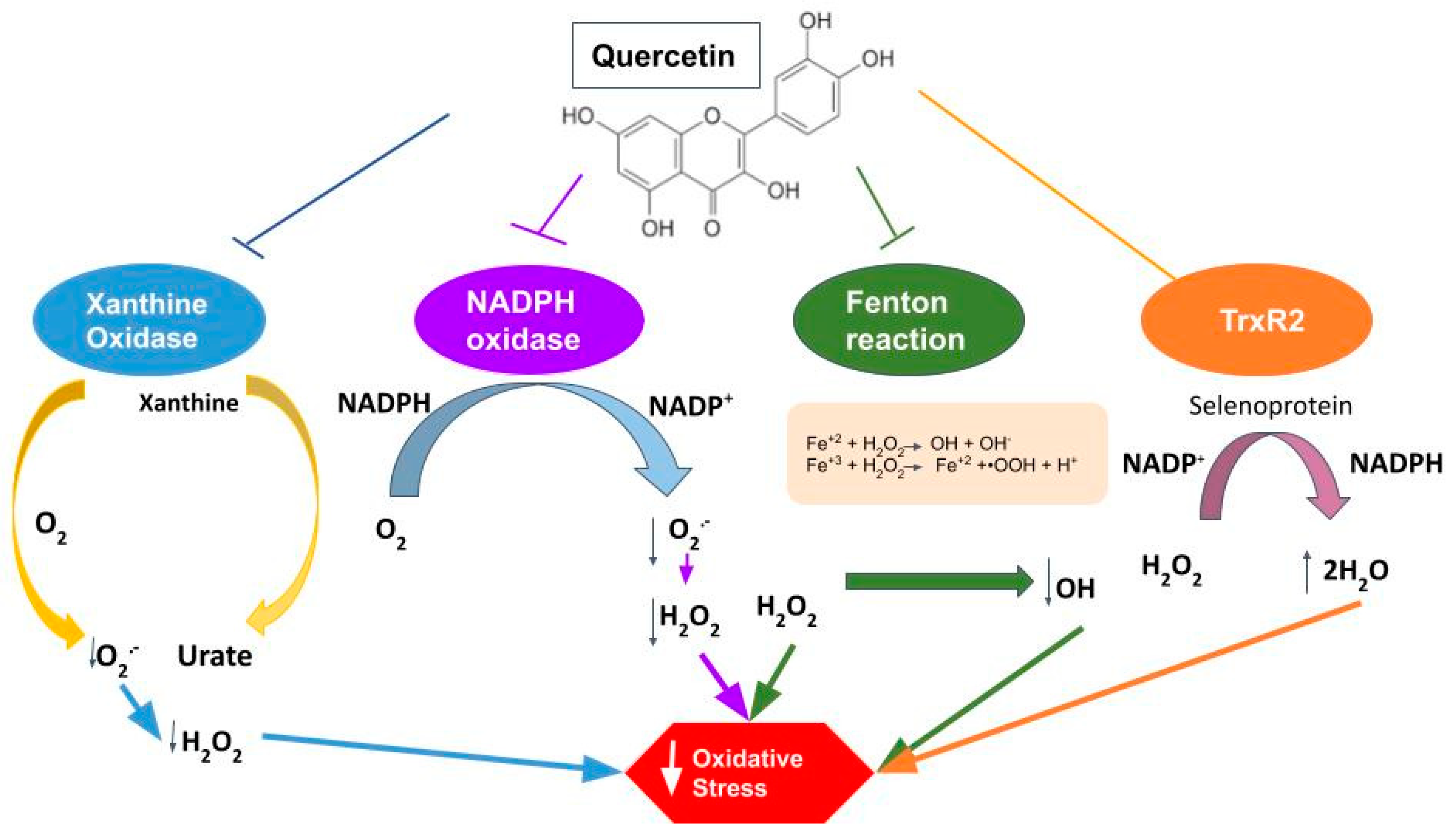
1.10.1. Properties of Quercetin against Oxidative Stress

Recent experiments with an animal model of hyperuricemia have demonstrated a reduction in xanthine oxidase expression and enzyme activity by Que treatment (100, 200, 400 mg/Kg) [9]. Xanthine oxidase is a molybdo-flavoenzyme found in various species. It is a homodimer with two symmetrical monomers. It has a great affinity for producing ROS because, in each of the monomers, there is a C-terminal molybdo protein that contains four redox centers. In turn, it has an N-terminal domain with two iron–sulfur centers and a central flavin adenine dinucleotide cofactor [118]. Thus, xanthine oxidase stimulates ROS production, which can end up causing oxidative stress injuries [119].
Similarly, Que can attenuate the expression of NOX2 [120]. The NADPH oxidase system is a multiprotein complex that produces ROS in different cells and tissues, being of great importance in phagocytic cells [121]. The NADPH oxidase enzyme is composed of several subunits, including the gp91-phox subunit and its counterparts which are commonly known as the NOX family (NOX1, NOX2, NOX3, NOX4, NOX5, DUOX1, and DUOX2) [122] that are found in almost all cells of the organism [123]. In the cardiovascular system, this complex is the main ROS producer, which is achieved through the transfer of an electron from NADPH to O2, resulting in NADP and O2●−. The newly produced O2●− will end up rapidly transforming into H2O2, characterized by being more stable and diffusible [124]. This leads us to think that if we initially inhibit the expression of the enzyme, we will reduce the production of H2O2 and, consequently, decrease the I/R damage produced by ROS in the myocardial tissue. In cardiomyocytes, NADPH oxidase can be activated by various mechanisms and stimulating factors [125]. Among them, one of the stimulants for the enzyme corresponds to I/R, with NOX2 and NOX4 being the ones more activated during myocardial I/R injury [82]. Therefore, the inhibitory effect of Que on NOX can potentially be a great target to ameliorate the injury induced by I/R.
In addition, Que combined with Fe2+/Cu+ will significantly inhibit Fenton reaction, another ROS source producing OH [126]. Fenton reaction consists of the electron transfer from Fe2+/Cu+ metallic ion to H2O2, which produces Fe3+/Cu2+, OH− and OH. It has been shown that OH has a very strong oxidability in acid solutions [127]. Then also, H2O2 can restore Fe2+/Cu+ from Fe3+/Cu2+, which produces HOO and H+. Fe2+/Cu+ can be oxidized by O2 again, which forms O2●−. In I/R damage, an acidic environment is created due to anaerobic glycolysis, which then facilitates the production of the Fenton reaction.
Selenium is an antioxidant and a component of selenoproteins, including glutathione peroxidases (GPX), thioredoxin reductases (TrxR) and methionine sulfoxide reductase 2, thereby modulating redox activity [128]. Accordingly, it has been observed that after Que administration at a dose of 50 mg/kg for eight weeks, the selenoprotein TrxR2 increased in the Que group compared to the control [82].
Quercetin is recognized as a significant antioxidant, preventing damage to cardiomyocytes as measured by oxidative stress, avoiding ROS increase and progression of damage after I/R. Moreover, ROS removal may increase NO bioavailability and restoration of endothelial function after I/R damage [129]. The mechanisms whereby Que exerts its antioxidant activity consist of eliminating ROS through its ability to react with the free radicals O2●−, HO, NO, alkoxy and peroxyl. It also occurs when the hydroxyl attached to the benzene ring transfers a hydrogen atom or an electron to the free radical, resulting in the formation of more stable molecules [130]. Quercetin also acts through indirect mechanisms, restoring inherent antioxidant systems of the body, such as enzymatic systems dependent or not on antioxidants, such as vitamins C and E, and reduced glutathione (GSH) [59], the latter decreasing after I/R injury, which indicates the depletion of the antioxidant system. Thus, it can eliminate ROS and restore the function of the intrinsic antioxidant system [131].

1.10.2. Anti-Inflammatory and Anti-Apoptotic Effects

Acute inflammatory reactions are intensified in cardiomyocytes following I/R injury. This is due to the neutrophilic granulocytes, which are the ones that explain most of the reduced exudation and even the diffusion of these neutrophils through the blood vessels. Thus, it can be generated more damage to the injured tissue [132]. In the I/R period, leukocytes can alter the function of endotheliocytes and reduce the relaxation capacity of the coronary arteries [133]. They also promote coagulation and pro-inflammatory effects that further aggravate the mechanical obstruction of capillaries. This will cause a decrease in blood flow and incomplete perfusion of the coronary arteries, which are the main causes of neutrophil conglutination in the vascular walls [134]. The binding of leukocytes to endotheliocytes plays an important role in the inflammatory reaction, which is accompanied by cardiomyocyte apoptosis. Increased permeability of the plasmalemma leads to cytochrome C release that further aggravates myocardial apoptosis [135]. In this context, Que has been shown to improve inflammation and apoptosis in the myocardium. A recent I/R injury model was made through the ligation of the left coronary artery, after which Que was administered (2, 10 and 20 mg/kg orally) and diltiazem (15 mg/kg orally) for five days. This treatment caused the inhibition of the expression of TNF-α, IL-6 and IL-1β (important biomarkers of inflammatory reactions) in serum and cardiomyocytes. This indicates that Que treatment after I/R can alleviate inflammation and apoptosis. In agreement with these findings, Que diminishes the infarct size, as shown with 2,3,4-triphenyltetrazolium chloride (TTC) staining, as well as myocardial contractility, demonstrating that Que is capable of mitigating the inflammation and restoring myocardial function [131].

1.10.3. Vasodilatory Effects

It is known that during a period of ischemia, the coronary arteries are usually obstructed by emboli. Although blood flow can be quickly restored through interventional therapies, there are multiple findings that show that G-protein-coupled receptors are altered after I/R injury [136]. The administration of Que has been shown to improve vasoconstriction caused by ET-1, one of the types of endothelin, which is one of the most potent vasoconstrictors distributed in the endothelium of blood vessels (as well as in many other tissues and cells) [137].

1.10.4. Associations between Quercetin and Other Antioxidants

Although Que acts by reducing oxidative stress, there are many mechanisms enhancing I/R damage that are still functioning. Therefore, it is proposed to associate Que with other antioxidants to produce synergy and further reduction of oxidative damage. There are multiple possible associations between Que and other antioxidants that could enhance the cardioprotective effect. One of the associations that have been studied is the combination between Que and α-tocopherol, which was studied in rats that were induced AMI by isoproterenol and were previously administered a combination of Que (10 mg/Kg) and α-tocopherol (10 mg/Kg) for 14 days. The combined pretreatment normalized all biochemical parameters and minimized ECG alterations. Therefore, Que and α-tocopherol exhibited enhanced cardioprotective effects against isoproterenol-induced cardiotoxicity because they eliminate free radicals, improve antioxidation, and maintain Ca2+ levels. Furthermore, the study showed that the combined pretreatment was more effective than the sole one [138]. There are other molecules with which the association with Que has been studied to demonstrate its cardio-supporting effect, such as lycopene, another natural phytocompound with antioxidant effects. In an animal model study in which cardiac toxicity was induced by isoproterenol (ISO), free radicals and oxidative stress increased in myocardial tissue. The combination of Que with lycopene prevented all the side effects of cardiotoxicity, significantly decreasing the myocardial damage, reducing oxidative stress, and slowing the levels of expression of antioxidant genes, a participant in pathways related to the Nrf2, HO-1, NQO1, GSTµ, SOD, SOD2, CAT and BCL-2 genes [139]. Evidence of the association of quercetin with clinical drugs was not found.

2. Discussion

Oxidative stress constitutes an essential mechanism of damage involved in I/R injury in acute myocardial infarction. Even if multiple individual antioxidants have been tested to prevent or minimize this damage, an effective treatment is not available. This problem might be related to the fact that oxidative stress results from different pathways that lead to an imbalance between ROS generation and antioxidant activity. Some of these mechanisms are mitochondrial dysfunction, activation of the inflammatory response, decreased antioxidant activity and activation of pro-oxidant enzymes, such as xanthine oxidase, uncoupled eNOS, NOX, and others. The comprehension of oxidative stress mechanisms makes us realize that the treatment alternatives based on monotherapy are not the appropriate solution, a view that has not been changed for more than a decade. It is of interest that several isolated beneficial effects could be joined to improve the cardioprotective effects occurring in I/R settings, such as percutaneous coronary intervention following AMI. Thus, some antioxidants are iron chelators (deferoxamine), some are ROS scavengers (N-acetylcysteine), some activate the Nrf2 antioxidant response pathway (naringenin, melatonin, sulforaphane), and others exert influence on alternative edges of ROS production, resulting in effects that could be synergic and beneficial (Table 1). In the case of resveratrol and quercetin, these molecules have several cardioprotective effects making them suitable to be included as part of a multitarget therapy.
It is important to note that resveratrol and quercetin molecular sites of action are still not fully elucidated, but there are many proven targets. In the case of resveratrol, activation of Nrf2 and SIRT1 occurs through increased antioxidant proteins expression and PGC-1α deacetylation [140]. In addition, resveratrol also activates the PI3K/AKT pathway during I/R injury, resulting in decreased mitochondrial pathway-mediated apoptosis [97]. As mentioned, resveratrol is also capable of reducing ferroptosis by decreasing Fe2+ concentration, which could be involved in inducing changes in the regulation of gene expression, but the exact target remains to be fully elucidated.
On the other hand, quercetin exerts its protective effects mainly through the inhibition of ROS-producing enzymes, such as NADPH oxidase and xanthine oxidase. The exact mechanism whereby this is achieved has been only partially described. A recent study shows that NOX may be inhibited for the enhanced expression of the antioxidant enzyme heme oxygenase-1 (HO-1) by quercetin [141]. In the case of xanthine oxidase, quercetin acts as a reversible inhibitor of the enzyme action [142].
Another relevant aspect is that there are some concerns about the water solubility of these compounds, which could make us question whether the suitability can be successfully administered intravenously. To solve this problem, different strategies have been designed to increase their water solubility. In the case of resveratrol, showing cardioprotective efficacy at low concentrations, it has been reported its administration with complexes with β-cyclodextrin and hydroxypropyl-β-cyclodextrin [143], complexes that could slow down resveratrol metabolism and increase bioavailability [144]. Furthermore, solubilization of resveratrol could be increased with micellar solutions of bile acids, an effect attributed to the smallest membranolytic potential achieved through the formation of bile acids derivatives with resveratrol [145]. On the other hand, multiple studies aim to increase Que solubility by various mechanisms and thus increase its application properties. One of them is the nanoformulation method, which improves solubility, bioavailability, and circulation time, among other effects [146]. In turn, Que solubility and its thermal degradation in ethanol expanded with CO2 and ethyl lactate were studied [147]. On the other hand, co-crystallization has recently gained attention as a means of improving the physicochemical characteristics of a compound, and each of these cocrystals exhibited pharmacokinetic properties that are far superior to those of Que alone [148]. Finally, all these solutions sustain the research that has been done about these two antioxidants and their potential cardioprotective effects. The improvement of solubility with different agents makes it possible to continue considering resveratrol as a suitable potential part of therapy against I/R injury. The evidence leads us to think that if the solubility of Que is increased, its therapeutic effects could be promisingly improved.

3. Concluding Remarks

Ischemia/reperfusion injury occurring in AMI patients undergoing PCI is a complex process involving pathophysiological cascades leading to cellular disturbances ranging from metabolic changes to cell death. Although oxidative stress is involved in several of these injury mechanisms, antioxidant therapeutic interventions so far have not been successful in protecting myocardial tissue against the harmful effects. Likely, the use of the most frequent monotherapies in a multifactorial process could explain the lack of consistency between the experimental studies and the clinical data. The paradigm of a multitarget therapy based on an association of different antioxidant molecules should be expected to improve the abrogation of oxidative damage and related effects derived from ROS burst, particularly occurring early at the onset of reperfusion. Thus, the administration of several antioxidant molecules could give rise to an additive or synergistic pharmacological effect. Some compounds, such as N-acetylcysteine, ascorbate and deferoxamine, have been tested as monotherapies in other clinical settings associated with increased ROS, showing lightly protective effects, as well as those caused by other naturally occurring antioxidant molecules. Among the latter, cardioprotective properties have been found in the case of resveratrol and Que, among others, having efficacy at low concentrations suitable to avoid adverse events. Therefore, these compounds could also be considered in this association, but following a better characterization of their pharmacokinetic properties, solubility at the required doses and studies about the appropriate design of formulations. Studies about associations of these compounds with others that have previously shown cardioprotective effects could be analyzed in isolated heart rat Langendorff model and, subsequently, in clinical trials aimed to reduce the infarct size due to the increased efficacy of this therapeutic association. This could lead to a better prognosis and quality of life after an acute myocardial infarction, decreasing complications, such as heart failure and arrhythmias, and also reducing the associated costs.
The evidence supports that resveratrol is safe to be used in low doses. It has strong antioxidant effects, in which Nrf2 and SIRT1 activation are involved, thereby increasing the antioxidant response and decreasing inflammation and ferroptosis. Taken together, these effects could be beneficial as part of a multitarget therapy. Nevertheless, further studies are still lacking to implement a therapeutic scheme that includes resveratrol involving association with other antioxidants, specifically with vitamin C, which inactivates the NOX, reducing ROS production, and the iron chelator deferoxamine, reducing ferroptosis and cell death. We propose resveratrol might show a synergic effect with these antioxidants due to a theoretical potentiation of oxidative stress reduction. Quercetin has multiple beneficial effects, particularly through its antioxidant activity could account for an enhanced cardioprotective effect.
To date, the association of resveratrol and other antioxidants has not been studied, as well as there is no evidence of the combination of resveratrol or quercetin with clinical drugs. This is a topic that could be interesting to investigate, considering the beneficial and antioxidant properties of some drugs, such as melatonin, rosuvastatin and N-acetylcysteine, that are exposed in Table 2. In this case, being synthetic antioxidants, it would be important to consider what happens with the side effects of the drug in use.
Even if treatment for I/R injury has not been found yet, resveratrol and quercetin constitute attractive natural pharmacological agents to be used in a combined therapy aimed to reduce cardiac tissue damage and infarct size due to their cardioprotective effects. Randomized double-blind clinical trials should be made to test the therapeutic efficacy as well as safety of these associations.

Author Contributions

R.R. Formulated the hypothesis and edited the manuscript before submission. C.R. Reviewed the literature, edited the figures, and resolved reviewers’ comments. D.S. Reviewed the literature and resolved reviewers’ comments. D.V.-H. Reviewed the literature, edited the figures, resolved reviewers’ comments, and edited the manuscript before submission. S.S. Reviewed the literature and added new insights about the topic. E.P. Reviewed the literature, added new information, and edited the manuscript before submission. B.B. Reviewed the literature and added new information to the manuscript. L.S. Formulated the hypothesis and edited the manuscript before submission. All authors reviewed the manuscript. All authors have read and agreed to the published version of the manuscript.

Funding

This study was supported by the Agencia Nacional de Investigación y Desarrollo (ANID), Fondo Nacional de Desarrollo Científico, Tecnológico y de Innovación Tecnológica, (FONDECYT), grant number 1211850. APC was funded by Luciano Saso.

Institutional Review Board Statement

Not applicable.

Informed Consent Statement

Not applicable.

Data Availability Statement

Not applicable.

Conflicts of Interest

The authors declare no conflict of interest.

References

  1. World Health Organization. Cardiovascular Diseases. 2019. Available online: https://www.who.int/health-topics/cardiovascular-diseases#tab=tab_1 (accessed on 10 November 2021).
  2. Khan, M.A.; Hashim, M.J.; Mustafa, H.; Baniyas, M.Y.; Al Suwaidi, S.K.B.M.; Alkatheeri, R.; Alblooshi, F.M.K.; Almatrooshi, M.E.A.H.; Alzaabi, M.E.H.; Al Darmaki, R.S.; et al. Global Epidemiology of Ischemic Heart Disease: Results from the Global Burden of Disease Study. Cureus 2020, 12, e9349. [Google Scholar] [CrossRef]
  3. Yellon, D.M.; Hausenloy, D.J. Myocardial reperfusion injury. N. Engl. J. Med. 2007, 357, 1121–1135. [Google Scholar] [CrossRef]
  4. Massalha, S.; Luria, L.; Kerner, A.; Roguin, A.; Abergel, E.; Hammerman, H.; Boulos, M.; Dragu, R.; Kapeliovich, M.R.; Beyar, R.; et al. Heart failure in patients with diabetes undergoing primary percutaneous coronary intervention. Eur. Heart J. Acute Cardiovasc. Care 2016, 5, 455–462. [Google Scholar] [CrossRef]
  5. Sinning, C.; Westermann, D.; Clemmensen, P. Oxidative stress in ischemia and reperfusion: Current concepts, novel ideas and future perspectives. Biomark Med. 2017, 11, 11031–11040. [Google Scholar] [CrossRef]
  6. Rodrigo, R.; González-Montero, J.; Sotomayor, C.G. Novel Combined Antioxidant Strategy against Hypertension, Acute Myocardial Infarction and Postoperative Atrial Fibrillation. Biomedicines 2021, 9, 620. [Google Scholar] [CrossRef]
  7. Davidson, S.M.; Ferdinandy, P.; Andreadou, I.; Bøtker, H.E.; Heusch, G.; Ibanez, B.; Ovize, M.; Schulz, R.; Yellon, D.M.; Hausenloy, D.J.; et al. Multitarget Strategies to Reduce Myocardial Ischemia/Reperfusion Injury: JACC Review Topic of the Week. J. Am. Coll. Cardiol. 2019, 73, 89–99. [Google Scholar] [CrossRef]
  8. Menezes-Rodrigues, F.S.; Errante, P.R.; Araújo, E.A.; Fernandes, M.P.P.; Silva, M.M.D.; Pires-Oliveira, M.; Scorza, C.A.; Scorza, F.A.; Taha, M.O.; Caricati-Neto, A. Cardioprotection stimulated by resveratrol and grape products prevents lethal cardiac arrhythmias in an animal model of ischemia and reperfusion. Acta Cir. Bras. 2021, 36, e360306. [Google Scholar] [CrossRef]
  9. Huang, J.; Wang, S.; Zhu, M.; Chen, J.; Zhu, X. Effects of genistein, apigenin, quercetin, rutin and astilbin on serum uric acid levels and xanthine oxidase activities in normal and hyperuricemic mice. Food Chem. Toxicol. 2011, 49, 1943–1947. [Google Scholar] [CrossRef]
  10. Rodrigo, R.; González, J.; Paoletto, F. The role of oxidative stress in the pathophysiology of hypertension. Hypertens. Res. 2011, 34, 431–440. [Google Scholar] [CrossRef] [Green Version]
  11. Valko, M.; Leibfritz, D.; Moncol, J.; Cronin, M.T.; Mazur, M.; Telser, J. Free radicals and antioxidants in normal physiological functions and human disease. Int. J. Biochem. Cell Biol. 2007, 39, 44–84. [Google Scholar] [CrossRef]
  12. Förstermann, U.; Xia, N.; Li, H. Roles of Vascular Oxidative Stress and Nitric Oxide in the Pathogenesis of Atherosclerosis. Circ. Res. 2017, 120, 713–735. [Google Scholar] [CrossRef]
  13. Nakamura, T.; Naguro, I.; Ichijo, H. Iron homeostasis and iron-regulated ROS in cell death, senescence and human diseases. Biochim. Biophys. Acta Gen. Subj. 2019, 1863, 1398–1409. [Google Scholar] [CrossRef]
  14. Thompson, R.C.; Allam, A.H.; Lombardi, G.P.; Wann, L.S.; Sutherland, M.L.; Sutherland, J.D.; Soliman, M.A.; Frohlich, B.; Mininberg, D.T.; Monge, J.M.; et al. Atherosclerosis across 4000 years of human history: The Horus study of four ancient populations. Lancet 2013, 381, 1211–1222. [Google Scholar] [CrossRef]
  15. Sanada, S.; Komuro, I.; Kitakaze, M. Pathophysiology of myocardial reperfusion injury: Preconditioning, postconditioning, and translational aspects of protective measures. Am. J. Physiol. Heart Circ. Physiol. 2011, 301, H1723–H1741. [Google Scholar] [CrossRef] [Green Version]
  16. Avkiran, M.; Marber, M.S. Na(+)/H(+) exchange inhibitors for cardioprotective therapy: Progress, problems and prospects. J. Am. Coll. Cardiol. 2002, 39, 747–753. [Google Scholar] [CrossRef]
  17. Rossi, A.E.; Dirksen, R.T. Sarcoplasmic reticulum: The dynamic calcium governor of muscle. Muscle Nerve 2006, 33, 715–731. [Google Scholar] [CrossRef]
  18. Sasaki, M.; Joh, T. Oxidative stress and ischemia-reperfusion injury in gastrointestinal tract and antioxidant, protective agents. J. Clin. Biochem. Nutr. 2007, 40, 1–12. [Google Scholar] [CrossRef] [Green Version]
  19. Raedschelders, K.; Ansley, D.M.; Chen, D.D. The cellular and molecular origin of reactive oxygen species generation during myocardial ischemia and reperfusion. Pharmacol. Ther. 2012, 133, 230–255. [Google Scholar] [CrossRef]
  20. Vásquez-Vivar, J.; Martásek, P.; Whitsett, J.; Joseph, J.; Kalyanaraman, B. The ratio between tetrahydrobiopterin and oxidized tetrahydrobiopterin analogues controls superoxide release from endothelial nitric oxide synthase: An EPR spin trapping study. Biochem. J. 2002, 362, 733–739. [Google Scholar] [CrossRef]
  21. Parra, P.; Rodrigo, R. Novel Antioxidant Therapy against Myocardial Ischemia–Reperfusion Injury during Percutaneous Coronary Angioplasty. In Free Radicals and Diseases; Ahmad, R., Ed.; InTech: London, UK, 2016. [Google Scholar]
  22. Choy, K.W.; Murugan, D.; Mustafa, M.R. Natural products targeting ER stress pathway for the treatment of cardiovascular diseases. Pharmacol. Res. 2018, 132, 119–129. [Google Scholar] [CrossRef]
  23. Cullinan, S.B.; Diehl, J.A. PERK-dependent activation of Nrf2 contributes to redox homeostasis and cell survival following endoplasmic reticulum stress. J. Biol. Chem. 2004, 279, 20108–20117. [Google Scholar] [CrossRef] [Green Version]
  24. Bernardi, P.; Vassanelli, S.; Veronese, P.; Colonna, R.; Szabó, I.; Zoratti, M. Modulation of the mitochondrial permeability transition pore. Effect of protons and divalent cations. J. Biol. Chem. 1992, 267, 2934–2939. [Google Scholar] [CrossRef]
  25. Griffiths, E.J.; Halestrap, A.P. Mitochondrial non-specific pores remain closed during cardiac ischaemia, but open upon reperfusion. Biochem. J. 1995, 307 Pt 1, 93–98. [Google Scholar] [CrossRef] [Green Version]
  26. Hausenloy, D.J.; Yellon, D.M. The mitochondrial permeability transition pore: Its fundamental role in mediating cell death during ischaemia and reperfusion. J. Mol. Cell. Cardiol. 2003, 35, 339–341. [Google Scholar] [CrossRef] [Green Version]
  27. Kim, J.S.; Jin, Y.; Lemasters, J.J. Reactive oxygen species, but not Ca2+ overloading, trigger pH- and mitochondrial permeability transition-dependent death of adult rat myocytes after ischemia-reperfusion. Am. J. Physiol. Heart Circ. Physiol. 2006, 290, H2024–H2034. [Google Scholar] [CrossRef]
  28. Ong, S.B.; Samangouei, P.; Kalkhoran, S.B.; Hausenloy, D.J. The mitochondrial permeability transition pore and its role in myocardial ischemia reperfusion injury. J. Mol. Cell. Cardiol. 2015, 78, 23–34. [Google Scholar] [CrossRef]
  29. Baines, C.P.; Molkentin, J.D. Adenine nucleotide translocase-1 induces cardiomyocyte death through upregulation of the pro-apoptotic protein Bax. J. Mol. Cell. Cardiol. 2009, 46, 969–977. [Google Scholar] [CrossRef] [Green Version]
  30. Padmavathi, G.; Ramkumar, K.M. MicroRNA mediated regulation of the major redox homeostasis switch, Nrf2, and its impact on oxidative stress-induced ischemic/reperfusion injury. Arch. Biochem. Biophys. 2021, 698, 108725. [Google Scholar] [CrossRef]
  31. Chen, R.; Zhang, Y.Y.; Lan, J.N.; Liu, H.M.; Li, W.; Wu, Y.; Leng, Y.; Tang, L.H.; Hou, J.B.; Sun, Q.; et al. Ischemic Postconditioning Alleviates Intestinal Ischemia-Reperfusion Injury by Enhancing Autophagy and Suppressing Oxidative Stress through the Akt/GSK-3β/Nrf2 Pathway in Mice. Oxid. Med. Cell Longev. 2020, 2020, 6954764. [Google Scholar] [CrossRef] [Green Version]
  32. Mariathasan, S.; Weiss, D.S.; Newton, K.; McBride, J.; O’Rourke, K.; Roose-Girma, M.; Lee, W.P.; Weinrauch, Y.; Monack, D.M.; Dixit, V.M. Cryopyrin activates the inflammasome in response to toxins and ATP. Nature 2006, 440, 228–232. [Google Scholar] [CrossRef]
  33. Cabrera-Fuentes, H.A.; Ruiz-Meana, M.; Simsekyilmaz, S.; Kostin, S.; Inserte, J.; Saffarzadeh, M.; Galuska, S.P.; Vijayan, V.; Barba, I.; Barreto, G.; et al. RNase1 prevents the damaging interplay between extracellular RNA and tumour necrosis factor-α in cardiac ischaemia/reperfusion injury. Thromb. Haemost. 2014, 112, 1110–1119. [Google Scholar] [CrossRef]
  34. Gloire, G.; Legrand-Poels, S.; Piette, J. NF-kappaB activation by reactive oxygen species: Fifteen years later. Biochem Pharmacol. 2006, 72, 1493–1505. [Google Scholar] [CrossRef]
  35. Djavaheri-Mergny, M.; Amelotti, M.; Mathieu, J.; Besançon, F.; Bauvy, C.; Souquère, S.; Pierron, G.; Codogno, P. NF-kappaB activation represses tumor necrosis factor-alpha-induced autophagy. J. Biol. Chem. 2006, 281, 30373–30382. [Google Scholar] [CrossRef] [Green Version]
  36. Rodrigo, R.; Prieto, J.C.; Castillo, R. Cardioprotection against ischaemia/reperfusion by vitamins C and E plus n-3 fatty acids: Molecular mechanisms and potential clinical applications. Clin. Sci. 2013, 124, 1–15. [Google Scholar] [CrossRef] [Green Version]
  37. Vinten-Johansen, J. Involvement of neutrophils in the pathogenesis of lethal myocardial reperfusion injury. Cardiovasc. Res. 2004, 61, 481–497. [Google Scholar] [CrossRef] [Green Version]
  38. Mann, D.L. Innate immunity and the failing heart: The cytokine hypothesis revisited. Circ. Res. 2015, 116, 1254–1268. [Google Scholar] [CrossRef] [Green Version]
  39. Nian, M.; Lee, P.; Khaper, N.; Liu, P. Inflammatory cytokines and postmyocardial infarction remodeling. Circ. Res. 2004, 94, 1543–1553. [Google Scholar] [CrossRef]
  40. Frangogiannis, N.G. Regulation of the inflammatory response in cardiac repair. Circ. Res. 2012, 110, 159–173. [Google Scholar] [CrossRef]
  41. Horckmans, M.; Ring, L.; Duchene, J.; Santovito, D.; Schloss, M.J.; Drechsler, M.; Weber, C.; Soehnlein, O.; Steffens, S. Neutrophils orchestrate post-myocardial infarction healing by polarizing macrophages towards a reparative phenotype. Eur. Heart J. 2017, 38, 187–197. [Google Scholar] [CrossRef] [Green Version]
  42. Lillo-Moya, J.; Rojas-Solé, C.; Muñoz-Salamanca, D.; Panieri, E.; Saso, L.; Rodrigo, R. Targeting Ferroptosis against Ischemia/Reperfusion Cardiac Injury. Antioxidants 2021, 10, 667. [Google Scholar] [CrossRef]
  43. Li, J.; Cao, F.; Yin, H.L.; Huang, Z.J.; Lin, Z.T.; Mao, N.; Sun, B.; Wang, G. Ferroptosis: Past, present and future. Cell Death Dis. 2020, 11, 88. [Google Scholar] [CrossRef] [PubMed]
  44. Friedmann Angeli, J.P.; Schneider, M.; Proneth, B.; Tyurina, Y.Y.; Tyurin, V.A.; Hammond, V.J.; Herbach, N.; Aichler, M.; Walch, A.; Eggenhofer, E.; et al. Inactivation of the ferroptosis regulator Gpx4 triggers acute renal failure in mice. Nat. Cell Biol. 2014, 16, 1180–1191. [Google Scholar] [CrossRef] [Green Version]
  45. Anderson, G.J.; Frazer, D.M. Current understanding of iron homeostasis. Am. J. Clin. Nutr. 2017, 106 (Suppl. 6), 1559S–1566S. [Google Scholar] [CrossRef] [PubMed] [Green Version]
  46. Mou, Y.; Wang, J.; Wu, J.; He, D.; Zhang, C.; Duan, C.; Li, B. Ferroptosis, a new form of cell death: Opportunities and challenges in cancer. J. Hematol. Oncol. 2019, 12, 34. [Google Scholar] [CrossRef] [PubMed] [Green Version]
  47. Eefting, F.; Rensing, B.; Wigman, J.; Pannekoek, W.J.; Liu, W.M.; Cramer, M.J.; Lips, D.J.; Doevendans, P.A. Role of apoptosis in reperfusion injury. Cardiovasc. Res. 2004, 61, 414–426. [Google Scholar] [CrossRef]
  48. Borutaite VJekabsone AMorkuniene RBrown, G.C. Inhibition of mitochondrial permeability transition prevents mitochondrial dysfunction, cytochrome c release and apoptosis induced by heart ischemia. J. Mol. Cell Cardiol. 2003, 35, 357–366. [Google Scholar]
  49. Saelens, X.; Festjens, N.; Vande Walle, L.; van Gurp, M.; van Loo, G.; Vandenabeele, P. Toxic proteins released from mitochondria in cell death. Oncogene 2004, 23, 2861–2874. [Google Scholar] [CrossRef] [Green Version]
  50. Rodrigo, R.; Libuy, M.; Feliú, F.; Hasson, D. Molecular basis of cardioprotective effect of antioxidant vitamins in myocardial infarction. Biomed. Res. Int. 2013, 2013, 437613. [Google Scholar] [CrossRef] [Green Version]
  51. Gasparetto, C.; Malinverno, A.; Culacciati, D.; Gritti, D.; Prosperini, P.G.; Specchia, G.; Ricevuti, G. Antioxidant vitamins reduce oxidative stress and ventricular remodeling in patients with acute myocardial infarction. Int. J. Immunopathol. Pharmacol. 2005, 18, 487–496. [Google Scholar] [CrossRef] [Green Version]
  52. Zhang, X.; Xiao, Z.; Yao, J.; Zhao, G.; Fa, X.; Niu, J. Participation of protein kinase C in the activation of Nrf2 signaling by ischemic preconditioning in the isolated rabbit heart. Mol. Cell. Biochem. 2013, 372, 169–179. [Google Scholar] [CrossRef]
  53. Shen, Y.; Liu, X.; Shi, J.; Wu, X. Involvement of Nrf2 in myocardial ischemia and reperfusion injury. Int. J. Biol. Macromol. 2019, 125, 496–502. [Google Scholar] [CrossRef] [PubMed]
  54. Cheng, L.; Jin, Z.; Zhao, R.; Ren, K.; Deng, C.; Yu, S. Resveratrol attenuates inflammation and oxidative stress induced by myocardial ischemia-reperfusion injury: Role of Nrf2/ARE pathway. Int. J. Clin. Exp. Med. 2015, 8, 10420–10428. [Google Scholar] [PubMed]
  55. Kobayashi, A.; Kang, M.I.; Okawa, H.; Ohtsuji, M.; Zenke, Y.; Chiba, T.; Igarashi, K.; Yamamoto, M. Oxidative stress sensor Keap1 functions as an adaptor for Cul3-based E3 ligase to regulate proteasomal degradation of Nrf2. Mol. Cell. Biol. 2004, 24, 7130–7139. [Google Scholar] [CrossRef] [PubMed] [Green Version]
  56. Villeneuve, N.F.; Lau, A.; Zhang, D.D. Regulation of the Nrf2-Keap1 antioxidant response by the ubiquitin proteasome system: An insight into cullin-ring ubiquitin ligases. Antioxid. Redox Signal. 2010, 13, 1699–1712. [Google Scholar] [CrossRef] [PubMed] [Green Version]
  57. Buendia, I.; Michalska, P.; Navarro, E.; Gameiro, I.; Egea, J.; León, R. Nrf2-ARE pathway: An emerging target against oxidative stress and neuroinflammation in neurodegenerative diseases. Pharmacol. Ther. 2016, 157, 84–104. [Google Scholar] [CrossRef]
  58. Hybertson, B.M.; Gao, B.; Bose, S.K.; McCord, J.M. Oxidative stress in health and disease: The therapeutic potential of Nrf2 activation. Mol. Aspects Med. 2011, 32, 234–246. [Google Scholar] [CrossRef]
  59. Liu, H.; Zhang, L.; Lu, S. Evaluation of antioxidant and immunity activities of quercetin in isoproterenol-treated rats. Molecules 2012, 17, 4281–4291. [Google Scholar] [CrossRef] [Green Version]
  60. Ding, S.; Yang, Y.; Mei, J. Protective Effects of L-Malate against Myocardial Ischemia/Reperfusion Injury in Rats. Evid. Based Complement. Altern. Med. 2016, 2016, 3803657. [Google Scholar] [CrossRef] [Green Version]
  61. Wang, Y.; Li, L.; Wang, Y.; Zhu, X.; Jiang, M.; Song, E.; Song, Y. New application of the commercial sweetener rebaudioside a as a hepatoprotective candidate: Induction of the Nrf2 signaling pathway. Eur. J. Pharmacol. 2018, 822, 128–137. [Google Scholar] [CrossRef]
  62. Jain, A.K.; Jaiswal, A.K. GSK-3beta acts upstream of Fyn kinase in regulation of nuclear export and degradation of NF-E2 related factor 2. J. Biol. Chem. 2007, 282, 16502–16510. [Google Scholar] [CrossRef] [Green Version]
  63. Wang, J.; Zhang, W.; Lv, C.; Wang, Y.; Ma, B.; Zhang, H.; Fan, Z.; Li, M.; Li, X. A novel biscoumarin compound ameliorates cerebral ischemia reperfusion-induced mitochondrial oxidative injury via Nrf2/Keap1/ARE signaling. Neuropharmacology 2020, 167, 107918. [Google Scholar] [CrossRef] [PubMed]
  64. López-Bernardo, E.; Anedda, A.; Sánchez-Pérez, P.; Acosta-Iborra, B.; Cadenas, S. 4-Hydroxynonenal induces Nrf2-mediated UCP3 upregulation in mouse cardiomyocytes. Free Radic. Biol. Med. 2015, 88, 427–438. [Google Scholar] [CrossRef] [PubMed]
  65. Sun, B.; Xu, Y.; Liu, Z.Y.; Meng, W.X.; Yang, H. Autophagy assuages myocardial infarction through Nrf2 signaling activation-mediated reactive oxygen species clear. Eur. Rev. Med. Pharmacol. Sci. 2020, 24, 7381–7390. [Google Scholar] [CrossRef] [PubMed]
  66. Cebova, M.; Pechanova, O. Protective Effects of Polyphenols against Ischemia/Reperfusion Injury. Molecules 2020, 25, 3469. [Google Scholar] [CrossRef]
  67. Hao, J.; Li, W.W.; Du, H.; Zhao, Z.F.; Liu, F.; Lu, J.C.; Yang, X.C.; Cui, W. Role of Vitamin C in Cardioprotection of Ischemia/Reperfusion Injury by Activation of Mitochondrial KATP Channel. Chem. Pharm. Bull. 2016, 64, 548–557. [Google Scholar] [CrossRef] [Green Version]
  68. Pucheu, S.; Coudray, C.; Tresallet, N.; Favier, A.; De Leiris, J. Effect of iron overload in the isolated ischemic and reperfused rat heart. Cardiovasc. Drugs Ther. 1993, 7, 701–711. [Google Scholar] [CrossRef]
  69. Chan, W.; Taylor, A.J.; Ellims, A.H.; Lefkovits, L.; Wong, C.; Kingwell, B.A.; Natoli, A.; Croft, K.D.; Mori, T.; Kaye, D.M.; et al. Effect of Iron Chelation on Myocardial Infarct Size and Oxidative Stress in ST-Elevation–Myocardial Infarction. Circ. Cardiovasc. Interv. 2012, 5, 270–278. [Google Scholar] [CrossRef] [Green Version]
  70. Liao, C.L.; Liu, Y.; Huang, M.Z.; Liu, H.Y.; Ye, Z.L.; Su, Q. Myocardial ischemia reperfusion injury is alleviated by curcumin-peptide hydrogel via upregulating autophagy and protecting mitochondrial function. Stem Cell Res. Ther. 2021, 12, 89. [Google Scholar] [CrossRef]
  71. Duan, W.; Yang, Y.; Yan, J.; Yu, S.; Liu, J.; Zhou, J.; Zhang, J.; Jin, Z.; Yi, D. The effects of curcumin post-treatment against myocardial ischemia and reperfusion by activation of the JAK2/STAT3 signaling pathway. Basic Res. Cardiol. 2012, 107, 263. [Google Scholar] [CrossRef]
  72. Jiang, L.; Yin, X.; Chen, Y.H.; Chen, Y.; Jiang, W.; Zheng, H.; Huang, F.Q.; Liu, B.; Zhou, W.; Qi, L.W.; et al. Proteomic analysis reveals ginsenoside Rb1 attenuates myocardial ischemia/reperfusion injury through inhibiting ROS production from mitochondrial complex I. Theranostics 2021, 11, 1703–1720. [Google Scholar] [CrossRef]
  73. Li, C.; Yang, P.; Jiang, Y.; Lin, Z.; Pu, Y.; Xie, L.; Sun, L.; Lu, D. Ginsenoside Rb1 attenuates cardiomyocyte apoptosis induced by myocardial ischemia reperfusion injury through mTOR signal pathway. Biomed. Pharmacother. 2020, 125, 109913. [Google Scholar] [CrossRef] [PubMed]
  74. Zheng, Q.; Bao, X.Y.; Zhu, P.C.; Tong, Q.; Zheng, G.Q.; Wang, Y. Ginsenoside Rb1 for Myocardial Ischemia/Reperfusion Injury: Preclinical Evidence and Possible Mechanisms. Oxid. Med. Cell Longev. 2017, 2017, 6313625. [Google Scholar] [CrossRef] [PubMed] [Green Version]
  75. Shanmugam, K.; Boovarahan, S.R.; Prem, P.; Sivakumar, B.; Kurian, G.A. Fisetin Attenuates Myocardial Ischemia-Reperfusion Injury by Activating the Reperfusion Injury Salvage Kinase (RISK) Signaling Pathway. Front. Pharmacol. 2021, 12, 566470. [Google Scholar] [CrossRef] [PubMed]
  76. Xu, S.; Wu, B.; Zhong, B.; Lin, L.; Ding, Y.; Jin, X.; Huang, Z.; Lin, M.; Wu, H.; Xu, D. Naringenin alleviates myocardial ischemia/reperfusion injury by regulating the nuclear factor-erythroid factor 2-related factor 2 (Nrf2) /System xc-/ glutathione peroxidase 4 (GPX4) axis to inhibit ferroptosis. Bioengineered 2021, 12, 10924–10934. [Google Scholar] [CrossRef] [PubMed]
  77. Ucar, B.I.; Ucar, G.; Saha, S.; Buttari, B.; Profumo, E.; Saso, L. Pharmacological Protection against Ischemia-Reperfusion Injury by Regulating the Nrf2-Keap1-ARE Signaling Pathway. Antioxidants 2021, 10, 823. [Google Scholar] [CrossRef]
  78. Silva-Palacios, A.; Ostolga-Chavarría, M.; Sánchez-Garibay, C.; Rojas-Morales, P.; Galván-Arzate, S.; Buelna-Chontal, M.; Pavón, N.; Pedraza-Chaverrí, J.; Königsberg, M.; Zazueta, C. Sulforaphane protects from myocardial ischemia-reperfusion damage through the balanced activation of Nrf2/AhR. Free Radic. Biol. Med. 2019, 143, 331–340. [Google Scholar] [CrossRef]
  79. Bellows, S.D.; Hale, S.L.; Simkhovich, B.Z.; Kay, G.L.; Kloner, R.A. Do antioxidant vitamins reduce infarct size following acute myocardial ischemia/reperfusion? Cardiovasc. Drugs Ther. 1995, 9, 117–123. [Google Scholar] [CrossRef]
  80. Dodd, S.; Dean, O.; Copolov, D.L.; Malhi, G.S.; Berk, M. N-acetylcysteine for antioxidant therapy: Pharmacology and clinical utility. Expert Opin. Biol. Ther. 2008, 8, 1955–1962. [Google Scholar] [CrossRef]
  81. Orhan, G.; Yapici, N.; Yuksel, M.; Sargin, M.; Şenay, Ş.; Yalçin, A.; Aykaç, Z.; Aka, S. Effects of N-acetylcysteine on myocardial ischemia-reperfusion injury in bypass surgery. Heart Vessel. 2006, 21, 42–47. [Google Scholar] [CrossRef]
  82. Zhang, J.; Huang, L.; Shi, X.; Yang, L.; Hua, F.; Ma, J.; Zhu, W.; Liu, X.; Xuan, R.; Shen, Y.; et al. Metformin protects against myocardial ischemia-reperfusion injury and cell pyroptosis via AMPK/NLRP3 inflammasome pathway. Aging 2020, 12, 24270–24287. [Google Scholar] [CrossRef]
  83. Calvert, J.W.; Gundewar, S.; Jha, S.; Greer, J.; Bestermann, W.; Tian, R.; Lefer, D. Acute metformin therapy confers cardioprotection against myocardial infarction via AMPK-eNOS-mediated signaling. Diabetes 2008, 57, 696–705. [Google Scholar] [CrossRef] [PubMed] [Green Version]
  84. Shi, Y.; Hou, S.A. Protective effects of metformin against myocardial ischemia reperfusion injury via AMPK dependent suppression of NOX4. Mol. Med. Rep. 2021, 24, 712. [Google Scholar] [CrossRef] [PubMed]
  85. Liu, C.W.; Yang, F.; Cheng, S.Z.; Liu, Y.; Wan, L.H.; Cong, H.L. Rosuvastatin postconditioning protects isolated hearts against ischemia-reperfusion injury: The role of radical oxygen species, PI3K-Akt-GSK-3β pathway, and mitochondrial permeability transition pore. Cardiovasc. Ther. 2017, 35, 3–9. [Google Scholar] [CrossRef] [PubMed]
  86. Yang, Y.F.; Wang, H.; Song, N.; Jiang, Y.H.; Zhang, J.; Meng, X.W.; Feng, X.M.; Liu, H.; Peng, K.; Ji, F.H. Dexmedetomidine Attenuates Ischemia/Reperfusion-Induced Myocardial Inflammation and Apoptosis Through Inhibiting Endoplasmic Reticulum Stress Signaling. J. Inflamm. Res. 2021, 14, 1217–1233. [Google Scholar] [CrossRef] [PubMed]
  87. Ibacache, M.; Sanchez, G.; Pedrozo, Z.; Galvez, F.; Humeres, C.; Echevarria, G.; Duaso, J.; Hassi, M.; Garcia, L.; Diaz-Araya, G.; et al. Dexmedetomidine preconditioning activates pro-survival kinases and attenuates regional ischemia/ reperfusion injury in rat heart. Biochim. Biophys. Acta 2012, 1822, 537–545. [Google Scholar] [CrossRef] [Green Version]
  88. Riquelme, J.A.; Westermeier, F.; Hall, A.R.; Vicencio, J.M.; Pedrozo, Z.; Ibacache, M.; Fuenzalida, B.; Sobrevia, L.; Davidson, S.M.; Yellon, D.M.; et al. Dexmedetomidine protects the heart against ischemia- reperfusion injury by an endothelial eNOS/NO dependent mechanism. Pharmacol. Res. 2016, 103, 318–327. [Google Scholar] [CrossRef] [Green Version]
  89. Lochner, A.; Marais, E.; Huisamen, B. Melatonin and cardioprotection against ischaemia/reperfusion injury: What’s new? A review. J. Pineal Res. 2018, 65, e12490. [Google Scholar] [CrossRef] [Green Version]
  90. Li, T.; Tan, Y.; Ouyang, S.; He, J.; Liu, L. Resveratrol protects against myocardial ischemia-reperfusion injury via attenuating ferroptosis. Gene 2021, 808, 145968. [Google Scholar] [CrossRef]
  91. Shaito, A.; Posadino, A.M.; Younes, N.; Hasan, H.; Halabi, S.; Alhababi, D.; Al-Mohannadi, A.; Abdel-Rahman, W.M.; Eid, A.H.; Nasrallah, G.K.; et al. Potential Adverse Effects of Resveratrol: A Literature Review. Int. J. Mol. Sci. 2020, 21, 2084. [Google Scholar] [CrossRef] [Green Version]
  92. Pastor, R.F.; Restani, P.; Di Lorenzo, C.; Orgiu, F.; Teissedre, P.L.; Stockley, C.; Ruf, J.C.; Quini, C.I.; Garcìa Tejedor, N.; Gargantini, R.; et al. Resveratrol, human health and winemaking perspectives. Crit. Rev. Food Sci. Nutr. 2019, 59, 1237–1255. [Google Scholar] [CrossRef]
  93. He, Y.; Fu, Y.; Xi, M.; Zheng, H.; Zhang, Y.; Liu, Y.; Zhao, Y.; Xi, J.; He, Y. Zn2+ and mPTP mediate resveratrol-induced myocardial protection from endoplasmic reticulum stress. Metallomics 2020, 12, 290–300. [Google Scholar] [CrossRef] [PubMed]
  94. Chen, C.; Zou, L.X.; Lin, Q.Y.; Yan, X.; Bi, H.L.; Xie, X.; Wang, S.; Wang, Q.S.; Zhang, Y.L.; Li, H.H. Resveratrol as a new inhibitor of immunoproteasome prevents PTEN degradation and attenuates cardiac hypertrophy after pressure overload. Redox Biol. 2019, 20, 390–401. [Google Scholar] [CrossRef] [PubMed]
  95. Sulaiman, M.; Matta, M.J.; Sunderesan, N.R.; Gupta, M.P.; Periasamy, M.; Gupta, M. Resveratrol, an activator of SIRT1, upregulates sarcoplasmic calcium ATPase and improves cardiac function in diabetic cardiomyopathy. Am. J. Physiol. Heart Circ. Physiol. 2010, 298, H833–H843. [Google Scholar] [CrossRef] [PubMed] [Green Version]
  96. Deng, M.; Wang, D.; He, S.; Xu, R.; Xie, Y. SIRT1 confers protection against ischemia/reperfusion injury in cardiomyocytes via regulation of uncoupling protein 2 expression. Mol. Med. Rep. 2017, 16, 7098–7104. [Google Scholar] [CrossRef]
  97. Yu, D.; Xiong, J.; Gao, Y.; Li, J.; Zhu, D.; Shen, X.; Sun, L.; Wang, X. Resveratrol activates PI3K/AKT to reduce myocardial cell apoptosis and mitochondrial oxidative damage caused by myocardial ischemia/reperfusion injury. Acta Histochem. 2021, 123, 151739. [Google Scholar] [CrossRef]
  98. Pompura, S.L.; Dominguez-Villar, M. The PI3K/AKT signaling pathway in regulatory T-cell development, stability, and function. J. Leukoc. Biol. 2018, 103, 1065–1076. [Google Scholar] [CrossRef]
  99. Zhang, X.; Huang, L.F.; Hua, L.; Feng, H.K.; Shen, B. Resveratrol protects myocardial apoptosis induced by ischemia-reperfusion in rats with acute myocardial infarction via blocking P13K/Akt/e-NOS pathway. Eur. Rev. Med. Pharmacol. Sci. 2019, 23, 1789–1796. [Google Scholar] [CrossRef]
  100. Song, H.P.; Chu, Z.G.; Zhang, D.X.; Dang, Y.M.; Zhang, Q. PI3K-AKT pathway protects cardiomyocytes against hypoxia-induced apoptosis by MitoKATP-Mediated mitochondrial translocation of pAKT. Cell. Physiol. Biochem. 2018, 49, 717–727. [Google Scholar] [CrossRef]
  101. Ge, L.; Li, C.; Wang, Z.; Zhang, Y.; Chen, L. Suppression of Oxidative Stress and Apoptosis in Electrically Stimulated Neonatal Rat Cardiomyocytes by Resveratrol and Underlying Mechanisms. J. Cardiovasc. Pharmacol. 2017, 70, 396–404. [Google Scholar] [CrossRef]
  102. Hu, Y.; Pan, H.; Peng, J.; He, J.; Tang, M.; Yan, S.; Rong, J.; Li, J.; Zheng, Z.; Wang, H.; et al. Resveratrol inhibits necroptosis by mediating the TNF-α/RIP1/RIP3/MLKL pathway in myocardial hypoxia/reoxygenation injury. Acta Biochim. Biophys. Sin. 2021, 53, 430–437. [Google Scholar] [CrossRef]
  103. Xing, Z.; He, Q.; Xiong, Y.; Zeng, X. Systematic Pharmacology Reveals the Antioxidative Stress and Anti-Inflammatory Mechanisms of Resveratrol Intervention in Myocardial Ischemia-Reperfusion Injury. Evid. Based Complement. Altern. Med. 2021, 2021, 5515396. [Google Scholar] [CrossRef] [PubMed]
  104. Song, X.; Liu, Z.; Zeng, R.; Shao, J.; Zheng, Y.; Ye, W. Resveratrol Alleviates Vascular Endothelial Damage Caused by Lower-Extremity Ischemia Reperfusion (I/R) through Regulating Keap1/Nrf2 Signaling-Mediated Oxidative Stress. Evid. Based Complement. Altern. Med. 2021, 2021, 5556603. [Google Scholar] [CrossRef] [PubMed]
  105. Yang, L.; Zhang, Y.; Zhu, M.; Zhang, Q.; Wang, X.; Wang, Y.; Zhang, J.; Li, J.; Yang, L.; Liu, J.; et al. Resveratrol attenuates myocardial ischemia/reperfusion injury through up-regulation of vascular endothelial growth factor B. Free Radic. Biol. Med. 2016, 101, 1–9. [Google Scholar] [CrossRef] [PubMed]
  106. Li, H.L.; Wang, A.B.; Huang, Y.; Liu, D.P.; Wei, C.; Williams, G.M.; Zhang, C.N.; Liu, G.; Liu, Y.Q.; Hao, D.L.; et al. Isorhapontigenin, a new resveratrol analog, attenuates cardiac hypertrophy via blocking signaling transduction pathways. Free Radic. Biol. Med. 2005, 38, 243–257. [Google Scholar] [CrossRef] [PubMed]
  107. Calabrese, E.J.; Mattson, M.P.; Calabrese, V. Resveratrol commonly displays hormesis: Occurrence and biomedical significance. Hum. Exp. Toxicol. 2010, 29, 980–1015. [Google Scholar] [CrossRef]
  108. Amri, A.; Chaumeil, J.C.; Sfar, S.; Charrueau, C. Administration of resveratrol: What formulation solutions to bioavailability limitations? J. Control. Release 2012, 158, 182–193. [Google Scholar] [CrossRef]
  109. Peng, W.; Xu, S.; Zhang, J.; Zhang, Y. Vitamin C Attenuates Sodium Fluoride-Induced Mitochondrial Oxidative Stress and Apoptosis via Sirt1-SOD2 Pathway in F9 Cells. Biol. Trace Elem. Res. 2019, 191, 189–198. [Google Scholar] [CrossRef]
  110. Abdullah, M.; Jamil, R.T.; Attia, F.N. Vitamin C (Ascorbic Acid); StatPearls: Treasure Island, FL, USA, 2021. [Google Scholar]
  111. Andres, S.; Pevny, S.; Ziegenhagen, R.; Bakhiya, N.; Schäfer, B.; Hirsch-Ernst, K.I.; Lampen, A. Safety Aspects of the Use of Quercetin as a Dietary Supplement. Mol. Nutr. Food Res. 2018, 62, 1700447. [Google Scholar] [CrossRef]
  112. Tawani, A.; Mishra, S.K.; Kumar, A. Structural insight for the recognition of G-quadruplex structure at human c-myc promoter sequence by flavonoid Quercetin. Sci. Rep. 2017, 7, 3600. [Google Scholar] [CrossRef] [Green Version]
  113. Suganthy, N.; Devi, K.P.; Nabavi, S.F.; Braidy, N.; Nabavi, S.M. Bioactive effects of quercetin in the central nervous system: Focusing on the mechanisms of actions. Biomed. Pharmacother. 2016, 84, 892–908. [Google Scholar] [CrossRef]
  114. Xu, D.; Hu, M.J.; Wang, Y.Q.; Cui, Y.L. Antioxidant Activities of Quercetin and Its Complexes for Medicinal Application. Molecules 2019, 24, 1123. [Google Scholar] [CrossRef] [PubMed] [Green Version]
  115. Zhang, L.; Dong, M.; Xu, G.; Tian, Y.; Tang, H.; Wang, Y. Metabolomics Reveals that Dietary Ferulic Acid and Quercetin Modulate Metabolic Homeostasis in Rats. J. Agric. Food Chem. 2018, 66, 1723–1731. [Google Scholar] [CrossRef] [PubMed]
  116. Jiang, W.; Zhang, H.; Wu, J.; Zhai, G.; Li, Z.; Luan, Y.; Garg, S. CuS@MOF-Based Well-Designed Quercetin Delivery System for Chemo-Photothermal Therapy. ACS Appl. Mater. Interfaces 2018, 10, 34513–34523. [Google Scholar] [CrossRef] [PubMed]
  117. Zhang, Y.M.; Zhang, Z.Y.; Wang, R.X. Protective Mechanisms of Quercetin Against Myocardial Ischemia Reperfusion Injury. Front. Physiol. 2020, 11, 956. [Google Scholar] [CrossRef]
  118. Wilson, A.J.; Gill, E.K.; Abudalo, R.A.; Edgar, K.S.; Watson, C.J.; Grieve, D.J. Reactive oxygen species signalling in the diabetic heart: Emerging prospect for therapeutic targeting. Heart 2018, 104, 293–299. [Google Scholar] [CrossRef]
  119. Malik, N.; Dhiman, P.; Sobarzo-Sanchez, E.; Khatkar, A. Flavonoids and Anthranquinones as Xanthine Oxidase and Monoamine Oxidase Inhibitors: A New Approach Towards Inflammation and Oxidative Stress. Curr. Top. Med. Chem. 2018, 18, 2154–2164. [Google Scholar] [CrossRef]
  120. Wan, L.L.; Xia, J.; Ye, D.; Liu, J.; Chen, J.; Wang, G. Effects of quercetin on gene and protein expression of NOX and NOS after myocardial ischemia and reperfusion in rabbit. Cardiovasc. Ther. 2009, 27, 28–33. [Google Scholar] [CrossRef]
  121. Arango, J.C.; Gámez, L.Y.; López, J.A. Sistema NADPH oxidasa: Nuevos retos y perspectivas. Iatreia 2010, 23, 362–372. [Google Scholar]
  122. Breitenbach, M.; Rinnerthaler, M.; Weber, M.; Breitenbach-Koller, H.; Karl, T.; Cullen, P.; Basu, S.; Haskova, D.; Hasek, J. The defense and signaling role of NADPH oxidases in eukaryotic cells. Wien. Med. Wochenschr. 2018, 168, 286–299. [Google Scholar] [CrossRef] [Green Version]
  123. Brandes, R.P.; Weissmann, N.; Schröder, K. Nox family NADPH oxidases: Molecular mechanisms of activation. Free Radic. Biol. Med. 2014, 76, 208–226. [Google Scholar] [CrossRef]
  124. Guichard, C.; Pedruzzi, E.; Fay, M.; Ben Mkaddem, S.; Coant, N.; Daniel, F.; Ogier-Denis, E. The Nox/Duox family of ROS-generating NADPH oxidases. Med. Sci. 2006, 22, 953–959. [Google Scholar] [CrossRef] [Green Version]
  125. Anilkumar, N.; Sirker, A.; Shah, A.M. Redox sensitive signaling pathways in cardiac remodeling, hypertrophy and failure. Front. Biosci. 2009, 14, 3168–3187. [Google Scholar] [CrossRef] [PubMed]
  126. Jomova, K.; Lawson, M.; Drostinova, L.; Lauro, P.; Poprac, P.; Brezova, V.; Michalik, M.; Lukeš, V.; Valko, M. Protective role of quercetin against copper(II)-induced oxidative stress: A spectroscopic, theoretical and DNA damage study. Food Chem. Toxicol. 2017, 110, 340–350. [Google Scholar] [CrossRef] [PubMed]
  127. Babenkova, I.V.; Osipov, A.N.; Teselkin, Y.O. The Effect of Dihydroquercetin on Catalytic Activity of Iron (II) Ions in the Fenton Reaction. Bull. Exp. Biol. Med. 2018, 165, 347–350. [Google Scholar] [CrossRef]
  128. Steinbrenner, H.; Speckmann, B.; Klotz, L.O. Selenoproteins: Antioxidant selenoenzymes and beyond. Arch. Biochem. Biophys. 2016, 595, 113–119. [Google Scholar] [CrossRef]
  129. Benito, S.; Lopez, D.; Sáiz, M.P.; Buxaderas, S.; Sánchez, J.; Puig-Parellada, P.; Mitjavila, M.T. A flavonoid-rich diet increases nitric oxide production in rat aorta. Br. J. Pharmacol. 2002, 135, 910–916. [Google Scholar] [CrossRef] [Green Version]
  130. Sarkar, A.; Middya, T.R.; Jana, A.D. A QSAR study of radical scavenging antioxidant activity of a series of flavonoids using DFT based quantum chemical descriptors--the importance of group frontier electron density. J. Mol. Model. 2012, 18, 2621–2631. [Google Scholar] [CrossRef]
  131. Tang, S.T.; Wang, F.; Shao, M.; Wang, Y.; Zhu, H.Q. MicroRNA-126 suppresses inflammation in endothelial cells under hyperglycemic condition by targeting HMGB1. Vascul. Pharmacol. 2017, 88, 48–55. [Google Scholar] [CrossRef]
  132. Rohrbach, S.; Troidl, C.; Hamm, C.; Schulz, R. Ischemia and reperfusion related myocardial inflammation: A network of cells and mediators targeting the cardiomyocyte. IUBMB Life 2015, 67, 110–119. [Google Scholar] [CrossRef] [Green Version]
  133. Akhlaghi, M.; Bandy, B. Mechanisms of flavonoid protection against myocardial ischemia-reperfusion injury. J. Mol. Cell. Cardiol. 2009, 46, 309–317. [Google Scholar] [CrossRef]
  134. Li, J.; Zhang, H.; Zhang, C. Role of inflammation in the regulation of coronary blood flow in ischemia and reperfusion: Mechanisms and therapeutic implications. J. Mol. Cell. Cardiol. 2012, 52, 865–872. [Google Scholar] [CrossRef] [PubMed] [Green Version]
  135. Bartekova, M.; Šimončíková, P.; Fogarassyová, M.; Ivanová, M.; Okruhlicová, L.; Tribulova, N.; Dovinová, I.; Barancik, M. Quercetin improves postischemic recovery of heart function in doxorubicin-treated rats and prevents doxorubicin-induced matrix metalloproteinase-2 activation and apoptosis induction. Int. J. Mol. Sci. 2015, 16, 8168–8185. [Google Scholar] [CrossRef] [PubMed] [Green Version]
  136. Skovsted, G.F.; Kruse, L.S.; Larsen, R.; Pedersen, A.F.; Trautner, S.; Sheykhzade, M.; Edvinsson, L. Heart ischaemia-reperfusion induces local up-regulation of vasoconstrictor endothelin ETB receptors in rat coronary arteries downstream of occlusion. Br. J. Pharmacol. 2014, 171, 2726–2738. [Google Scholar] [CrossRef] [PubMed] [Green Version]
  137. Kaoukis, A.; Deftereos, S.; Raisakis, K.; Giannopoulos, G.; Bouras, G.; Panagopoulou, V.; Papoutsidakis, N.; Cleman, M.W.; Stefanadis, C. The role of endothelin system in cardiovascular disease and the potential therapeutic perspectives of its inhibition. Curr. Top. Med. Chem. 2013, 13, 95–114. [Google Scholar] [CrossRef]
  138. Punithavathi, V.R.; Stanely Mainzen Prince, P. The cardioprotective effects of a combination of quercetin and α-tocopherol on isoproterenol-induced myocardial infarcted rats. J. Biochem. Mol. Toxicol. 2011, 25, 28–40. [Google Scholar] [CrossRef]
  139. Chen, L.; Wu, X.; Wang, W.; Wang, X.; Ma, J. Quercetin with lycopene modulates enzymic antioxidant genes pathway in isoproterenol cardiotoxicity in rats. Libyan J. Med. 2021, 16, 1943924. [Google Scholar] [CrossRef]
  140. Kim, E.N.; Lim, J.H.; Kim, M.Y.; Ban, T.H.; Jang, I.A.; Yoon, H.E.; Park, C.W.; Chang, Y.S.; Choi, B.S. Resveratrol, an Nrf2 activator, ameliorates aging-related progressive renal injury. Aging 2018, 10, 83–99. [Google Scholar] [CrossRef] [Green Version]
  141. Luo, M.; Tian, R.; Yang, Z.; Peng, Y.Y.; Lu, N. Quercetin suppressed NADPH oxidase-derived oxidative stress via heme oxygenase-1 induction in macrophages. Arch. Biochem. Biophys. 2019, 671, 69–76. [Google Scholar] [CrossRef]
  142. Zhang, C.; Wang, R.; Zhang, G.; Gong, D. Mechanistic insights into the inhibition of quercetin on xanthine oxidase. Int. J. Biol. Macromol. 2018, 112, 405–412. [Google Scholar] [CrossRef]
  143. Lucas-Abellán, C.; Fortea, I.; López-Nicolás, J.M.; Nunez-Delicado, E. Cyclodextrins as resveratrol carrier system. Food Chem. 2007, 104, 39–44. [Google Scholar] [CrossRef]
  144. Das, S.; Lin, H.S.; Ho, P.C.; Ng, K.Y. The impact of aqueous solubility and dose on the pharmacokinetic profiles of resveratrol. Pharm. Res. 2008, 25, 2593–2600. [Google Scholar] [CrossRef] [PubMed]
  145. Atanacković, M.; Posa, M.; Heinle, H.; Gojković-Bukarica, L.; Cvejić, J. Solubilization of resveratrol in micellar solutions of different bile acids. Colloids Surf B Biointerfaces 2009, 72, 148–154. [Google Scholar] [CrossRef] [PubMed]
  146. Hesari, M.; Mohammadi, P.; Khademi, F.; Shackebaei, D.; Momtaz, S.; Moasefi, N.; Farzaei, M.H.; Abdollahi, M. Current Advances in the Use of Nanophytomedicine Therapies for Human Cardiovascular Diseases. Int. J. Nanomed. 2021, 16, 3293–3315. [Google Scholar] [CrossRef] [PubMed]
  147. Cunico, L.P.; Cobo, A.M.; Al-Hamimi, S.; Turner, C. Solubility and Thermal Degradation of Quercetin in CO2-Expanded Liquids. Molecules 2020, 25, 5582. [Google Scholar] [CrossRef] [PubMed]
  148. Smith, A.J.; Kavuru, P.; Wojtas, L.; Zaworotko, M.J.; Shytle, R.D. Cocrystals of quercetin with improved solubility and oral bioavailability. Mol. Pharm. 2011, 8, 1867–1876. [Google Scholar] [CrossRef]
Figure 1. Representation of mechanisms involved in Ischemia/Reperfusion damage. The increase in intracellular calcium concentration is related to the activation of xanthine oxidase, a pro-oxidant enzyme. The increase in intramitochondrial calcium and the pH increase during reperfusion lead to the opening of mPTP and the loss of mitochondrial transmembrane potential, associated with ROS production. Reactive oxygen species activate the NFκB transcription factor, promoting inflammation and neutrophil migration to the injury zone and increasing ROS production by NADPH oxidase. Another ROS source is the uncoupled eNOS. Reactive oxygen species peroxidize lipids, proteins, and DNA, triggering cell death, together with apoptosis that happens from the release of cytochrome c through mPTP and ferroptosis that occurs in the context of a decrease in GSH and GPX4 activity, with accumulation of lipid peroxidation products.
Molecules 27 02564 g001
Figure 2. Schematic representation of the different cardioprotective effects of antioxidants and related molecules.
Molecules 27 02564 g002
Figure 3. Trans resveratrol Structure (3,5,4′-trihydroxy-trans-stilbene).
Molecules 27 02564 g003
Figure 4. Representation of resveratrol cardioprotective effect. Resveratrol activates the Nrf2 signaling pathway, increasing the expression of antioxidant enzymes. It also reduces the expression of pro-apoptotic proteins, such as caspase 3, via the PI3K/Akt pathway. Other effects are the reduction of inflammation and ferroptosis, which has been associated with reduced expression of TNFα, NfκB, TfR1, and increased activity of GPX4 after administration of resveratrol. The activation of SIRT1 is related to a decrease in intramitochondrial calcium concentration, which diminishes mitochondrial ROS production.
Molecules 27 02564 g004
Figure 5. Quercetin Structure (3,30,40,5,7-pentahydroxyflavone).
Molecules 27 02564 g005
Figure 6. Representation of quercetin cardioprotective effect. Quercetin acts through the inhibition of the enzymes xanthine oxidase, NADPH oxidase and the Fenton reaction, mechanisms leading to a decrease in ROS production. In addition, it acts by activating the selenoprotein TrxR2, which has a powerful antioxidant effect by inactivating ROS. All these mechanisms lead to a decrease in oxidative stress.
Molecules 27 02564 g006
Table 1. Natural Antioxidants.
MoleculeCellular MechanismsMain EffectsReferences
Vitamin CReduction of ROS burst
Down-regulation of NOX activity and prevention of eNOS uncoupling
Reduction of intracellular Ca2+ overload
Prevention of mitochondrial membrane potential depolarization
Antioxidant[36,67]
DeferoxamineIron ChelationAntioxidant[68,69]
CurcuminActivation of JAK/STAT3 signaling pathwayAnti-inflammatory
Anti-apoptotic
Antioxidant
[70,71]
Ginsenoside Rb1Inhibition of ROS production in mitochondrial complex l (NADH dehydrogenase)
Possible activation of Anti-apoptotic mTOR pathway
Increasing GSH and NO expression
Antioxidant
Anti-inflammatory
Anti-apoptotic
[72,73,74]
FisetinActivation of PI3K/Akt/GSK-3β pathway
Reversion of mitochondrial dysfunction
Antioxidant[75]
NaringeninRegulation of Nrf2 pathway and inhibition of ferroptosisFerroptosis Inhibitor[76]
SulforaphaneRegulation of Nrf2 pathwayAntioxidant[77,78]
Table 2. Synthetic antioxidants.
MoleculeCellular MechanismsMain EffectsReferences
N-AcetylcysteineGSH donation
Increase in microvascular blood flow
ROS Scavenger
Antioxidant
Anti-inflammatory
[79,80,81]
MetforminAMPK-dependent NOX4 suppression and eNOS activation
Suppression of NLRP3 inflammasome activation
Antioxidant
Anti-inflammatory
Anti-apoptotic
[82,83,84]
RosuvastatinActivation of Akt and GSK-3β
Enhancement of superoxide dismutase activity
Inhibition of mPTP opening
Up-regulation of PPAR-γ and UCP2
Antioxidant
Anti-inflammatory
[85]
DexmedetomidineDown-regulation of ERS signaling pathways (PERK, CHOP, IREI)
Increase of NO production via PI3K signaling
Anti-inflammatory
Anti-apoptotic
[86,87,88]
MelatoninFree radical scavenging
Activation of Nrf2 pathway
Reduction of NFκB binding to DNA
Inhibition of iNOS and cyclooxygenase expressions
Antioxidant
Anti-inflammatory
[89]
Publisher’s Note: MDPI stays neutral with regard to jurisdictional claims in published maps and institutional affiliations.

