
Preparation, Characterization, and Pharmacological Investigation of Withaferin-A Loaded Nanosponges for Cancer Therapy; In Vitro, In Vivo and Molecular Docking Studies

 , , , and
Abstract
:1. Introduction
2. Results and Discussion
2.1. Physical Characterization
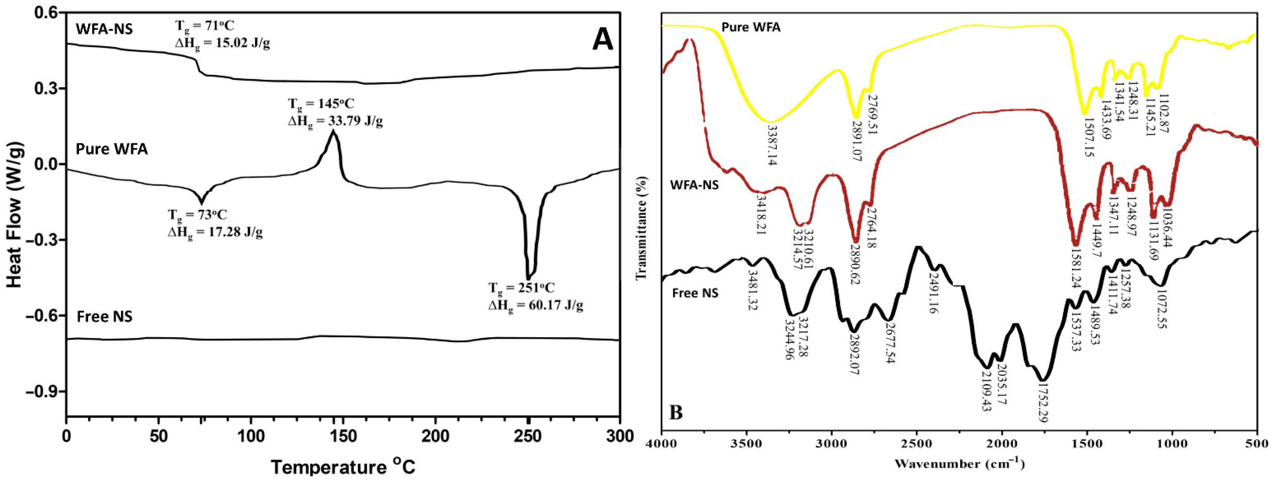
2.1.1. Differential Scanning Calorimetric (DSC) Analysis
2.1.2. Fourier Transform Infra-Red (FTIR) Spectroscopic Analysis
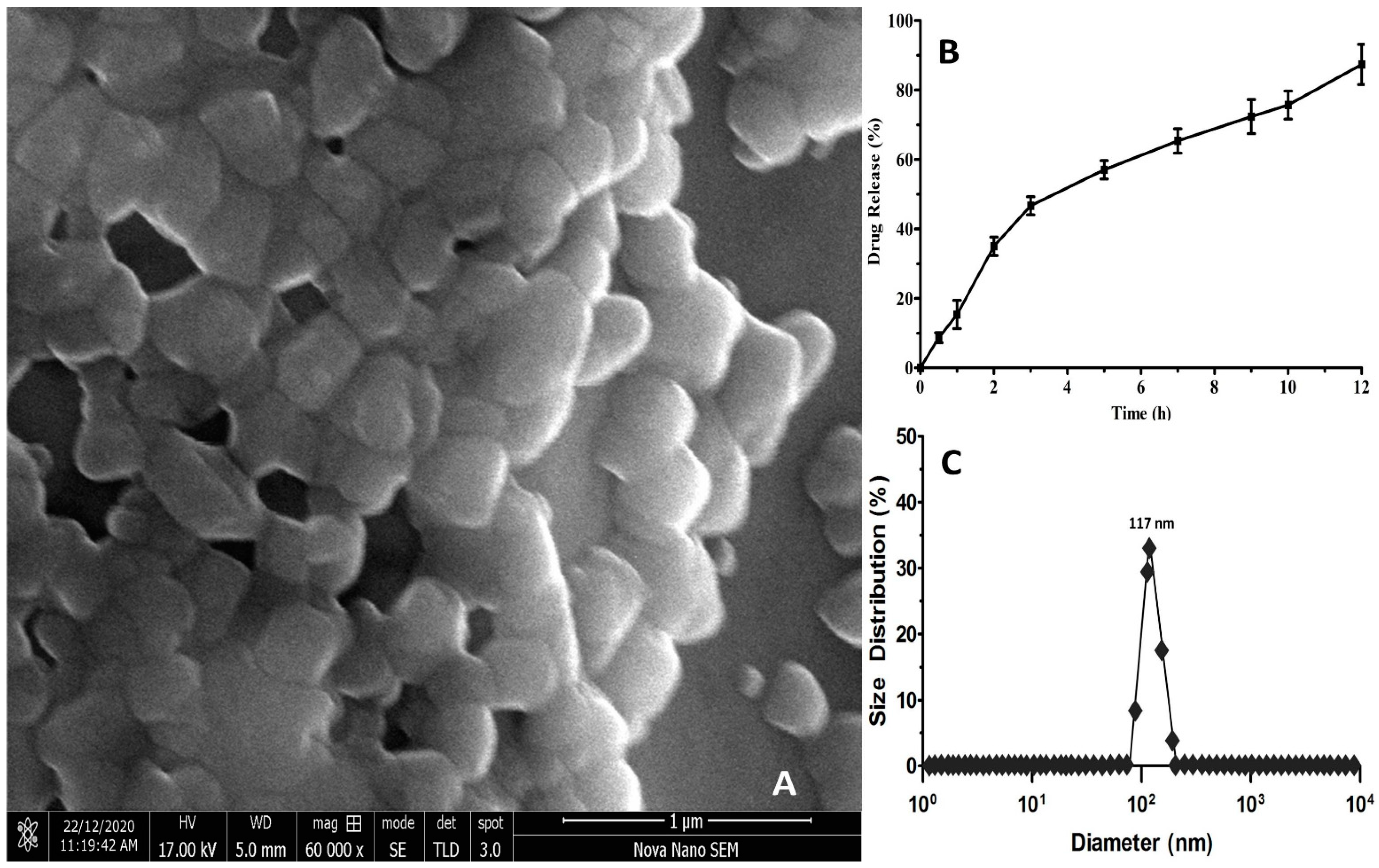
2.1.3. Scanning Electron Microscopic (SEM) Analysis
2.1.4. Estimation of Nanosponges Hydrodynamic Diameter
2.1.5. Drug Release Kinetics Studies
2.1.6. Entrapment Efficiency (EE)
2.2. Pharmacological Characterization
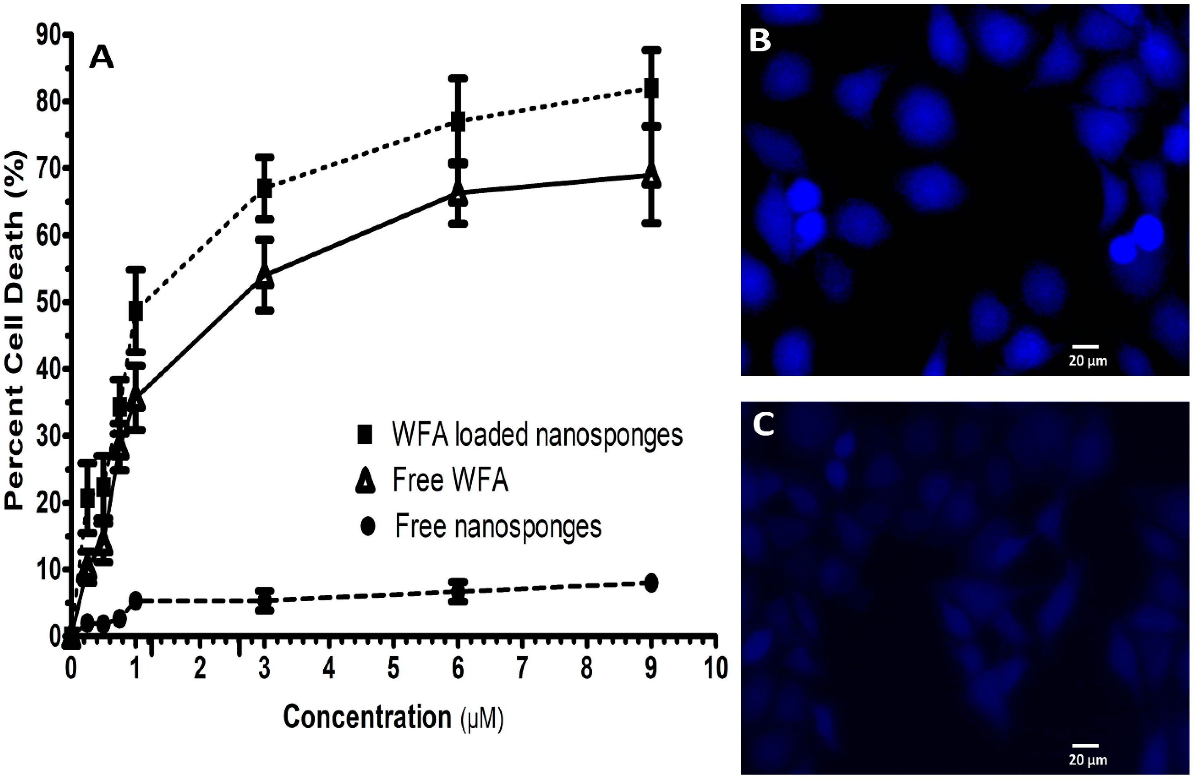
2.2.1. Anticancer Activity (SRB Dye Assay)
2.2.2. DAPI Staining
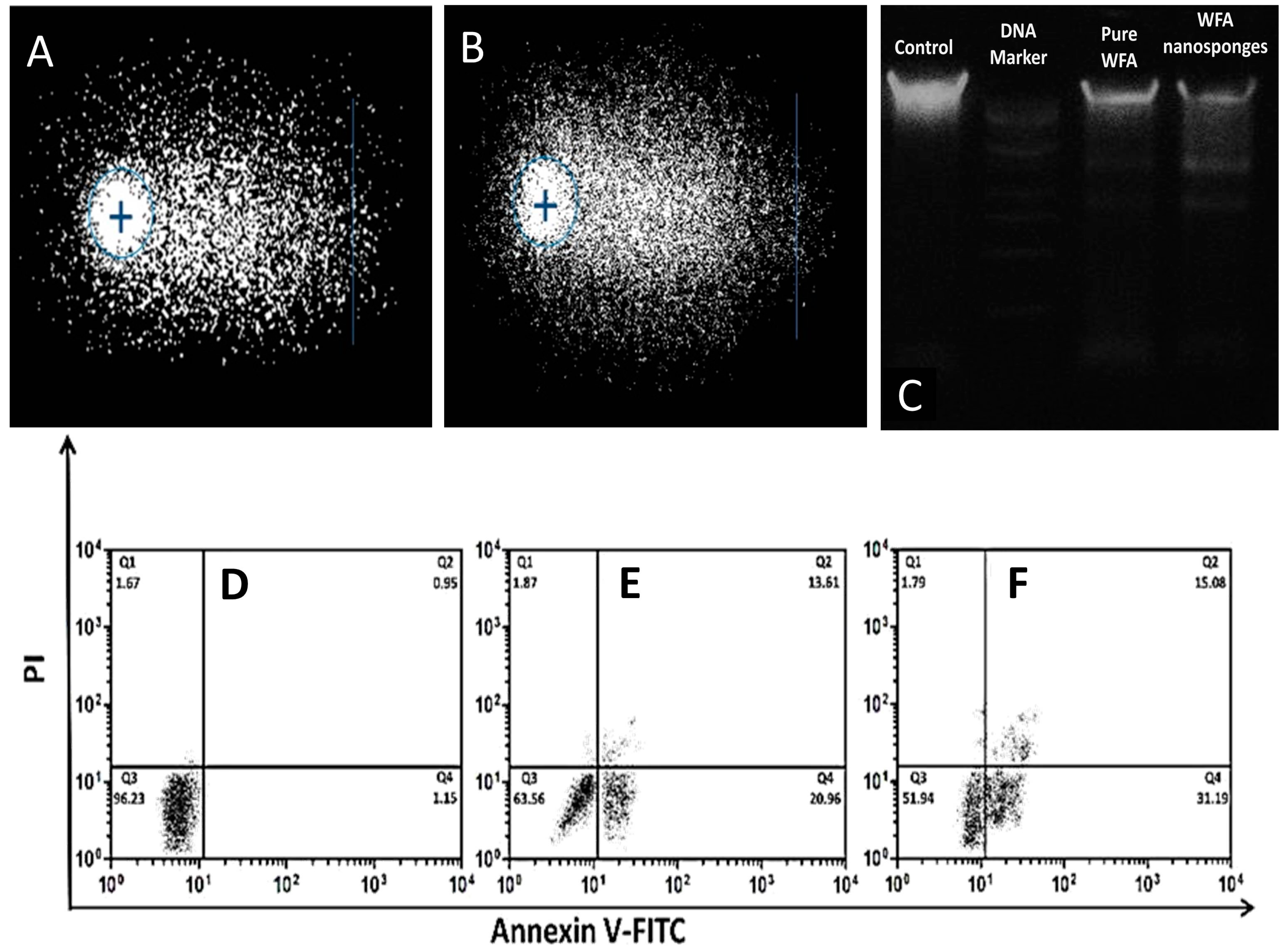
2.2.3. Genotoxicity Assessment
2.2.4. DNA Fragmentation
2.2.5. Flow Cytometry Analysis
2.3. Animal Studies
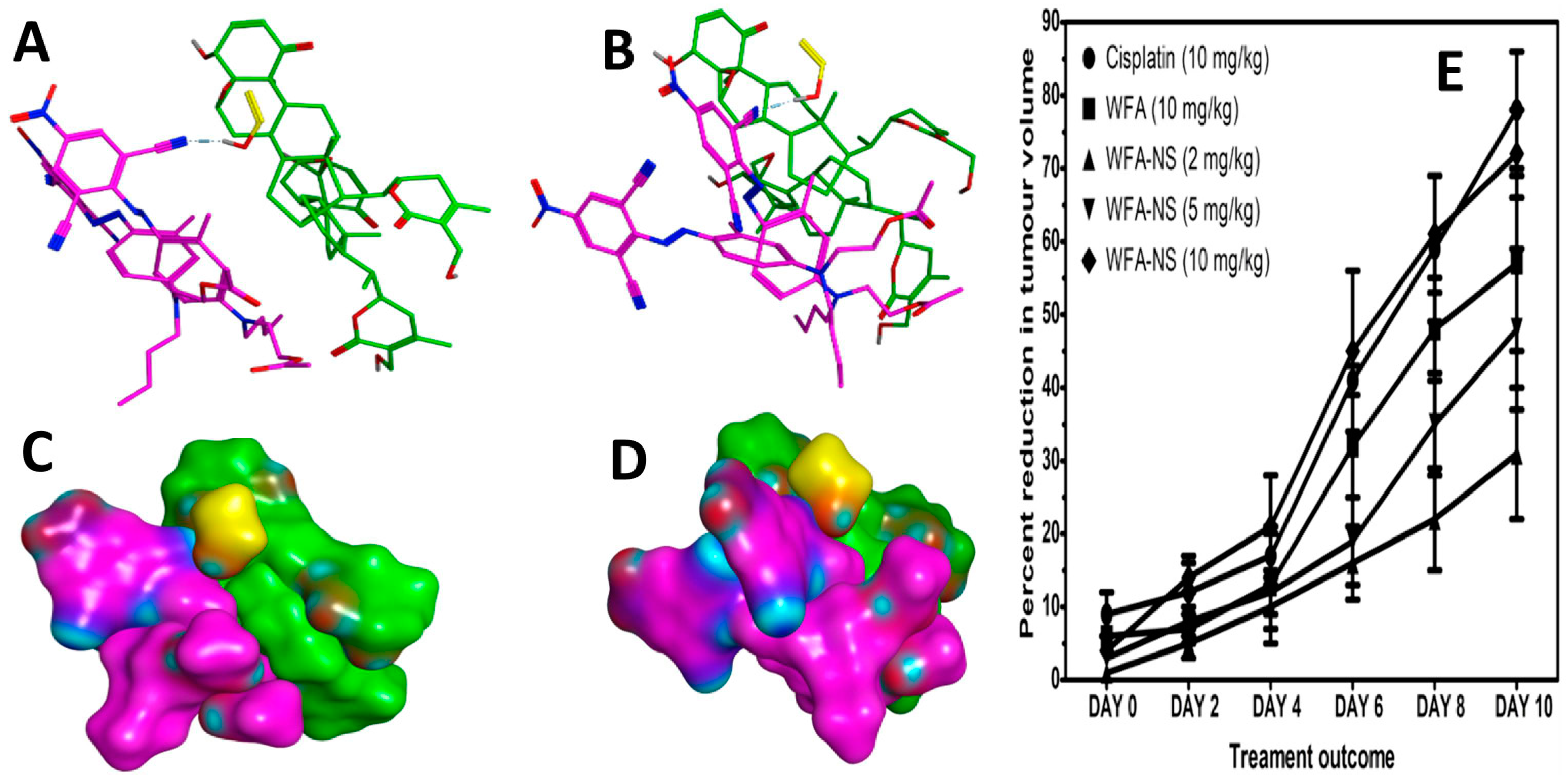
In Vivo Studies
2.4. In Silico Studies
Molecular Docking Studies
3. Materials and Methods
3.1. Preparation of Withaferin-A Loaded Nanosponges
3.2. Characterization of Withaferin-A Loaded Nanosponges
3.2.1. Differential Scanning Calorimetric (DSC) Analysis
3.2.2. Fourier Transform Infra-Red (FTIR) Spectroscopic Analysis
3.2.3. Scanning Electron Microscopic (SEM) Analysis
3.2.4. Estimation of Nanosponges Hydrodynamic Diameter
3.2.5. Study of Drug Release Kinetics
3.2.6. Entrapment Efficiency
3.2.7. Anticancer Activity (SRB Dye Assay)
3.2.8. DAPI Staining
3.2.9. Genotoxicity Assessment
3.2.10. DNA Fragmentation
3.2.11. Flow Cytometry Analysis
3.2.12. In Vivo Studies
3.2.13. Molecular Docking Studies
3.2.14. Statistical Analysis
4. Conclusions
Author Contributions
Funding
Institutional Review Board Statement
Informed Consent Statement
Data Availability Statement
Acknowledgments
Conflicts of Interest
Sample Availability
References
- Majolo, F.; de Oliveira Becker Delwing, L.K.; Marmitt, D.J.; Bustamante-Filho, I.C.; Goettert, M.I. Medicinal plants and bioactive natural compounds for cancer treatment: Important advances for drug discovery. Phytochem. Lett. 2019, 31, 196–207. [Google Scholar] [CrossRef]
- Gross, A.H.; Cromwell, J.; Fonteyn, M.; Matulonis, U.A.; Hayman, L.L. Hopelessness and complementary therapy use in patients with ovarian cancer. Cancer Nurs. 2013, 36, 256–264. [Google Scholar] [CrossRef]
- Ohnishi, S.; Takeda, H. Herbal medicines for the treatment of cancer chemotherapy-induced side effects. Front. Pharmacol. 2015, 6, 14. [Google Scholar] [CrossRef] [PubMed] [Green Version]
- Mir, B.A.; Khazir, J.; Mir, N.A.; Hasan, T.-U.; Koul, S. Botanical, chemical and pharmacological review of Withania somnifera (Indian ginseng): An ayurvedic medicinal plant. Indian J. Drugs Dis. 2012, 1, 147–160. [Google Scholar]
- Afroz, M.; Akter, S.; Ahmed, A.; Rouf, R.; Shilpi, J.A.; Tiralongo, E.; Sarker, S.D.; Göransson, U.; Uddin, S.J. Ethnobotany and antimicrobial peptides from plants of the solanaceae family: An update and future prospects. Front. Pharmacol. 2020, 11, 565. [Google Scholar] [CrossRef] [PubMed]
- Mishra, L.C.; Singh, B.B.; Dagenais, S. Scientific basis for the therapeutic use of Withania somnifera (ashwagandha): A review. Altern. Med. Rev. 2000, 5, 334–346. [Google Scholar]
- Vashi, R.; Patel, B.M.; Goyal, R.K. Keeping abreast about Ashwagandha in breast cancer. J. Ethnopharmacol. 2020, 269, 113759. [Google Scholar] [CrossRef]
- Dutta, R.; Khalil, R.; Green, R.; Mohapatra, S.S.; Mohapatra, S. Withania somnifera (Ashwagandha) and withaferin A: Potential in integrative oncology. Int. J. Mol. Sci. 2019, 20, 5310. [Google Scholar] [CrossRef] [PubMed] [Green Version]
- Aalinkeel, R.; Hu, Z.; Nair, B.B.; Sykes, D.E.; Reynolds, J.L.; Mahajan, S.D.; Schwartz, S.A. Genomic analysis highlights the role of the JAK-STAT signaling in the anti-proliferative effects of dietary flavonoid—‘Ashwagandha’ in prostate cancer cells. Evid. Based Complement. Alternat. Med. 2010, 7, 177–187. [Google Scholar] [CrossRef] [Green Version]
- Lee, H.-E.; Shin, J.-A.; Jeong, J.H.; Jeon, J.-G.; Lee, M.-H.; Cho, S.-D. Anticancer activity of A shwagandha against human head and neck cancer cell lines. J. Oral Pathol. Med. 2016, 45, 193–201. [Google Scholar] [CrossRef]
- Yu, Y.; Katiyar, S.P.; Sundar, D.; Kaul, Z.; Miyako, E.; Zhang, Z.; Kaul, S.C.; Reddel, R.R.; Wadhwa, R. Withaferin-A kills cancer cells with and without telomerase: Chemical, computational and experimental evidences. Cell Death Dis. 2017, 8, e2755. [Google Scholar] [CrossRef] [Green Version]
- Berghe, W.V.; Sabbe, L.; Kaileh, M.; Haegeman, G.; Heyninck, K. Molecular insight in the multifunctional activities of Withaferin A. Biochem. Pharmacol. 2012, 84, 1282–1291. [Google Scholar] [CrossRef]
- Lee, I.-C.; Choi, B.Y. Withaferin-A—A natural anticancer agent with pleitropic mechanisms of action. Int. J. Mol. Sci. 2016, 17, 290. [Google Scholar] [CrossRef] [PubMed] [Green Version]
- Harding, M.C.; Sloan, C.D.; Merrill, R.M.; Harding, T.M.; Thacker, B.J.; Thacker, E.L. Transitions from Heart Disease to Cancer as the Leading Cause of Death in US States, 1999–2016. Prev. Chronic Dis. 2018, 15, E158. [Google Scholar] [CrossRef] [Green Version]
- Mattiuzzi, C.; Lippi, G. Current cancer epidemiology. J. Epidemiol. Glob. Health 2019, 9, 217–222. [Google Scholar] [CrossRef] [PubMed] [Green Version]
- Sung, H.; Ferlay, J.; Siegel, R.L.; Laversanne, M.; Soerjomataram, I.; Jemal, A.; Bray, F. Global cancer statistics 2020: GLOBOCAN estimates of incidence and mortality worldwide for 36 cancers in 185 countries. CA Cancer J. Clin. 2021, 71, 209–249. [Google Scholar] [CrossRef] [PubMed]
- Ginsburg, O.; Bray, F.; Coleman, M.P.; Vanderpuye, V.; Eniu, A.; Kotha, S.R.; Sarker, M.; Huong, T.T.; Allemani, C.; Dvaladze, A.; et al. The global burden of women’s cancers: A grand challenge in global health. Lancet 2017, 389, 847–860. [Google Scholar] [CrossRef]
- Sankaranarayanan, R.; Ramadas, K.; Qiao, Y.-L. Managing the changing burden of cancer in Asia. BMC Med. 2014, 12, 3. [Google Scholar] [CrossRef] [Green Version]
- Arbyn, M.; Weiderpass, E.; Bruni, L.; de Sanjosé, S.; Saraiya, M.; Ferlay, J.; Bray, F. Estimates of incidence and mortality of cervical cancer in 2018: A worldwide analysis. Lancet Glob. Health 2020, 8, e191–e203. [Google Scholar] [CrossRef] [Green Version]
- Francies, F.Z.; Hull, R.; Khanyile, R.; Dlamini, Z. Breast cancer in low-middle income countries: Abnormality in splicing and lack of targeted treatment options. Am. J. Cancer Res. 2020, 10, 1568–1591. [Google Scholar]
- Sharma, A.; Madhunapantula, S.V.; Robertson, G.P. Toxicological considerations when creating nanoparticle-based drugs and drug delivery systems. Expert Opin. Drug Metab. Toxicol. 2012, 8, 47–69. [Google Scholar] [CrossRef]
- Hubbell, J.A.; Chilkoti, A. Nanomaterials for drug delivery. Science 2012, 337, 303–305. [Google Scholar] [CrossRef]
- Bolmal, U.B.; Manvi, F.V.; Kotha, R.; Palla, S.S.; Paladugu, A.; Reddy, K.R. Recent advances in nanosponges as drug delivery system. Int. J. Pharm. Sci. Nanotechnol. 2013, 6, 1934–1944. [Google Scholar] [CrossRef]
- Shanuja, J.; Singh, K.; Nandhini, R.S.; Palanivelu, J. Nanosponges: In Perspective to Therapeutic Medicine. In Nanotechnology in Medicine; Springer: Berlin/Heidelberg, Germany, 2021; pp. 87–104. [Google Scholar]
- Jilsha, G.; Viswanad, V. Nanosponges: A novel approach of drug delivery system. Int. J. Pharm. Sci. Rev. Res. 2013, 19, 119–123. [Google Scholar]
- Pawar, A.Y. Nanosponges: A novel drug delivery system. Asian Journal of Pharmaceutics (AJP): Free full text articles from Asian. J. Pharm. 2016, 10, 456–463. [Google Scholar]
- Chaudhary, A.; Nagaich, U.; Gulati, N.; Sharma, V.K.; Khosa, R.L.; Partapur, M.U. Enhancement of solubilization and bioavailability of poorly soluble drugs by physical and chemical modifications: A recent review. J. Adv. Pharm. Educ. Res. 2012, 2, 32–67. [Google Scholar]
- Patil, T.S.; Nalawade, N.A.; Kakade, V.K.; Kale, S.N. Nanosponges: A novel targeted drug delivery for cancer treatment. Int. J. Adv. Res. Dev. 2017, 2, 55–62. [Google Scholar]
- Jin, Y.; Wen, J.; Garg, S.; Liu, D.; Zhou, Y.; Teng, L.; Zhang, W. Development of a novel niosomal system for oral delivery of Ginkgo biloba extract. Int. J. Nanomed. 2013, 8, 421–430. [Google Scholar] [CrossRef] [Green Version]
- Deb, T.K.; Ramireddy, B.; Moin, A.; Shivakumar, H.G. In vitro-in vivo evaluation of xanthan gum and eudragit inter polyelectrolyte complex based sustained release tablets. Int. J. Pharm. Investig. 2015, 5, 65–72. [Google Scholar] [CrossRef] [Green Version]
- Zhang, M.; Li, H.; Lang, B.; O’Donnell, K.; Zhang, H.; Wang, Z.; Dong, Y.; Wu, C.; Williams, R.O., 3rd. Formulation and delivery of improved amorphous fenofibrate solid dispersions prepared by thin film freezing. Eur. J. Pharm. Biopharm. 2012, 82, 534–544. [Google Scholar] [CrossRef]
- Moghassemi, S.; Hadjizadeh, A. Nano-niosomes as nanoscale drug delivery systems: An illustrated review. J. Control. Release 2014, 185, 22–36. [Google Scholar] [CrossRef]
- Salunke, A.; Upmanyu, N. Formulation, Development and Evaluation of Budesonide Oral Nano-sponges Using DOE Approach: In Vivo Evidences. Adv. Pharm. Bull. 2021, 11, 286–294. [Google Scholar] [CrossRef] [PubMed]
- Amer, R.I.; El-Osaily, G.H.; Gad, S.S. Design and optimization of topical terbinafine hydrochloride nanosponges: Application of full factorial design, in vitro and in vivo evaluation. J. Adv. Pharm. Technol. Res. 2020, 11, 13–19. [Google Scholar] [CrossRef] [PubMed]
- Shah, N.; Gohil, D.Y.; Seth, A.K.; Aundhia, C.J.; Patel, S.S. Development of Risedronate Sodium-loaded Nanosponges by Experimental Design: Optimization and in vitro Characterization. Indian J. Pharm. Sci. 2019, 81, 309–316. [Google Scholar] [CrossRef]
- Zidan, A.S.; Ibrahim, M.M.; Megrab, N.A.E. Optimization of methotrexate loaded niosomes by Box–Behnken design: An understanding of solvent effect and formulation variability. Drug Dev. Ind. Pharm. 2017, 43, 1450–1459. [Google Scholar] [CrossRef] [PubMed]
- Xie, J.; Wang, C.-H. Paclitaxel-loaded biodegradable nanoparticles developed by direct dialysis and electrodydrodynamic atomization methods. In Proceedings of the AIChE Annual Meeting, Austin, TX, USA, 7–12 November 2004; pp. 1097–1106. [Google Scholar]
- Moghassemi, S.; Parnian, E.; Hakamivala, A.; Darzianiazizi, M.; Vardanjani, M.M.; Kashanian, S.; Larijani, B.; Omidfar, K. Uptake and transport of insulin across intestinal membrane model using trimethyl chitosan coated insulin niosomes. Mater. Sci. Eng. C Mater. Biol. Appl. 2015, 46, 333–340. [Google Scholar] [CrossRef]
- Hajizadeh, M.R.; Najmeh Parvaz, N.; Barani, M.; Khoshdel, A.; Fahmidehkar, M.A.; Mahmoodi, M.; Torkzadeh-Mahani, M. Diosgenin-loaded niosome as an effective phytochemical nanocarrier: Physicochemical characterization, loading efficiency, and cytotoxicity assay. DARU J. Pharm. Sci. 2019, 27, 329–339. [Google Scholar] [CrossRef]
- Danaei, M.; Dehghankhold, M.; Ataei, S.; Davarani, F.H.; Javanmard, R.; Dokhani, A.; Khorasani, S.; Mozafari, M.R. Impact of particle size and polydispersity index on the clinical applications of lipidic nanocarrier systems. Pharmaceutics 2018, 10, 57. [Google Scholar] [CrossRef] [Green Version]
- Cheewatanakornkool, K.; Niratisai, S.; Manchun, S.; Dass, C.R.; Sriamornsak, P. Characterization and in vitro release studies of oral microbeads containing thiolated pectin–doxorubicin conjugates for colorectal cancer treatment. Asian J. Pharm. Sci. 2017, 12, 509–520. [Google Scholar] [CrossRef]
- Guinedi, A.S.; Mortada, N.D.; Mansour, S.; Hathout, R.M. Preparation and evaluation of reverse-phase evaporation and multilamellar niosomes as ophthalmic carriers of acetazolamide. Int. J. Pharm. 2005, 306, 71–82. [Google Scholar] [CrossRef]
- Jahromi, L.P.; Ghazali, M.; Ashrafi, H.; Azadi, A. A comparison of models for the analysis of the kinetics of drug release from PLGA-based nanoparticles. Heliyon 2020, 6, e03451. [Google Scholar] [CrossRef] [Green Version]
- Salamanca, C.H.; Yarce, C.J.; Moreno, R.A.; Prieto, V.; Recalde, J. Natural gum-type biopolymers as potential modified nonpolar drug release systems. Carbohydr. Polym. 2018, 189, 31–38. [Google Scholar] [CrossRef]
- Sharma, R.; Pathak, K. Polymeric nanosponges as an alternative carrier for improved retention of econazole nitrate onto the skin through topical hydrogel formulation. Pharm. Dev. Technol. 2011, 16, 367–376. [Google Scholar] [CrossRef]
- Abbas, N.; Parveen, K.; Hussain, A.; Latif, S.; Zaman, S.U.; Shah, P.A.; Ahsan, M. Nanosponge-based hydrogel preparation of fluconazole for improved topical delivery. Trop. J. Pharm. Res. 2019, 18, 215–222. [Google Scholar] [CrossRef]
- Vajrabhaya, L.-O.; Korsuwannawong, S. Cytotoxicity evaluation of a Thai herb using tetrazolium (MTT) and sulforhodamine B (SRB) assays. J. Anal. Sci. Technol. 2018, 9, 15. [Google Scholar] [CrossRef] [Green Version]
- Orellana, E.A.; Kasinski, A.L. Sulforhodamine B (SRB) assay in cell culture to investigate cell proliferation. Bio Protocol. 2016, 6, e1984. [Google Scholar] [CrossRef] [Green Version]
- McKenna, M.K.; Gachuki, B.W.; Alhakeem, S.S.; Oben, K.N.; Rangnekar, V.M.; Gupta, R.C.; Bondada, S. Anti-cancer activity of withaferin A in B-cell lymphoma. Cancer Biol. Ther. 2015, 16, 1088–1098. [Google Scholar] [CrossRef] [Green Version]
- Alemi, A.; Reza, J.Z.; Haghiralsadat, F.; Jaliani, H.Z.; Karamallah, M.H.; Hosseini, S.A.; Karamallah, S.H. Paclitaxel and curcumin coadministration in novel cationic PEGylated niosomal formulations exhibit enhanced synergistic antitumor efficacy. J. Nanobiotechnol. 2018, 16, 28. [Google Scholar] [CrossRef] [PubMed]
- Shaker, D.S.; Shaker, M.A.; Hanafy, M.S. Cellular uptake, cytotoxicity and in-vivo evaluation of Tamoxifen citrate loaded niosomes. Int. J. Pharm. 2015, 493, 285–294. [Google Scholar] [CrossRef] [PubMed]
- Geetha, R.; Ashokkumar, T.; Tamilselvan, S.; Govindaraju, K.; Sadiq, M.; Singaravelu, G. Green synthesis of gold nanoparticles and their anticancer activity. Cancer Nanotechnol. 2013, 4, 91–98. [Google Scholar] [CrossRef] [PubMed] [Green Version]
- Chiani, M.; Norouzian, D.; Shokrgozar, M.A.; Azadmanesh, K.; Najmafshar, A.; Mehrabi, M.R.; Akbarzadeh, A. Folic acid conjugated nanoliposomes as promising carriers for targeted delivery of bleomycin. Artif. Cells Nanomed. Biotechnol. 2018, 46, 757–763. [Google Scholar] [CrossRef] [PubMed]
- Taleblou, N.; Sirousazar, M.; Hassan, Z.M.; Khaligh, S.G. Capecitabine-loaded anti-cancer nanocomposite hydrogel drug delivery systems: In vitro and in vivo efficacy against the 4T1 murine breast cancer cells. J. Biomater. Sci. Polym. Ed. 2020, 31, 72–92. [Google Scholar] [CrossRef] [PubMed]
- Guo, R.; Cheng, Y.; Ding, D.; Li, X.; Zhang, L.; Jiang, X.; Liu, B. Synthesis and antitumoral activity of gelatin/polyoxometalate hybrid nanoparticles. Macromol. Biosci. 2011, 11, 839–847. [Google Scholar] [CrossRef]
- Kakar, S.S.; Jala, V.R.; Fong, M.Y. Synergistic cytotoxic action of cisplatin and withaferin A on ovarian cancer cell lines. Biochem. Biophys. Res. Commun. 2012, 423, 819–825. [Google Scholar] [CrossRef] [Green Version]
- Kakar, S.S.; Ratajczak, M.Z.; Powell, K.S.; Moghadamfalahi, M.; Miller, D.M.; Batra, S.K.; Singh, S.K. Withaferin a alone and in combination with cisplatin suppresses growth and metastasis of ovarian cancer by targeting putative cancer stem cells. PLoS ONE 2014, 9, e107596. [Google Scholar] [CrossRef]
- Yin, X.; Yang, G.; Ma, D.; Su, Z. Inhibition of cancer cell growth in cisplatin-resistant human oral cancer cells by withaferin-A is mediated via both apoptosis and autophagic cell death, endogenous ROS production, G2/M phase cell cycle arrest and by targeting MAPK/RAS/RAF signalling pathway. J. BUON 2020, 25, 332–337. [Google Scholar]
- Babazadeh, A.; Zeinali, M.; Hamishehkar, H. Nano-phytosome: A developing platform for herbal anti-cancer agents in cancer therapy. Curr. Drug Targets 2018, 19, 170–180. [Google Scholar] [CrossRef]
- Aftab, S.; Shah, A.; Nadhman, A.; Kurbanoglu, S.; Ozkan, S.A.; Dionysiou, D.D.; Shukla, S.S.; Aminabhavi, T.M. Nanomedicine: An effective tool in cancer therapy. Int. J. Pharm. 2018, 540, 132–149. [Google Scholar] [CrossRef]
- Dong, P.; Rakesh, K.P.; Manukumar, H.M.; Mohammed, Y.H.S.; Karthik, C.S.; Sumathi, S.; Mallu, P.; Qin, H.-L. Innovative nano-carriers in anticancer drug delivery-a comprehensive review. Bioorg. Chem. 2019, 85, 325–336. [Google Scholar] [CrossRef] [PubMed]
- Chemical Computing Group’s Molecular Operating Environment (MOE) MOE 2019. 0201. Available online: http://www.chemcomp.com/MOEMolecular_Operating_Environment.htm (accessed on 11 January 2020).
- Shah, H.S.; Usman, F.; Ashfaq–Khan, M.; Khalil, R.; Ul-Haq, Z.; Mushtaq, A.; Qaiser, R.; Iqbal, J. Preparation and characterization of anticancer niosomal withaferin–A formulation for improved delivery to cancer cells: In vitro, in vivo, and in silico evaluation. J. Drug Deliv. Sci. Technol. 2020, 59, 101863. [Google Scholar] [CrossRef]
- Wasilewska, K.; Winnicka, K. Ethylcellulose–a pharmaceutical excipient with multidirectional application in drug dosage forms development. Materials 2019, 12, 3386. [Google Scholar] [CrossRef] [Green Version]
- Ahmed, M.M.; Fatima, F.; Anwer, M.K.; Ansari, M.J.; Das, S.S.; Alshahrani, S.M. Development and characterization of ethyl cellulose nanosponges for sustained release of brigatinib for the treatment of non-small cell lung cancer. J. Polym. Eng. 2020, 40, 823–832. [Google Scholar] [CrossRef]
- Pushpalatha, R.; Selvamuthukumar, S.; Kilimozhi, D. Cross-linked, cyclodextrin-based nanosponges for curcumin delivery-Physicochemical characterization, drug release, stability and cytotoxicity. J. Drug Deliv. Sci. Technol. 2018, 45, 45–53. [Google Scholar] [CrossRef]
- Vichai, V.; Kirtikara, K. Sulforhodamine B colorimetric assay for cytotoxicity screening. Nat. Protoc. 2006, 1, 1112–1116. [Google Scholar] [CrossRef]
- Priyadarsini, R.V.; Murugan, R.S.; Maitreyi, S.; Ramalingam, K.; Karunagaran, D.; Nagini, S. The flavonoid quercetin induces cell cycle arrest and mitochondria-mediated apoptosis in human cervical cancer (HeLa) cells through p53 induction and NF-κB inhibition. Eur. J. Pharmacol. 2010, 649, 84–91. [Google Scholar] [CrossRef]
- Olive, P.L.; Banáth, J.P. The comet assay: A method to measure DNA damage in individual cells. Nat. Protoc. 2006, 1, 23–29. [Google Scholar] [CrossRef]
- Singh, N.P.; McCoy, M.T.; Tice, R.R.; Schneider, E.L. A simple technique for quantitation of low levels of DNA damage in individual cells. Exp. Cell Res. 1988, 175, 184–191. [Google Scholar] [CrossRef] [Green Version]
- Liu, X.; Zou, H.; Slaughter, C.; Wang, X. DFF, a heterodimeric protein that functions downstream of caspase-3 to trigger DNA fragmentation during apoptosis. Cell 1997, 89, 175–184. [Google Scholar] [CrossRef] [Green Version]
- Lin, G.-J.; Jiang, G.-B.; Xie, Y.-Y.; Huang, H.-L.; Liang, Z.-H.; Liu, Y.-J. Cytotoxicity, apoptosis, cell cycle arrest, reactive oxygen species, mitochondrial membrane potential, and Western blotting analysis of ruthenium (II) complexes. J. Biol. Inorg. Chem. 2013, 18, 873–882. [Google Scholar] [CrossRef]
- Md Yousof Ali, M.Y.; Zaib, S.; Rahman, M.M.; Jannat, S.; Iqbal, J.; Park, S.K.; Chang, M.S. Didymin, a dietary citrus flavonoid exhibits anti-diabetic complications and promotes glucose uptake through the activation of PI3K/Akt signaling pathway in insulin-resistant HepG2 cells. Chem. Biol. Interact. 2019, 305, 180–194. [Google Scholar] [CrossRef]


| Analysis | Results |
|---|---|
| Diameter hydrodynamic | 117 ± 4 nm |
| Entrapment Efficiency (%) | 85 ± 11 |
| Zeta Potential (mV) | −39.02 ± 5.71 |
| Poly dispersity Index(PDI) | 0.389 ± 0.091 |
| Zero-order | 0.8734 |
| First-order | 0.9867 |
| Higuchi Model | 0.9806 |
| Korsemeyer Peppas, n value | 0.9713, 0.324 |
| Percent Reduction in Tumor Volume | |||||
|---|---|---|---|---|---|
| Cisplatin (C) 10 mg/kg | WFA (D) 10 mg/kg | WFA-NS (E) 2 mg/kg | WFA-NS (F) 5 mg/kg | WFA-NS (G) 10 mg/kg | |
| DAY0 | 9 ± 3 | 6 ± 1 | 1 ± 1 | 3 ± 1 | 4 ± 1 |
| DAY2 | 12 ± 5 | 7 ± 2 | 5 ± 3 | 8 ± 2 | 14 ± 2 |
| DAY4 | 17 ± 3 | 13 ± 8 | 10 ± 6 | 12 ± 3 | 21 ± 7 |
| DAY6 | 41 ± 2 | 32 ± 11 | 16 ± 7 | 19 ± 6 | 45 ± 11 |
| DAY8 | 59 ± 10 | 48 ± 7 | 22 ± 11 | 35 ± 7 | 61 ± 8 |
| DAY10 | 78 ± 8 | 57 ± 12 | 31 ± 9 | 48 ± 11 | 72 ± 6 |
| Group | Type of Treatment |
|---|---|
| A | Water for injection in cancerous mice (WFI) |
| B | Free nanosponges (NS) for cancerous mice |
| C | Cancerous mice were given 10 mg/kg cisplatin |
| D | WFA (10 mg/kg) treated cancerous mice |
| E | WFA-NS (2 mg/kg) treated cancerous mice |
| F | WFA-NS (5 mg/kg) treated cancerous mice |
| G | WFA-NS (10 mg/kg) treated cancerous mice |
Publisher’s Note: MDPI stays neutral with regard to jurisdictional claims in published maps and institutional affiliations. |
© 2021 by the authors. Licensee MDPI, Basel, Switzerland. This article is an open access article distributed under the terms and conditions of the Creative Commons Attribution (CC BY) license (https://creativecommons.org/licenses/by/4.0/).
Share and Cite
Shah, H.S.; Nasrullah, U.; Zaib, S.; Usman, F.; Khan, A.; Gohar, U.F.; Uddin, J.; Khan, I.; Al-Harrasi, A. Preparation, Characterization, and Pharmacological Investigation of Withaferin-A Loaded Nanosponges for Cancer Therapy; In Vitro, In Vivo and Molecular Docking Studies. Molecules 2021, 26, 6990. https://doi.org/10.3390/molecules26226990
Shah HS, Nasrullah U, Zaib S, Usman F, Khan A, Gohar UF, Uddin J, Khan I, Al-Harrasi A. Preparation, Characterization, and Pharmacological Investigation of Withaferin-A Loaded Nanosponges for Cancer Therapy; In Vitro, In Vivo and Molecular Docking Studies. Molecules. 2021; 26(22):6990. https://doi.org/10.3390/molecules26226990
Chicago/Turabian StyleShah, Hamid Saeed, Usman Nasrullah, Sumera Zaib, Faisal Usman, Ajmal Khan, Umar Farooq Gohar, Jalal Uddin, Imtiaz Khan, and Ahmed Al-Harrasi. 2021. "Preparation, Characterization, and Pharmacological Investigation of Withaferin-A Loaded Nanosponges for Cancer Therapy; In Vitro, In Vivo and Molecular Docking Studies" Molecules 26, no. 22: 6990. https://doi.org/10.3390/molecules26226990