
Synthesis and Evaluation of Antioxidant Properties of 2-Substituted Quinazolin-4(3H)-ones


Faculty of Pharmacy, University of Ljubljana, Aškerčeva cesta 7, SI-1000 Ljubljana, Slovenia
*
Author to whom correspondence should be addressed.
Molecules 2021, 26(21), 6585; https://doi.org/10.3390/molecules26216585
Submission received: 13 October 2021
/
Revised: 28 October 2021
/
Accepted: 28 October 2021
/
Published: 30 October 2021
(This article belongs to the Special Issue Measurement of Antioxidant Activity: Advances and Perspectives)
Abstract
:Quinazolinones represent an important scaffold in medicinal chemistry with diverse biological activities. Here, two series of 2-substituted quinazolin-4(3H)-ones were synthesized and evaluated for their antioxidant properties using three different methods, namely DPPH, ABTS and TEACCUPRAC, to obtain key information about the structure–antioxidant activity relationships of a diverse set of substituents at position 2 of the main quinazolinone scaffold. Regarding the antioxidant activity, ABTS and TEACCUPRAC assays were more sensitive and gave more reliable results than the DPPH assay. To obtain antioxidant activity of 2-phenylquinazolin-4(3H)-one, the presence of at least one hydroxyl group in addition to the methoxy substituent or the second hydroxyl on the phenyl ring in the ortho or para positions is required. An additional ethylene linker between quinazolinone ring and phenolic substituent, present in the second series (compounds 25a and 25b), leads to increased antioxidant activity. Furthermore, in addition to antioxidant activity, the derivatives with two hydroxyl groups in the ortho position on the phenyl ring exhibited metal-chelating properties. Our study represents a successful use of three different antioxidant activity evaluation methods to define 2-(2,3-dihydroxyphenyl)quinazolin-4(3H)-one 21e as a potent antioxidant with promising metal-chelating properties.
1. Introduction
Quinazoline is a nitrogen containing fused heterocycle, and with additional carbonyl linkage it forms two different quinazolinones (either 4(3H)-quinazolinone or 2(1H)-quinazolinone) [1]. Quinazolinones represent an important scaffold in medicinal chemistry due to their synthetic accessibility and diverse in vitro and in vivo pharmacological activities [2,3]. Many synthetic and natural quinazoline-based drugs have been used clinically to treat diverse pathological conditions. The most known drugs among 4(3H)-quinazolinones are the triazole antifungal drug albaconazole (1), the antihyperglicemic agent balaglitazone (2), the antimalarial agent febrifugine (3), the antihypertensive agent quinethazone (4) and the quinazolines with GABAergic activity (e.g., methaqualone (5), afloqualone (6) and diproqualone (7)) (Figure 1a) [1].
The antioxidant properties were described for structurally diverse quinazolines (Figure 1b), such as 2-pentylquinazolin-4(3H)-one derivatives (e.g., 8) [4], heterocyclic quinazoline-4-one derivatives [5,6] (e.g., 9 [6]), N-(pyrazin-2-yl)-2-[(4-oxo-3-(4-sulfamoylphenyl)-3,4-dihydroquinazolin-2-yl)thio]acetamide (10) [7], quinazolinone-1,3,4-oxadiazole conjugates (e.g., 11) [8], and a series of 2,3-disubstituted-2,3-dihydro-quinazolin-4(1H)-one-derived [9,10] and quinazolinone-derived Schiff′s bases (e.g., 12) [11,12]. Furthermore, antioxidant activity was also reported for 2-thioxobenzo[g]quinazoline derivatives (e.g., 13) [13], bis(2,3-dihydroquinazolin-4(1H)-one derivatives [14], 2-(chloromethyl)-3-(4-methyl-6-oxo-5-[(E)-phenyldiazenyl]-2-thioxo-5,6-dihydropyrimidine-1(2H)-yl)quinazoline-4(3H)-ones (e.g., 14) [15], guanine-based (E)-2-(2-(pyridin-2-ylmethylene)hydrazinyl)quinazolin-4(3H)-ones [16], quinazolinone-based hydrazones (e.g., 15) [17] and iodinated quinazolinones bearing a benzensulfonamide moiety (e.g., 16) [18].
There are many different analytical methods for assessing the antioxidant capacity. They can be divided into three distinctive groups, namely spectrometry, electrochemical techniques and chromatography (GC and HPLC) [19,20]. Spectrometric techniques are the most commonly used assays because they are rapid, simple and easily accessible. They are based on measuring the change in absorbance or fluorescence of an indicator containing solution after the addition of the antioxidant [21]. As indicators, compounds capable of detecting hydrogen atom transfer (e.g., α,α-azabisizobutronytril, fluorescein, luminol) or single electron transfer (e.g., Cu2+, Fe3+, 2,2′-azinobis(3-ethylbenzthiazolin-6-sulfonic acid (ABTS), 2,2-di(4-tert-octylphenyl)1-picrylhydrazyl) [20]) are used. A widely used assay for the determination of antioxidant potential is the DPPH (2,2-diphenyl-1-picryl-hydrazyl-hydrate) free radical scavenging method, measuring the reduction of odd electrons on nitrogen atoms in DPPH (by receiving a hydrogen atom from antioxidants) to the corresponding hydrazine [22]. When a transition metal (such as copper) is used as an indicator, the total antioxidant capacity is determined. For example, the commonly used cupric reducing antioxidant capacity (CUPRAC) assay is based on the reduction of cupric (Cu2+) to cuprous (Cu+) and the absorbance of the formed Cu(I)-neocuproine chelate is measured [23]. On the other hand, cyclic voltammetry and bi/amperometry are the most commonly used electrochemical methods, whereas GC and HPLC are most often applied for antioxidant separation and detection in complex samples [19].
Recently, we discovered the antioxidant, cytotoxic, and protective effects of three different quinazolinones in lipopolysaccharide murine microglia and hydrogen peroxide mouse neuroblastoma-2a cells [24]. Two quinazolinones with antioxidant activity (i.e., 17 and 18, Figure 1) possessed an aromatic substituent with a hydroxyl group at position 2 of the main quinazolin-4(3H)-one ring. Herein, we decided to synthesize and investigate the antioxidant properties of two series of 2-substituted quinazolin-4(3H)-ones using three different antioxidant methods and compared them to the known structural analogs of phenolic antioxidants. Furthermore, their ability to chelate metal ions was also determined. Based on the results obtained, we gained some key information about the structure–antioxidant activity relationships of 2-substituted quinazolin-4(3H)-ones and defined quinazolinone with potent antioxidant activity and promising metal-chelating properties.
2. Results and Discussion
The synthesis of 2-aryl-quinazolin-4(3H)-ones was performed according to the previously published procedures [25,26]. Briefly, in an open flask, the corresponding aldehydes 19a–l reacted with antranilamide (20) in DMSO via aerobic oxidative cyclization to the corresponding quinazolinones 21a–l (Scheme 1) [25,26]. The synthesis of (E)-2-(4-hydroxystyryl)quinazolin-4(3H)-one (25a) and (E)-2-(4-hydroxy-3-methoxystyryl)quinazolin-4(3H)-one (25b) have not yet been described. Thus, we decided to use derivatives of cinnamaldehyde and prepared the compounds 25a–b using antranilamide (20) via the aforementioned procedure. (E)-3-(4-hydroxyphenyl)acrylaldehyde (24a) and (E)-3-(4-hydroxy-3-methoxyphenyl)acrylaldehyde (24b) were synthesized from the corresponding cinnamic acids 22a and 22b (i.e., p-coumaric and ferulic acids, respectively) [27]. Firstly, the coupling of a carboxylic group with N,O-dimethylhydroxylamine using 1-ethyl-3-(3-dimethylaminopropyl)carbodiimide (EDC) afforded Weinreb amides 23a–b, which were further reduced with diisobutylaluminium hydride (DIBAL) to yield the cinnamaldehyde derivatives 24a and 24b, which reacted with antranilamide (20) in DMSO to form the final products 25a and 25b.
The antioxidant activity of synthesized quinazolin-4(3H)-ones 21a–l and 25a–b was firstly screened by one of the most commonly used methods, i.e., DPPH assay (Table 1, Figure 2a). The most potent radical scavenging activity was observed for three dihydroxy-substituted quinazolinones, i.e., 21e, 21g and 21h with EC50 values of 7.5, 7.4 and 7.2 μM, respectively. The second hydroxyl group needs to be in the ortho or para position, since the meta derivative (compound 21f) loses most of its scavenging properties. This is in accordance with literature where increased antioxidant activity of phenolic compounds was reported if a second hydroxyl group is introduced in the ortho or para positions [28]. The majority of compounds with only one hydroxyl group did not show any activity with the exception of 21j, 21l and 25b, which possessed additional methoxy substituent in the ortho or para position according to the hydroxyl group. It is known from the literature that the antioxidant activity of monophenols is significantly enhanced by one or two methoxy substituents in the ortho position relative to the hydroxyl group [28]. Comparing 21l and 25b, the only difference between them was the ethylene linker between the quinazolinone and benzene rings, which led to higher scavenging potency (approximately a 16.5-fold difference). Some compounds (such as 21b, 21d and 21f) showed lower radical scavenging properties, as expected, according to their phenolic structure. Thus, we believe that the more appropriate assays for the determination of the antioxidant properties of 2-substituted quinazolin-4(3H)-ones are the ABTS and TEACCUPRAC assays (Table 1, Figure 2).
In the ABTS assay (Table 1) we were able to determine the EC50 values of monohydroxy derivatives in the range from 23.0 to 69.9 μM, with the meta derivative 21c being the most potent. Among dihydroxy derivatives, there was no significant difference in potency (all EC50s were around 8 μM), while the EC50s for methoxy derivatives were in the range from 15.3 to 20.1 μM, with the most potent compounds 21k and 21l possessing the methoxy group in the ortho position. As mentioned previously, electron-donating groups (such as MeO) on the phenol ring significantly affected the antioxidant activity by decreasing the O−H bond dissociation enthalpy (BDE) of the phenol, leading to increased antioxidant activity [29]. However, the position of the methoxy group relative to the hydroxyl is important, since lower EC50 values (higher potency) were obtained in cases of ortho or para methoxy derivatives (21k and 21j, respectively) compared to the meta derivative 21i. This is in accordance with literature data where higher antioxidant properties were reported for natural and synthetic phenolic compounds with electron-donating groups in ortho or para positions [28,29,30]. The most potent compound was 25b possessing an ethylene linker between quinazolinone and ortho-methoxyphenol moiety. The conjugated double bond also contributes to higher antioxidant properties due to the resonance stabilization effect on the phenoxyl radical [31], which was formed in the reaction between quinazolinone and ABTS (Figure 2).
In TEACCUPRAC assay (Table 1), compound 21e exhibited the highest antioxidant capacity, with a TEAC value of 3.46. The other two dihydroxy derivatives, namely 21g and 21h, also showed good antioxidant properties (TEAC value of 2.62 and 2.74, respectively). Again, two hydroxyl groups in the ortho or para position according to each other, are the most optimal substitutions, since meta derivative 21f showed much lower antioxidant capacity in the TEACCUPRAC assay. Among the methoxy derivatives, 21j was the most potent antioxidant, whereas 25b exhibited similar antioxidant properties to Trolox. The comparison of p-hydroxyphenyl derivative 21d (TEAC value of 0.0315) and 4-hydroxystyryl derivative 25a (TEAC value of 0.539) stresses the importance of the additional ethylene linker, leading to increased antioxidant potency due to the resonance stabilization of the formed phenoxyl radical. A similar pattern was also observed in the previously discussed ABTS assay.
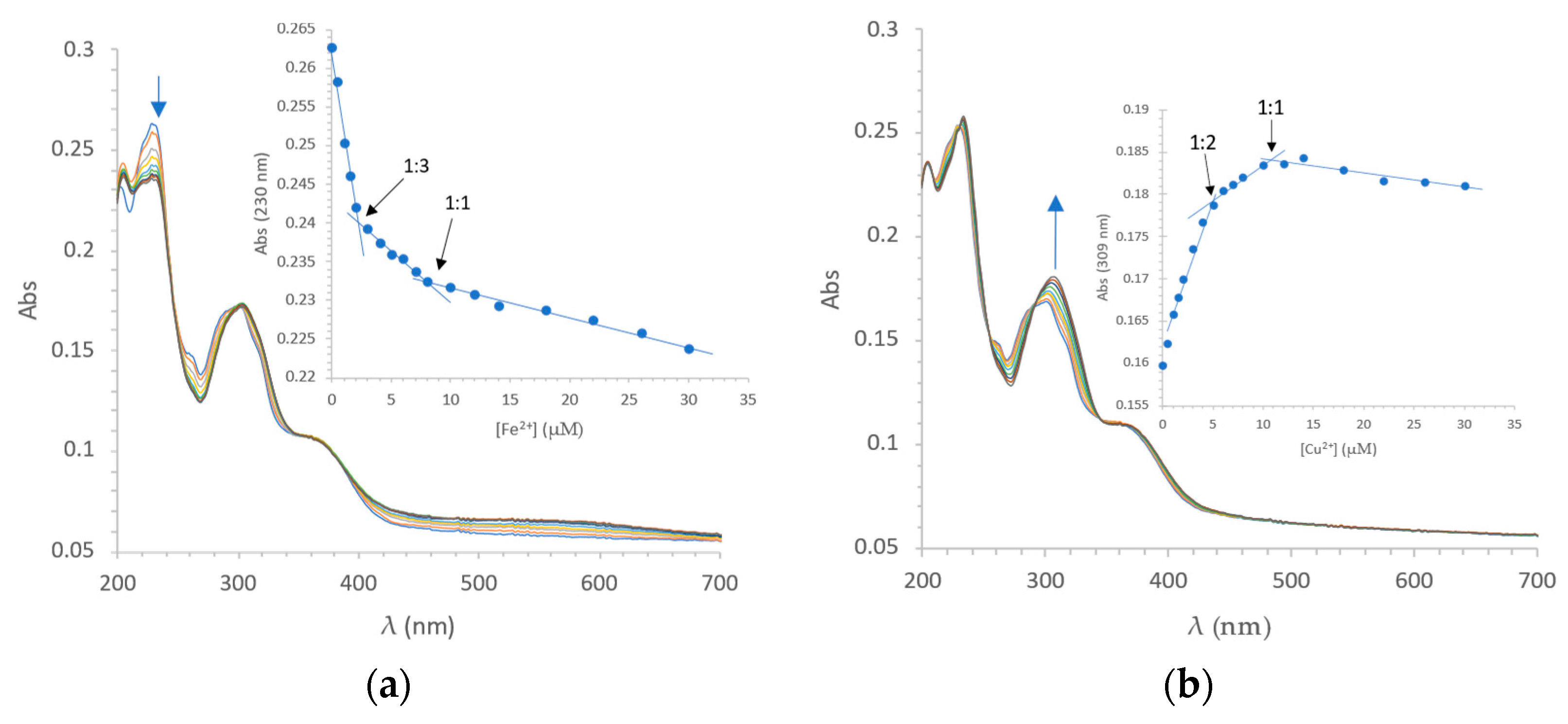
In addition to three different antioxidant activity measurement assays ability of chelating metal ions (i.e., Fe2+ and Cu2+) of 2-substituted quinazolin-4(3H)-ones was also evaluated (Figure 3, Supplementary Materials, Figures S1–S14). Preliminary screening was performed by comparing the UV-Vis spectra of 10 mM solutions with spectra after the addition of 5, 10 and 20 mM Fe2+ or Cu2+ (Figures S1–S14). If the spectrum did not change shape and a slight dilution effect was seen, it was concluded that the compound does not bind metal ions. Two compounds, namely 21e and 21h, show pronounced chelation properties. Compound 21e was selected for further study because of potent antioxidant activity (Table 1) and significant metal-chelating properties (Figure 3).
UV-Vis spectroscopic titration of 21e with Fe2+ was performed in 20 mM KPB at pH 7.2. With the addition of Fe2+, the free 21e absorption (λmax = 230 nm) rapidly decreased and the newly formed Fe2+-complex band red shifted around 300 nm. The presence of a clear isosbestic point (λmax = 301 nm) suggested the formation of the 21e-Fe2+ complex (Figure 3a). A plot of 21e-Fe2+ complex absorption at 230 nm against the Fe2+ concentration is displayed in Figure 3a. The titration curve displays the formation of 1:3 and 1:1 Fe2+: 21e complexes.
Similarly, complex formation between 21e and Cu2+ was studied. The addition of Cu2+ to 21e produced a new band at 309 nm, of higher intensity than the 301 nm band of 21e-Fe2+. The Cu2+ was partially reduced to Cu+, and the catechol was oxidised to the orthoquinone derivative of 21e (Figure S16). A plot of the 21e-Cu2+ complex absorption at 309 nm against the Cu concentration is presented in Figure 3b. The titration curve displays the formation of 1:2 and 1:1 Cu: 21e complexes, which was confirmed by ESI-high resolution mass spectrometry measurements (Figure S15).
3. Materials and Methods
3.1. Chemistry
The reagents and solvents were obtained from commercial sources (Sigma-Aldrich, Acros Organics, Alfa Aesar, TCI, Merck) and used without further purification. Thin-layer chromatography (TLC) on silica gel plates (Merck DC Fertigplatten Kieselgel 60 GF254) was used to monitor the reaction. TLC spots were visualized under UV light and/or stained with the appropriate dyeing agents (Iron(III) chloride, 2,4-dinitrophenylhydrazine, bromocresol green). Flash column chromatography was performed on Merck silica gel 60 (mesh size, 70–230). Yields refer to the purified products and were not optimized. The 1H and 13C NMR spectra were recorded at 295 K in DMSO-d6 on a Bruker Avance III NMR spectrometer equipped with a broadband decoupling inverse 1H probe. The coupling constants (J) were in Hz, and the splitting patterns were designated as: s, singlet; br s, broad singlet; d, doublet; dd, double doublet; t, triplet; dt, double triplet; ddd, double of doublet of doublet; and m, multiplet. The mass spectra and high-resolution mass measurements were performed at the Faculty of Pharmacy, University of Ljubljana, on an ADVION Expression CMSL mass spectrometer (Advion Inc., New York, NY, USA) and an Exactive TM Plus Orbitrap mass spectrometer (Thermo Fischer Scientific Inc., Waltham, MA, USA), respectively.
General Procedure for the Synthesis of Quinazolinones 21a–l
Quinazolinones 21a–l were synthesized according to the previously reported procedures, with some modifications [25]. Appropriate aldehyde 19a–l (1.2 equiv.) and anthranilamide (20) (1.0 equiv.) were dissolved in DMSO (5 mL). The reaction mixture was stirred at 100 °C in an open flask for 24–48 h and cooled to room temperature. Up to 100 mL of water was added to form the precipitate, which was collected by filtration and washed with water and methanol. If the product was not pure according to thin-layer chromatography, it was further recrystallized from ethanol.
- 2-phenylquinazolin-4(3H)-one (21a): synthesized from benzaldehyde 19a (1.2 mmol, 0.122 mL) and anthranilamide 20 (1.0 mmol, 0.136 g). Yield 81%; white crystals. Rf = 0.76 (DCM/MeOH = 15/1 v/v); 1H NMR (400 MHz, DMSO-d6): δ (ppm) = 7.52–7.61 (m, 4H), 7.75–7.77 (m, 1H), 7.83–7.87 (m, 1H), 8.16–8.21 (m, 3H), 12.57 (s, 1H); 13C NMR (100 MHz, DMSO-d6): δ (ppm) = 120.93, 125.84, 126.61, 127.49, 127.74, 128.61, 131.4, 132.67, 134.63, 148.70, 152.31, 162.24; HRMS (ESI+) m/z calc. for C14H11N2O [M + H]+ 223.08659, found 223.08642; IR (ATR): ν cm−1 = 3063, 1661, 1599, 1557, 1474, 1336, 1290, 1190, 1143, 1102, 1024, 940, 822, 765, 687, 617, 536.
- 2-(2-hydroxyphenyl)quinazolin-4(3H)-one (21b): synthesized from salicylaldehyde 19b (1.2 mmol, 0.126 mL) and anthranilamide 20 (1.0 mmol, 0.136 g). Yield 74%; yellow-white crystals. Rf = 0.52 (EtOAc/n-hexane = 1/1 v/v); 1H NMR (400 MHz, DMSO-d6): δ (ppm) = 6.95–7.03 (m, 2H), 7.44–7.58 (m, 2H), 7.77–7.90 (m, 2H), 8.16–8.25 (m, 2H); 12.49 (s, 1H), 13.81 (s, 1H); 13C NMR (100 MHz, DMSO-d6): δ (ppm) = 113.71, 117.87, 118.81, 120.73, 126.03, 126.96, 127.68, 133.71, 135.02, 146.11, 153.69, 160.02, 161.37; HRMS (ESI+) m/z calc. for C14H11N2O2 [M + H]+ 239.08150, found 239.08085; IR (ATR): ν cm−1 = 3099, 1666, 1604, 1559, 1511, 1491, 1460, 1438, 1396, 1330, 1299, 1251, 1226, 1166, 1146, 1124, 1068, 1040, 1020, 951, 874, 825, 793, 760, 686.
- 2-(3-hydroxyphenyl)quinazolin-4(3H)-one (21c): synthesized from 3-hydroxybenzaldehyde 19c (1.2 mmol, 0.146 g) and anthranilamide 20 (1.0 mmol, 0.136 g). Yield 67%; yellow-white crystals. Rf = 0.18 (EtOAc/n-hexane = 1/1 v/v); 1H NMR (400 MHz, DMSO-d6): δ (ppm) = 6.98–7.00 (m, 1H), 7.33–7.37 (m, 1H), 7.50–7.54 (m, 1H), 7.60–7.62 (m, 2H), 7.72–7.74 (m, 1H), 7.82–7.86 (m, 1H), 8.15–8.17 (m, 1H), 9.79 (s, 1H), 12.46 (s, 1H); 13C NMR (100 MHz, DMSO-d6): δ (ppm) = 114.55, 118.32, 118.49, 120.98, 125.83, 126.51, 127.44, 129.66, 134.01, 134.58, 148.70, 152.32, 157.49, 162.17; HRMS (ESI+) m/z calc. for C14H11N2O2 [M + H]+ 239.08150, found 239.08067; IR (ATR): ν cm−1 = 3193, 3067, 1657, 1604, 1560, 1510, 1468, 1443, 1370, 1337, 1298, 1245, 1214, 1138, 1091, 1019, 998, 972, 886, 823, 802, 770, 719, 671.
- 2-(4-hydroxyphenyl)quinazolin-4(3H)-one (21d): synthesized from 4-hydroxybenzaldehyde 19d (1.2 mmol, 0.146 g) and anthranilamide 20 (1.0 mmol, 0.136 g). Yield 50%; white solid. Rf = 0.34 (EtOAc/n-hexane = 2/1 v/v); 1H NMR (400 MHz, DMSO-d6): δ (ppm) = 6.89 (d, J = 8.6 Hz, 2H), 7.46 (ddd, J = 7.8, 7.1, 1.2 Hz, 1H), 7.68 (d, J = 7.6 Hz, 1H), 7.79 (ddd, J = 8.3, 6.9, 1.4 Hz, 1H), 8.07–8.13 (m, 3H), 10.17 (s, 1H), 12.32 (s, 1H); 13C NMR (100 MHz, DMSO-d6): δ (ppm) = 115.32, 120.54, 123.17, 125.78, 125.89, 127.16, 129.55, 134.48, 149.01, 152.08, 160.51, 162.29; HRMS (ESI+) m/z calc. for C14H11N2O2 [M + H]+ 239.0821, found 239.0824; IR (ATR): ν cm−1 = 3183, 3069, 2915, 2594, 1656, 1602, 1577, 1556, 1520, 1488, 1451, 1431, 1376, 1344, 1324, 1310, 1286, 1258, 1233, 1182, 1150, 1107, 1082, 1027.
- 2-(2,3-dihydroxyphenyl)quinazolin-4(3H)-one (21e): synthesized from 2,3-dihydroxybenzaldehyde 19e (1.2 mmol, 0.166 g) and anthranilamide 20 (1.0 mmol, 0.136 g). Yield 71%; yellow crystals. Rf = 0.25 (DCM/MeOH = 15/1 v/v); 1H NMR (400 MHz, DMSO-d6): δ (ppm) = 6.76–6.80 (m, 1H), 6.98–7.00 (m, 1H), 7.52–7.57 (m, 1H), 7.70–7.76 (m, 2H), 7.85–7.89 (m, 1H), 8.15–8.17 (m, 1H), 9.24 (s, 1H), 12.44 (s, 1H), 14.01 (s, 1H); 13C NMR (100 MHz, DMSO-d6): δ (ppm) = 113.99, 117.90, 118.77, 119.38, 121.14, 126.30, 126.57, 127.41, 135.55, 146.42, 146.99, 149.89, 154.70, 161.87; HRMS (ESI+) m/z calc. for C14H11N2O3 [M + H]+ 255.07642, found 255.07582; IR (ATR): ν cm−1 = 3435, 3107, 1667, 1612, 1584, 1570, 1508, 1445, 1370, 1332, 1280, 1214, 1176, 1145, 1076, 1002, 915, 815, 769, 731, 626, 548, 533.
- 2-(2,4-dihydroxyphenyl)quinazolin-4(3H)-one (21f): synthesized from 2,4-dihydroxybenzaldehyde 19f (1.2 mmol, 0.166 m) and anthranilamide 20 (1.0 mmol, 0.136 g). Yield 45%; yellow-white crystals. Rf = 0.30 (DCM/MeOH = 15/1 v/v); 1H NMR (400 MHz, DMSO-d6): δ (ppm) = 6.35–6.40 (m, 2H), 7.46–7.50 (m, 1H), 7.66–7.68 (m, 1H), 7.80–7.84 (m, 1H), 8.10–8.13 (m, 2H), 10.28 (s, 1H), 12.27 (s, 1H), 14.24 (s, 1H); 13C NMR (100 MHz, DMSO-d6): δ (ppm) = 103.85, 105.50, 108.10, 120.66, 125.91, 126.52, 126.67, 129.53, 135.44, 146.69, 154.46, 161.93, 163.05, 163.13; HRMS (ESI+) m/z calc. for C14H11N2O3 [M + H]+ 255.07642, found 255.07610; IR (ATR): ν cm−1 = 3195, 1670, 1605, 1525, 1441, 1334, 1287, 1230, 1179, 1148, 1067, 1018, 979, 949, 823, 761, 684, 622.
- 2-(2,5-dihydroxyphenyl)quinazolin-4(3H)-one (21g): synthesized from 2,5-dihydroxybenzaldehyde 19g (1.2 mmol, 0.166 g) and anthranilamide 20 (1.0 mmol, 0.136 g). Yield 56%; brown crystals. Rf = 0.26 (DCM/MeOH = 15/1 v/v); 1H NMR (400 MHz, DMSO-d6): δ (ppm) = 6.85–6.95 (m, 2H), 7.51–7.56 (m, 1H), 7.63–7.63 (m, 1H), 7.73–7.75 (m, 1H), 7.83–7.87 (m, 1H), 8.14–8.16 (m, 1H), 9.12 (s, 1H), 12.32 (s, 1H), 12.65 (s, 1H); 13C NMR (100 MHz, DMSO-d6): δ (ppm) = 113.88, 114.88, 118.80, 121.18, 121.99, 126.46, 126.77, 127.22, 135.38, 147.23, 150.08, 152.63, 153.78, 161.76; HRMS (ESI+) m/z calc. for C14H11N2O3 [M + H]+ 255.07642, found 255.07617; IR (ATR): ν cm−1 = 3198, 3093, 1609, 1561, 1509, 1480, 1368, 1326, 1302, 1251, 1200, 1124, 979, 917, 873, 816, 767, 678, 621, 526.
- 2-(3,4-dihydroxyphenyl)quinazolin-4(3H)-one (21h): synthesized from 3,4-dihydroxybenzaldehyde 19h (1.2 mmol, 0.166 g) and anthranilamide 20 (1.0 mmol, 0.136 g). Yield 68%; yellow-white crystals. Rf = 0.16 (DCM/MeOH = 15/1 v/v); 1H NMR (400 MHz, DMSO-d6): δ (ppm) = 6.84–6.86 (m, 1H), 7.44–7.48 (m, 1H), 7.55–7.58 (m, 1H), 7.66–7.70 (m, 2H), 7.78–7.80 (m, 1H), 8.11–8.13 (m, 1H), 9.34 (s, 1H), 9.69 (s, 1H), 12.25 (s, 1H); 13C NMR (100 MHz, DMSO-d6): δ (ppm) = 115.64, 115.74, 120.07, 121.07, 124.08, 126.30, 126.34, 127.61, 134.97, 145.84, 149.52 (2C), 152.70, 162.76; HRMS (ESI+) m/z calc. for C14H11N2O3 [M + H]+ 255.07642, found 255.07608; IR (ATR): ν cm−1 = 3455, 3035, 1643, 1602, 1528, 1468, 1403, 1290, 1249, 1199, 1149, 1116, 1078, 978, 859, 770, 686, 645, 585, 525.
- 2-(2-hydroxy-4-methoxyphenyl)quinazolin-4(3H)-one (21i): synthesized from 2-hydroxy-4-methoxybenzaldehyde 19i (1.2 mmol, 0.183 g) and anthranilamide 20 (1.0 mmol, 0.136 g). Yield 83%; light orange crystals. Rf = 0.24 (EtOAc/n-hexane = 1/1 v/v); 1H NMR (400 MHz, DMSO-d6): δ (ppm) = 3.81 (s, 3H), 6.52–6.56 (m, 2H), 7.48–7.52 (m, 1H), 7.69–7.71 (m, 1H), 7.81–7.85 (m, 1H), 8.12–8.21 (m, 2H), 12.37 (s, 1H), 14.42 (s, 1H); 13C NMR (100 MHz, DMSO-d6): δ (ppm) = 55.45, 101.74, 106.11, 106.48, 120.32, 125.48, 126.02, 126.42, 128.76, 135.00, 145.69, 153.79, 161.40, 162.70, 163.67; HRMS (ESI+) m/z calc. for C15H13N2O3 [M + H]+ 269.09207, found 269.09146; IR (ATR): ν cm−1 = 3077, 1670, 1605, 1562, 1525, 1501, 1464, 1405, 1337, 1252, 1211, 1182, 1155, 1131, 1069, 1032, 966, 945, 859, 818, 763, 689.
- 2-(2-hydroxy-5-methoxyphenyl)quinazolin-4(3H)-one (21j): synthesized from 2-hydroxy-5-methoxybenzaldehyde 19j (1.2 mmol, 0.183 g) and anthranilamide 20 (1.0 mmol, 0.136 g). Yield 71%; dark yellow crystals. Rf = 0.49 (EtOAc/n-hexane = 1/1 v/v); 1H NMR (400 MHz, DMSO-d6): δ (ppm) = 3.81 (s, 3H), 6.93–6.95 (m, 1H), 7.06–7.09 (m, 1H), 7.53–7.57 (m, 1H), 7.75–7.78 (m, 2H), 7.84–7.88 (m, 1H), 8.15–8.17 (m, 1H), 12.58 (s, 1H), 13.48 (s, 1H); 13C NMR (100 MHz, DMSO-d6): δ (ppm) = 55.82, 110.11, 112.97, 118.93, 120.65, 121.78, 126.01, 126.94, 135.02, 146.08, 151.69, 153.62, 154.40, 161.48; HRMS (ESI+) m/z calc. for C15H13N2O3 [M + H]+ 269.09207, found 269.09125; IR (ATR): ν cm−1 = 3094, 2989, 2837, 1660, 1611, 1562, 1508, 1487, 1456, 1426, 1392, 1332, 1294, 1253, 1223, 1144, 1046, 967, 912, 878, 853, 833, 813, 764, 677.
- 2-(3-hydroxy-4-methoxyphenyl)quinazolin-4(3H)-one (21k): synthesized from 3-hydroxy-4-methoxybenzaldehyde 19k (1.1 mmol, 0.167 g) and anthranilamide 20 (1.0 mmol, 0.136 g). Yield 51%; light yellow crystals. Rf = 0.12 (EtOAc/n-hexane = 1/1 v/v); 1H NMR (400 MHz, DMSO-d6): δ (ppm) = 3.87 (s, 3H), 7.05–6.08 (m, 1H), 7.47–7.51 (m, 1H), 7.68–7.72 (m, 3H), 7.80–7.84 (m, 1H), 8.12–8.14 (m, 1H), 9.40 (s, 1H), 12.33 (s, 1H); 13C NMR (100 MHz, DMSO-d6): δ (ppm) = 111.45, 114.73, 119.33, 120.62, 124.89, 125.83, 126.09, 127.01, 134.54, 146.431, 148.68, 150.73, 152.07, 162.30; HRMS (ESI+) m/z calc. for C15H13N2O3 [M + H]+ 269.09207, found 269.09118; IR (ATR): ν cm−1 = 3532, 2974, 1652, 1603, 1577, 1514, 1490, 1437, 1344, 1292, 1256, 1215, 1196, 1142, 1107, 1075, 1021, 925, 887, 864, 832, 734, 712, 689.
- 2-(4-hydroxy-3-methoxyphenyl)quinazolin-4(3H)-one (21l): synthesized from 4-hydroxy-3-methoxybenzaldehyde 19l (1.1 mmol, 0.167 g) and anthranilamide 20 (1.0 mmol, 0.136 g). Yield 71%; yellow-white solid. Rf = 0.30 (EtOAc/n-hexane = 1/1 v/v); 1H NMR (400 MHz, DMSO-d6): δ (ppm) = 3.90 (s, 3H), 6.92 (d, J = 8.3 Hz, 1H), 7.44–7.48 (m, 1H), 7.69 (d, J = 7.9 Hz, 1H), 7.74–7.79 (m, 2H), 7.80 (d, J = 1.8 Hz, 1H), 8.12 (dd, J = 7.8, 0.8 Hz, 1H) 9.78 (s, 1H), 12.36 (s, 1H); 13C NMR (100 MHz, DMSO-d6): δ (ppm) = 55.83, 111.37, 115.47, 120.64, 121.55, 123.45, 125.88, 126.00, 127.28, 134.57, 147.54, 149.07, 150.00, 152.09, 162.44; HRMS (ESI-) m/z calc. for C15H11N2O3 [M − H]− 267.07752, found 267.07741; IR (ATR): ν cm−1 = 3488, 3170, 3128, 3087, 3012, 2972, 2944, 2847, 1659, 1610, 1573, 1522, 1481, 1458, 1444, 1344, 1284, 1247, 1211, 1173, 1147, 1119, 1072, 1027, 1018, 966, 895, 864, 820, 765.
Quinazolinones 25a–b were synthesized from cinnamic acids 22a–b according to the previously reported procedures, with some modifications [25,27]:
- (E)-3-(4-hydroxyphenyl)-N-methoxy-N-methylacrylamide (23a): N,O-dimethylhydroxylamine hydrochloride (2.93 g, 30.0 mmol) was suspended in N,N-dimethylformamide (10 mL). After the addition of N,N-diisopropylethylamine (4.53 mL, 26.0 mmol) and 4-(dimethylamino)pyridine (0.367 g, 3.0 mmol), p-coumaric acid 22a (3.28 g, 20.0 mmol) and N-(3-dimethylaminopropyl)-N′-ethylcarbodiimide hydrochloride (3.93 g, 20.5 mmol) were added. The reaction mixture was stirred at room temperature for 24 h. Then, ethyl acetate (300 mL) was added and the obtained solution was washed with 1 M hydrochloric acid (2 × 100 mL), saturated sodium hydrogen carbonate solution (2 × 100 mL) and brine (100 mL). The layers were separated, and the organic phase was dried over anhydrous sodium sulfate, filtered and concentrated in vacuo to obtain (E)-3-(4-hydroxyphenyl)-N-methoxy-N-methylacrylamide (23a) as a white solid [27] in 80% yield. Rf = 0.44 (EtOAc/n-hexane = 2/1 v/v); 1H NMR (400 MHz, DMSO-d6): δ (ppm) = 3.18 (s, 3H), 3.72 (s, 3H), 6.79–6.82 (m, 2H), 6.89 (d, J = 15.7 Hz, 1H), 7.48 (d, J = 15.7 Hz, 1H), 7.52–7.55 (m, 2H), 9.94 (s, 1H); HRMS (ESI-) m/z calc. for C11H12NO3 [M − H]− 206.08227, found 206.08187; IR (ATR): ν cm−1 = 3078, 3006, 2941, 2812, 2690, 2621, 1638, 1569, 1509, 1439, 1384, 1279, 1267, 1236, 1205, 1164, 1144, 1098, 1027, 1000, 975, 942, 826, 766.
- (E)-3-(4-hydroxy-3-methoxyphenyl)-N-methoxy-N-methylacrylamide (23b): N,O-dimethylhydroxylamine hydrochloride (2.93 g, 30.0 mmol) was suspended in N,N-dimethylformamide (10 mL). After the addition of N,N-diisopropylethylamine (4.53 mL, 26.0 mmol) and 4-(dimethylamino)pyridine (0.367 g, 3.0 mmol), p-ferulic acid 22b (3.88 g, 20.0 mmol) and N-(3-dimethylaminopropyl)-N′-ethylcarbodiimide hydrochloride (3.93 g, 20.5 mmol) were added. The reaction mixture was stirred at room temperature for 72 h. Then, ethyl acetate (300 mL) was added, and the obtained solution was washed with 1 M hydrochloric acid (2 × 100 mL), saturated sodium hydrogen carbonate solution (2 × 100 mL) and brine (100 mL). The layers were separated, and the organic phase was dried over anhydrous sodium sulfate, then filtered and concentrated in vacuo. The crude product was purified over a short column of silica gel (EtOAc/n-hexane = 2/1 as an eluent) to obtain (E)-3-(4-hydroxy-3-methoxyphenyl)-N-methoxy-N-methylacrylamide (23b) as a white solid [27] in 77% yield. Rf = 0.35 (EtOAc/n-hexane = 2/1 v/v); 1H NMR (400 MHz, DMSO-d6): δ (ppm) = 3.19 (s, 3H), 3.73 (s, 3H), 3.83 (s, 3H), 6.81 (d, J = 8.1 Hz, 1H), 6.92 (d, J = 15.7 Hz, 1H), 7.14 (dd, J = 8.2, 1.9 Hz, 1H), 7.26 (d, J = 1.9 Hz, 1H), 7.50 (d, J = 15.7 Hz, 1H), 9.52 (s, 1H); HRMS (ESI-) m/z calc. for C12H14NO4 [M − H]− 236.09283, found 236.09269; IR (ATR): ν cm−1 = 3288, 3007, 2940, 2835, 1647, 1610, 1597, 1587, 1509, 1465, 1452, 1427, 1382, 1279, 1265, 1230, 1200, 1160, 1148, 1120, 1097, 1034, 999, 979, 952, 835, 805, 729.
- (E)-3-(4-hydroxyphenyl)acrylaldehyde (24a): The solution of (E)-3-(4-hydroxyphenyl)-N-methoxy-N-methylacrylamide 23a (1.243 g, 6.0 mmol) in anhydrous tetrahydrofuran (50 mL) was cooled to −78 °C. 1 M solution of diisobutylaluminium hydride in hexane (12.0 mL, 12.0 mmol), previously cooled to −78 °C, was added dropwise to the reaction mixture via a cannula. The solution was stirred for an hour at −78 °C before excess DIBAL was quenched by dropwise addition of ethyl acetate (5 mL). The solution was stirred for additional half an hour and the cooling bath was then removed. Ethyl acetate (150 mL) was added to the resulting reaction mixture, which was then washed with 1 M hydrochloric acid (3 × 50 mL) and brine (50 mL). The organic phase was dried over anhydrous sodium sulfate and concentrated in vacuo to obtain crude (E)-3-(4-hydroxyphenyl)acrylaldehyde 24a (0.741 g) as a yellow solid [27], which was used in the next reaction without further purification. Rf = 0.69 (EtOAc/n-hexane = 2/1 v/v); 1H NMR (400 MHz, DMSO-d6): δ (ppm) = 6.66 (dd, J = 15.8, 7.9 Hz, 1H), 6.82–6.86 (m, 2H), 7.58–7.61 (m, 2H), 7.61 (d, J = 15.8 Hz, 1H), 9.58 (d, J = 7.9 Hz, 1H), 10.21 (s, 1H); HRMS (ESI+) m/z calc. for C9H9O2 [M + H]+ 149.05971, found 149.05972; IR (ATR): ν cm−1 = 3078, 2824, 1638, 1598, 1573, 1510, 1460, 1383, 1323, 1282, 1236, 1205, 1170, 1137, 1105, 1001, 971, 815, 759.
- (E)-3-(4-hydroxy-3-methoxyphenyl)acrylaldehyde (24b): The solution of (E)-3-(4-hydroxy-3-methoxyphenyl)-N-methoxy-N-methylacrylamide 23b (1.424 g, 6.0 mmol) in anhydrous tetrahydrofuran (50 mL) was cooled to −78 °C. 1 M solution of diisobutylaluminium hydride in hexane (12.0 mL, 12.0 mmol), previously cooled to −78 °C, was added dropwise to the reaction mixture via a cannula. The solution was stirred for an hour at −78 °C before excess DIBAL was quenched by dropwise addition of ethyl acetate (5 mL). The solution was stirred for an additional half an hour and the cooling bath was then removed. Ethyl acetate (150 mL) was added to the resulting reaction mixture, which was then washed with 1 M hydrochloric acid (3 × 50 mL) and brine (50 mL). The organic phase was dried over anhydrous sodium sulfate and concentrated in vacuo to obtain crude (E)-3-(4-hydroxy-3-methoxyphenyl)acrylaldehyde 24b (0.909 g) as a yellow solid [27], which was used in the next reaction without further purification. Rf = 0.65 (EtOAc/n-hexane = 2/1 v/v); 1H NMR (400 MHz, DMSO-d6): δ (ppm) = 3.82 (s, 3H), 6.74 (dd, J = 15.7, 7.9 Hz, 1H), 6.84 (d, J = 8.2 Hz, 1H), 7.17 (dd, J = 8.2, 1.9 Hz, 1H), 7.34 (d, J = 1.9 Hz, 1H), 7.59 (d, J = 15.7 Hz, 1H), 9.58 (d, J = 7.9 Hz, 1H), 9.82 (s, 1H); HRMS (ESI+) m/z calc. for C10H11O3 [M + H]+ 179.07027, found 179.07006; IR (ATR): ν cm−1 = 3351, 2982, 2840, 2736, 1730, 1660, 1621, 1583, 1510, 1464, 1430, 1373, 1282, 1203, 1161, 1116, 1028, 969, 809, 760, 743.
- (E)-2-(4-hydroxystyryl)quinazolin-4(3H)-one (25a): To a solution of (E)-3-(4-hydroxyphenyl)acrylaldehyde 24a (0.741 g, 5.0 mmol) in DMSO (20 mL), anthranilamide 20 (0.681 g, 5.0 mmol) was added and stirred at 100 °C in an open flask for 24 h. After cooling to room temperature, 20 mL of water was added to form the precipitate, which filtered off and recrystallized from ethanol. The solid was suspended in 5 mL of acetone and filtered off to obtain pure (E)-2-(4-hydroxystyryl)quinazolin-4(3H)-one (25a) as a yellow solid in 16% yield. Rf = 0.54 (EtOAc/n-hexane = 2/1 v/v); 1H NMR (400 MHz, DMSO-d6): δ (ppm) = 6.78 (d, J = 16.1 Hz, 1H), 6.82–6.86 (m, 2H), 7.42−7.47 (m, 1H), 7.48–7.52 (m, 2H), 7.64 (d, J = 7.9 Hz, 1H), 7.78 (d, J = 8.5, 7.2, 1.6 Hz, 1H), 7.87 (d, J = 16.1 Hz, 1H), 8.09 (dd, J = 7.9, 1.2 Hz, 1H), 9.97 (1H), 12.23 (s, 1H); 13C NMR (100 MHz, DMSO-d6): δ (ppm) = 116.00, 117.36, 120.93, 125.86, 125.89, 126.11, 126.99, 129.54, 134.48, 138.58, 149.26, 151.95, 159.33, 161.81; HRMS (ESI+) m/z calc. for C16H13N2O2 [M + H]+ 265.09715 found 265.09653; IR (ATR): ν cm−1 = 3191, 3144, 2934, 2585, 1667, 1645, 1601, 1577, 1558, 1515, 1470, 1440, 1364, 1342, 1322, 1309, 1285, 1270, 1251, 1209, 1170, 1141, 1102, 1009, 970, 818, 766.
- (E)-2-(4-hydroxy-3-methoxystyryl)quinazolin-4(3H)-one (25b): To a solution of (E)-3-(4-hydroxyphenyl)acrylaldehyde 24b (0.891 g, 5.0 mmol) in DMSO (20 mL), anthranilamide 20 (0.681 g, 5.0 mmol) was added and stirred at 100 °C in an open flask for 24 h. After cooling to room temperature, 30 mL of water was added to form the precipitate, which was recrystallize from ethanol to obtain pure (E)-2-(4-hydroxy-3-methoxystyryl)quinazolin-4(3H)-one (25b) as a yellow solid in 30% yield. Rf = 0.25 (EtOAc/n-hexane = 2/1 v/v); 1H NMR (400 MHz, DMSO-d6): δ (ppm) = 3.84 (s, 3H), 6.81–6.85 (m, 2H), 7.09 (dd, J = 8.1, 1.2 Hz, 1H), 7.25 (d, J = 1.2 Hz, 1H), 7.42–7.47 (m, 1H), 7.63 (d, J = 8.1 Hz, 1H), 7.76–7.81 (m, 1H), 7.87 (d, J = 16.1 Hz, 1H), 8.09 (dd, J = 7.9, 1.6 Hz, 1H), 9.58 (1H), 12.19 (s, 1H); 13C NMR (100 MHz, DMSO-d6): δ (ppm) = 55.62, 110.87, 115.82, 117.69, 120.92, 121.94, 125.88, 126.60, 126.96, 134.49, 138.84, 148.00, 148.79, 149.26, 151.95, 161.81; HRMS (ESI+) m/z calc. for C17H15N2O3 [M + H]+ 295.10772, found 295.10715; IR (ATR): ν cm−1 = 3184, 3049, 2987, 2933, 2832, 1679, 1644, 1601, 1573, 1560, 1518, 1469, 1447, 1425, 1345, 1326, 1279, 1257, 1244, 1203, 1164, 1119, 1038, 1005, 960, 886, 834, 758.
3.2. Antioxidant Activity Determined by DPPH•, ABTS and CUPRAC Assays
Free-radical scavenging activity was evaluated employing the DPPH• and ABTS assays. In our previous study [32], we found that polyphenols are slow-reacting antioxidants; thus, we extended the incubation period in the DPPH and ABTS assays to 90 min. The 2,2-Diphenyl-1-picrylhydrazyl radical (DPPH•) was dissolved in methanol (150 µL, 140 µM) and added to 150 µL methanol solution of the tested compound (62.5–500 µM), methanol (negative control) or α-tocopherol (positive control) in each well of a flat-bottomed 96-well microliter plate (TPP, Tissue Culture Test Plates). The reaction between DPPH• and the tested compound was then monitored at λ = 517 nm using a Synergy H4 Hybrid Multi-Mode Microplate Reader (Bio-Tek Instruments, Inc) at t = 20 °C in the dark after 90 min. Each set of experiments was performed in triplicate.
For the ABTS assay, a slightly modified procedure described in the literature [33] was used. To 10 mL of 7 mM stock solution of 2,2′-azinobis(3-ethylbenzothiazoline-6-sulfonic acid ammonium salt) (ABTS) was added 178 µL 140 mM solution of potassium persulfate. Working solution was allowed to react for 16 h at room temperature in the dark. The solution was than diluted by mixing 1 mL ABTS•+ with 32 mL of ethanol (96%) to obtain an absorbance of 1.1 units at 734 nm after mixing with the same volume of ethanol. Solutions of tested compounds and solution of a standard (Trolox) were freshly prepared in 96% ethanol at 1 mM concentration. ABTS•+ solution (150 µL, 215 µM) was added to 150 µL ethanol solution of the tested compound (2.5–100 µM) or ethanol (negative control) in each well of a flat-bottomed 96-well microliter plate (TPP, Tissue Culture Test Plates). The reaction between ABTS•+ and tested compound was then monitored at λ = 734 nm by using a Synergy H4 Hybrid Multi-Mode Microplate Reader (Bio-Tek Instruments, Inc) at T = 20 °C in the dark after 90 min. Each set of experiments was performed in triplicate.
Trolox equivalent antioxidant capacity (TEACCUPRAC) of compounds (21a–l, 25a–b) was determined using its Cu2+ reducing capability in the presence on neocuproine by the CUPRAC method [34]. Solution compounds (21a–l, 25a–b) and the solution of the standard (Trolox) were freshly prepared in 96% ethanol at 1 mM concentration. To a test tube, 1 mL each of CuCl2 (10 mM in water), neocuproine (7.5 mM in 96% ethanol) and ammonium acetate buffer (pH 7, 1 mM in water) solutions were added. Compound (or standard) solution (x mL) and water (1.10 − x) mL were added to the mixture to obtain the final volume 4.1 mL. The tubes were closed by parafilm, and the mixtures were vortexed and incubated for 60 min at room temperature. Absorbance at 450 nm was recorded against a reagent blank using the UV-Vis spectrophotometer (Agilent Cary 3500 UV-Vis spectrophotometer with the Compact Peltier UV-Vis Module). The molar absorptivity (ε) for each antioxidant was calculated from the slope of the calibration line by plotting absorbance versus concentration (the calibration curve obtained can be found in the Supplementary material). TEACCUPRAC was calculated by dividing the molar absorptivity of the tested compound (21a–l, 25a–b) by that of Trolox.
3.3. UV-Vis Spectroscopic Studies
UV-Vis spectra were recorded using the mentioned UV–Vis spectrophotometer at 25 °C. Titration experiments were performed by sequential additions of 0.5–12 μL of metal ion solution (1 mM stock solution of ammonium iron(II) sulfate hexahydrate or copper(II) chloride, freshly made in 0.1 M HCl) to the same 3 mL compound solution in a quartz cuvette (10 μM prepared from 1 mM stock solution in MeOH). The mixture was equilibrated at 25 °C for 10 min. All titrations were performed in 20 mM KPB buffer at pH 7.2 [35].
4. Conclusions
Two series of 2-substituted quinazolin-4(3H)-ones 21a–l and 25a–b were synthesized from anthranilamide (20) and corresponding aldehydes (19a–l). All synthesized compounds were evaluated for their antioxidant properties in three different methods, namely DPPH, ABTS and TEACCUPRAC assays. We found that the ABTS and TEACCUPRAC assays are more appropriate for antioxidant activity evaluation of 2-substituted quinazolin-4(3H)-ones. To gain antioxidant activity, the presence of at least one hydroxyl group on the aromatic substituent at position 2 of the main quinazolin-4(3H)-ones scaffold is required. The addition of a methoxy substituent or the second hydroxyl group in the ortho or para position relative to the hydroxyl group significantly increases the antioxidant activity. The most potent antioxidants from the first series are 2,3-, 2,5 and 3,4-dihydroxy derivatives 21e, 21g and 21h, respectively. The second series represent two compounds with additional ethylene linker between quinazolinone ring and phenolic substituent, namely 4-hydroxystyryl derivatives 25a and 25b, which are more potent antioxidants than 4-hydroxyphenyl counterparts 21d and 21l. In addition to high antioxidant activity, quinazolinones 21e and 21h also exhibited significant metal-chelating properties.
Supplementary Materials
The following are available online. Supporting Information data include Figure S1: Spectrophotometric titration of 10 μM 21a with Cu2+ (a) and Fe2+ (b) (0 (blue), 5 (orange), 10 (gray), 20 (ocher) μM) in 20 mM KPB buffer, pH 7.2, 25 °C), Figure S2: Spectrophotometric titration of 10 μM 21b with Cu2+ (a) and Fe2+ (b) (0 (blue), 5 (orange), 10 (gray), 20 (ocher) μM) in 20 mM KPB buffer, pH 7.2, 25 °C), Figure S3: Spectrophotometric titration of 10 μM 21c with Cu2+ (a) and Fe2+ (b) (0 (blue), 5 (orange), 10 (gray), 20 (ocher) μM) in 20 mM KPB buffer, pH 7.2, 25 °C), Figure S4: Spectrophotometric titration of 10 μM 21d with Cu2+ (a) and Fe2+ (b) (0 (blue), 5 (orange), 10 (gray), 20 (ocher) μM) in 20 mM KPB buffer, pH 7.2, 25 °C), Figure S5: Spectrophotometric titration of 10 μM 21e with Cu2+ (a) and Fe2+ (b) (0 (blue), 5 (orange), 10 (gray), 20 (ocher) μM) in 20 mM KPB buffer, pH 7.2, 25 °C), Figure S6: Spectrophotometric titration of 10 μM 21f with Cu2+ (a) and Fe2+ (b) (0 (blue), 5 (orange), 10 (gray), 20 (ocher) μM) in 20 mM KPB buffer, pH 7.2, 25 °C), Figure S7: Spectrophotometric titration of 10 μM 21g with Cu2+ (a) and Fe2+ (b) (0 (blue), 5 (orange), 10 (gray), 20 (ocher) μM) in 20 mM KPB buffer, pH 7.2, 25 °C), Figure S8: Spectrophotometric titration of 10 μM 21h with Cu2+ (a) and Fe2+ (b) (0 (blue), 5 (orange), 10 (gray), 20 (ocher) μM) in 20 mM KPB buffer, pH 7.2, 25 °C), Figure S9: Spectrophotometric titration of 10 μM 21i with Cu2+ (a) and Fe2+ (b) (0 (blue), 5 (orange), 10 (gray), 20 (ocher) μM) in 20 mM KPB buffer, pH 7.2, 25 °C), Figure S10: Spectrophotometric titration of 10 μM 21j with Cu2+ (a) and Fe2+ (b) (0 (blue), 5 (orange), 10 (gray), 20 (ocher) μM) in 20 mM KPB buffer, pH 7.2, 25 °C), Figure S11: Spectrophotometric titration of 10 μM 21k with Cu2+ (a) and Fe2+ (b) (0 (blue), 5 (orange), 10 (gray), 20 (ocher) μM) in 20 mM KPB buffer, pH 7.2, 25 °C), Figure S12: Spectrophotometric titration of 10 μM 21l with Cu2+ (a) and Fe2+ (b) (0 (blue), 5 (orange), 10 (gray), 20 (ocher) μM) in 20 mM KPB buffer, pH 7.2, 25 °C), Figure S13: Spectrophotometric titration of 10 μM 25a with Cu2+ (a) and Fe2+ (b) (0 (blue), 5 (orange), 10 (gray), 20 (ocher) μM) in 20 mM KPB buffer, pH 7.2, 25 °C), Figure S14: Spectrophotometric titration of 10 μM 25b with Cu2+ (a) and Fe2+ (b) (0 (blue), 5 (orange), 10 (gray), 20 (ocher) μM) in 20 mM KPB buffer, pH 7.2, 25 °C), Figure S15: Electrospray mass spectrum of solution Cu2+ and 21e (10 μM each, 1:1) in methanol/water (1:1,v/v), Figure S16: Electrospray mass spectrum of solution Cu2+ and 21e (10 μM each, 1:1) in methanol/water (1:1,v/v). Peak at m/z = 253 belongs to oxidised 21e orthoquinone.
Author Contributions
Conceptualization, J.M., M.H. and M.S.; methodology, J.M. and M.S.; validation, J.M. and L.S.; formal analysis, J.M., M.H., L.S. and M.S.; investigation, J.M. and L.S.; resources, J.M. and L.S.; data curation, J.M, L.S. and M.S.; writing—original draft preparation, J.M., M.H. and M.S.; writing—review and editing, M.S.; visualization, J.M., M.H. and M.S.; supervision, J.M.; project administration, M.S.; funding acquisition, J.M. and M.S. All authors have read and agreed to the published version of the manuscript.
Funding
This research was funded by the Slovenian Research Agency, research core funding No. P1-0208, Grant number J1-1709 to J.M.
Informed Consent Statement
Not applicable.
Data Availability Statement
Data is contained within the article and supplementary material. Other data are available on request from the corresponding author.
Acknowledgments
The authors acknowledge Maja Frelih for HRMS measurements and Zala Škulj.
Conflicts of Interest
The authors declare no conflict of interest.
Sample Availability
Samples of the compounds are available from the authors.
References
- S.Auti, P.; George, G.; T.Paul, A. Recent Advances in the Pharmacological Diversification of Quinazoline/Quinazolinone Hybrids. RSC Adv. 2020, 10, 41353–41392. [Google Scholar] [CrossRef]
- Alagarsamy, V.; Chitra, K.; Saravanan, G.; Solomon, V.R.; Sulthana, M.T.; Narendhar, B. An Overview of Quinazolines: Pharmacological Significance and Recent Developments. Eur. J. Med. Chem. 2018, 151, 628–685. [Google Scholar] [CrossRef]
- Hameed, A.; Al-Rashida, M.; Uroos, M.; Ali, S.A.; Arshia; Ishtiaq, M.; Khan, K.M. Quinazoline and Quinazolinone as Important Medicinal Scaffolds: A Comparative Patent Review (2011–2016). Expert Opin. Ther. Pat. 2018, 28, 281–297. [Google Scholar] [CrossRef] [PubMed]
- El-Sayed, A.A.; Ismail, M.F.; Amr, A.E.-G.E.; Naglah, A.M. Synthesis, Antiproliferative, and Antioxidant Evaluation of 2-Pentylquinazolin-4(3H)-One(Thione) Derivatives with DFT Study. Molecules 2019, 24, 3787. [Google Scholar] [CrossRef] [Green Version]
- Shahida, P.; Syed, M.S.; Khalid, M.K.; Muhammad, I.C. Antioxidant and ROS Inhibitory Activities of Heterocyclic 2-Aryl-4(3H)-Quinazolinone Derivatives. Lett. Drug Des. Discov. 1969, 18, 1. [Google Scholar]
- Hamada, N.M.M.; Elgawad, A.A. Experimental and Computational Study of Antioxidant Activities of Synthetic Heterocyclic Quinazoline-4-One Derivatives. Am. J. Org. Chem. 2019, 9, 14–24. [Google Scholar]
- Soliman, A.M.; Karam, H.M.; Mekkawy, M.H.; Ghorab, M.M. Antioxidant Activity of Novel Quinazolinones Bearing Sulfonamide: Potential Radiomodulatory Effects on Liver Tissues via NF-ΚB/PON1 Pathway. Eur. J. Med. Chem. 2020, 197, 112333. [Google Scholar] [CrossRef]
- Zicane, D.; Tetere, Z.; Mierina, I.; Turks, M.; Ravina, I.; Leonciks, A. Synthesis of Quinazolinone-1,3,4-Oxadiazole Conjugates and Studies of Their Antibacterial and Antioxidant Activity. J. Chem. Pharm. Res. 2014, 6, 1153–1158. [Google Scholar]
- Hricovíniová, J.; Hricovíniová, Z.; Kozics, K. Antioxidant, Cytotoxic, Genotoxic, and DNA-Protective Potential of 2,3-Substituted Quinazolinones: Structure—Activity Relationship Study. Int. J. Mol. Sci. 2021, 22, 610. [Google Scholar] [CrossRef]
- Hricovíniová, Z.; Hricovíni, M.; Kozics, K. New Series of Quinazolinone Derived Schiff’s Bases: Synthesis, Spectroscopic Properties and Evaluation of Their Antioxidant and Cytotoxic Activity. Chem. Pap. 2018, 72, 1041–1053. [Google Scholar] [CrossRef]
- Rakesh, K.P.; Manukumar, H.M.; Gowda, D.C. Schiff’s Bases of Quinazolinone Derivatives: Synthesis and SAR Studies of a Novel Series of Potential Anti-Inflammatory and Antioxidants. Bioorg. Med. Chem. Lett. 2015, 25, 1072–1077. [Google Scholar] [CrossRef] [PubMed] [Green Version]
- Ullas, B.J.; Rakesh, K.P.; Shivakumar, J.; Gowda, D.C.; Chandrashekara, P.G. Multi-Targeted Quinazolinone-Schiff’s Bases as Potent Bio-Therapeutics. Results Chem. 2020, 2, 100067. [Google Scholar] [CrossRef]
- Al-Salahi, R.; Taie, H.A.A.; Bakheit, A.H.; Marzouk, M.; Almehizia, A.A.; Herqash, R.; Abuelizz, H.A. Antioxidant Activities and Molecular Docking of 2-Thioxobenzo[g]Quinazoline Derivatives. Pharm. Rep. 2019, 71, 695–700. [Google Scholar] [CrossRef]
- Sivaguru, P.; Parameswaran, K.; Lalitha, A. Antioxidant, Anticancer and Electrochemicspectrum did not change shape and a slightal Redox Properties of New Bis(2,3-Dihydroquinazolin-4(1H)-One) Derivatives. Mol. Divers. 2017, 21, 611–620. [Google Scholar] [CrossRef] [PubMed]
- Kumar, A.; Sharma, P.; Kumari, P.; Lal Kalal, B. Exploration of Antimicrobial and Antioxidant Potential of Newly Synthesized 2,3-Disubstituted Quinazoline-4(3H)-Ones. Bioorg. Med. Chem. Lett. 2011, 21, 4353–4357. [Google Scholar] [CrossRef]
- Lazou, M.; Tarushi, A.; Gritzapis, P.; Psomas, G. Transition Metal Complexes with a Novel Guanine-Based (E)-2-(2-(Pyridin-2-Ylmethylene)Hydrazinyl)Quinazolin-4(3H)-One: Synthesis, Characterization, Interaction with DNA and Albumins and Antioxidant Activity. J. Inorg. Biochem. 2020, 206, 111019. [Google Scholar] [CrossRef]
- Haghighijoo, Z.; Firuzi, O.; Hemmateenejad, B.; Emami, S.; Edraki, N.; Miri, R. Synthesis and Biological Evaluation of Quinazolinone-Based Hydrazones with Potential Use in Alzheimer’s Disease. Bioorg. Chem. 2017, 74, 126–133. [Google Scholar] [CrossRef] [PubMed]
- Soliman, A.M.; Mekkawy, M.H.; Karam, H.M.; Higgins, M.; Dinkova-Kostova, A.T.; Ghorab, M.M. Novel Iodinated Quinazolinones Bearing Sulfonamide as New Scaffold Targeting Radiation Induced Oxidative Stress. Bioorg. Med. Chem. Lett. 2021, 42, 128002. [Google Scholar] [CrossRef]
- Pisoschi, A.M.; Negulescu, G.P. Methods for Total Antioxidant Activity Determination: A Review. Biochem. Anal. Biochem. 2012, 01. [Google Scholar] [CrossRef] [Green Version]
- Munteanu, I.G.; Apetrei, C. Analytical Methods Used in Determining Antioxidant Activity: A Review. Int. J. Mol. Sci. 2021, 22, 3380. [Google Scholar] [CrossRef]
- Methods for Determining the Efficacy of Radical-Trapping Antioxidants-ScienceDirect. Available online: https://www.sciencedirect.com/science/article/abs/pii/S0891584915000271?via%3Dihub (accessed on 13 October 2021).
- Kedare, S.B.; Singh, R.P. Genesis and Development of DPPH Method of Antioxidant Assay. J. Food Sci. Technol. 2011, 48, 412–422. [Google Scholar] [CrossRef] [Green Version]
- Özyürek, M.; Güçlü, K.; Tütem, E.; Başkan, K.S.; Erçağ, E.; Çelik, S.E.; Baki, S.; Yıldız, L.; Karaman, Ş.; Apak, R. A Comprehensive Review of CUPRAC Methodology. Anal. Methods 2011, 3, 2439–2453. [Google Scholar] [CrossRef]
- Neeranjini, N.; Chia-Wei, P.; Matej, S.; Luciano, S.; Vikineswary, S. Synthesized 2-Trifluoromethylquinazolines and Quinazolinones Protect BV2 and N2a Cells against LPS- and H2O2-Induced Cytotoxicity. Med. Chem. 2021, 17, 623–629. [Google Scholar]
- Hrast, M.; Rožman, K.; Jukič, M.; Patin, D.; Gobec, S.; Sova, M. Synthesis and Structure–Activity Relationship Study of Novel Quinazolinone-Based Inhibitors of MurA. Bioorg. Med. Chem. Lett. 2017, 27, 3529–3533. [Google Scholar] [CrossRef] [PubMed]
- Kim, N.Y.; Cheon, C.-H. Synthesis of Quinazolinones from Anthranilamides and Aldehydes via Metal-Free Aerobic Oxidation in DMSO. Tetrahedron Lett. 2014, 55, 2340–2344. [Google Scholar] [CrossRef]
- Zhu, Y.; Mohammadi, A.; Ralph, J. Facile Synthesis of 4-Hydroxycinnamaldehydes. Bioenergy Res. 2012, 5, 407–411. [Google Scholar] [CrossRef]
- Cuvelier, M.-E.; Richard, H.; Berset, C. Comparison of the Antioxidative Activity of Some Acid-Phenols: Structure-Activity Relationship. Biosci. Biotechnol. Biochem. 1992, 56, 324–325. [Google Scholar] [CrossRef] [Green Version]
- Lee, C.Y.; Sharma, A.; Semenya, J.; Anamoah, C.; Chapman, K.N.; Barone, V. Computational Study of Ortho-Substituent Effects on Antioxidant Activities of Phenolic Dendritic Antioxidants. Antioxidants 2020, 9, 189. [Google Scholar] [CrossRef] [Green Version]
- Ali, H.M.; Abo-Shady, A.; Sharaf Eldeen, H.A.; Soror, H.A.; Shousha, W.G.; Abdel-Barry, O.A.; Saleh, A.M. Structural Features, Kinetics and SAR Study of Radical Scavenging and Antioxidant Activities of Phenolic and Anilinic Compounds. Chem. Cent. J. 2013, 7, 53. [Google Scholar] [CrossRef] [PubMed] [Green Version]
- Sova, M. Antioxidant and Antimicrobial Activities of Cinnamic Acid Derivatives. Mini. Rev. Med. Chem. 2012, 12, 749–767. [Google Scholar] [CrossRef]
- Losada-Barreiro, S.; Sova, M.; Mravljak, J.; Saso, L.; Bravo-Díaz, C. Synthesis, In Vitro Antioxidant Properties and Distribution of a New Cyanothiophene-Based Phenolic Compound in Olive Oil-In-Water Emulsions. Antioxidants 2020, 9, 623. [Google Scholar] [CrossRef]
- Thaipong, K.; Boonprakob, U.; Crosby, K.; Cisneros-Zevallos, L.; Hawkins Byrne, D. Comparison of ABTS, DPPH, FRAP, and ORAC Assays for Estimating Antioxidant Activity from Guava Fruit Extracts. J. Food Compos. Anal. 2006, 19, 669–675. [Google Scholar] [CrossRef]
- Apak, R.; Güçlü, K.; Özyürek, M.; Karademir, S.E. Novel Total Antioxidant Capacity Index for Dietary Polyphenols and Vitamins C and E, Using Their Cupric Ion Reducing Capability in the Presence of Neocuproine: CUPRAC Method. J. Agric. Food Chem. 2004, 52, 7970–7981. [Google Scholar] [CrossRef] [PubMed]
- Guo, M.; Perez, C.; Wei, Y.; Rapoza, E.; Su, G.; Bou-Abdallah, F.; Chasteen, N.D. Iron-Binding Properties of Plant Phenolics and Cranberry’s Bio-Effects. Dalton Trans. 2007, 43, 4951–4961. [Google Scholar] [CrossRef] [PubMed] [Green Version]
Figure 1.
(a) Representative quinazolin-4(3H)-one-based drugs 1–10; (b) representative examples of diverse quinazolin-4(3H)-ones 8–18 with antioxidant activity.
Figure 1.
(a) Representative quinazolin-4(3H)-one-based drugs 1–10; (b) representative examples of diverse quinazolin-4(3H)-ones 8–18 with antioxidant activity.

Scheme 1.
Reagents and conditions: (a) DMSO, 100 °C, 24–48 h; (b) CH3NHOCH3 × HCl, DMF, DIPEA, DMAP, EDC × HCl, rt, 24−72 h; (c) THF, DIBAL, −78 °C, 1 h; (d) anthranilamide (20), DMSO, 100 °C, 24 h.
Scheme 1.
Reagents and conditions: (a) DMSO, 100 °C, 24–48 h; (b) CH3NHOCH3 × HCl, DMF, DIPEA, DMAP, EDC × HCl, rt, 24−72 h; (c) THF, DIBAL, −78 °C, 1 h; (d) anthranilamide (20), DMSO, 100 °C, 24 h.

Figure 2.
(a) Schematic presentation of DPPH and ABTS assay of quinazolin-4(3H)-ones; (b) schematic presentation of TEACCUPRAC assay of quinazolin-4(3H)-ones.
Figure 2.
(a) Schematic presentation of DPPH and ABTS assay of quinazolin-4(3H)-ones; (b) schematic presentation of TEACCUPRAC assay of quinazolin-4(3H)-ones.

Figure 3.
Spectrophotometric titration of 10 μM 21e with (a) Fe2+ (0, 0.5, 1, 1.5, 2, 3, 4, 5, 6, 7, 8, 10, 12, 14, 18, 22, 26 and 30 μM) in 20 mM KPB buffer, pH 7.2. Inset, titration curve: absorbance change at 230 nm versus Fe2+. (b) Cu2+ (0, 0.5, 1, 1.5, 2, 3, 4, 5, 6, 7, 8, 10, 12, 14, 18, 22, 26 and 30 μM) in 20 mM KPB buffer, pH 7.2. Inset, titration curve: absorbance change at 309 nm versus Cu2+. Only the first nine absorption curves are shown.


Table 1.
Antioxidant activity measurements of synthesized quinazolin-4(3H)-ones 21a–l and 25a–b.
 | Antioxidant Activity | |||
|---|---|---|---|---|
| DPPH 1 | ABTS 2 | CUPRAC | ||
| Compound | Substituent (R) | EC50 (µM) | EC50 (µM) | TEACCUPRAC |
| 21a |  | N.A. 3 | N.A. | N.A. |
| 21b |  | N.A. 3 | 26.7 ± 2.0 | 0.0345 |
| 21c |  | N.A. 3 | 23.0 ± 1.1 | 0.0191 |
| 21d |  | N.A. 3 | 69.9 ± 0.9 | 0.0315 |
| 21e |  | 7.5 ± 0.5 | 9.04 ± 0.44 | 3.46 |
| 21f |  | 936.3 ± 47.1 | 8.38 ± 0.12 | 0.586 |
| 21g |  | 7.4 ± 0.2 | 8.36 ± 0.38 | 2.62 |
| 21h |  | 7.2 ± 0.2 | 10.40 ± 0.26 | 2.74 |
| 21i |  | N.D. 4 | 20.1 ± 1.0 | 0.0906 |
| 21j |  | 130.0 ± 37.7 | 17.4 ± 0.7 | 1.31 |
| 21k |  | N.D. 4 | 16.7 ± 1.8 | 0.792 |
| 21l |  | 527.7 ± 15.1 | 15.3 ± 1.3 | 0.815 |
| 25a |  | N.D.4 | 22.5 ± 1.1 | 0.539 |
| 25b |  | 32.4 ± 1.2 | 6.67 ± 0.62 | 1.01 |
1 Positive control: α-tocopherol, EC50 = 14.2 ± 0.7 µM. 2 Positive control: trolox, EC50 = 17.6 ± 0.4 µM. 3 N.A. = not active (100% of DPPH, 0% of radical scavenging activity). 4 N.D. = EC50 could not be determined, % of radical scavenging at 500 μM was lower than 30%.
Publisher’s Note: MDPI stays neutral with regard to jurisdictional claims in published maps and institutional affiliations. |
© 2021 by the authors. Licensee MDPI, Basel, Switzerland. This article is an open access article distributed under the terms and conditions of the Creative Commons Attribution (CC BY) license (https://creativecommons.org/licenses/by/4.0/).
Share and Cite
MDPI and ACS Style
Mravljak, J.; Slavec, L.; Hrast, M.; Sova, M. Synthesis and Evaluation of Antioxidant Properties of 2-Substituted Quinazolin-4(3H)-ones. Molecules 2021, 26, 6585. https://doi.org/10.3390/molecules26216585
AMA Style
Mravljak J, Slavec L, Hrast M, Sova M. Synthesis and Evaluation of Antioxidant Properties of 2-Substituted Quinazolin-4(3H)-ones. Molecules. 2021; 26(21):6585. https://doi.org/10.3390/molecules26216585
Chicago/Turabian StyleMravljak, Janez, Lara Slavec, Martina Hrast, and Matej Sova. 2021. "Synthesis and Evaluation of Antioxidant Properties of 2-Substituted Quinazolin-4(3H)-ones" Molecules 26, no. 21: 6585. https://doi.org/10.3390/molecules26216585