


Unveiling the Secrets of an Artwork through Non-Invasive Investigations—Case Study of a 19th-Century Female Portrait

1
Conservation and Restoration Department, University of Art and Design Cluj-Napoca, 31 Unirii Street, 400098 Cluj-Napoca, Romania
2
National Institute of Research and Development for Optoelectronics, 409 Atomiștilor Street, 077125 Măgurele, Romania
*
Author to whom correspondence should be addressed.
Minerals 2023, 13(9), 1193; https://doi.org/10.3390/min13091193
Submission received: 22 June 2023
/
Revised: 31 July 2023
/
Accepted: 5 September 2023
/
Published: 11 September 2023
(This article belongs to the Special Issue Multidisciplinary Research for the Monitoring and Preventive Conservation of Cultural Heritage)
Abstract
:This article aims to present the results of the investigations performed on a 19th-century oil painting on canvas belonging to the Conservation and Restoration Department of the University of Art and Design in Cluj-Napoca, Romania. The artwork depicting the portrait of a lady originating from an important Irish noble family (Judith Bunbury, 1785–1861) has been investigated using only non-invasive methods. The investigation protocol included digital photography in different light sources at different wavelengths, which was used to document the current condition of the painting, UV fluorescence, which highlighted the previous improper retouches and the presence of a varnish coat, Infrared reflectography, which brought to light the underdrawing made by the artist and X-ray Fluorescence (XRF) spectroscopy, an elemental analysis technique which indicated the chemical composition of the pigments, suggesting the use of lead white, a barium white pigment, yellow and red ochre, vermilion, cobalt blue, and a manganese-based brown pigment. The results of this entirely non-invasive investigation approach helped in choosing the most appropriate conservation and restoration methodology for the artwork.
1. Introduction
The process of investigating an artwork is a very important preliminary step made to reveal precious information about the technique, materials, and conservation state of the painting. The results of the investigations are useful to determine the manufacturing technique and the color palette of an artist and can also be useful for determining the proper conservation and restoration methodology.
After determining the materials used in creating the painting, the main aim throughout the conservation and restoration process is the use of compatible, preferably historically accurate, materials, which in time under different microclimate conditions will tend to age and react similarly to the original ones constituting the painting. In this way, the creation and amplification of additional physical tensions inside the layers is avoided.
The subject of this study is an oil painting on canvas from Ireland and created by an anonymous artist, most likely sometime in the middle of the 19th century (no signature or inscriptions were found). The artwork was acquired in 2022 from a British auction house by a private Romanian art collector and brought into the Conservation and Restoration Department of the University of Art and Design in Cluj-Napoca to be restored. Due to the poor state of conservation of the painting, a non-invasive investigation protocol has been performed. An inscription on the back of the painting suggests that it represents the Portrait of Judith Bunbury, born in 1782 in Ireland and died in 1861 in Cheltenham, England, and wife of Hans Allen, a Royal Irish Artillery colonel (as indicated by the “R.A.” inscription on the back of the painting). The portrait (genre-portraiture) belongs to the Early Victorian period, it is inspired by realism and it depicts an ethical essence because it shows the person depicted with a written text under the arm [1].
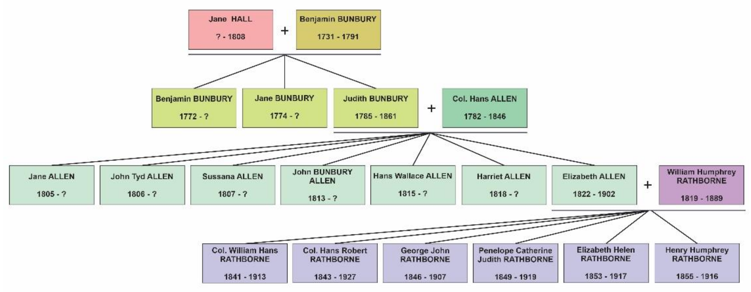
Based on the research done by J. Ferran [2], Judith was Capt. Benjamin Bunbury and Jane Hall’s daughter and had seven children with Hans Allen: Jane Allen, John Tyd Allen, Sussana Allen, John Bunbury Allen, Hans Wallace Allen, Harriet Allen, and Elizabeth Allen [2]. Otherwise, J. Ferran lists her birth date as around 1785, which contradicts the inscription from the back of the painting.
A scrap of paper was found attached on the verso of the painting with the following text: “For Hans Rathborne from aunt Hal/r…”. Based on the family tree (Figure 1), Judith’s daughter Elizabeth Allen together with her husband, Capt. William Humphrey Rathborne, had two sons named Hans Rathborne: col. William Hans Rathborne (1841–1913) and lt. col. Hans Robert Rathborne (1843–1927). By the Victorian era traditions, the first name, and essentially the one used in society, of the firstborn male child (in this case William) was named after the father’s father, while the second male child was named after the mother’s father. Therefore, we can conclude that Col. Hans Robert was the one who received the painting as a gift probably from Aunt Harriet, born in 1818. The Bunbury family dates back to the 12th century, and it was formed in the Kingdom of England but had Norman origins.
We thus understand that the figure represented in the painting is descended from this historical family and due to the social and economic status of the family maintained over time, we assume that the artwork was painted by a significant painter. The quality of the painting technique states the same fact.
The study aimed to gather information about the materials and technique of the artist, in a completely non-invasive manner, in view of designing the optimal restoration process. Another goal of the paper was to establish a time frame for the painting, by using the information gathered from the investigation protocol, correlated with the available written historical information and research regarding the fashion attire of the sitting figure.
2. Materials and Methods
Before the conservation and restoration process of artwork, investigations and written/visual documentation are carried out to create a record of the painting’s initial condition and characteristics. Nowadays, it has become common practice to set up multi-analytical non-invasive investigation protocols, to reduce as much as possible the invasive methods of investigation. For the painting under discussion, to minimize any potential harm to the studied object, which was in a poor state of conservation, all the investigations conducted were non-invasive. The original canvas of the artwork was very fragile due to the improper microclimate in which it was held before it entered the conservation and restoration studio. It was perforated in eight spots of which seven have been improperly “consolidated” with thick layers of overpainting on the front (more probably oil paint) and unsuitable cotton fabric patches with glue on the back. The paint layer adhered strongly to the canvas and only some small areas were flaking. Chromatic (yellowing and darkening) and textural (craquelures) alterations of the varnish layer took place due to aging and oxidation. The investigation protocol was designed to accomplish the main goals of the restorers: to determine the most appropriate way of treatment, to obtain the best results possible after the treatment, and to thoroughly document all steps of this process.
A Nikon D700 camera (12.1 megapixels and 35 mm full-frame sensor with AF-S NIKKOR 24–70 mm lens) was used to record high-resolution photographs of the painting under different illumination conditions. The spatial resolution at the acquisition was 4256 × 2832 pixels and the objective was set at f/3.5 diaphragm aperture and 50 mm focal distance. First, the artwork was examined in natural illumination. The uniformly scattered lighting was provided naturally, coming through the windows. This investigation was done right after the artwork entered the doors of the studio and helped us better understand its current condition. Second, the artwork was observed in transillumination, by pointing a powerful source of light (35 W, 98 daylight LED, 5000 °K) on the back of the painting (at 60–70 cm distance), to bring to light the inner features of the painting. This was followed by the examination of the painting in raking light, which highlighted the topography of the artwork, including uneven tensions of the support, degradation due to previous interventions, and density of the brush strokes. This non-damaging process consisted of placing the same source of light on the side of the painting to accentuate and reveal surface details such as texture, brushstrokes, and irregularities that may not be distinguishable under frontal lighting conditions. By positioning the source of light at a low angle (between 0 and 20°) to the surface of the artwork, shadows that emphasize the irregularities of the surface are created. The artwork can be examined from different angles by moving the light source around the studied object. Finally, ultraviolet (UV) fluorescence images were recorded, by placing an ultraviolet lamp in front of the artwork. In this way, the materials found on the surface of the artwork undergo temporary modifications called fluorescence. This phenomenon makes them easier to identify because each material has a distinguishable fluorescent property. The examination was done using the Luminaire Cts Art Lux 100 LV portable lamp with 8 UV LED lights, 365 nm wavelength. To collect only the fluorescent radiation, a 77 mm Hoya UV-blocking filter for the camera lens was used.
Infrared reflectography (IR) is a non-invasive technique of linear optical imaging that consists of producing an infrared image between 1000 and 1100 nm electromagnetic radiation of the investigated artwork. This technique reveals features underlying the pictorial layer, such as underdrawings, signatures, compositional and hidden pictorial changes (pentimento) made by the artist, and sometimes traces of subsequent overpainting [3]. The infrared photographs revealing the underdrawing of the painting were taken using Cts’s System for reflectography Mod. Mir 10 New, which is composed of a MICRO IR10.2 color camera (6 Mpixel resolution) for infrared reflectography with a sensor sensitive to the infrared part of the spectrum, a 12.5 mm f1/8 lens (RICOH TV Lens), and an infrared filter (IR1100). The illumination sources were two common incandescent lamps directed from the sides at approximately 5 m distance, radiating enough infrared rays for our purpose.
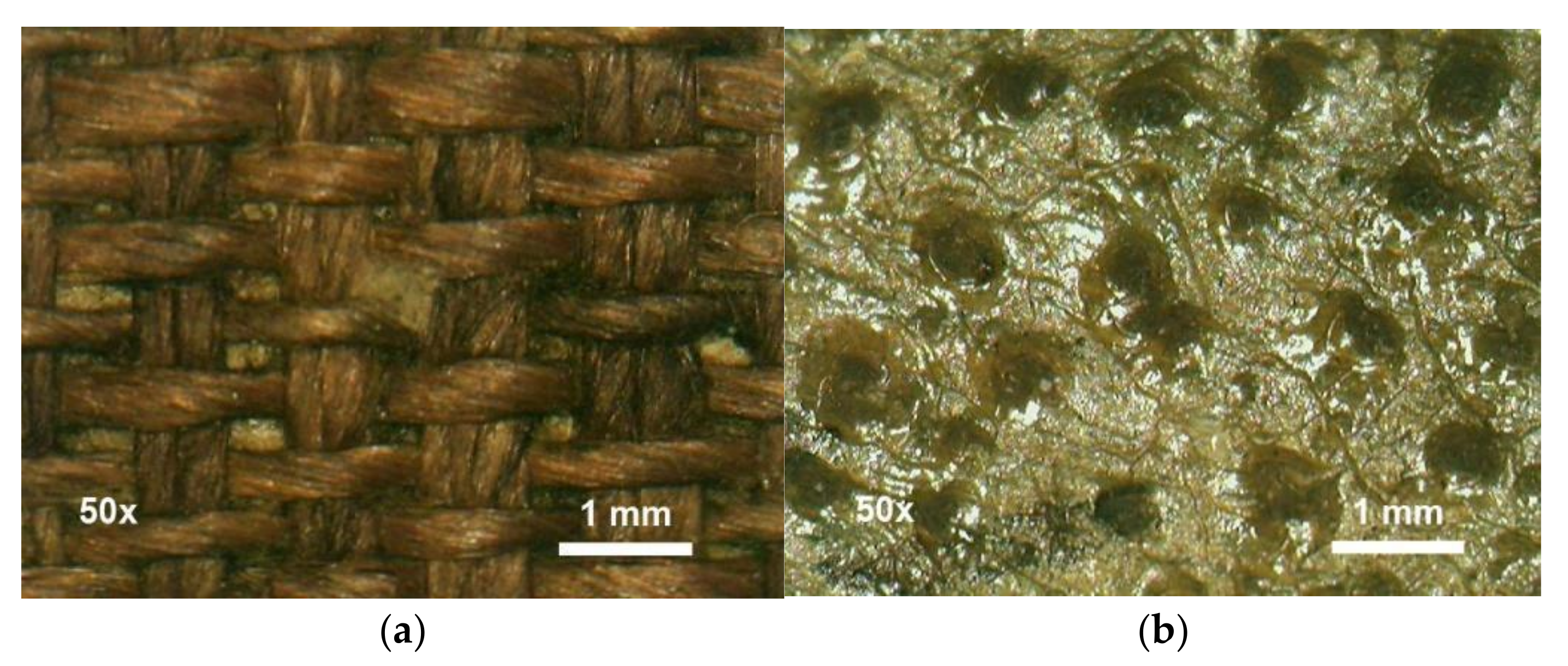
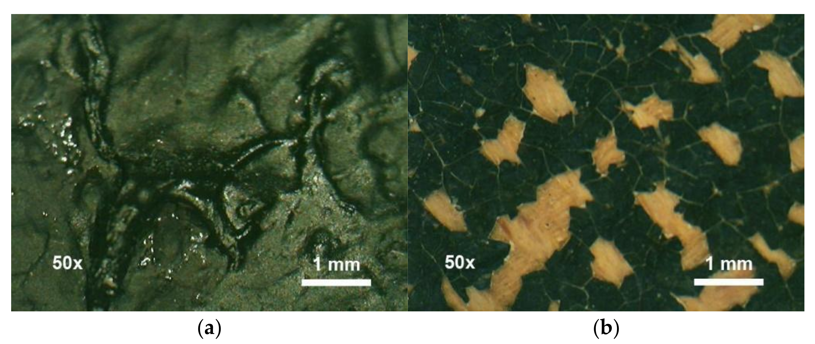
The microscopic investigations were performed directly on the painting using the portable video microscope (CTS Videomicroscope I-Scope USB); for the data collection, Moritex Viewer 2.2 was used. Thanks to its integrated LED illumination system and capacity of magnifying the image up to 200×, a close observation of the artwork has been provided and important characteristics have been revealed such as textures and degradation phenomena. In the investigation process, 50× (FOV 4.5 mm) and 200× (FOV 1 mm) magnifying lenses were used.
The elemental composition of the inorganic pigments was determined using the X-ray Fluorescence spectrometry method. This analysis was performed using a portable handheld energy-dispersive spectrometer Bruker TRACER III-SD. The equipment was operated in hand-held mode, at ~2 mm distance from the painting surface, at 40 kV tube voltage, 11 µA current intensity, for 5 s analysis time, without filtering of the beam. Identification of elements was performed with ARTAX software (Bruker, AXS GmbH, Mannheim, Germany) using standard Bayesian deconvolution, allowing a semi-quantitative approach. Data plotting was done using OriginPro 2021 (OriginLab Corporation, Northampton, MA, USA). The main advantage of using this portable instrument is that it provides a non-invasive, non-destructive analysis of pigments and inorganic materials in artworks.
All the investigations mentioned above represent a very important step before the restoration process because they are meant to reveal important information about the materials that have been used by the artist and about its technique, helping the conservator understand the artwork to approach suitable treatments.
3. Results and Discussion
3.1. Art Historical Analysis
The 63.5 × 76 cm painting depicts the bust portrait of an elderly lady with her right hand raised near the chest, holding a golden Victorian lorgnette (reading glasses with a handle). Her elbow rests upon an open book or some sheets of paper placed on a table, reflecting the typical conventions of the Victorian era. The figure is dressed in a black mourning ensemble, called “widow’s weeds”. Several elements, such as the strength of the clothing elements, jewelry, and also the fact that she is represented with glasses in her hand and a text under her elbow resting on a desk, point towards the idea that the woman represented in this painting was a well-educated, wealthy person.
Although the elderly lady is looking at the viewer with some semblance of warmth, she still conveys a sense of dignity and authority, rather than a friendly demeanor. This was common place in Victorian England, where society looked down on smiles and grins, which were associated with madness or lewdness.
The chromatic palette of the painting is represented by warm colors, especially brown and ochre of various intensities. The white color of the lace is warmed with ochre and the garment is close to black. The background color is also a lighter brown. The color of the face and hand is warm; the dominant color is yellow ochre, with different lighter or darker shades. The painter applied an intense red color for the coloring of the lips and more faded redness in the cheeks. It is obvious that the painting was realized in an enclosed space, with the light source illuminating the person probably being a lamp or candles.
Regarding the period in which the portrait was painted, some information can be deduced from written archive information. If Hans Robert Rathborne was born in 1843 (as mentioned in Burke’s Peerage, a British genealogical publisher [4]), then the painting was commissioned at a later date. Considering that Judith died in 1861, and if the portrait was made using the real-life model, it could mean that the portrait was painted between 1843 and 1861. Nevertheless, if it was commissioned after her death (painted after an earlier painting or photograph) then it could have been commissioned before the last couple of decades of the 19th century. Although Aunt Harriet’s time of death is unknown, considering she was born in 1818, her death probably occurred long before the turn of the century.
3.2. Visual Analysis
3.2.1. Examination in Natural Light
Generally, one of the first steps is to observe the painting in visible light [5]. The documentation of the artwork’s state of conservation from the moment it enters the studio is an important step in the restoration process. One way to document its condition is by taking high-resolution photographs in good lighting. When photographing the artwork in direct lighting, the light falls uniformly across the entire surface of the painting.
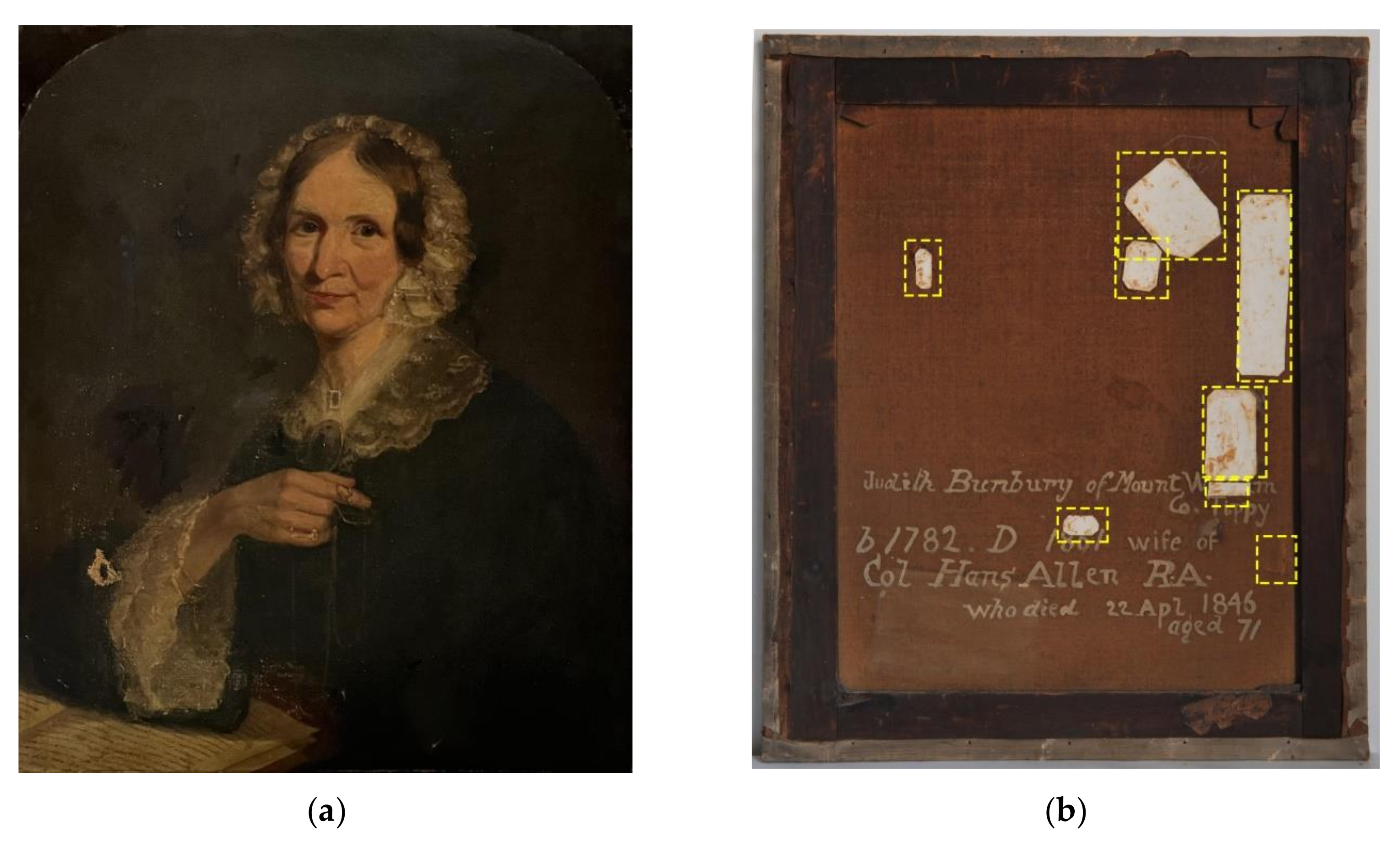
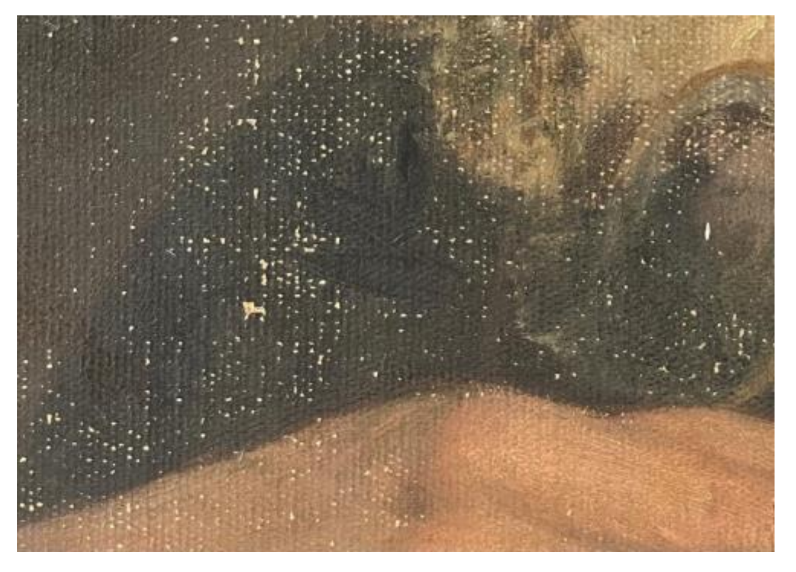
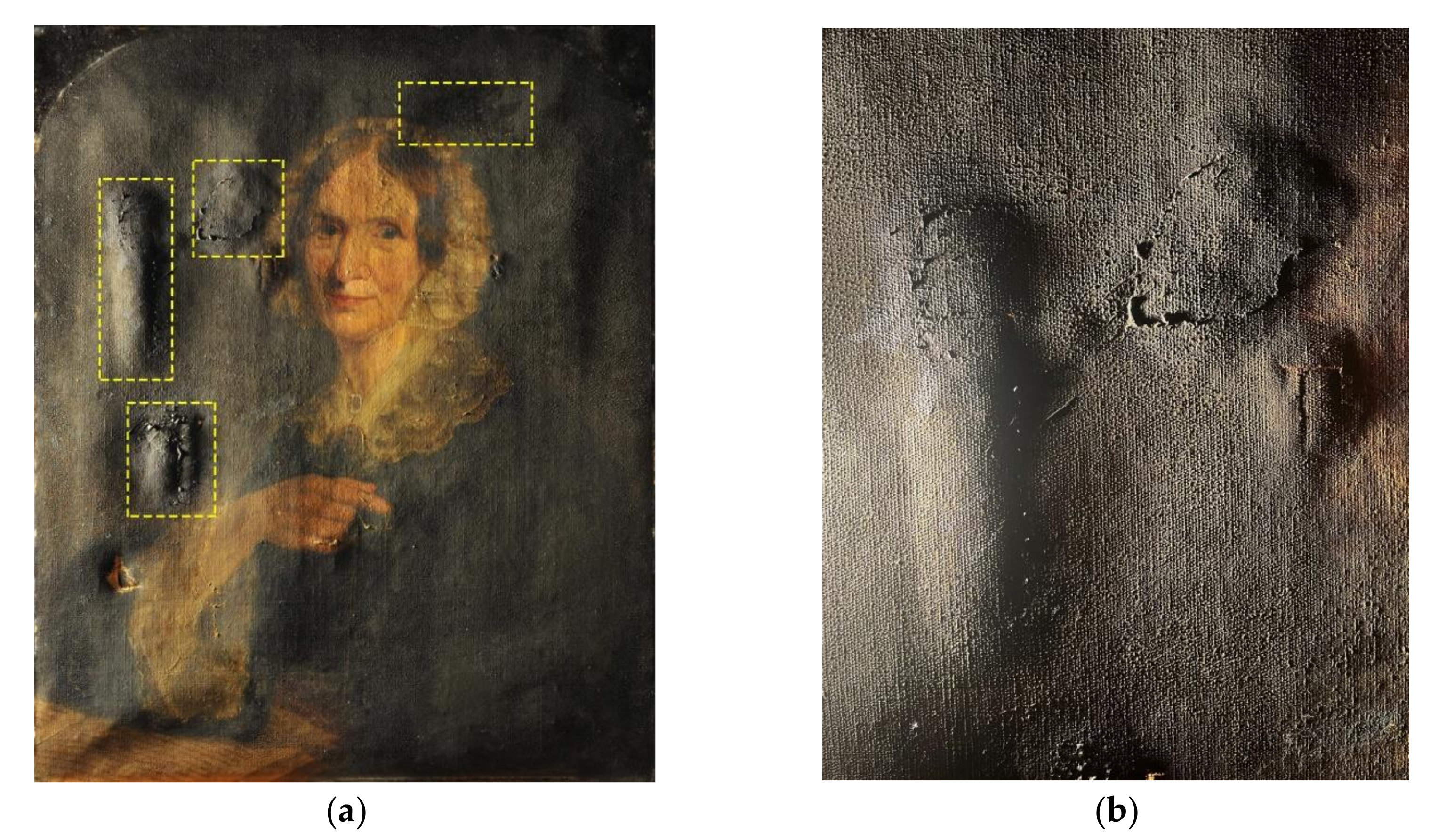
After photographing the artwork in visible light (Figure 2), it was noticed that the textile support is made out of a single piece of linen fabric that has been perforated in eight spots, mainly in the background area. Of these, seven perforations have been improperly consolidated with a thick layer of oil paint (overpaint) on the front and with unsuitable patches on the back (indicated in Figure 2b by yellow dashed boxes). The oil content of the overpaint has been proven by performing an oil test on an isolated area using a saturated solution of Sudan Black B. The patches were mechanically unsuitable due to the animal glue used to adhere them to the holes and tears. The fact that the adhesive is protein-based (animal glue) was proven by a protein test performed on a patch using an acid fuchsin reagent. The glue, which lost its elasticity and contracted over time, caused some aesthetically disturbing bulges on the front of the painting and distinctive craquelure in the paint layer. The fact that one of the perforations has not been consolidated indicates that it might be more recent than the others. Additionally, it could be noticed that both the paint layer and the varnish layer were evenly applied. The paint layer has strong adhesion to the preparation layer, with only a few areas on the shoulder and above the right hand in which lacunae and flaking were seen (Figure 3). Due to aging, chromatic (yellowing and darkening) and textural (craquelures) alterations of the varnish layer took place. The chromatic alterations are probably photo-induced due to the fact that the edges of the painting layer that were covered by the frame did not show as severe an alteration as the areas exposed to light and oxygen.
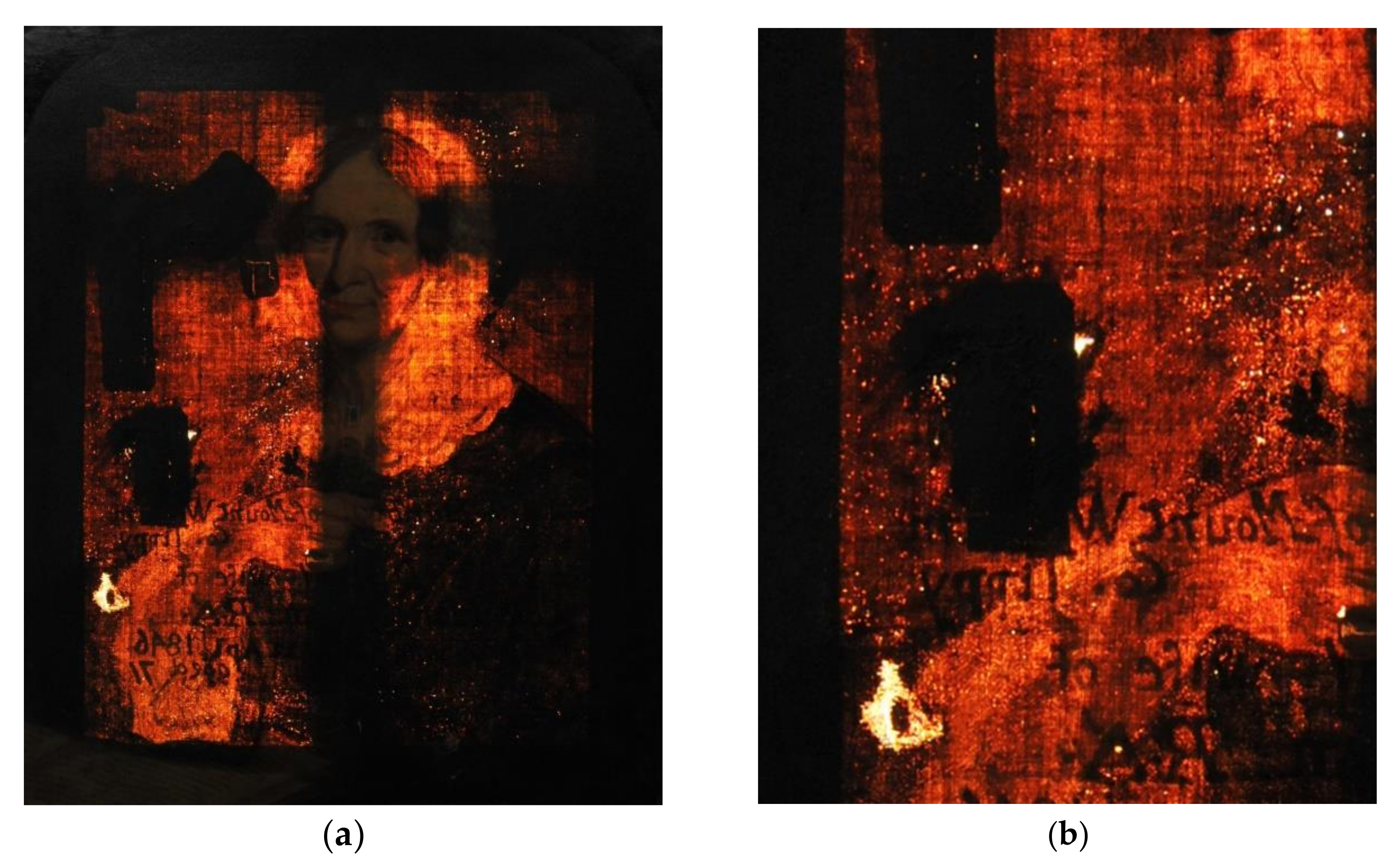
3.2.2. Transillumination
This type of investigation consists of placing a source of light on the back of the painting. It is commonly referred to as “backlighting”. Shining the light through an object helps analyze its internal features and reveals details that may not be visible through other examination methods.
This investigation method proved to be very effective in highlighting the damages and imperfections of the canvas, technical aspects, and previous interventions. The most visible flaws seen under this type of investigation were the perforations and the improper patching done on the back using thick cotton fabric, as can be seen in Figure 4a,b. Although patching was commonly used for lining and locally stabilizing gaps in paintings, their presence on the canvas can induce unwanted effects, such as contraction or swelling. The animal glue, which lost its elasticity, became brittle and contracted over time, causing bulges on the front of the painting and deformations of the textile support. Furthermore, it caused the appearance of distinctive craquelure in the paint layer in these areas [6].
Transillumination also revealed details about the painting technique, such as the fact that some areas were painted in light coats of paint and others were applied in a pasty manner. Backlighting is also important to see if the added paint exceeds the margins of the existing lacunae. In this case, it showed that the overpainting was not limited to the edges of the lacunae, covering large areas of the original painting layer as well.
3.2.3. Observation in Raking Light
Raking light is a non-destructive investigation method used to examine the surface of different types of artworks such as wall, panel, or canvas paintings. This type of investigation highlights the tridimensional aspect of the surface of the artwork providing information regarding the technique and conservation state. It allows conservators to see the painting from different angles and to observe how the light interacts with its surface.
As can be seen in Figure 5a,b, the examination of the painting in raking light highlighted the thick layers of oil paint used to improperly retouch the painting and the deformations and bulges of the canvas caused by the patches on the back (alterations highlighted by the yellow dashed boxes). The areas painted in a pasty manner are also easily distinguished under the radiant source of lighting.
3.3. Imaging Analysis
3.3.1. Videomicroscopy
Videomicroscopy is a non-invasive investigation technique that does not imply sampling and provides a close in situ observation of the artwork highlighting the main characteristics of the technique and materials used by the artist and also the structural damages [7].
Thanks to its incorporated lighting system, the I-scope equipment generated detailed images of the examined elements. Therefore, the I-scope provided a close view of the canvas highlighting its texture and its warp and weft (Figure 6a), the varnish coat and its patinated, agglutinated look caused by aging and light/sun exposure (Figure 6b), of the improperly made retouches by a previous restorer (Figure 7a), and of the flaking paint layer (Figure 7b). In most areas, the paint layers separated, while the ground layer remained adhered to the textile support. This might have been caused due to loss of adhesion between the ground and paint layers. If the ground layer was applied early on to the canvas, but the painting took place sometime later, the excessive drying of the ground layer (containing lead, which has a drying effect) and an insufficient quantity of oil binder in the subsequent paint layer could have caused this form of flaking. Another explanation could be damage caused by humidity. The exchange of moisture between the front layers and the textile support of the painting occurs through the craquelure, and thus, moisture may accumulate between the paint and ground layer. The moisture may have softened the glue and ground layer, while the paint layers remained inelastic. This may have led to the lifting and separation of the layers.
3.3.2. UV Fluorescence Imaging
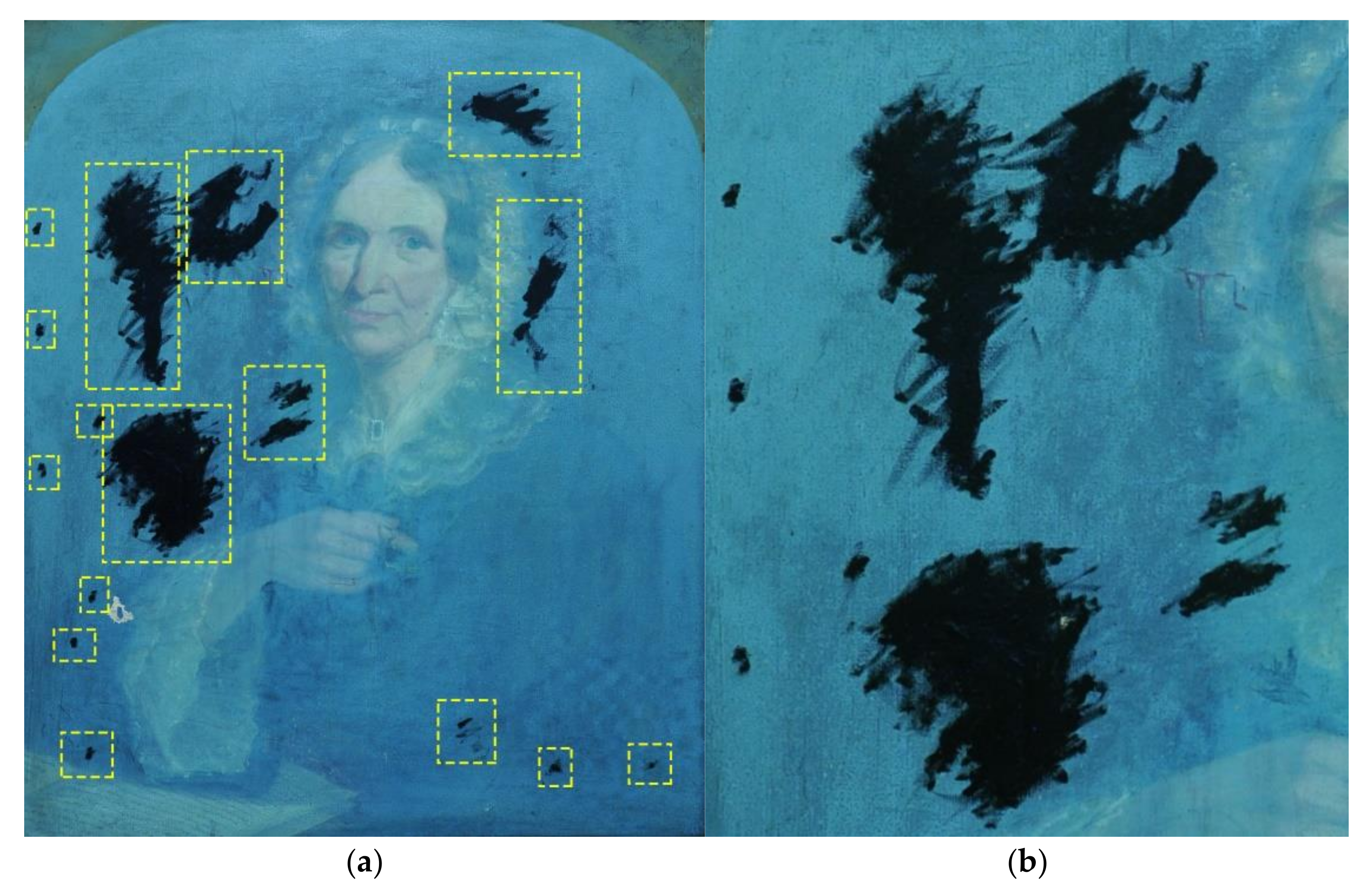
Even if the varnish of a painting can be easily observed in good lighting, using UV fluorescence helps to determine how homogenous the application of the varnish layer is [8]. It also allows us to see where retouching was done using similar colors. The varnish of a painting provides a protective role for the painting acting as a barrier to dirt, smoke, and injury caused by possible mechanical scratches. It also holds an aesthetic value providing depth and clarity to the colors [9]. Furthermore, the varnish layer protects against atmospheric pollutants, such as chlorine or sulfur emissions that can alter the color of certain pigments. It also offers protection against ultraviolet rays (which can cause the oxidation of the oil-based binder, as well as the discoloration of some organic dyes or the alteration of some inorganic pigments) and infrared rays (which, by heating the substrate, can cause the loss of mechanical resistance within the layers), originating from natural and/or artificial light sources. Putting the artwork under this method of investigation revealed that the painting has an evenly applied layer of varnish and some retouches that are improperly made (highlighted in Figure 8 by the yellow dashed boxes). The darker areas that can be seen in Figure 8, which can be easily distinguished from the original paint, correspond to areas where retouching has been made. Also, the greenish tint under UV light indicates the use of a natural resin varnish coat. The retouches were made in a single layer of paint, meaning they were applied during a single restoration intervention when the patches were also applied. The retouches did not cause any color change in the proximity of the retouched areas.
3.3.3. Infrared Reflectography (IR)
Infrared reflectography is a non-invasive optical technique that consists of producing an infrared image of the investigated artwork that reveals features underlying the pictorial layer. Infrared reflectography is one of the routine methods of examining paintings since 1950 and it is the tool of choice for researching the underdrawings. The underdrawing is the drawing that the artist made on the already prepared canvas for painting, it was then covered by subsequent paint layers. Depending on their chemical composition, certain materials absorb the IR radiation (they appear dark), while others reflect it, meaning that they appear brighter, thus the underdrawings, compositional changes, signatures, or underlying brushstrokes may become visible. Usually, iron, aluminum, manganese, barium, copper, cobalt, and graphite-based pigments and materials (for example earth pigments, ochres, cobalt blue, malachite, and azurite) absorb the radiation and usually appear dark. Cadmium, zinc, chromium, and titan-based pigments usually transmit infrared radiation and reflect it quite well (for example, cadmium yellows and reds, and zinc and titan white), thus appearing bright. Lead, tin, mercury, and sulfur-based pigments strongly reflect the radiation and appear very bright (for example lead white, vermillion, and ultramarine). Of course, the reflection of the infrared radiation depends on the thickness of the paint layer as well as on the quantitative composition of the various elements. While these features are not conclusive, they can be used as a starting point along with other analytical investigations in identifying some of the pigments used.
Sometimes superimposition of the IR images occurs when both the paint layers and the underdrawings can be seen, thus making it more difficult to interpret the images [10].
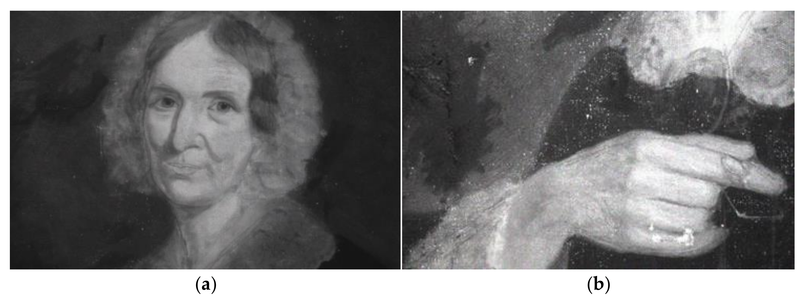
The use of IR allowed the underdrawing of the painting to be seen, which would not have been possible without the naked eye. The underdrawing made in graphite was obvious under the portrait, under the bonnet, under the neck area, and under the hand (Figure 9).
There was not any signature of the painter found on the painting. This fact may be due to several reasons. One reason could be that the painter signed the painting on an additional piece of paper attached to the canvas. Another reason, likely the most plausible one, is related to the fact that the studied painting is a commissioned work. In the Victorian era, some artworks were left unsigned because they were not for sale in front of an audience, they were not showcased to the public at soirees or in great halls. As it is in this case, the painting was a commissioned work probably by Aunt Harriet, who offered it as an exclusive gift to Hans Robert Rathborne, in memory of his grandmother, Judith Bunbury. Portraits received as gifts often sat in a personal library or a private study, meaning they were not intended for the public eye, but rather for a private audience.
3.4. Elemental Analysis
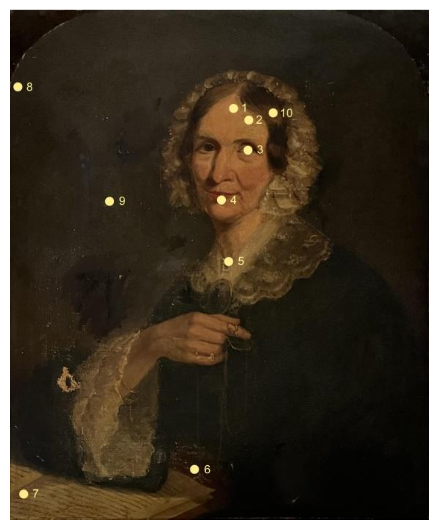
X-ray Fluorescence (XRF) spectrometry is a non-destructive method of investigation that allows obtaining an accurate and quick identification of inorganic materials and has been widely used in the study of art objects in past decades [11]. XRF studies have been performed on different types of artworks such as manuscripts, wall paintings, wood panels, and canvas paintings [12,13,14,15,16]. For the aim of this study, the investigation was carried out on 10 distinct analysis zones, covering the entire color range, as can be seen in Figure 10.
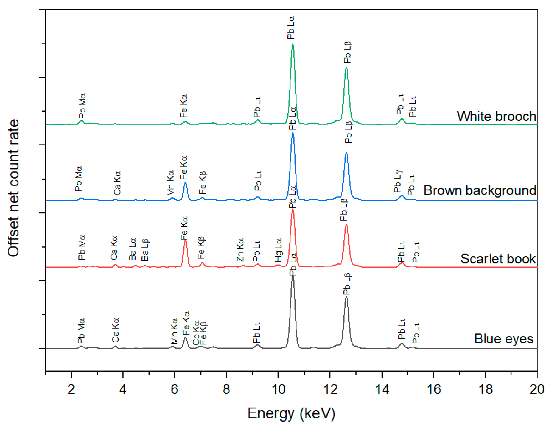
Figure 11 shows the XRF spectra for some representative areas: blue (area 3), white (area 5), scarlet (area 6), and brown (area 9). As can be seen from the figure, the acquired spectra were dominated by the high lead (Pb) lines, present in all analyzed areas. Lead has been used since ancient times to produce variations of white, red, or yellow pigments [11,17]. However, XRF alone cannot differentiate between different pigments with similar elemental composition. Moreover, it cannot determine where the lead signal is coming from [11], and given the fact that lead was found in all analyzed areas, the signal may be coming from an underlayer [18,19]. Also, based on the fact that the painting in question is an oil painting, it could have been used as a drier for the binder [20]. So, the presence of the lead lines could be due to one or more of the previously mentioned reasons.
The fact that, aside from the lead lines, no other significant elements were detected in the spectrum of the white area (5), suggests that lead white could have been used as a white pigment.
In addition, similar lead intensities were found for area 8 (lacuna in the background), which sustains the idea that lead was used in the ground layer as well. Besides lead, calcium (Ca) was another element identified in every analyzed area. The use of lead white mixed with calcium carbonate in primers was frequently used until the 20th century [21] because of its qualities: it offers good and luminous coverage in oil, has a drying effect on the oil medium, and also provides a low oil absorption, thus counteracting the uneven absorbency of the overlapping paint layers in time.
This information was crucial in further restoration interventions, namely at the filling of lacunae, where the choice of the filler would consist of calcium carbonate with a white pigment mixed with animal glue.
For the background (areas 8 and 9), the manganese line (Mn Kα) could be seen, which points towards the use of brown manganese-based pigment.
The spectrum corresponding to area 6 shows both slightly higher iron (Fe) lines, indicating the use of an iron-based pigment, and traces of mercury (Hg), as can be seen in Figure 10 (scarlet book). Although the high lead lines made it difficult to assess the presence of sulfur, which appears in the same energy range as the Pb Mα lines, the occurrence of mercury traces indicates the use of the mercuric sulfide pigment (HgS). This is commonly referred to as cinnabar or its synthetic variant, vermilion [17], a pigment that is known to degrade with time, leading to the darkening of its original color. In Europe, both natural (from the cinnabar mineral) and synthetic vermilion were used simultaneously [22] until the end of the 19th century.
Another interesting aspect is the fact that traces of barium (Ba) and zinc (Zn) were evident in the spectrum of area 6 but also appeared at lower levels in some of the other areas.
The presence of Pb, along with Zn and Ba indicates the artist’s technique of combining multiple white pigments to obtain the desired tones. The combination of zinc and barium could come from lithopone, a mixture of zinc sulfide and barium sulfate (ZnS∙BaSO4), introduced in the second half of the 19th century [17,23], but it could also point towards the use of zinc white, which is zinc oxide, and barium sulfate, known as Blanc Fixe [24,25].
XRF analysis also revealed the presence of cobalt traces in the spectrum recorded from the area of the eye (3). While only traces of cobalt could be seen, correlated with the visual aspect, it can infer the use of a blue cobalt-based pigment. XRF spectroscopy alone cannot directly indicate which type of pigment could have been used, as several blue cobalt-based pigments were in use during the 19th century. However, the absence of certain elements, such as As, Sn, Zn, or Cr can rule out the possibility of certain pigments, including cerulean blue, dark cobalt blue, smalt, cobalt chromite, or cobalt spinel, thus leaving the most probable pigment as cobalt blue, which has been used since the beginning of the 19th century [26].
4. Conclusions
The investigation campaign of the artwork was a crucial preliminary step that unveiled valuable information about its complexity, conservation state, technique, previous interventions (such as improper retouches and patches), and chemical composition of its materials.
The examination in natural light played a pivotal role in this investigation protocol revealing information about the general state of the painting and its flaws. By shining light through the back of the artwork, hidden damage, weaknesses like perforations, technical aspects, and previous improper restorations, which have led to the structural deterioration of the painting, were highlighted. It also provided an insight view into the artist’s working process. The artwork’s topography and texture were highlighted thanks to the raking light investigation in which shadows were generated by the low-angle positioned source of light. This method of investigation allowed the conservators to appreciate and better understand the artistic techniques that were used in the process of creating this artwork, represented by a combination of light and pasty brushstrokes. The use of UV lighting proved to be a valuable investigation technique that made the improper previous retouches obvious, with several darker areas, which correspond to newer materials. Moreover, the use of a natural resin varnish was indicated by the greenish tint of the UV images. Videomicroscopy was useful in evidencing the uniformly applied varnish layer, which had an agglutinated look, with chromatic alterations due to light exposure. The underdrawings were highlighted with the help of IR reflectography, in the upper half and middle of the drawing, in the bonnet, neck, and hand areas. The XRF analysis revealed the chemical composition of the used inorganic pigments: white areas showed lead, along with barium and zinc, red areas showed both iron and mercury, the brown background showed the presence of a manganese-based pigment, while the blue was obtained with a cobalt-based pigment, and the preparation layer showed both calcium and lead.
In summary, the investigation protocol carried out on the artwork significantly enhanced the understanding of it by gathering information about its condition, previous interventions, technique, and chemical composition. Of course, although not available at the time of the investigation, the methodology would be complete by adding some portable, non-invasive spectroscopic techniques, such as Raman or Fourier-transform infrared spectroscopy, for the identification of the molecular composition of the varnish, the glue used for patching or the impregnation layers, if any. There is no debate that a molecular analysis would have completed the investigations, but overall, the results obtained were, in this case, sufficient for the start of the restoration process, as they have helped experts understand the painting’s historical and cultural context to approach a targeted restoration methodology. For instance, for the filling of the lacunae, the filler will consist of calcium carbonate, and animal glue with added titanium white, to match as closely as possible to the appearance and properties of the original ground layer. Additionally, by identifying some of the pigments, the use of acids or bases during cleaning interventions has become safe as they will not cause any color alteration.
Furthermore, by establishing a connection between the materials used by the artist and the written information available about the Bunbury family, in correlation with the painting technique used and the fashion attire worn by the model, the team successfully narrowed down the time frame in which the artwork was painted: the portrait was commissioned between 1843 and possibly 1861.
Author Contributions
Conceptualization, A.R. and L.G.; methodology, A.R.; investigation, A.R., L.G. and A.B.; resources, A.R.; writing—original draft preparation, A.R., A.B. and M.A.; writing—review and editing, L.G. and A.R. All authors have read and agreed to the published version of the manuscript.
Funding
This work was carried out through the project UpUAD funded by the National Council for the Financing of Higher Education, grant number CNFIS-FDI-2023-F-0468, and through the Core Program within the National Research Development and Innovation Plan 2022–2027, project no. PN 23 05, and Program 1—Development of the national research-development system, Subprogram 1.2—Institutional performance—Projects to finance the excellent RDI, Contract no. 18PFE/30.12.2021, carried out with the support of MCID.
Data Availability Statement
Data are available from the corresponding author upon reasonable request.
Acknowledgments
Not applicable.
Conflicts of Interest
The authors declare no conflict of interest.
References
- Crowther, P. Awakening Beauty. The Crowther-Oblack Collection of Victorian Art. Available online: https://www.victorianweb.org/painting/awakeningbeauty/style.html (accessed on 10 June 2023).
- Ferran, J. Legacy 9.0. Family Tree. Available online: https://www.monchique.com/Ochanoff/ohanov/ochanoff/7001.htm (accessed on 10 June 2023).
- Amat, A.; Miliani, C.; Brunetti, B.G. Non-Invasive Multi-Technique Investigation of Artworks: A New Tool for on-the-Spot Data Documentation and Analysis. J. Cult. Herit. 2013, 14, 23–30. [Google Scholar] [CrossRef]
- Burke, B. A Genealogical and Heraldic History of the Landed Gentry of Ireland; Harrison & Sons: London, UK, 1912. [Google Scholar]
- Van Aspren de Boer, J.R.J. An Introduction to the Scientific Examination of Paintings. In Scientific Examination of Early Netherlandish Painting; Aspren De Boer, J.R.J., Filedt Kok, J.P., Dishoeck, F., Eds.; Brill: Leiden, The Netherlands, 1975. [Google Scholar]
- Teixeira, A.C. Canvas Support Impregnation Materials and Techniques: A Study of Portuguese Painting and Its Conservation Issues. CeROArt 2016. [Google Scholar] [CrossRef]
- Mureșan, T. Videomicroscopia Portabilă În Analiza Picturii Murale. Caiet. Restaurării 2013, 170–177. [Google Scholar]
- Rauca-Bencze, F. Metodologii Istorice de Dublare a Picturilor În Ulei Pe Suport Textil. Studiu de Caz- Pelerinii Din Emaus Din Colecția Muzeului de Artă Cluj-Napoca. Caiet. Restaurării 2019, 1, 208–223. [Google Scholar]
- Vandivere, A.; Wadum, J.; van den Berg, K.J.; van Loon, A. From ‘Vermeer Illuminated’ to ‘The Girl in the Spotlight’: Approaches and Methodologies for the Scientific (Re-)Examination of Vermeer’s Girl with a Pearl Earring. Herit. Sci. 2019, 7, 66. [Google Scholar] [CrossRef]
- Pinna, D.; Galeotti, M.; Mazzeo, R. Scientific Examination for the Investigation of Paintings: A Handbook for Conservator-Restorers. In Scientific Examination for the Investigation of Painting: A Handbook for Conservator-Restorers; Centro Di: Florence, Italy, 2009; ISBN 978-88-7038-474-1. [Google Scholar]
- Shugar, N.A.; Mass, L.J. Handheld XRF for Art and Archaeology; Leuven University Press: Leuven, Belgium, 2012. [Google Scholar]
- Cucci, C.; Bracci, S.; Casini, A.; Innocenti, S.; Picollo, M.; Stefani, L.; Rao, I.G.; Scudieri, M. The Illuminated Manuscript Corale 43 and Its Attribution to Beato Angelico: Non-Invasive Analysis by FORS, XRF and Hyperspectral Imaging Techniques. Microchem. J. 2018, 138, 45–57. [Google Scholar] [CrossRef]
- Antunes, V.; Valadas, S.; Serrão, V.; Carvalho, M.L.; Candeias, A.; Mirão, J.; Cardoso, A.; Pessanha, S.; Manso, M. Josefa d’ Óbidos Workshop from Panel to Canvas. Multianalytical Approach to Materials and Technical Evolution of the Most Significant Portuguese Painting Workshop of the 17 Th Century. J. Mol. Struct. 2019, 1188, 31–41. [Google Scholar] [CrossRef]
- Cortea, I.M.; Ghervase, L.; Ratoiu, L.; Rădvan, R. Application of Spectroscopic and Hyperspectral Imaging Techniques for Rapid and Nondestructive Investigation of Jewish Ritual Parchment. Front. Mater. 2020, 7, 601339. [Google Scholar] [CrossRef]
- Kokiasmenou, E.; Caliri, C.; Kantarelou, V.; Germanos Karydas, A.; Romano, F.P.; Brecoulaki, H. Macroscopic XRF Imaging in Unravelling Polychromy on Mycenaean Wall-Paintings from the Palace of Nestor at Pylos. J. Archaeol. Sci. Rep. 2020, 29, 102079. [Google Scholar] [CrossRef]
- Klisińska-Kopacz, A.; Obarzanowski, M.; Frączek, P.; Moskal-del Hoyo, M.; Gargano, M.; Goslar, T.; Chmielewski, F.; Dudała, J.; del Hoyo-Meléndez, J.M. An Analytical Investigation of a Wooden Panel Painting Attributed to the Workshop of Lucas Cranach the Elder. J. Cult. Herit. 2022, 55, 185–194. [Google Scholar] [CrossRef]
- Eastaugh, N.; Walsh, V.; Chaplin, T.; Siddall, R. Pigment Compendium: A Dictionary and Optical Microscopy of Historical Pigments; Eastaugh, N., Walsh, V., Chaplin, T., Siddall, R., Eds.; Elsevier: Amsterdam, The Netherlands, 2008. [Google Scholar]
- Duran, A.; Siguenza, M.B.; Franquelo, M.L.; de Haro, M.C.J.; Justo, A.; Perez-Rodriguez, J.L. Murillo’s Paintings Revealed by Spectroscopic Techniques and Dedicated Laboratory-Made Micro X-ray Diffraction. Anal. Chim. Acta 2010, 671, 1–8. [Google Scholar] [CrossRef] [PubMed]
- Pięta, E.; Proniewicz, E.; Szmelter-Fausek, B.; Olszewska-Świetlik, J.; Proniewicz, L.M. Pigment Characterization of Important Golden Age Panel Paintings of the 17th Century. Spectrochim. Acta Part A Mol. Biomol. Spectrosc. 2015, 136 Pt B, 594–600. [Google Scholar] [CrossRef]
- Laporte, L.; Ducouret, G.; Gobeaux, F.; Lesaine, A.; Hotton, C.; Bizien, T.; Michot, L.; de Viguerie, L. Rheo-SAXS Characterization of Lead-Treated Oils: Understanding the Influence of Lead Driers on Artistic Oil Paint’s Flow Properties. J. Colloid Interface Sci. 2023, 633, 566–574. [Google Scholar] [CrossRef] [PubMed]
- Stols-Witlox, M. Twentieth-Century Grounds. In Conservation of Easel Paintings; Stoner, J.H., Rushfield, R., Eds.; Routledge: London, UK, 2012; pp. 168–188. [Google Scholar]
- Gettens, R.J.; Feller, R.L.; Chase, W.T. Vermilion and Cinnabar. In Artists’ Pigments. A Handbook of Their History and Characteristics, Volume 2; Roy, A., Ed.; National Gallery of Art: Washington, DC, USA, 2012; pp. 159–182. [Google Scholar]
- Macchia, A.; Schuberthan, L.M.; Ferro, D.; Colasanti, I.A.; Montorsi, S.; Biribicchi, C.; La Russa, M.F. Analytical Investigations of XIX–XX Century Paints: The Study of Two Vehicles from the Museum for Communications of Frankfurt. Molecules 2023, 28, 2197. [Google Scholar] [CrossRef] [PubMed]
- Valentini, F.; De Angelis, S.; Marinelli, L.; Zaratti, C.; Colapietro, M.; Tarquini, O.; Macchia, A. Multianalytical Non-Invasive Characterization of ‘Mater Boni Consilii’ Iconography Oil Painting. Heritage 2023, 6, 3499–3513. [Google Scholar] [CrossRef]
- Veiga, A.R. Oil Painting on Tinplate by Francisco José Resende. CeROArt 2010. [Google Scholar] [CrossRef]
- Gettens, R.J.; Stout, G.L. Painting Materials: A Short Encyclopaedia; Dover Publications, Inc.: New York, NY, USA, 1966. [Google Scholar]
Figure 1.
The Bunbury family tree.

Figure 2.
The portrait of Judith Bunbury—photographs of the front (a) and back (b) of the painting in visible light.
Figure 2.
The portrait of Judith Bunbury—photographs of the front (a) and back (b) of the painting in visible light.

Figure 3.
Flaking and lacunae of painting layer, photographed in visible light.

Figure 4.
Photography with transilluminating light; overview (a) and detail (b).

Figure 5.
Photography with raking light; overview (a) and detail (b).

Figure 6.
The texture of the canvas (a) and of the varnish coat (b).

Figure 7.
Defects on the surface: improperly made retouch (a) and flaking (b).

Figure 8.
Photography in ultraviolet light; overview (a) and detail (b).

Figure 9.
Infrared photographs showing the underdrawings made in graphite: portrait area (a); right-hand area (b).
Figure 9.
Infrared photographs showing the underdrawings made in graphite: portrait area (a); right-hand area (b).

Figure 10.
Visible light photography with the points analyzed with XRF (numbers represent analysis areas).
Figure 10.
Visible light photography with the points analyzed with XRF (numbers represent analysis areas).

Figure 11.
XRF spectra of selected areas.

Disclaimer/Publisher’s Note: The statements, opinions and data contained in all publications are solely those of the individual author(s) and contributor(s) and not of MDPI and/or the editor(s). MDPI and/or the editor(s) disclaim responsibility for any injury to people or property resulting from any ideas, methods, instructions or products referred to in the content. |
© 2023 by the authors. Licensee MDPI, Basel, Switzerland. This article is an open access article distributed under the terms and conditions of the Creative Commons Attribution (CC BY) license (https://creativecommons.org/licenses/by/4.0/).
Share and Cite
MDPI and ACS Style
Rauca, A.; Ghervase, L.; Berdie, A.; Agachi, M. Unveiling the Secrets of an Artwork through Non-Invasive Investigations—Case Study of a 19th-Century Female Portrait. Minerals 2023, 13, 1193. https://doi.org/10.3390/min13091193
AMA Style
Rauca A, Ghervase L, Berdie A, Agachi M. Unveiling the Secrets of an Artwork through Non-Invasive Investigations—Case Study of a 19th-Century Female Portrait. Minerals. 2023; 13(9):1193. https://doi.org/10.3390/min13091193
Chicago/Turabian StyleRauca, Adrian, Luminița Ghervase, Antonia Berdie, and Matei Agachi. 2023. "Unveiling the Secrets of an Artwork through Non-Invasive Investigations—Case Study of a 19th-Century Female Portrait" Minerals 13, no. 9: 1193. https://doi.org/10.3390/min13091193
Note that from the first issue of 2016, this journal uses article numbers instead of page numbers. See further details here.