Article

European Population of Pectobacterium punjabense: Genomic Diversity, Tuber Maceration Capacity and a Detection Tool for This Rarely Occurring Potato Pathogen

1
French Federation of Seed Potato Growers (FN3PT-inov3PT), 43-45 Rue de Naples, 75008 Paris, France
2
Institute for Integrative Biology of the Cell, Université Paris-Saclay, CEA, CNRS, 91198 Gif-sur-Yvette, France
3
IGEPP, Agrocampus Ouest, INRAe, University Rennes 1, F-35653 Le Rheu, France
4
Laboratory of Plant Protection and Biotechnology, Intercollegiate Faculty of Biotechnology, University of Gdansk and Medical University of Gdansk, Abrahama 58, 80-307 Gdansk, Poland
5
Department of Pharmaceutical Microbiology, Faculty of Pharmacy, Medical University of Gdansk, Al. Gen. Hallera 107, 80-416 Gdansk, Poland
6
Biointeractions and Plant Health, Wageningen University & Research, P.O. Box 16, 6700 AA Wageningen, The Netherlands
*
Authors to whom correspondence should be addressed.
Microorganisms 2021, 9(4), 781; https://doi.org/10.3390/microorganisms9040781
Submission received: 19 March 2021 / Revised: 2 April 2021 / Accepted: 6 April 2021 / Published: 8 April 2021
(This article belongs to the Special Issue Dickeya and Pectobacterium: Ecology, Pathology and Plant Protection)

Abstract

:
Enterobacteria belonging to the Pectobacterium and Dickeya genera are responsible for soft rot and blackleg diseases occurring in many crops around the world. Since 2016, the number of described species has more than doubled. However, some new species, such as Pectobacterium punjabense, are often poorly characterized, and little is known about their genomic and phenotypic variation. Here, we explored several European culture collections and identified seven strains of P. punjabense. All were collected from potato blackleg symptoms, sometimes from a long time ago, i.e., the IFB5596 strain isolated almost 25 years ago. We showed that this species remains rare, with less than 0.24% of P. punjabense strains identified among pectinolytic bacteria present in the surveyed collections. The analysis of the genomic diversity revealed the non-clonal character of P. punjabense species. Furthermore, the strains showed aggressiveness differences. Finally, a qPCR Taqman assay was developed for rapid and specific strain characterization and for use in diagnostic programs.

1. Introduction

Bacteria belonging to the Pectobacterium and Dickeya genera are causal agents of soft rots in a wide variety of host plants [1]. The agricultural and economic impact ranks these pathogens classified within the group of soft rot Pectobacteriaceae (SRP) [2] among the most studied phytopathogenic bacteria [3]. They produce extracellular enzymes degrading plant cell walls [4,5] causing the breakdown of plant tissues. Several of the SRP can generate blackleg symptoms, characterized by a blackening of the stem base [6]. The losses can be significant [7] and, to date, chemical, physical or biocontrol applications have shown limited efficacy. Therefore, prophylactic methods such seed sanitation and breeding potato cultivars to improve natural resistance are still highly recommended [8].
Since the wide-scale use of next generation sequencing (NGS) for strain characterization, it has become clear that the genetic diversity of SRP is very high [9]. Analysis of NGS data resulted in the elevation of different subspecies to the species level [10] and also in the description of new species. As a consequence, the number of species belonging to these two genera has increased from five Pectobacterium spp. and seven Dickeya spp. before 2016 to 18 Pectobacterium spp. and 12 Dickeya spp. until now [11,12]. The NGS analysis further resulted in the revision and refining of the taxonomic position of strains maintained in laboratory or reference collections, sometimes for a long time [10,13]. The isolates were collected from different backgrounds, including diseased plant samples and surface waters [14,15,16,17,18,19,20].
Historically, the isolates collected in temperate regions from blackleg-diseased plants were identified as Erwinia atroseptica, Erwinia carotovora subsp. wasabiae and Erwinia chrysanthemi [21,22,23]. On the basis of the current taxonomy, P. atrosepticum, P. brasiliense, P. parmentieri, P. versatile, P. polaris and P. carotovorum are the most prevalent Pectobacterium species responsible for soft rot and/or blackleg diseases [24,25,26,27,28,29,30]. It should be noted, however, that the ability of pectinolytic bacteria to grow as well as develop blackleg and soft rot symptoms depends on many environmental factors such as temperature and humidity, in a species-dependent way [31]. Concerning Dickeya spp., D. dianthicola and the clonal pathogen D. solani are still the only two species isolated from blackleg symptoms in European potato fields [32,33,34].
The ability to detect all blackleg causing strains is of high importance. Molecular assays have been developed already for specific detection of many Pectobacterium and Dickeya species [35,36,37,38,39,40,41,42,43,44]. Nevertheless, detection methods are lacking for some of the more recently described species such as P. polaris [15], P. versatile [10] or P. punjabense [45].
Our study focused on P. punjabense species. The type strain P. punjabense SS95T was isolated in 2017 from blackleg symptoms sampled in a Pakistani potato field. The average nucleotide identity and in silico DNA–DNA hybridization values allowed one to distinguish this strain as belonging to a new species, taxonomically close to P. parmentieri and P. wasabiae. In addition, other genomic analyzes showed that several genes of P. punjabense SS95T involved in siderophore transport and metabolite uptake are absent in P. parmentieri and P. wasabiae genomes [45]. Since this initial description, there are no records of the pathogen from other countries. We investigated the distribution, the genetic diversity and phenotypic variation of Pectobacterium punjabense in Europe by the analysis of strains deposited in culture collections. We also describe the development of a qPCR TaqMan assay for specific detection of P. punjabense.

2. Materials and Methods

2.1. Bacterial Strains

In order to assess the presence of P. punjabense in European potato soft rot Pectobacteriaceae collections, strains of the French RNS collection from inov3PT, the Dutch IPO collection from Wageningen University and Research and the Polish IFB collection from the Intercollegiate Faculty of Biotechnology were screened by sequencing of the housekeeping genes gapA and dnaX [46,47] or gyrA, rpoA, rpoS and recA for the Polish collection [14,48,49]. Four strains, namely, P9A19a, RNS08.28, IPO3715 and IFB5596 (Table 1), all originating from blackleg symptoms and isolated on CVP medium [50], showed gapA and dnaX sequences similar to those of the P. punjabense SS95 type strain. These strains, stored in glycerol at −80 °C, were plated on Tryptone Yeast extract (TY) rich medium and were used in all following steps of this work. Seven type strains of other Pectobacterium species, namely, P. polonicum DPMP315T, P. wasabiae CFBP3304T, P. parmentieri RNS08.42.1aT, P. betavasculorum NCPPB2795T, P. zantedeschiae 9MT, P. peruviense IFB5232T and P. atrosepticum CFBP1526T, were also used as reference in genomic and phenotypic comparative assays.

2.2. Genetic and Genomic Diversity

2.2.1. Genomic Resource

The draft genomes of the four P. punjabense candidates were sequenced using a MiSeq Paired-End Illumina technology. Then, paired-end reads of each strain were assembled using the de novo assembly tool of CLC genomics workbench 10.1.1 (https://digitalinsights.qiagen.com accessed on 12 February 2021). The draft genome sequences were deposited in GenBank to which accession numbers have been assigned. The genome sequences representative of the seven Pectobacterium species closest to P. punjabense, as well as those of P. punjabense SS95T, were used as references in the comparative analyses (Table 1).

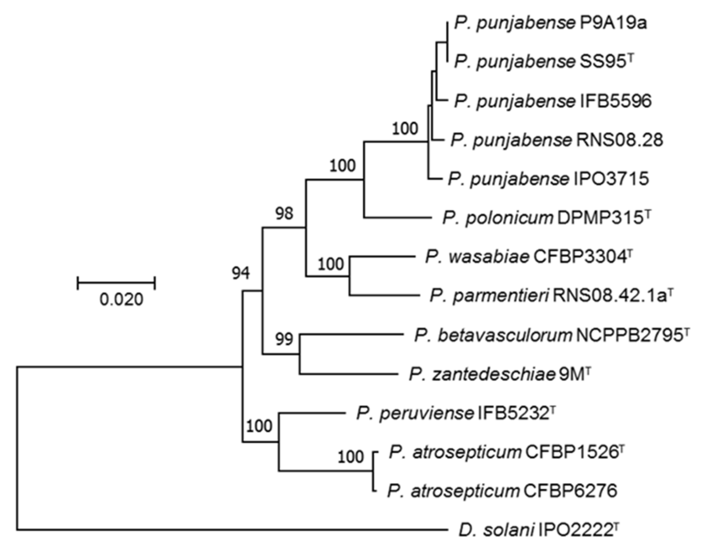
2.2.2. Multi-Locus Sequence Analysis (MLSA)

The sequences of the fourteen housekeeping genes (acnA, fusA, gapA, glyA, groEL, gyrB, mdh, mtlD, purA, recA, rplB, rpoD, rpoS, secY) of P. punjabense candidates and reference type strains, recovered from the corresponding genome sequences, were concatenated, aligned and used to build a phylogenetic tree using the maximum composite likelihood method with MEGA X [51]. Bootstrap values were calculated from 1000 replicate iterations.

2.2.3. Average Nucleotide Identity (ANI), In Silico DNA–DNA Hybridization (DDH) and Single Nucleotide Polymorphism (SNP) Analysis

The average nucleotide identity (ANI) calculator of the EzBioCloud server [52] was used for genome comparison and to confirm the taxonomic assignation of each P. punjabense candidate genome. In addition, the Genome-to-Genome Distance Calculator of the Leibniz Institute DSMZ [53] was used to calculate the DNA–DNA hybridization (DDH) value in order to obtain a second robust parameter for each genome comparison. Then, read mapping of each P. punjabense candidate to the P. punjabense SS95T reference genome was performed. Finally, the variant detection tool of CLC genomics workbench 10.1.1 (https://digitalinsights.qiagen.com accessed on 12 February 2021) was used to estimate the number of SNPs for each P. punjabense candidate, with the P. punjabense SS95T genome as a reference.

2.2.4. BRIG Analysis

A sisual comparison of genome homology was performed using BRIG (BLAST Ring Image Generator) [54]. P. punjabense SS95T was used as the reference genome and was compared to the other genomes of P. punjabense. The analysis was performed with the default settings.

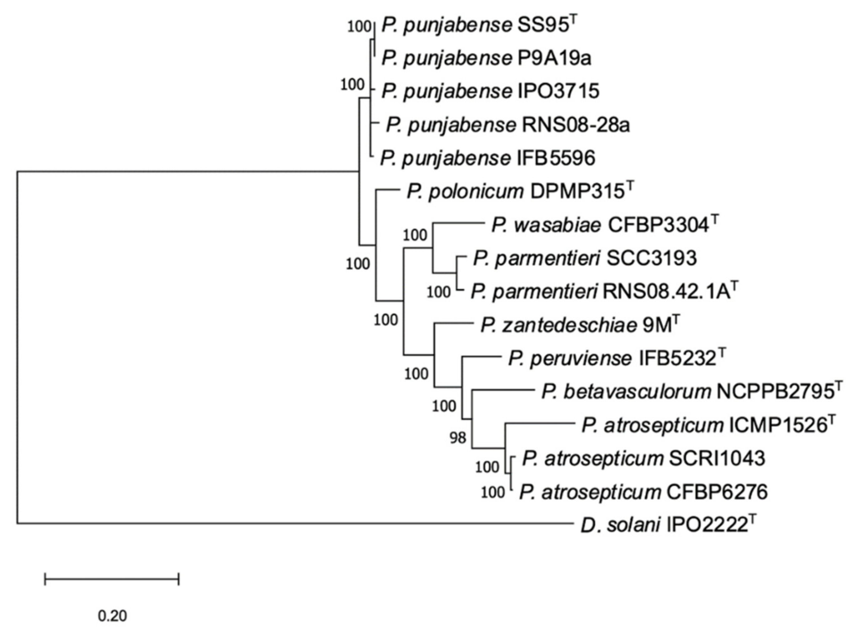
2.3. Core Protein Phylogeny

A core phylogenetic analysis conducted from the protein sequences deduced from the concatenated core genes shared by all the genomes was compared with the clustering obtained in the multi-locus sequence analysis (MLSA). The phylogenomic analysis was performed with the PhyloPhlAn computational pipeline (https://huttenhower.sph.harvard.edu/phylophlan accessed on 18 January 2021), which uses the most conserved universal proteins from full proteomes to extract the phylogenetic signal [55]. The maximum likelihood tree was built based on 399 protein sequences. The core protein analysis was performed using the genomic sequences from the type and reference strains of the most closely related members of the genus Pectobacterium.

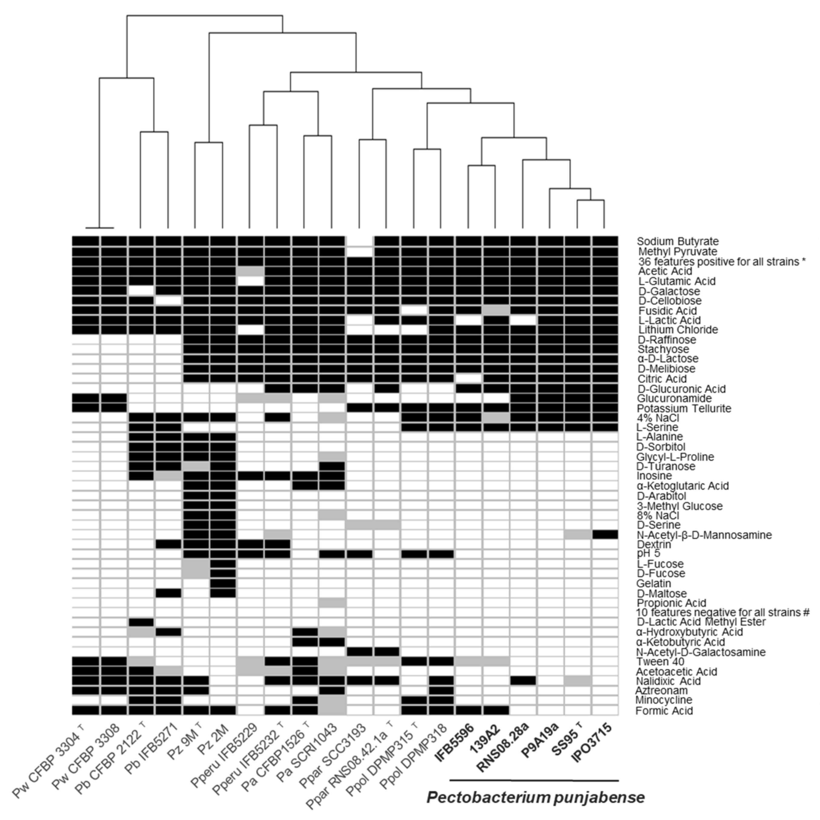
2.4. Carbon Source Utilization Profiles

P. punjabense strains (IFB5596 and 139A2, both co-isolated from the same sample in 1996, as well as RNS0828a, P9A19a and IPO3715) and strains of other closely related species such as P. atrosepticum (SCRI1043, ICMP1526T), P. betavasculorum (CFBP2122T, CFBP5271), P. peruviense (IFB5232T, IFB9229), P. parmentieri (SCC3193, IFB5322), P. wasabiae (CFBP3304T, CFBP3308) and P. zantedeschiae (9MT and 2M) were analyzed for the usage of different carbon sources using GEN III plates, and the results were compared with BIOLOG utilities. The assays were performed according to the manufacturers’ instructions.

2.5. Fatty Acid Methyl Esters (FAME) Composition

The whole-cell derived fatty acids were isolated directly from bacteria growing on TSA plates for 2 days at 28 °C. After saponification, methylation, extraction and washing according to the procedure described by Sasser et al. [56], the obtained fatty acid methyl esters (FAME) were analyzed using a gas chromatography-FID detector (Agilent 7820A) and an Ultra 2-HP capillary column (cross-linked 5% phenyl methyl silicone, 25 m, 0.22 mm id and 0.33 μm film thickness). A standard No. 1200-A solution (MIDI Inc., Newark, DE, USA) was used to calibrate the GC. Qualitative and quantitative analyses of the fatty acids were performed using the TSBA library (ver. 6.2B) and Sherlock Microbial Identification System software (MIDI Inc., Newark, DE, USA).

2.6. Tuber Maceration Assay

Assays were conducted in order to compare the tuber maceration capacity of P. punjabense to that of other Pectobacterium reference strains. In addition to P. punjabense P9A19a, RNS08.28, IPO3715 and SS95T, three P. punjabense isolates RNS16-153-1A, RNS18-61 and RNS18-78 were also used in the tuber maceration assay. These three isolates were collected in 2016 or 2018 from blackleg symptoms during field surveys in France and were identified as P. punjabense based on dnaX and gapA housekeeping gene sequences (Figure S1) in the course of this work. The tuber assay was adapted from previous works, as described in Blin et al. [34]. Inoculum suspensions of each of the tested strains were prepared from overnight cultures in TY broth and calibrated at 108 cfu/mL. Two pipette tips containing 10 µL inoculum suspension each were driven into the flesh of surface-disinfected potato tubers (cv. Bintje). Eight tubers per strain were thus inoculated for a total of sixteen inoculation points per strain. A negative control with 10 µL of NaCl 0.8% was used.
Inoculated potato tubers were incubated at 25 °C in the dark in humid chambers with water-saturated air. After five days, tubers were cut across inoculation points and symptom severity was scored based on a six-class visual scale according to rot extension. To each class, a coefficient was assigned whose value increased with symptom severity (0, 0.2, 0.4, 0.6, 0.8 and 1). A pathological index (from 0 to 100) representative of the aggressiveness of each strain was calculated as follows:
P a t h o l o g i c a l   i n d e x =   ( n u m b e r   o f   t u b e r s   p e r   c l a s s   ×   c l a s s   c o e f f i c i e n t ) T o t a l   o f   t u b e r s × 100
Firstly, the data were analyzed using a Kruskal–Wallis non parametric rank test. Secondly, a pairwise post hoc Tukey test was performed to discriminate strains if significant differences were detected by the Kruskal–Wallis rank test.

2.7. qPCR TaqMan Assay

In order to find “specific regions” present in P. punjabense and absent in strains of the three closest species P. polonicum, P. parmentieri and P. wasabiae, successive Illumina-read mapping was performed with the CLC genomics workbench 10.1.1. In detail, Illumina reads of P. punjabense RNS08.28 were successively mapped (length fraction = 0.5; similarity = 0.8) on the five assembled P. punjabense genomes. Mapping reads were collected and then successively mapped on P. parmentieri RNS08.42.1aT, P. parmentieri WPP163, P. parmentieri SCC3193, P. parmentieri IFB5408, P. parmentieri IFB5427, P. parmentieri IFB5432, P. parmentieri IFB5441, P. parmentieri IFB5485, P. parmentieri IFB5486, P. parmentieri IFB5604, P. parmentieri IFB5619, P. wasabiae CFBP3304T and P. polonicum DPMP315T public genomes. Only the non-mapping reads were retained and de novo assembled to generate P. punjabense-specific contigs (minimal cut-off fixed at 2 kb).
Using these P. punjabense “specific regions”, primers and probes were designed using Primer3 [57]. In silico specificity of candidates was tested by a local blast on all the Pectobacterium genomes of the study, as well as with the Standard Nucleotide BLAST on the NCBI Nucleotide collection database. A qPCR TaqMan assay specific for P. punjabense was developed on a LightCycler96 Instrument real-time PCR system (Roche Life Science). The amplicon has an expected size of 217 bp and is located in a gene coding for a hypothetical protein. Amplification reactions were performed with 10 µL of FastStart Essential DNA Probes Master 2x (reference 06402682001, Roche Life Science), 3 µL of 3.33 µM primers P. punjabense 2b-F TCCTTCAGCCAGAGAACCAG, 3 µL of 3.33 µM primers P. punjabense 2b-R AACAACAATACCGGCAAGTGG, 2 µL of 2 µM probe P. punjabense 2b TGCAGGCCTTGTAACTCCGCT labelled with a 5′ reporter dye (FAM-6-carboxyfluorescein (FAM)) and a 3′-end quencher dye (BHQ1), synthesized by Eurofins Genomics (Ebersberg, Germany). Finally, 2 µL of template DNA was added to a final volume of 20 µL. Cycling parameters were 95 °C for 10 min, 40 cycles of 95 °C for 10 s and 60 °C for 1 min. Taqman assays were performed on a large number of strains to test the specificity of the tool. Pure DNA samples were prepared using the MasterPure™ Complete DNA kit (Lucigen) and diluted at 1 ng/µL. A serial dilution of the P. punjabense SS95T extracted DNA ranging from 2 ng to 2.5 fg was used to determine the sensitivity of the Taqman assay. Each quantity was tested in triplicate.

3. Results

3.1. Identification and Diversity of P. punjabense Candidates

The MLSA phylogenic tree including strains tentatively identified as P. punjabense derived from European collections and type strains from phylogenetically related Pectobacterium species was built from the concatenated sequences of fourteen housekeeping genes extracted from genomes. All P. punjabense candidates grouped together with the type strain P. punjabense SS95. The resulting cluster presented a robust bootstrap value (Figure 1). The core protein phylogeny confirmed the status of P9A19a, RNS08.28, IPO3715 and IFB5596 as P. punjabense strains (Figure 2).
The ANI and DDH values were calculated to consolidate the assignation of the European isolates P9A19a, RNS08.28, IPO3715 and IFB5596 to P. punjabense species. When the ANI and DDH values were calculated between P. punjabense genomes (Table 2), all values were higher than the accepted cut-off value for species delineation: 95 for ANI values and 70 for DDH [58,59]. The ANI and DDH values of P. punjabense isolates ranged from 98.6 to 100 and from 88.6 to 100, respectively. These infra-specific variations highlight the non-clonal character of P. punjabense strains. The ANI and DDH parameters also confirmed that P. polonicum DPMP315 is closely related to P. punjabense strains, with a mean ANI value calculated at 93.9 and a DDH of 54.9. Then, P. parmentieri RNS08.42.1a and P. wasabiae CFBP3304 are the two other strains closest to the P. punjabense strains with ANI values ranging between 91.0 and 91.5 and DDH values between 43.3 and 44.4.
In addition, a variant detection analysis to estimate the number of SNPs between each P. punjabense strain and the type strain SS95 yielded approximately 35,000 SNPs for RNS08.28 and IPO3715. For IFB5596, approximately 33,000 SNPs were found. Surprisingly, no SNP was found for P9A19a, suggesting a clonal origin of these Pakistani and French isolates. Finally, pairwise ANI values, SNP analysis and also BRIG analysis used for genome comparison (Figure S2) confirmed that the four European P. punjabense strains are genetically different.

3.2. Physiological and Structural Characteristics

The physiological and biochemical features of all the P. punjabense strains were very similar in the BIOLOG GEN III plates system (Figure 3). None of the single carbon sources alone could discriminate P. punjabense from the other Pectobacterium species. Noticeably, all P. punjabense strains assimilated D-glucuronic acid and L-serine as the sole carbon source, while most other strains tested could not. Only P. punjabense, P. parmentieri and P. polonicum grew in the presence of the inorganic compound potassium tellurite. P. punjabense strains displayed a high sensitivity for different antibiotic compounds such as nalidixic acid, aztreonam, minocycline and formic acid compared with the other species. In particular, the two strains of P. betavasculorum were able to grow in the presence of each of these four antibiotic compounds.
Fatty acid methyl esterase (FAME) analysis revealed the presence of in total 28 fatty acids in the biomass of strains analyzed. Among these, 22 were detected in P. punjabense strains, from which 18 were present in all of the analyzed P. punjabense strains. Six out of 28 detected fatty acids were absent in this species. The most prevalent are the following fatty acids: 12:0, 16:0, 17:1 ω8c, 17:0, 12:0 aldehyde/unknown 10.928/16:1 iso I/14:0 3OH, 16:1 ω7c/16:1 ω6c, 18:1 ω7c/18:1 ω6c (Table S1). Overall, the fatty acid contents of P. punjabense strains were homogenous and similar to those of other Pectobacterium strains. However, fatty acids 17:0 cyclo, 19:0 iso and 19:0 cyclo ω8c were absent in the P. punjabense strains but present in the most closely related P. polonicum DPMP315 strain.

3.3. Occurrence of P. Punjabense in Collections
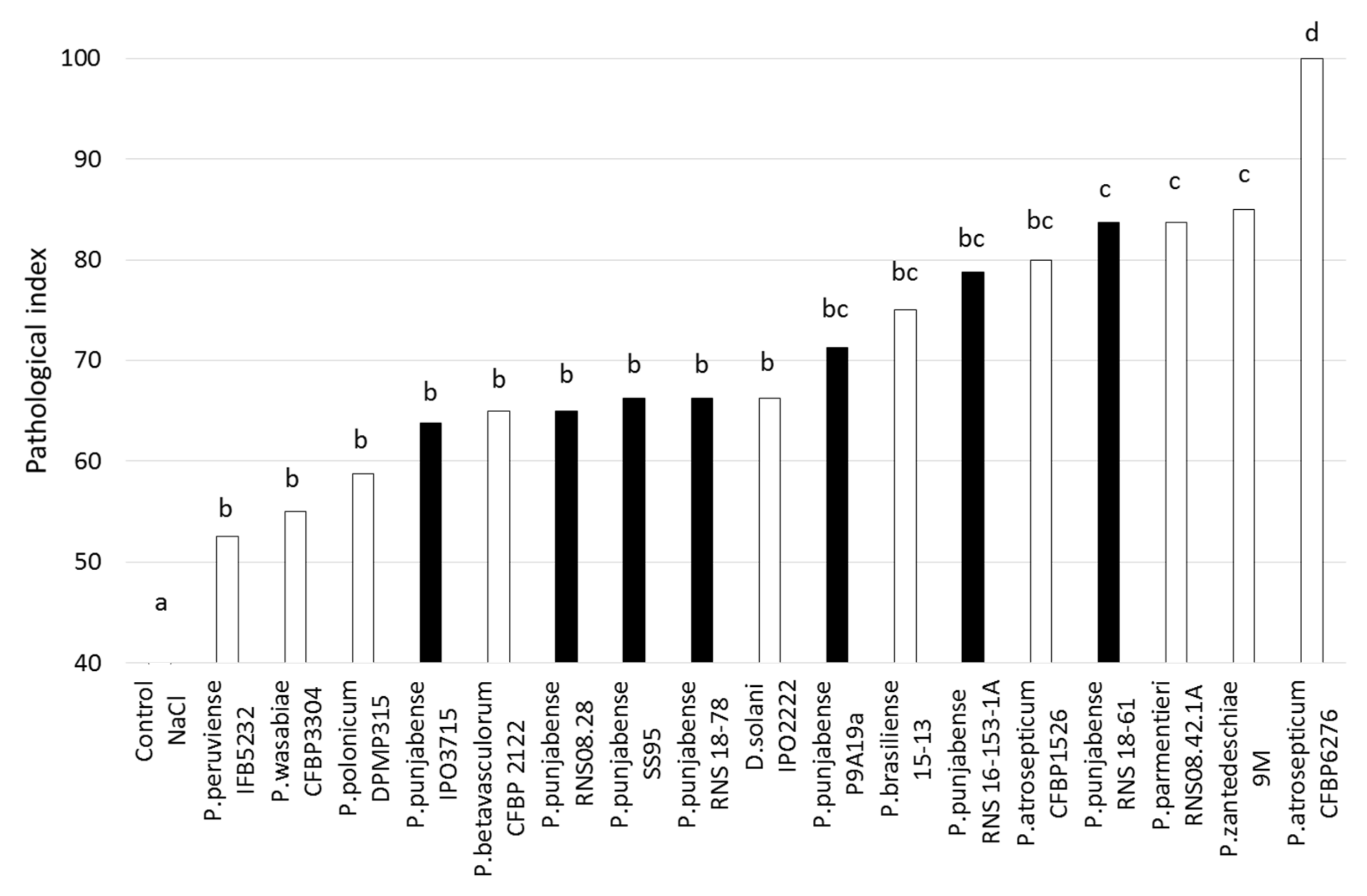
The three bacterial collections used in our study gather SRP collected over many years from different sources, although strains from blackleg=diseased plants are overrepresented. For collection, a CVP medium has been used that allows the isolation of all SRP with largely the same efficiency [50]. We therefore could make an estimate on the isolation incidence of P. punjabense relative to other SRP strains (Table 3). For example, over the five sampling campaigns between 2015 and 2019, 1663 strains were deposited in the RNS collection, including only four P. punjabense strains, all collected from different fields. In the IPO collection, among 1012 SRP characterized between 1963 and 2020, only one P. punjabense strain was identified. From 2031 strains sampled in Poland between 1996 and 2014 (including 1500 strains isolated in 1996), four P. punjabense isolates were identified, but all were isolated from the same blackleg diseased plant, and they showed identical housekeeping genes sequences.

3.4. Aggressiveness

The P. punjabense strains varied in their tuber maceration capacity (Figure 4). For strains IPO3715, RNS08-28, SS95T and RNS18-78, the pathological index was significantly lower than that of RNS 18-61, which had an index similar to that of the reference strain P. zantedeschiae 9MT, previously described as a strain with a high maceration capacity [18]. Strains P9A19a and RNS16-153-1a presented a pathological index of 71 and 79, respectively, indicating an intermediate aggressiveness level compared to the other strains of the panel. Finally, none of the P. punjabense strains tested showed a potential maceration capacity equivalent to the referent aggressive strain P. atrosepticum CFBP6276 under the conditions of the experiment.

3.5. Development of a qPCR TaqMan Assay Specific for P. Punjabense

The qPCR Taqman 2b assay gave a strong signal (Ct values ranging from 17.58 to 20.29) with DNA extracted from the eight P. punjabense strains (the five strains used for the genomic work and the three additional isolates included in the pathogenicity assay), but not with DNA from 59 different Pectobacterium and Dickeya species, all calibrated at 1 ng/µL (Table 4).
To determine the threshold level, a ten-fold serial dilution of P. punjabense SS95T DNA ranging from 2 ng to 2.5 fg was tested. The detection threshold of the qPCR Taqman 2b was obtained at 20 fg of P. punjabense DNA, with a mean detection value of 34.52 Ct and a standard deviation of 0.98 (Table 5). The analysis of raw curves generated by the LightCycler96 Instrument real-time PCR system allowed fixing the efficiency of the qPCR reaction at 1.99, which is very close to the expected theoretical value of 2.00.

4. Discussion

The recent development of next generation sequencing technologies considerably improved knowledge of bacterial genomics including those of the genus Pectobacterium. It became evident that this genus is highly diverse, and NGS analysis also led to improved identification of strains in culture collections and the description of several new Pectobacterium species.
This is the case of P. punjabense species, whose type strain SS95 was isolated from blackleg diseased potatoes in Pakistan in 2017. Until now, SS95 was the only P. punjabense strain described [45]. By exploring strains of different European work collections on the basis of sequences of housekeeping genes homology, we uncovered seven strains assignable to P. punjabense, and the genomes of four of them were sequenced. MLSA (Figure 1) and core phylogenic protein analysis (Figure 2), as well as ANI and DDH genomic parameters (Table 2), confirmed that these four new strains indeed belong to P. punjabense. The P. punjabense strains could not be distinguished from other species tested on the basis of the fatty acid profile (Table S1).
The strains RNS08-28 and P9A19a were isolated in France in 2008 and 2015, respectively, while strain IPO3715 was isolated in the Netherlands in 2013. The oldest strain in our sample, IFB5596, isolated in 1996, comes from a Polish potato field. These data provide interesting information on the prevalence of this species. First, P. punjabense is present in Europe and is not limited to Pakistan, where it was initially described. Furthermore, the Polish strain IFB5596 indicates that P. punjabense has been present in Europe for at least 25 years but has always gone unnoticed due to a lack of specific detection tools. All strains identified as P. punjabense were isolated from blackleg diseased plants, but they remain very rare among isolated strains; in the three collections analyzed, the isolation frequency of P. punjabense strains varied from 0.05 to 0.24% of the total number of SRP strains isolated during the sampling period (Table 3).
The genomic data of the P. punjabense strains also provided valuable information on the diversity within this species. In particular, the ANI comparisons between P. punjabense strains showed values ranging from 98.6 to 100.0 (Table 2), which is comparable to those observed in the closely related species P. parmentieri, with values ranging from 98.92 to 99.97 [60]. Surprisingly, the two P. punjabense strains SS95T and P9A19a showed exactly the same genomic sequences (ANI of 100.0; DDH of 100.0; no SNP found) although they were isolated in two distant countries and in two different years. In addition, no seed potatoes of the cultivar from which strain P9A19a was isolated are exported to Pakistan. There is no information on the cultivar from which the Pakistani strain SS95 was isolated, but there is no trade of Pakistani seed potatoes into Europe. As a result, no available data link these two strains to a possible common event. If we exclude the comparison between SS95T and P9A19a, the average ANI for the other P. punjabense comparisons is 98.8 ± 0.1 and the mean DDH value is 89.3 ± 0.8. The relatively high genomic diversity is consistent with the finding that the pathogen has been present for a long time in Europe [61]. This hypothesis is reinforced by the high number of SNPs observed when comparing the different strains of P. punjabense identified to the type strain (more than 30,000). Therefore, the situation of P. punjabense over time is clearly distinct from that of the emergence of the clonal pathogen D. solani in European potato fields [33], where less than 100 SNPs were observed between two D. solani genomes [62].
With our current knowledge, the question of the P. punjabense reservoir remains unresolved. Indeed, the low frequency of P. punjabense found among isolates collected from blackleg symptoms, combined with the genomic diversity observed, suggests that P. punjabense is present in other niches beside potato plants, but no reference is available to support this hypothesis. In recent surveys of water from French rivers, no P. punjabense have been isolated [63], while P. versatile [10] or P. aquaticum [17] were recovered. P. punjabense was also not found within the French Collection for Plant-associated Bacteria (CIRM-CFBP), which includes over 265 Pectobacterium strains isolated from 1944 to 2020 and which represent isolates from a high diversity of plants [28]. Furthermore, the low incidence of P. punjabense strains sampled from blackleg-diseased plants in comparison to other species of Pectobacterium, such as P. brasiliense, P. parmentieri or P. atrosepticum, indicates that the potato plant is probably not the preferred host for this species.
The P. punjabense isolates tested were able to macerate potato tubers, indicating that they can cause soft rot during storage of tubers. For most strains investigated in this study, the ability of P. punjabense to cause potato blackleg has not been determined yet, but in a field experiment with vacuum-infiltrated seed tubers, strain IPO3715 did not result in diseased plants, whereas in the same experiment P. brasiliense showed a high disease incidence of 80% [64]. Therefore, further investigations are needed to assess the host range of P. punjabense.
Testing phenotypic characteristics using BIOLOG plates revealed that the P. punjabense strains have a lower capacity to grow in the presence of most of the antibiotic compounds tested, compared to the other pectinolytic bacteria of the panel (Figure 3). These data suggest that in complex environments, such as field soil where microbial competition is intense and phytosanitary products are widely used, P. punjabense could be less competitive [65]. This could partly explain the small number of strains collected in fields.
To date, no specific molecular tools are available to detect P. punjabense [37,38]. They showed a negative or a weak signal around the expected value at 434 bp and additional nonspecific bands with the generic PCR Y1-Y2 assay widely used to detect Pectobacterium sp. [35]. In order to clarify this, the alignment of the pectate lyase sequences of the strains of the panel revealed six mismatches between the nucleotides of the Y1 primer and the corresponding pectate lyase region in P. punjabense strains (Figure S3); this probably explains the negative PCR result observed with the Y1-Y2 primers. This information could also partly explain why P. punjabense strains remained poorly identified, sometimes for many years. In case of negative amplification obtained with the generic Y1-Y2 primers, some isolates have possibly been assimilated to contaminants. The Taqman 2b developed in this study has shown both its specificity and its sensitivity towards the P. punjabense species and will be helpful for rapid identification, seed testing, surveys and studies on epidemiology and disease management.
Overall, this study provides a first comprehensive look at P. punjabense in Europe and provides also information on its diversity. The data have shown that it has been present for almost 25 years, but the low frequency of isolation combined with the lack of specific identification tools have prevented its recognition. The Taqman 2b assay developed in this study will enable further studies in the potato ecosystem, including the capacity to cause blackleg and the host range.

Supplementary Materials

The following are available online at https://www.mdpi.com/article/10.3390/microorganisms9040781/s1, Figure S1: The gapA molecular phylogenetic analysis for taxonomic assignation of P. punjabense candidates. The evolutionary history was inferred by using the maximum likelihood method. There were a total of 846 positions in the final dataset. Bootstrap values were calculated from 1000 replicate iterations. Evolutionary analyses were conducted in MEGA X, Figure S2: BLAST comparisons of four P. punjabense genomes sequenced against the P. punjabense SS95T genome performed with the BRIG application, Figure S3: Y1 sequences alignment with different Pectobacterium strains. Nucleotide represented in grey corresponds to that of the reference sequence (primer Y1). Nucleotide in bold red represents a mismatch with that of the reference sequence, Table S1: The percentage of total amount of fatty acids detected in the cells of five P. punjabense strains (SS95T, IFB5596, RNS08.28, P9A19a, IPO3715), P. polonicum DPMP315T, P. wasabiae (CFBP3304T, CFBP3308), P. parmentieri (RNS08.42.1AT, SCC3193), P. atrosepticum (CFBP1526T, CFBP6276), P. peruviense (IFB5232T), P. zantedeschiae (9MT) and P. betavasculorum (CFBP2122T).

Author Contributions

Conceptualization, V.H.; methodology, J.C., A.L., M.W. and V.H.; formal analysis, J.C., M.W.; investigation, J.C., A.L., M.W. and K.W.; resources, A.L., M.W., P.D., J.v.d.W., D.A., D.F. and V.H.; data curation, J.C., M.W., J.v.d.W.; writing—original draft preparation, J.C; writing—review and editing, J.C., M.W., J.v.d.W., D.A., D.F. and V.H.; visualization, J.C. and M.W.; supervision, V.H.; project administration, V.H. All authors have read and agreed to the published version of the manuscript.

Funding

This research was funded by the French Federation of Seed Potato Growers (FN3PT) and by the National Science Centre, Poland, project Opus 9 [2015/17/B/NZ9/01730].

Data Availability Statement

Data presented in this study are available in the present manuscript and its supplementary information file.

Conflicts of Interest

The authors declare no conflict of interest.

References

  1. Ma, B.; Hibbing, M.E.; Kim, H.-S.; Reedy, R.M.; Yedidia, I.; Breuer, J.; Breuer, J.; Glasner, J.D.; Perna, N.T.; Kelman, A.; et al. Host range and molecular phylogenies of the soft rot enterobacterial genera Pectobacterium and Dickeya. Phytopathology 2007, 97, 1150–1163. [Google Scholar] [CrossRef] [PubMed] [Green Version]
  2. Adeolu, M.; Alnajar, S.; Naushad, S.; Gupta, R.S. Genome-based phylogeny and taxonomy of the “Enterobacteriales”: Proposal for Enterobacterales ord. nov. divided into the families Enterobacteriaceae, Erwiniaceae fam. nov., Pectobacteriaceae fam. nov., Yersiniaceae fam. nov., Hafniaceae fam. nov., Morganellaceae fam. nov., and Budviciaceae fam. nov. Int. J. Syst. Evol. Microbiol. 2016, 66, 5575–5599. [Google Scholar]
  3. Mansfield, J.; Genin, S.; Magori, S.; Citovsky, V.; Sriariyanum, M.; Ronald, P.; Dow, M.; Verdier, V.; Beer, S.V.; Machado, M.A.; et al. Top 10 plant pathogenic bacteria in molecular plant pathology. Mol. Plant Pathol. 2012, 13, 614–629. [Google Scholar] [CrossRef] [PubMed] [Green Version]
  4. Barras, F.; van Gijsegem, F.; Chatterjee, A.K. Extracellular enzymes and pathogenesis of soft-rot Erwinia. Annu. Rev. Phytopathol. 1994, 32, 201–234. [Google Scholar] [CrossRef]
  5. Toth, I.K.; Bell, K.S.; Holeva, M.C.; Birch, P.R. Soft rot erwiniae: From genes to genomes. Mol. Plant Pathol. 2003, 4, 17–30. [Google Scholar] [CrossRef]
  6. Pérombelon, M.C.M. Potato diseases caused by soft rot erwinias: An overview of pathogenesis. Plant Pathol. 2002, 51, 1–12. [Google Scholar] [CrossRef]
  7. Charkowski, A.O. The changing face of bacterial soft-rot diseases. Annu. Rev. Phytopathol. 2018, 56, 269–288. [Google Scholar] [CrossRef]
  8. Czajkowski, R.; Pérombelon, M.C.M.; van Veen, J.A.; van der Wolf, J.M. Control of blackleg and tuber soft rot of potato caused by Pectobacterium and Dickeya species: A review. Plant Pathol. 2011, 60, 999–1013. [Google Scholar] [CrossRef]
  9. Pitman, A.R.; Wright, P.J.; Galbraith, M.D.; Harrow, S.A. Biochemical and genetic diversity of pectolytic Enterobacteria causing soft rot disease of potatoes in New Zealand. Australas. Plant Pathol. 2008, 37, 559–568. [Google Scholar] [CrossRef]
  10. Portier, P.; Pédron, J.; Taghouti, G.; Fischer-Le Saux, M.; Caullireau, E.; Bertrand, C.; Laurent, A.; Chawki, K.; Oulghazi, S.; Moumni, M.; et al. Elevation of Pectobacterium carotovorum subsp. odoriferum to species level as Pectobacterium odoriferum sp. nov., proposal of Pectobacterium brasiliense sp. nov. and Pectobacterium actinidiae sp. nov., emended description of Pectobacterium carotovorum and description of Pectobacterium versatile sp. nov., isolated from streams and symptoms on diverse plants. Int. J. Syst. Evol. Microbiol. 2019, 69, 3207–3216. [Google Scholar] [PubMed]
  11. Hugouvieux-Cotte-Pattat, N.; Brochier-Armanet, C.; Flandrois, J.P.; Reverchon, S. Dickeya poaceiphila sp. nov., a plant pathogenic bacterium isolated from sugar cane (Saccharum officinarum). Int. J. Syst. Evol. Microbiol. 2020, 70, 4508–4514. [Google Scholar] [CrossRef]
  12. Pasanen, M.; Waleron, M.; Schott, T.; Cleenwerck, I.; Misztak, A.; Waleron, K.; Pritchard, L.; Bakr, R.; Degefu, Y.; van der Wolf, J.; et al. Pectobacterium parvum sp. nov., having a Salmonella SPI-1-like Type III secretion system and low virulence. Int. J. Syst. Evol. Microbiol. 2020, 70, 2440–2448. [Google Scholar]
  13. Khayi, S.; Cigna, J.; Chong, T.M.; Quêtu-Laurent, A.; Chan, K.-G.; Hélias, V.; Faure, D. Transfer of the potato plant isolates of Pectobacterium wasabiae to Pectobacterium parmentieri sp. nov. Int. J. Syst. Evol. Microbiol. 2016, 66, 5379–5383. [Google Scholar] [CrossRef]
  14. Waleron, M.; Misztak, A.; Waleron, M.; Franczuk, M.; Wielgomas, B.; Waleron, K. Transfer of Pectobacterium carotovorum subsp. carotovorum strains isolated from potatoes grown at high altitudes to Pectobacterium peruviense sp. nov. Syst. Appl. Microbiol. 2018, 41, 85–93. [Google Scholar]
  15. Dees, M.W.; Lysoe, E.; Rossmann, S.; Perminow, J.; Brurberg, M.B. Pectobacterium polaris sp. nov., isolated from potato (Solanum tuberosum). Int. J. Syst. Evol. Microbiol. 2017, 67, 5222–5229. [Google Scholar] [CrossRef]
  16. Waleron, M.; Misztak, A.; Waleron, M.; Franczuk, M.; Jonca, J.; Wielgomas, B.; Mikiciński, A.; Popović, T.; Waleron, K. Pectobacterium zantedeschiae sp. nov. a new species of a soft rot pathogen isolated from Calla lily (Zantedeschia spp.). Syst. Appl. Microbiol. 2019, 42, 275–283. [Google Scholar] [CrossRef]
  17. Pédron, J.; Bertrand, C.; Taghouti, G.; Portier, P.; Barny, M.A. Pectobacterium aquaticum sp. nov., isolated from waterways. Int. J. Syst. Evol. Microbiol. 2019, 69, 745–751. [Google Scholar] [CrossRef] [PubMed]
  18. Waleron, M.; Misztak, A.; Waleron, M.; Jonca, J.; Furmaniak, M.; Waleron, K. Pectobacterium polonicum sp. nov. isolated from vegetable fields. Int. J. Syst. Evol. Microbiol. 2019, 69, 1751–1759. [Google Scholar] [CrossRef] [PubMed]
  19. Oulghazi, S.; Pédron, J.; Cigna, J.; Lau, Y.Y.; Moumni, M.; Van Gijsegem, F.; Chan, K.G.; Faure, D. Dickeya undicola sp. nov., a novel species for pectinolytic isolates from surface waters in Europe and Asia. Int. J. Syst. Evol. Microbiol. 2019, 69, 2440–2444. [Google Scholar] [CrossRef]
  20. Hugouvieux-Cotte-Pattat, N.; Jacot-des-Combes, C.; Briolay, J. Dickeya lacustris sp. nov., a water-living pectinolytic bacterium isolated from lakes in France. Int. J. Syst. Evol. Microbiol. 2019, 69, 721–726. [Google Scholar] [CrossRef] [PubMed]
  21. Perombelon, M.C.M.; Kelman, A. Ecology of the Soft Rot Erwinias. Annu. Rev. Phytopathol. 1980, 18, 361–387. [Google Scholar] [CrossRef]
  22. Pitman, A.R.; Harrow, S.A.; Visnovsky, S.B. Genetic characterization of Pectobacterium wasabiae causing soft rot disease of potato in New Zealand. Eur. J. Plant Pathol. 2010, 126, 423–435. [Google Scholar] [CrossRef]
  23. Gardan, L.; Gouy, C.; Christen, R.; Samson, R. Elevation of three subspecies of Pectobacterium carotovorum to species level: Pectobacterium atrosepticum sp. nov., Pectobacterium betavasculorum sp. nov. and Pectobacterium wasabiae sp. nov. Int. J. Syst. Evol. Microbiol. 2003, 53, 381–391. [Google Scholar] [CrossRef]
  24. De Boer, S.H.; Li, X.; Ward, L. Pectobacterium spp. associated with bacterial stem rot syndrome of potato in Canada. Phytopathology 2012, 102, 937–947. [Google Scholar] [CrossRef] [Green Version]
  25. De Werra, P.; Bussereau, F.; Keiser, A. First report of potato blackleg caused by Pectobacterium carotovorum subsp. brasiliense in Switzerland. Plant Dis. 2015, 99, 551. [Google Scholar] [CrossRef]
  26. Waleron, M.; Waleron, K.; Lojkowska, E. Occurrence of Pectobacterium wasabiae in potato field samples. Eur. J. Plant Pathol. 2013, 137, 149–158. [Google Scholar] [CrossRef] [Green Version]
  27. Zoledowska, S.; Motyka, A.; Zukowska, D.; Sledz, W.; Lojkowska, E. Population structure and biodiversity of Pectobacterium parmentieri isolated from potato fields in temperate climate. Plant Dis. 2018, 102, 154–164. [Google Scholar] [CrossRef] [PubMed] [Green Version]
  28. Portier, P.; Pédron, J.; Taghouti, G.; Dutrieux, C.; Barny, M.-A. Updated Taxonomy of Pectobacterium Genus in the CIRM-CFBP Bacterial Collection: When Newly Described Species Reveal “Old” Endemic Population. Microorganisms 2020, 8, 1441. [Google Scholar] [CrossRef] [PubMed]
  29. Waleron, M.; Misztak, A.; Jońca, J.; Furmaniak, M.; Waleron, M.M.; Waleron, K. First report of ‘Candidatus Pectobacterium maceratum’ causing soft rot of potato in Poland. Plant Dis. 2019, 103, 1409. [Google Scholar] [CrossRef]
  30. Waleron, M.; Misztak, A.; Jońca, J.; Waleron, K. First Report of Pectobacterium polaris Causing Soft Rot of Potato in Poland. Plant Dis. 2019, 103, 144. [Google Scholar] [CrossRef]
  31. Lebecka, R.; Flis, B.; Murawska, Z. Comparison of temperature effects on the in vitro growth and disease development in potato tubers inoculated with bacteria Pectobacterium atrosepticum, P. carotovorum subsp. carotovorum and Dickeya solani. J. Phytopathol. 2018, 166, 654–662. [Google Scholar] [CrossRef]
  32. Samson, R.; Legendre, J.B.; Christen, R.; Fischer-Le Saux, M.; Achouak, W.; Garden, L. Transfer of Pectobacterium chrysanthemi and Brenneria paradisiaca to the genus Dickeya gen. nov. as Dickeya chrysanthemi comb. nov. and Dickeya paradisiaca comb. nov. and delineation of four novel species, Dick. Int. J. Syst. Evol. Microbiol. 2005, 55, 1415–1427. [Google Scholar] [CrossRef]
  33. Toth, I.K.; van der Wolf, J.M.; Saddler, G.; Lojkowska, E.; Hélias, V.; Pirhonen, M.; Tsror Lahkim, L.; Elphinstone, J.G. Dickeya species: An emerging problem for potato production in Europe. J. Plant Pathol. 2011, 60, 385–399. [Google Scholar] [CrossRef]
  34. Blin, P.; Robic, K.; Khayi, S.; Cigna, J.; Munier, E.; Dewaegeneire, P.; Laurent, A.; Jaszczyszyn, Y.; Hong, K.W.; Chan, K.G.; et al. Pattern and causes of the establishment of the invasive bacterial potato pathogen Dickeya solani and of the maintenance of the resident pathogen D. dianthicola. Mol. Ecol. 2020. [Google Scholar] [CrossRef]
  35. Darrasse, A.; Priou, S.; Kotoujansky, A.; Bertheau, Y. PCR and restriction fragment length polymorphism of a pel gene as a tool to identify Erwinia carotovora in relation to potato diseases. Appl. Environ. Microbiol. 1994, 60, 1437–1443. [Google Scholar] [CrossRef] [Green Version]
  36. Nassar, A.; Darrasse, A.; Lemattre, M.; Kotoujansky, A.; Dervin, C.; Vedel, R.; Bertheau, Y. Characterization of Erwinia chrysanthemi by pectinolytic isozyme polymorphism and restriction fragment length polymorphism analysis of PCR- amplified fragments of pel genes. Appl. Environ. Microbiol. 1996, 62, 2228–2235. [Google Scholar] [CrossRef] [PubMed] [Green Version]
  37. Czajkowski, R.; Pérombelon, M.C.M.; Jafra, S.; Lojkowska, E.; Potrykus, M.; van der Wolf, J.M.; Sledz, W. Detection, identification and differentiation of Pectobacterium and Dickeya species causing potato blackleg and tuber soft rot: A review. Ann. Appl. Biol. 2015, 166, 18–38. [Google Scholar] [CrossRef] [PubMed] [Green Version]
  38. Van der Wolf, J.M.; Cahill, G.; Van Gijsegem, F.; Hélias, V.; Humphris, S.; Li, X.S.; Lojkowska, E.; Pritchard, L. Isolation, Detection and Characterization of Pectobacterium and Dickeya Species. In Plant Diseases Caused by Dickeya and Pectobacterium Species, 1st ed.; Van Gijsegem, F., van der Wolf, J.M., Toth, I.K., Eds.; Springer: Cham, Switzerland, 2021; pp. 149–173. [Google Scholar]
  39. Frechon, D.; Exbrayat, P.; Helias, V.; Hyman, L.J.; Jouan, B.; Llop, P.; Lopez, M.M.; Payet, N.; Perombelon, M.C.M.; Toth, I.K.; et al. Evaluation of a PCR kit for the detection of Erwinia carotovora subsp. atroseptica on potato tubers. Potato Res. 1998, 41, 163–173. [Google Scholar] [CrossRef]
  40. Pritchard, L.; Humphris, S.; Saddler, G.S.; Parkinson, N.M.; Bertrand, V.; Elphinstone, J.G.; Toth, I.K. Detection of phytopathogens of the genus Dickeya using a PCR primer prediction pipeline for draft bacterial genome sequences. Plant Pathol. 2012, 62, 587–596. [Google Scholar] [CrossRef]
  41. Kim, M.H.; Cho, M.S.; Kim, B.K.; Choi, H.J.; Hahn, J.H.; Kim, C.K.; Kang, M.J. Quantitative real-time polymerase chain reaction assay for detection of Pectobacterium wasabiae using YD repeat protein gene-based primers. Plant Dis. 2012, 96, 253–257. [Google Scholar] [CrossRef] [PubMed] [Green Version]
  42. van der Wolf, J.M.; de Haan, E.G.; Kastelein, P.; Krijger, M.; de Haas, B.H.; Velvis, H.; Mendes, O.; Kooman-Gersmann, M.; van der Zouwen, P.S. Virulence of Pectobacterium carotovorum subsp. brasiliense on potato compared with that of other Pectobacterium and Dickeya species under climatic conditions prevailing in the Netherlands. Plant Pathol. 2017, 66, 571–583. [Google Scholar]
  43. Duarte, V.; de Boer, S.H.; Ward, L.J.; de Oliveira, A.M. Characterization of atypical Erwinia carotovora strains causing blackleg of potato in Brazil. J. Appl. Microbiol. 2004, 96, 535–545. [Google Scholar] [CrossRef] [PubMed]
  44. Van der Wolf, J.M.; de Haas, B.H.; van Hoof, R.; de Haan, E.G.; van den Bovenkamp, G.W. Development and evaluation of Taqman assays for the differentiation of Dickeya (sub)species. Eur. J. Plant Pathol. 2014, 138, 695–709. [Google Scholar] [CrossRef]
  45. Sarfraz, S.; Riaz, K.; Oulghazi, S.; Cigna, J.; Sahi, S.T.; Khan, S.H.; Faure, D. Pectobacterium punjabense sp. nov., isolated from blackleg symptoms of potato plants in Pakistan. Int. J. Syst. Evol. Microbiol. 2018, 68, 3551–3556. [Google Scholar] [CrossRef] [PubMed]
  46. Cigna, J.; Dewaegeneire, P.; Beury, A.; Gobert, V.; Faure, D. A gapA PCR-sequencing assay for Identifying the Dickeya and Pectobacterium potato pathogens. Plant Dis. 2017, 101, 1278–1282. [Google Scholar] [CrossRef] [PubMed] [Green Version]
  47. Chawki, K.; Quêtu-Laurent, A.A.; Taghouti, G.; Caullireau, E.; Fischer-Le Saux, M.; Le Hingrat, Y.; Andrivon, D.; Portier, P.; Helias, V. The Pectobacterium complex: Diversity and phylogeny. Phytopathology 2018, 108, S1.307. [Google Scholar]
  48. Waleron, M.; Waleron, K.; Podhajska, A.J.; Lojkowska, E. Genotyping of bacteria belonging to the former Erwinia genus by PCR-RFLP analysis of a recA gene fragment. Microbiology 2002, 148, 583–595. [Google Scholar] [CrossRef] [PubMed] [Green Version]
  49. Waleron, M.; Waleron, K.; Geider, K.; Lojkowska, E. Application of RFLP analysis of recA, gyrA and rpoS gene fragments for rapid differentiation of Erwinia amylovora from Erwinia strains isolated in Korea and Japan. Eur. J. Plant Pathol. 2008, 121, 161–172. [Google Scholar] [CrossRef]
  50. Hélias, V.; Hamon, P.; Huchet, E.; van der Wolf, J.M.; Andrivon, D. Two new effective semiselective crystal violet pectate media for isolation of Pectobacterium and Dickeya. Plant Pathol. 2012, 61, 339–345. [Google Scholar] [CrossRef]
  51. Kumar, S.; Stecher, G.; Li, M.; Knyaz, C.; Tamura, K. MEGA X: Molecular Evolutionary Genetics Analysis across computing platforms. Mol. Biol. Evol. 2018, 35, 1547–1549. [Google Scholar] [CrossRef]
  52. Yoon, S.H.; Ha, S.M.; Lim, J.M.; Kwon, S.J.; Chun, J. A large-scale evaluation of algorithms to calculate average nucleotide identity. Antonie van Leeuwenhoek 2017, 110, 1281–1286. [Google Scholar] [CrossRef]
  53. Meier-Kolthoff, J.P.; Auch, A.F.; Klenk, H.P.; Göker, M. Genome sequence-based species delimitation with confidence intervals and improved distance functions. BMC Bioinform. 2013, 14, 60. [Google Scholar] [CrossRef] [Green Version]
  54. Alikhan, N.F.; Petty, N.K.; Ben Zakour, N.L.; Beatson, S.A. BLAST Ring Image Generator (BRIG): Simple prokaryote genome comparisons. BMC Genom. 2011, 12, 402. [Google Scholar] [CrossRef] [PubMed] [Green Version]
  55. Segata, N.; Börnigen, D.; Morgan, X.C.; Huttenhower, C. PhyloPhlAn is a new method for improved phylogenetic and taxonomic placement of microbes. Nat. Commun. 2013, 4, 2304. [Google Scholar] [CrossRef] [PubMed]
  56. Sasser, M. Bacterial Identification by Gas Chromatographic Analysis of Fatty Acid Methyl Esters (GC-FAME); Technical Note #101; MIDI Inc.: Newark, DE, USA, 1990. [Google Scholar]
  57. Untergasser, A.; Cutcutache, I.; Koressaar, T.; Ye, J.; Faircloth, B.C.; Remm, M.; Rozen, S.G. Primer3—New capabilities and interfaces. Nucleic Acids Res. 2012, 40, e115. [Google Scholar] [CrossRef] [PubMed] [Green Version]
  58. Kim, M.; Oh, H.S.; Park, S.C.; Chun, J. Towards a taxonomic coherence between average nucleotide identity and 16S rRNA gene sequence similarity for species demarcation of prokaryotes. Int. J. Syst. Evol. Microbiol. 2014, 64, 346–351. [Google Scholar] [CrossRef] [PubMed]
  59. Auch, A.F.; von Jan, M.; Klenk, H.P.; Göker, M. Digital DNA-DNA hybridization for microbial species delineation by means of genome-to-genome sequence comparison. Stand Genom. Sci. 2010, 2, 117–134. [Google Scholar] [CrossRef] [PubMed] [Green Version]
  60. Zoledowska, S.; Motyka-Pomagruk, A.; Sledz, W.; Mengoni, A.; Lojkowska, E. High genomic variability in the plant pathogenic bacterium Pectobacterium parmentieri deciphered from de novo assembled complete genomes. BMC Genom. 2018, 19, 751. [Google Scholar] [CrossRef] [PubMed] [Green Version]
  61. Achtman, M.; Wagner, M. Microbial diversity and the genetic nature of microbial species. Nat. Rev. Microbiol. 2008, 6, 431–440. [Google Scholar] [CrossRef]
  62. Khayi, S.; Blin, P.; Pédron, J.; Chong, T.M.; Chan, K.G.; Moumni, M.; Hélias, V.; Van Gijsegem, F.; Faure, D. Population genomics reveals additive and replacing horizontal gene transfers in the emerging pathogen Dickeya solani. BMC Genom. 2015, 16, 788. [Google Scholar] [CrossRef] [PubMed] [Green Version]
  63. Barny, M.A.; (Institute of Ecology and Environmental Sciences, Sorbonne University, IEES, Paris, France). Personal communication, 2020.
  64. Vreeburg, R.; (Netherlands General Inspection Service, NAK, Emmeloord, The Netherlands). Personal communication, 2020.
  65. Hibbing, M.E.; Fuqua, C.; Parsek, M.R.; Peterson, S.B. Bacterial competition: Surviving and thriving in the microbial jungle. Nat. Rev. Microbiol. 2010, 8, 15–25. [Google Scholar]
Figure 1. Multi-locus sequence analysis of P. punjabense strains and the most closely related species of the genus Pectobacterium. Maximum likelihood tree built with 14 housekeeping genes (acnA, fusA, gapA, glyA, groEL, gyrB, mdh, mtlD, purA, recA, rplB, rpoD, rpoS, secY) using MEGA X [51]. D. solani IPO2222 (CP015137) was used as an outgroup. Bootstrap values were calculated from 1000 replicates.
Microorganisms 09 00781 g001
Figure 2. The phylogenomic analysis of P. punjabense strains and the most closely related species of the genus Pectobacterium, based on 399 most conserved universal proteins. The maximum likelihood tree was constructed using the PhyloPhlAn computational pipeline (https://huttenhower.sph.harvard.edu/phylophlan accessed on 18 January 2021). The gene sequences of D. solani IPO2222 (CP015137) were used as an outgroup. Bootstrap values were calculated from 1000 replicates.
Microorganisms 09 00781 g002
Figure 3. Results of the BIOLOG assay with the strains panel of P. punjabense and other closely related species.
Microorganisms 09 00781 g003
Figure 4. Soft rot index in a potato tuber assay comparing P. punjabense strains (in black) to other representative Pectobacterium strains (in white). Different lowercase letters above bars indicate statistically significant differences (Tukey HSD test, p = 0.05).
Microorganisms 09 00781 g004
Table 1. P. punjabense strains selected for draft genome sequencing and additional representative Pectobacterium genomes used for genetic analysis.
StrainYear of IsolationOriginIsolation SourceGenBank Accession Number
P. punjabense P9A19a 12015FrancePotato blackleg symptomJADARA000000000
P. punjabense RNS08.28 12008FrancePotato blackleg symptomJADARB000000000
P. punjabense IPO3715 12013The NetherlandsPotato blackleg symptomJADDMS000000000
P. punjabense IFB5596 11996PolandPotato blackleg symptomLXFY00000000
P. punjabense SS95T2017PakistanPotato blackleg symptomPYSO00000000.1
P. polonicum DPMP315T2016PolandGroundwaterRJTN00000000.1
P. wasabiae CFBP3304T1985JapanHorseradishCP015750.1
P. parmentieri RNS08.42.1aT2008FrancePotato blackleg symptomCP015749.1
P. betavasculorum NCPPB2795T1972USASugar beet soft rotJQHM00000000.1
P. zantedeschiae 9MT2005PolandCalla lily tuberNWTM00000000.1
P. peruviense IFB5232T1970sPeruPotato plantsLXFV00000000.1
P. atrosepticum CFBP1526T1957United KingdomPotato blackleg symptomALIV00000000.1
1 Draft genome was sequenced and assembled in this study.
Table 2. Average nucleotide identity (ANI) and DNA–DNA hybridization (DDH) values for each genome comparison. Comparison between P. punjabense strains are marked in green.
StrainCode(1)(2)(3)(4)(5)(6)(7)(8)(9)(10)(11)(12)(13)
P. punjabense SS95(1) 100.088.789.488.654.644.143.338.338.538.637.337.3DDH
P. punjabense P9A19a(2)100.0 88.789.488.754.644.243.338.338.538.637.337.3
P. punjabense RNS08.28a(3)98.798.7 91.089.555.144.443.638.238.438.737.337.4
P. punjabense IPO3715(4)98.898.798.9 90.055.144.443.738.438.638.837.337.5
P. punjabense IFB5596(5)98.798.698.898.8 54.944.343.338.438.538.737.637.5
P. polonicum DPMP315(6)93.993.893.993.993.9 43.142.838.238.338.237.237.5
P. parmentieri RNS08.42.1a(7)91.391.491.591.491.491.0 54.939.539.639.637.637.8
P. wasabiae CFBP3304(8)91.191.091.391.291.291.093.9 40.040.339.937.938.5
P. atrosepticum CFBP1526(9)89.489.389.489.589.389.389.890.0 96.054.043.847.0
P. atrosepticum CFBP6276(10)89.489.489.489.589.489.389.990.199.5 54.244.047.1
P. peruviense IFB5232(11)89.689.689.589.689.689.489.890.093.793.7 44.247.3
P. zantedeschiae 9M(12)89.189.089.089.089.289.189.289.491.391.291.4 44.4
P. betavasculorum CFBP2122(13)89.089.089.189.189.089.089.189.491.991.992.191.4
ANI
Table 3. Occurrence of P. punjabense strains in collections.
CollectionIsolation PeriodTotal Number of SRP Strains in Collection 1P. punjabense Strains Identified 2Frequency of P. punjabense Strains (%)
RNS2015–2019166340.24
IPO1963–2020101210.10
IFB1996–2014203110.05
1 Number of characterized soft rot Pectobacteriaceae (Dickeya and Pectobacterium) strains; 2 corresponding to different isolates (i.e., coming from different origins and/or with different housekeeping gene sequences).
Table 4. Specificity of the qPCR Taqman 2b for detection of Pectobacterium punjabense on DNA extracted from strains belonging to various Pectobacterium and Dickeya species.
SpeciesStrainIsolation SourceGeographical Origin,
Year of Isolation
Detection in Taqman Assay
(Ct Value)
Pectobacterium atrosepticum
P. atrosepticumCFBP1526TSolanum tuberosumUK, 1957-
P. atrosepticumCFBP6276Solanum tuberosumFrance, 1999-
P. atrosepticumCFBP1453Solanum lycopersicumFrance, 1973-
P. atrosepticumCFBP1527Solanum tuberosumUSA, 1973-
P. atrosepticumCFBP5394Solanum tuberosumAlgeria, 1999-
P. atrosepticumCFBP3139SoilUK, 1962-
P. atrosepticumCFBP7375Solanum tuberosumSyria, 2004-
Pectobacterium parmentieri
P. parmentieriRNS08-42-1ATSolanum tuberosumFrance, 2008-
P. parmentieriCFBP1338Solanum tuberosumUK, 1970s-
P. parmentieriCFBP1342Solanum tuberosumUK, 1970s-
P. parmentieriCFBP5382Solanum tuberosumNetherlands, 1997-
P. parmentieriSS90Solanum tuberosumPakistan, 2017-
P. parmentieriSCC3193Solanum tuberosumFinland, 1980s-
Pectobacterium wasabiae
P. wasabiaeCFBP 3304TEutrema wasabiJapan, 1985-
Pectobacterium punjabense
P. punjabenseSS95TSolanum tuberosumPakistan, 201717.58
P. punjabenseIPO3715Solanum tuberosumNetherlands, 201317.78
P. punjabenseRNS08-28Solanum tuberosumFrance, 200820.29
P. punjabenseRNS16-153Solanum tuberosumFrance, 201619.19
P. punjabenseRNS18-61Solanum tuberosumFrance, 201819.53
P. punjabenseRNS18-78Solanum tuberosumFrance, 201819.94
P. punjabenseP9A19aSolanum tuberosumFrance, 201517.63
P. punjabenseIFB5596Solanum tuberosumPoland, 199618.61
Pectobacterium cacticida
P. cacticidaCFBP3628TCarnegiea giganteaUSA, 1944-
P. cacticidaCFBP3217Carnegiea giganteaUSA, 1959-
P. cacticidaCFBP3219Carnegiea giganteaUSA, 1966-
Pectobacterium peruviense
P. peruvienseIFB5232TSolanum tuberosumPeru, 1970s-
Pectobacterium brasiliense
P. brasilienseRNS13-47-1ASolanum tuberosumFrance, 2013-
P. brasilienseCFBP5837WaterSpain, 1990s-
Pectobacterium carotovorum
P. carotovorumICMP 5702TSolanum tuberosumDenmark, 1952-
P. carotovorumCFBP5374Solanum tuberosumCanada, 1994-
Pectobacterium aroidearum
P. aroidearumCFBP8168TZantedeschia aethiopicaSouth Africa, 1959-
Pectobacterium odoriferum
P. odoriferumNCPPB 3839TCichorium intybusFrance, 1978-
P. odoriferumCFBP5668Cichorium intybusFrance, 1983-
Pectobacterium zantedeschiae
P. zantedeschiae9MTZantedeschia aethiopicaPoland, 2005-
Pectobacterium betavasculorum
P. betavasculorumCFBP2122TBeta vulgarisUSA, 1972-
Pectobacterium polonicum
P. polonicumDPMP315TGroundwaterPoland, 2016-
Pectobacterium fontis
P. fontisM022TWaterfallMalaysia, 2013-
Pectobacterium versatile
P. versatileSS96Solanum tuberosumPakistan, 2017-
P. versatileS4.16.03.3FSolanum tuberosumMorocco, 2016-
P. versatileS4.16.03.3ISolanum tuberosumMorocco, 2016-
P. versatileRNS98-1Solanum tuberosumFrance, 1998-
Pectobacterium polaris
P. polarisS4.16.03.2BSolanum tuberosumMorocco, 2016-
P. polarisSS28Solanum tuberosumPakistan, 2017-
Dickeya dianthicola
D. dianthicolaNCPPB 453TDianthus caryophyllusUK, 1956-
D. dianthicolaCFBP2015Solanum tuberosumFrance, 1975-
D. dianthicolaCFBP1888Solanum tuberosumFrance, 1978-
D. dianthicolaMIE32Solanum tuberosumIsrael, ?-
D. dianthicolaMIE33Pelargonium capitatumSwitzerland, 1988-
D. dianthicolaMIE34Solanum tuberosumSwitzerland, 2013-
Dickeya dadantii
D. dadantiiCFBP3695Zea maysCuba, 1987-
D. dadantii3937Saintpaulia ionanthaFrance, 1977-
D. dadantiiCFBP2051Dieffenbachia sp. USA, 1957-
Dickeya solani
D. solaniIPO 2222TSolanum tuberosumNetherlands, 2007-
D. solaniRNS05-1-2ASolanum tuberosumFrance, 2005-
D. solaniAm3aSolanum tuberosumFrance, 2015-
D. solaniMK16River waterUK, ?-
D. solaniRNS07-7-3BSolanum tuberosumFrance, 2007-
D. solaniCC3239Solanum tuberosumUK, ?-
Dickeya paradisiaca
D. paradisiacaCFBP4178TMusa paradisiacaColombia, 1970-
Dickeya zeae
D. zeaeCFBP3707WaterIsrael, 1986-
Dickeya chrysanthemi
D. chrysanthemiCFBP3704Cynara scolymus L.Reunion island, 1986-
D. chrysanthemiCFBP6689Cichorium endiviaFrance, 2002-
Dickeya undicola
D. undicola2B12TLake waterMalaysia, 2014-
D. undicolaFVG1Fresh waterFrance, 2017-
D. undicolaFVG10Fresh waterFrance, 2016-
Dickeya fangzhongdai
D. fangzhongdaiCFBP8607TPyrus pyrifoliaChina, 2009-
D. fangzhongdaiB16Phalaenopsis sp. Slovenia, 2010-
-: no amplification; Ct value: cycle threshold value, defined as the number of cycles required for the fluorescent signal to exceed the detection threshold.
Table 5. Sensitivity results observed with qPCR Taqman 2b on a Standard curve of P. punjabense SS95T DNA.
DNA ConcentrationRepetitionCt ValueCt MeanStandard Deviation
2 ng117.5417.550.07
217.48
317.62
200 pg120.9721.020.04
221.05
321.03
20 pg124.5724.540.03
224.53
324.51
2 pg127.8727.820.05
227.78
327.82
200 fg131.2131.470.23
231.56
331.64
20 fg133.9634.520.98
235.65
333.96
10 fg136.7535.241.32
234.64
334.33
5 fg1-NDND
2-
3-
2.5 fg1-NDND
237.16
3-
Control1-NDND
2-
3-
ND: not determined. -: no amplification.
Publisher’s Note: MDPI stays neutral with regard to jurisdictional claims in published maps and institutional affiliations.

