Article

Genetic Diversity and Expression of Intimin in Escherichia albertii Isolated from Humans, Animals, and Food

1
National Key Laboratory of Intelligent Tracking and Forecasting for Infectious Diseases, National Institute for Communicable Disease Control and Prevention, Chinese Center for Disease Control and Prevention, Beijing 102206, China
2
Division of Laboratory Medicine, Oslo University Hospital, 0372 Oslo, Norway
*
Author to whom correspondence should be addressed.
Microorganisms 2023, 11(12), 2843; https://doi.org/10.3390/microorganisms11122843
Submission received: 17 October 2023 / Revised: 16 November 2023 / Accepted: 21 November 2023 / Published: 23 November 2023

Abstract

:
Escherichia albertii (E. albertii) is an emerging diarrheagenic pathogen associated with sporadic infections and human gastroenteric outbreaks. The eae gene, which encodes intimin in the locus of enterocyte effacement (LEE) operon, contributes to the establishment of the attaching and effacing (A/E) lesion. Increasing collection of E. albertii strains from various sources has resulted in a rapid increase in the number of eae subtypes. This study systematically investigated the prevalence and genetic diversity of eae among E. albertii strains isolated from humans, animals, and food. The eae gene was present in 452/459 (98.5%) strains and 23 subtypes were identified including two novel subtypes, named eae-α11 and η3. The eae-σ subtype was the most predominant among humans, animals, and food-derived strains, while eae-γ3, τ, and α11 were unique in human-derived strains. Additionally, the LEE island was also analyzed at genomic, transcriptional, and functional levels through genomic analysis, quantitative reverse transcription PCR, and HEp-2 cell adherence assays, respectively. The eae transcript levels were variable and associated with eae subtypes. Three different adherence patterns, including localized adherence-like (LAL), diffuse adherence (DA), and detachment (DE), were observed among E. albertii strains. This study demonstrated a high diversity of functional intimin in E. albertii strains isolated from humans, animals, and food. Further in vivo and in vitro studies are warranted to better elucidate the role of intimin or LEE in different genetic backgrounds.

1. Introduction

Escherichia albertii (E. albertii), a Gram-negative foodborne gastrointestinal pathogen, is the newest member of the attaching and effacing (A/E) morphotype of bacterial pathogens [1]. This lesion is characterized by the intimate adherence between the bacteria and the host cell, leading to the destruction of microvilli and the formation of pedestal-like structures beneath the adherent bacteria [2]. The genetic basis for the formation of the A/E lesion is a chromosomal pathogenicity island named locus of enterocyte effacement (LEE). The LEE is a ~35 kb genetic element that encodes a type III secretion system (T3SS), the outer membrane protein intimin and its translocated receptor Tir, as well as some secreted effectors that are linked to virulence [3]. In addition to LEE, cytolethal distending toxin (CDT) genes and Shiga toxin 2 genes (stx2a and stx2f) also contribute to the pathogenesis of E. albertii [4]. The cdt operon contains three adjacent genes, cdtA, cdtB, and cdtC. CdtB acts as an active subunit with DNase I activity, CdtA and CdtC facilitate the binding of CDT to receptor molecules on susceptible cells and entry of CdtB into the cytoplasm [5]. Currently, the cdtB gene has been divided into six subtypes (cdtB-I to cdtB-VI) in E. albertii strains [6]. The stx gene encoding for Shiga toxins has been found in certain strains of E. albertii, which is closely related to clinically significant E. albertii infection [7,8].
E. albertii was reported to be responsible for six human outbreaks in Japan from 2003 to 2015 [9]. Clinical symptoms caused by E. albertii infection are similar to those caused by enteropathogenic E. coli (EPEC), typically including watery diarrhea, dehydration, abdominal pain, vomiting, and fever [9]. In 2004, large-scale mortality of finch species (Carduelis flammea) occurred in Alaska, and E. albertii was identified as the probable etiology [10]. In recent years, E. albertii strains have been identified widely in avian, mammal species, raw meats, and humans [11,12,13,14]. However, the close association of animal or food vehicles with human infections remains unclear.
The intimin encoded by eae mediates bacterial attachment to epithelial cells [3]. The entire length of the eae gene is approximately 2800 nucleotides. The eae gene has been divided into several subtypes based on the diversity of the 3′ region, which has been identified to be the intimin cell-binding domain (Int280a) [15]. Recent surveys of E. albertii strains from various sources have identified several eae subtypes based on sequence variations [6,16]. Importantly, some of the intimin subtypes identified in E. albertii strains have been novel or rare subtypes in E. coli. Further study is required to better understand the diversity and functions of intimin subtypes in E. albertii. In this study, we investigated the prevalence and genetic diversity of the intimin gene among E. albertii strains isolated from diverse sources, and analyzed LEE island at both genomic and functional levels.

2. Materials and Methods

2.1. Isolates Collection

A total of 205 whole genome sequenced E. albertii strains from China were used in this study, including 201 strains previously reported [6,12], one newly sequenced strain ESA302 in this study, and three collected from the NCBI database (https://www.ncbi.nlm.nih.gov/datasets/, accessed on 9 October 2022). The genomic DNA of strain ESA302 was extracted using the Wizard Genomic DNA purification kit (Promega, WI, USA). Fragment libraries of the genomic DNA were generated using the Universal DNAseq Library Prep Kit (KAITAI-BIO, Hangzhou, China) and sequenced using the combined methods of the PacBio Sequel (Pacific Biosciences, Menlo Park, CA, USA) and Illumina NovaSeq 6000 platform (Illumina, San Diego, CA, USA). After filtering out low-quality reads, the clean data were de novo assembled into a complete genome using Unicycler v0.4.8 [17]. In addition, the whole genome sequences of 254 E. albertii strains from humans (n = 139), animals (n = 114), and food (n = 1) isolated in different countries from 1954 to 2022 were downloaded from the NCBI database. QUAST v5.2.0 was used to assess the quality of genomes [18]. One reported specific gene (EAKF1_ch4033) of E. albertii was used to confirm the species level of all genomes used in this study, with 70% coverage and 90% identity [6,19]. All 459 E. albertii genomes used in this study are listed in Table S1.

2.2. E. albertii O-Antigen Genotyping, stx2, and cdtB Subtyping

The E. albertii O-antigen genotypes (EAOgs) were performed by BLASTN (https://ftp.ncbi.nlm.nih.gov/blast/executables/blast+/LATEST/, accessed on 3 January 2023) search using the nucleotide sequences of the 42 primer pairs described by Ooka et al. [20]. Only 100% matching was assigned to a given genotype. To predict the cdtB and stx2a/stx2f genes, an in-house subtyping database was created with the ABRicate 1.0.1 (https://github.com/tseemann/abricate, accessed on 3 January 2023) by including representative nucleotide sequences of all identified subtypes. The assemblies were then searched against the subtyping database. The reference sequences for cdtB and stx2 genes (stx2a and stx2f) were summarized in Table S2.

2.3. eae Subtyping and Polymorphisms Analysis

To predict the intimin subtypes of each E. albertii strain, representative nucleotide sequences were downloaded and organized from GenBank according to Ooka et al. and Luo et al. [6,16]. The BLASTN search with an identity of ≥95% and coverage of ≥70% was used to determine the intimin subtypes. A 95% nucleotide sequence identity cutoff value and phylogenetic tree structure were used to define a novel subtype as described previously [15]. Then, the complete eae sequences of all E. albertii isolates were extracted from assembles, and aligned using the MAFFT program (https://mafft.cbrc.jp/alignment/software/, accessed on 10 January 2023) [21]. The eae genotype (GT) based on eae sequence polymorphism was used to determine the diversity within each eae subtype [15].

2.4. Locus of Enterocyte Effacement (LEE) Analyses

The nucleotide sequences of LEE elements were manually located through annotations and then extracted by software UGENE version 46 [22] from all complete genomes. The genetic structure of LEE elements was visualized by Easyfig version 2.2.2 [23]. Alignments of the nucleotide sequences of LEE were created using MAFFT [21], and neighbor-joining trees were constructed using MEGA 11 [24] with default settings and visualized using phytools v1.0 (https://github.com/liamrevell/phytools.git, accessed on 11 January 2023) [25].

2.5. Pangenomes Analysis

Genome assemblies were annotated using Prokka v1.14.6 [26], and pangenomes of E. albertii strains were then calculated from genome annotations using Roary version 3.13.0 (https://github.com/sanger-pathogens/Roary, accessed on 5 December 2022) [27] with the command: roary -s -e -mafft *.gff. Pangenomes consist of a complete set of core and accessory genes in all analyzed isolates.

2.6. mRNA Expression Level of LEE-Related Genes

Bacteria were grown in 5 mL of Luria–Bertani (LB) with shaking (180 rpm) at 37 °C to reach an optical density at 600 nm (OD600) of 0.5. Total RNA was extracted from the bacterial cultures using RNeasy Mini Kit (Qiagen, Valencia, CA, USA) according to the manufacturer’s instructions. Next, the total RNA amount was determined by a NanoDrop 2000 spectrophotometer (Thermo Fisher Scientific, Waltham, MA, USA). Quantitative reverse transcription PCR (RT-qPCR) was accomplished using HiScript II One Step qRT-PCR SYBR Green Kit (Vazyme, Nanjing, China) with eae, ler primers (0.4 μM) and gapA universal primers (0.4 μM), respectively (Supplementary Table S3). The RT-qPCR cycle parameters were as follows: 50 °C (15 min), 95 °C (30 s), 40 cycles of 95 °C (10 s), 55 °C (30 sec-read fluorescence), and followed by melt curve analysis. Each experiment was calculated with three technical replicates. The relative difference in gene expression was calculated using the 2−ΔΔCT method [28]. The E. albertii type strain NBRC 107761, 27 E. albertii isolates (10 strains from animals, 9 strains from food, 8 strains from humans), typical EPEC strain E2348/69, and three atypical EPEC isolates were tested. The bar plots were visualized using the web application Chiplot [29].

2.7. Cell Adherence Assays

The HEp-2 adherence patterns of E. albertii strains were determined according to the method described by Cravioto et al. [30]. Briefly, bacteria strains were grown in LB at 37 °C to OD600 = 0.5. HEp-2 cells cultivated for 48 h in 24-well plates containing coverslips were infected with bacterial strains at a multiplicity of infection (MOI) of 1:100. After 3 h of incubation at 37 °C, preparations were washed with phosphate-buffered saline (PBS), fixed with methanol, stained with Giemsa stain, and examined by light microscopy. When the adherence pattern was weak or negative, a new preparation was made and examined after a 6 h incubation period. Light microscopy was used to classify adherence patterns as previously described: localized adherence (LA)—large, compact microcolonies visualized after 3 h of interaction; localized adherence-like (LAL)—looser bacteria clusters than LA and identified after 6 h of interaction; aggregative adherence (AA)—a “stacked-brick” arrangement; diffuse adherence (DA)—bacteria attached in a randomly scattered manner; nonadherent (NA)—without strains adherent to cell; detachment (DE)—cell detached from the dishes [31]. A total of 32 strains were selected in this section, including 28 E. albertii strains and four E. coli strains that served as different adhesion types: tEPEC E2348/69 for LA, aEPEC 019 for LAL, EAEC 042 for AA, and E. coli HB101 for NA.

2.8. Statistical Analyses

Fisher’s exact test was used to analyze the association between eae subtypes and their distribution in different sources. The statistical significance was determined by SPSS Statistics26, and p value < 0.05 was considered statistically significant.

3. Results

3.1. Prevalence of eae, cdtB, and stx2f Genes in E. albertii Strains

Among the contained E. albertii strains, 452/459 (98.5%) strains were positive for the eae gene, and 23 subtypes were detected (Table 1). The cdtB gene was present in 455/459 (99.1%) strains, belonging to four cdtB subtypes. The predominant cdtB subtypes were cdtB-II and cdtB-VI, accounting for 60.2% (274/455) and 24.8% (113/455), respectively. Notably, there were 47/455 (10.3%) strains possessing two cdtB subtypes each, e.g., cdtB-I/II, cdtB-I/VI, or cdtB-II/IV. None carried cdtB-III or cdtB-V subtype. Additionally, the stx2f gene was detected in 52/459 (11.3%) strains, while stx2a was absent in all strains (Table S1).

3.2. Prevalence of E. albertii O-Antigen Genotypes

Among 459 strains, 422 strains were classified into 40 different E. albertii O-antigen genotypes, and 37 strains were untypable. The most predominant E. albertii O-antigen genotype was EAOg4 (101/459, 22.0%), followed by EAOg1 (73/459, 15.9%) and EAOg2 (27/459, 5.9%) (Table S1).

3.3. Diversity and Subtypes of eae in E. albertii Strains from Different Sources

A total of 23 eae subtypes were identified, with eae-σ (n = 186), ρ (n = 47), and ε3 (n = 29) being the dominant subtypes (Table 1). The eae-σ subtype was predominant among human-, animal-, and food-derived strains, accounting for 29.2% (43/147), 28.0% (42/150), and 62.3% (101/162), respectively. The eae-α8, α10, β3, β4, ε1, ε3, ε4, λ2, σ, σ2, ι2, ο1, ν, and τ subtypes were associated with cdtB-II subtype with a significant difference (p < 0.01). In addition, two novel eae subtypes named eae-α11 and η3 were defined based on the sequence similarity and phylogenetic relationship (Figure S1). The eae-α11 and η3 were presented in strains derived from humans and animals, respectively.
In human-derived strains, 22 eae subtypes were detected. The predominant eae subtypes were eae-σ (43/147, 29.2%) and ι2 (15/146, 10.3%). The eae-ι2 was mainly present in human-derived strains, accounting for 83.3% (15/18). In addition, eae-γ3 (n = 3), τ (n = 6), and α11 (n = 4) were only present in human-derived strains (Table 1).
In animal-derived strains, 19 eae subtypes were detected. The predominant eae subtypes were eae-σ (42/150, 28.0%), α9 (20/150, 13.3%), and σ2 (16/150, 10.7%). eae-α9, ε4, and σ2 were mainly present in animal-derived strains (p < 0.001), accounting for 80.0% (20/25), 92.3% (12/13), and 80.0% (16/20), respectively (Table 1).
In food-derived strains, 7 eae subtypes were detected. The predominant subtypes were eae-σ (101/162, 62.3%), ρ (36/162, 22.2%), and ε3 (15/162, 9.3%). The eae-ρ was mainly present in food-derived strains, accounting for 76.6% (36/47). The prevalence of eae-ρ in food was significantly higher than in humans or animals (p < 0.001) (Table 1).

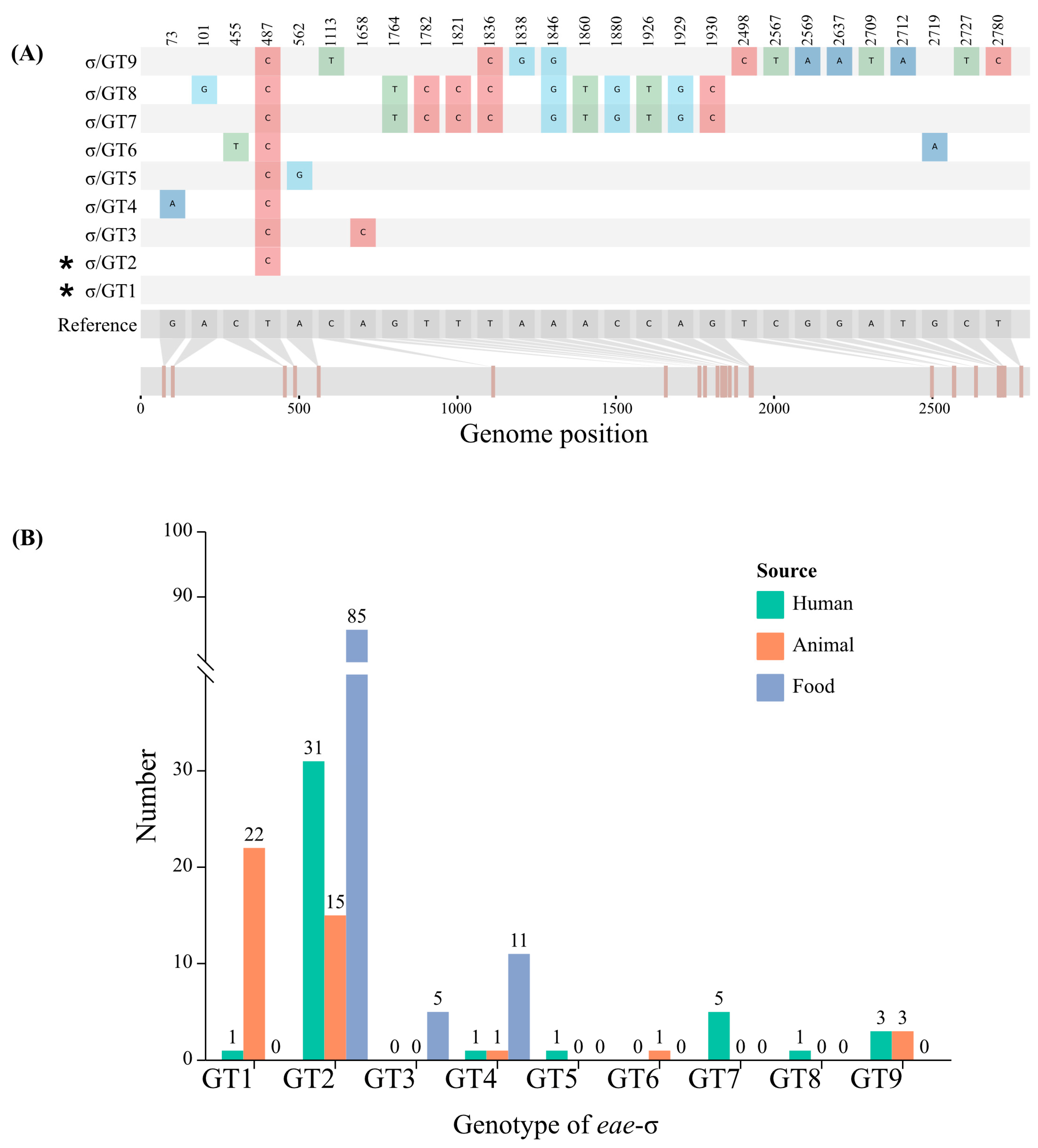
3.4. Genotypes of eae Subtype and Its Correlation with Sources

The predominant eae subtypes σ, ρ, ε3, and α9 were also analyzed to determine the diversity within each eae subtype (Figure S2). The reference sequence of each subtype was assigned as genotype 1 (GT1) and summarized in Table S2.
Among the 186 eae-σ strains, 8 genotypes (σ/GT2-GT9) were identified using eae-σ sequence AJ781125 as reference. Compared to σ/GT1, σ/GT2 had a synonymous substitution at location 487 (T to C) and other genotypes were non-synonymous (Figure 1A). The σ/GT2 was the major genotype in eae-σ strains and was associated with food-derived strains (Figure 1B).
Among the 47 eae-ρ strains, 5 genotypes (ρ/GT2-GT6) were defined based on the eae-ρ reference sequence (DQ523613). The mutations ρ/GT2-GT6 were all non-synonymous, with each of them displaying two non-synonymous substitutions at locations 2188 (A to T) and 2242 (C to A). These substitutions resulted in the change from isoleucine to leucine and from glutamine to lysine, respectively. ρ/GT4 was the predominant genotype in eae-ρ strains and was associated with animal-derived strains (Figure S2).
Among the 29 eae-ε3 strains, 7 genotypes (ε3/GT2-GT8) were defined based on the eae-ε3 reference sequence (AJ7876649). All variants of eae-ε3 were non-synonymous substitutions. The major genotype was ε3/GT2, which was associated with food-derived strains (Figure S2).
Among the 25 eae-α9 strains, 8 genotypes (α9/GT2-GT9) were defined based on the eae-α9 reference sequence (GCA_003860365.1). Except for α9/GT3 and GT5, other variants of eae-α9 were non-synonymous. α9/GT3 had only one synonymous substitution at location 489 (A to C), while α9/GT5 had two synonymous substitutions at locations 2466 (C to A) and 2733 (T to C). The major genotype was α9/GT6, which was associated with animal-derived strains (Figure S2).

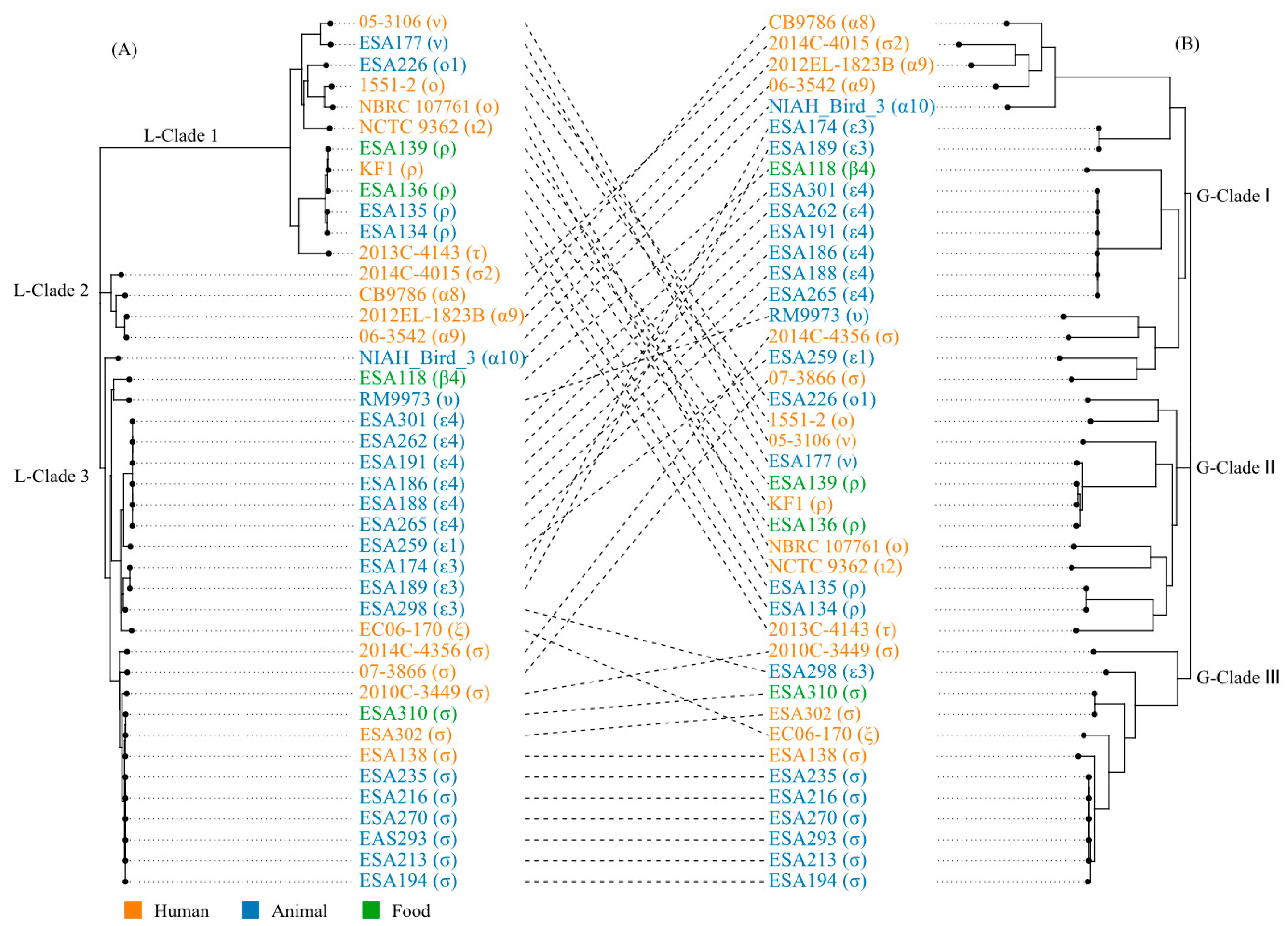
3.5. The Locus of Enterocyte Effacement in E. albertii

The complete genomes of 40 E. albertii strains were selected to characterize the LEE elements. The LEE elements were composed of 41 open reading frames organized in six major operons. Meta-alignment revealed that LEE elements were conserved between E. albertii and the other A/E members (Figure S3). In all 40 E. albertii strains, the LEE elements were integrated into the tRNA-pheU loci and had a length of 34–35 kb, with 81–100% similarity among each other. A neighbor-joining phylogenetic tree based on the complete nucleic acid sequences of the LEE elements was then constructed. According to the topological structure and evolutionary distance, the phylogenetic tree based on the sequences of LEE elements was divided into three main clades (L-Clade 1, 2, and 3) (Figure 2A). Strains with the same eae subtypes clustered together. L-Clades 1 and 3 contained strains from humans, animals, and food, while Clade 2 only harbored strains from humans. Within L-Clade 1, the predominant eae subtypes were eae-ο, ο1, ι2, ν, τ, and ρ. L-Clade 2 mainly consisted of eae-α8, α9, and σ2 subtypes. L-Clade 3 predominantly possessed eae-α10, ε1, ε3, ε4, υ, β4, ξ, and σ subtypes.
To explore concordance between LEE phylogenetic and pan-genome tree, a pan-genome tree was constructed using the 40 complete genomes of E. alberti, which was compared to that of LEE. The pan-genome tree also formed three major clades, namely G-Clade I, II, and III. Each clade contained strains derived from humans, animals, and food. The result indicated a significant divergence relationship for LEE, which was reflected in the tanglegrams (Figure 2). Only L-Clade 1 of the LEE phylogenetic tree was correlated with G-Clade II of the pangenome tree. L-Clade 2 of the LEE phylogenetic tree was correlated with a portion of G-Clade I of the pangenome tree. L-Clade 3 of the LEE phylogenetic tree was correlated with both G-Clades I and III of the pangenome tree.

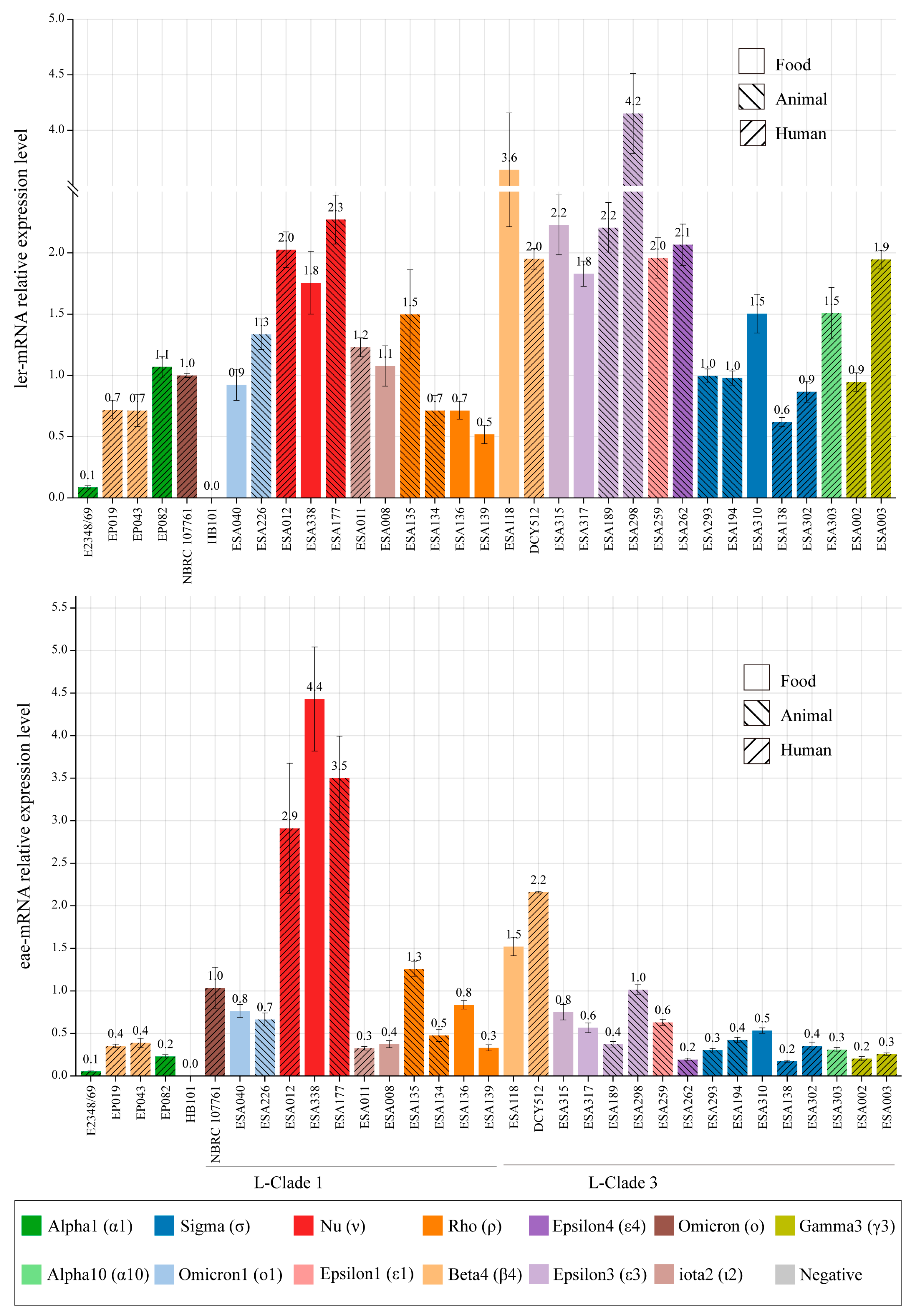
3.6. LEE Genes Expression in Different Strains

The ler gene, encoded in the LEE1 operon, is the master transcriptional factor of the six LEE operons [3]. The eae gene, which encodes intimin in the LEE5 operon, contributes to the establishment of the attaching and effacing (A/E) lesion [1]. The expression levels of ler and eae genes were evaluated using qRT-PCR assays. Compared to strain NBRC 107761, all E. albertii isolates presented ler and eae transcript levels varying from 0.5 to 4.2-fold and 0.2 to 4.4-fold, respectively. Strain ESA298 isolated from animal source with eae-ε3 subtypes displayed the highest transcript level of ler gene, while its transcript level of eae gene was relatively low. A significantly higher expression level of the eae gene was observed in eae-ν strains (eg., ESA012, ESA138, and ESA177), with the transcript level ranging from 2.9 to 4.4-fold. However, no significant differences in ler transcript levels among strains with different eae subtypes or sources were found (Figure 3).

3.7. Adherence Patterns of E. albertii Isolates

In this study, 28 E. albertii strains belonging to 13 eae subtypes were investigated for their adherence to cultivated HEp-2 cells, and strain NBRC 107761 was used as a reference strain. All 28 E. albertii strains were different from the tEPEC strain E2348/69, which showed LA in 3 h-assay. Twelve strains (42.9%) displayed a LAL pattern and ten strains (35.7%) displayed a DA pattern, while six (21.4%) strains resulted in cell detachment (DE) in 6 h-assay (Table 2). Among six strains with DE patterns, five strains carried cdtB-VI and one carried cdtB-II. Human-derived strains exhibited LAL (6/8, 75%) or DA (2/8, 25%) patterns, while animal and food-derived strains exhibited three patterns. Additionally, none of the gene transcripts (ler or eae) showed significant differences among strains with LAL, DA, and DE patterns (Table 2 and Figure 3).

4. Discussion

Intimin, encoded by eae gene, plays a crucial role in the development of A/E lesions by inducing the effacement of microvilli and forming actin pedestals [15]. Currently, at least 30 eae subtypes have been defined in E. coli [15]. Previous studies indicated that eae subtypes are correlated with host specificity and tissue tropism in E. coli [15,32]. For example, the eae-γ1 appeared to be the most frequent subtype among O157:H7 strains isolated from patients with bloody diarrhea (BD) and hemolytic uremic syndrome (HUS) [15]. In atypical EPEC strains, eae-β1 was found to be significantly prevalent in strains derived from diarrheal patients, while eae-ζ3 was commonly observed in strains derived from cattle [32]. The correlation between different eae subtypes and disease severity or hosts could be considered in the risk assessment of bacterial infections.
Increasing collection of E. albertii from various sources has resulted in a rapid increase in the number of eae subtypes. Some intimin subtypes identified in E. albertii strains have been novel or rare subtypes in E. coli. In this study, E. albertii from diverse sources exhibited diversity in the eae subtypes. Based on the sequence polymorphism, a total of 23 eae subtypes were identified among 459 E. albertii strains. Two novel subtypes, eae-α11 and η3, were named. Moreover, the distribution of eae subtypes varied among human, animal, and food-derived strains. For instance, eae-γ, τ, and α11 subtypes were found exclusively in human-derived strains. eae-α9, ε4, and σ2 were predominantly present in animal-derived strains, while eae-ρ and σ were primarily found in food-derived strains. Furthermore, each eae subtype possessed different genotypes. ρ/GT4 and α9/GT6 were associated with animal-derived strains that carried stx2f genes, which was relevant to clinical disease in finches and mild symptoms in humans [7,33].
The LEE island provides the genetic basis of observed A/E lesions [34]. It was initially identified in EPEC, which was a major cause of diarrhea in infants and young children worldwide [35]. Subsequently, it was found to occur in EHEC, rabbit diarrheagenic E. coli (RDEC), the murine pathogen Citrobacter rodentium, and later in E. albertii [36]. The regulation of LEE island has been intensively investigated in A/E pathogens, including in E. albertii type strain Albert 19982T (=NBRC 107761) [2]. In this study, the regulatory landscapes of diverse E. albertii strains were explored. The LEE elements in E. albertii strains were highly conserved, and their evolution was not synchronized with the genomic evolution. These findings were consistent with previous studies [34,37]. Moreover, the LEE elements in different strains exhibited diverse transcription and adherence patterns, which suggested their involvement in the pathogenic process. No significant differences in ler transcript levels among strains with different eae subtypes or sources were found, while a significantly higher expression level of the eae gene was observed in eae-ν strains. LAL was the most frequent adherence pattern among E. albertii strains, whereas DA and DE patterns were found in lower frequencies. Human-derived strains were more likely to exhibit LAL patterns, which might be relevant to the pathogenic process of colonization [38,39]. There might be other factors that could influence the transcription and expression of LEE elements, such as specific genes, plasmids, and other mobile elements. For instance, the LA pattern of tEPEC was highly related to the bundle-forming pilus encoded by EPEC adherence factor (EAF) plasmid. Moreover, some strains caused cell detachment which might be related to the subtype of cdtB-VI or other toxins [5]. Nevertheless, further studies were required to understand the in vivo pathogenicity of strain-specific eae variants, LEE transcription, and cell adherence.
In conclusion, we described the genetic diversity of eae gene in E. albertii strains isolated from different sources and identified two novel eae subtypes. Most eae subtypes were distributed among human, animal, and food-derived strains, while some subtypes showed host preference. The sequence and organization of LEE island among E. albertii isolates were relatively conserved, but the expression of ler and eae genes in different isolates was variable. Additionally, the LAL pattern represented a virulence property of E. albertii strains, especially human-derived strains. However, many strains exhibit DA or DE patterns. Further in vivo and in vitro studies are underway to understand the role of LEE in different genetic backgrounds.

Supplementary Materials

The following supporting information can be downloaded at: https://www.mdpi.com/article/10.3390/microorganisms11122843/s1, Supplementary Figure S1: Neighbor-joining (NJ) tree of 40 different subtypes of eae gene; Supplementary Figure S2: Phylogenetic relationships of 44 different eae sequences including 6 predominant eae subtypes in this study; Supplementary Figure S3: The genetic structure of LEE; Supplementary Table S1: E. albertii isolates used in this study; Supplementary Table S2: The accession numbers of eae, cdtB, and stx2a/stx2f subtypes used in this study; Supplementary Table S3: RT-qPCR primers used in this study. Reference [40] is cited in the supplementary materials.

Author Contributions

Conceptualization, Q.L. and Y.X.; Data curation, Q.L.; Formal analysis, Q.L.; Funding acquisition, Y.X.; Investigation, X.Y., H.S., H.W., X.S. and P.Z.; Methodology, Q.L., X.Y., H.S. and X.B.; Project administration, Y.X.; Resources, X.Y., H.S., H.W., X.S., P.Z. and X.B.; Software, Q.L. and X.Y.; Supervision, Y.X.; Validation, Y.X.; Visualization, Q.L.; Writing—original draft, Q.L.; Writing—review & editing, X.Y., H.S., H.W., X.S., P.Z., X.B. and Y.X. All authors have read and agreed to the published version of the manuscript.

Funding

This work was supported by the National Natural Science Foundation of China (82072254 & 81772152) and the National Key Research and Development Program of China (2021YFC2301105).

Data Availability Statement

The genomes of strain ESA302 were submitted to GenBank under the accession numbers GCA_032680885.1.

Conflicts of Interest

The authors declare no conflict of interest.

References

  1. Gomes, T.A.T.; Ooka, T.; Hernandes, R.T.; Yamamoto, D.; Hayashi, T.; Donnenberg, M.S. Escherichia albertii pathogenesis. EcoSal Plus 2020, 9, 10.1128. [Google Scholar] [CrossRef] [PubMed]
  2. Egan, M.; Critelli, B.; Cleary, S.P.; Marino, M.; Upreti, C.; Kalman, D.; Bhatt, S. Transcriptional and posttranscriptional regulation of the locus of enterocyte effacement in Escherichia albertii. Microb. Pathog. 2019, 135, 103643. [Google Scholar] [CrossRef] [PubMed]
  3. Gaytán, M.O.; Martínez-Santos, V.I.; Soto, E.; González-Pedrajo, B. Type three secretion system in attaching and effacing pathogens. Front. Cell. Infect. Microbiol. 2016, 6, 129. [Google Scholar] [CrossRef]
  4. Muchaamba, F.; Barmettler, K.; Treier, A.; Houf, K.; Stephan, R. Microbiology and epidemiology of Escherichia albertii-an emerging elusive foodborne pathogen. Microorganisms 2022, 10, 875. [Google Scholar] [CrossRef]
  5. Pons, B.; Vignard, J.; Mirey, G. Cytolethal distending toxin subunit B: A review of structure–function relationship. Toxins 2019, 11, 595. [Google Scholar] [CrossRef]
  6. Luo, L.; Wang, H.; Payne, M.J.; Liang, C.; Bai, L.; Zheng, H.; Zhang, Z.; Zhang, L.; Zhang, X.; Yan, G.; et al. Comparative genomics of Chinese and international isolates of Escherichia albertii: Population structure and evolution of virulence and antimicrobial resistance. Microb. Genom. 2021, 7, 000710. [Google Scholar] [CrossRef]
  7. Bengtsson, R.J.; Baker, K.S.; Cunningham, A.A.; Greig, D.R.; John, S.K.; Macgregor, S.K.; Seilern-Moy, K.; Spiro, S.; Chong, C.C.; De Silva, P.M.; et al. The genomic epidemiology of Escherichia albertii infecting humans and birds in Great Britain. Nat. Commun. 2023, 14, 1707. [Google Scholar] [CrossRef]
  8. Brandal, L.T.; Tunsjø, H.S.; Ranheim, T.E.; Løbersli, I.; Lange, H.; Wester, A.L.; Burnham, C.-A.D. Shiga toxin 2a in Escherichia albertii. J. Clin. Microbiol. 2015, 53, 1454–1455. [Google Scholar] [CrossRef]
  9. Masuda, K.; Ooka, T.; Akita, H.; Hiratsuka, T.; Takao, S.; Fukada, M.; Inoue, K.; Honda, M.; Toda, J.; Sugitani, W.; et al. Epidemiological aspects of Escherichia albertii outbreaks in Japan and genetic characteristics of the causative pathogen. Foodborne Pathog. Dis. 2020, 17, 144–150. [Google Scholar] [CrossRef]
  10. Oaks, J.L.; Besser, T.E.; Walk, S.T.; Gordon, D.M.; Beckmen, K.B.; Burek, K.A.; Haldorson, G.J.; Bradway, D.S.; Ouellette, L.; Rurangirwa, F.R.; et al. Escherichia albertii in wild and domestic birds. Emerg. Infect. Dis. 2010, 16, 638–646. [Google Scholar] [CrossRef]
  11. Barmettler, K.; Biggel, M.; Treier, A.; Muchaamba, F.; Vogler, B.R.; Stephan, R. Occurrence and characteristics of Escherichia albertii in wild Birds and poultry flocks in Switzerland. Microorganisms 2022, 10, 2265. [Google Scholar] [CrossRef] [PubMed]
  12. Liu, Q.; Bai, X.; Yang, X.; Fan, G.; Wu, K.; Song, W.; Sun, H.; Chen, S.; Chen, H.; Xiong, Y. Identification and genomic characterization of Escherichia albertii in migratory birds from Poyang Lake, China. Pathogens 2022, 12, 9. [Google Scholar] [CrossRef]
  13. Hinenoya, A.; Wang, H.; Patrick, E.M.; Zeng, X.; Cao, L.; Li, X.-P.; Lindsey, R.L.; Gillespie, B.; He, Q.; Yamasaki, S.; et al. Longitudinal surveillance and comparative characterization of Escherichia albertii in wild raccoons in the United States. Microbiol. Res. 2022, 262, 127109. [Google Scholar] [CrossRef]
  14. Barmettler, K.; Biggel, M.; Treier, A.; Muchaamba, F.; Stephan, R. Livestock as possible reservoir of Escherichia albertii in Switzerland. Schweiz. Arch. Tierheilkd. 2023, 165, 299–306. [Google Scholar] [CrossRef] [PubMed]
  15. Hua, Y.; Bai, X.; Zhang, J.; Jernberg, C.; Chromek, M.; Hansson, S.; Frykman, A.; Yang, X.; Xiong, Y.; Wan, C.; et al. Molecular characteristics of eae-positive clinical Shiga toxin-producing Escherichia coli in Sweden. Emerg. Microbes Infect. 2020, 9, 2562–2570. [Google Scholar] [CrossRef]
  16. Ooka, T.; Seto, K.; Kawano, K.; Kobayashi, H.; Etoh, Y.; Ichihara, S.; Kaneko, A.; Isobe, J.; Yamaguchi, K.; Horikawa, K.; et al. Clinical significance of Escherichia albertii. Emerg. Infect. Dis. 2012, 18, 488–492. [Google Scholar] [CrossRef]
  17. Wick, R.R.; Judd, L.M.; Gorrie, C.L.; Holt, K.E. Unicycler: Resolving bacterial genome assemblies from short and long sequencing reads. PLoS Comput. Biol. 2017, 13, e1005595. [Google Scholar] [CrossRef]
  18. Gurevich, A.; Saveliev, V.; Vyahhi, N.; Tesler, G. QUAST: Quality assessment tool for genome assemblies. Bioinformatics 2013, 29, 1072–1075. [Google Scholar] [CrossRef]
  19. Lindsey, R.L.; Garcia-Toledo, L.; Fasulo, D.; Gladney, L.M.; Strockbine, N. Multiplex polymerase chain reaction for identification of Escherichia coli, Escherichia albertii and Escherichia fergusonii. J. Microbiol. Methods 2017, 140, 1–4. [Google Scholar] [CrossRef]
  20. Ooka, T.; Seto, K.; Ogura, Y.; Nakamura, K.; Iguchi, A.; Gotoh, Y.; Honda, M.; Etoh, Y.; Ikeda, T.; Sugitani, W.; et al. O-antigen biosynthesis gene clusters of Escherichia albertii: Their diversity and similarity to Escherichia coli gene clusters and the development of an O-genotyping method. Microb. Genom. 2019, 5, 000314. [Google Scholar] [CrossRef]
  21. Rozewicki, J.; Li, S.; Amada, K.M.; Standley, D.M.; Katoh, K. MAFFT-DASH: Integrated protein sequence and structural alignment. Nucleic Acids Res. 2019, 47, W5–W10. [Google Scholar] [CrossRef]
  22. Rose, R.; Golosova, O.; Sukhomlinov, D.; Tiunov, A.; Prosperi, M. Flexible design of multiple metagenomics classification pipelines with UGENE. Bioinformatics 2018, 35, 1963–1965. [Google Scholar] [CrossRef]
  23. Sullivan, M.J.; Petty, N.K.; Beatson, S.A. Easyfig: A genome comparison visualizer. Bioinformatics 2011, 27, 1009–1010. [Google Scholar] [CrossRef] [PubMed]
  24. Tamura, K.; Stecher, G.; Kumar, S. MEGA11: Molecular evolutionary genetics analysis version 11. Mol. Biol. Evol. 2021, 38, 3022–3027. [Google Scholar] [CrossRef]
  25. Revell, L.J. phytools: An R package for phylogenetic comparative biology (and other things). Methods Ecol. Evol. 2012, 3, 217–223. [Google Scholar] [CrossRef]
  26. Seemann, T. Prokka: Rapid prokaryotic genome annotation. Bioinformatics 2014, 30, 2068–2069. [Google Scholar] [CrossRef]
  27. Page, A.J.; Cummins, C.A.; Hunt, M.; Wong, V.K.; Reuter, S.; Holden, M.T.G.; Fookes, M.; Falush, D.; Keane, J.A.; Parkhill, J. Roary: Rapid large-scale prokaryote pan genome analysis. Bioinformatics 2015, 31, 3691–3693. [Google Scholar] [CrossRef]
  28. Rao, X.; Huang, X.; Zhou, Z.; Lin, X. An improvement of the 2^(−delta delta CT) method for quantitative real-time polymerase chain reaction data analysis. Biostat. Bioinform. Biomath. 2013, 3, 71–85. [Google Scholar]
  29. Xie, J.; Chen, Y.; Cai, G.; Cai, R.; Hu, Z.; Wang, H. Tree Visualization By One Table (tvBOT): A web application for visualizing, modifying and annotating phylogenetic trees. Nucleic Acids Res. 2023, 51, W587–W592. [Google Scholar] [CrossRef]
  30. Cravioto, A.; Gross, R.J.; Scotland, S.M.; Rowe, B. An adhesive factor found in strains of Escherichia coli belonging to the traditional infantile enteropathogenic serotypes. Curr. Microbiol. 1979, 3, 95–99. [Google Scholar] [CrossRef]
  31. Govindarajan, D.K.; Viswalingam, N.; Meganathan, Y.; Kandaswamy, K. Adherence patterns of Escherichia coli in the intestine and its role in pathogenesis. Med. Microecol. 2020, 5, 100025. [Google Scholar] [CrossRef]
  32. Xu, Y.; Bai, X.; Zhao, A.; Zhang, W.; Ba, P.; Liu, K.; Jin, Y.; Wang, H.; Guo, Q.; Sun, H.; et al. Genetic diversity of intimin gene of atypical enteropathogenic Escherichia coli isolated from human, animals and raw meats in China. PLoS ONE 2016, 11, e0152571. [Google Scholar] [CrossRef] [PubMed]
  33. Den Ouden, A.; Greig, D.R.; Rodwell, E.V.; Tripodo, F.; Olonade, I.; Swift, C.; Jenkins, C. Escherichia coli encoding Shiga toxin subtype Stx2f causing human infections in England, 2015–2022. J. Med. Microbiol. 2023, 72, 001707. [Google Scholar] [CrossRef]
  34. Ingle, D.J.; Tauschek, M.; Edwards, D.J.; Hocking, D.M.; Pickard, D.J.; Azzopardi, K.I.; Amarasena, T.; Bennett-Wood, V.; Pearson, J.S.; Tamboura, B.; et al. Evolution of atypical enteropathogenic E. coli by repeated acquisition of LEE pathogenicity island variants. Nat. Microbiol. 2016, 1, 15010. [Google Scholar] [CrossRef] [PubMed]
  35. Mare, A.D.; Ciurea, C.N.; Man, A.; Tudor, B.; Moldovan, V.; Decean, L.; Toma, F. Enteropathogenic Escherichia coli—A summary of the literature. Gastroenterol. Insights 2021, 12, 28–40. [Google Scholar] [CrossRef]
  36. Franzin, F.M.; Sircili, M.P. Locus of enterocyte effacement: A pathogenicity island involved in the virulence of enteropathogenic and enterohemorragic Escherichia coli subjected to a complex network of gene regulation. Biomed. Res. Int. 2015, 2015, 534738. [Google Scholar] [CrossRef]
  37. Ooka, T.; Ogura, Y.; Katsura, K.; Seto, K.; Kobayashi, H.; Kawano, K.; Tokuoka, E.; Furukawa, M.; Harada, S.; Yoshino, S.; et al. Defining the genome features of Escherichia albertii, an emerging enteropathogen closely related to Escherichia coli. Genome Biol. Evol. 2015, 7, 3170–3179. [Google Scholar] [CrossRef]
  38. Pereira, A.; Giugliano, L. Adhesion of diarrheagenic Escherichia coli and inhibition by glycocompounds engaged in the mucosal innate immunity. Biology 2013, 2, 810–831. [Google Scholar] [CrossRef]
  39. Serapio-Palacios, A.; Finlay, B.B. Dynamics of expression, secretion and translocation of type III effectors during enteropathogenic Escherichia coli infection. Curr. Opin. Microbiol. 2020, 54, 67–76. [Google Scholar] [CrossRef]
  40. Yang, X.; Sun, H.; Fan, R.; Fu, S.; Zhang, J.; Matussek, A.; Xiong, Y.; Bai, X. Genetic diversity of the intimin gene (eae) in non-O157 Shiga toxin-producing Escherichia coli strains in China. Sci. Rep. 2020, 10, 3275. [Google Scholar] [CrossRef]
Figure 1. (A) The nucleotide differences among eae-σ GTs. The asterisk represented the synonymous mutation. (B) The distribution of eae-σ GTs among strains isolated from different sources.
Microorganisms 11 02843 g001
Figure 2. Tanglegram comparison between a phylogenetic tree based on sequences of LEE elements and pan-genome tree. (A) Neighbor-joining phylogenetic tree based on the sequences of the LEE elements. The tree was divided into three main clusters (L-Clades 1, 2, and 3). (B) A pan-genome tree is based on the presence or absence of the gene in the pan-genome. The tree is also divided into three clusters (G-Clades I, II, and III) according to topological structure and evolutionary distance. The colors of the leaves indicated the source of strains. Lines between trees link the same strains and crossing lines indicate a lack of similarity in the two trees.
Microorganisms 11 02843 g002
Figure 3. Relative transcription of ler and eae genes as determined by qRT-PCR.
Microorganisms 11 02843 g003
Table 1. Intimin subtypes of E. albertii strains isolated from humans, animals, and food.
SubtypesHumanAnimalFoodTotalp Value
alpha10 (α10)1080180.001 *
alpha8 (α8)640100.019 *
alpha9 (α9)520025<0.001 *
beta3 (β3)380110.004 *
beta4 (β4)633120.692
epsilon1 (ε1)14050.064
epsilon3 (ε3)5915290.113
epsilon4 (ε4)112013<0.001 *
gamma3 (γ3)30030.031 *
iota2 (ι2)151218<0.001 *
lambda2 (λ2)42060.069
lambda3 (λ3)10010.317
nu (ν)52180.160
omicron (ο)740110.008 *
omicron1 (ο1)12470.574
rho (ρ)923647<0.001 *
sigma (σ)4342101186<0.001 *
sigma2 (σ2)416020<0.001 *
tau (τ)60060.001 *
xi (ξ)43070.077
ypsilon (υ)12030.312
alpha11 (α11)40040.01 *
eta3 (η3)03030.067
Negative33060.077
Total147150162459-
* Statistically significant difference.
Table 2. HEp-2 adherence assays of different E. albertii strains.
Strain NameSourcesDetail of Sourceseae SubtypescdtB SubtypesCell Adherence
E2348/69HumanFecesα1-LA3h
EAEC 042HumanFeces--AA3h
aEPEC EP019HumanFecesβ4-LAL
HB101LabLab--NA
NBRC 107761HumanFecesοcdtB-VILAL
ESA040FoodDuck intestineο1cdtB-VIDA
ESA226AnimalTBGο1cdtB-VIDA
ESA012HumanFecesνcdtB-IIDA
ESA338FoodSwine meatνcdtB-VIDA
ESA177AnimalLWGνcdtB-VIDA
ESA011HumanFecesι2cdtB-IIDA
ESA008FoodDuck intestineι2cdtB-VILAL
ESA135AnimalBatρcdtB-VIDE
ESA134AnimalBatρcdtB-VIDE
ESA136FoodChicken intestineρcdtB-VIDE
ESA139FoodDuck intestineρcdtB-VIDE
ESA118FoodDuck intestineβ4cdtB-IILAL
DCY512HumanFecesβ4cdtB-I/IILAL
ESA315FoodChicken intestineε3cdtB-IIDA
ESA317FoodChicken meatε3cdtB-IILAL
ESA189AnimalEWε3cdtB-IIDE
ESA298AnimalEWε3cdtB-IILAL
ESA259AnimalEWε1cdtB-IILAL
ESA262AnimalNPε4cdtB-VIDE
ESA293AnimalEWσcdtB-IIDA
ESA194AnimalTBGσcdtB-IIDA
ESA310FoodChicken intestineσcdtB-IILAL
ESA138HumanFecesσcdtB-IILAL
ESA302HumanFecesσcdtB-IILAL
ESA303HumanBloodstreamα10cdtB-I/IILAL
ESA002HumanFecesγ3cdtB-VIDA
ESA003HumanFecesγ3cdtB-VILAL
‘-’, absent; LWG, Lesser white-fronted goose (Anser erythropus); TBG, Taiga bean goose (Anser fabalis); EW, Eurasian wigeon (Mareca penelope); GWG, Greater white-fronted goose (Anser albifrons); NP, Northern Pintail (Anas acuta); LAL: localized-like adherence; NA: nonadherent; AA: aggregative adherence; DA: diffuse adherence; LA: localized adherence; DE, detachment.
Disclaimer/Publisher’s Note: The statements, opinions and data contained in all publications are solely those of the individual author(s) and contributor(s) and not of MDPI and/or the editor(s). MDPI and/or the editor(s) disclaim responsibility for any injury to people or property resulting from any ideas, methods, instructions or products referred to in the content.

