
In Vitro and In Silico Anti-Picornavirus Triterpene Alkanoic Acid Ester from Saudi Collection of Rhazya stricta Decne

 , ,
, ,
Abstract
:1. Introduction
2. Materials and Methods
2.1. General
2.2. Plant Materials
2.3. Extraction and Fractionation
2.4. Chromatographic Purification
2.4.1. Acetylation of 1 and 2
2.4.2. Alkaline Hydrolysis of 1 and 2
2.4.3. Acetylation of 1b
2.5. Anti-Viral Assay
2.5.1. Preparations of the Total Extract, Fractions, and Pure Isolates for the In Vitro Assay
2.5.2. Determination of Samples Cytotoxicity on BHK (Baby Hamster Kidney) Cells
2.5.3. MTT Assay Protocol
2.6. Molecular Docking Analysis
2.6.1. Preparation of Ligand Structures
2.6.2. Retrieval and Preparation of Target Protein Structure
2.6.3. In Silico Molecular Docking
3. Results
3.1. Compounds Characterization
3.2. Anti-viral Assay
3.2.1. Determination of Samples Cytotoxicity on BHK Cells
3.2.2. MTT Assay Protocol
3.3. Molecular Docking Analysis
4. Discussion
5. Conclusions
Supplementary Materials
Author Contributions
Funding
Institutional Review Board Statement
Informed Consent Statement
Data Availability Statement
Acknowledgments
Conflicts of Interest
References
- Bukhari, N.A.; Al-Otaibi, R.A.; Ibhrahim, M.M. Phytochemical and taxonomic evaluation of Rhazya stricta in Saudi Arabia. Saudi J. Biol. Sci. 2017, 24, 1513–1521. [Google Scholar] [CrossRef] [Green Version]
- Gilani, S.A.; Kikuchi, A.; Shinwari, Z.K.; Khattak, Z.I.; Watanabe, K.N. Phytochemical, pharmacological and ethnobotanical studies of Rhazya stricta Decne. Phytother. Res. 2007, 21, 301–307. [Google Scholar] [CrossRef]
- Yahya, M.A. Saudi Plants: A Phytochemical and Biological Approach; King Abdulaziz City for Science and Technology: Riyah, Saudi Arabia, 1990. [Google Scholar]
- Bashir, A.K.; Abdalla, A.A.; Hassan, E.S.; Wasfi, I.A.; Amiri, M.A.; Crabb, T.A. Alkaloids with antimicrobial activity from the root of Rhazya stricta Decn. growing in United Arab Emirates. Arab. Gulf J. Sci. Res. 1994, 12, 119–131. [Google Scholar]
- Tanira, M.; Ali, B.; Bashir, A.; Chandranath, I. Some pharmacologic and toxicologic studies on rhazya stricta decne in rats, mice and rabbits. Gen. Pharmacol. Vasc. Syst. 1996, 27, 1261–1267. [Google Scholar] [CrossRef]
- Read, M. Plants of Dhofar (The Southern Region of Oman, Traditional Economic and Medicinal Uses) Anthony G. Miller and Miranda Morris The Office of the Adviser for Conservation of the Environment, Diwan of Royal Court, Sultanate of Oman. 1988, 361pp., HB. In UK, available from Holmes McDougall Ltd., Allander House, 137–141 Leith Walk, Edinburgh EH6 8NS. £37.50 inc postage. Oryx 2009, 24, 232. [Google Scholar]
- Dymock, W.; Warden, C.J.H.; Hooper, D. Pharmacographia Indica; Kegan Paul, Trench, Trubner & Co.: London, UK, 1890. [Google Scholar]
- Ahmad, H.; Bhatti, G.R.; Latif, A. Medicinal flora of the thar desert: An overview of problems and their feasible solutions. Zonas Áridas 2006, 8, 73–84. [Google Scholar]
- Ahmed, M. Checklist of Medicinal Flora of Tehsil Isakhel, District Mianwali-Pakistan. Ethnobot. Leafl. 2006, 2006, 10. [Google Scholar]
- Jamal, S.M.; Belsham, G.J. Foot-and-mouth disease: Past, present and future. Vet. Res. 2013, 44, 116. [Google Scholar] [CrossRef] [Green Version]
- Grubman, M.J.; Baxt, B. Foot-and-mouth disease. Clin. Microbiol. Rev. 2004, 17, 465–493. [Google Scholar] [CrossRef] [Green Version]
- Knowles, N.J.; Samuel, A.R. Molecular epidemiology of foot-and-mouth disease virus. Virus Res. 2003, 91, 65–80. [Google Scholar] [CrossRef]
- Kahn, C.N.; Line, S. The Mercks Veterinary Manual, 9th ed.; Merck and Company Incorporated: White House Station, NJ, USA, 2005. [Google Scholar]
- Alexandersen, S.; Kitching, R.P.; Mansley, L.M.; Donaldson, A.I. Clinical and laboratory investigations of five outbreaks of foot-and-mouth disease during the 2001 epidemic in the United Kingdom. Vet. Rec. 2003, 152, 489–496. [Google Scholar] [CrossRef] [PubMed]
- Radostitis, O.M.; Gay, C.C.; Blood, D.C.; Hinchcliff, K.W. Veterinary Medicine: A Textbook of the Diseases of Cattle, Sheep, Pigs, Goats and Horses, 9th ed.; W.B. Saunders Company: Sarasota, FL, USA; Harcourt Publishers: London, UK, 2000. [Google Scholar]
- Knight-Jones, T.J.; Rushton, J. The economic impacts of foot and mouth disease-what are they, how big are they and where do they occur? Prev. Vet. Med. 2013, 112, 161–173. [Google Scholar] [CrossRef] [PubMed] [Green Version]
- Gakuya, D.W.; Mulei, C.M.; Wekesa, S.B. Use of ethnoveterinary remedies in the management of foot and mouth disease lesions in a dairy herd. Afr. J. Tradit. Complement. Altern. Med. 2011, 8, 165–169. [Google Scholar] [CrossRef] [PubMed] [Green Version]
- Theerawatanasirikul, S.; Lueangaramkul, V.; Thangthamniyom, N.; Chankeeree, P.; Semkum, P.; Lekcharoensuk, P. Andrographolide and Deoxyandrographolide Inhibit Protease and IFN-Antagonist Activities of Foot-and-Mouth Disease Virus 3Cpro. Animals 2022, 12, 1995. [Google Scholar] [CrossRef]
- Abdel-Kader, M.S.; Rehman, N.U.; Aldosari, A.F.; Almutib, F.S.; Al Muwinea, A.I.; Saeedan, A.S. Bronchodilator Secondary Metabolites from Rhazya stricta Decne Aerial Parts. Separations 2022, 9, 412. [Google Scholar] [CrossRef]
- Theerawatanasirikul, S.; Lekcharoensuk, P. Virtual Screening of Natural Compounds Targeting Proteases of Coronaviruses and Picornaviruses. In Methods in Pharmacology and Toxicology; Roy, K., Ed.; Springer: New York, NY, USA, 2021. [Google Scholar]
- Khairy, A.; Hammoda, H.M.; Celik, I.; Zaatout, H.H.; Ibrahim, R.S. Discovery of potential natural dihydroorotate dehydrogenase inhibitors and their synergism with brequinar via integrated molecular docking, dynamic simulations and in vitro approach. Sci. Rep. 2022, 12, 19037. [Google Scholar] [CrossRef]
- Reham, S.; El-Mezayen, I.N.S.; Khairy, A.; Zaatout, H.H.; Hammoda, H.M.; Metwally, A.M. Biologically-Guided Isolation of Natural Lead Antithyroid Drug from Medicago sativa L. Sprouts and Its Toxic Profile in Comparison with Propylthiouracil. J. Food Drug Anal. 2020, 28, 407–448. [Google Scholar]
- Wang, J.; Xu, C.; Wong, Y.; Li, Y.; Liao, F.; Jiang, T.; Tu, Y. Artemisinin, the Magic Drug Discovered from Traditional Chinese Medicine. Engineering 2019, 5, 32–39. [Google Scholar] [CrossRef]
- Jin, Q.; Ko, H.J.; Chang, Y.-S.; Woo, E.-R. Chemical constituents from the aerial parts of Aster yomena. Nat. Prod. Sci. 2013, 19, 269–274. [Google Scholar]
- Dekebo, A.; Dagne, E.; Gautun, O.; Aasen, A. Triterpenes from the resin of Boswellia neglecta. Bull. Chem. Soc. Ethiop. 2002, 16, 87–90. [Google Scholar] [CrossRef] [Green Version]
- Abdullahi, S.; Musa, A.; Abdullahi, M.; Sule, M.; Sani, M. Isolation of Lupeol from the Stem-bark of Lonchocarpus sericeus (Papilionaceae). Sch. Acad. J. Biosci. 2013, 1, 18–19. [Google Scholar]
- Ben nejma, A.; Besbes, M.; Guérineau, V.; Touboul, D.; Ben jannet, H.; Hamza, M. Isolation and structure elucidation of acetylcholinesterase lipophilic lupeol derivatives inhibitors from the latex of the Tunisian Periploca laevigata. Arab. J. Chem. 2017, 10, S2767–S2772. [Google Scholar] [CrossRef]
- Furukawa, S.; Takagi, N.; Ikeda, T.; Ono, M.; Nafady, A.M.; Nohara, T.; Sugimoto, H.; Doi, S.; Yamada, H. Two novel long-chain alkanoic acid esters of lupeol from alecrim-propolis. Chem. Pharm. Bull. (Tokyo) 2002, 50, 439–440. [Google Scholar] [CrossRef] [PubMed] [Green Version]
- Ododo, M.M.; Choudhury, M.K.; Dekebo, A.H. Structure elucidation of β-sitosterol with antibacterial activity from the root bark of Malva parviflora. SpringerPlus 2016, 5, 1210. [Google Scholar] [CrossRef] [Green Version]
- Kim, D.-H.; Han, K.-M.; Chung, I.-S.; Kim, D.-K.; Kim, S.-H.; Kwon, B.-M.; Jeong, T.-S.; Park, M.-H.; Ahn, E.-M.; Baek, N.-I. Triterpenoids from the Flower of Campsis grandiflora K. Schum. as Human Acyl-CoA: Cholesterol Acyltransferase Inhibitors. Arch. Pharm. Res. 2005, 28, 550–556. [Google Scholar] [CrossRef]
- Noviany, N.; Osman, H. Structure Elucidation of Betulinic Acid from Sesbania grandiflora Root. J. Phys. J. Phys. Conf. Ser. 2021, 1751, 012090. [Google Scholar] [CrossRef]
- Mason, P.W.; Grubman, M.J.; Baxt, B. Molecular basis of pathogenesis of FMDV. Virus Res. 2003, 91, 9–32. [Google Scholar] [CrossRef]
- Gao, Y.; Sun, S.-Q.; Guo, H.-C. Biological function of Foot-and-mouth disease virus non-structural proteins and non-coding elements. Virol. J. 2016, 13, 107. [Google Scholar] [CrossRef] [Green Version]
- Birtley, J.R.; Knox, S.R.; Jaulent, A.M.; Brick, P.; Leatherbarrow, R.J.; Curry, S. Crystal structure of foot-and-mouth disease virus 3C protease. New insights into catalytic mechanism and cleavage specificity. J. Biol. Chem. 2005, 280, 11520–11527. [Google Scholar] [CrossRef] [Green Version]
- Asiamah, I.; Obiri, S.A.; Tamekloe, W.; Armah, F.A.; Borquaye, L.S. Applications of molecular docking in natural products-based drug discovery. Sci. Afr. 2023, 20, e01593. [Google Scholar] [CrossRef]
- Waterhouse, A.; Bertoni, M.; Bienert, S.; Studer, G.; Tauriello, G.; Gumienny, R.; Heer, F.T.; De Beer, T.A.P.; Rempfer, C.; Bordoli, L.; et al. SWISS-MODEL: Homology modelling of protein structures and complexes. Nucleic Acids Res. 2018, 46, W296–W303. [Google Scholar] [CrossRef] [PubMed] [Green Version]
- Darshani, P.; Sen Sarma, S.; Srivastava, A.K.; Baishya, R.; Kumar, D. Anti-viral triterpenes: A review. Phytochem. Rev. 2022, 21, 1761–1842. [Google Scholar] [CrossRef] [PubMed]
- Wassel, M.M.S.; Gamal El-Din, W.M.; Ahmed Ragab, A.; Elhag Ali, G.A.M.; Ammar, Y.A. Antiviral Activity of Adamantane-Pyrazole Derivatives Against Foot and Mouth Disease Virus Infection in vivo and in vitro with Molecular Docking Study. J. Appl. Vet. Sci. 2020, 5, 37–46. [Google Scholar]


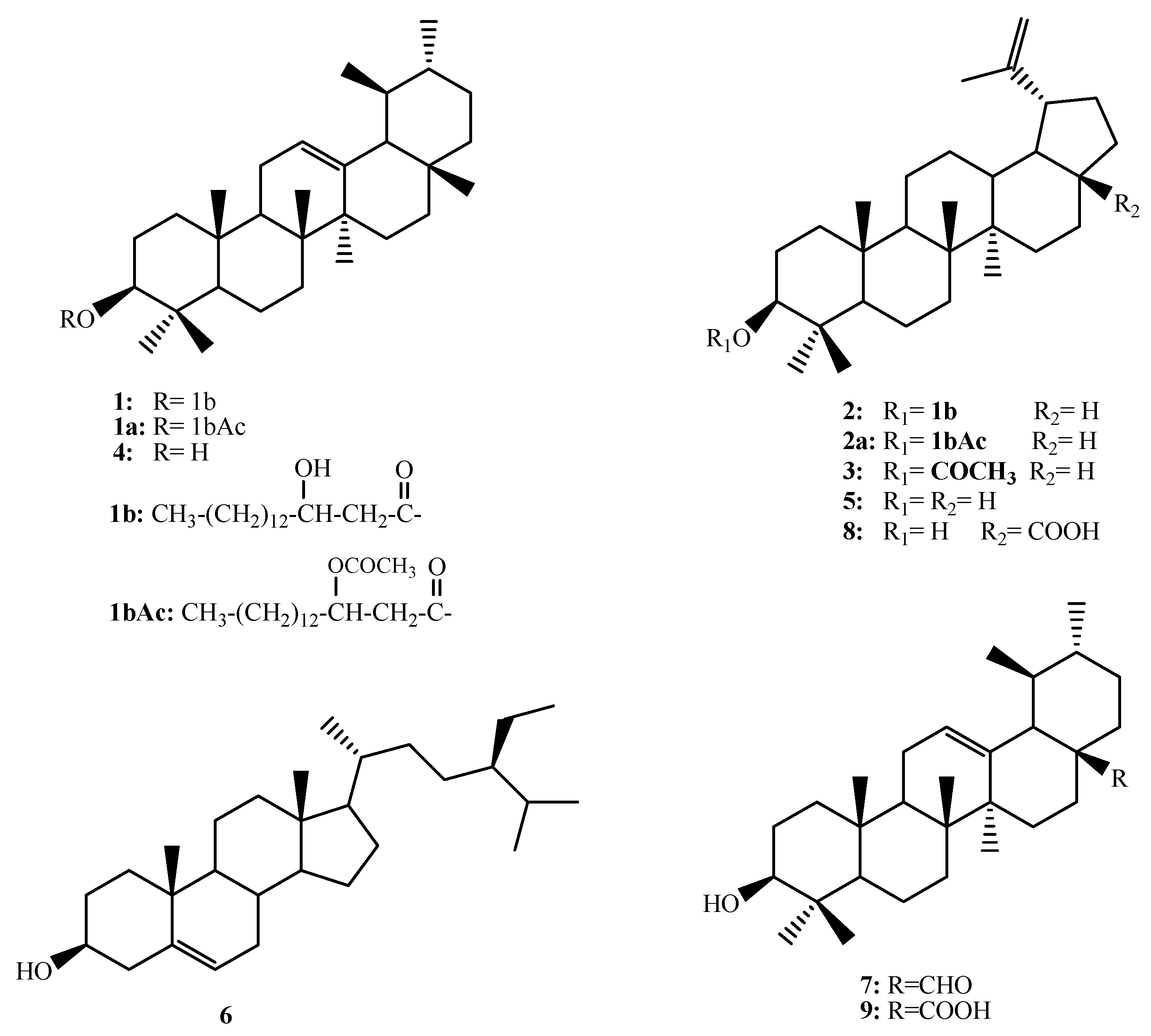
| 1 | 1a | 2 | 2a | 1b | |
|---|---|---|---|---|---|
| 3 | 4.46 (dd, 4.5, 11.7) | 4.71 (dd, 4.4, 11.8) | 4.70 (dd, 4.5, 11.8) | 4.72 (dd, 4.5, 11.9) | - |
| 12 | 5.18 bs | 5.18 bs | Overlapped | Overlapped | - |
| 29 | Overlapped | Overlapped | 4.73 bs, 4.85 bs | 4.74 bs, 4.87 bs | - |
| 2′ | 2.44 (5.1, 15.1) 2.56 (dd, 7.7, 15.1) | 2.44 (5.1, 15.1) 2.56 (dd, 7.7, 15.1) | 2.33 (d, 6.0) | 2.44 (Overlap) 2.56 (dd, 7.7, 15.1) | 2.39 (dd, 10.0, 15.9) 2.49 (bd, 15.9) |
| 3′ | 3.78 m | 5.46 (p, 6.0) | 4.02 m | 5.47 (p, 6.8) | 3.96 bs |
| 16′ | Overlapped | Overlapped | Overlapped | Overlapped | 0.81 (t, 6.5) |
| CH3-CO | - | 1.75 s | - | 1.71 s | - |
| Pos. | 1 | 1a | 2 | 2a | Pos. | 1 | 1a | 2 | 2a | 1b |
|---|---|---|---|---|---|---|---|---|---|---|
| 1 | 38.09 | 38.02 | 38.17 | 38.13 | 21 | 31.39 | 31.00 | 29.50 | 29.53 | |
| 2 | 25.28 | 27.37 | 27.52 | 27.50 | 22 | 41.67 | 41.60 | 40.00 | 40.00 | |
| 3 | 80.82 | 80.74 | 80.71 | 80.71 | 23 | 27.95 | 28.38 | 27.84 | 27.76 | |
| 4 | 36.61 | 39.94 | 37.68 | 38.13 | 24 | 16.83 | 16.75 | 16.00 | 16.02 | |
| 5 | 55.30 | 55.22 | 55.27 | 55.28 | 25 | 15.61 | 15.53 | 16.58 | 16.58 | |
| 6 | 18.26 | 18.18 | 18.17 | 18.16 | 26 | 16.86 | 16.78 | 15.84 | 15.82 | |
| 7 | 32.91 | 32.83 | 35.60 | 35.60 | 27 | 23.36 | 23.29 | 14.50 | 14.49 | |
| 8 | 42.09 | 42.00 | 40.75 | 40.73 | 28 | 28.83 | 28.75 | 17.90 | 17.91 | |
| 9 | 47.61 | 47.45 | 50.21 | 50.17 | 29 | 17.55 | 17.47 | 109.71 | 109.75 | |
| 10 | 37.63 | 37.25 | 36.89 | 36.87 | 30 | 21.40 | 21.33 | 19.20 | 19.19 | |
| 11 | 23.68 | 23.60 | 20.80 | 20.77 | 1′ | 172.33 | 169.57 | 172.33 | 169.55 | 177.92 |
| 12 | 124.63 | 124.55 | 23.76 | 23.77 | 2′ | 40.75 | 39.79 | 41.89 | 39.48 | 41.19 |
| 13 | 139.58 | 139.50 | 38.03 | 38.00 | 3′ | 68.16 | 70.60 | 67.98 | 70.59 | 68.12 |
| 14 | 40.02 | 42.00 | 42.76 | 42.74 | 4′ | 34.14 | 34.06 | 36.81 | 34.06 | 36.50 |
| 15 | 28.20 | 28.13 | 25.19 | 25.16 | 5′ | 32.10 | 32.02 | 32.00 | 32.02 | 31.96 |
| 16 | 26.79 | 26.71 | 34.21 | 34.18 | 6′–14′ | 29.56–29.94 | 29.49–29.82 | 29.47–29.85 | 29.49–29.86 | 24.70–29.74 |
| 17 | 33.81 | 33.74 | 42.90 | 42.91 | 15′ | 22.88 | 22.81 | 22.78 | 22.81 | 22.73 |
| 18 | 59.16 | 59.08 | 48.32 | 48.30 | 16′ | 14.15 | 14.08 | 14.03 | 14.08 | 14.17 |
| 19 | 39.57 | 39.49 | 48.05 | 48.08 | C H3-CO | 20.52 | 20.52 | - | ||
| 20 | 39.86 | 39.63 | 150.34 | 150.37 | CH3-CO | 169.37 | 169.34 | - |
| ID | Dilution 1:2 | Viability % | Toxicity % | CC50 (ug/mL) | MNTC |
|---|---|---|---|---|---|
| BHK | ug/mL | 100 | 0 | - | |
| RST | 1000 | 14.4 | 85.6 | 342.4 | 62.5 |
| 500 | 27.1 | 72.9 | |||
| 250 | 53.8 | 46.2 | |||
| 125 | 94.1 | 5.9 | |||
| 62.5 | 100.0 | 0 | |||
| 31.25 | 100.0 | 0 | |||
| 15.625 | 100.0 | 0 | |||
| 7.812 | 100.0 | 0 | |||
| RSP | 1000 | 22.6 | 77.4 | 653.3 | 62.5 |
| 500 | 22.6 | 77.4 | |||
| 250 | 52.4 | 47.6 | |||
| 125 | 95.2 | 4.8 | |||
| 62.5 | 100.0 | 0 | |||
| 31.25 | 100.0 | 0 | |||
| 15.625 | 100.0 | 0 | |||
| 7.812 | 100.0 | 0 | |||
| RSC | 1000 | 8.8 | 91.2 | 46.1 | 7.8 |
| 500 | 9.1 | 90.9 | |||
| 250 | 17.8 | 82.2 | |||
| 125 | 26.4 | 73.6 | |||
| 62.5 | 35.6 | 64.4 | |||
| 31.25 | 54.3 | 45.7 | |||
| 15.625 | 90.8 | 9.2 | |||
| 7.812 | 100 | 0 | |||
| RSE | 1000 | 25.3 | 74.7 | 699.2 | 250 |
| 500 | 60.1 | 39.9 | |||
| 250 | 99.6 | 0.4 | |||
| 125 | 100.0 | 0 | |||
| 62.5 | 98.4 | 1.6 | |||
| 31.25 | 99.9 | 0.1 | |||
| 15.625 | 101.0 | 0 | |||
| 7.812 | 100.0 | 0 | |||
| RSH | 1000 | 21.1 | 78.9 | 593.0 | 62.5 |
| 500 | 46.1 | 53.9 | |||
| 250 | 77.3 | 22.7 | |||
| 125 | 91.0 | 9.0 | |||
| 62.5 | 100.0 | 0 | |||
| 31.25 | 99.9 | 0.1 | |||
| 15.625 | 99.5 | 0.5 | |||
| 7.812 | 100.0 | 0 |
| Test | Coc. ug/mL | Viability | Toxicity | Viral Activity % | Anti-viral Effect % |
|---|---|---|---|---|---|
| BHK | 100 | 0 | |||
| FMD | 41.4 | 58.6 | 100 | 0 | |
| RST | 62.5 | 42.83626 | 57.16374 | 97.5 | 2.5 |
| RSP | 125 | 65.0 | 35.0 | 59.9 | 40.1 |
| RSC | 7.812 | 38.9 | 61.1 | 104.2 | 0 |
| RSE | 250 | 41.1 | 58.9 | 100.5 | 0 |
| RSH | 62.5 | 41.2 | 58.8 | 100.2 | 0 |
| 1 | 500 | 71.8 | 28.2 | 49.0 | 51.0 |
| 2 | 125 | 62.9 | 37.1 | 64.5 | 35.5 |
| 3 | 125 | 62.7 | 37.3 | 65 | 35.0 |
| 4 | 500 | 51.9 | 48.1 | 83.7 | 16.3 |
| 5 | 250 | 46.9 | 53.1 | 92.4 | 7.6 |
| 6 | 250 | 60.5 | 39.5 | 68.7 | 31.3 |
| 7 | 250 | 60.8 | 39.2 | 68.2 | 31.8 |
| 8 | 125 | 57.5 | 42.5 | 74.0 | 26.0 |
| 9 | 125 | 52 | 48 | 83.4 | 16.6 |
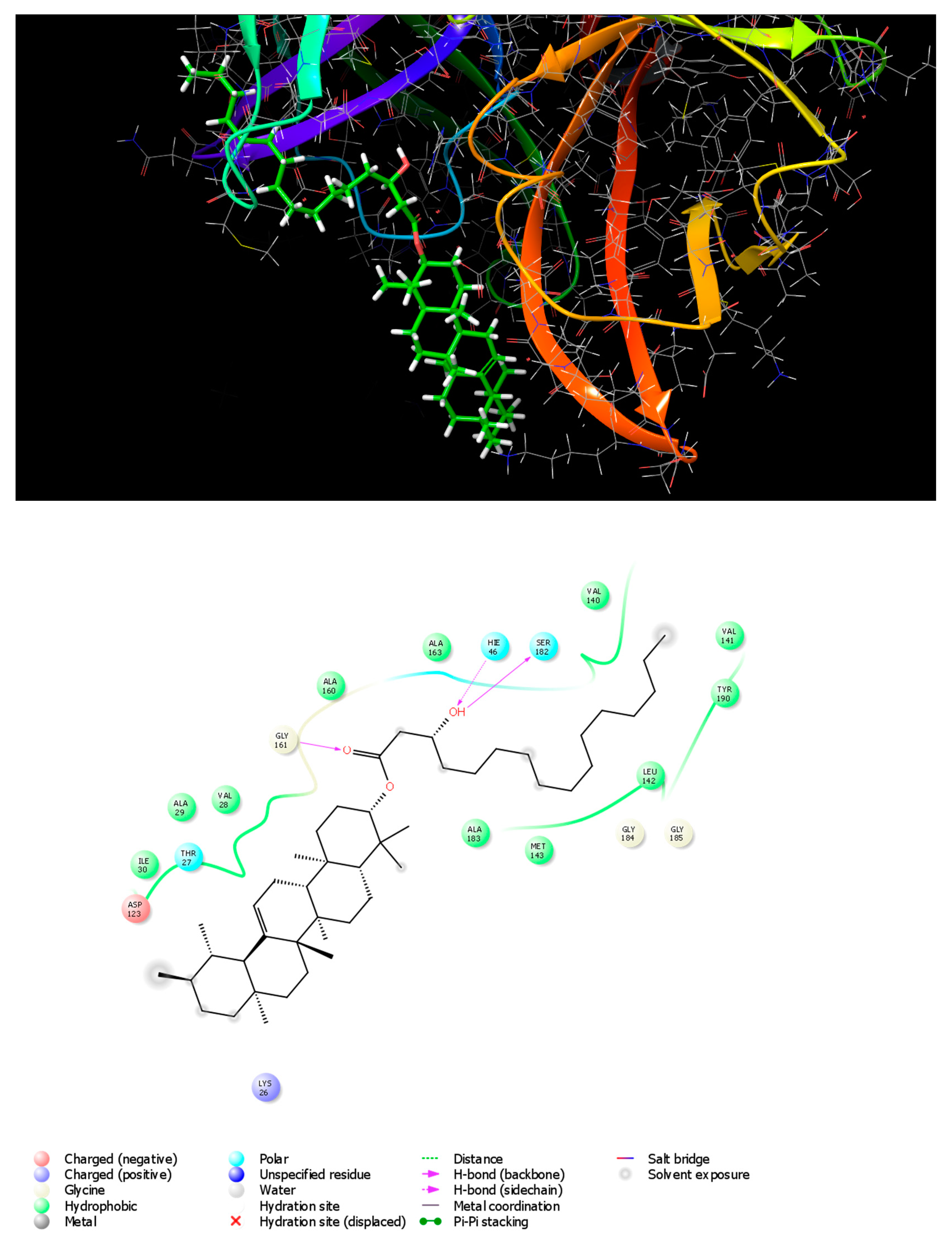
| Compound | Docking Score(Kcal mol−1) | Interacting Residues | |
|---|---|---|---|
| Type of Binding Interactions | Amino Acid Residues Involved in Protein–Ligand Interaction | ||
| Glycyrrhizic acid | −5.894 | Hydrogen bonding (backbone) | THR158, GLY161 |
| Hydrogen bonding (side chain) | HIE46 | ||
| Polar interaction | HIE46, THR158, SER182, ASN186 | ||
| Hydrophobic interaction | ALA29, ILE30, PRO114, MET143, ALA157, ALA160, TYR162, ALA163, ALA183 | ||
| Charged (positive) ionic interaction | ARG159 | ||
| Ribavirin | −5.853 | Hydrogen bonding (backbone) | THR158, GLY161 |
| Hydrogen bonding (side chain) | HIE46, HIE181 | ||
| Polar interaction | HIE46, THR158, HIE181, SER182 | ||
| Hydrophobic interaction | ALA29, ILE30, CYS31, ALA160, TYR162, ALA163, ALA183 | ||
| Charged (positive) ionic interaction | ARG159 | ||
| Rhazyin A | −5.048 | Hydrogen bonding (backbone) | GLY161, SER182 |
| Hydrogen bonding (side chain) | HIE46 | ||
| Polar interaction | THR27, HIE46, SER182 | ||
| Hydrophobic interaction | VAL28, ALA29, ILE30, VAL140, VAL141, LEU142, MET143, ALA160, ALA163, ALA183, TYR190 | ||
| Charged (negative) ionic interaction | ASP123 | ||
| Charged (positive) ionic interaction | LYS26 | ||
| Procrim A | −4.76 | Hydrogen bonding (backbone) | ILE30, GLY161 |
| Hydrogen bonding (side chain) | HIE46 | ||
| Polar interaction | THR27, HIE46, HIE181, SER182, ASN186 | ||
| Hydrophobic interaction | LEU21, VAL28, ALA29, ILE30, CYS31, PRO44, LEU47, MET143, ALA160, TYR162, ALA163, ALA183 | ||
| Charged (negative) ionic interaction | GLU50, ASP123 | ||
| Charged (positive) ionic interaction | ARG159 | ||
| Lupeol acetate | −3.986 | Hydrogen bonding (backbone) | MET143 |
| Hydrogen bonding (side chain) | |||
| Polar interaction | HIE46, SER182 | ||
| Hydrophobic interaction | VAL28, ALA29, ILE30, LEU142, MET143, ALA160, ALA183, TYR190 | ||
| Charged (negative) ionic interaction | GLU50 | ||
| Ursaldehyde | −3.508 | Hydrogen bonding (backbone) | ILE30 |
| Polar interaction | HIE46 | ||
| Hydrophobic interaction | LEU21, ALA29, ILE30, CYS31, PRO44, LEU47, MET143, ALA160, ALA163 | ||
| Charged (negative) ionic interaction | GLU50, ASP144 | ||
| β-Sitosterol | −3.503 | Hydrogen bonding (backbone) | ILE30 |
| Polar interaction | HIE46, HIE181, SER182, ASN186 | ||
| Hydrophobic interaction | ALA29, ILE30, CYS31, VAL140, VAL141, LEU142, MET143, ALA160, TYR162, ALA163, ALA183, VAL188, TYR190 | ||
| Charged (positive) ionic interaction | ARG159 | ||
| Betulenic acid | −3.404 | Hydrogen bonding (backbone) | VAL28, GLY184 |
| Polar interaction | HIE46, SER182 | ||
| Hydrophobic interaction | VAL28, ALA29, ILE30, LEU142, MET143, MET148, ALA160, ALA183 | ||
| Charged (negative) ionic interaction | GLU50, ASP144, ASP146 | ||
| Polar interaction | HIE46, SER182, | ||
| Hydrophobic interaction | LEU21, ALA29, ILE30, CYS31, PRO44, LEU47, ALA60, LEU142, MET143, ALA163, ALA183 | ||
| Charged (negative) ionic interaction | GLU50, ASP144 | ||
| Hydrogen bonding (side chain) | HIE181 | ||
| Polar interaction | HIE46, THR158, HIE181, SER182 | ||
| Hydrophobic interaction | VAL28, ALA29, ILE30, MET143, ALA163, ALA160, TYR162, ALA183 | ||
| Charged (negative) ionic interaction | GLU50 | ||
| Charged (positive) ionic interaction | ARG159 | ||
| Glycine interaction | GLY161, GLY184 | ||
| Lupeol | −2.777 | Hydrogen bonding (backbone) | VAL28 |
| Polar interaction | HIE46, SER182 | ||
| Hydrophobic interaction | VAL28, ALA29, ILE30, LEU142, MET143, MET148, ALA183, ALA160 | ||
| Charged (negative) ionic interaction | GLU50, ASP144, ASP146 | ||
Disclaimer/Publisher’s Note: The statements, opinions and data contained in all publications are solely those of the individual author(s) and contributor(s) and not of MDPI and/or the editor(s). MDPI and/or the editor(s) disclaim responsibility for any injury to people or property resulting from any ideas, methods, instructions or products referred to in the content. |
© 2023 by the authors. Licensee MDPI, Basel, Switzerland. This article is an open access article distributed under the terms and conditions of the Creative Commons Attribution (CC BY) license (https://creativecommons.org/licenses/by/4.0/).
Share and Cite
Abdel-Kader, M.S.; Almutib, F.S.; Aldosari, A.F.; Soliman, G.A.; Elzorba, H.Y.; Alqarni, M.H.; Ibrahim, R.S.; Zaatout, H.H. In Vitro and In Silico Anti-Picornavirus Triterpene Alkanoic Acid Ester from Saudi Collection of Rhazya stricta Decne. Metabolites 2023, 13, 750. https://doi.org/10.3390/metabo13060750
Abdel-Kader MS, Almutib FS, Aldosari AF, Soliman GA, Elzorba HY, Alqarni MH, Ibrahim RS, Zaatout HH. In Vitro and In Silico Anti-Picornavirus Triterpene Alkanoic Acid Ester from Saudi Collection of Rhazya stricta Decne. Metabolites. 2023; 13(6):750. https://doi.org/10.3390/metabo13060750
Chicago/Turabian StyleAbdel-Kader, Maged S., Fahad S. Almutib, Abdullah F. Aldosari, Gamal A. Soliman, Hisham Y. Elzorba, Mohammed H. Alqarni, Reham S. Ibrahim, and Hala H. Zaatout. 2023. "In Vitro and In Silico Anti-Picornavirus Triterpene Alkanoic Acid Ester from Saudi Collection of Rhazya stricta Decne" Metabolites 13, no. 6: 750. https://doi.org/10.3390/metabo13060750