Article

Fillet Fish Fortified with Algal Extracts of Codium tomentosum and Actinotrichia fragilis, as a Potential Antibacterial and Antioxidant Food Supplement

by
Mohamed S. M. Abd El Hafez
1,2,*,
Sarah H. Rashedy
1,
Neveen M. Abdelmotilib
3,
Hala E. Abou El-Hassayeb
1,
João Cotas
4 and
Leonel Pereira
4,*
1
National Institute of Oceanography and Fisheries, NIOF, Cairo 11516, Egypt
2
Center of Excellence for Drug Preclinical Studies (CE-DPS), Pharmaceutical and Fermentation Industries Development Center (PFIDC), City of Scientific Research and Technological Applications (SRTA-City), New Borg El-Arab City 21934, Egypt
3
Department of Food Technology, Arid Lands Cultivation Research Institute (ALCRI), City of Scientific Research and Technological Applications (SRTA-CITY), New Borg El-Arab City 21934, Egypt
4
MARE—Marine and Environmental Sciences Centre, Department of Life Sciences, University of Coimbra, Calçada Martim de Freitas, 3000-456 Coimbra, Portugal
*
Authors to whom correspondence should be addressed.
Mar. Drugs 2022, 20(12), 785; https://doi.org/10.3390/md20120785
Submission received: 17 November 2022 / Revised: 8 December 2022 / Accepted: 16 December 2022 / Published: 17 December 2022
(This article belongs to the Special Issue Characterization of Bioactive Components in Edible Algae 3rd Edition)

Abstract

:
With respect to the potential natural resources in the marine environment, marine macroalgae or seaweeds are recognized to have health impacts. Two marine algae that are found in the Red Sea, Codium tomentosum (Green algae) and Actinotrichia fragilis (Red algae), were collected. Antibacterial and antioxidant activities of aqueous extracts of these algae were evaluated in vitro. Polyphenols from the extracts were determined using HPLC. Fillet fish was fortified with these algal extracts in an attempt to improve its nutritional value, and sensory evaluation was performed. The antibacterial effect of C. tomentosum extract was found to be superior to that of A. fragilis extract. Total phenolic contents of C. tomentosum and A. fragilis aqueous extract were 32.28 ± 1.63 mg/g and 19.96 ± 1.28 mg/g, respectively, while total flavonoid contents were 4.54 ± 1.48 mg/g and 3.86 ± 1.02 mg/g, respectively. Extract of C. tomentosum demonstrates the highest antioxidant activity, with an IC50 value of 75.32 ± 0.07 μg/mL. The IC50 of L-ascorbic acid as a positive control was 22.71 ± 0.03 μg/mL. The IC50 values for inhibiting proliferation on normal PBMC cells were 33.7 ± 1.02 µg/mL and 51.0 ± 1.14 µg/mL for C. tomentosum and A. fragilis, respectively. The results indicated that both algal aqueous extracts were safe, with low toxicity to normal cells. Interestingly, fillet fish fortified with C. tomentosum extract demonstrated the greatest overall acceptance score. These findings highlight the potential of these seaweed species for cultivation as a sustainable and safe source of therapeutic compounds for treating human and fish diseases, as well as effective food supplements and preservatives instead of chemical ones after performing in vivo assays.

Graphical Abstract

1. Introduction

Marine natural products are extremely useful substances for use in food additives and drug discovery as a result of their broad variety of bioactivities [1,2,3]. Marine organisms use powerful secondary metabolites to prevent the growth of invading neighbors and attract food. Such survival conditions stimulate the production of an extremely abundant variety of biologically active substances [4,5,6,7]. Secondary metabolites isolated from different alga are playing an important role as lead components, natural medicine or nutraceuticals in drug discovery research and pharmaceutical industries and natural food preservatives [8,9]. Recently, due to the resistance of different pathogenic bacteria and pests to antibiotics and insecticidal agents, finding new active components against these health and environmental problems is one of the major areas of medical and agricultural research. The peculiarity of the marine environment and the high biological activity of marine natural products make algal metabolites a fascinating source for finding new antimicrobial and insecticidal compound [10,11,12].
Seaweeds are large and diverse groups of plants that are rich in active metabolites and a source of novel ingredients for functional foods. Nutritional studies on seaweeds indicate that brown, green and red seaweeds possess good nutritional quality and could be used as an alternative source of dietary fiber, protein and minerals. Seaweeds are also considered as a source of bioactive compounds as they are able to produce a great variety of secondary metabolites, characterized by a wide range of biological activities such as antimicrobial, anti-inflammatory, antiviral as well as antitumor activities [13,14,15]. Moreover, many studies show that some algae extracts display substantial antioxidant activities. Antioxidant substances in seaweeds contribute to the endogenous defense mechanism against external stressful conditions. Antioxidant properties of some red, brown and green algae extracts have shown that they vary in proportion to the content of antioxidative compounds [16,17].
Algae are a unique raw material for the production of a number of substances with a wide range of useful properties. Their composition is characterized by the specific content of minerals, pigments, lipids, polyphenols, proteins, amino acids, cellulose, polysaccharides, etc. One of the most significant groups of compounds that determines the biomedical importance of marine algae is polyphenols. The largest proportion of phenolic compounds in green and red algae is represented by bromophenols, phenolic acids and flavonoids [18]. Seaweed phenolic compounds are metabolites that are scientifically defined as molecules with hydroxylated aromatic rings [19,20,21]. These phytochemicals have a diverse chemical structure, ranging from simple moieties to large molecular polymers. The shikimate or acetate pathway is the main metabolic pathway which generates these phytochemicals [22,23,24]. Bromophenols, flavonoids, phenolic acids, phenolic terpenoids and mycosporine-like amino acids account for the majority of phenolic chemicals found in green and red seaweeds [25,26,27,28]. These compounds are classified as secondary metabolites because they are protective agents that are produced in response to various stimuli and serve as seaweed defense mechanisms against herbivory and UV exposure [29].
The majority of phenolic compounds have anti-diabetic, anti-inflammatory, antimicrobial, antiviral, anti-allergic, antioxidant, antiphotoaging, antipruritic, hepatoprotective, hypotension, neuroprotective and anticancer properties [19,28,30,31,32,33,34,35,36,37,38,39,40,41]. Given their many bioactivities, seaweeds are ideal candidates for the creation of goods or components utilized in commercial applications such as medicines, cosmetics, functional foods and even bioactive food packaging films to preserve food quality [38,40,42,43,44]. Because of their structural similarities and proclivity to react with other chemicals, phenolic compounds are extremely difficult to extract quantitatively on an industrial scale [19]. Thus, seaweed extracts can be a solution to reduce costs and attain economic feasibility at a large scale.
Therefore, the aim of the present study was to determine the polyphenol content and antioxidant and antibacterial activities of Codium tomentosum (Chlorophyta) and Actinotrichia fragilis (Rhodophyta) from the Red Sea (Egypt). Furthermore, we fortified fillet fish with these algal extracts in an attempt to improve nutritional values and food safety while maintaining a pleasant taste.

2. Results and Discussion

In the present study, the aqueous extracts of C. tomentosum and A. fragilis were investigated for their antioxidant and antimicrobial activity, and their polyphenol contents were also investigated to provide clarification of the chemical constituents. Macroalgae are ecologically and biologically important natural sources. They are an important basis for therapeutically useful substances. Due to their biological and chemical variations, the marine environment may be a source of novel types of antimicrobial agents and biologically active compounds.

2.1. Antibacterial Activity and MIC Determination

Antibacterial activities expressed as inhibition zone diameters of algal aqueous extracts against the tested bacterial strains are shown in Table 1. The antibacterial effects of C. tomentosum aqueous extract were found to be superior to those of A. fragilis extract in the experiment.
The minimum inhibitory concentration (MIC) is an essential factor that assesses microorganism resistance and sensitivity to specific substances. The MIC of C. tomentosum and A. fragilis against the bacterial strains is shown in Table 2.
Seaweeds are potential renewable resources of bioactive compounds with diverse beneficial effects. Several studies reported that bioactive secondary metabolites isolated from various seaweed exhibit potential to be used as antimicrobial molecules. Nevertheless, species belonging to the genus Codium have been the least investigated among all members of Chlorophyta for their biological activities and their possible use in food and biomedical applications. Aqueous extracts of C. tomentosum and A. fragilis were subjected to antibacterial assay against a wide array of bacterial pathogens. The selected bacteria are among the most common causes of foodborne and infectious diseases. They showed an extended spectrum of inhibitory activity against all the bacterial pathogens [45].
In addition, there are reported studies that describe the antibacterial capability (derived from secondary and primary metabolites) of seaweeds against medically important pathogenic bacteria. As previously shown in many studies, marine macroalgae have antimicrobial components that inhibit the growth of some bacteria. It has also been reported that the efficacy of macroalgae extracts against microorganisms is mostly influenced by factors such as location and seasonality. Another study of macroalgae showed a high percentage of species with antimicrobial activity, 73% in the case of Chlorophyta (green algae), 69% in Rhodophyta (red algae) and 53% in Phaeophyceae (brown algae) [46]. Rajauria et al. used aqueous phenolic extract to demonstrate that Himanthalia elongata have antimicrobial activity against Listeria monocytogenes ATCC 19115, Salmonella abony NCTC 6017, Enterococcus faecalis ATCC 7080 and Pseudomonas aeruginosa ATCC 27853 [47].
The findings of the present investigation are analogous to those observed by Elkhateeb et al. [48], who reported that the crude extract of C. tomentosum showed strong antimicrobial activity against two Gram-positive bacteria, Staphylococcus aureus ATCC 6538 and Bacillus subtilis ATCC 6633, as well as two Gram-negative bacteria, Escherichia coli ATCC 19404 and Vibrio alginolyticus MK 170250.
However, in other studies such as that of Koz et al. [46], antibacterial activity of Codium fragile extract against E. aerogenes, E. coli and B. subtilis was observed. Antibacterial activity of C. intricatum extract in opposition to S. aureus and MRSA is similar to that reported from methanol extracts of Codium species (C. tomentosum, C. tomentosum, C. dichotomum and C. fragile), where high inhibitory activity was noted [45]. Pasdaran et al. [10] reported the antibacterial activity of A. fragilis volatile oil against Pseudomonas aeruginosa, E. coli and Staphylococcus aureus. Salem et al. [49] reported the antibacterial activity of A. fragilis extracts against E. coli, S. aureus, E. feacalis, Salmonella sp., B. cereus and P. aeruginosa. Alghazeer et al. [50] reported that extracts from C. tomentosum possess in vitro antibacterial activity against eight bacterial strains, namely S. aureus, B. subtilis, Bacillus spp., S. epidermidis, S. typhi, E. coli, P. aeruginosa and klebsiella spp., similar to the findings of this study.
The sensitivity of a specific kind of bacteria to the activity of bioactive substances found in the algal extracts is attributed to the difference in structure and composition of the cell walls. Gram-positive bacteria are marked by dense peptidoglycan in the outer layer of the cell wall, while Gram-negative bacteria have a composite, multilayered cell wall structure that makes the entry of bioactive compounds more difficult [51].

2.2. Antioxidant Activity

Phenolic compounds are key antioxidant and antibacterial agents with numerous benefits for disease prevention and human health. Flavonoids are natural substances with a polyphenolic structure; as a result, they have antibacterial and antioxidant activity and can help prevent illnesses including Alzheimer’s disease, cancer and atherosclerosis [52]. Total phenolic and flavonoid contents of Codium tomentosum and Actinotrichia fragilis are shown in Table 3.
Total phenolic contents of C. tomentosum and A. fragilis aqueous extract were 32.28 ± 1.63 mg/g and 19.96 ± 1.28 mg/g of extract, respectively, while TFCs were 4.54 ± 1.48 mg/g and 3.86 ± 1.02 mg/g of extract, respectively. TFC’s significance comes mostly from its redox characteristics that might account for its antibacterial and antioxidant activities against a variety of microorganisms and free radicals. Furthermore, the action of TFC as an antioxidant agent is closely related to the hydroxyl groups in its structures, which are responsible for scavenging lipid peroxy-radicals, singlet oxygen, superoxide anion and free radical stabilization [53,54,55].
The DPPH is a significant metric for determining an extract’s antioxidant activity. The IC50 is the extract concentration needed to scavenge 50% of the DPPH radicals. The IC50 values of C. tomentosum and A. fragilis aqueous extracts are presented in Table 4. The better the antioxidant properties, the smaller the IC50 value. According to the results, C. tomentosum aqueous extract demonstrates the highest antioxidant activity, with an IC50 value of 75.32 ± 0.07 μg/mL. The IC50 of L-ascorbic acid as a positive control was 22.71 ± 0.03 μg/mL.
Basically, antioxidants delay oxidation and reduce oxidative damage, which is a significant causative factor in the development of many chronic diseases. Oxidative stress is an important factor in the pathogenesis of various diseases such as atherosclerosis, cancer and aging. Naturally generated reactive oxygen species can attack cell components and then exert several types of biological damage and oxidative stress [56,57,58]. Antioxidants protect against these reactions which occur in vital systems and increase shelf-life when added to lipids and lipid-containing foods [46,52]. Although synthetic antioxidants such as butylatedhydroxyanisole, butylatedhydroxytoluene and propyl gallate have been used for many years, they have started to be restricted in recent years because of their carcinogenicity. Thus, there is a gradual increase in the investigations to identify new natural antioxidants [46,52].
Seaweeds are considered to be important sources of antioxidants. The results from antioxidant activity screening in the extracts suggested that algae extracts have radical scavenging inhibitor activity. The findings of the present investigation are similar to those observed by Alghazeer et al. [16], who reported that C. tomentosum extracts possess antioxidant and antiproliferative activities which might be helpful in preventing or slowing the progress of various oxidative stress-related disorders. The findings of the present investigation are similar to those observed by Baskaran et al. [59], who reported that methanol crude extracts of A. fragilis contain different potential antioxidant compounds able to scavenge different types of free radicals. 
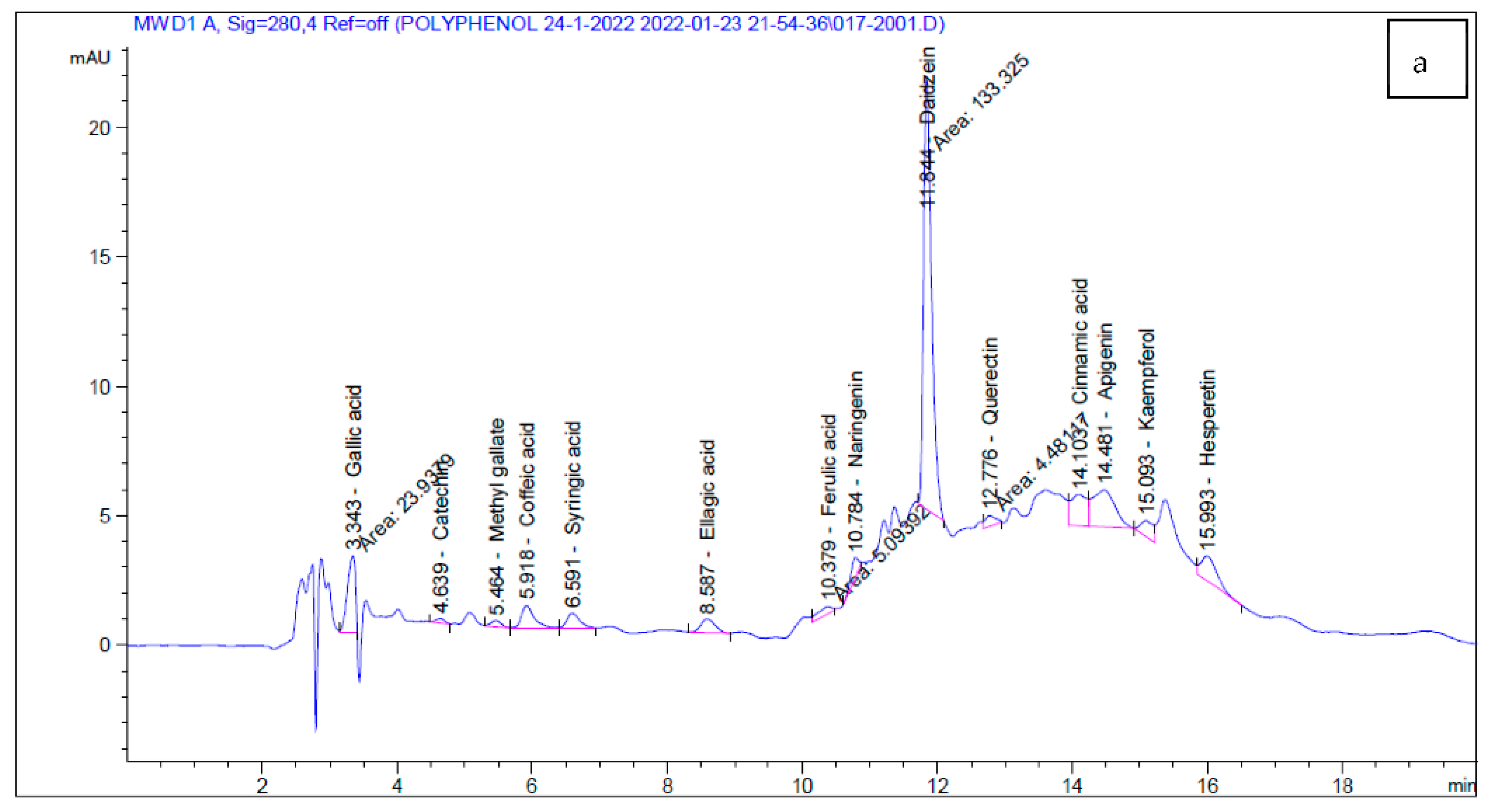
Acanthophora specifera flavonoid separation reveals a combination of chlorogenic acid (69.64%), caffeic acid (12.86%), vitexin-rahmnose (12.35%), quercetin (1.41%) and catechol (0.59%) [60]. The antioxidant activity of the flavonoid-enriched extract has been proven to be very high [61]
Phenolic compounds have exceptional antioxidant properties due to their capacity to function as chelating agents with reactive oxygen species, avoiding oxidative stress and cell damage [29,62]. As a result, scavenging of oxidants is critical for disease prevention, and phenolic compounds found in seaweeds are particularly helpful as a natural supply of antioxidant agents [52]. The study of Agregán et al. [63] demonstrated that aqueous polyphenolic extracts of Ascophyllum nodosum, Bifurcaria bifurcata and Fucus vesiculosus have antioxidant activity, which stabilize the canola oil oxidation level.
Nonetheless, synthetic ingredient limits in the food business may represent a tipping point for the use of seaweed compounds as safe replacements [64], since they also exhibit anti-microbial activity against key food spoilage and food pathogenic microorganisms [65]. Seaweed phenolic antioxidant extracts have been used to improve oxidative stability and to preserve or boost the intrinsic quality and nutritional content of foods [66,67]. The antioxidant potential is useful in the food industry not only for nutraceutical compounds on functional food products, where they are indisputably valuable for health improvement (as food supplements), but also to extend the shelf-life period when used in processed food (functional foods) [68,69]. Furthermore, the antibacterial activity of seaweed phenolics suggests that they can be valuable in the food industry [70].

2.3. Cytotoxicity Assay

The influence of various concentrations of Codium tomentosum and Actinotrichia fragilis aqueous extracts on PBMC viability are given in Table 5 and Table 6. The IC50 value for inhibiting proliferation on normal PBMC cells was 33.7 ± 1.02 µg/mL and 51.0 ± 1.14 µg/mL for C. tomentosum and Actinotrichia fragilis, respectively. The results indicated that both algal aqueous extracts were safe with low toxicity to normal cells and could be used as novel and effective food preservatives instead of chemical ones after performing in vivo assays, acting also as nutraceuticals [52].
The current study’s findings were consistent with those published by Alghazeer et al. [16], who reported that seaweeds rich in bioactive compounds may be used in anticancer drug research programs. However, further investigations are essential to reveal the molecular mechanisms of the anticancer activities of these algae.

2.4. HPLC Analysis

To identify bioactive compounds in C. tomentosum and A. fragilis aqueous extracts, high-performance liquid chromatography (HPLC) was used. The HPLC chromatogram of standards and phenolic compounds of C. tomentosum and A. fragilis aqueous extracts are shown in Table 7 and Figure 1.
The HPLC analysis results confirmed the presence of several polyphenolic compounds when compared to the HPLC standard chromatogram. These findings agreed with those of Tanna et al. [36], who revealed phenolic, flavonoid and amino acid compositions in some species of seaweeds that are promising as functional food ingredients or dietary supplements for daily intake.
HPLC analysis results of C. tomentosum and A. fragilis extracts confirmed the in vitro antioxidant and antimicrobial studies, which demonstrated their biochemical components. These active components are also considered the main reason for free radical scavenging activities of our target seaweeds, demonstrating the nutraceutical potential of these extracts and also highlighting them as a raw source of interesting natural compounds with pharmacological potential [52].

2.5. Sensory Evaluation of Fillet Fish Fortified with Algal Extracts

Regarding the bioactivity and biochemical potential of these two extracts, it is important to understand if they can be used as food additives and applied in the food industry [52,71]. Table 8 shows the sensory evaluation scores of fillet fish fortified with algal extracts. According to the findings, fillet fish fortified with algal extracts has been accepted as having high nutritional value for human diet. Interestingly, fillet fish fortified with C. tomentosum extract demonstrated the greatest overall acceptance score, as well as the highest antibacterial and antioxidant activity.
Water extracts were used because they are safer solvents than other organic solvents. Water extracts contain a variety of constituents, including polysaccharides and polyphenols, but we focused on polyphenols in this study because they are powerful antioxidants and antibacterial agents, as reported previously by several studies. This important observation will be made in subsequent work.
In this preliminary work, we treated the fillet fish with aqueous extracts mixed with spices immediately before frying in oil. Only sensory evaluation was performed on the fillet fish fortified with algal extracts in an attempt to improve its nutritional value in comparison to our standard fillet fish diets.
Our future studies will focus on fortifying fillet fish or yogurt with natural seaweed extracts as natural preservatives instead of chemical ones. pH values, titratable acidities (TAs), total soluble solids, water holding capacity (WHC), thiobarbituric acid reactive substances (TBARS), lipid oxidation, protein oxidation (modified DNPH carbonyl assay) and microbiological properties will be evaluated over 30 days of storage.
The constraints on synthetic components in the food business may be a tipping moment for the use of seaweed compounds as safe replacements [64], as they also exhibit antimicrobial properties against major food spoilage and pathogenic microbes [65]. Seaweed phenolic antioxidant extracts have been used to improve oxidative stability and to preserve or boost the intrinsic quality and nutritional content of foods [66,67].
The antioxidant potential is beneficial in the food business not only as nutraceutical compounds in functional food items, where they are undeniably valuable for health improvement (as food supplements), but also to extend the shelf-life period when included in processed foods (functional foods) [68,69]. Furthermore, the antibacterial properties of seaweed phenolics suggest that they may be valuable in the food business [70].
Furthermore, bromophenols enriched extract from Ulva lactuca and Pterocladiella capillacea were investigated as “marine flavour” agents in farmed fish and other aquatic organisms, because farming final products can differ in flavor from wild catches, and this can be incorporated as a feed ingredient or a seaweed bromophenol-enriched sauce [72].

3. Materials and Methods

3.1. Algae Collection

Two marine algae species, Codium tomentosum Stackhouse, a green marine macroalgae (phylum Chlorophyta, class Ulvophyceae), and Actinotrichia fragilis Forsskal, Borgesen, a red marine macroalgae (phylum Rhodophyta, class Florideophyceae), were harvested in summer 2021 from the tidal zone of the Red Sea shore at Hurghada, Egypt, between latitude 27°28.15′ N and longitude 33°77.13′ E (Figure 2). The seaweed species were identified under a stereo microscope according to their morphological characteristics with taxonomic references following the descriptions of Aleem [73]. The two macroalgal species were collected randomly from the site in the summer season. Because of the large amount of collected algae used in the extraction, no reproductive stage was recorded.

3.2. Preparation of Algal Extracts

About 0.5 kg of each alga was washed with water, shade-dried and powdered in a mixer grinder. Hot water (80 °C) was used to extract the compounds from the samples (1:2, w/v) for one hour before separating the filtrate and residue. The extraction was repeated three times under similar conditions. Last, freeze-drying the filtrate yielded powdered algae aqueous extracts. The extracts were then kept at −20 °C for subsequent study.

3.3. Antibacterial Activity and MIC Determination

Agar well diffusion tests described by Abd El Hafez et al. [5] and Kadaikunnan et al. [74] were applied to detect the action of the extracts against the reference pathogenic strains—Gram-negative bacteria Klebsiella pneumonia (ATCC700603) and E. coli (ATCC25922), and Gram-positive bacteria Staphylococcus aureus (ATCC6538) and Streptococcus pyogenes (EMCC1772)—obtained from City of Scientific Research and Technological Applications, by using 100 μL of the inoculums (1 × 10⁸ CFU/mL). Using descending concentrations, the minimum inhibitory concentration (MIC) of algal extracts toward the reference strains was established. In sterile saline, growing culture suspensions of the reference pathogenic strains were prepared and adapted to a concentration of 106 cells/mL. The reference pathogenic strain suspension was inoculated onto a plate of nutrient agar (Oxoid, U.K.) and left to dry for 15 min at room temperature. The algal extracts were serially diluted, and 100 μL of each dilution was independently applied to each well. The plates were incubated for 24 h at 37 °C before the MIC values were recorded. For each culture algal extract, the test was performed in triplicate.

3.4. Determination of Total Phenolic Contents

The total phenolic content was determined using the Folin–Ciocalteu reagent following [75] using gallic acid as a reference. Each sample was evaluated in three replicates. A 0.1 mL aliquot of Folin–Ciocalteu reagent was added to 0.1 mL of reconstituted extract. Then, 2.0 mL saturated sodium carbonate (7%) was added to the mixture after 15 min. For 30 min, the mixture was left at room temperature. TPC was determined using a spectrophotometer at 760 nm with gallic acid as a reference. The linear regression equation generated from the standard gallic acid calibration curve was used to calculate TPC as milligrams of gallic acid equivalent per gram sample. Each sample was evaluated in triplicate.

3.5. Determination of Total Flavonoid Contents

The total flavonoid contents were determined using a colorimetric method described by Sakanaka et al. [76]. First, 1.0 mL of the sample and 4.0 mL of water were mixed in a flask. Then, 0.75 mL of 5% sodium nitrite and 0.150 mL of 10% aluminum chloride were added to the mixture. After 5 min at room temperature, 0.5 mL of 1 M sodium hydroxide was added. A UV/VIS spectrophotometer was used to measure the absorbance at 510 nm. The results were expressed as milligram catechol equivalent per gram sample from the standard catechol calibration curve.

3.6. Antioxidant Potentials and DPPH Radical Scavenging Activity

The extracts’ free radical scavenging activity was determined using the DPPH method described by Catarino et al. [77]. Ascorbic acid was used as a standard with different ranges (10–100 μg/mL) to generate the standard curve. The absorbance was measured at 517 nm using a spectrophotometer. The activity of DPPH radical scavenging was determined as mg ascorbic acid equivalent (AAE)/g dry sample. The percentage of DPPH radical scavenging activity was calculated using the following Equation (1):
DPPH   radical   scavenging   activity   ( %   inhibition ) = Abs control   Abs sample   Abs control   × 100  

3.7. HPLC Analysis

The phenolic profile of algal aqueous extracts was screened using an HPLC (Agilent 1260 series). An Eclipse C18 column with (4.6 mm × 250 mm i.d., 5 μm) was used and its temperature was kept constant at 40 °C. The mobile phase was water (A) and 0.05% trifluoroacetic acid in acetonitrile (B) and the volume of injection was 5 μL. The flow rate was 0.9 mL/min and the multi-wavelength detector was monitored at 280 nm.

3.8. Cytotoxicity Assay

In a 96-well tissue culture plate, the IC50 value of algal extracts on peripheral blood mononuclear cells (PBMCs) was defined using the neutral red cytotoxicity assay [78,79]. Extract concentrations of 250, 125, 62.5, 31.2, 15.6 and 7.8 µg/mL were obtained using double-fold serial dilutions. The cells in the wells were individually treated with various concentrations of extract, 150 µL in every well except for the blank. The following Formula (2) was utilized to determine the percentage of cytotoxicity (inhibition):
%   Inhibition = O . D   Control O . D   Treatment O . D   Control

3.9. Sensory Evaluation of Fillet Fish Fortified with Algal Extracts

A total of 1 kg of fillet fish was treated by adding 1% of powdered algae aqueous extracts (C. tomentosum and A. fragilis) and then fried in oil. The samples were divided into four treatments: Treatment 1 (T1) as control (without extracts), Treatment 2 (T2) with 1% of C. tomentosum extract, Treatment 3 (T3) with 1% of A. fragilis extract and Treatment 4 (T4) as a mix with 0.5% of C. tomentosum and 0.5% of A. fragilis extracts.
The sensory evaluation panel included 15 members from the Food Technology Department and other departments, Arid Lands Cultivation Research Institute (ACRI), City of Scientific Research and Technological Applications (SRTA-City). The panel members were asked to observe some items (appearance, consistency, tenderness, flavor and overall eating quality) and give scores from 1 to 7 (excellent 7; very good 6; good 5; medium 4; fair 3; poor 2; very poor 1).

3.10. Statistical Analysis

All data collected were analyzed using one-way ANOVA, Fisher’s grouping test Minitab® (Version 16) software, n = 3, and p < 0.05 revealed a significant difference. Significant means were compared by Duncan’s post hoc multiple comparison test.

4. Conclusions

It is well established that seaweeds contain various phytochemicals with diverse biological activities, are considered safe for human consumption and have significant health benefits. The current study provides approximate data on the phytochemical constituents of marine seaweeds Codium tomentosum and Actinotrichia fragilis aqueous extracts for use as food supplements fortified in fillet fish. Bioactive compounds found in seaweeds are awaiting a significant breakthrough to be used as natural antibacterial and antioxidant supplements in various food products. Moreover, these seaweed extracts have antibacterial activity against bacterial infections, lending credence to their traditional use and implying a future role for these seaweeds in combating microbial populations. The nutritional and physiological benefits of seaweeds suggest that they may be introduced to our regular diets as natural preservatives to promote health and immune system function, as well as enhance new pharmaceutical and food applications.
These results demonstrate that C. tomentosum aqueous extract can be further exploited. There is a need to advance our understanding of this interesting extract’s impact after fortified fillet consumption to understand if the bioactivity is maintained, improved or lowered.

Author Contributions

Conceptualization, M.S.M.A.E.H., S.H.R., N.M.A. and H.E.A.E.-H.; methodology, M.S.M.A.E.H., S.H.R., N.M.A. and H.E.A.E.-H.; validation, J.C. and L.P.; formal analysis, M.S.M.A.E.H., S.H.R., N.M.A., H.E.A.E.-H. and J.C.; investigation, M.S.M.A.E.H., S.H.R., N.M.A. and H.E.A.E.-H.; writing—original draft preparation, M.S.M.A.E.H., S.H.R., N.M.A. and H.E.A.E.-H.; writing—review and editing, J.C. and L.P.; visualization, J.C. and L.P.; supervision, S.H.R. and L.P. All authors have read and agreed to the published version of the manuscript.

Funding

This research received no external funding.

Institutional Review Board Statement

Not applicable.

Informed Consent Statement

Not applicable.

Data Availability Statement

Data are available from authors.

Acknowledgments

João Cotas and Leonel Pereira are grateful for the support of the FCT—Foundation for Science and Technology, I.P., within the scope of the project LA/P/0069/2020 granted to the Associate Laboratory ARNET, UIDB/04292/2020 granted to MARE—Marine and Environmental Sciences Centre.

Conflicts of Interest

The authors declare no conflict of interest.

References

  1. El Feky, S.E.; Abd El Hafez, M.S.M.; Abd El Moneim, N.A.; Ibrahim, H.A.H.; Okbah, M.A.; Ata, A.; El Sedfy, A.S.; Hussein, A. Cytotoxic and Antimicrobial Activities of Two New Sesquiterpenoids from Red Sea Brittle Star Ophiocoma dentata. Sci. Rep. 2022, 12, 8209. [Google Scholar] [CrossRef]
  2. Shinde, P.; Banerjee, P.; Mandhare, A. Marine Natural Products as Source of New Drugs: A Patent Review (2015–2018). Expert Opin. Ther. Pat. 2019, 29, 283–309. [Google Scholar] [CrossRef]
  3. Ghosh, S.; Sarkar, T.; Pati, S.; Kari, Z.A.; Edinur, H.A.; Chakraborty, R. Novel Bioactive Compounds From Marine Sources as a Tool for Functional Food Development. Front. Mar. Sci. 2022, 9, 832957. [Google Scholar] [CrossRef]
  4. Abd El Hafez, M.S.M.; El Aziz Okbah, M.A.; Ibrahim, H.A.H.; Hussein, A.A.E.R.; El Moneim, N.A.A.; Ata, A. First Report of Steroid Derivatives Isolated from Starfish Acanthaster planci with Anti-Bacterial, Anti-Cancer and Anti-Diabetic Activities. Nat. Prod. Res. 2022, 36, 5545–5552. [Google Scholar] [CrossRef]
  5. Abd El Hafez Et Al, M.S. Biological Activities of Secondary Metabolites from Turbinaria triquetra (Phaeophyceae), Hypnea cornuta (Florideophyceae), and Ulva prolifera (Ulvophyceae) Methanolic Extracts. Egypt. J. Aquat. Biol. Fish. 2022, 26, 1227–1246. [Google Scholar] [CrossRef]
  6. Pereira, A.G.; Fraga-Corral, M.; Garcia-Oliveira, P.; Lourenço-Lopes, C.; Carpena, M.; Prieto, M.A.; Simal-Gandara, J. The Use of Invasive Algae Species as a Source of Secondary Metabolites and Biological Activities: Spain as Case-Study. Mar. Drugs 2021, 19, 178. [Google Scholar] [CrossRef]
  7. Lomartire, S.; Cotas, J.; Pacheco, D.; Marques, J.C.; Pereira, L.; Gonçalves, A.M.M. Environmental Impact on Seaweed Phenolic Production and Activity: An Important Step for Compound Exploitation. Mar. Drugs 2021, 19, 245. [Google Scholar] [CrossRef]
  8. Abd El Hafez, M.S.M.; ElKomy, R.G.; Saleh, H.; Aboul-Ela, H.M. Extracts of the Green Algae Ulva Prolifera Possess Antioxidant and Antibacterial Activities In Vitro. Egypt. J. Aquat. Biol. Fish. 2020, 24, 267–280. [Google Scholar] [CrossRef]
  9. Shah, S.A.A.; Hassan, S.S.U.; Bungau, S.; Si, Y.; Xu, H.; Rahman, M.H.; Behl, T.; Gitea, D.; Pavel, F.-M.; Corb Aron, R.A.; et al. Chemically Diverse and Biologically Active Secondary Metabolites from Marine Phylum Chlorophyta. Mar. Drugs 2020, 18, 493. [Google Scholar] [CrossRef]
  10. Pasdaran, A.; Hamedi, A.; Mamedov, N.A. Antibacterial and Insecticidal Activity of Volatile Compounds of Three Algae Species of Oman Sea. Int. J. Second. Metab. 2016, 3, 66–73. [Google Scholar] [CrossRef]
  11. Sanjeewa, K.K.A.; Kim, E.-A.; Son, K.-T.; Jeon, Y.-J. Bioactive Properties and Potentials Cosmeceutical Applications of Phlorotannins Isolated from Brown Seaweeds: A Review. J. Photochem. Photobiol. B Biol. 2016, 162, 100–105. [Google Scholar] [CrossRef]
  12. Salehi, B.; Sharifi-Rad, J.; Seca, A.M.; Pinto, D.C.; Michalak, I.; Trincone, A.; Mishra, A.P.; Nigam, M.; Zam, W.; Martins, N. Current Trends on Seaweeds: Looking at Chemical Composition, Phytopharmacology, and Cosmetic Applications. Molecules 2019, 24, 4182. [Google Scholar] [CrossRef] [Green Version]
  13. Abd El Hafez, M.S.M.; Abd El-Wahab, M.G.; Seadawy, M.G.; El-Hosseny, M.F.; Beskales, O.; Saber Ali Abdel-Hamid, A.; El Demellawy, M.A.; Ghareeb, D.A. Characterization, In-Silico, and In-Vitro Study of a New Steroid Derivative from Ophiocoma dentata as a Potential Treatment for COVID-19. Sci. Rep. 2022, 12, 5846. [Google Scholar] [CrossRef]
  14. Ganesan, A.R.; Tiwari, U.; Rajauria, G. Seaweed Nutraceuticals and Their Therapeutic Role in Disease Prevention. Food Sci. Hum. Wellness 2019, 8, 252–263. [Google Scholar] [CrossRef]
  15. Pradhan, B.; Bhuyan, P.P.; Patra, S.; Nayak, R.; Behera, P.K.; Behera, C.; Behera, A.K.; Ki, J.-S.; Jena, M. Beneficial Effects of Seaweeds and Seaweed-Derived Bioactive Compounds: Current Evidence and Future Prospective. Biocatal. Agric. Biotechnol. 2022, 39, 102242. [Google Scholar] [CrossRef]
  16. Alghazeer, R.; Howell, N.K.; El-Naili, M.B.; Awayn, N. Anticancer and Antioxidant Activities of Some Algae from Western Libyan Coast. Nat. Sci. 2018, 10, 1–24. [Google Scholar] [CrossRef] [Green Version]
  17. Gheda, S.; Naby, M.A.; Mohamed, T.; Pereira, L.; Khamis, A. Antidiabetic and Antioxidant Activity of Phlorotannins Extracted from the Brown Seaweed Cystoseira compressa in Streptozotocin-Induced Diabetic Rats. Environ. Sci. Pollut. Res. 2021, 28, 22886–22901. [Google Scholar] [CrossRef]
  18. Besednova, N.N.; Andryukov, B.G.; Zaporozhets, T.S.; Kryzhanovsky, S.P.; Kuznetsova, T.A.; Fedyanina, L.N.; Makarenkova, I.D.; Zvyagintseva, T.N. Algae Polyphenolic Compounds and Modern Antibacterial Strategies: Current Achievements and Immediate Prospects. Biomedicines 2020, 8, 342. [Google Scholar] [CrossRef]
  19. Mekinić, I.G.; Skroza, D.; Šimat, V.; Hamed, I.; Čagalj, M.; Perković, Z.P. Phenolic Content of Brown Algae (Pheophyceae) Species: Extraction, Identification, and Quantification. Biomolecules 2019, 9, 244. [Google Scholar] [CrossRef] [Green Version]
  20. Cotas, J.; Leandro, A.; Pacheco, D.; Gonçalves, A.M.M.; Pereira, L. A Comprehensive Review of the Nutraceutical and Therapeutic Applications of Red Seaweeds (Rhodophyta). Life 2020, 10, 19. [Google Scholar] [CrossRef]
  21. Swanson, B.G. Tannins and Polyphenols. In Encyclopedia of Food Sciences and Nutrition; Caballero, B., Ed.; Academic Press: Cambridge, MA, USA, 2003; pp. 5729–5733. [Google Scholar]
  22. Bravo, L. Polyphenols: Chemistry, Dietary Sources, Metabolism, and Nutritional Significance. Nutr. Rev. 2009, 56, 317–333. [Google Scholar] [CrossRef]
  23. Shahidi, F.; Naczk, M. Phenolics in Food and Nutraceuticals; CRC Press: Boca Raton, FL, USA, 2003; ISBN 9780367395094. [Google Scholar]
  24. Dai, J.; Mumper, R.J. Plant Phenolics: Extraction, Analysis and Their Antioxidant and Anticancer Properties. Molecules 2010, 15, 7313–7352. [Google Scholar] [CrossRef]
  25. Wells, M.L.; Potin, P.; Craigie, J.S.; Raven, J.A.; Merchant, S.S.; Helliwell, K.E.; Smith, A.G.; Camire, M.E.; Brawley, S.H. Algae as Nutritional and Functional Food Sources: Revisiting Our Understanding. J. Appl. Phycol. 2017, 29, 949–982. [Google Scholar] [CrossRef]
  26. Heo, S.J.; Park, E.J.; Lee, K.W.; Jeon, Y.J. Antioxidant Activities of Enzymatic Extracts from Brown Seaweeds. Bioresour. Technol. 2005, 96, 1613–1623. [Google Scholar] [CrossRef]
  27. Corona, G.; Coman, M.M.; Guo, Y.; Hotchkiss, S.; Gill, C.; Yaqoob, P.; Spencer, J.P.E.; Rowland, I. Effect of Simulated Gastrointestinal Digestion and Fermentation on Polyphenolic Content and Bioactivity of Brown Seaweed Phlorotannin-Rich Extracts. Mol. Nutr. Food Res. 2017, 61, 1700223. [Google Scholar] [CrossRef] [Green Version]
  28. Gómez-Guzmán, M.; Rodríguez-Nogales, A.; Algieri, F.; Gálvez, J. Potential Role of Seaweed Polyphenols in Cardiovascular-Associated Disorders. Mar. Drugs 2018, 16, 250. [Google Scholar] [CrossRef] [Green Version]
  29. Audibert, L.; Fauchon, M.; Blanc, N.; Hauchard, D.; Ar Gall, E. Phenolic Compounds in the Brown Seaweed Ascophyllum Nodosum: Distribution and Radical-Scavenging Activities. Phytochem. Anal. 2010, 21, 399–405. [Google Scholar] [CrossRef]
  30. Chen, Y.; Lin, H.; Li, Z.; Mou, Q. The Anti-Allergic Activity of Polyphenol Extracted from Five Marine Algae. J. Ocean Univ. China 2015, 14, 681–684. [Google Scholar] [CrossRef]
  31. Murray, M.; Dordevic, A.; Ryan, L.; Bonham, M. The Impact of a Single Dose of a Polyphenol-Rich Seaweed Extract on Postprandial Glycaemic Control in Healthy Adults: A Randomised Cross-Over Trial. Nutrients 2018, 10, 270. [Google Scholar] [CrossRef] [Green Version]
  32. Pangestuti, R.; Kim, S.-K. Neuroprotective Effects of Marine Algae. Mar. Drugs 2011, 9, 803–818. [Google Scholar] [CrossRef]
  33. Holdt, S.L.; Kraan, S. Bioactive Compounds in Seaweed: Functional Food Applications and Legislation. J. Appl. Phycol. 2011, 23, 543–597. [Google Scholar] [CrossRef]
  34. Liu, M.; Hansen, P.E.; Lin, X. Bromophenols in Marine Algae and Their Bioactivities. Mar. Drugs 2011, 9, 1273–1292. [Google Scholar] [CrossRef] [Green Version]
  35. Thomas, N.V.; Kim, S.K. Potential Pharmacological Applications of Polyphenolic Derivatives from Marine Brown Algae. Environ. Toxicol. Pharmacol. 2011, 32, 325–335. [Google Scholar] [CrossRef]
  36. Tanna, B.; Brahmbhatt, H.R.; Mishra, A. Phenolic, Flavonoid, and Amino Acid Compositions Reveal That Selected Tropical Seaweeds Have the Potential to Be Functional Food Ingredients. J. Food Process. Preserv. 2019, 43, e14266. [Google Scholar] [CrossRef]
  37. Nwosu, F.; Morris, J.; Lund, V.A.; Stewart, D.; Ross, H.A.; McDougall, G.J. Anti-Proliferative and Potential Anti-Diabetic Effects of Phenolic-Rich Extracts from Edible Marine Algae. Food Chem. 2011, 126, 1006–1012. [Google Scholar] [CrossRef]
  38. Lopes, G.; Sousa, C.; Silva, L.R.; Pinto, E.; Andrade, P.B.; Bernardo, J.; Mouga, T.; Valentão, P. Can Phlorotannins Purified Extracts Constitute a Novel Pharmacological Alternative for Microbial Infections with Associated Inflammatory Conditions? PLoS ONE 2012, 7, e31145. [Google Scholar] [CrossRef]
  39. Urquiaga, I.; Leighton, F. Plant Polyphenol Antioxidants and Oxidative Stress. Biol. Res. 2000, 33, 55–64. [Google Scholar] [CrossRef]
  40. Pereira, L. Therapeutic and Nutritional Uses of Algae; CRC Press: Boca Raton, FL, USA, 2018; ISBN 9781498755382. [Google Scholar]
  41. Ravikumar, S.; Jacob Inbaneson, S.; Suganthi, P. Seaweeds as a Source of Lead Compounds for the Development of New Antiplasmodial Drugs from South East Coast of India. Parasitol. Res. 2011, 109, 47–52. [Google Scholar] [CrossRef]
  42. Maqsood, S.; Benjakul, S.; Shahidi, F. Emerging Role of Phenolic Compounds as Natural Food Additives in Fish and Fish Products. Crit. Rev. Food Sci. Nutr. 2013, 53, 162–179. [Google Scholar] [CrossRef]
  43. Panzella, L.; Napolitano, A. Natural Phenol Polymers: Recent Advances in Food and Health Applications. Antioxidants 2017, 6, 30. [Google Scholar] [CrossRef]
  44. Leandro, A.; Pereira, L.; Gonçalves, A.M.M. Diverse Applications of Marine Macroalgae. Mar. Drugs 2020, 18, 17. [Google Scholar] [CrossRef] [Green Version]
  45. Arguelles, E.D.L.R. Evaluation of Nutritional Composition and in Vitro Antioxidant and Antibacterial Activities of Codium intricatum Okamura from Ilocos Norte (Philippines). Jordan J. Biol. Sci. 2020, 13, 375–382. [Google Scholar]
  46. Koz, F.F.Y.; Yavasoglu, N.U.K.; Demirel, Z.; Sukatar, A.; Ozdemir, G. Antioxidant and Antimicrobial Activities of Codium fragile (Suringar) Hariot (Chlorophyta) Essential Oil and Extracts. Asian J. Chem. 2009, 21, 1197–1209. [Google Scholar]
  47. Rajauria, G.; Jaiswal, A.K.; Abu-Gannam, N.; Gupta, S. Antimicrobial, antioxidant and free radical-scavenging capacity of brown seaweed Himanthalia elongata from western coast of Ireland. J. Food Biochem. 2013, 37, 322–335. [Google Scholar] [CrossRef]
  48. Elkhateeb, M.I.; El-Bitar, A.M.H.; Saleh, S.R.; Abdelreheem, A.M.A. Evaluation of Bioactive Phytochemical Characterization, Antioxidant, Antimicrobial, and Antihemolytic Properties of Some Seaweeds Collected from Red Sea Coast, Egypt. Egypt. J. Aquat. Biol. Fish. 2021, 25, 417–436. [Google Scholar] [CrossRef]
  49. Salem, W.M.; Galal, H.; Nasr El-deen, F. Screening for Antibacterial Activities in Some Marine Algae from the Red Sea (Hurghada, Egypt). Afr. J. Microbiol. Res. 2011, 5, 2160–2167. [Google Scholar] [CrossRef] [Green Version]
  50. Alghazeer, R.; Whida, F.; Abduelrhman, E.; Gammoudi, F.; Naili, M. In Vitro Antibacterial Activity of Alkaloid Extracts from Green, Red and Brown Macroalgae from Western Coast of Libya. Afr. J. Biotechnol. 2013, 12, 7086–7091. [Google Scholar]
  51. Eldrin De Los Reyes, A.; Arsenia Basaran, S. Bioactive Properties of Sargassum siliquosum J. Agardh (Fucales, Ochrophyta) and Its Potential as Source of Skin-Lightening Active Ingredient for Cosmetic Application. J. Appl. Pharm. Sci. 2020, 10, 51–58. [Google Scholar] [CrossRef]
  52. Cotas, J.; Leandro, A.; Monteiro, P.; Pacheco, D.; Figueirinha, A.; Gonçalves, A.M.M.; da Silva, G.J.; Pereira, L. Seaweed Phenolics: From Extraction to Applications. Mar. Drugs 2020, 18, 384. [Google Scholar] [CrossRef]
  53. Olugbami, J.O.; Gbadegesin, M.A.; Odunola, O.A. In Vitro Evaluation of the Antioxidant Potential, Phenolic and Flavonoid Contents of the Stem Bark Ethanol Extract of Anogeissus leiocarpus. Afr. J. Med. Med. Sci. 2014, 43, 101–109. [Google Scholar]
  54. Hazra, B.; Biswas, S.; Mandal, N. Antioxidant and Free Radical Scavenging Activity of Spondias pinnata. BMC Complement. Altern. Med. 2008, 8, 63. [Google Scholar] [CrossRef] [Green Version]
  55. Christodoulou, M.C.; Orellana Palacios, J.C.; Hesami, G.; Jafarzadeh, S.; Lorenzo, J.M.; Domínguez, R.; Moreno, A.; Hadidi, M. Spectrophotometric Methods for Measurement of Antioxidant Activity in Food and Pharmaceuticals. Antioxidants 2022, 11, 2213. [Google Scholar] [CrossRef]
  56. Nita, M.; Grzybowski, A. The Role of the Reactive Oxygen Species and Oxidative Stress in the Pathomechanism of the Age-Related Ocular Diseases and Other Pathologies of the Anterior and Posterior Eye Segments in Adults. Oxidative Med. Cell. Longev. 2016, 2016, 3164734. [Google Scholar] [CrossRef] [Green Version]
  57. Juan, C.A.; Pérez de la Lastra, J.M.; Plou, F.J.; Pérez-Lebeña, E. The Chemistry of Reactive Oxygen Species (ROS) Revisited: Outlining Their Role in Biological Macromolecules (DNA, Lipids and Proteins) and Induced Pathologies. Int. J. Mol. Sci. 2021, 22, 4642. [Google Scholar] [CrossRef]
  58. Sharifi-Rad, M.; Anil Kumar, N.V.; Zucca, P.; Varoni, E.M.; Dini, L.; Panzarini, E.; Rajkovic, J.; Tsouh Fokou, P.V.; Azzini, E.; Peluso, I.; et al. Lifestyle, Oxidative Stress, and Antioxidants: Back and Forth in the Pathophysiology of Chronic Diseases. Front. Physiol. 2020, 11, 694. [Google Scholar] [CrossRef]
  59. Baskaran, K.; Rathi, M.A.; Nirmaladevi, N. Free Radical Scavenging Activity of Methanolic Extract of Marine Red Algae Actinotrichia fragilis. Asian J. Pharm. Pharmacol. 2019, 5, 876–883. [Google Scholar] [CrossRef]
  60. Jeyaprakash, R.R.K. HPLC Analysis of Flavonoids in Acanthophora specifera (Red Seaweed) Collected from Gulf of Mannar, Tamilnadu, India. Int. J. Sci. Res. 2017, 6, 69–72. [Google Scholar]
  61. Bilanglod, A.; Petthongkhao, S.; Churngchow, N. Phenolic and Flavonoid Contents Isolated from the Red Seaweed, Acanthophora spicifera. In Proceedings of the 31st Annual Meeting of the Thai Society for Biotechnology and International Conference (TSB2019), Phuket, Thailand, 10–12 November 2019; pp. 189–196. [Google Scholar]
  62. Ferreres, F.; Lopes, G.; Gil-Izquierdo, A.; Andrade, P.B.; Sousa, C.; Mouga, T.; Valentão, P. Phlorotannin Extracts from Fucales Characterized by HPLC-DAD-ESI-MS n: Approaches to Hyaluronidase Inhibitory Capacity and Antioxidant Properties. Mar. Drugs 2012, 10, 2766–2781. [Google Scholar] [CrossRef] [Green Version]
  63. Agregán, R.; Munekata, P.E.; Domínguez, R.; Carballo, J.; Franco, D.; Lorenzo, J.M. Proximate Composition, Phenolic Content and In Vitro Antioxidant Activity of Aqueous Extracts of the Seaweeds Ascophyllum nodosum, Bifurcaria bifurcata and Fucus vesiculosus. Effect of Addition of the Extracts on the Oxidative Stability of Canola Oil Unde. Food Res. Int. 2017, 99, 986–994. [Google Scholar] [CrossRef]
  64. Freile-Pelegrín, Y.; Robledo, D. Bioactive Phenolic Compounds from Algae. Bioact. Compd. Mar. Foods Plant Anim. Sources 2013, 6, 113–129. [Google Scholar] [CrossRef]
  65. Gupta, S.; Abu-Ghannam, N. Recent Developments in the Application of Seaweeds or Seaweed Extracts as a Means for Enhancing the Safety and Quality Attributes of Foods. Innov. Food Sci. Emerg. Technol. 2011, 12, 600–609. [Google Scholar] [CrossRef]
  66. Apostolidis, E.; Kwon, Y.I.; Shetty, K. Inhibition of Listeria monocytogenes by Oregano, Cranberry and Sodium Lactate Combination in Broth and Cooked Ground Beef Systems and Likely Mode of Action through Proline Metabolism. Int. J. Food Microbiol. 2008, 128, 317–324. [Google Scholar] [CrossRef]
  67. Lin, Y.T.; Labbe, R.G.; Shetty, K. Inhibition of Listeria monocytogenes in Fish and Meat Systems by Use of Oregano and Cranberry Phytochemical Synergies. Appl. Environ. Microbiol. 2004, 70, 5672–5678. [Google Scholar] [CrossRef] [Green Version]
  68. Wang, T.; Jónsdóttir, R.; Kristinsson, H.G.; Thorkelsson, G.; Jacobsen, C.; Hamaguchi, P.Y.; Ólafsdóttir, G. Inhibition of Haemoglobin-Mediated Lipid Oxidation in Washed Cod Muscle and Cod Protein Isolates by Fucus vesiculosus Extract and Fractions. Food Chem. 2010, 123, 321–330. [Google Scholar] [CrossRef]
  69. Chakraborty, K.; Joseph, D.; Praveen, N.K. Antioxidant Activities and Phenolic Contents of Three Red Seaweeds (Division: Rhodophyta) Harvested from the Gulf of Mannar of Peninsular India. J. Food Sci. Technol. 2015, 52, 1924–1935. [Google Scholar] [CrossRef]
  70. Eom, S.H.; Kim, Y.M.; Kim, S.K. Antimicrobial Effect of Phlorotannins from Marine Brown Algae. Food Chem. Toxicol. 2012, 50, 3251–3255. [Google Scholar] [CrossRef]
  71. Leandro, A.; Pacheco, D.; Cotas, J.; Marques, J.C.; Pereira, L.; Gonçalves, A.M.M. Seaweed’s Bioactive Candidate Compounds to Food Industry and Global Food Security. Life 2020, 10, 140. [Google Scholar] [CrossRef]
  72. Whitfield, F.B.; Helidoniotis, F.; Shaw, K.J.; Svoronos, D. Distribution of Bromophenols in Species of Marine Algae from Eastern Australia. J. Agric. Food Chem. 1999, 47, 2367–2373. [Google Scholar] [CrossRef]
  73. Aleem, A.A. Contribution to the Study of the Marine Algae of the Red Sea. III—Marine Algae from Obhor, in the Vicinity of Jeddah. Bull. Fac. Sci. K.A.U. Jeddah 1978, 2, 99–118. [Google Scholar]
  74. Kadaikunnan, S.; Rejiniemon, T.S.; Khaled, J.M.; Alharbi, N.S.; Mothana, R. In-Vitro Antibacterial, Antifungal, Antioxidant and Functional Properties of Bacillus amyloliquefaciens. Ann. Clin. Microbiol. Antimicrob. 2015, 14, 9. [Google Scholar] [CrossRef] [Green Version]
  75. Singleton, V.L.; Orthofer, R.; Lamuela-Raventós, R.M. Analysis of Total Phenols and Other Oxidation Substrates and Antioxidants by Means of Folin-Ciocalteu Reagent. In Methods in Enzymology; ACADEMIC PRESS, INC.: Cambridge, MA, USA, 1999; pp. 152–178. [Google Scholar]
  76. Sakanaka, S.; Tachibana, Y.; Okada, Y. Preparation and Antioxidant Properties of Extracts of Japanese Persimmon Leaf Tea (Kakinoha-Cha). Food Chem. 2005, 89, 569–575. [Google Scholar] [CrossRef]
  77. Catarino, M.D.; Silva, A.M.S.; Saraiva, S.C.; Sobral, A.J.F.N.; Cardoso, S.M. Characterization of Phenolic Constituents and Evaluation of Antioxidant Properties of Leaves and Stems of Eriocephalus africanus. Arab. J. Chem. 2018, 11, 62–69. [Google Scholar] [CrossRef] [Green Version]
  78. Panda, S.; Ravindran, B. Isolation of Human PBMCs. Bio-Protocol 2013, 3, e323. [Google Scholar] [CrossRef]
  79. Repetto, G.; del Peso, A.; Zurita, J.L. Neutral Red Uptake Assay for the Estimation of Cell Viability/Cytotoxicity. Nat. Protoc. 2008, 3, 1125–1131. [Google Scholar] [CrossRef]
Figure 1. HPLC chromatograms of (a) Codium tomentosum extract, (b) Actinotrichia fragilis extract and (c) standards.
Marinedrugs 20 00785 g001aMarinedrugs 20 00785 g001b
Figure 2. The collected marine algae: (a) Codium tomentosum (green algae) and (b) Actinotrichia fragilis (red algae).
Marinedrugs 20 00785 g002
Table 1. Antibacterial potential of algal extracts.
StrainsInhibition Zone Diameter (mm) 1
Codium tomentosum
(Green Algae)
Actinotrichia fragilis (Red Algae)
Gram-positive bacteria
Staphylococcus aureus (ATCC25923)22 ± 0.04 a18 ± 0.05 b
Streptococcus pyogenes (EMCC1772)20 ± 0.01 a16 ± 0.01 b
Gram-negative bacteria
Escherichia coli (ATCC25922)14 ± 0.08 a12 ± 0.05 b
Klebsiella pneumonia (ATCC700603)18 ± 0.02 a14 ± 0.07 b
1 All results are expressed as the means ± standard deviation; n = 3. Diameter includes 5 mm well diameter. Different letters (a and b) indicate significant differences at p ≤ 0.05.
Table 2. Minimum inhibitory concentration (MIC) of algal extracts against pathogenic bacteria indicated as a zone of inhibition for each concentration.
StrainsMIC (mg/mL) 1
Codium tomentosumActinotrichia fragilis
Gram-positive bacteria
Staphylococcus aureus (ATCC25923)0.4 ± 0.05 b0.8 ± 0.03 a
Streptococcus pyogenes (EMCC1772)0.4 ± 0.04 b0.8 ± 0.02 a
Gram-negative bacteria
Escherichia coli (ATCC25922)0.8 ± 0.01 b1.2 ± 0.03 a
Klebsiella pneumonia (ATCC700603)0.6 ± 0.04 b1.2 ± 0.01 a
1 Diameter includes 5 mm well diameter. MIC—minimum inhibition concentration (mg/mL). Different letters (a and b) indicate significant differences at p ≤ 0.05.
Table 3. Total phenolic and flavonoid contents of Codium tomentosum and Actinotrichia fragilis.
Test 1Codium tomentosumActinotrichia fragilis
Total phenolic content (mg/g of extract)32.28 ± 1.63 a19.96 ± 1.28 b
Total flavonoid content
(mg/g of extract)
4.54 ± 1.48 c3.86 ± 1.02 d
1 Means in the same column followed by different lowercase letters are significantly different (p < 0.05).
Table 4. Antioxidant activity of Codium tomentosum and Actinotrichia fragilis as measured by DPPH assay.
ExtractsDPPH (IC50) μg/mL
Ascorbic acid22.71 ± 0.03 c
Codium tomentosum75.32 ± 0.07 b
Actinotrichia fragilis94.43 ± 0.02 a
IC50 (μg/mL): Inhibitory concentrations at which 50% of DPPH radicals are scavenged. Means in the same column followed by different lowercase letters are significantly different (p < 0.05).
Table 5. The influence of various concentrations of Codium tomentosum aqueous extract on PBMC viability.
Concentration (µg/mL)Inhibition %Viability %
2506931
1256931
62.56436
31.23070
15.62971
7.82773
IC50 = 33.7 ± 1.02 µg/mL
Table 6. The influence of various concentrations of Actinotrichia fragilis aqueous extract on PBMC viability.
Concentration (µg/mL)Inhibition %Viability %
2507327
1256040
62.54753
31.23961
15.63763
7.81189
IC50 = 51.0 ± 1.14 µg/mL
Table 7. Phenolic compounds of algal extracts identified by HPLC.
Phenolic CompoundsCodium tomentosumActinotrichia fragilis
Conc. (µg/g)
Gallic acid174.32303.68
Chlorogenic acidND29.50
Catechin29.9287.10
Methyl gallate12.910.00
Caffeic acid70.2112.79
Syringic acid50.030.00
Pyro catecholND0.00
RutinND22.73
Ellagic acid232.69190.62
Coumaric acidND0.00
VanillinND3.36
Ferulic acid26.1315.99
Naringenin61.0923.77
Daidzein791.3910.47
Quercetin48.0365.78
Cinnamic acid35.516.10
Apigenin224.8830.28
Kaempferol77.280.00
Hesperetin75.900.00
ND: Not detected.
Table 8. Sensory evaluation of fillet fish fortified with algal extracts.
AppearanceConsistencyTendernessFlavor (Odor and Taste)Overall Acceptance
T1 (Control)4.60 ± 1.06 a5.07 ± 1.03 a3.53 ± 1.69 b4.13 ± 1.64 a4.53 ± 1.18 b
T2 (1% of C. tomentosum extract)5.80 ± 1.15 a5.67 ± 1.18 a5.13 ± 1.41 a5.60 ± 1.63 a5.93 ± 1.10 a
T3 (1% A. fragilis extract)4.73 ± 1.16 a4.87 ± 1.19 a4.80 ± 1.61 ab4.67 ± 1.11 a4.80 ± 0.77 b
T4 (0.5% of C. tomentosum and 0.5% of A. fragilis extracts)5.13 ± 1.41 a5.67 ± 1.05 a5.40 ± 1.59 a5.33 ± 1.39 a5.47 ± 1.45 a
Means in the same column followed by different lowercase letters are significantly different (p < 0.05).
Publisher’s Note: MDPI stays neutral with regard to jurisdictional claims in published maps and institutional affiliations.

