
Deep-Sea Natural Products from Extreme Environments: Cold Seeps and Hydrothermal Vents
by
 and
and
Mengjing Cong
1,2, 
Xiaoyan Pang
1,3,
Kai Zhao
1,
Yue Song
1,2,
Yonghong Liu
1,2,3 and
Junfeng Wang
1,2,3,* 1
CAS Key Laboratory of Tropical Marine Bio-Resources and Ecology, Guangdong Key Laboratory of Marine Materia Medica, Innovation Academy of South China Sea Ecology and Environmental Engineering, South China Sea Institute of Oceanology, Chinese Academy of Sciences, Guangzhou 510301, China
2
University of Chinese Academy of Sciences, 19 Yuquan Road, Beijing 100049, China
3
Southern Marine Science and Engineering Guangdong Laboratory (Guangzhou), Guangzhou 511458, China
*
Author to whom correspondence should be addressed.
Mar. Drugs 2022, 20(6), 404; https://doi.org/10.3390/md20060404
Submission received: 27 April 2022
/
Revised: 8 June 2022
/
Accepted: 14 June 2022
/
Published: 19 June 2022
(This article belongs to the Special Issue Bioactive Compounds from the Deep-Sea-Derived Microorganisms)
Abstract
:The deep sea has been proven to be a great treasure for structurally unique and biologically active natural products in the last two decades. Cold seeps and hydrothermal vents, as typical representatives of deep-sea extreme environments, have attracted more and more attention. This review mainly summarizes the natural products of marine animals, marine fungi, and marine bacteria derived from deep-sea cold seeps and hydrothermal vents as well as their biological activities. In general, there were 182 compounds reported, citing 132 references and covering the literature from the first report in 1984 up to March 2022. The sources of the compounds are represented by the genera Aspergillus sp., Penicillium sp., Streptomyces sp., and so on. It is worth mentioning that 90 of the 182 compounds are new and that almost 60% of the reported structures exhibited diverse bioactivities, which became attractive targets for relevant organic synthetic and biosynthetic studies.
1. Introduction
Extreme environments refer to areas close to the limits of life, such as cold seeps, hydrothermal vents, polar and hot regions, or marine areas with high salinity [1]. Because of the extreme conditions of pressure, temperature, or high concentrations of toxic elements, unique organisms are more likely to appear. Compared with other ecosystems, extreme environments have not been fully developed and utilized, due to the limited conditions and difficult sampling. In recent years, with the progress of technology and the further exploration of the deep sea, scientists have gradually realized the uniqueness of natural products from extreme environments [2].
Cold seeps are typical deep-sea, chemosynthetically driven ecosystems, characterized by methane-rich fluid emissions and distinctive sulfur oxidation–reduction reactions, which lead to a high abundance of specialized cold-seep microorganisms [3]. The temperature of cold seeps is 2–4 °C, basically the same as the temperature around the seafloor. Microorganisms and animals from deep-sea cold seeps, which could be a new source of biomedically important compounds, due to their unique habitat, are only beginning to be investigated. The great potential for natural product discovery in deep-sea cold seep organisms will undoubtedly accelerate the investigation of new drugs [4].
Hydrothermal vents are formed when water heated in the Earth’s crust and magma are forced explosively to the surface through rock fissures in volcanic regions. Since ocean hydrothermal vents are among the most dynamic environments on Earth, secondary metabolite diversity of this extreme environment is considerably high [5]. With advances in sample collecting techniques, deep-sea hydrothermal vents might be potential hot spots for natural product discovery [6].
Therefore, this review covers papers on metabolites isolated from deep-sea extreme environments, including cold seeps and hydrothermal vents, using databases such as SciFinder, Web of Science, and so on. The structures of these compounds and details of the source organisms and depth of collection are presented along with relevant biological activities of the metabolites and synthetic studies. A total number of 182 compounds are presented in this review, with 132 cited references.
2. Cold Seeps
2.1. Marine Animals
Marine animals generally contain high proportions of n-3 polyunsaturated fatty acids (PUFAs) [7], in particular, long-chain PUFAs, such as DHA and EPA. There is increasing evidence that specific dietary patterns including, for example, n-3 PUFAs may be beneficial in reducing breast cancer risk [8,9]. However, some bivalve symbiotic bacteria were found to contain a novel n-4 or n-7 family, which appears to be an adaptation to the extremely high pressure and low temperature of seawater [10].
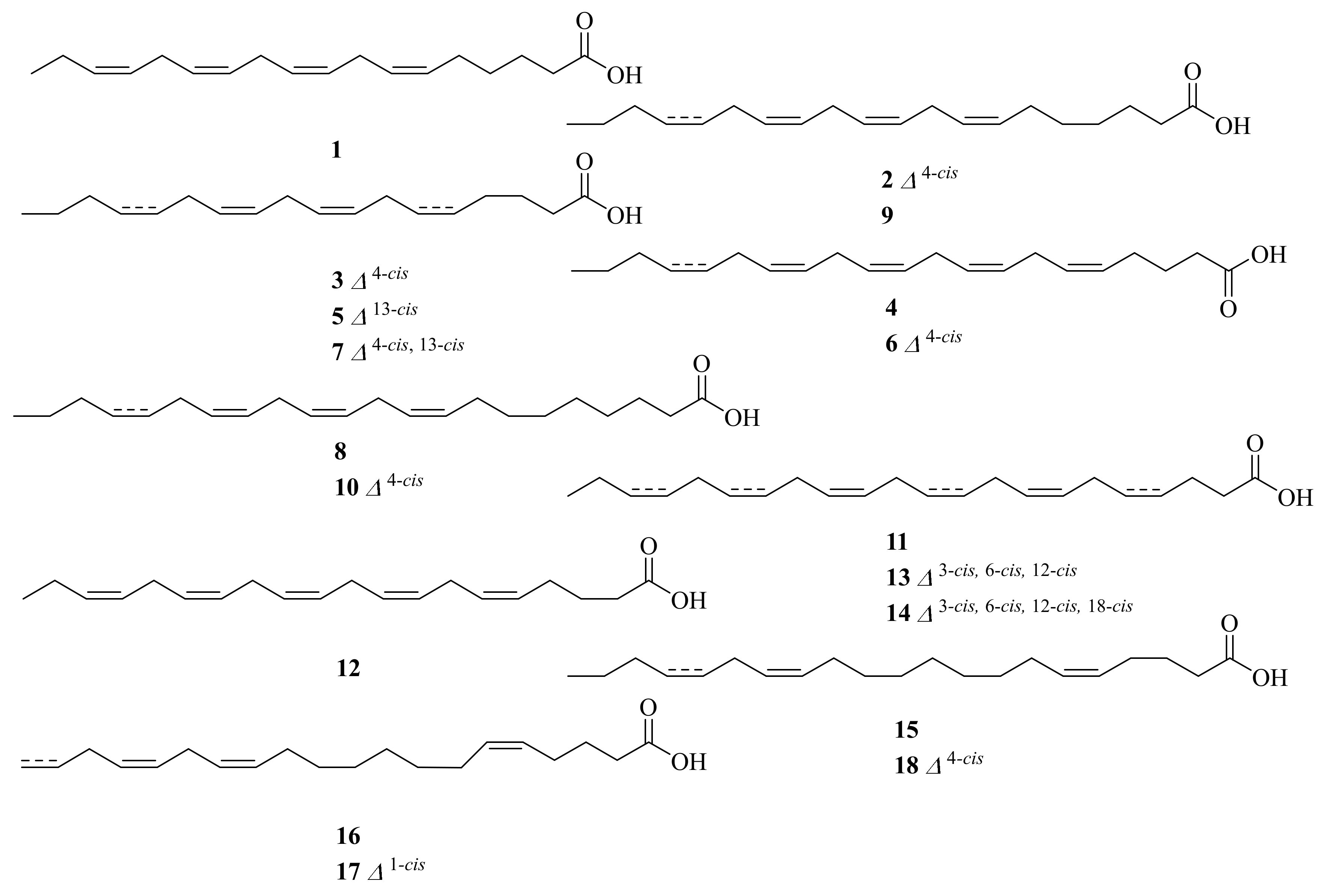
Novel fatty acids (1–10) (Figure 1) were purified from the two cold-seep-derived mussels Bathymodiolus japonicus and B. platifrons, collected at a depth of 1209 m at latitude 35°18′ N and longitude 139°13′ E in the Northern Pacific Ocean and a depth of 978 m at latitude 27°47′ N and longitude 126°54′ E in the East China Sea. The major PUFAs in the two mussels belong to unusual n-4 and n-7 methylene-interrupted PUFAs. B. japonicus and B. platifrons could maintain fluidity in plasma membrane lipids by accumulation of n-4 family methylene-interrupted PUFAs [11].
The cold-seep clam Calyptogena phaseoliformis, collected in the Japan Trench at a depth of 6354–6367 m, yielded eight novel fatty acids (11–18) (Figure 1). They were determined by gas chromatography–mass spectrometry analysis of 4,4-dimethyloxazoline derivatives. The major fatty acids present in C. phaseoliformis lipids belong to the n-4 family non-methylene-interrupted PUFAs [12].
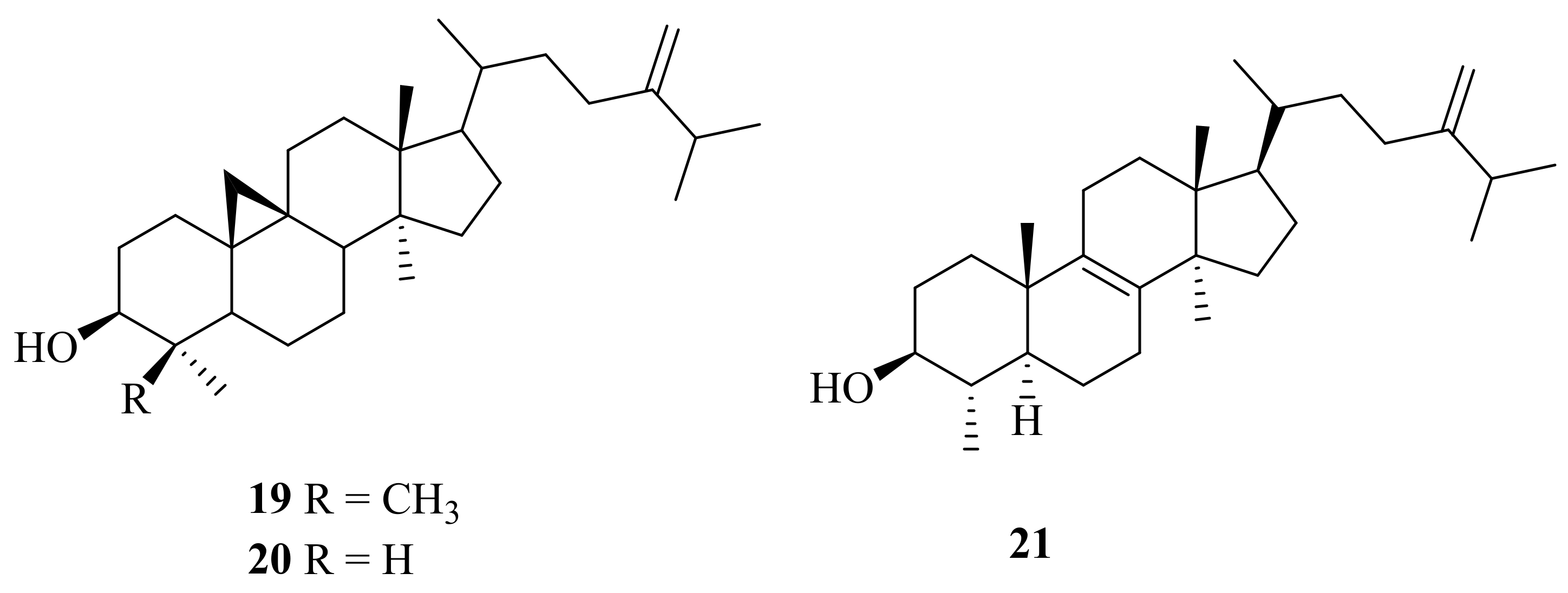
From the cold-seep bivalve Calyptogena soyoae, which was collected at a depth of 1100 m in Sagami Bay, three sterols (19–21) (Figure 2) were isolated [13]. Among them, 24-methylenecycloartanol (19) had shown many biological activities, such as significant anti-diabetic activity [14], strong activity against 12-O-tetradecanoylphorbol-13-acetate (TPA)-induced inflammation [15], and promising inhibition of growth of human breast cancer (MCF-7), with an IC50 value of 16.93 μM [16]. Cycloeucalenol (20) had been reported in the form of cycloeucalenol trans-ferulate in rice germ and was also found in Tinospora cordifolia or Guduchi. Its biological effects include cardiotonic [17], anti-fungal [18], and anti-inflammatory activities [15]. Compound 21 was evaluated for its cytotoxicity against MCF-7 and MDA-MB231, which clearly inhibited cell growth, with IC50 values of 29.33 ± 1.52 and 41.81 ± 2.42 μM, respectively [19].
2.2. Marine Fungi
2.2.1. Aspergillus sp.
Aspergillus is one of the most common and important genera of fungus. It has attracted more and more scientists’ attention, with a variety of active secondary metabolites [20,21].
A deep-sea-derived fungus, Aspergillus insuetus SD-512, which was obtained from cold seep sediments collected at a depth of 1331 m, yielded three new ophiobolin sesterterpenoids (22–24) and three new farnesylated phthalide derivatives, farnesylemefuranones D–F (30–32), along with five known ophiobolin analogs (25–29) (Figure 3). Of them, compound 24 displayed broad-spectrum antibacterial activities with minimum inhibitory concentration (MIC) values ranging from 4 to 32 μg/mL [22]. Compound 26 was found to be active against Escherichia coli, with inhibitory diameters of 10 mm [23]. Compounds 25–27 were evaluated for cytotoxic activity against murine L5178Y lymphoma cells. However, none of them showed significant activity [24]. Compounds 28 and 29 were firstly isolated from extracts of Emericella variecolor GF10, which was separated from marine sediment [25]. Compound 28 showed potent cytotoxicity, with GI50 (growth inhibition) values ranging from 0.20 to 0.30 µM, against six cancer cell lines, HCT-15, NUGC-3, NCI-H23, ACHN, PC-3, and MDA-MB-231 [26]. In addition, the total synthesis of (−)-6-epi-ophiobolin N (28) was reported [27]. 6-epi-Ophiobolin G (29) exhibited potent cytotoxic activity against HepG2, with an IC50 value of 0.37 μM [28]. Compound 30 exhibited inhibitory effects against the aquatic pathogens Vibrio vulnificus QDIO-4 and Vibrio alginolyticus QDIO-7, with a MIC value of 4 μg/mL, while compound 32 showed further activity against the aquatic bacteria Vibrio vulnificus QDIO-4, Vibrio alginolyticus QDIO-7, and Edwardsiella tarda QDIO-8, with a MIC value of 4 μg/mL [22].
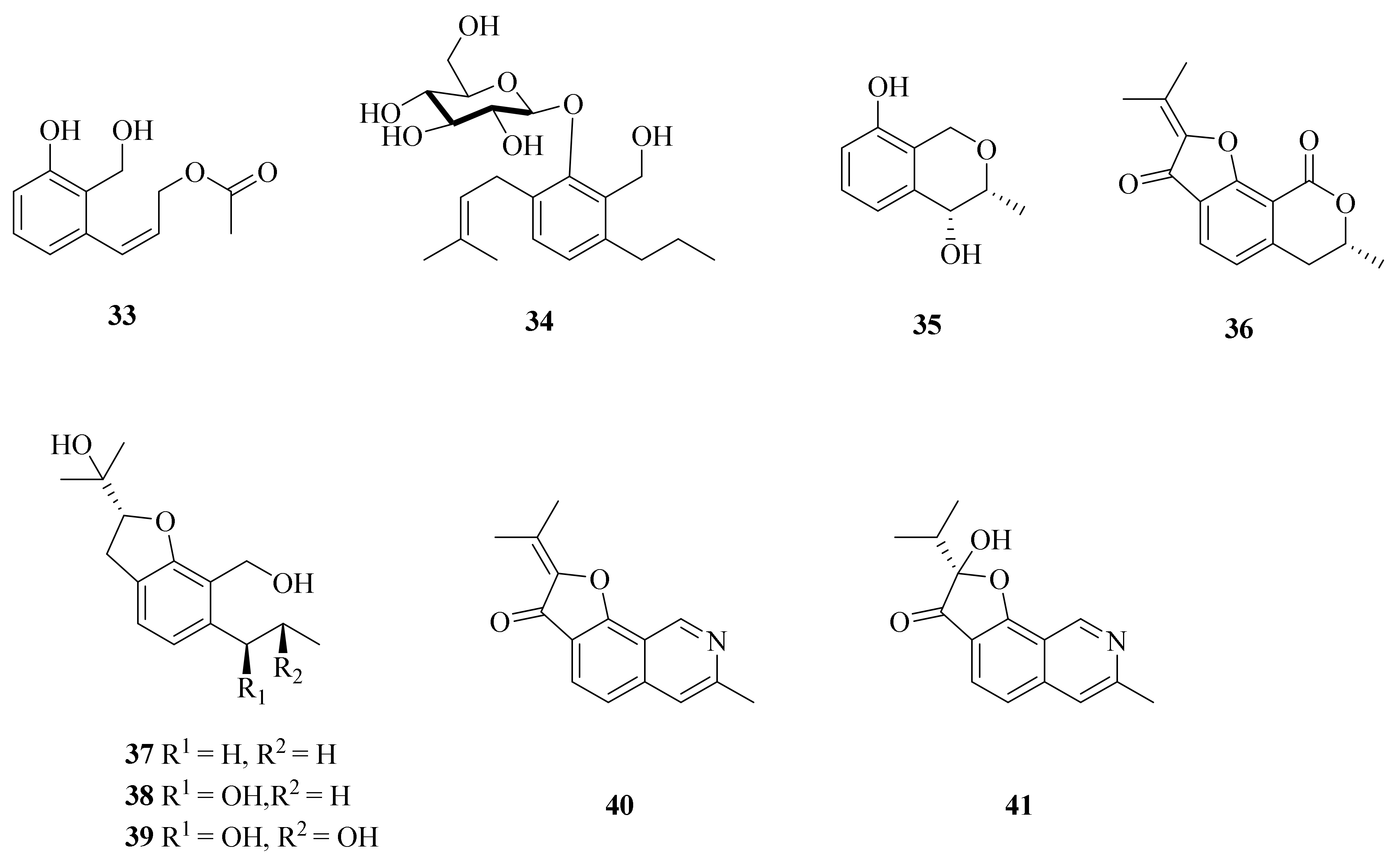
Moreover, Aspergillus insuetus SD-512 also yielded one new phenol derivative—acetylpeniciphenol (33)—along with eight known analogs (34–41) (Figure 4) [3]. Compound 33 was tested for antibacterial activities against six human or aquatic pathogens, while it exhibited an inhibitory effect against Edwardsiella tarda, Vibrio alginolyticus, and V. vulnificus, with MIC values of 4, 8, and 8 μg/mL, respectively [3]. Compound 34 displayed no significant activity in inhibiting LPS-induced NO production in RAW264.7 macrophages [29]. The biosynthetic pathway of penicisochroman E (35) was clarified; it involves epoxidation and cyclization followed by dehydration and subsequent hydrogenation [30]. (−)-Brassicadiol (37) exhibited cytotoxicity against both cancerous and non-cancerous (Vero) cells, with IC50 values ranging from 66.3 to 113.3 μM [31]. The synthesis of (−)-brassicadiol (37) was also described [32]. One study showed that daldinin C (38) was firstly isolated from cultures of the ascomycete Daldinia concentrica [33]. The anti-HIV activity of daldinin C (38) was tested, but the results were negative [34]. Penicisochroman I (39) showed weak cytotoxicity against KB and NCI-H187 cells [31]. TMC-120B (40) and TMC-120C (41) were observed to significantly lower PTZ-induced seizures in the larval zebrafish PTZ seizure model [35]. Because compound 41 has significant activity, its total synthesis route was also studied [36].
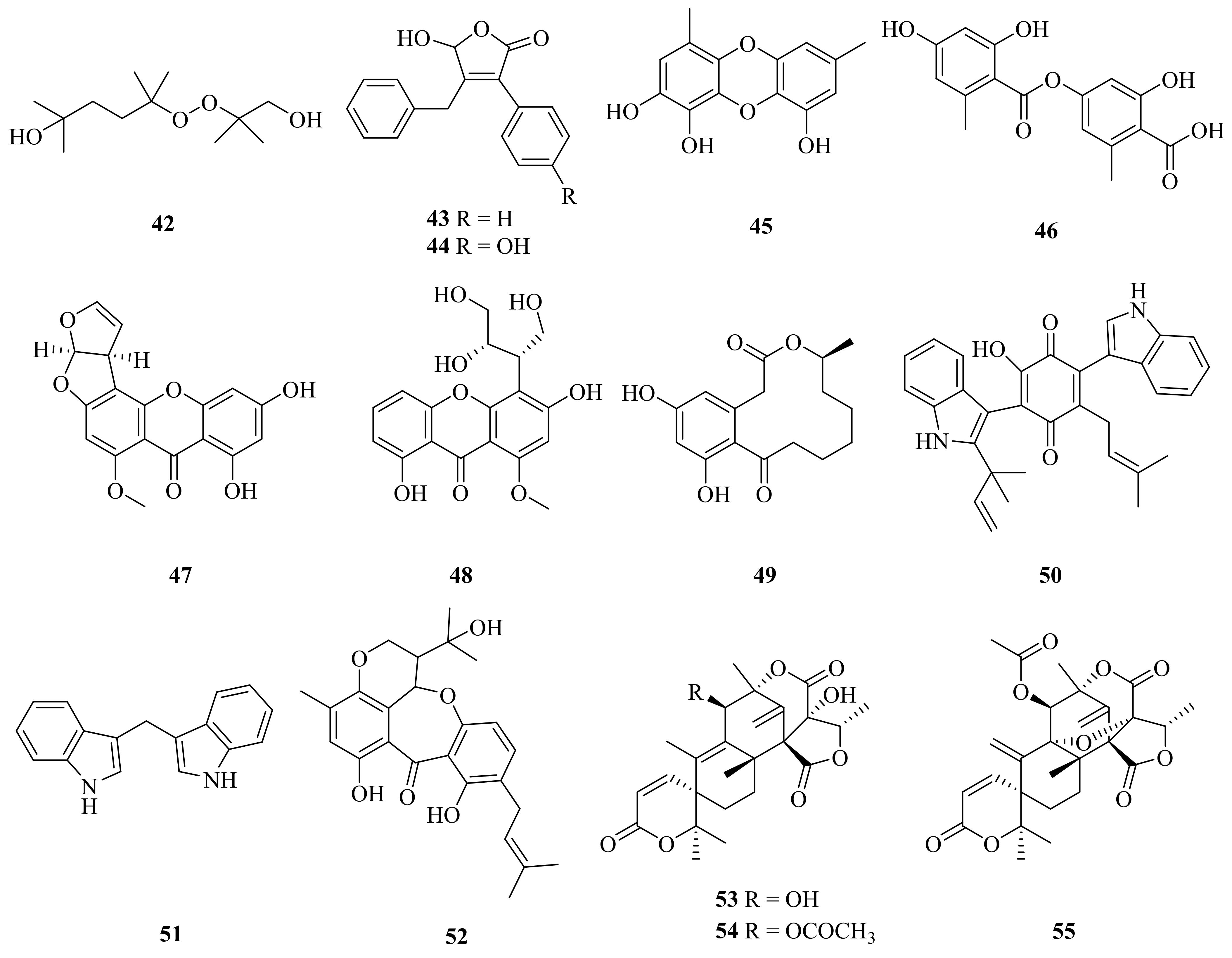
A new acyclic peroxide derivative, asperoxide A (42), and 13 known compounds (43–55) (Figure 5) were reported in 2020 from the deep-sea cold-seep species Aspergillus nidulans SD-531. All of the isolated compounds were tested for antimicrobial activities against human and aquatic bacteria as well as plant pathogenic fungi. Compounds 42–51 exhibited antimicrobial activities against some of the tested strains, with MIC values ranging from 2 to 64 μg/mL [37]. An improved synthesis of microperfuranone (43) (six steps, 56% yield) was reported [38]. 9-Hydroxymicroperfuranone (44) was also isolated from the fungus Emericella quadrilineata IFM42027 [39]. Compound 45 displayed the strongest antibacterial activities among the tested samples and may be a promising natural antimicrobial agent [37]. Lecanoric acid (46) exhibited potent free radical scavenging activity and showed significant Nrf2 activation [40]. Sterigmatocystin (47) displayed promising antibacterial activity, especially on Pseudomonas aeruginosa, with a MIC of 125 μg/mL [41]. Sterigmatocystin (47) also showed cytotoxic activities against HepG2, Hela, MCF-7, and HT-29, with IC50 values of 12.50 ± 0.89 μM, 11.50 ± 0.99 μM, 6.76 ± 0.31 μM, and 8.16 ± 0.39 μM, respectively [42]. Curvularin (48) was active against fungi and numerous cancer cell lines [43], and the total synthesis of curvularin (48) was achieved through a ring-closing-metathesis-based construction of the macrocyclic framework [44]. Terrequinone A (50) was found to be cytotoxic, with IC50 values ranging from 5.40 to 13.90 µM against four cancer cell lines (NCI-H460, MCF-7, SF-268, and MIA Pa Ca-2) and normal human primary fibroblast cells (WI-38) [45]. 3,3′-Diindolylmethane (51) showed many biological activities, such as extensive anticancer activity [46,47], adipogenesis properties [48], and an antioxidant function [49]. Compound 52 displayed acetylcholinesterase (AchE) inhibitory activity, with an IC50 value of 0.40 µM [50]. Compound 53 was evaluated for its cytotoxicity toward HTB-176 human lymphoma cells, with an IC50 of 10 ± 3.92 μM. Compound 53 also demonstrated significant antibacterial activity against P. aeruginosa [51]. In addition, one study reported the mechanistic details of the enzyme-catalyzed, stereospecific spiro-lactone ring-forming reaction to produce austinol (53) [52]. Compound 54 exhibited considerable cytotoxicity against HL-60 and SU-DHL-4 tumor cell lines, with IC50 values of 18.9 and 25.6 μM, respectively [53]. Compounds 53–55 also exhibited potent neuraminidase inhibitory activity [54].
2.2.2. Penicillium sp.
Penicillium fungi have received remarkable interest as an important source of novel natural products encompassing diverse chemical structures and bioactive properties [55,56].
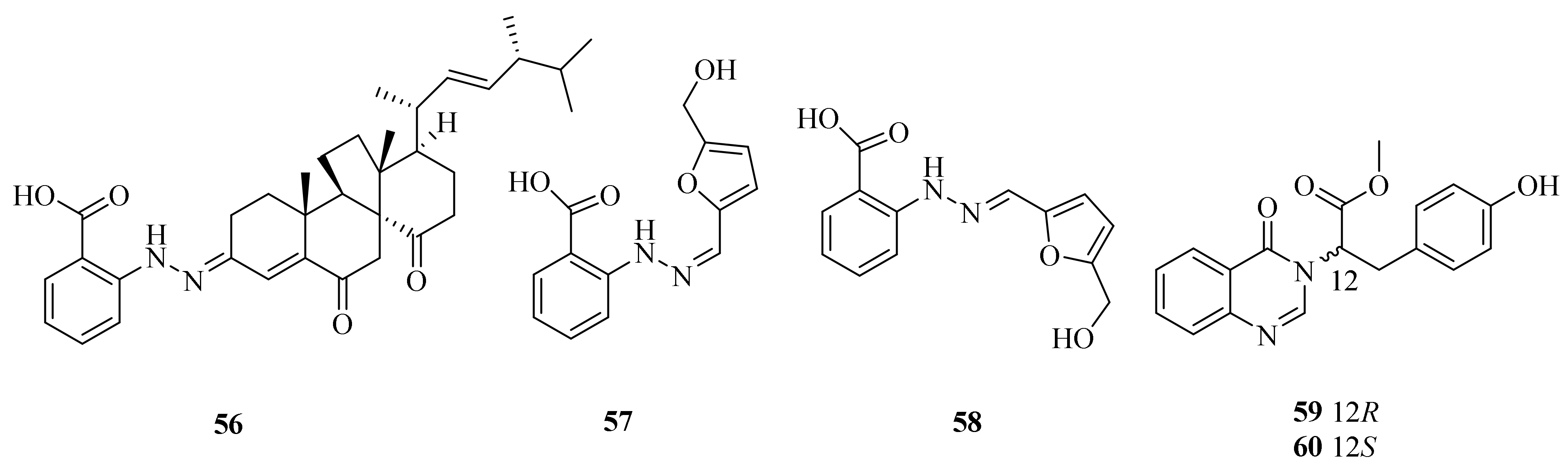
The fungus Penicillium oxalicum, obtained from a deep-sea cold seep, was found to produce three new phenylhydrazones, penoxahydrazones A–C (56–58), and two new quinazolines, penoxazolones A (59) and B (60) (Figure 6). Compounds 56, 59, and 60 could inhibit Chattonella marina, Heterosigma akashiwo, and Prorocentrum donghaiense, with IC50 values ranging from 0.57 to 9.1 µg/mL. Isolates 56, 59, and 60 also showed moderate inhibition against V. harveyi and V. parahaemolyticus, with inhibition zone diameters exceeding 10 mm at 20 µg/dis [57].
2.2.3. Cladosporium sp.
Marine-associated Cladosporium species have attracted considerable interest because of their ability to produce a wide array of metabolites, including alkaloids, macrolides, diketopiperazines, pyrones, tetralones, sterols, phenolics, terpenes, and lactones, that possess versatile bioactivities [58,59].
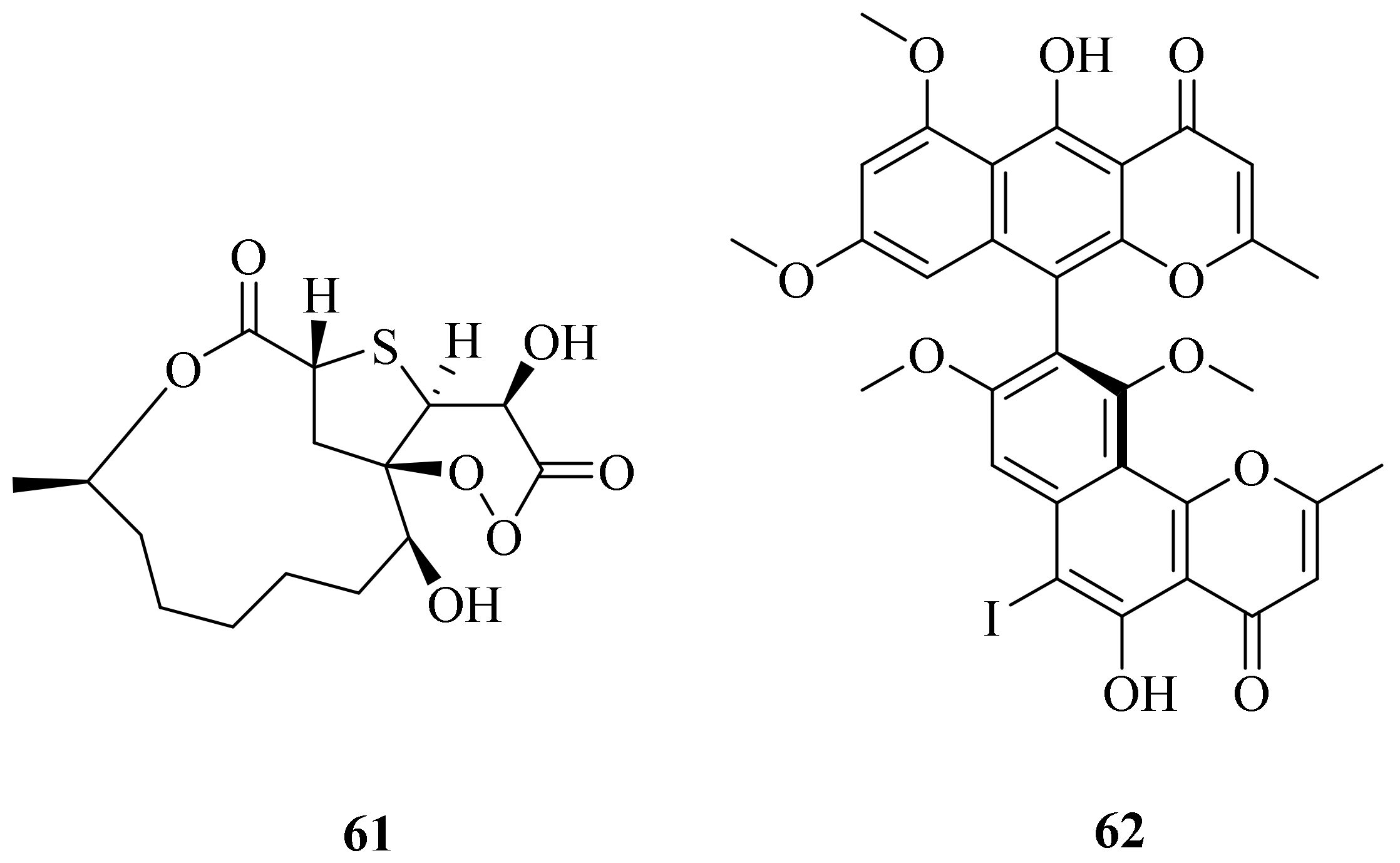
Cladosporioidin A (61), which possesses a novel sulfur and peroxy-bridged twelve-membered macrolide, and a new iodinated dimeric naphtho-γ-pyrone, (aS)-6-iodofonsecinone A (62) (Figure 7), were obtained from a cold seep isolate (8–1) of Cladosporium cladosporioides. Compound 61 was found to exhibit weak antibacterial ability against three bacteria (Vibrio harveyi, V. anguillarum, and Pseudoalteromonas citrea), with inhibitory zone diameters of 7.0, 7.0, and 8.0 mm, respectively. Compound 62 appeared to be the most potent against P. citrea, with an IC50 value of 0.61 μg/mL [60].
2.2.4. Curvularia sp.
Secondary metabolites of the genus Curvularia revealed fascinating biological activities, including anti-malarial, anti-biofouling, anti-larval, and anti-inflammatory activities [61].
The deep-sea cold-seep endozoic fungus Curvularia verruculosa CS-129, retrieved from an area in the South China Sea, has yielded a new cytochalasin dimer—verruculoid A (63)—three new cytochalasin derivatives (64, 66, and 68), and a synthetic product obtained as a natural product for the first time (69) together with four known analogs (65, 67, 70, and 71) (Figure 8). Compound 63 displayed activity against the human pathogenic bacterium Escherichia coli (MIC = 2 μg/mL) [62]. Cytochalasin B (65) had the best effect on the actin cytoskeleton [63]. Cytochalasin B6 (67) was firstly isolated from a jellyfish-derived fungus, Phoma sp., and showed moderate cytotoxicity [64]. Compounds 68, 70, and 71 showed cytotoxicity against HCT-116, HepG-2, and MCF-7, with IC50 values from 5.2 to 12 μM [62]. Deoxaphomin (71) also exerted the most marked inhibitory effects on the growth of six cancer cell lines: the human OE21 esophageal, U373 glioblastoma, SKMEL28 melanoma, A549 non-small cell lung cancer, mouse B16F10 melanoma, and human HS683 oligodendroglioma cell lines [65].
2.3. Marine Bacteria
2.3.1. Streptomyces sp.
Streptomyces sp. Have well-developed branching hyphae, and more than 1000 species have been reported, mainly distributed in soil. They are attractive microbial cell factories that have industrial capabilities to produce a wide array of bioactive secondary metabolites [66,67].
A cold-seep-derived actinomycete belonging to the Streptomyces olivaceus OUCLQ19-3 genus was found to contain two new (72 and 73) and six known (74–79) (Figure 9) dixiamycins. In the antibacterial test, compounds 72–79 exhibited significant growth inhibition against several multi-drug-resistant (MDR) strains, with MIC values ranging from 0.78 to 6.25 μg/mL; among these, 72, 73, and 76–79 were more potent than the positive control tetracycline [4]. Dixiamycins A (77) and B (76) are the first examples of atropisomerism naturally occurring in N–N-coupled atropo-diastereomers [68]. A unique method of electrochemical dimerization of carbazoles and carbolines enabled the first total synthesis of dixiamycin B (76) [69]. Sulfadixiamycin A (79) was found to have selective yet moderate antimycobacterial properties, with a MIC value of 25 mg/mL [70].
2.3.2. Halomonas sp.
Halomonas is a kind of Gram-negative bacterium which has strong adaptability and a wide range of adaptability to temperature, salinity, and oxygen. It may have important application values in sewage treatment and bioremediation [71].
An immune-enhancing exopolysaccharide, EPS2E1 (80), was reported in 2021 from a cold-seep bacterium, Halomonas sp. 2E1, which was collected in the South China Sea (119°17′ 04.956″ E, 22°06′58.384″ N; 1142 m deep). Structural analysis showed that the backbone mainly consisted of →2)-Man-(α-1→ and →2, 6)-Man-(α-1→ in a ratio of 2.45:1.00. The chain contained →4)-Glc-(α-1→, →6)-Man-(α-1→and→3)-Glc-(β-1→). EPS2E1 exhibits the potential to be an immunopotentiator, because it could significantly increase the production of NO, COX-2, TNF-α, IL-1β, and IL-6 by activating the MAPK and NF-κB pathways on RAW264.7 macrophages. [62,72].
2.3.3. Vibrio sp.
Bacteria belonging to the Vibrio family are short in shape and named for their curve-like arcs. They are usually found in freshwater or seawater and also in the intestines of humans or fish. Some species are pathogenic to fish or humans [73]. Vibrio species can produce compounds with attractive biological activities, including antibacterial, anticancer, and antivirulence activities [74].
In 2021, the isolation of a novel exopolysaccharide, EPS364 (81), was reported from a deep-sea cold-seep fungus, Vibrio alginolyticus 364, obtained in the South China Sea (119°17′05.3940″ E, 22°06′58.7264″ N). EPS364 consisted of mannose, glucosamine, gluconic acid, galactosamine, and arabinose in a molar ratio of 5:9:3.4:0.5:0.8. Notably, EPS364 exhibited a significant antitumor activity, inducing apoptosis, dissipation of the mitochondrial membrane potential (MMP), and generation of reactive oxygen species (ROS) in Huh7.5 liver cancer cells, which suggests that EPS364 is a promising antitumor agent for pharmacotherapy [75].
2.3.4. Bacillus sp.
Marine Bacillus species produce versatile secondary metabolites, including lipopeptides, polypeptides, fatty acids, polyketides, and coumarins. These structurally diverse compounds exhibit a wide range of biological activities [76].
A bacterial strain isolated from the cold-seep-derived fungus Bacillus sp. CS30 which was collected in the South China Sea in October 2017 (119°17′09.655″ E, 22°06′5.169″ N), exhibited strong growth inhibition against M. grisea. Two purified antifungal agents were isolated which belong to the surfactin family and were named surfactin CS30-1 and surfactin CS30-2 (82 and 83). Both of them showed antifungal activity, since they could induce the generation of reactive oxygen species (ROS) and caused serious damage to the cell wall and cytoplasm [77].
2.4. Others
Three novel series of non-isoprenoidal dialkyl glycerol diethers were tentatively identified in carbonate crusts precipitated from methane-rich bottom-waters and pore-waters associated with Mediterranean mud volcanoes (84–86) (Figure 10). All of the reported sedimentary compounds represent the first detailed report on the occurrence of alkyl diethers in a non-thermophilic setting, and the cyclopropyl and cyclohexyl moieties as observed in the series I and II components are unique for ether lipids [78].
3. Hydrothermal Vents
3.1. Marine Animal
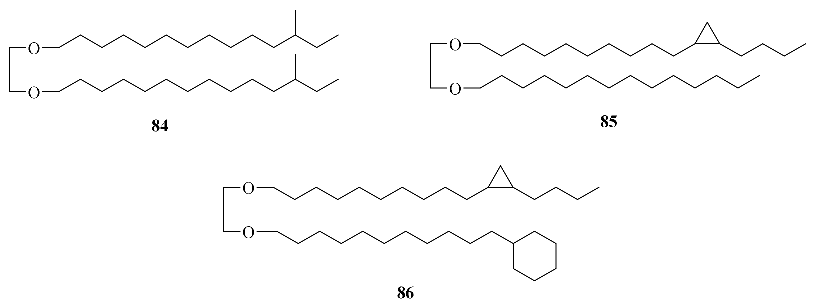
Three sterols were isolated (87–89) (Figure 11) from the species of bivalve Bathymodiolus septemdierum, which was collected in 2004 at a depth of 1244 m from hydrothermal vents at Myojin Knoll, Japan. Their unique feeding modes and metabolism of nutrients make the structures of their natural products more novel [13]. Compound 88 showed allelopathic activity against Lactuca sativa seedlings and autotoxic activity against A. hoantchy seedlings [79]. The total synthesis of 5α, 6β-dihydroxystigmastan-3-O-β-glycopyranoside (89) was reported [80].
3.2. Marine Fungi
3.2.1. Penicillium sp.
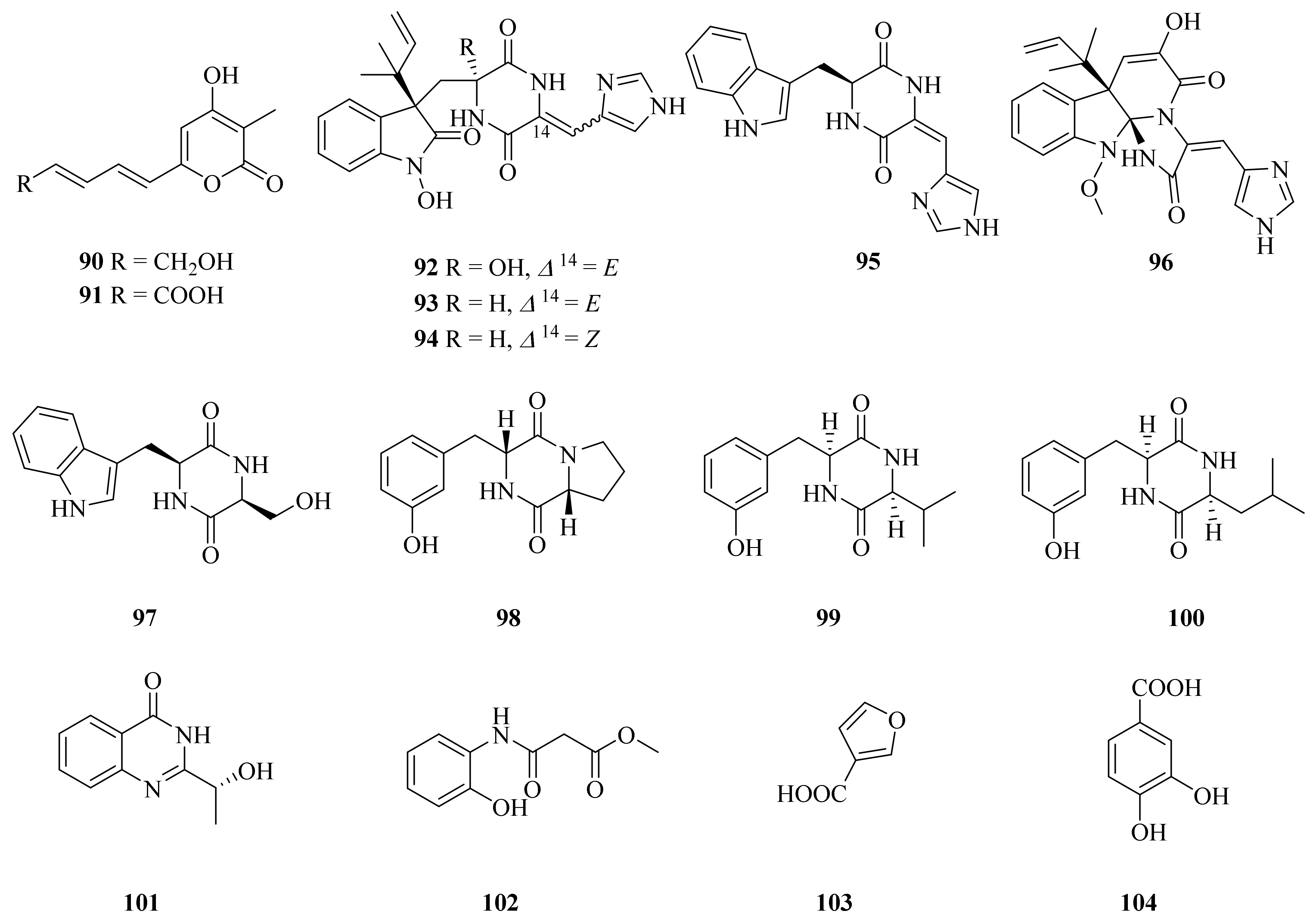
In 2020, Han et al. described the isolation of three new compounds (90–92) along with twelve known compounds (93–104) (Figure 12) from a deep-sea hydrothermal fungus, Penicillium chrysogenum SCSIO 07007, collected from the Western Atlantic (126.8983° E, 27.7875° N) at a depth of 1028 m. Of them, chrysopyrones A and B (90 and 91) showed obvious inhibitory activities against protein tyrosine phosphatase 1B (PTP1B), with IC50 values of 9.32 and 27.8 μg/mL, respectively [81]. Meleagrin (96) exhibited a variety of activities, such as antitumor [82], cytotoxic [83], antibiofilm, and antifouling activities [84]. Cyclo (Trp-Ser) (97) displayed antibacterial activity against Escherichia coli, Chromobacterium violaceum CV026, Pseudomonas aeruginosa PA01, Staphylococcus aureus, and Candida albicans, with MIC values ranging from 3.2 to 6.4 mg/mL [85]. Cyclo (Pro-Tyr) (98) exhibited weak antibacterial activity against X. axonopodis pv. citri and R. solanacearum but showed a MIC of 31.25 μg/mL [86]. The biosynthesis of chrysogine (100) was proven to be related to a candidate NRPS cluster comprising five additional genes named chry2–6 gene clusters [87]. 2-Furoic acid (103) was shown to be effective in lowering both serum cholesterol and serum triglyceride levels, significantly in rats with an elevation of HDL cholesterol levels at 20 mg/kg/day orally [88]. 3,4-Dihydroxybenzoic acid (104) may be an important phenolic compound in regulating root formation in P. cynaroides cuttings [89].
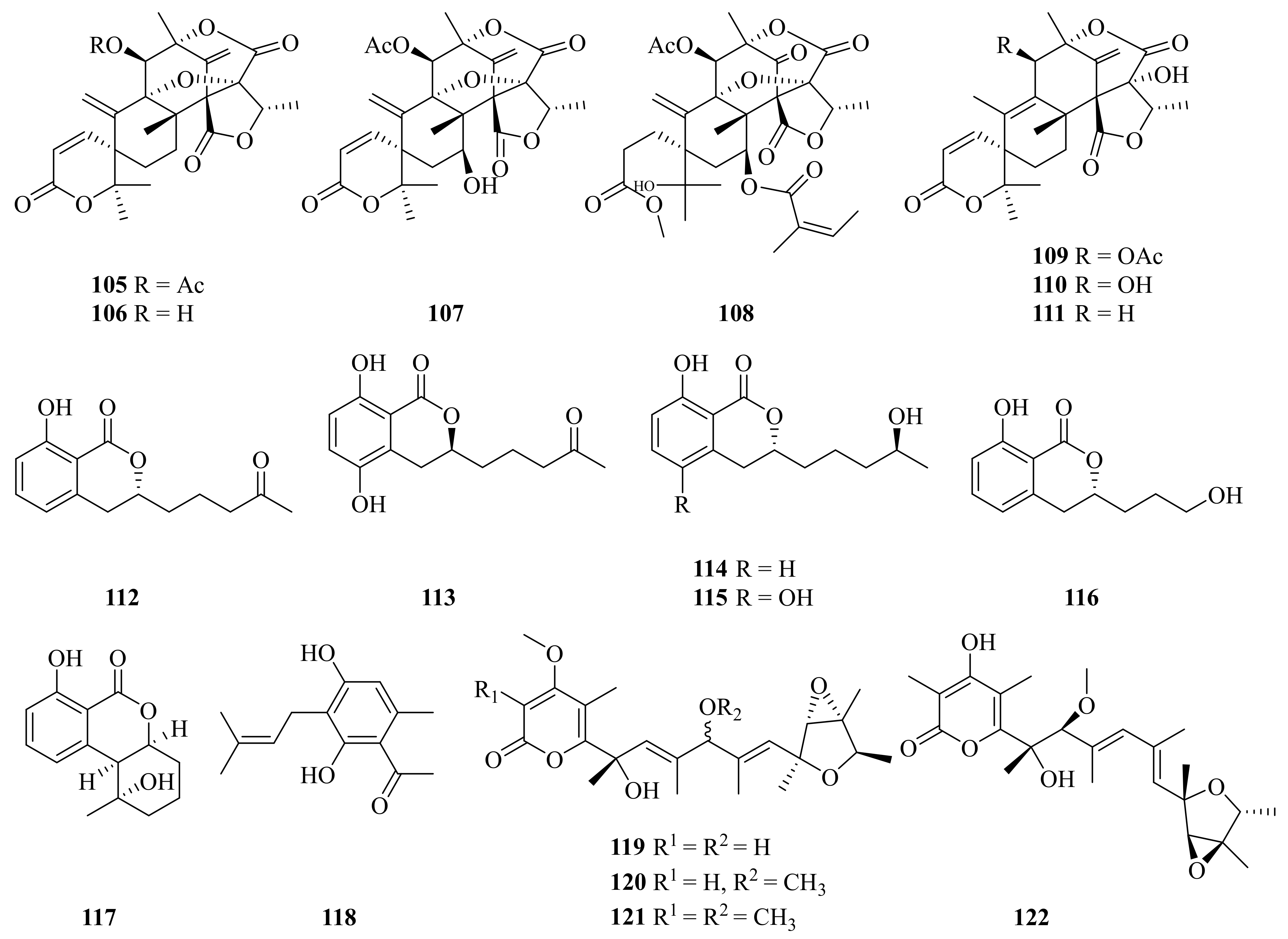
Five new compounds (108, 113, 115–117) together with eight known compounds (105–107, 109–112, 114, and 118) (Figure 13) were obtained from Penicillium sp. Y-5-2, which was collected in May 2014 from Kueishantao, off Taiwan. New compounds 113, 115, and 117 revealed inhibitory activities against E. coli at MIC values around 32 μg/mL [90]. Dehydroaustin (105) was an attractive natural insecticide with a LC50 value of 2.9 ppm [91]. Compounds 105 and 106 showed acetylcholinesterase (AchE) inhibitory activity, with IC50 values of 0.40 and 3.00 μM, respectively [50]. Dehydroaustinol (106) and austin (109) displayed considerable cytotoxicity against the HL-60 and SU-DHL-4 tumor cell lines, with IC50 values ranging from 18.9 to 27.8 μM [53]. Austinol (110) exhibited strong antibacterial activity against the P. aeruginosa bacterial strain, with a MIC value of 0.13 ± 0.4 µg/mL [51]. Aspergillumarins A (112) and B (114) showed weak antibacterial activity against Staphylococcus aureus and Bacillus subtilis at a concentration of 50 μg/mL [92]. Pestalotionol (118) showed potent antibiotic activity against Staphylococcus aureus and Bacillus subtilis, with MIC values of 8 and 2 μg/mL, respectively [90]. Compound 118 also showed weak anti-inflammatory activity by measuring the nitric oxide (NO) production in lipopolysaccharide (LPS)-activated RAW264.7 macrophages [93].
In 2020, Pan and colleagues isolated four verrucosidin derivatives (119–122) (Figure 13) from the sulfur-derived fungus Penicillium sp. Y-50-10, collected in the Kueishantao hydrothermal vents off Taiwan [94]. Compounds 119–122 showed activity against Bacillus subtilis, with MIC values of 32 μg/mL [95].
3.2.2. Aspergillus sp.
In 2016, the strain Aspergillus sp. WU 243, collected from the digestive gland of Xenograpsus testudinatus, a unique type of crab which dwells in the Kueishantao hydrothermal vents off Taiwan, was reported to contain a novel hybrid polyketide-terpenoid, aspergstressin (123), and four known compounds (124–127) (Figure 14) [96]. Cyclo-(Try-Phe) (125) can be used as a plant growth regulator; it exhibited different biological activities against the tested plants [97]. Cordyol C (126) exhibited significant anti-HSV-1 activity, with an IC50 value of 1.3 μg/mL, and cytotoxic activity against BC and NCI-H187 cancer cell lines, with IC50 values of 8.65 and 3.72 μg/mL, respectively [98]. Cordyol C (126) was also a toxic compound against HeLa cells, with an IC50 value of 35.29 ± 1.55 mM [99]. Sydowic acid (127) was assessed in murine leukemia P-388 cells and showed potential cytotoxicity, with an IC50 value of 20.30 μg/mL [100].
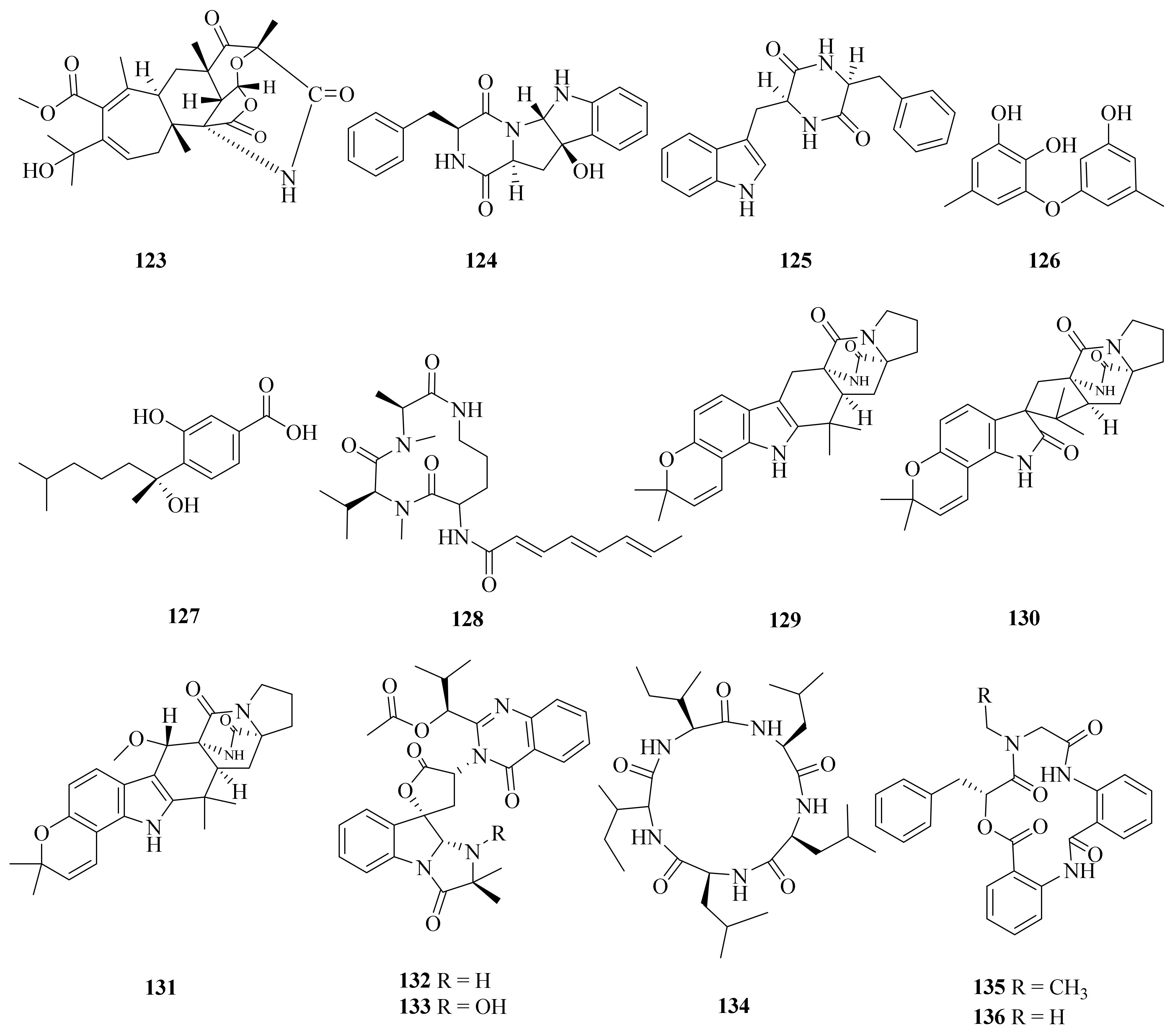
Four secondary metabolites (128–131) (Figure 14) were isolated from the hydrothermal fungus Aspergillus sclerotiorum C10WU, which was collected from Kueishantao, Taiwan. Stress metabolite 128 was reported to possess insecticidal activities and show cytotoxic effects against human cervical carcinoma [101]. Stephacidin A (129) is proposed as a biosynthetic precursor to notoamide B in various Aspergillus species. Following a strategy based on doubly 13C-labeled stephacidin A (129), it could undergo bio-transformation to notoamide B (130) [102]. In addition, the total synthesis of the natural indole alkaloid notoamide F (131) was reported [103].
A hydrothermal fungus Aspergillus clavatus C2WU, which was also collected from Kueishantao, Taiwan, yielded two secondary metabolites (132 and 133) (Figure 14). Notably, deoxytryptoquivaline (132) showed strong binding to three targets, SARS-CoV-2 main protease and spike glycoprotein and human angiotensin-converting enzyme 2. Therefore, it has promise for being further investigated as a possible multitarget drug against COVID-19 [104]. Aspergillus clavatus C2WU also yielded a unique new cyclopeptide, clavatustide C (134) (Figure 15), which was produced as a stress metabolite in response to abiotic stress elicitation by one of the hydrothermal vent’s fluid components, Zn [105]. Moreover, two novel cyclodepsipeptides, namely, clavatustides A (135) and B (136) (Figure 15), were also purified from Aspergillus clavatus C2WU. Clavatustides A (135) and B (136) displayed antitumor activity by suppressing the proliferation of hepatocellular carcinoma (HCC) cell lines (HepG2, SMMC-7721, and BEL-7402), inducing an accumulation of HepG2 cells in G1 phase and a reduction in cells in S phase [106]. The enantiopure synthesis of clavatustides A (135) and B (136) was accomplished by a seven-step synthetic protocol starting from commercially available (R)-phenyllactic acid [107].
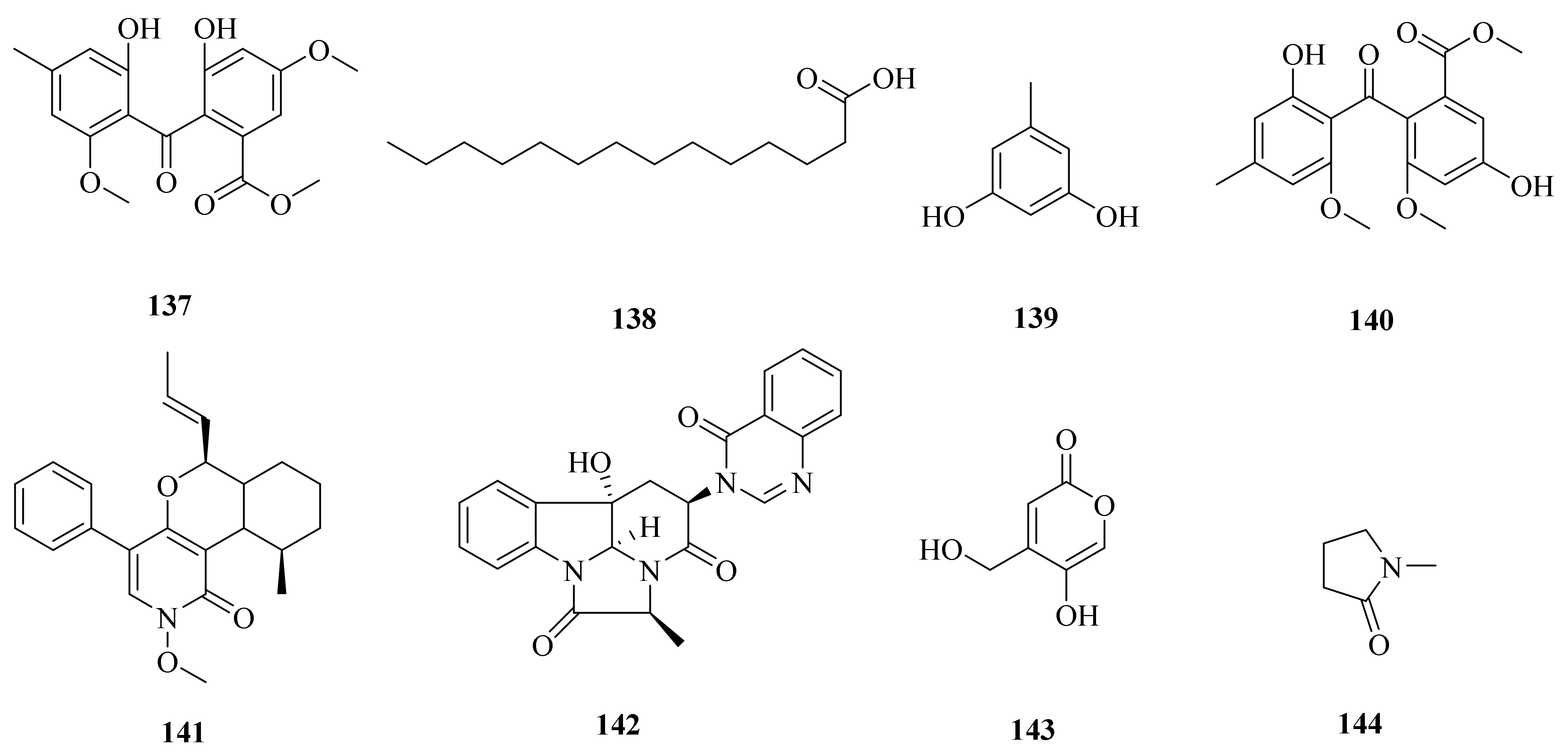
One new compound (137) and seven known compounds (138–144) (Figure 15) were obtained from Aspergillus sp. YQ-13, collected from the sediment of Kueishantao hydrothermal vents off Taiwan [108]. Notably, myristic acid (138) showed various biological activities, for example, specifically blocking T cell antigen receptor CD3-induced Ca2+ mobilization in T cells [109]; exhibiting antibacterial activity [110]; and reducing type 2 diabetes risk [111]. Orcinol (139) exhibited remarkable antioxidant activity; its free radical scavenging rate can reach up to 80% of 20 mg/mL [112]. Compounds 137 and 139 were tested by the methods of DPPH and FRAP assays, showing moderate antioxidant activities [108]. 1,2-seco-Trypacidin (140) exhibited a weak inhibitory effect on Helicobacter pylori 159, with a MIC of 16 μg/mL [113]. Leporin A (141) and chaetominine (142) exhibited antibiotic activity, with MIC values around 1 to 25 μg/mL against Bacillus subtilis, Klebsiella pneumoniae, methicillin-resistant Staphylococcus aureus (MRSA), Pseudomonas aeruginosa, Staphylococcus aureus, Escherichia coli, and Acinetobacter Bauman [108]. 4-(Hydroxymethyl)-5-hydroxy-2H-pyran-2-one (143) induced the production of cAMP in a dose-dependent manner, which indicated that 143 might be a possible ligand of GPR12 [114]. Compound 143 also has significant antioxidant activity, with an IC50 value of 59.5 µM [115], and weak inhibition of bacterial growth [116].
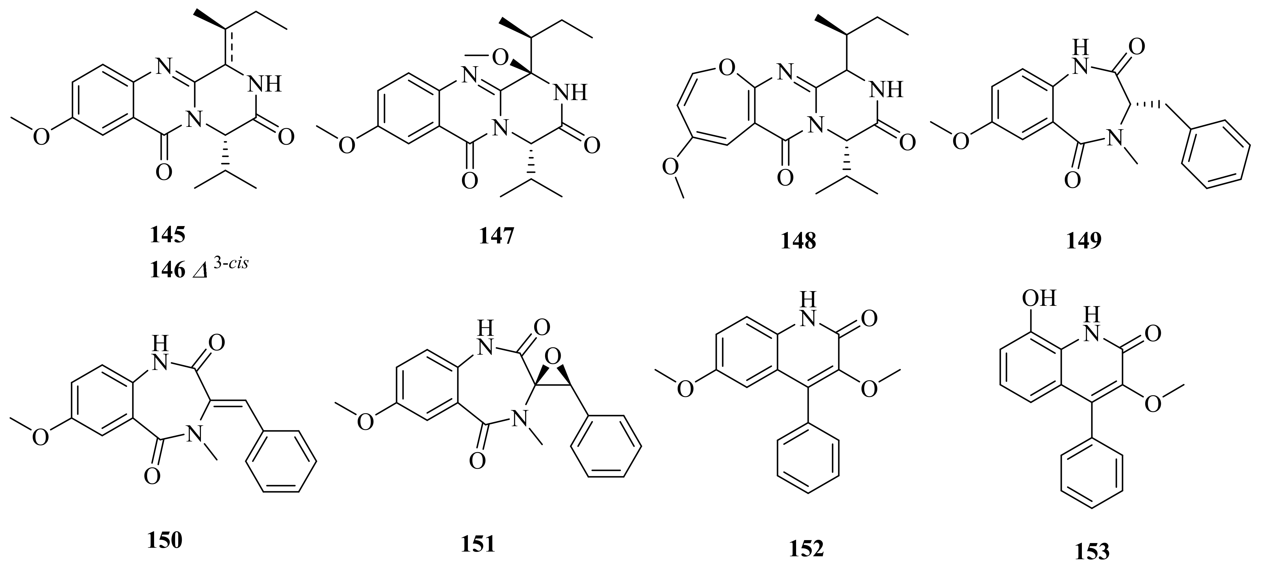
Three new quinazoline derivatives (145–147), one new oxepine-containing natural product (148), four new cyclopenin derivatives (149–151 and 153), and one known compound (152) (Figure 16) were isolated from an ethyl acetate extract of a hydrothermal vent crab belonging to the genus Aspergillus versicolor XZ-4, collected from the Taiwan Kueishantao. Compounds 149 and 151–153 revealed inhibitory activities against E. coli at MIC values around 32 μg/mL [117]. 3,6-O-Dimethylviridicatin (152) was firstly isolated from the deep-sea-derived fungus Aspergillus versicolor SCSIO 05879 [118].
3.2.3. Graphostroma sp.
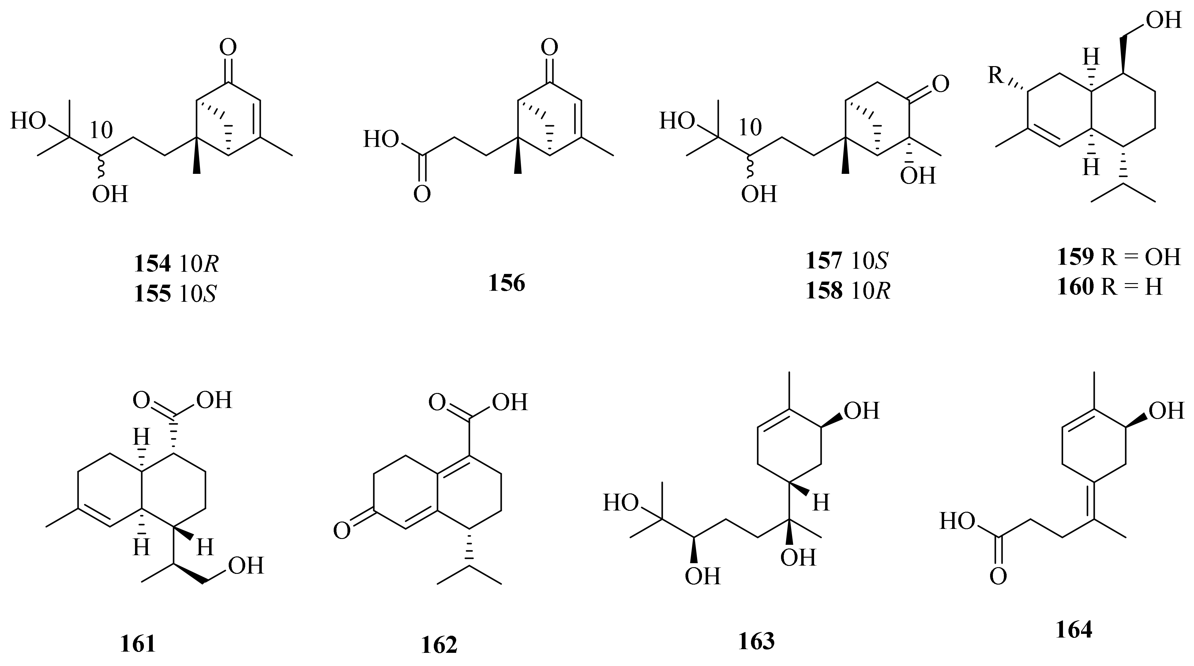
In 2017, the fungus Graphostroma sp. MCCC 3A00421, collected from a deep-sea hydrothermal sulfide deposit of the Atlantic Ocean (13.36° W, 15.17° S, at a depth of −2721 m), was reported to contain 11 sesquiterpene compounds (154–164) (Figure 17). Two of them are structurally connected (154 and 155), and nine are new compounds (156–164) [119]. Among them, compounds 154 and 155 were evaluated for their anticancer activity but had no significant effect against HL-60, A-549, MCF-7, SMMC-7721, and SW-480 human cancer cell lines [120]. Khusinol B (159) showed more significant anti-inflammatory activity than the positive control (aminoguanidine), with an IC50 value of 17 µM. In addition, compound 159 also showed weak anti-allergic activity, with an IC50 value of 150 µM [119].
3.3. Marine Bacteria
3.3.1. Streptomyces sp.
Hydrothermal vent microorganisms have a unique metabolic mechanism, because they have to withstand and respond to heavy metal concentrations [121]. A novel antibiotic (165) (Figure 18) was produced by Streptomyces sp. WU20, which was isolated from the metal-rich hydrothermal vents in Kueishantao, Taiwan. Compound 165 exhibited antimicrobial activity against Bacillus subtilis, with a MIC of around 32 μg/mL [122].
3.3.2. Geobacillus sp.
Geobacillus is a Gram-positive bacterium, rod-shaped, and either paired or chained, and its optimum growth temperature is 65–70 degrees [123].
In 2017, the bacterium Geobacillus sp. E263, collected from a deep-sea hydrothermal vent in the East Pacific, was reported to contain a novel quinoid compound (166) (Figure 18). The research indicated that 2-amino-6-hydroxy-[1,4]-benzoquinone (166) could trigger the apoptosis of gastric cancer cells and breast cancer cells by inducing the accumulation of intracellular reactive oxygen species [124].
3.3.3. Halomonas sp.
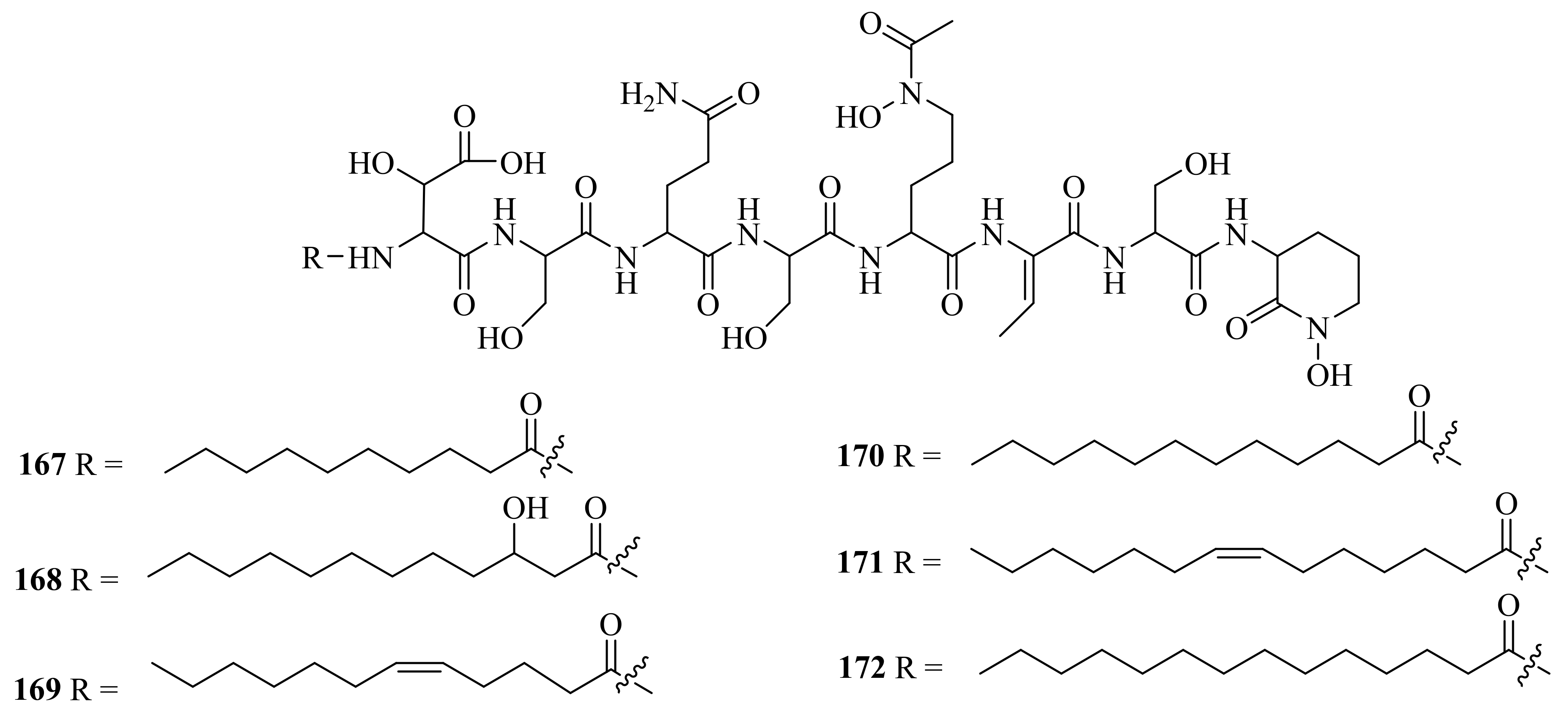
Six new amphiphilic siderophores, loihichelins A-F (167–172) (Figure 19), were obtained from cultures of the deep-sea hydrothermal vent and sulfide rock bacterium Halomonas sp. LOB-5, which was collected from Marker 17 (depth of 1714 m) at Loihi Seamount. These siderophores showed a potential role in the promotion of Mn(II) and Fe(II) oxidation [125]. In addition, the reports on loihichelins A-F were the first publications on new natural products from ocean hydrothermal vent environments.
3.3.4. Vibrio sp.
An exopolysaccharide was produced under laboratory conditions by Vibrio diabolicus, a bacterium retrieved from a deep-sea hydrothermal vent in the East Pacific Rise (12°48.13′ N, 103°56.30′ W) (173). Structural analysis showed that the polysaccharide consists of a linear tetrasaccharide repeating unit with the following structure: →3)-β-D-Glcp NAc-(1→4)-β-D-GlcpA-(1→4)-β-D-GlcpA-(1→4)-α-D-Galp Nac-(1→ [126].
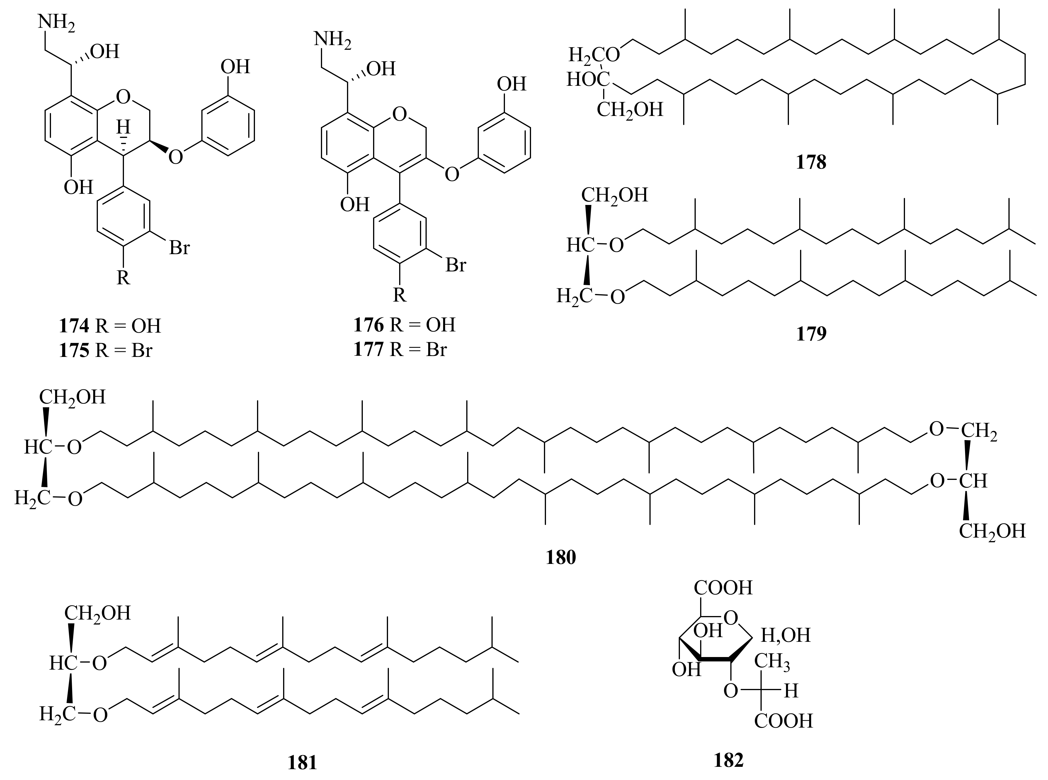
The bacterium Thermovibrio ammonifican, collected from a culture from marine hydrothermal vents in the East Pacific Rise (9°50′ N, 104°189′ W) at a depth of 2500 m, was found to contain four hydroxyethyl amine chromene derivatives, ammonificins A-D (174–177) (Figure 20) [127]. Ammonificins C (174) and D (175) could induce apoptosis at 2 μM and 3 μM, respectively (the control, staurosporine at 0.1 μM) [128].
3.3.5. Methanococcus sp.
3.3.6. Thermococcus sp.
3.3.7. Alteromonas sp.
4. Comprehensive Overview and Outlook
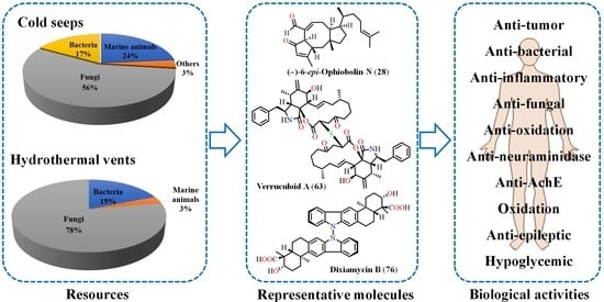
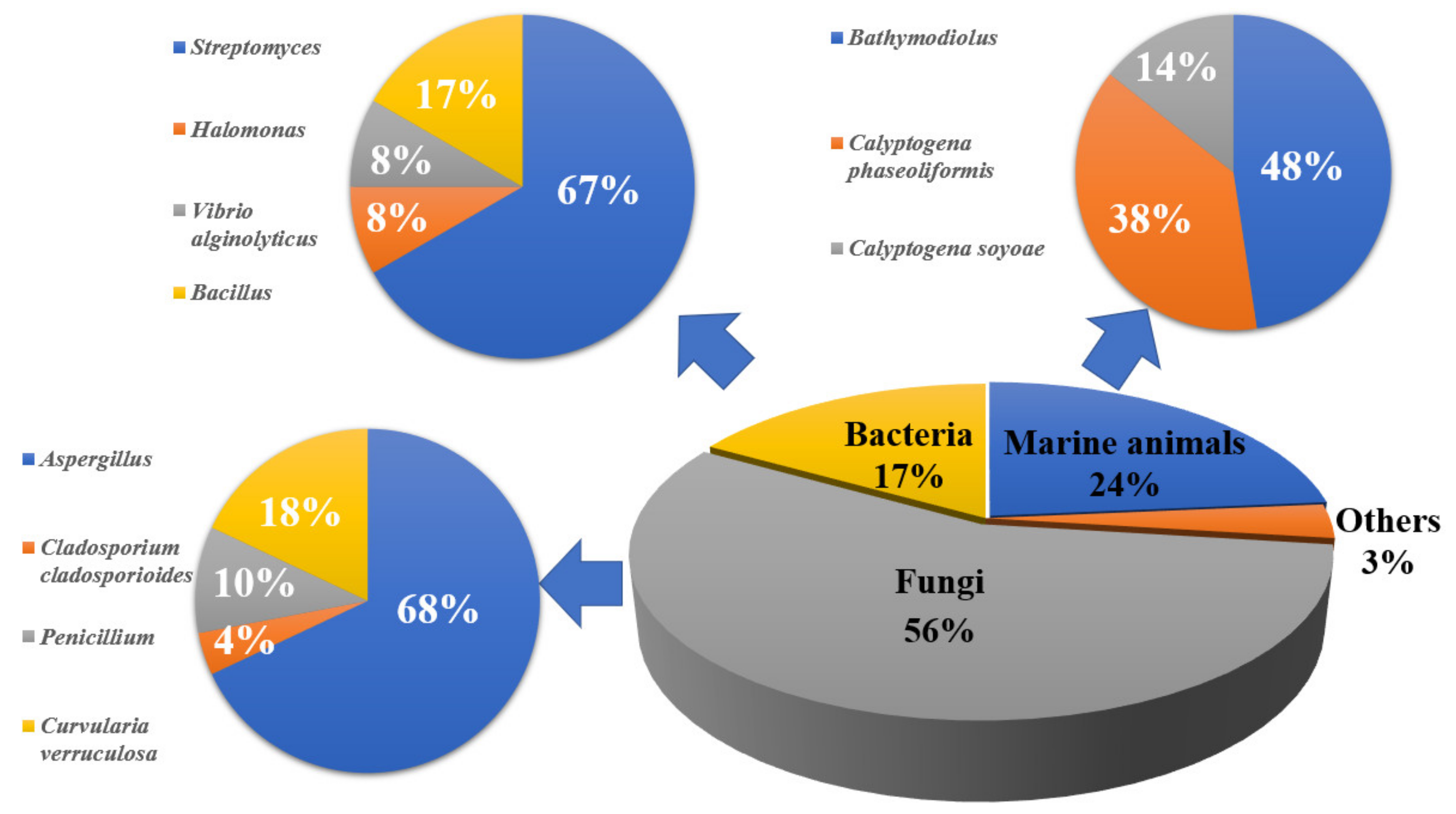
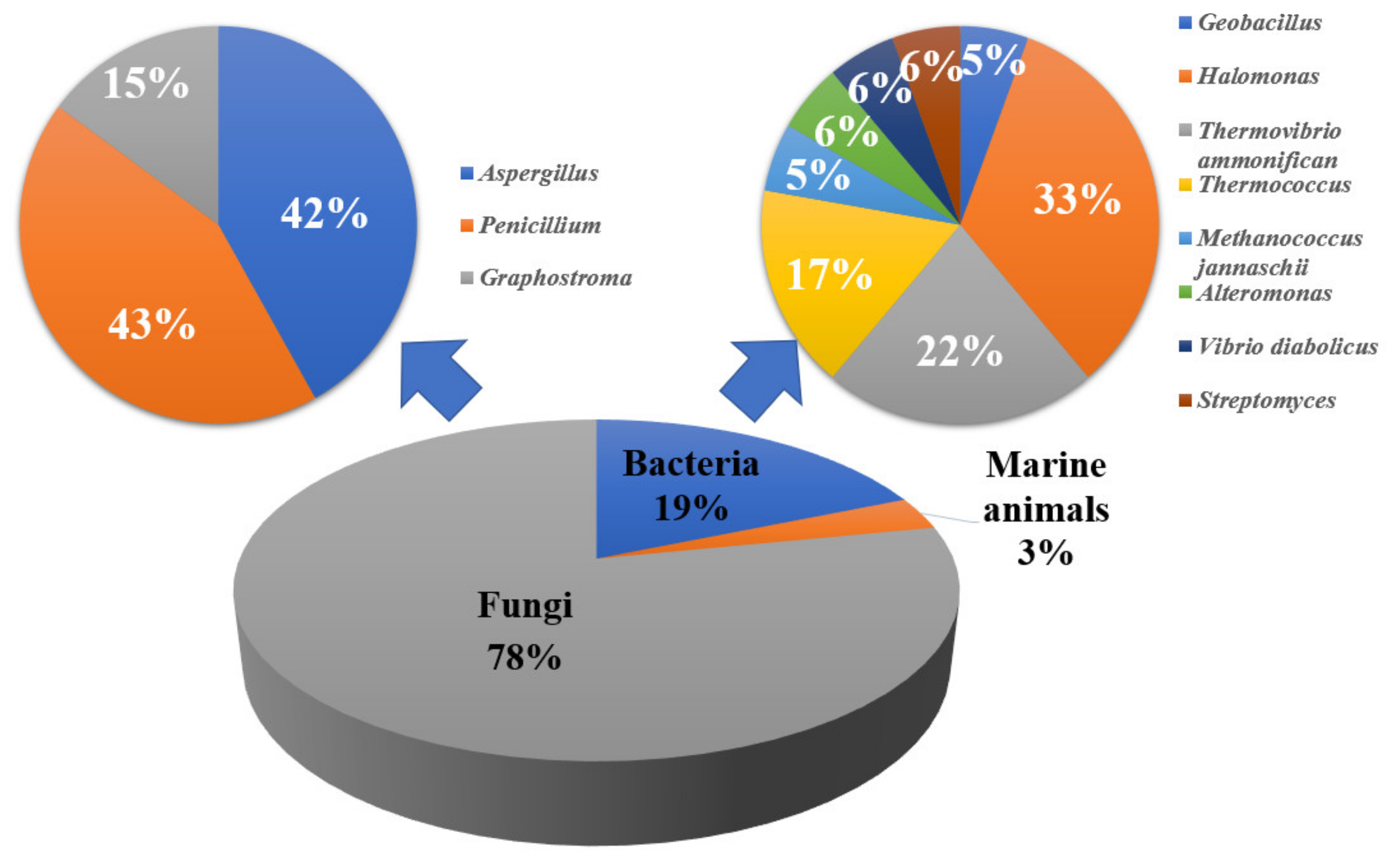
We provide a comprehensive overview of the sources and bioactivities of the 182 natural products from the deep-sea extreme environments described up to March 2022. It was observed that cold-seep-derived compounds could be divided into four parts, namely, marine animals (24%), fungi (56%), bacteria (17%), and others (3%). In general, they mainly come from Aspergillus, Bathymodiolus, and Curvularia, according to the number of compounds (Figure 21), suggesting that these genera would be subjected to the focus of future research. The secondary metabolites isolated from hydrothermal vents are found in three parts. At the domain level, 78% of the natural products were derived from fungi, while 19% originated from bacteria, among which Aspergillus and Penicillium were the main source of natural products (Figure 22).
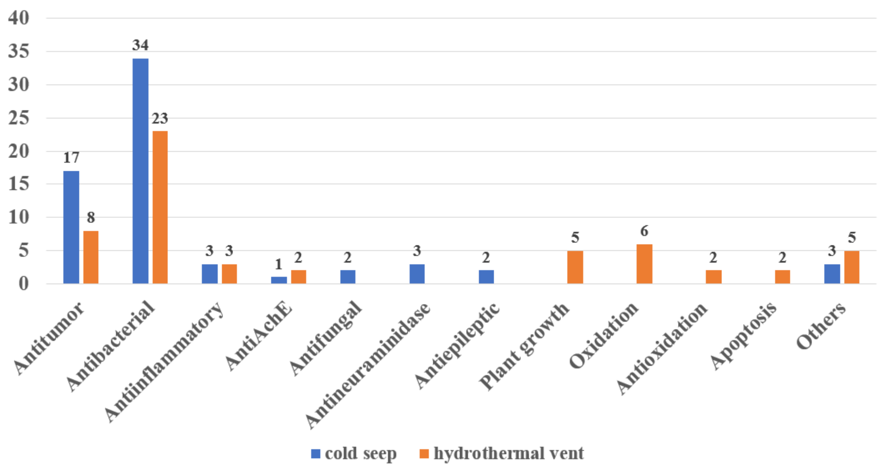
By comparing and analyzing the activities of secondary metabolites derived from cold seeps and hydrothermal vents, it was found that almost 60% of the 182 compounds had biological activities, and their activities were diverse (Figure 23). Among them, antibacterial and antitumor activities are reported most frequently. Some cold-seep-derived compounds also have antifungal and anti-epileptic activities, while hydrothermal vent-derived natural products also include plant growth regulation and oxidant activities. In general, that secondary metabolites derived from cold seeps and hydrothermal vents have novel and diverse biological activities may be due to their extreme and special environments.
5. Conclusions
There were 86 natural products isolated from cold seeps, while 96 secondary metabolites were isolated from hydrothermal vents. The sources of the compounds are represented by the genera Aspergillus sp., Penicillium sp., and so on. There are 90 new compounds among the 182 compounds. Around 60% of the deep-sea natural products were reported to possess bioactivity. For example, an exopolysaccharide, EPS364 (81), from cold-seep Vibrio alginolyticus 364, was investigated for its mechanism of inhibiting the growth and adhesion of liver cancer cells, which has proved to be the basis for a promising anticancer drug [75]. A hydrothermal vent-derived compound, deoxytryptoquivaline (142), showed strong binding to three important targets of SARS-CoV-2 and so has promise for being further investigated as a possible multitarget drug against COVID-19 [104]. These novel and diverse activities indicate that deep-sea extreme environments might facilitate the production of functional natural products. Moreover, the total synthesis or biosynthesis of some compounds was described. For example, the total synthesis pathway of (−)-6-epi-ophiobolin N (28), which was isolated from cold-seep sediments, was reported [27]. Dixiamycins A (77) and B (76), which were separated from a cold-seep environment sample, were reported in an unusual oxidative cyclization strategy for tailoring indolosesquiterpene biosynthesis [132] and in a possible route for total synthesis [69], respectively. These synthesized compounds either have a wide range of sources, diverse activities, or unique molecular skeletons rarely discovered in nature. This further indicates that the natural products derived from extreme environments, such as cold seeps and hydrothermal vents, have great potential and are a treasure to be further developed.
Author Contributions
Conceptualization, J.W., M.C. and Y.L.; writing—original draft preparation, M.C.; review and editing, J.W., M.C., X.P., K.Z. and Y.S. All authors have read and agreed to the published version of the manuscript.
Funding
This work was financially supported by the Guangdong MEPP Funds (No. GDNRC [2021]48) to J.W., Finance Science and Technology Project of Hainan Province (ZDKJ202018) to J.W., Guangdong Local Innovation Team Program (2019BT02Y262) to J.W., Key Special Project for Introduced Talents Team of Southern Marine Science and Engineering Guangdong Laboratory (Guangzhou) (GML2019ZD0406) to Y.L., National Natural Science Foundation of China (Nos. 41776169, 42006084, and 21772210) to J.W., X.P. and Y.L., and Project from the Institute of South China Sea Ecology and Environmental Engineering, CAS (ISEE2018PY04) to Y.L.
Conflicts of Interest
The authors declare no conflict of interest.
References
- Rampelotto, P.H. Extremophiles and Extreme Environments. Life 2013, 3, 482–485. [Google Scholar] [CrossRef] [PubMed]
- Brown, P.D.; Lawrence, A.L. The Importance of Asking “How and Why?” In Natural Product Structure Elucidation. Nat. Prod. Rep. 2017, 34, 1193–1202. [Google Scholar] [CrossRef] [PubMed] [Green Version]
- Chi, L.P.; Li, X.M.; Wan, Y.P.; Li, Y.H.; Li, X.; Wang, B.G. Two New Phenol Derivatives from the Cold Seep-Derived Fungus Aspergillus insuetus SD-512. Chem. Biodivers. 2021, 18, e2100512. [Google Scholar] [CrossRef] [PubMed]
- Jin, E.; Li, H.; Liu, Z.; Xiao, F.; Li, W. Antibiotic Dixiamycins from a Cold-Seep-Derived Streptomyces olivaceus. J. Nat. Prod. 2021, 84, 2606–2611. [Google Scholar] [CrossRef] [PubMed]
- Pettit, R.K. Culturability and Secondary Metabolite Diversity of Extreme Microbes: Expanding Contribution of Deep Sea and Deep-Sea Vent Microbes to Natural Product Discovery. Mar. Biotechnol. 2011, 13, 1–11. [Google Scholar] [CrossRef] [PubMed]
- Thornburg, C.C.; Zabriskie, T.M.; McPhail, K.L. Deep-Sea Hydrothermal Vents: Potential Hot Spots for Natural Products Discovery? J. Nat. Prod. 2010, 73, 489–499. [Google Scholar] [CrossRef] [PubMed]
- Avila, E.D.; Gallegos, J.L.V.; Cruz, M.G.; Dehesa, A.Z. Omega-3 Polyunsaturated Fatty Acids Supplemented Diet and Its Preventive Effect on Tumor Growth in Nude Mice. Nutr. Clin. Diet. Hosp. 2018, 38, 16–21. [Google Scholar]
- Celik, M.; Diler, A.; Kucukgulmez, A. A Comparison of the Proximate Compositions and Fatty Acid Profiles of Zander (Sander lucioperca) from Two Different Regions and Climatic Conditions. Food Chem. 2005, 92, 637–641. [Google Scholar] [CrossRef]
- Saito, H.; Seike, Y.; Ioka, H.; Osako, K.; Tanaka, M.; Takashima, A.; Keriko, J.M.; Kose, S.; Souza, J.C.R. High Docosahexaenoic Acid Levels in Both Neutral and Polar Lipids of a Highly Migratory Fish: Thunnus tonggol (Bleeker). Lipids 2005, 40, 941–953. [Google Scholar] [CrossRef]
- Delong, E.F.; Yayanos, A.A. Adaptation of the Membrane-Lipids of a Deep-Sea Bacterium to Changes in Hydrostatic-Pressure. Science 1985, 228, 1101–1102. [Google Scholar] [CrossRef]
- Saito, H. Unusual Novel n-4 Polyunsaturated Fatty Acids in Cold-Seep Mussels (Bathymodiolus japonicus and Bathymodiolus platifrons), Originating from Symbiotic Methanotrophic Bacteria. J. Chromatogr. A 2008, 1200, 242–254. [Google Scholar] [CrossRef]
- Saito, H. Identification of Novel n-4 Series Polyunsaturated Fatty Acids in a Deep-Sea Clam, Calyptogena phaseoliformis. J. Chromatogr. A. 2007, 1163, 247–259. [Google Scholar] [CrossRef]
- Kawai, S.; Takada, Y.; Tsuchida, S.; Kado, R.; Kimura, J. Sterols from Bivalves Calyptogena soyoae and Bathymodiolus septemdierum Living in Deep Sea. Fish. Sci. 2007, 73, 902–906. [Google Scholar] [CrossRef]
- Nair, A.N.S.; Nair, R.V.R.; Nair, A.P.R.; Nair, A.S.; Thyagarajan, S.; Johnson, A.J.; Baby, S. Antidiabetes Constituents, Cycloartenol and 24-Methylenecycloartanol, from Ficus krishnae. PLoS ONE 2020, 15, e0235221. [Google Scholar] [CrossRef]
- Akihisa, T.; Yasukawa, K.; Yamaura, M.; Ukiya, M.; Kimura, Y.; Shimizu, N.; Arai, K. Triterpene Alcohol and Sterol Ferulates from Rice Bran and Their Anti-Inflammatory Effects. J. Agric. Food Chem. 2000, 48, 2313–2319. [Google Scholar] [CrossRef]
- Pulipati, S.; Babu, P.S.; Dommati, H. Phytochemical, in-Silico Analysis and Anticancer Activity of a Bioactive Principle Isolated from Amaranthus tricolor (L). Res. J. Biotechnol. 2021, 16, 122–133. [Google Scholar]
- Kongkathip, N.; Dhumma-upakorn, P.; Kongkathip, B.; Chawananoraset, K.; Sangchomkaeo, P.; Hatthakitpanichakul, S. Study on Cardiac Contractility of Cycloeucalenol and Cycloeucalenone Isolated from Tinospora crispa. J. Ethnopharmacol. 2002, 83, 95–99. [Google Scholar] [CrossRef]
- Ekhuemelo, D.O.; Agbidye, F.S.; Anyam, J.V.; Ekhuemelo, C.; Igoli, J.O. Antifungal Activity of Compounds Obtained from Sawdust and Stem Bark of Sasswood Tree (Erythrophleum suaveolens) on Wood Rot Fungi. J. Appl. Sci. Environ. Manag. 2019, 23, 1685–1690. [Google Scholar] [CrossRef] [Green Version]
- Aghaei, M.; Yazdiniapour, Z.; Ghanadian, M.; Zolfaghari, B.; Lanzotti, V.; Mirsafaee, V. Obtusifoliol Related Steroids from Euphorbia sogdiana with Cell Growth Inhibitory Activity and Apoptotic Effects on Breast Cancer Cells (MCF-7 and MDA-MB231). Steroids 2016, 115, 90–97. [Google Scholar] [CrossRef]
- Lee, Y.M.; Kim, M.J.; Li, H.; Zhang, P.; Bao, B.Q.; Lee, K.J.; Jung, J.H. Marine-Derived Aspergillus Species as a Source of Bioactive Secondary Metabolites. Mar. Biotechnol. 2013, 15, 499–519. [Google Scholar] [CrossRef]
- Zhang, X.L.; Li, Z.; Gao, J.T. Chemistry and Biology of Secondary Metabolites from Aspergillus Genus. J. Nat. Prod. 2018, 8, 275–304. [Google Scholar] [CrossRef]
- Chi, L.P.; Li, X.M.; Wan, Y.P.; Li, X.; Wang, B.G. Ophiobolin Sesterterpenoids and Farnesylated Phthalide Derivatives from the Deep Sea Cold-Seep-Derived Fungus Aspergillus insuetus SD-512. J. Nat. Prod. 2020, 83, 3652–3660. [Google Scholar] [CrossRef]
- Liu, X.H.; Miao, F.P.; Qiao, M.F.; Cichewicz, R.H.; Ji, N.Y. Terretonin, Ophiobolin, and Drimane Terpenes with Absolute Configurations from an Algicolous Aspergillus ustus. Rsc. Advances 2013, 3, 588–595. [Google Scholar] [CrossRef]
- Liu, H.B.; Edrada-Ebel, R.; Ebel, R.; Wang, Y.; Schulz, B.; Draeger, S.; Muller, W.E.G.; Wray, V.; Lin, W.H.; Proksch, P. Ophiobolin Sesterterpenoids and Pyrrolidine Alkaloids from the Sponge-Derived Fungus Aspergillus ustus. Helv. Chim. Acta. 2011, 94, 623–631. [Google Scholar] [CrossRef]
- Wei, H.; Itoh, T.; Kinoshita, M.; Nakai, Y.; Kurotaki, M.; Kobayashi, M. Cytotoxic Sesterterpenes, 6-epi-ophiobolin G and 6-epi-ophiobolin N, from Marine Derived Fungus Emericella variecolor GF10. Tetrahedron 2004, 60, 6015–6019. [Google Scholar] [CrossRef]
- Choi, B.K.; Trinh, P.T.H.; Lee, H.S.; Choi, B.W.; Kang, J.S.; Ngoc, N.T.D.; Van, T.T.T.; Shin, H.J. New Ophiobolin Derivatives from the Marine Fungus Aspergillus flocculosus and Their Cytotoxicities against Cancer Cells. Mar. Drugs 2019, 17, 346. [Google Scholar] [CrossRef] [Green Version]
- Brill, Z.G.; Grover, H.K.; Maimone, T.J. Enantioselective Synthesis of an Ophiobolin Sesterterpene Via a Programmed Radical Cascade. Science 2016, 352, 1078–1082. [Google Scholar] [CrossRef] [Green Version]
- Wang, Q.X.; Bao, L.; Yang, X.L.; Liu, D.L.; Guo, H.; Dai, H.Q.; Song, F.H.; Zhang, L.X.; Guo, L.D.; Li, S.J.; et al. Ophiobolins P-T, Five New Cytotoxic and Antibacterial Sesterterpenes from the Endolichenic Fungus Ulocladium sp. Fitoterapia 2013, 90, 220–227. [Google Scholar] [CrossRef]
- Wen, H.L.; Zang, Y.; Zhu, Q.H.; Ouyang, S.; Luo, J.J.; Luo, N.H.; Zhu, H.C.; Zhang, Y.H. Two New Phenolic Glucosides from Marine-Derived Fungus Aspergillus sp. Nat. Prod. Res. 2020, 34, 1–7. [Google Scholar] [CrossRef]
- Trisuwan, K.; Rukachaisirikul, V.; Sukpondma, Y.; Phongpaichit, S.; Preedanon, S.; Sakayaroj, J. Furo [3,2-H]Isochroman, Furo [3,2-H]Isoquinoline, Isochroman, Phenol, Pyranone, and Pyrone Derivatives from the Sea Fan-Derived Fungus Penicillium sp. PSU-F40. Tetrahedron 2010, 66, 4484–4489. [Google Scholar] [CrossRef]
- Bunbamrung, N.; Intaraudom, C.; Boonyuen, N.; Rachtawee, P.; Laksanacharoen, P.; Pittayakhajonwut, P. Penicisochromans from the Endophytic Fungus Penicillium sp. BCC18034. Phytochem. Lett. 2014, 10, 13–18. [Google Scholar] [CrossRef]
- Kuramochi, K.; Tsubaki, K. Synthesis and Structural Characterization of Natural Benzofuranoids. J. Nat. Prod. 2015, 78, 1056–1066. [Google Scholar] [CrossRef] [PubMed]
- Shao, H.J.; Qin, X.D.; Dong, Z.J.; Zhang, H.B.; Liu, J.K. Induced Daldinin a, B, C with a New Skeleton from Cultures of the Ascomycete Daidinia concentrica. J. Antibiot. 2008, 61, 115–119. [Google Scholar] [CrossRef]
- Qin, X.D.; Dong, Z.J.; Liu, J.K.; Yang, L.M.; Wang, R.R.; Zheng, Y.T.; Lu, Y.; Wu, Y.S.; Zheng, Q.T. Concentricolide, an Anti-HIV Agent from the Ascomycete Daldinia concentrica. Helv. Chim. Acta. 2006, 89, 127–133. [Google Scholar] [CrossRef]
- Copmans, D.; Kildgaard, S.; Rasmussen, S.A.; Slezak, M.; Dirkx, N.; Partoens, M.; Esguerra, C.V.; Crawford, A.D.; Larsen, T.O.; de Witte, P.A.M. Zebrafish-Based Discovery of Antiseizure Compounds from the North Sea: Isoquinoline Alkaloids TMC-120A and TMC-120B. Mar. Drugs 2019, 17, 607. [Google Scholar] [CrossRef] [Green Version]
- Haidar, A.K.; Kjeldsen, N.D.; Troelsen, N.S.; Previtali, V.; Lundquist, K.P.; Larsen, T.O.; Clausen, M.H. A Concise Total Synthesis of the Fungal Isoquinoline Alkaloid TMC-120B. Molecules 2022, 27, 521. [Google Scholar] [CrossRef]
- Lü, F.; Li, X.; Chi, L.; Meng, L.; Wang, B. A New Acyclic Peroxide from Aspergillus nidulans SD-531, a Fungus Obtained from Deep-Sea Sediment of Cold Spring in the South China Sea. J. Oceanol. Limnol. 2020, 38, 1225–1232. [Google Scholar] [CrossRef]
- Parker, A.N.; Lock, M.J.; Hutchison, J.M. Synthesis of 4-Benzyl-3-Phenylbutenolide Natural Products. Tetrahedron Lett. 2013, 54, 5322–5324. [Google Scholar] [CrossRef]
- Fujimoto, H.; Asai, T.; Kim, Y.P.; Ishibashi, M. Nine Constituents Including Six Xanthone-Related Compounds Isolated from Two Ascomycetes, Gelasinospora santi-florii and Emericella quadrilineata, Found in a Screening Study Focused on Immunomodulatory Activity. Chem. Pharm. Bull. 2006, 54, 550–553. [Google Scholar] [CrossRef] [Green Version]
- Wu, Z.; Wang, Y.; Liu, D.; Proksch, P.; Yu, S.; Lin, W. Antioxidative Phenolic Compounds from a Marine-Derived Fungus Aspergillus versicolor. Tetrahedron 2016, 72, 50–57. [Google Scholar] [CrossRef]
- Elsayed, H.E.; Kamel, R.A.; Ibrahim, R.R.; Abdel-Razek, A.S.; Shaaban, M.A.; Frese, M.; Sewald, N.; Ebrahim, H.Y.; Moharram, F.A. Cytotoxicity, Antimicrobial, and in Silico Studies of Secondary Metabolites from Aspergillus sp. Isolated from Tecoma stans (L.) Juss. Ex Kunth Leaves. Front. Chem. 2021, 9, 760083. [Google Scholar] [CrossRef]
- Gao, S.; Tian, W.J.; Liao, Z.J.; Wang, G.H.; Zeng, D.Q.; Liu, X.Z.; Wang, X.Y.; Zhou, H.; Chen, H.F.; Lin, T. Chemical Constituents from Endophytic Fungus Annulohypoxylon cf. stygium in Leaves of Anoectochilus roxburghii. Chem. Biodivers. 2020, 17, e2000424. [Google Scholar] [CrossRef]
- Xie, L.W.; Ouyang, Y.C.; Zou, K.; Wang, G.H.; Chen, M.J.; Sun, H.M.; Dai, S.K.; Li, X. Isolation and Difference in Anti-Staphylococcus aureus Bioactivity of Curvularin Derivates from Fungus Eupenicillium sp. Appl. Biochem. Biotechnol. 2009, 159, 284–293. [Google Scholar] [CrossRef]
- Mohapatra, D.; Rahaman, H.; Pal, R.; Gurjar, M. Total Synthesis of (S)-(-)-Curvularin: A Ring-Closing-Metathesis-Based Construction of the Macrocyclic Framework. Synlett 2008, 2008, 1801–1804. [Google Scholar] [CrossRef]
- He, J.; Wijeratne, E.M.K.; Bashyal, B.P.; Zhan, J.X.; Seliga, C.J.; Liu, M.P.X.; Pierson, E.E.; Pierson, L.S.; VanEtten, H.D.; Gunatilaka, A.A.L. Cytotoxic and Other Metabolites of Aspergillus Inhabiting the Rhizosphere of Sonoran Desert Plants. J. Nat. Prod. 2004, 67, 1985–1991. [Google Scholar] [CrossRef]
- Kim, S.M. Cellular and Molecular Mechanisms of 3,3′-Diindolylmethane in Gastrointestinal Cancer. Int. J. Mol. Sci. 2016, 17, 1155. [Google Scholar] [CrossRef] [Green Version]
- Rahman, K.W.; Li, Y.; Wang, Z.; Sarkar, S.H.; Sarkar, F.H. Gene Expression Profiling Revealed Survivin as a Target of 3,3′-Diindolylmethane-Induced Cell Growth Inhibition and Apoptosis in Breast Cancer Cells. Cancer Res. 2006, 66, 4952–4960. [Google Scholar] [CrossRef] [Green Version]
- Lee, J.; Yue, Y.; Park, Y.; Lee, S.H. 3,3′-Diindolylmethane Suppresses Adipogenesis Using Ampkalpha-Dependent Mechanism in 3t3-L1 Adipocytes and Caenorhabditis elegans. J. Med. Food 2017, 20, 646–652. [Google Scholar] [CrossRef]
- Li, Y.; Kong, D.; Ahmad, A.; Bao, B.; Sarkar, F.H. Antioxidant Function of Isoflavone and 3,3′-Diindolylmethane: Are They Important for Cancer Prevention and Therapy? Antioxid. Redox Signal. 2013, 19, 139–150. [Google Scholar] [CrossRef] [Green Version]
- Long, Y.H.; Cui, H.; Liu, X.L.; Xiao, Z.E.; Wen, S.T.; She, Z.G.; Huang, X.S. Acetylcholinesterase Inhibitory Meroterpenoid from a Mangrove Endophytic Fungus Aspergillus sp. 16-5c. Molecules 2017, 22, 727. [Google Scholar] [CrossRef] [Green Version]
- Orfali, R.; Perveen, S. New Bioactive Metabolites from the Thermophilic Fungus Penicillium sp. Isolated from Ghamiqa Hot Spring in Saudi Arabia. J. Chem. 2019, 2019, 7162948. [Google Scholar] [CrossRef] [Green Version]
- Matsuda, Y.; Awakawa, T.; Wakimoto, T.; Abe, I. Spiro-Ring Formation Is Catalyzed by a Multifunctional Dioxygenase in Austinol Biosynthesis. J. Am. Chem. Soc. 2013, 135, 10962–10965. [Google Scholar] [CrossRef] [PubMed]
- Mo, S.; Yin, J.; Ye, Z.; Li, F.; Lin, S.; Zhang, S.; Yang, B.; Yao, J.; Wang, J.; Hu, Z.; et al. Asperanstinoids A–E: Undescribed 3,5-Dimethylorsellinic Acid-Based Meroterpenoids from Aspergillus calidoustus. Phytochemistry 2021, 190, 112892. [Google Scholar] [CrossRef] [PubMed]
- Li, X.; Li, L.; Li, X.M.; Li, H.L.; Konuklugil, B.; Wang, B.G. Ustusaustin A: A New Neuraminidase Inhibitory Meroterpene from the Ascidian-Derived Endophytic Fungus Aspergillus ustus TK-5. Nat. Prod. Res. 2021, 35, 4939–4944. [Google Scholar] [CrossRef]
- Assaf, C.E.; Zetina-Serrano, C.; Tahtah, N.; El Khoury, A.; Atoui, A.; Oswald, I.P.; Puel, O.; Lorber, S. Regulation of Secondary Metabolism in the Penicillium Genus. Int. J. Mol. Sci. 2020, 21, 9462. [Google Scholar] [CrossRef]
- Yang, X.L.; Liu, J.P.; Mei, J.H.; Jiang, R.; Tu, S.Z.; Deng, H.F.; Liu, J.; Yang, S.M.; Li, J. Origins, Structures, and Bioactivities of Secondary Metabolites from Marine-Derived Penicillium Fungi. Rev. Med. Chem. 2021, 21, 2000–2019. [Google Scholar] [CrossRef]
- Liu, Y.P.; Fang, S.T.; Shi, Z.Z.; Wang, B.G.; Li, X.N.; Ji, N.Y. Phenylhydrazone and Quinazoline Derivatives from the Cold-Seep-Derived Fungus Penicillium oxalicum. Mar. Drugs 2020, 19, 9. [Google Scholar] [CrossRef]
- Salvatore, M.M.; Andolfi, A.; Nicoletti, R. The Genus Cladosporium: A Rich Source of Diverse and Bioactive Natural Compounds. Molecules 2021, 26, 3959. [Google Scholar] [CrossRef]
- Mohamed, G.A.; Ibrahim, S.R.M. Untapped Potential of Marine-Associated Cladosporium Species: An Overview on Secondary Metabolites, Biotechnological Relevance, and Biological Activities. Mar. Drugs 2021, 19, 645. [Google Scholar] [CrossRef]
- Li, C.-P.; Song, Y.-P.; Wang, B.-G.; Ji, N.-Y. Sulfurated and Iodinated Metabolites from the Cold-Seep Fungus Cladosporium cladosporioides 8-1. Tetrahedron Lett. 2022, 93, 153689. [Google Scholar] [CrossRef]
- Khiralla, A.; Spin, R.; Saliba, S.; Laurain-Mattar, D. Diversity of Natural Products of the Genera Curvularia and Bipolaris. Fungal Biol. Rev. 2019, 33, 101–122. [Google Scholar] [CrossRef]
- Hu, X.Y.; Wang, C.Y.; Li, X.M.; Yang, S.Q.; Li, X.; Wang, B.G.; Si, S.Y.; Meng, L.H. Cytochalasin Derivatives from the Endozoic Curvularia verruculosa CS-129, a Fungus Isolated from the Deep-Sea Squat Lobster Shinkaia crosnieri Living in the Cold Seep Environment. J. Nat. Prod. 2021, 84, 3122–3130. [Google Scholar] [CrossRef]
- Kretz, R.; Wendt, L.; Wongkanoun, S.; Luangsa-Ard, J.J.; Surup, F.; Helaly, S.E.; Noumeur, S.R.; Stadler, M.; Stradal, T.E.B. The Effect of Cytochalasans on the Actin Cytoskeleton of Eukaryotic Cells and Preliminary Structure(-)Activity Relationships. Biomolecules 2019, 9, 73. [Google Scholar] [CrossRef] [Green Version]
- Kim, E.L.; Wang, H.; Park, J.H.; Hong, J.; Choi, J.S.; Im, D.S.; Chung, H.Y.; Jung, J.H. Cytochalasin Derivatives from a Jellyfish-Derived Fungus Phoma sp. Bioorg. Med. Chem. Lett. 2015, 25, 2096–2099. [Google Scholar] [CrossRef]
- Van Goietsenoven, G.; Mathieu, V.; Andolfi, A.; Cimmino, A.; Lefranc, F.; Kiss, R.; Evidente, A. In Vitro Growth Inhibitory Effects of Cytochalasins and Derivatives in Cancer Cells. Planta Med. 2011, 77, 711–717. [Google Scholar] [CrossRef]
- Lee, N.; Hwang, S.; Lee, Y.; Cho, S.; Palsson, B.; Cho, B.K. Synthetic Biology Tools for Novel Secondary Metabolite Discovery in Streptomyces. J. Microbiol. Biotechnol. 2019, 29, 667–686. [Google Scholar] [CrossRef] [Green Version]
- Pham, V.T.; Nguyen, C.T.; Dhakal, D.; Nguyen, H.T.; Kim, T.S.; Sohng, J.K. Recent Advances in the Heterologous Biosynthesis of Natural Products from Streptomyces. Appl. Sci. 2021, 11, 1851. [Google Scholar] [CrossRef]
- Zhang, Q.; Mándi, A.; Li, S.; Chen, Y.; Zhang, W.; Tian, X.; Zhang, H.; Li, H.; Zhang, W.; Zhang, S.; et al. N-N-Coupled Indolo-Sesquiterpene Atropo-Diastereomers from a Marine-Derived Actinomycete. Eur. J. Org. Chem. 2012, 2012, 5256–5262. [Google Scholar] [CrossRef]
- Rosen, B.R.; Werner, E.W.; O’Brien, A.G.; Baran, P.S. Total Synthesis of Dixiamycin B by Electrochemical Oxidation. J. Am. Chem. Soc. 2014, 136, 5571–5574. [Google Scholar] [CrossRef]
- Baunach, M.; Ding, L.; Willing, K.; Hertweck, C. Bacterial Synthesis of Unusual Sulfonamide and Sulfone Antibiotics by Flavoenzyme-Mediated Sulfur Dioxide Capture. Angew. Chem. Inter. Ed. 2015, 54, 13279–13283. [Google Scholar] [CrossRef]
- El-Garawani, I.M.; El-Sabbagh, S.M.; Abbas, N.H.; Ahmed, H.S.; Eissa, O.A.; Abo-Atya, D.M.; Khalifa, S.A.M.; El-Seedi, H.R. A Newly Isolated Strain of Halomonas sp. (Ha1) Exerts Anticancer Potential Via Induction of Apoptosis and G(2)/M Arrest in Hepatocellular Carcinoma (HEPG2) Cell Line. Sci. Rep. 2020, 10, 14076. [Google Scholar] [CrossRef]
- Wang, Q.; Wei, M.; Zhang, J.; Yue, Y.; Wu, N.; Geng, L.; Sun, C.; Zhang, Q.; Wang, J. Structural Characteristics and Immune-Enhancing Activity of an Extracellular Polysaccharide Produced by Marine Halomonas sp. 2E1. Int. J. Biol. Macromol. 2021, 183, 1660–1668. [Google Scholar] [CrossRef]
- Fredslund, F.; Borchert, M.S.; Poulsen, J.C.N.; Mortensen, S.B.; Perner, M.; Streit, W.R.; Lo Leggio, L. Structure of a Hyperthermostable Carbonic Anhydrase Identified from an Active Hydrothermal Vent Chimney. Enzyme Microb. Technol. 2018, 114, 48–54. [Google Scholar] [CrossRef] [Green Version]
- Mansson, M.; Gram, L.; Larsen, T.O. Production of Bioactive Secondary Metabolites by Marine Vibrionaceae. Mar. Drugs 2011, 9, 1440–1468. [Google Scholar] [CrossRef] [Green Version]
- Wang, Y.; Liu, G.; Liu, R.; Wei, M.; Zhang, J.; Sun, C. EPS364, a Novel Deep-Sea Bacterial Exopolysaccharide, Inhibits Liver Cancer Cell Growth and Adhesion. Mar. Drugs 2021, 19, 171. [Google Scholar] [CrossRef]
- Mondol, M.A.M.; Shin, H.J.; Islam, M.T. Diversity of Secondary Metabolites from Marine Bacillus Species: Chemistry and Biological Activity. Mar. Drugs 2013, 11, 2846–2872. [Google Scholar] [CrossRef] [Green Version]
- Wu, S.; Liu, G.; Zhou, S.; Sha, Z.; Sun, C. Characterization of Antifungal Lipopeptide Biosurfactants Produced by Marine Bacterium Bacillus sp. CS30. Mar. Drugs 2019, 17, 199. [Google Scholar] [CrossRef] [Green Version]
- Pancost, R.D.; Bouloubassi, I.; Aloisi, G.; Damste, J.S.S.; The Medinaut Shipboard Scientific Party. Three Series of Non-Isoprenoidal Dialkyl Glycerol Diethers in Cold-Seep Carbonate Crusts. Org. Geochem. 2001, 32, 695–707. [Google Scholar] [CrossRef]
- Guo, K.; He, X.; Yan, Z.; Li, X.; Ren, X.; Pan, L.; Qin, B. Allelochemicals from the Rhizosphere Soil of Cultivated Astragalus hoantchy. J. Agric. Food Chem. 2016, 64, 3345–3352. [Google Scholar] [CrossRef]
- Mori, K.; Fukamatsu, K.; Kido, M. Pheromone Synthesis, 152. Synthesis of Blattellastanoside-a and Blattellastanoside-B, Chlorinated Steroid Glucosides Isolated as the Aggregation Pheromone of the German-Cockroach, Blattella-germanica L. Liebigs Ann. Chem. 1993, 1993, 665–670. [Google Scholar] [CrossRef]
- Han, W.; Cai, J.; Zhong, W.; Xu, G.; Wang, F.; Tian, X.; Zhou, X.; Liu, Q.; Liu, Y.; Wang, J. Protein Tyrosine Phosphatase 1b (PTP1B) Inhibitorsfrom the Deep-Sea Fungus Penicillium chrysogenum SCSIO 07007. Bioorg. Chem. 2020, 96, 103646. [Google Scholar] [CrossRef] [PubMed]
- Du, L.; Feng, T.; Zhao, B.; Li, D.; Cai, S.; Zhu, T.; Wang, F.; Xiao, X.; Gu, Q. Alkaloids from a Deep Ocean Sediment-Derived Fungus Penicillium sp. and Their Antitumor Activities. J. Antibiot. 2010, 63, 165–170. [Google Scholar] [CrossRef] [PubMed] [Green Version]
- Hamed, A.; Abdel-Razek, A.S.; Araby, M.; Abu-Elghait, M.; El-Hosari, D.G.; Frese, M.; Soliman, H.S.M.; Stammler, H.G.; Sewald, N.; Shaaban, M. Meleagrin from Marine Fungus Emericella Dentata NQ45: Crystal Structure and Diverse Biological Activity Studies. Nat. Prod. Res. 2021, 35, 3830–3838. [Google Scholar] [CrossRef] [PubMed]
- He, F.; Han, Z.; Peng, J.; Qian, P.Y.; Qi, S.H. Antifouling Indole Alkaloids from Two Marine Derived Fungi. Nat. Prod. Commun. 2013, 8, 329–332. [Google Scholar] [CrossRef] [Green Version]
- Sun, S.; Dai, X.; Sun, J.; Bu, X.; Weng, C.; Li, H.; Zhu, H. A Diketopiperazine Factor from Rheinheimera aquimaris QSI02 Exhibits Anti-Quorum Sensing Activity. Sci. Rep. 2016, 6, 39637. [Google Scholar] [CrossRef] [Green Version]
- Wattana-Amorn, P.; Charoenwongsa, W.; Williams, C.; Crump, M.P.; Apichaisataienchote, B. Antibacterial Activity of Cyclo(L-Pro-L-Tyr) and Cyclo(D-Pro-L-Tyr) from Streptomyces sp. Strain 22-4 against Phytopathogenic Bacteria. Nat. Prod. Res. 2016, 30, 1980–1983. [Google Scholar] [CrossRef] [Green Version]
- Wollenberg, R.D.; Saei, W.; Westphal, K.R.; Klitgaard, C.S.; Nielsen, K.L.; Lysoe, E.; Gardiner, D.M.; Wimmer, R.; Sondergaard, T.E.; Sorensen, J.L. Chrysogine Biosynthesis Is Mediated by a Two-Module Nonribosomal Peptide Synthetase. J. Nat. Prod. 2017, 80, 2131–2135. [Google Scholar] [CrossRef]
- Hall, I.H.; Wong, O.T.; Reynolds, D.J.; Chang, J.J. The Hypolipidemic Effects of 2-Furoic Acid in Sprague-Dawley Rats. Arch. Pharm. 1993, 326, 15–23. [Google Scholar] [CrossRef]
- Wu, H.C.; du Toit, E.S.; Reinhardt, C.F.; Rimando, A.M.; van der Kooy, F.; Meyer, J.J.M. The Phenolic, 3,4-Dihydroxybenzoic Acid, Is an Endogenous Regulator of Rooting in Protea cynaroides. Plant Growth Regul. 2007, 52, 207–215. [Google Scholar] [CrossRef] [Green Version]
- Pan, C.Q.; Shi, Y.T.; Auckloo, B.N.; ul Hassan, S.S.; Akhter, N.; Wang, K.W.; Ye, Y.; Chen, C.T.A.; Tao, X.Y.; Wu, B. Isolation and Antibiotic Screening of Fungi from a Hydrothermal Vent Site and Characterization of Secondary Metabolites from a Penicillium Isolate. Mar. Biotechnol. 2017, 19, 469–479. [Google Scholar] [CrossRef]
- Geris, R.; Rodrigues-Fo, E.; da Silva, H.H.G.; da Silva, I.G. Larvicidal Effects of Fungal Meroterpenoids in the Control of Aedes aegypti L. the Main Vector of Dengue and Yellow Fever. Chem. Biodivers. 2008, 5, 341–345. [Google Scholar] [CrossRef]
- Li, S.; Wei, M.; Chen, G.; Lin, Y. Two New Dihydroisocoumarins from the Endophytic Fungus Aspergillus sp. Collected from the South China Sea. Chem. Nat. Compd. 2012, 48, 371–373. [Google Scholar] [CrossRef]
- Chang, H.S.; Lin, C.H.; Chen, Y.S.; Wang, H.C.; Chan, H.Y.; Hsieh, S.Y.; Wu, H.C.; Cheng, M.J.; Yuan, G.F.; Lin, S.Y.; et al. Secondary Metabolites of the Endophytic Fungus Lachnum abnorme from Ardisia cornudentata. Int. J. Mol. Sci. 2016, 17, 1512. [Google Scholar] [CrossRef] [Green Version]
- Pan, C.; Shi, Y.; Auckloo, B.N.; Chen, C.-T.A.; Chen, X.; Wu, X.; Wu, B. Four Verrucosidin Derivatives Isolated from the Hydrothermal Vent Sulfur-Derived Fungus Penicillium sp. Y-50-10. Chem. Nat. Compd. 2018, 54, 253–256. [Google Scholar] [CrossRef]
- Pan, C.; Shi, Y.; Auckloo, B.N.; Chen, X.; Chen, C.T.; Tao, X.; Wu, B. An Unusual Conformational Isomer of Verrucosidin Backbone from a Hydrothermal Vent Fungus, Penicillium sp. Y-50-10. Mar. Drugs 2016, 14, 156. [Google Scholar] [CrossRef] [Green Version]
- Ding, C.; Wu, X.; Auckloo, B.N.; Chen, C.T.; Ye, Y.; Wang, K.; Wu, B. An Unusual Stress Metabolite from a Hydrothermal Vent Fungus Aspergillus sp. WU 243 Induced by Cobalt. Molecules 2016, 21, 105. [Google Scholar] [CrossRef] [Green Version]
- Kimura, Y.; Tani, K.; Kojima, A.; Sotoma, G.; Okada, K.; Shimada, A. Cyclo-(L-Tryptophyl-L-Phenylalanyl), a Plant Growth Regulator Produced by the Fungus Penicillium sp. Phytochemistry 1996, 41, 665–669. [Google Scholar] [CrossRef]
- Bunyapaiboonsri, T.; Yoiprommarat, S.; Intereya, K.; Kocharin, K. New Diphenyl Ethers from the Insect Pathogenic Fungus Cordyceps sp. BCC 1861. Chem. Pharm. Bull. 2007, 55, 304–307. [Google Scholar] [CrossRef] [Green Version]
- Pimjuk, P.; Mongkolthanaruk, W.; Suwannasai, N.; Senawong, T.; Tontapha, S.; Amornkitbumrung, V.; McCloskey, S. A New A-Pyrone Derivative from Annulohypoxylon stygium SWUF09-030. J. Asian Nat. Prod. Res. 2020, 23, 1182–1188. [Google Scholar] [CrossRef]
- Riga, R.; Happyana, N.; Holisotan Hakim, E. Sesquiterpenes Produced by Pestalotiopsis microspora HF 12440 Isolated from Artocarpus heterophyllus. Nat. Prod. Res. 2020, 34, 2229–2231. [Google Scholar] [CrossRef]
- Myokei, R.; Sakurai, A.; Chang, C.F.; Kodaira, Y.; Takahashi, N.; Tamura, S. Aspochracin, a New Insecticidal Metabolite of Aspergillus ochraceus Part I. Isolation, Structure and Biological Activities. Agric. Biol. Chem. 1969, 33, 1491–1500. [Google Scholar]
- Finefield, J.M.; Kato, H.; Greshook, T.J.; Sherman, D.H.; Tsukamoto, S.; Williams, R.M. Biosynthetic Studies of the Notoamides: Isotopic Synthesis of Stephacidin a and Incorporation into Notoamide B and Sclerotiamide. Org. Lett. 2011, 13, 3802–3805. [Google Scholar] [CrossRef] [Green Version]
- Zhang, B.X.; Zheng, W.F.; Wang, X.Q.; Sun, D.Q.; Li, C.Z. Total Synthesis of Notoamides F, I, and R and Sclerotiamide. Angew. Chem. Int. Ed. 2016, 55, 10435–10438. [Google Scholar] [CrossRef] [PubMed]
- Ismail, E.M.O.A.; Shantier, S.W.; Mohammed, M.S.; Musa, H.H.; Osman, W.; Mothana, R.A.; Gupta, L. Quinoline and Quinazoline Alkaloids against COVID-19: An in Silico Multitarget Approach. J. Chem. 2021, 2021, 3613268. [Google Scholar] [CrossRef]
- Ye, P.; Shen, L.; Jiang, W.; Ye, Y.; Chen, C.T.; Wu, X.; Wang, K.; Wu, B. Zn-Driven Discovery of a Hydrothermal Vent Fungal Metabolite Clavatustide C, and an Experimental Study of the Anti-Cancer Mechanism of Clavatustide B. Mar. Drugs 2014, 12, 3203–3217. [Google Scholar] [CrossRef] [PubMed] [Green Version]
- Jiang, W.; Ye, P.; Chen, C.T.; Wang, K.; Liu, P.; He, S.; Wu, X.; Gan, L.; Ye, Y.; Wu, B. Two Novel Hepatocellular Carcinoma Cycle Inhibitory Cyclodepsipeptides from a Hydrothermal Vent Crab-Associated Fungus Aspergillus clavatus C2WU. Mar. Drugs 2013, 11, 4761–4772. [Google Scholar] [CrossRef]
- Chettu, S.K.; Madhu, R.B.; Raolji, G.B.; Babu, K.R.; Rao, N.S.K.; Gopalakrishnan, S.; Ismail, A.; Reddy, G.B.; Shafi, S. First Total Synthesis of Cyclodepsipeptides Clavatustide a and B and Their Enantiomers. RSC Adv. 2016, 6, 61555–61565. [Google Scholar] [CrossRef]
- Tao, Q.; Ding, C.; Auckloo, B.N.; Wu, B. Bioactive Metabolites from a Hydrothermal Vent Fungus Aspergillus sp. YQ-13. Nat. Prod. Commun. 2018, 13, 571–573. [Google Scholar] [CrossRef] [Green Version]
- Nordstrom, T.; Lindqvist, C.; Stahls, A.; Mustelin, T.; Andersson, L.C. Inhibition of CD3-Induced Ca2+ Signals in Jurkat T-Cells by Myristic Acid. Cell Calcium 1991, 12, 449–455. [Google Scholar] [CrossRef]
- Chen, X.; Zhao, X.; Deng, Y.; Bu, X.; Ye, H.; Guo, N. Antimicrobial Potential of Myristic Acid against Listeria monocytogenes in Milk. J. Antibiot. 2019, 72, 298–305. [Google Scholar] [CrossRef]
- Takato, T.; Iwata, K.; Murakami, C.; Wada, Y.; Sakane, F. Chronic Administration of Myristic Acid Improves Hyperglycaemia in the Nagoya-Shibata-Yasuda Mouse Model of Congenital Type 2 Diabetes. Diabetologia 2017, 60, 2076–2083. [Google Scholar] [CrossRef] [Green Version]
- Zhou, X.; Yang, C.L.; Meng, Q.F.; Cui, Y.; Wang, Y.D.; Chen, X.; Fu, S.B. Investigation of Chemical Compounds and Dpph Radical Scavenging Activity of Oudemansiella raphanipes (Agaricomycetes) Based on Fermentation. Int. J. Med. Mushrooms 2020, 22, 299–304. [Google Scholar] [CrossRef]
- Fu, Z.; Liu, Y.; Xu, M.; Yao, X.; Wang, H.; Zhang, H. Absolute Configuration Determination of Two Diastereomeric Neovasifuranones a and B from Fusarium oxysporum R1 by a Combination of Mosher’s Method and Chiroptical Approach. J. Fungi 2021, 8, 40. [Google Scholar] [CrossRef]
- Lin, A.; Lu, X.; Fang, Y.; Zhu, T.; Gu, Q.; Zhu, W. Two New 5-Hydroxy-2-Pyrone Derivatives Isolated from a Marine-Derived Fungus Aspergillus flavus. J. Antibiot. 2008, 61, 245–249. [Google Scholar] [CrossRef]
- Pagning, A.L.N.; Tamokou, J.-d.-D.; Khan, M.L.; Ali, M.I.; Hameed, A.; Ngnokam, D.; Tapondjou, L.A.; Kuiate, J.-R.; Ali, M.S. Antimicrobial, Antioxidant and Butyrylcholinesterase Inhibition Activities of Extracts and Isolated Compounds from Scadoxus pseudocaulus and Semi-Synthetic Farrerol Derivatives. S. Afr. J. Bot. 2016, 102, 166–174. [Google Scholar] [CrossRef]
- Happi, G.M.; Kouam, S.F.; Talontsi, F.M.; Nkenfou, C.N.; Longo, F.; Zühlke, S.; Douanla-Meli, C.; Spiteller, M. A New Dimeric Naphtho-Γ-Pyrone from an Endophytic Fungus Aspergillus niger Akrn Associated with the Roots of Entandrophragma congoënse Collected in Cameroon. Z. Nat. B 2015, 70, 625–630. [Google Scholar] [CrossRef]
- Pan, C.Q.; Shi, Y.T.; Chen, X.G.; Chen, C.T.A.; Tao, X.Y.; Wu, B. New Compounds from a Hydrothermal Vent Crab-Associated Fungus Aspergillus versicolor XZ-4. Org. Biomol. Chem. 2017, 15, 1155–1163. [Google Scholar] [CrossRef]
- Wang, J.; He, W.; Huang, X.; Tian, X.; Liao, S.; Yang, B.; Wang, F.; Zhou, X.; Liu, Y. Antifungal New Oxepine-Containing Alkaloids and Xanthones from the Deep-Sea-Derived Fungus Aspergillus versicolor SCSIO 05879. J. Agric. Food Chem. 2016, 64, 2910–2916. [Google Scholar] [CrossRef]
- Niu, S.; Xie, C.-L.; Zhong, T.; Xu, W.; Luo, Z.-H.; Shao, Z.; Yang, X.-W. Sesquiterpenes from a Deep-Sea-Derived Fungus Graphostroma sp. MCCC 3a00421. Tetrahedron 2017, 73, 7267–7273. [Google Scholar] [CrossRef]
- Wu, Z.-Y.; Wu, Y.; Chen, G.-D.; Hu, D.; Li, X.-X.; Sun, X.; Guo, L.-D.; Li, Y.; Yao, X.-S.; Gao, H. Xylariterpenoids a–D, Four New Sesquiterpenoids from the Xylariaceae Fungus. RSC Adv. 2014, 4, 54144–54148. [Google Scholar] [CrossRef]
- Holden, J. Microbe–Metal Interactions in Marine Hydrothermal Environments. Curr. Opin. Chem. Biol. 2003, 7, 160–165. [Google Scholar] [CrossRef]
- Shi, Y.; Pan, C.; Auckloo, B.N.; Chen, X.; Chen, C.A.; Wang, K.; Wu, X.; Ye, Y.; Wu, B. Stress-Driven Discovery of a Cryptic Antibiotic Produced by Streptomyces sp. WU20 from Kueishantao Hydrothermal Vent with an Integrated Metabolomics Strategy. Appl. Microbiol. Biotechnol. 2017, 101, 1395–1408. [Google Scholar] [CrossRef] [PubMed]
- Gurumurthy, D.M.; Neelagund, S.E. Molecular Characterization of Industrially Viable Extreme Thermostable Novel Alpha-Amylase of Geobacillus sp. ISO5 Isolated from Geothermal Spring. J. Pure Appl. Microbiol. 2012, 6, 1759–1773. [Google Scholar]
- Xu, C.; Sun, X.; Jin, M.; Zhang, X. A Novel Benzoquinone Compound Isolated from Deep-Sea Hydrothermal Vent Triggers Apoptosis of Tumor Cells. Mar. Drugs 2017, 15, 200. [Google Scholar] [CrossRef] [Green Version]
- Homann, V.V.; Sandy, M.; Tincu, J.A.; Templeton, A.S.; Tebo, B.M.; Butler, A. Loihichelins A-F, a Suite of Amphiphilic Siderophores Produced by the Marine Bacterium Halomonas LOB-5. J. Nat. Prod. 2009, 72, 884–888. [Google Scholar] [CrossRef] [Green Version]
- Rougeaux, H.; Kervarec, N.; Pichon, R.; Guezennec, J. Structure of the Exopolysaccharide of Vibrio diabolicus Isolated from a Deep-Sea Hydrothermal Vent. Carbohydr. Res. 1999, 322, 40–45. [Google Scholar] [CrossRef]
- Andrianasolo, E.H.; Haramaty, L.; Rosario-Passapera, R.; Bidle, K.; White, E.; Vetriani, C.; Falkowski, P.; Lutz, R. Ammonificins a and B, Hydroxyethylamine Chroman Derivatives from a Cultured Marine Hydrothermal Vent Bacterium, Thermovibrio ammonificans. J. Nat. Prod. 2009, 72, 1216–1219. [Google Scholar] [CrossRef] [Green Version]
- Andrianasolo, E.H.; Haramaty, L.; Rosario-Passapera, R.; Vetriani, C.; Falkowski, P.; White, E.; Lutz, R. Ammonificins C and D, Hydroxyethylamine Chromene Derivatives from a Cultured Marine Hydrothermal Vent Bacterium, Thermovibrio ammonificans. Mar. Drugs 2012, 10, 2300–2311. [Google Scholar] [CrossRef] [Green Version]
- Comita, P.B.; Gagosian, R.B.; Pang, H.; Costello, C.E. Structural Elucidation of a Unique Macrocyclic Membrane Lipid from a New, Extremely Thermophilic, Deep-Sea Hydrothermal Vent Archaebacterium, Methanococcus jannaschii. J. Biol. Chem. 1984, 259, 15234–15241. [Google Scholar] [CrossRef]
- Gonthier, I.; Rager, M.N.; Metzger, P.; Guezennec, J.; Largeau, C. A Di-O-Dihydrogeranylgeranyl Glycerol from Thermococcus S 557, a Novel Ether Lipid, and Likely Intermediate in the Biosynthesis of Diethers in Archaea. Tetrahedron Lett. 2001, 42, 2795–2797. [Google Scholar] [CrossRef] [Green Version]
- Dubreucq, G.; Domon, B.; Fournet, B. Structure Determination of a Novel Uronic Acid Residue Isolated from the Exopolysaccharide Produced by a Bacterium Originating from Deep Sea Hydrothermal Vents. Carbohydr. Res. 1996, 290, 175–181. [Google Scholar] [CrossRef]
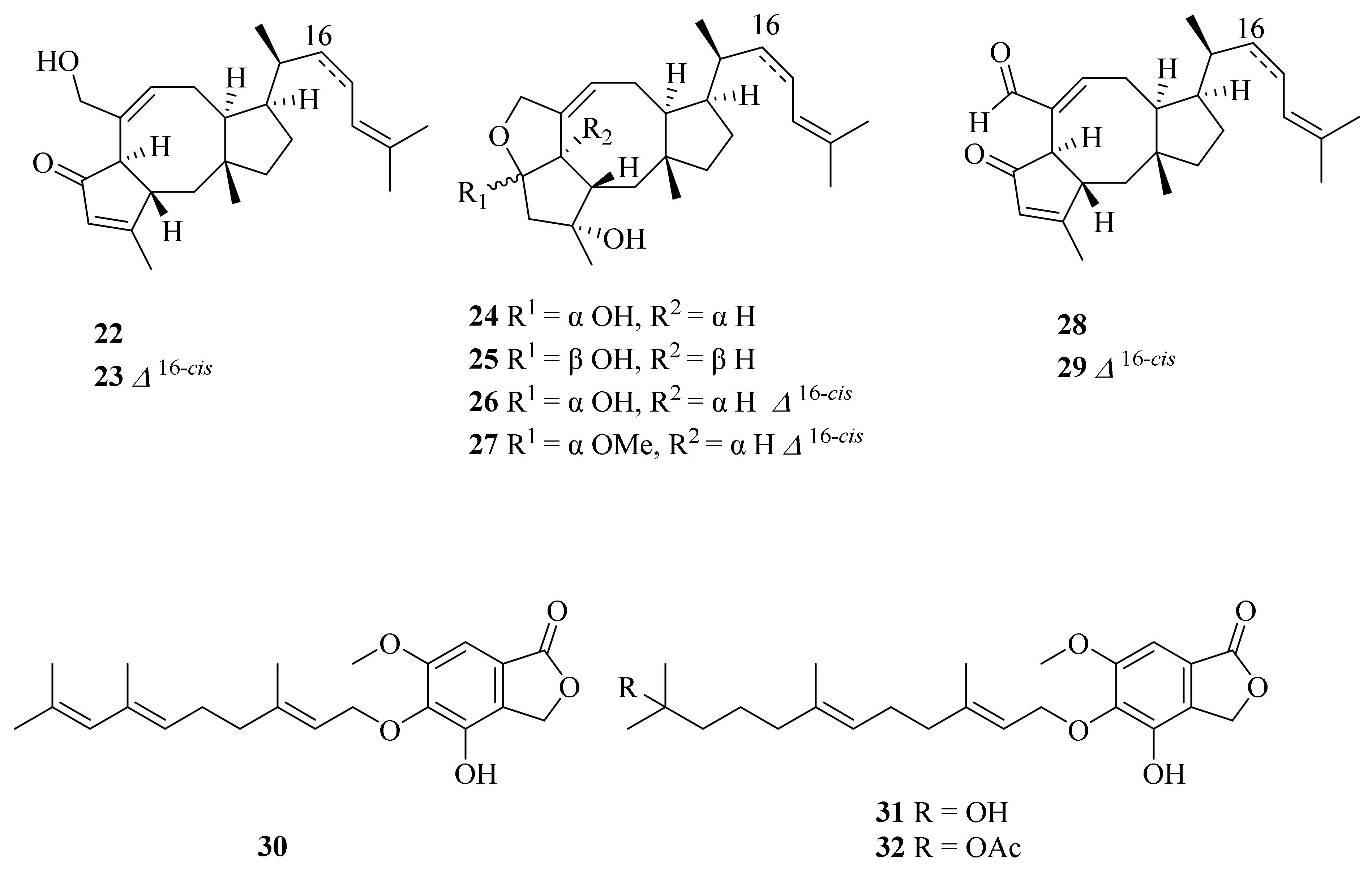
- Li, H.; Zhang, Q.; Li, S.; Zhu, Y.; Zhang, G.; Zhang, H.; Tian, X.; Zhang, S.; Ju, J.; Zhang, C. Identification and Characterization of Xiamycin a and Oxiamycin Gene Cluster Reveals an Oxidative Cyclization Strategy Tailoring Indolosesquiterpene Biosynthesis. J. Am. Chem. Soc. 2012, 134, 8996–9005. [Google Scholar] [CrossRef]
Figure 1.
The chemical structures of compounds (1–18).

Figure 2.
The chemical structures of compounds (19–21).

Figure 3.
The chemical structures of compounds (22–32).

Figure 4.
The chemical structures of compounds (33–41).

Figure 5.
The chemical structures of compounds (42–55).

Figure 6.
The chemical structures of compounds (56–60).

Figure 7.
The chemical structures of compounds (61–62).

Figure 8.
The chemical structures of compounds (63–71).

Figure 9.
The chemical structures of compounds (72–79).

Figure 10.
The chemical structures of compounds (84–86).

Figure 11.
The chemical structures of compounds (87–89).

Figure 12.
The chemical structures of compounds (90–104).

Figure 13.
The chemical structures of compounds (105–122).

Figure 14.
The chemical structures of compounds (123–136).

Figure 15.
The chemical structures of compounds (137–144).

Figure 16.
The chemical structures of compounds (145–153).

Figure 17.
The chemical structures of compounds (154–164).

Figure 18.
The chemical structures of compounds (165–166).

Figure 19.
The chemical structures of compounds (167–172).

Figure 20.
The chemical structures of compounds (174–182).

Figure 21.
The sources of reported natural products from cold seeps.

Figure 22.
The sources of reported natural products from hydrothermal vents.

Figure 23.
The bioactivities of the natural products from cold seeps and hydrothermal vents.

Publisher’s Note: MDPI stays neutral with regard to jurisdictional claims in published maps and institutional affiliations. |
© 2022 by the authors. Licensee MDPI, Basel, Switzerland. This article is an open access article distributed under the terms and conditions of the Creative Commons Attribution (CC BY) license (https://creativecommons.org/licenses/by/4.0/).
Share and Cite
MDPI and ACS Style
Cong, M.; Pang, X.; Zhao, K.; Song, Y.; Liu, Y.; Wang, J. Deep-Sea Natural Products from Extreme Environments: Cold Seeps and Hydrothermal Vents. Mar. Drugs 2022, 20, 404. https://doi.org/10.3390/md20060404
AMA Style
Cong M, Pang X, Zhao K, Song Y, Liu Y, Wang J. Deep-Sea Natural Products from Extreme Environments: Cold Seeps and Hydrothermal Vents. Marine Drugs. 2022; 20(6):404. https://doi.org/10.3390/md20060404
Chicago/Turabian StyleCong, Mengjing, Xiaoyan Pang, Kai Zhao, Yue Song, Yonghong Liu, and Junfeng Wang. 2022. "Deep-Sea Natural Products from Extreme Environments: Cold Seeps and Hydrothermal Vents" Marine Drugs 20, no. 6: 404. https://doi.org/10.3390/md20060404
Note that from the first issue of 2016, this journal uses article numbers instead of page numbers. See further details here.