Article

Molecular Correlation between Larval, Deutonymph and Adult Stages of the Water Mite Arrenurus (Micruracarus) Novus

by
Pedro María Alarcón-Elbal
1,
Ricardo García-Jiménez
2,
María Luisa Peláez
2,
Jose Luis Horreo
3,* and
Antonio G. Valdecasas
2
1
Instituto de Medicina Tropical & Salud Global (IMTSAG), Universidad Iberoamericana (UNIBE), 22333 Santo Domingo, Dominican Republic
2
Museo Nacional de Ciencias Naturales, CSIC. C/José Gutiérrez Abascal, 2, 28006 Madrid, Spain
3
UMIB Research Unit of Biodiversity (UO, CSIC, PA), C/Gonzalo Gutiérrez de Quirós s/n, 33600 Mieres, Spain
*
Author to whom correspondence should be addressed.
Life 2020, 10(7), 108; https://doi.org/10.3390/life10070108
Submission received: 27 May 2020 / Revised: 5 July 2020 / Accepted: 7 July 2020 / Published: 9 July 2020
(This article belongs to the Special Issue Molecular Phylogenetics and Mitochondrial Evolution)

Abstract

:
The systematics of many groups of organisms has been based on the adult stage. Morphological transformations that occur during development from the embryonic to the adult stage make it difficult (or impossible) to identify a juvenile (larval) stage in some species. Hydrachnidia (Acari, Actinotrichida, which inhabit mainly continental waters) are characterized by three main active stages—larval, deutonymph and adult—with intermediate dormant stages. Deutonymphs and adults may be identified through diagnostic morphological characters. Larvae that have not been tracked directly from a gravid female are difficult to identify to the species level. In this work, we compared the morphology of five water mite larvae and obtained the molecular sequences of that found on a pupa of the common mosquito Culex (Culex) pipiens with the sequences of 51 adults diagnosed as Arrenurus species and identified the undescribed larvae as Arrenurus (Micruracarus) novus. Further corroborating this finding, adult A. novus was found thriving in the same mosquito habitat. We established the identity of adult and deutonymph A. novus by morphology and by correlating COI and cytB sequences of the water mites at the larval, deutonymph and adult (both male and female) life stages in a particular case of ‘reverse taxonomy’. In addition, we constructed the Arrenuridae phylogeny based on mitochondrial DNA, which supports the idea that three Arrenurus subgenera are ‘natural’: Arrenurus, Megaluracarus and Micruracarus, and the somewhat arbitrary distinction of the species assigned to the subgenus Truncaturus.

1. Introduction

The systematics of many groups of organisms has traditionally been based on the adult stage [1]. For species showing a deep morphological transformation from the embryonic to the adult stage, due to a change in habitat or different behavior (parasitic and free-living stages) it may be difficult, if not impossible, to identify the juvenile stage without a previous correlation with the corresponding adult.
Members of the clade Hydrachnidia (Acari Actinotrichida) mainly inhabit continental waters [2] and are characterized by three main active life stages—larval, deutonymph and adult—with intermediate dormant stages [3]. Deutonymphs and adults share a characteristic morphology and may be identified through diagnostic characters. Larval morphology is very different from that of the two other stages. The standard procedure to match the larvae of a species with its corresponding adult is to track them directly from a fertilized female. In short, a fertilized female is kept in a small vial with a strip of paper or another substrate until they oviposit eggs and larvae emerge [4,5]. It is hard to identify the species level of larvae that have not been tracked in this way with certainty.
The water mite genus Arrenurus has a worldwide distribution, except for the perpetual snow regions, but there are no cosmopolitan species; rather, each main area upholds its own set of species. There are 152 Arrenurus in Europe and, of these, about 30% have a wide distribution [6,7,8]. As in other Hydrachnidia genera, Arrenurus deutonymphs and adults share diagnostic characters, however, the larval stage has a distinct morphology and requires independent morphological characterization. Water mite larvae usually attach to insects at preimaginal stages before they hatch, using adults as vector to disperse [9]. Adults and larvae of the same species, therefore, may be found in very different locations in their life cycle. Some Arrenurus species have occasionally been recorded as an ectoparasite of aquatic insects at the juvenile stage, either nymphs or larvae [2]. Some have even been recorded in insect exuviae [6,10]. Given these challenges, species identification of isolated Arrenurus larvae is fraught with uncertainty.
In this work, we characterized Arrenurus sp. larvae that were attached to larvae and a pupa of the common mosquito Culex (Culex) pipiens and cross-correlate their sequences with those of 51 adult Arrenurus species. One of these species, Arrenurus (Micruracarus) novus George, 1884, was in the same mosquito habitat from which the Arrenurus larvae were isolated. We compare cytochrome C oxidase subunit I (COI) and cytochrome B (cytB) DNA sequences of the water mite larvae with those of A. novus deutonymphs and adults (male and female) and other Arrenurus species, in order to establish the identity of the water mite larvae in a particular case of ‘reverse taxonomy’ [11].

2. Materials and Methods

2.1. Taxon Sampling

Water mite and mosquito specimens were both collected during a survey of mosquitoes at preimaginal stages carried out every two weeks between May and October of 2012 at Acequia del Caminàs (UTM 30S 741855.92 E 4324431.48), a semiartificial irrigation channel within the marsh waters of the Xeraco and Xeresa wetlands (Valencia, Spain; Figure 1). For more details on the characteristics of the area, see Alarcón-Elbal et al. [12]. Further collection efforts were carried out in May 2019 without success.
We used a quick dipping technique [13] with a standard 500-mL mosquito dipper (BioQuip Products Inc., Rancho Dominguez, CA, USA). Culicids at preimaginal stages were sorted manually at the laboratory and then transferred to a 60 °C water bath for a minute and then to absolute ethanol. Culicid specimens with ectoparasites and free-living water mites in the same sample were both preserved in absolute ethanol and stored at −20 °C.
Preservation in absolute ethanol ensures that both microscopic and molecular techniques can be used on any water mite specimen collected from potential hosts or directly from the field [14,15]. Both the culicids and the water mite specimens were morphologically identified, and subsequently used for DNA extraction.

2.2. Morphological Identification of Culicids

Morphological identification of culicids at preimaginal stages were made to the species level based on external morphology and diagnostic characters related with chaetotaxy, as described by Schaffner et al. [16]. Specimens were examined under a Nikon SMZ-1B Stereozoom microscope.

2.3. Morphological Identification of Water Mites

2.3.1. Rearing of Adult Female Water Mites

In an attempt to rear larvae, six live adult Arrenurus sp. females were collected in the field and then kept under laboratory conditions in individual transparent poliestirene plastic containers (diameter 10–15 mm, height 15 mm) with a piece of paper and mineral water at room temperature for 14–15 days [4]. Following the monitoring period, the females were preserved in absolute ethanol for morphological identification of the species level.

2.3.2. Morphological Identification of Water Mite Adults, Deutonymphs and Larvae

Morphological identification of the adult and deutonymph water mite specimens collected from the field site was performed using the diagnostic characters originally described by George [17] and subsequently revised by Viets [18] and Gerecke [2].
The study of larval water mite specimens was based on four specimens found anchored on the body of a culicid larva (L1–L4 in Table 1) and one larva found on a culicid pupa (L5). The culicid larva, pupa and attached water mite larvae were examined first under a Bausch and Lomb stereomicroscope and then with laser scanning confocal microscopy (LSCM, Leica TCS SPE). The host attachment site, where the water mite larval mouthparts were embedded, was recorded and the larvae were individually and carefully detached using fine tweezers and transferred to a drop of glycerol on a microscope slide for LSCM stack acquisition. After imaging, the five larvae were then processed for molecular analyses.

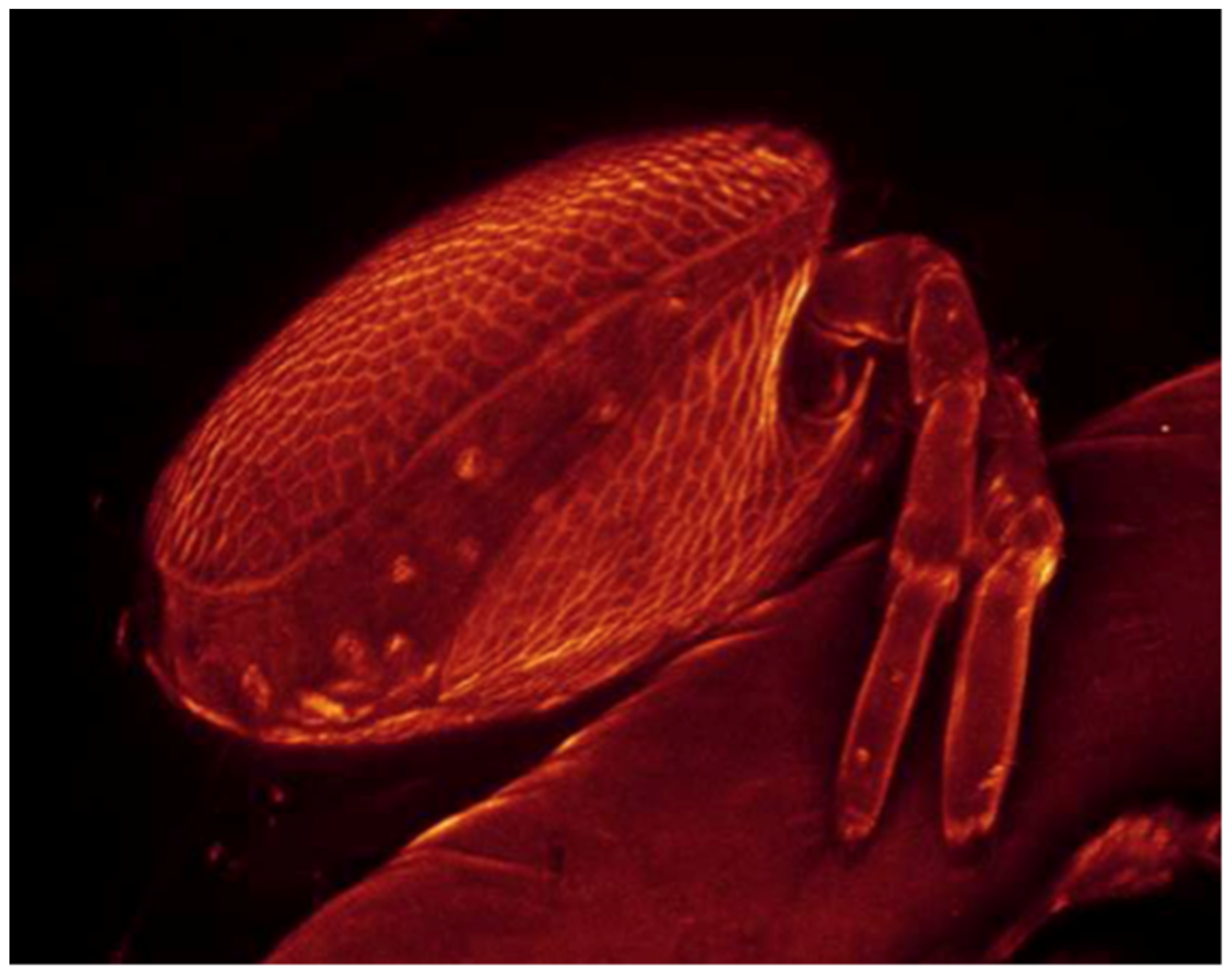
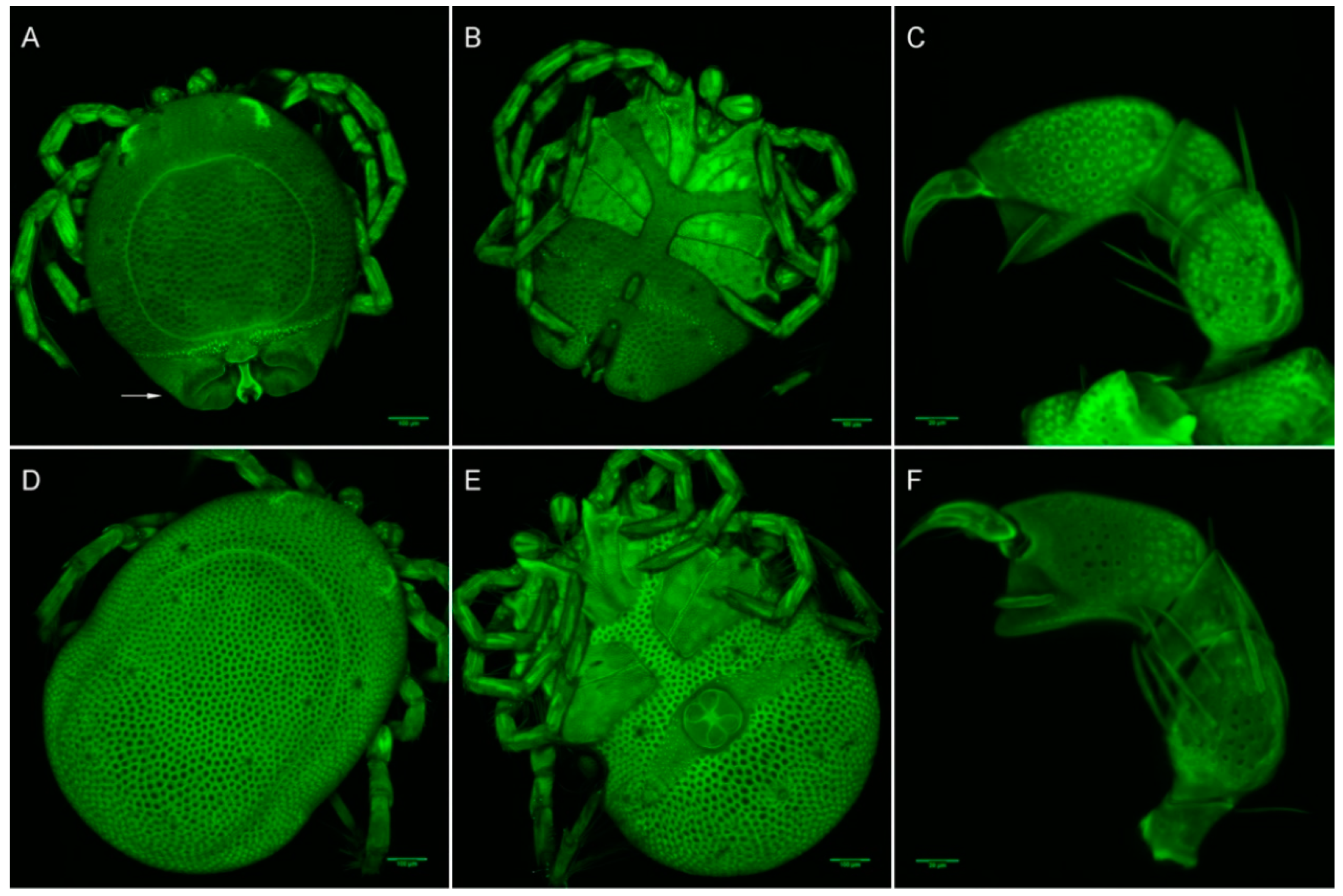
2.3.3. Laser Scanning Confocal Microscopy Imaging

Laser scanning confocal microscopy (LSCM, Leica TCS SPE) was employed to acquire z-stack images with the following objectives: 10×/0.30 NA, 20×/0.70 NA, 40×/1.25 NA oil immersion and 63x/1.30 NA glycerol immersion. The samples were subjected to an excitation wavelength of 488 nm and an emission range between 520 and 660 nm. For further information on the stack acquisition procedure, see Valdecasas and Abad [14]. Serial images were processed with Amira (ver. 5.4.3) and ImageJ/Fiji to obtain maximum 2D projections and Voltex volume renderings. Different parameters of visualization were used with Voltex to best highlight the characters of interest. Fiji/ImageJ were used to take all measurements directly from the image stacks. As all the material was used for the subsequent molecular analyses, no morphological vouchers could be kept. Therefore, a photographic voucher comprised of the full set of original unaltered stacks is stored in the confocal collection at the National Museum of Natural Sciences of Spain (CSIC–MNCN).

2.4. Molecular Analyses

A total of nine water mite samples—three adults, one deutonymph and five larvae—were processed for molecular analyses. DNA extraction, PCR amplification and sequencing were performed as previously described) [15]. The COI gene was amplified using the primer pair LCO1490 and HC02198 [19]. For the larval water mite samples (n = 5), the internal primer COI-AV-F (5′-ATAAGATTTTGACTTCTYCC-3′) and HC02198 (360 basepairs length) were used because the complete amplification of the gene was not successful. CytB was amplified using the primer pair COB-F/R [20]. Sequencing reactions were performed using ABI BigDye® v3.1 Cycle Sequencing Kit (Applied Biosystems) and run on an ABI 3100 Genetic Analyzer. Sequence chromatograms were checked for accuracy and edited using Sequencher® version 5.0 (Gene Codes Corporation, Ann Arbor, MI, USA). DNA sequences were deposited in GenBank (accession numbers MT598102 to MT598106 for COI and MT607634 to MT607637 for cytB).
The sequences of both gene fragments were analyzed for all three life cycle stages in order to molecularly correlate the undescribed larvae with the diagnosed adults and deutonymphs. Genetic distances were estimated using the Kimura-2-Parameter (K2P) distance model [21], as implemented in MEGA v.7 [22].
Additionally, COI and cytB sequences of Arrenurus species were searched in GenBank (https://www.ncbi.nlm.nih.gov/genbank/; accessed 21 May 2020) in order to estimate the genetic distances among all available sequences, including the new ones of this study. The distances of the GenBank sequences were calculated using MEGA. A Maximum-Likelihood phylogenetic tree of COI sequences was constructed with the IQ-TREE web server (http://iqtree.cibiv.univie.ac.at) with the following settings: automatic detection of substitution model, perturbation strength of 0.5, IQ-TREE stopping rule of 100, ultrafast bootstrap analysis (1000 replicates) and SH-aLRT single branch tests (1000 replicates). For this tree, two outgroup sequences were used: Torrenticola lundbladi (GenBank accession number JX629050) and Lebertia maderigena (KX421869).

3. Results

3.1. Morphological Identification of Culicids

All of the culicid specimens were identified as Culex (Culex) pipiens Linnaeus, 1758. The principal and reliable characters for diagnosis at the larval stage are the syphon index, the syphon/saddle index, the branch number of seta one on abdominal segments III–IV, 1a–S tuft, 1b–S tuft and syphon shape [23]. One culicid larva and one pupa were parasitized with four and one water mite larvae, respectively (Figure 2, Figure 3 and Figure 4).

3.2. Morphological and Molecular Identification of Water Mites

3.2.1. Rearing of Adult Female Water Mites

Six Arrenurus females were maintained for 14–15 days, but no eggs or larvae were observed during this period. We were not able to ascertain whether the females were gravid and if the lack of eggs and larvae was due to unsuitable laboratory conditions.

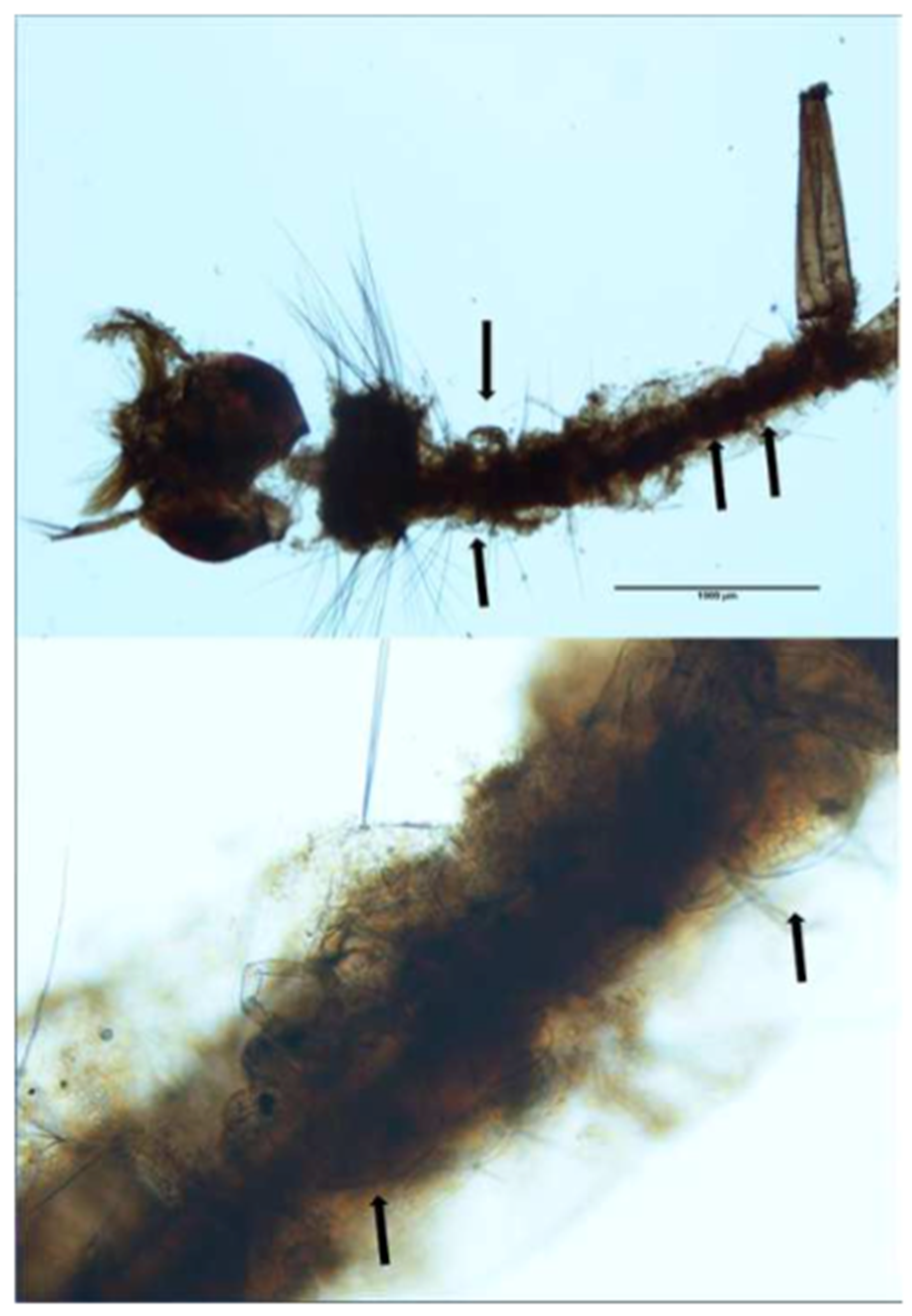
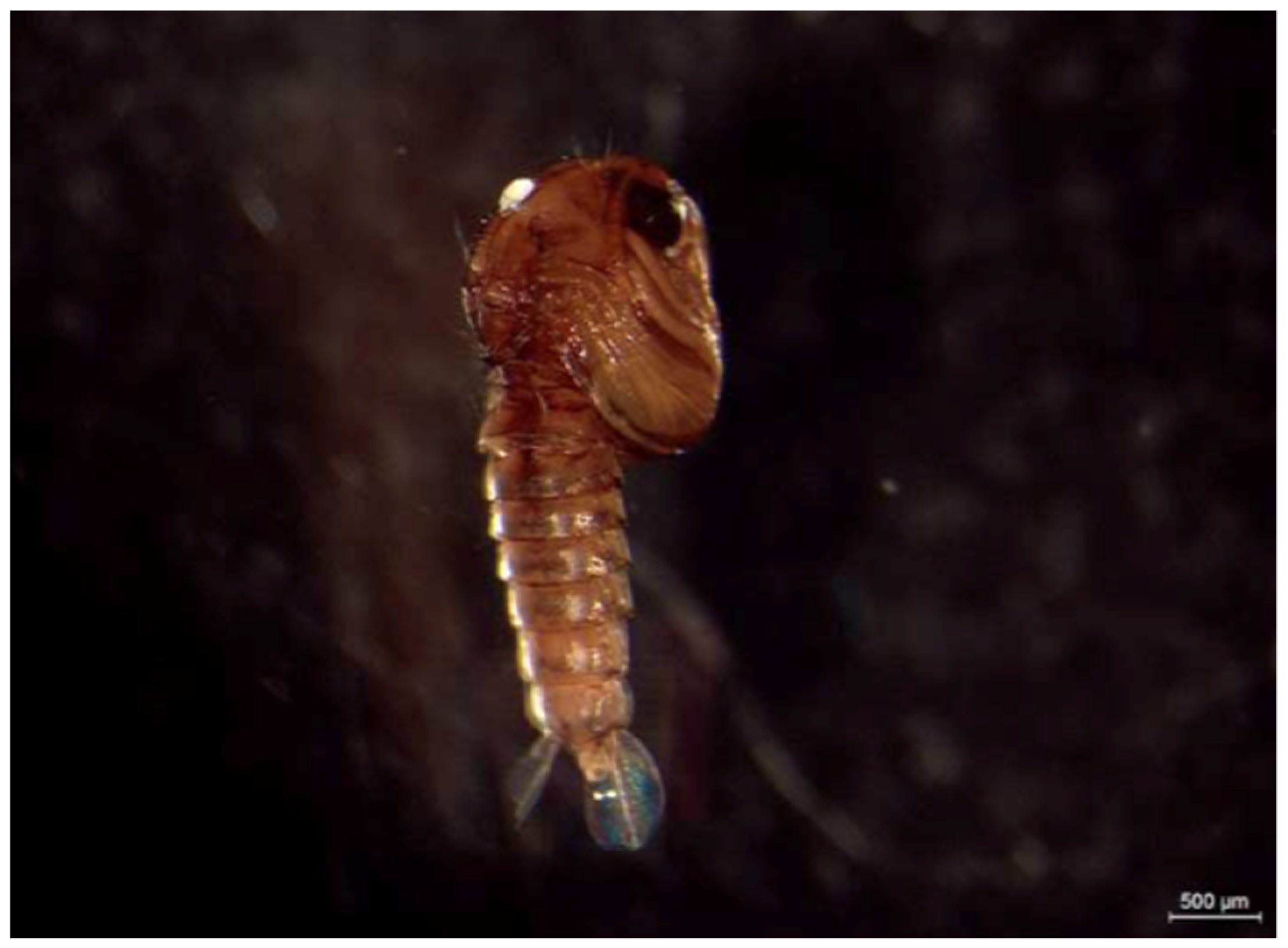
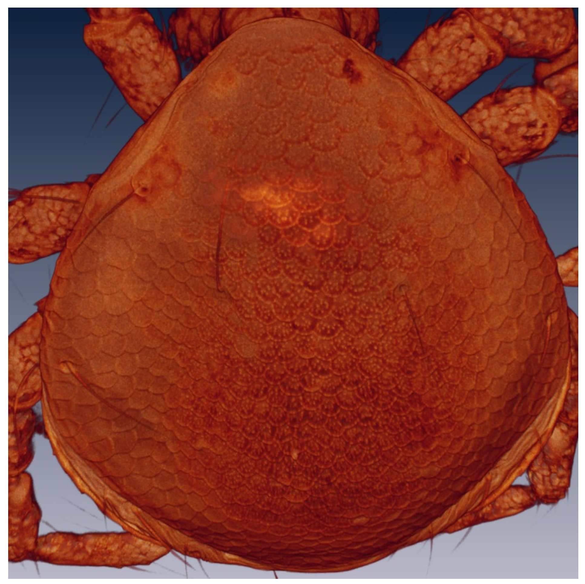
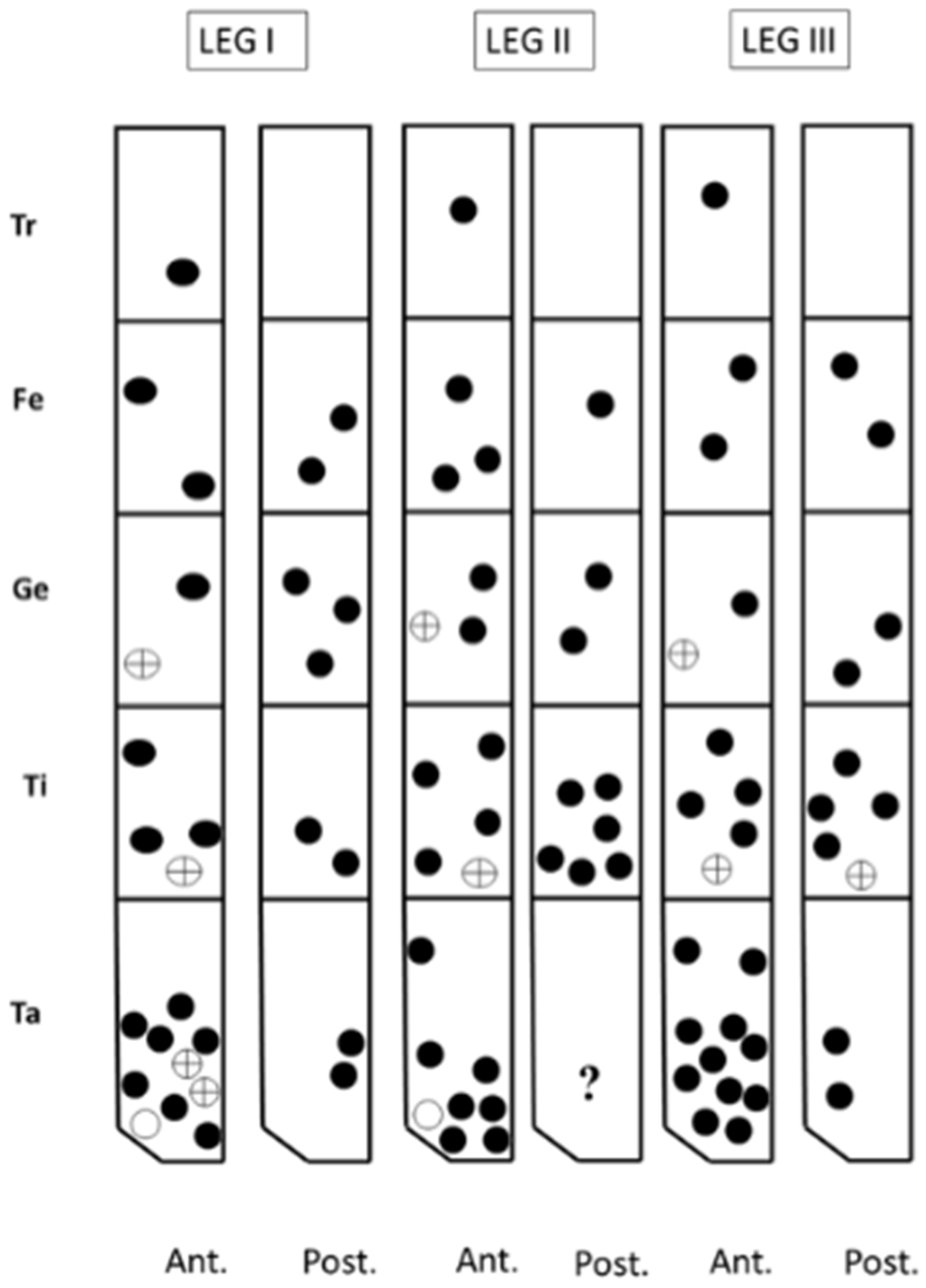
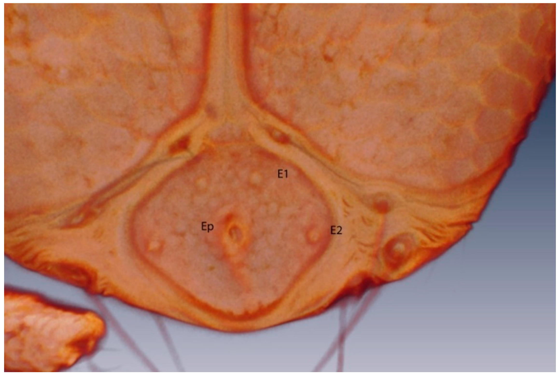
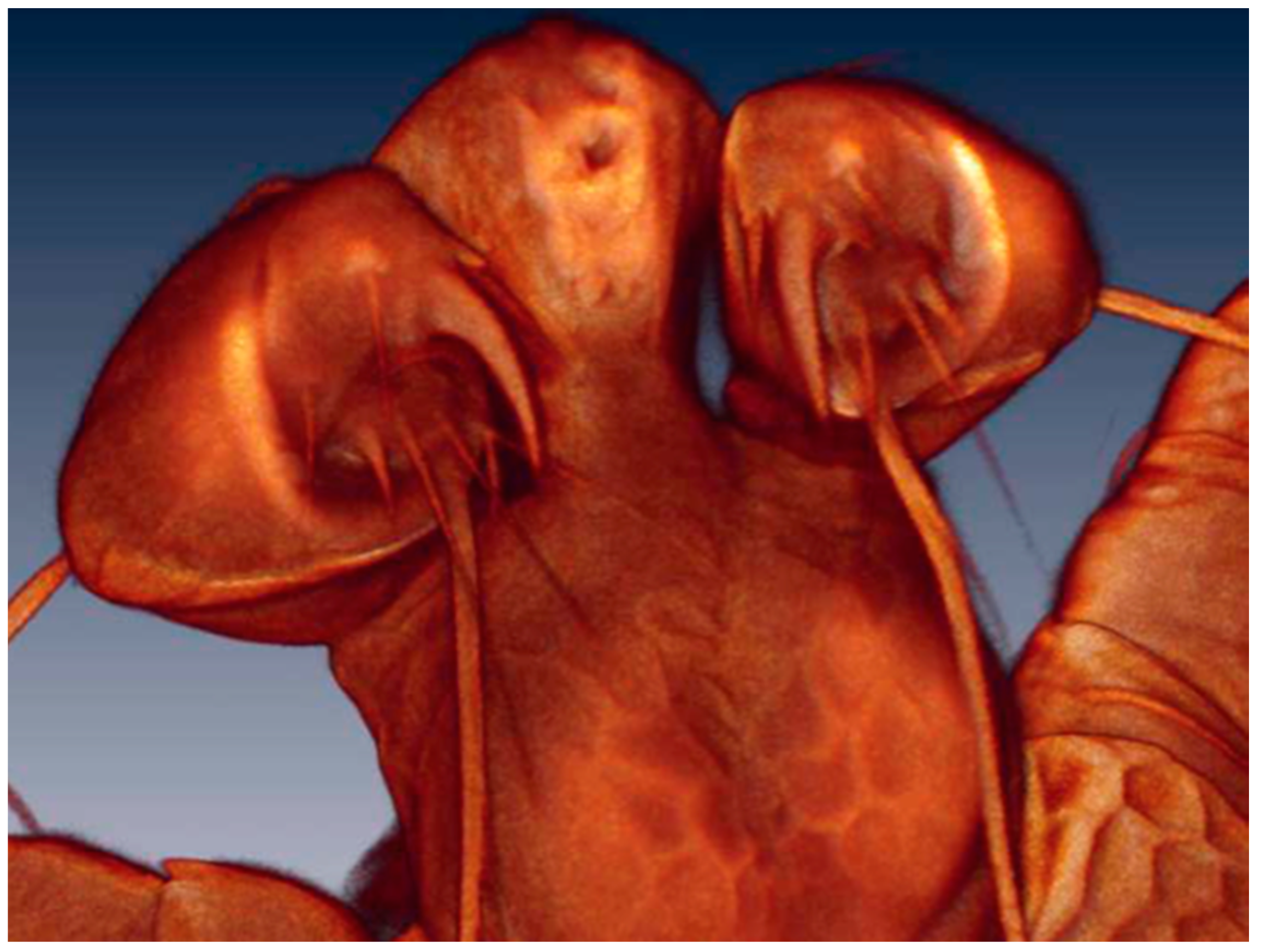
3.2.2. Larval Description

The description of the water mite larvae is preliminary: confocal microscopy could not resolve some of the extremely fine morphological details, an issue faced by other researchers of water mite larvae [7]. Therefore, scanning electron microscopy analyses are necessary for a complete description. The terminology used here follows that of Zawal [7]. Figure 5 and Figure 6 show a Voltex projection of the ventral and the dorsal habitus of a larva, respectively. The projections do not indicate morphometric distances, and the scales are only an approximation of size. Table 1 and Table 2 include some standard morphometric data taken independently of the Voltex projections. Figure 7 shows a schematic of the approximate distribution of setae on the leg segments.
Arrenurus novus larvae (see molecular analysis below) may be distinguished from other previously described Micruracarus larvae [7] by the following set of characters: dorsal plate ovoid, narrowed anteriorly; coxas distinct, ratio lateral length of coxal plate I, II and III: 5:3:4; a hexagonal excretory plate perimeter and the excretory pore in line or slightly above E2 setae (Figure 8). Total length of Leg1 < Leg II = Leg III.
The number of setae on the pedipalp segments are in agreement with Zawal’s description of Arrenurus larvae [7]: none on P-I; one on P-II; two on P-III, one long and thick and the other short; four on P-IV, three thin and one thick; and a solenoid and seven setae on P-V (Figure 9).

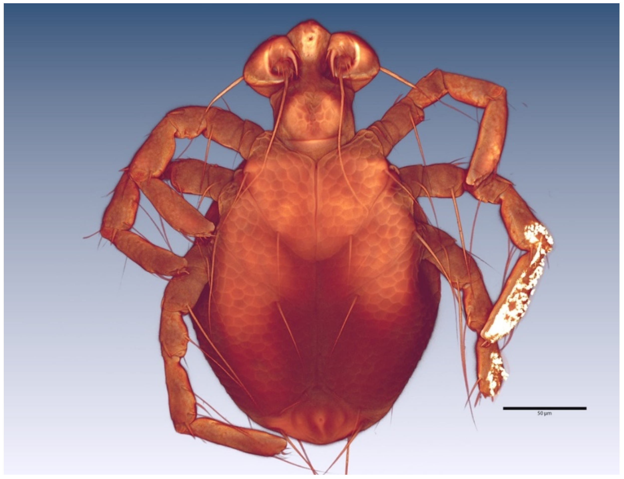
3.2.3. Morphological Identification of Adult and Deutonymph Water Mites

Seven male and six female adults plus two deutonymphs were identified as A. novus, a species found in standing waters throughout Europe [2]. Among the main diagnostic characters identifying A. novus adults are the petiole in the male (see arrow in Figure 10A), the setation of male and female palps, and the shape and length of cauda (Figure 10). The deutonymph diagnosis was based on palp shape and setation and habitat co-occurrence (Figure 11): approximate body length: 526 µm; body width: 467 µm; P-1 without setae; P-II with three medial setae; P-III with one medial setae; PIV uncate and a strong distal setae and PV with a basal fine setae.

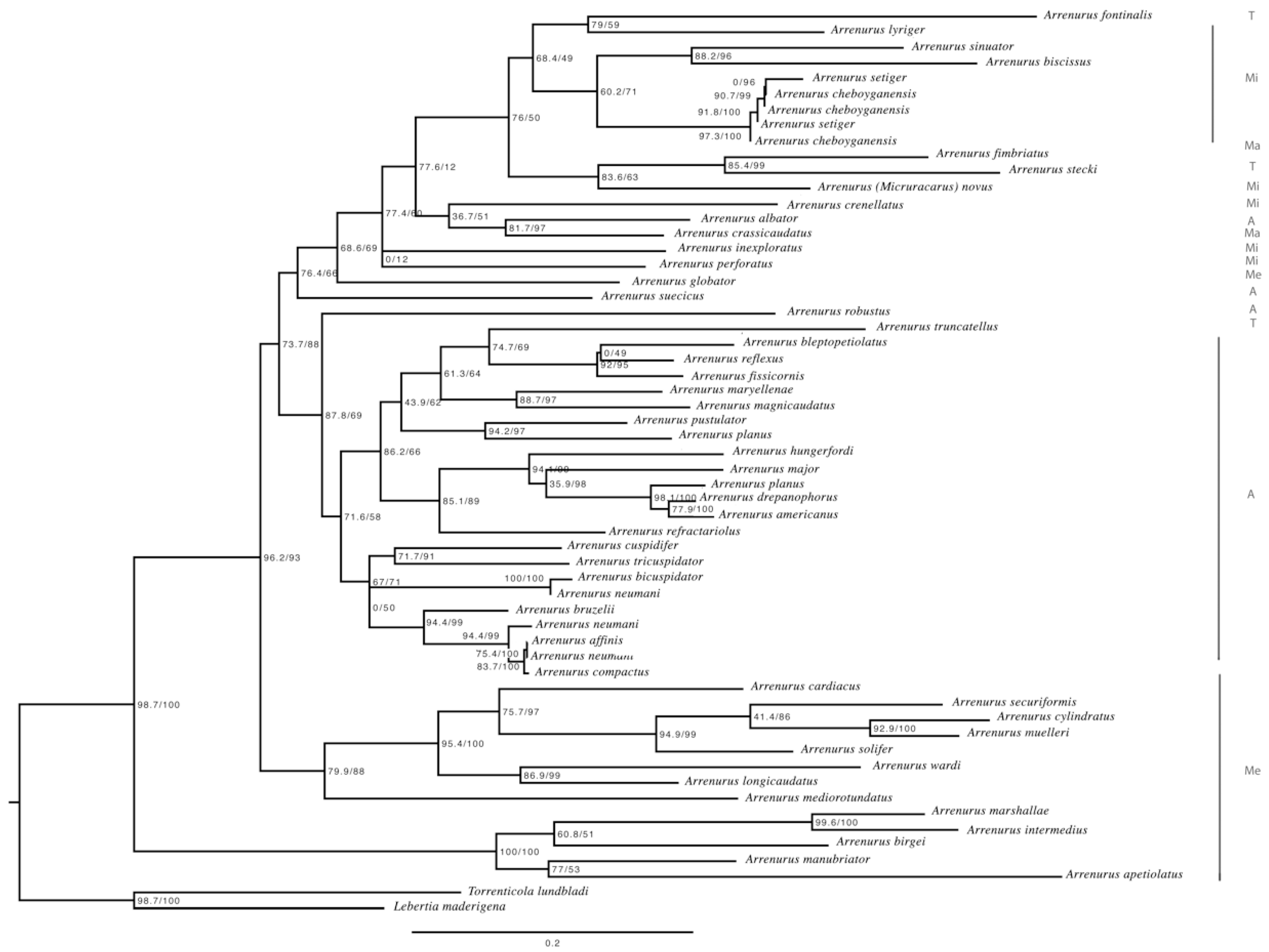
3.3. Molecular Analyses

GenBank identified 990 Arrenurus barcoding COI sequences but no Arrenurus cytB sequences (probably because cytB is not generally used for species delimitation). These sequences belonged to 51 species of the genus plus A. novus, whose sequence is reported here for the first time (genetic distances among species are shown in Table S1 and phylogenetic tree in Figure 12). The mean genetic distances among the 52 Arrenurus species was 16.4% (standard deviation = 4.1). The here-amplified COI sequences from all the different A. novus life-cycle stages were all the same.
Very few phylogenetic information about Arrenurus currently exist [24]. The here-constructed COI phylogenetic tree (Figure 12, Material S1) may be divided into three main arms: the upper branch is dominated by species belonging to the subgenus Micruracarus, with occasional incursions of species belonging to the subgenera Arrenurus, Truncaturus, Megaluracarus and Micrarrenurus. In this upper branch is located Arrenurus (Micruracarus) novus. The intermediate branch of the tree is composed exclusively of species of the subgenus Arrenurus, except for a sequence belonging to subgenus Truncaturus. The lower section of the tree consists exclusively of species belonging to the sub-genus Megaluracarus, with European and American representatives.
The 360-bp COI fragment was successfully amplified only from one larvae; however, no genetic variation was found among the sequences of this region (two adults, one deutonymph and one larvae). Genetic differences were, however, observed in the 330-bp cytB fragment (Table 3). One of the adults and all five larvae shared the same cytB haplotype, whereas the deutonymph and the other adult shared a different haplotype (Table 3).

4. Discussion

The number of molecular sequences available for Hydrachnidia has increased substantially in recent years. GenBank list 6450 Hydrachnidia nucleotide sequences (as of 21 May 2020). We did not search the BOLD Systems database for two main reasons: many of the Arrenurus sequences are privately held, and it was recently shown that some water mite sequences do not agree with their species assignment [25]. Of the GenBank sequences, almost 70% are identified to the generic level. Some new species have been described with morphological and molecular data [15,26]. For the genus Arrenurus, there are 1071 sequences (990 of them belonging to COI gene) but only 198, representing 51 taxa, are at the species level. These were employed to the genetic distance calculations. All of the Arrenurus sequences in GenBank identified to the species level are based on the adult stage; sequences obtained from larvae are only given a generic rank and are frequently singled out as operational taxonomic units (e.g. [27]). There is a need for more species-level sequences based on different life cycle stages to widen the usefulness of this type of data to address ecological, biogeographical and evolutionary questions. We identified the adult and deutonymph water mite specimens as A. novus, a broadly distributed species found from the Western Palearctic to the Afrotropic, commonly in standing waters [2,25]. However, according to Gerecke et al. [2], it has been “little reported in Europe”.
In the present study, we combined morphological and molecular techniques to associate undescribed larvae with corresponding adults and deutonymphs for the first time in water mites. Associations among life cycle stages are especially relevant for this genus given that less than 20% of arrenurid larvae are known [28]. To link undescribed larval specimens with other life stages (i.e., deutonymph and adult), we employed a DNA-based identification method in which sequences are considered as standardized comparative characters that can be used to support and integrate morphological datasets [29]. Even in cases with very small genetic distances, these data can be useful for species identification [30]. The results of our analyses of two mitochondrial genes (COI and cytB), which proved to be informative, clearly support the same conclusion: the larvae belong to the species A. novus. The larval COI sequences were compared with 52 adult Arrenurus species sequences (51 were from GenBank; the A. novus sequences, from one adult male, two adult females, one deutonymphs and one larvae, are provided for the first time here). Genetic distances among the three A. novus life-cycle stages were 0.000 (the same DNA sequence), confirming the co-specificity of the larvae, deutonymph and adults. The distance of A. novus with other Arrenurus species ranged between 9.8 with A. setiger and 24.3 with A. megalurus (see their phylogenetic relationships in Figure S1). The cytB sequences of the larvae and one of the adults were identical and highly similar to those of the other adult and the deutonymph (which were identical; see Table 3), strongly supporting the conclusion that they belong to the same species. It is important to note here that we are studying only mitochondrial DNA, thus, in the case of hybrids, we would be only describing the maternal species of the larvae. In addition, the phylogenetic tree based on COI mitochondrial information (Figure 12) supports the idea that three Arrenurus subgenera are ‘natural’—Arrenurus, Megaluracarus and Micruracarus—and the somewhat arbitrary distinction of the species assigned to the subgenus Truncaturus. This topology differs from the only other Arrenurus phylogeny published [24] and points to the need for more molecular data in order to clarify their phylogenetic relationships.
This study demonstrates the usefulness of an integrative approach to resolve the taxonomic uncertainty of water mites at the larval stage. Furthermore, it appears to be a promising approach for identifying larvae at the species level in terms of both reliability and speed, in comparison with rearing or hatching approaches. With these data, we can also begin to elucidate host–parasite interactions between a specific water mite species and its host(s) with greater detail. To date, a host of A. novus larvae was unknown [2]. Indeed, only a few studies have described a host-parasite interaction between a Culex species and an Arrenurus species (e.g. [6,10,31,32]). Arrenurus is the genus most reported to parasitize mosquitoes, evidence of its flexibility regarding host specificity [33]. Despite this, recent findings suggest that these mites prefer Culex species [34]. Aside from reducing host fitness by piercing their exoskeleton to feed on hemolymph, each host–parasite association has its own characteristics, which are determined mainly by the size of the partners, intrinsic defence mechanisms and environmental conditions [35]. Parasitic mites may play a significant role in the biological control of mosquitos in wetlands, especially of adult populations. Therefore, more observations and experimental data of water mite species are needed to better understand their host–parasite interactions, as well as to incorporate the use of molecular techniques in their identification, particularly at the larval stage.

Supplementary Materials

The following are available online at https://www.mdpi.com/2075-1729/10/7/108/s1, Table S1: K2P distances among the COI sequences of the 52 Arrenurus species included in the analysis. The final alignment was 659 bp. Arrenurus novus is indicated in bold. Material S1: Maximum Likelihood phylogenetic tree of the Arrenurus COI sequences employed in this study and the outgroups (Torrenticola lundbladi and Lebertia maderigena) in newick format.

Author Contributions

Field data sampling and culicids identification, P.M.A.-E.; Bright field microscopy and Laser Scanning Confocal Microscopy imaging, and identification of water mites, A.G.V.; Molecular extraction, amplification and sequencing R.G.-J.; Genbank searches and downloading and molecular analysis, M.L.P. and J.L.H.; all the authors contributed to the writing and reviewed the last version of the manuscript. All authors have read and agreed to the published version of the manuscript.

Funding

J.L.H. was supported by the Regional Government of Asturias (reference SV-16-UMB-1).

Acknowledgments

We thank Annie Machordom for access to the Systematics Molecular Laboratory of the MNCN.

Conflicts of Interest

The authors declare no conflict of interest. The funders had no role in the design of the study; in the collection, analyses, or interpretation of data; in the writing of the manuscript, or in the decision to publish the results.

References

  1. Thorp, J.H.; Rogers, D.C.; Dimmick, W.W. Introduction to invertebrates of inland waters. In Freshwater Invertebrates, 4th ed.; Elsevier BV: Amsterdam, The Netherlands, 2015; pp. 1–19. [Google Scholar]
  2. Gerecke, R.; Gledhill, T.; Pešić, V.; Smit, H. Süßwasserfauna von Mitteleuropa, Bd. 7/2-3 Chelicerata; Springer Spektrum: Heidelberg, Germany, 2016. [Google Scholar]
  3. Smith, I.M.; Cook, D.R.; Smith, B.P. Water mites (Hydrachnidiae) and other arachnids. In Ecology and Classification of North American Freshwater Invertebrates; Elsevier BV: Amsterdam, The Netherlands, 2010; pp. 485–586. [Google Scholar]
  4. Cook, W.J.; Smith, B.P.; Brooks, R.J. Allocation of reproductive effort in female Arrenurus spp. water mites (Acari: Hydrachnidia; Arrenuridae). Oecologia 1989, 79, 184–188. [Google Scholar] [CrossRef]
  5. Tuzovsky, P. A new water mite species of the genus Arrenurus Dugès, 1834 (Acariformes: Hydrachnidia: Arrenuridae) from Eastern Palaearctic. Acarina 2012, 20, 173–179. [Google Scholar]
  6. Zawal, A. Phoresy and parasitism: Water mite larvae of the genus Arrenurus (Acari: Hydrachnidia) on Odonata from lake Binowskie (NW Poland). Biol. Lett. 2006, 43, 257–276. [Google Scholar]
  7. Zawal, A. Morphological characteristics of water mite larvae of the genus Arrenurus Duges, 1834, with notes on the phylogeny of the genus and an identification key. Zootaxa 2008, 1765, 1–75. [Google Scholar] [CrossRef]
  8. Zawal, A. Morphology of the larval stages of Arrenurus affinis Koenike, 1887, A. neumani Piersig, 1895, and A. vietsi Koenike, 1911 (Acari: Hydrachnidia). Genus 2008, 19, 161–169. [Google Scholar]
  9. Bohonak, A.J.; Smith, B.P.; Thornton, M. Distributional, morphological and genetic consequences of dispersal for temporary pond water mites. Freshw. Boil. 2004, 49, 170–180. [Google Scholar] [CrossRef]
  10. Lanciani, C.A. Sexual bias in host selection by parasitic mites of the mosquito anopheles crucians (Diptera: Culicidae). J. Parasitol. 1988, 74, 768. [Google Scholar] [CrossRef]
  11. Markmann, M.; Tautz, D. Reverse taxonomy: An approach towards determining the diversity of meiobenthic organisms based on ribosomal RNA signature sequences. Philos. Trans. R. Soc. B: Boil. Sci. 2005, 360, 1917–1924. [Google Scholar] [CrossRef] [Green Version]
  12. Alarcón-Elbal, P.M.; Murillo, J.M.S.; Estrella, S.D.; Ruiz-Arrondo, I.; Prieto, R.P.; Curdi, J.L. Asociación de vector del VNO e hidrófito invasor: Culex pipiens Linnaeus, 1758 y Ludwigia grandiflora (Michaux) Greuter and Burdet en el marjal de Xeraco-Xeresa, Valencia. Anales Biol. 2013, 35, 17–27. [Google Scholar] [CrossRef]
  13. Service, M.W. A critical review of procedures for sampling populations of adult mosquitoes. Bull. Entomol. Res. 1977, 67, 343–382. [Google Scholar] [CrossRef]
  14. Valdecasas, A.G.; Abad, A. Morphological confocal microscopy in arthropods and the enhancement of autofluorescence after proteinase K extraction. Microsc. Microanal. 2010, 17, 109–113. [Google Scholar] [CrossRef] [PubMed]
  15. Pešić, V.; Valdecasas, A.G.; García-Jimenez, R. Simultaneous evidence for a new species of Torrenticola Piersig, 1896 (Acari, Hydrachnidia) from Montenegro. Zootaxa 2012, 3515, 38–50. [Google Scholar] [CrossRef]
  16. Schaffner, E.; Angel, G.; Geoffroy, B.; Hervy, J.P.; Rhaiem, A.; Brunhes, J. Les Moustiques d’Europe: Logiciel d’Identification et d’Enseignement = The Mosquitoes of Europe: An Identification and Training Programme; IRD Editions & EID Méditerranée: Montpellier, France, 2001. [Google Scholar]
  17. George, C.F. The British fresh-water mites. Hardwicke’s Sci. 1884, 20, 80–81. [Google Scholar]
  18. Viets, K. Zur kenntnis dert Hydracarinen-Fauna von Spanien. Arch. Hydrobiol. 1930. [Google Scholar]
  19. Folmer, O.; Black, M.; Hoeh, W.; Lutz, R.; Vrijenhoek, R. DNA primers for amplification of mitochondrial cytochrome c oxidase subunit I from diverse metazoan invertebrates. Mol. Mar. Biol. Biotechnol. 1994, 3, 294–299. [Google Scholar] [CrossRef] [PubMed]
  20. Ernsting, B.R.; Edwards, D.D.; Aldred, K.J.; Fites, J.S.; Neff, C.R. Mitochondrial genome sequence of Unionicola foili (Acari: Unionicolidae): A unique gene order with implications for phylogenetic inference. Exp. Appl. Acarol. 2009, 49, 305–316. [Google Scholar] [CrossRef]
  21. Kimura, M. A simple method for estimating evolutionary rates of base substitutions through comparative studies of nucleotide sequences. J. Mol. Evol. 1980, 16, 111–120. [Google Scholar] [CrossRef]
  22. Kumar, S.; Stecher, G.; Tamura, K. MEGA7: Molecular evolutionary genetics analysis version 7.0 for bigger datasets. Mol. Boil. Evol. 2016, 33, 1870–1874. [Google Scholar] [CrossRef] [Green Version]
  23. Azari-Hamidian, S.; Harbach, R.E. Keys to the adult females and fourth-instar larvae of the mosquitoes of Iran (Diptera: Culicidae). Zootaxa 2009, 2078, 1–33. [Google Scholar] [CrossRef]
  24. Wieçel, M. Effects of the Evolution of Intromission on Courtship Complexity and Male and Female Morphology: Water Mites of the Genus Arrenurus (Acari; Hydrachnida) from Europe and North America. Ph.D. Thesis, Adam Mickiewicz University, Poznan, Poland, 2016. [Google Scholar]
  25. Valdecasas, A.G.; García-Jiménez, R.; Marín, M. Two rare water mite species (Acari, Parasitengona, Hydrachnidia) new for the Iberian Peninsula. Rev. Ibérica Aracnol. 2019, 35, 33–37. [Google Scholar]
  26. Pešić, V.; Smit, H. Neumania kyrgyzica sp. nov. a new water mite from Kyrgyzstan based on morphological and molecular data (Acari, Hydrachnidia: Unionicolidae). Syst. Appl. Acarol. 2017, 22, 885. [Google Scholar] [CrossRef]
  27. Mlynarek, J.J.; Knee, W.; Forbes, M.R. Explaining susceptibility and resistance to a multi-host parasite. Evol. Boil. 2013, 41, 115–122. [Google Scholar] [CrossRef]
  28. Martin, P. Wassermilben (Hydrachnidia, Acari) und Insekten: Ein Überblick über eine selten betrachtete Beziehung. Entomol. Heute 2008, 20, 45–75. [Google Scholar]
  29. Scorrano, S.; Aglieri, G.; Boero, F.; Dawson, M.N.; Piraino, S. Unmasking Aurelia species in the Mediterranean Sea: An integrative morphometric and molecular approach. Zool. J. Linn. Soc. 2016, 180, 243–267. [Google Scholar] [CrossRef]
  30. Horreo, J.L.; Ardura, A.; Pola, I.G.; Martinez, J.L.; Garcia-Vazquez, E. Universal primers for species authentication of animal foodstuff in a single polymerase chain reaction. J. Sci. Food Agric. 2012, 93, 354–361. [Google Scholar] [CrossRef] [PubMed]
  31. Mullen, G.R. Water mites of the subgenus Truncaturus (Arrenuridae, Arrenurus) in North America. Ithaca Agric. Entomol. 1976, 6, 1–35. [Google Scholar]
  32. Kirkhoff, C.J.; Simmons, T.W.; Hutchinson, M.; Simmons, T.W. Adult mosquitoes parasitized by larval water mites in Pennsylvania. J. Parasitol. 2013, 99, 31–39. [Google Scholar] [CrossRef] [PubMed]
  33. Dos Santos, E.B.; Favretto, M.A.; Dos Santos Costa, S.G.; Navarro-Silva, M.A. Mites (Acari: Trombidiformes) parasitizing mosquitoes (Diptera: Culicidae) in an Atlantic Forest area in southern Brazil with a new mite genus country record. Exp. Appl. Acarol. 2016, 69, 323–333. [Google Scholar] [CrossRef] [PubMed] [Green Version]
  34. Atwa, A.A.; Bilgrami, A.L.; Al-Saggaf, A.I. Host–parasite interaction and impact of mite infection on mosquito population. Rev. Bras. Entomol. 2017, 61, 101–106. [Google Scholar] [CrossRef]
  35. Gerson, U.; Smiley, R.L.; Ochoa, R. Mites (Acari) for Pest Control; Blackewell: Malden, MA, USA, 2003. [Google Scholar]
Figure 1. Map of the sampling site (Xeraco, Valencia, Spain).
Life 10 00108 g001
Figure 2. Culex pipiens larva with Arrenurus sp. larvae attached (arrows) and detail.
Life 10 00108 g002
Figure 3. Culex pipiens pupa with Arrenurus sp. larva attached (white dot).
Life 10 00108 g003
Figure 4. Culex pipiens pupa with Arrenurus sp. larva attached (laser scanning confocal microscopy (LSCM) image).
Life 10 00108 g004
Figure 5. Arrenurus (Micruracarus) novus larva, ventral view (LSCM).
Life 10 00108 g005
Figure 6. Arrenurus (Micruracarus) novus larva, dorsal view (LSCM).
Life 10 00108 g006
Figure 7. ASchematic showing the distribution of setae on the leg segments of an Arrenurus (Micruracarus) novus larva. Tr: trochanter; Fe: femur; Ge: genu; Ti: tibia; Ta: tarsus. Life 10 00108 i001 Round setae; Life 10 00108 i002 solenidum; Life 10 00108 i003 other setae.
Life 10 00108 g007
Figure 8. Arrenurus (Micruracarus) novus larva, excretory plate (LSCM).
Life 10 00108 g008
Figure 9. Arrenurus (Micruracarus) novus larva, pedipalp (LSCM).
Life 10 00108 g009
Figure 10. Arrenurus (Micruracarus) novus adults (LSCM). (A) male, dorsal; (B) male, ventral; (C) male, palp; (D) female, dorsal; (E) female, ventral; (F) female, palp.
Life 10 00108 g010
Figure 11. Ventral view of Arrenurus (Micruracarus) novus deutonymph.
Life 10 00108 g011
Figure 12. Maximum Likelihood phylogenetic tree of the Arrenurus C oxidase subunit I (COI) sequences employed in this study and the outgroups (Torrenticola lundbladi and Lebertia maderigena). Node numbers show SH-aLRT (left) and bootstrap (right) support values (1000 replicates). Letters at right show subgenus acronyms: Arrenurus (A), Micruracarus (Mi), Truncaturus (T), Megaluracarus (Me) and Micrarrenurus (Ma).
Life 10 00108 g012
Table 1. Standard measurements (see [8]) of the larval body morphology (in µm; only an approximation due to specimen orientation on the slide).
L1L2L3L4L5
Body length197182190204200
Body width175168168164156
Dorsal plate length190186190185185
Dorsal plate width171152160148147
CpI medial margin length5755615762
CpII medial margin length2730302726
CpIII medial margin length3842444042
Distance between C1 and CpI median margin2119231820
Distance between C4 and CpIII median margin2827242729
Distance between C1 and C24036403839
Excretory pore plate length2527262229
Excretory pore plate width3230323031
Distance between Exp and Expp posterior margin1115151213
PIII length2721263131
Length of PIV claw2723141617
Table 2. Standard measurements of the larval leg morphology in µm.
Larvae TrochanterFemurGenuTibiaTarsus
L1leg12521294255
leg23025304657
leg33025304657
L2leg12321294049
leg22325304459
leg32723254857
L3leg13023303849
leg23032274257
leg33434274457
L4leg12923274455
leg22125294653
leg33229294659
L5leg12423274041
leg22922264560
leg33025304031
Table 3. K2P distances among the adult, deutonymph and larval Arrenurus sp. cytB sequences analyzed in this study (alignment: 330 bp).
K2PAdult 1Adult 2NymphLarvae
Adult 1-
Adult 20.006-
Nymph0.0060.000-
Larvae0.0000.0060.006-

