
Primary Mucinous Cystadenocarcinoma of the Breast Intermixed with Pleomorphic Invasive Lobular Carcinoma: The First Report of This Rare Association

 , , , , and
Abstract
:1. Introduction
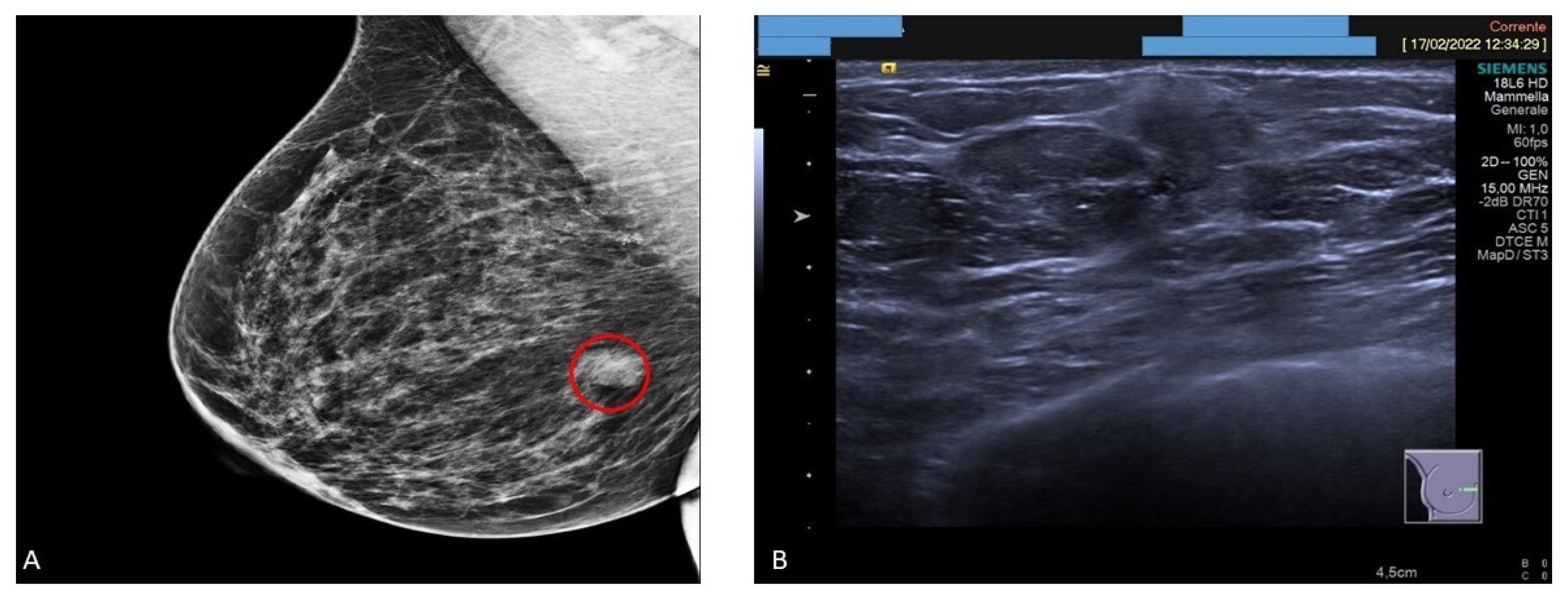
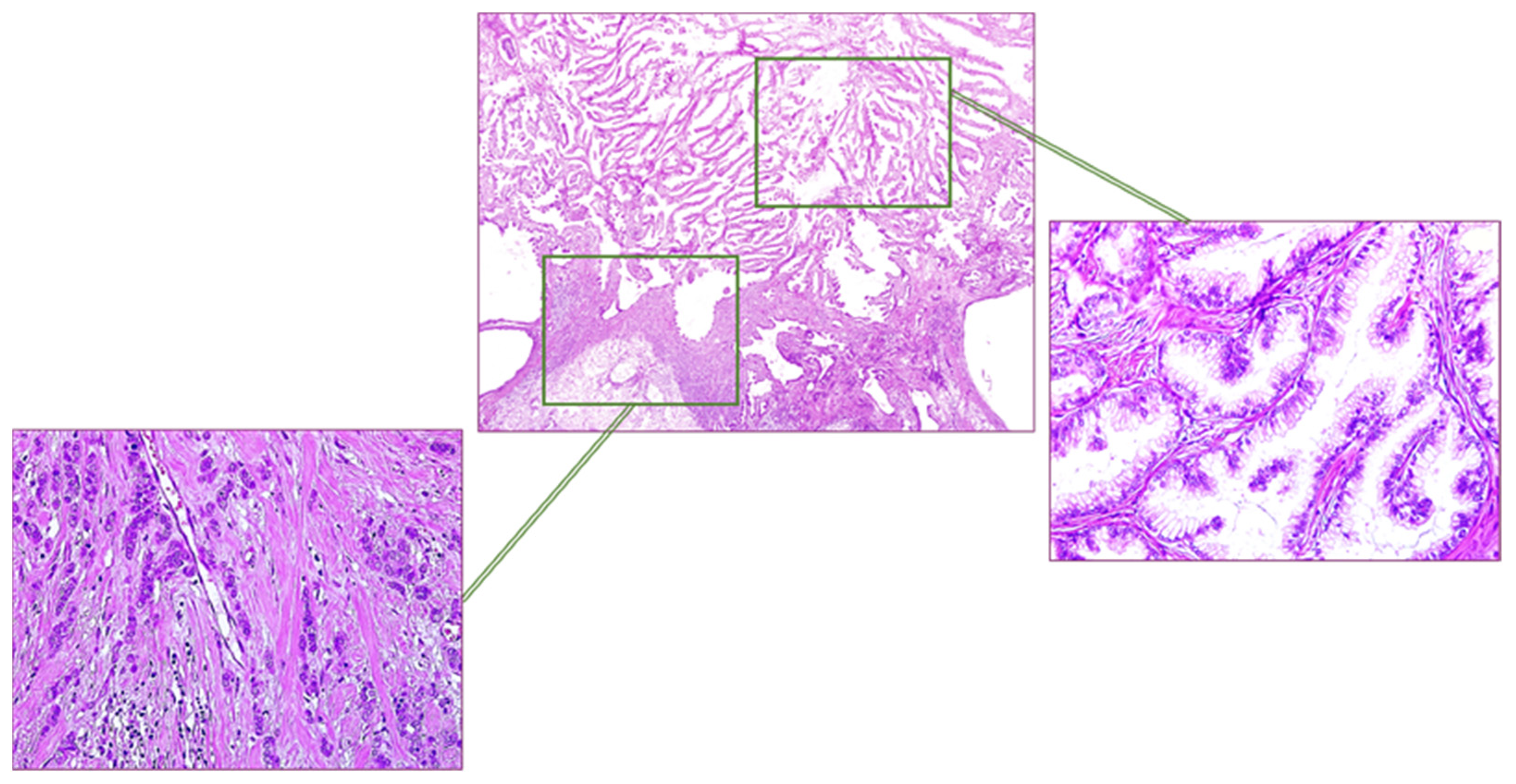
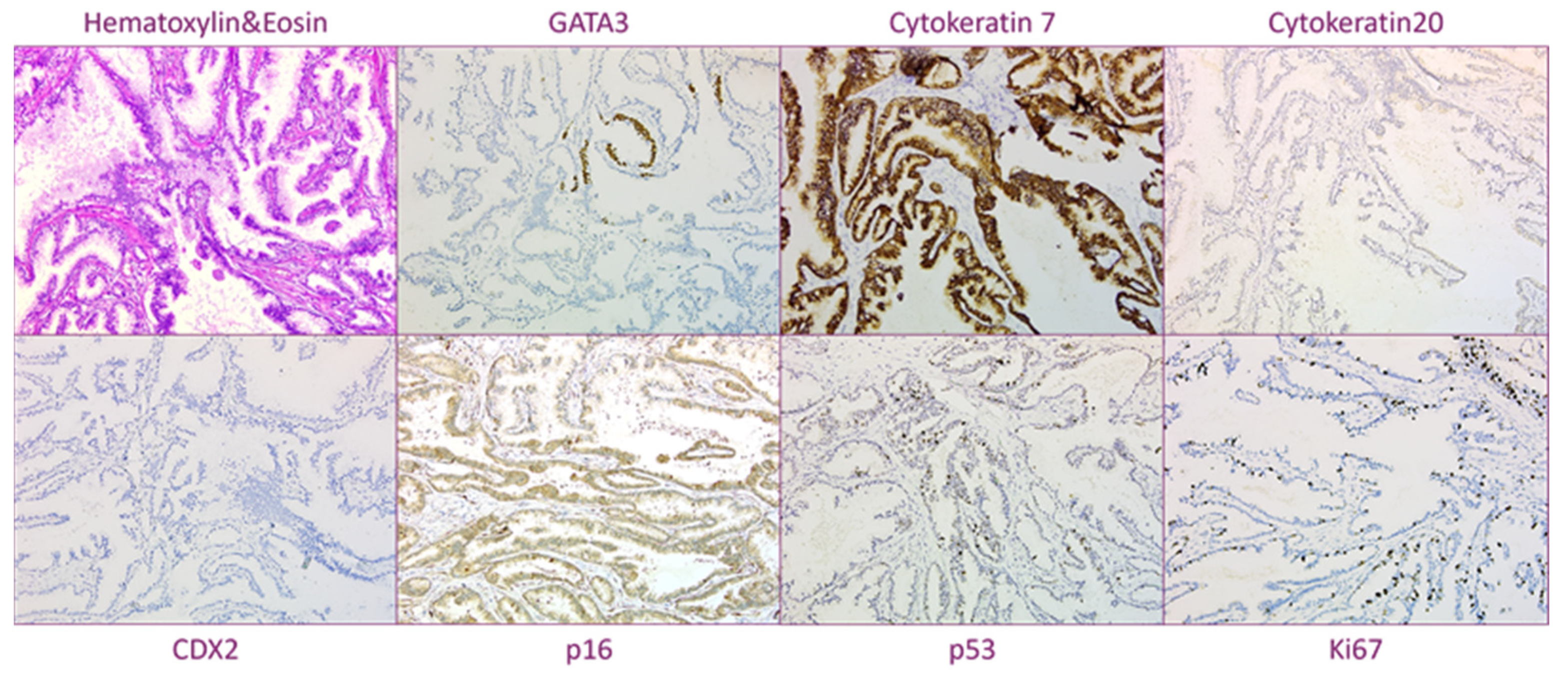
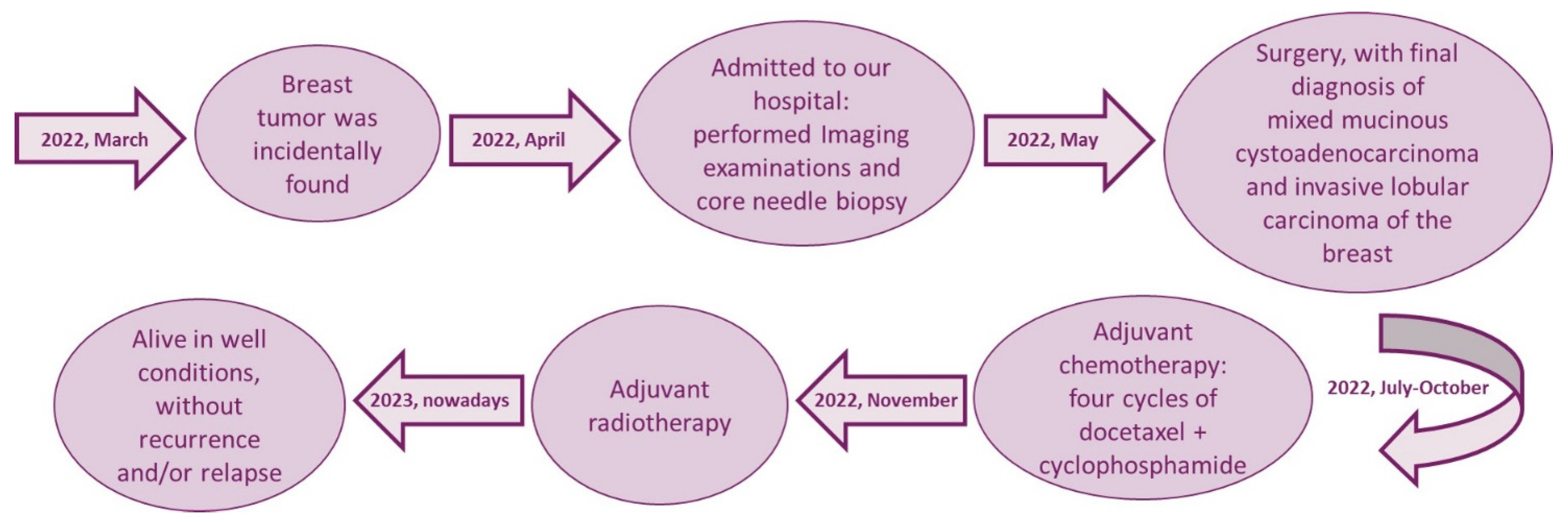
2. Case Report
3. Discussion
4. Conclusions
Author Contributions
Funding
Institutional Review Board Statement
Informed Consent Statement
Conflicts of Interest
References
- Rosen, P.P.; Scott, M. Cystic hypersecretory duct carcinoma of the breast. Am. J. Surg. Pathol. 1984, 8, 31–42. [Google Scholar] [CrossRef]
- Koenig, C.; Tavassoli, F.A. Mucinous Cystadenocarcinoma of the Breast. Am. J. Surg. Pathol. 1998, 22, 698–703. [Google Scholar] [CrossRef] [PubMed]
- Domoto, H.; Terahata, S.; Yamazaki, T.; Sato, K.; Takeo, H.; Tamai, S. Mucinous cystadenocarcinoma of the breast showing sulfomucin production. Histopathology 2000, 36, 567–569. [Google Scholar] [CrossRef] [PubMed]
- Honma, N.; Sakamoto, G.; Ikenaga, M.; Kuroiwa, K.; Younes, M.; Takubo, K. Mucinous cystadenocarcinoma of the breast: A case report and review of the liter-ature. Arch. Pathol. Lab. Med. 2003, 127, 1031–1033. [Google Scholar] [CrossRef]
- Chen, W.Y.; Chen, C.S.; Chen, H.C.; Hung, Y.J.; Chu, J.S. Mucinous cystadenocarcinoma of the breast coexisting with infiltrating ductal carci-noma. Pathol. Int. 2004, 54, 781–786. [Google Scholar] [CrossRef] [PubMed]
- Coyne, J.D.; Irion, L. Mammary mucinous cystadenocarcinoma. Histopathology 2006, 49, 659–660. [Google Scholar] [CrossRef]
- Lee, S.H.; Chaung, C.R. Mucinous metaplasia of breast carcinoma with macrocystic transformation resembling ovarian mucinous cystadenocarcinoma in a case of synchronous bilateral infiltrating ductal carcinoma. Pathol. Int. 2008, 58, 601–605. [Google Scholar] [CrossRef]
- Rakici, S.Y.; Gönüllü, G.; Gürsel, B.; Yıldız, L.; Bayrak, I.K.; Yücel, I. Mucinous Cystadenocarcinoma of the Breast with Estrogen Receptor Expression: A Case Report and Review of the Literature. Case Rep. Oncol. 2009, 2, 210–216. [Google Scholar] [CrossRef] [PubMed]
- Gulwani, H.; Bhalla, S. Mucinous cystadenocarcinoma: A rare primary malignant tumor of the breast. Indian J. Pathol. Microbiol. 2010, 53, 200–202. [Google Scholar] [CrossRef]
- Petersson, F.; Pang, B.; Thamboo, T.P.; Putti, T.C. Mucinous cystadenocarcinoma of the breast with amplification of the HER2-gene con-firmed by FISH: The first case reported. Hum. Pathol. 2010, 41, 910–913. [Google Scholar] [CrossRef]
- Sentani, K.; Tashiro, T.; Uraoka, N.; Aosaki, Y.; Yano, S.; Takaeko, F.; Yasui, W. Primary mammary mucinous cystadenocarcinoma: Cytological and histological findings. Diagn. Cytopathol. 2011, 40, 624–628. [Google Scholar] [CrossRef] [PubMed]
- Li, X.; Peng, J.; Zhang, Z.; Zhang, Y. Mammary Mucinous Cystadenocarcinoma. Breast J. 2012, 18, 282–283. [Google Scholar] [CrossRef]
- Deng, Y.; Xue, D.; Wang, X.; Xu, S.; Ao, Q.; Hu, Z.; Wang, G. Mucinous cystadenocarcinoma of the breast with a basal-like immunophenotype. Pathol. Int. 2012, 62, 429–432. [Google Scholar] [CrossRef]
- Kim, S.E.; Park, J.H.; Hong, S.; Koo, J.S.; Jeong, J.; Jung, W.-H. Primary Mucinous Cystadenocarcinoma of the Breast: Cytologic Finding and Expression of MUC5 Are Different from Mucinous Carcinoma. Korean J. Pathol. 2012, 46, 611. [Google Scholar] [CrossRef] [PubMed]
- Lin, D.L.; Hu, J.L.; Shao, S.H.; Sun, D.M.; Wang, J.G. Primary mucinous cystadenocarcinoma of the breast with endocervical like mucinous epi-thelium. Breast Care 2013, 8, 445–447. [Google Scholar] [CrossRef] [PubMed] [Green Version]
- Kucukzeybek, B.B.; Yigit, S.; Sari, A.A.; Rezanko, T.; Durak, E.; Sadullahoglu, C. Primary mucinous cystadenocarcinoma of the breast with amplification of the HER2 gene confirmed by FISH—Case report and review of the literature. Pol. J. Pathol. 2014, 65, 70–73. [Google Scholar] [CrossRef] [PubMed]
- Witherspoon, L.E.; Oxenhandler, R.W. A Rare Tumor: Mucinous Cystadenocarcinoma of the Breast. Am. Surg. 2015, 81, E106–E108. [Google Scholar] [CrossRef]
- Seong, M.; Ko, E.Y.; Han, B.-K.; Cho, S.Y.; Cho, E.Y.; Lee, S.K.; Lee, J.E. Radiologic Findings of Primary Mucinous Cystadenocarcinoma of the Breast: A Report of Two Cases and a Literature Review. J. Breast Cancer 2016, 19, 330–333. [Google Scholar] [CrossRef] [Green Version]
- Koufopoulos, N.; Goudeli, C.; Syrios, J.; Filopoulos, E.; Khaldi, L. Mucinous Cystadenocarcinoma of the Breast: The Challenge of Diagnosing a Rare Entity. Rare Tumors 2017, 9, 98–100. [Google Scholar] [CrossRef] [Green Version]
- Jain, E.; Kumar, A.; Jain, R.; Sharma, S. Primary mucinous cystadenocarcinoma of the breast coexisting with invasive ductal car-cinoma: A case report and review of the literature. Int. J. Clin. Exp. Med. 2017, 10, 7256–7260. [Google Scholar]
- Nayak, A.; Bleiweiss, I.J.; Dumoff, K.; Bhuiya, T.A. Mucinous Cystadenocarcinoma of the Breast: Report of 2 Cases Including One with Long-Term Local Recurrence. Int. J. Surg. Pathol. 2018, 26, 749–757. [Google Scholar] [CrossRef] [PubMed]
- Sun, M.; Su, S.; Liu, Q.; Li, Q.; Wang, K. Mammary synchronous mucinous cystadenocarcinoma and columnar cell mucinous carcinoma: A case report. Int. J. Clin. Exp. Pathol. 2020, 13, 2381–2386. [Google Scholar] [PubMed]
- Valdespino, V.E.; Matamoros, I.L.; Valle, M.L.; Rangel, M.M.; Ramon, H.M.; Gomez, V.V. Mucinous cystadenocarcinoma of the breast, case report. Obstet. Gynecol. Rep. 2019, 3, 7016. [Google Scholar] [CrossRef]
- Jain, E.; Kumar, A.; Jain, R.; Sharma, S. Primary Mucinous Cystadenocarcinoma of the Breast: A Rare Case Report with Review of Literature. Int. J. Surg. Pathol. 2021, 29, 740–746. [Google Scholar] [CrossRef]
- Nikbakhsh, N.; Hosseini, A.; Ghorbani, H.; Arefisigaroudi, N.; Davarian, A. Mucinous cystadenocarcinoma of the breast in a 69-year-old woman with positive hormone receptors, the first case reported. Casp. J. Intern. Med. 2021, 12 (Suppl. S2), S444–S446. [Google Scholar]
- Wang, X.; Li, Y.; Zhao, P.; Jia, H.; Dong, X.; Zhang, L.; Wang, C. Primary mucinous cystadenocarcinoma of the breast: A clinicopathologic analysis of one case and review of the literature. Int. J. Clin. Exp. Pathol. 2020, 13, 2562–2568. [Google Scholar]
- Kaur, K.; Shah, A.; Gandhi, J.; Trivedi, P. Mucinous cystadenocarcinoma of the breast: A new entity with broad differentials-a case report. J. Egypt. Natl. Cancer Inst. 2022, 34, 9. [Google Scholar] [CrossRef]
- Lin, L.H.; Hernandez, O.; Zhu, K.; Guth, A.; Cotzia, P.; Darvishian, F. Genetic profile of primary mucinous cystadeno-carcinoma of the breast-A case report. Breast J. 2021, 27, 731–734. [Google Scholar] [CrossRef]
- Moatasim, A.; Mamoon, N. Primary breast mucinous adenocarcinoma and review of Literature. Cureus 2022, 14, e23098. [Google Scholar] [PubMed]
- Zuo, C.; Xie, J. Mixed primary mucinous cystadenocarcinoma and invasive ductal carcinoma of the breast: A case report and literature review. Transl. Cancer Res. 2022, 11, 4455–4464. [Google Scholar] [CrossRef]
- Lei, T.; Shi, Y.Q.; Chen, T.B. Mammary mucinous cystadenocarcinoma with long-term follow-up: Molecular information and literature review. Diagn. Pathol. 2023, 18, 13. [Google Scholar] [CrossRef]
- Wen, H.Y.; Desmedt, C.; Reis- Filho, J.S.; Schmit, F. Mucinous cystadenocarcinoma. In WHO Classification of Breast Tumours, 5th ed.; Lokuhetty, D., Ed.; International Agency for Research on Cancer: Lyon, France, 2019; pp. 126–127. [Google Scholar]
- Dundr, P.; Singh, N.; Nožičková, B.; Němejcová, K.; Bártů, M.; Stružinská, I. Primary mucinous ovarian tumors vs. ovarian metastases from gastrointestinal tract, pancreas and biliary tree: A review of current problematics. Diagn. Pathol. 2021, 16, 20. [Google Scholar] [CrossRef]
- Hoon Tan, P.; Ellis, I.; Allison, K.; Brogi, E.; Fox, S.B.; Lakhani, S.; Lazar, A.J.; Morris, E.A.; Sahin, A.; Salgado, R.; et al. The 2019 World Health Organization classification of tumours of the breast. Histopathology 2020, 77, 181–185. [Google Scholar] [CrossRef] [PubMed] [Green Version]
- Assarzadegan, N.; Gonzalo, D.H.; Leon, M.E.; Asirvatham, J.R. Immunohistochemical Expression of CDX2 and SATB2 in Primary Breast Carcinomas. Appl. Immunohistochem. Mol. Morphol. 2020, 28, e53. [Google Scholar] [CrossRef] [PubMed]
- Creytens, D. Immunohistochemical SATB2 Expression in Breast Carcinomas: A Note of Caution. Appl. Immunohistochem. Mol. Morphol. 2020, 28, e53. [Google Scholar] [CrossRef]
- Lau, S.K.; Weiss, L.M.; Chu, P.G. Differential expression of MUC1, MUC2, and MUC5AC in carcinomas of various sites: An immunohistochemical study. Am. J. Clin. Pathol. 2004, 122, 61–69. [Google Scholar] [CrossRef]
- Nguyen, M.D.; Plasil, B.; Wen, P.; Frankel, W.L. Mucin profiles in signet ring cell carcinoma. Arch. Pathol. Lab. Med. 2006, 130, 799–804. [Google Scholar] [CrossRef]
- Chu, P.G.; Chung, L.; Weiss, L.M.; Lau, S.K. Determining the site of origin of mucinous adenocarcinoma: An im-munohistochemical study of 175 cases. Am. J. Surg. Pathol. 2011, 35, 1830–1836. [Google Scholar] [CrossRef] [PubMed]
- Hanski, C.; Hofmeier, M.; Schmitt-Gräff, A.; Riede, E.; Hanski, M.L.; Borchard, F.; Sieber, E.; Niedobitek, F.; Foss, H.-D.; Stein, H.; et al. Overexpression or ectopic expression of MUC2 is the common property of mucinous carcinomas of the colon, pancreas, breast, and ovary. J. Pathol. 1997, 182, 385–391. [Google Scholar] [CrossRef]
- Matsukita, S.; Nomoto, M.; Kitajima, S.; Tanaka, S.; Goto, M.; Irimura, T.; Kim, Y.S.; Sato, E.; Yonezawa, S. Expression of mucins (MUC1, MUC2, MUC5AC and MUC6) in mucinous carcinoma of the breast: Comparison with invasive ductal carcinoma. Histopathology 2003, 42, 26–36. [Google Scholar] [CrossRef] [PubMed]
- Melamed, M.R.; Robbins, G.F.; Foote, F.W., Jr. Prognostic significance of gelations mammary carcinoma. Cancer 1961, 14, 699–704. [Google Scholar] [CrossRef] [PubMed]


| Case | Age (Years) | Size (cm) | DCIS | ER/PR/HER2 | CK7/CK20 | Nodal Metastasis | Treatment | Follow-Up |
|---|---|---|---|---|---|---|---|---|
| 1 [1] | 79 | 6 | + | NA | NA/NA | - | Mastectomy | DOD, 24 months |
| 2 [2] | 54 | 19 | - | -/-/NA | +/- | + | Mastectomy, Lymph node dissection | ANED, 24 months |
| 3 [2] | 67 | 2.3 | + | -/-/NA | +/- | - | Mastectomy, Lymph node dissection | ANED, 22 months |
| 4 [2] | 49 | 8.5 | + | -/-/NA | +/- | - | Mastectomy, Lymph node dissection, Chemotherapy, Radiotherapy | ANED, 11 months |
| 5 [2] | 61 | 0.8 | - | -/-/NA | +/- | - | Lumpectomy, Lymph node dissection | NA |
| 6 [3] | 74 | 10 | + | -/NA/NA | +/- | - | Mastectomy, Lymph node dissection | ANED, 24 months |
| 7 [4] | 96 | 2 | - | -/-/- | NA/NA | + | Lumpectomy, Lymph node dissection | ANED, 46 months |
| 8 [5] | 65 | 3 | + | -/-/- | +/+ (focal) | - | Mastectomy, Lymph node dissection, Chemotherapy | ANED, 8 months |
| 9 [6] | 51 | 4 | - | -/-/NA | +/- | - | Lumpectomy | NA |
| 10 [7] | 55 | 2.5 | + | -/-/-/ | +/- | - | Lumpectomy | ANED, 6 months |
| 11 [8] | 52 | 10 | - | +/-/- | -/- | - | Mastectomy, Lymph node dissection | ANED, 24 months |
| 12 [9] | 61 | 3 | - | -/-/- | NA/- | - | Mastectomy, Lymph node dissection | ANED, 6 months |
| 13 [10] | 73 | 4.5 | + | -/-/2+ | +/- | - | Mastectomy, Lymph node dissection | NA |
| 14 [11] | 65 | 3 | + | -/-/NA | +/- | + | Partial mastectomy, Lymph node dissection | ANED, 6 months |
| 15 [12] | 52 | 6.5 | - | -/-/- | +/- | - | NA | NA |
| 16 [13] | 41 | 7 | + | -/-/- | +/- | - | Mastectomy, Lymph node dissection | ANED, 24 months |
| 17 [14] | 59 | 0.9 | + | -/-/2+ | +/- | - | Partial mastectomy, lymph node dissection, Chemotherapy | ANED, 3 months |
| 18 [15] | 62 | 5.6 | - | -/-/- | +/- | - | Mastectomy, Lymph node dissection | ANED, 15 months |
| 19 [16] | 55 | 2 | + | -/-/2+ | +/- | - | Mastectomy, Lymph node dissection, Chemotherapy, Radiotherapy | ANED, 10 months |
| 20 [17] | 91 | 7.5 | + | -/-/- | +/- | - | Mastectomy, Radiotherapy | DOD, 14 months |
| 21 [18] | 59 | 2 | - | -/-/3+ | NA/NA | - | Partial mastectomy | NA |
| 22 [18] | 50 | 2.2 | - | -/-/- | NA/NA | - | Partial mastectomy | NA |
| 23 [19] | 63 | 1.6 | - | -/-/- | +/- | + | Lumpectomy, Lymph node dissection, Chemotherapy, Radiotherapy | ANED, 48 months |
| 24 [20] | 58 | 4.5 | + | -/-/- | +/- | - | Mastectomy, Lymph node dissection | ANED, 6 months |
| 25 [21] | 68 | 6.2 | + | -/-/- | +/- | - | Lumpectomy, Sentinel lymph node dissection | ANED, 3 months |
| 26 [21] | 59 | 2 | + | -/-/- | +/- | - | Lumpectomy, lymph node dissection | Alive with local recurrence after 96 months |
| 27 [22] | 56 | 2 | - | +/+/- | -/- | - | Mastectomy, Lymph node dissection | ANED, 3 months |
| 28 [23] | 68 | 4 | NA | -/-/- | +/- | - | Mastectomy, Lymph node dissection | ANED, 21 months |
| 29 [24] | 45 | 4.3 | + | -/-/- | +/- | - | Lumpectomy, Mastectomy, Lymph node dissection, Chemotherapy, Radiotherapy | ANED, 6 months |
| 30 [25] | 69 | 2 | + | +/+/- | NA | - | NA | NA |
| 31 [26] | 66 | 2.5 | + | -/-/- | +/- | - | Mastectomy, Lymph node dissection | ANED, 13 months |
| 32 [27] | 65 | 18 | - | -/-/3+ | +/focal + | NA | Mastectomy | ANED, 6 months |
| 33 [28] | 72 | 0.9 | - | -/-/- | +/- | - | Mastectomy, Lymph node dissection | ANED, 16 moths |
| 34 [29] | 61 | 3.5 | + | +/+/- | +/- | + | Mastectomy, Lymph node dissection | ANED, 8 months |
| 35 [30] | 61 | 2.5 | + | -/-/- | +/- | - | Mastectomy, Lymph node dissection | ANED, 10 months |
| 36 [31] | 59 | 3 | - | -/-/- | +/- | - | Lumpectomy, Lymph node dissection | ANED, 108 months |
| Present Case | 41 | 1.5 | - | +/-/- | +/NA | Isolated Tumor Cells | Partial mastectomy, sentinel lymph node dissection | ANED, 11 months |
Disclaimer/Publisher’s Note: The statements, opinions and data contained in all publications are solely those of the individual author(s) and contributor(s) and not of MDPI and/or the editor(s). MDPI and/or the editor(s) disclaim responsibility for any injury to people or property resulting from any ideas, methods, instructions or products referred to in the content. |
© 2023 by the authors. Licensee MDPI, Basel, Switzerland. This article is an open access article distributed under the terms and conditions of the Creative Commons Attribution (CC BY) license (https://creativecommons.org/licenses/by/4.0/).
Share and Cite
Vegni, F.; D’Alessandris, N.; Santoro, A.; Angelico, G.; Scaglione, G.; Carlino, A.; Arciuolo, D.; Valente, M.; Sfregola, S.; Natale, M.; et al. Primary Mucinous Cystadenocarcinoma of the Breast Intermixed with Pleomorphic Invasive Lobular Carcinoma: The First Report of This Rare Association. J. Pers. Med. 2023, 13, 948. https://doi.org/10.3390/jpm13060948
Vegni F, D’Alessandris N, Santoro A, Angelico G, Scaglione G, Carlino A, Arciuolo D, Valente M, Sfregola S, Natale M, et al. Primary Mucinous Cystadenocarcinoma of the Breast Intermixed with Pleomorphic Invasive Lobular Carcinoma: The First Report of This Rare Association. Journal of Personalized Medicine. 2023; 13(6):948. https://doi.org/10.3390/jpm13060948
Chicago/Turabian StyleVegni, Federica, Nicoletta D’Alessandris, Angela Santoro, Giuseppe Angelico, Giulia Scaglione, Angela Carlino, Damiano Arciuolo, Michele Valente, Stefania Sfregola, Maria Natale, and et al. 2023. "Primary Mucinous Cystadenocarcinoma of the Breast Intermixed with Pleomorphic Invasive Lobular Carcinoma: The First Report of This Rare Association" Journal of Personalized Medicine 13, no. 6: 948. https://doi.org/10.3390/jpm13060948